Embed Size (px)
DESCRIPTION
Understanding Anatomy and Physiology
Citation preview

PARTV
Continuity
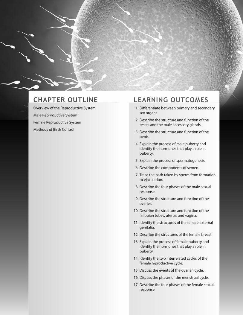
CHAPTER OUTLINEOverview of the Reproductive System
Male Reproductive System
Female Reproductive System
Methods of Birth Control
LEARNING OUTCOMES1. Differentiate between primary and secondary
sex organs.
2. Describe the structure and function of the
testes and the male accessory glands.
3. Describe the structure and function of the
penis.
4. Explain the process of male puberty and
identify the hormones that play a role in
puberty.
5. Explain the process of spermatogenesis.
6. Describe the components of semen.
7. Trace the path taken by sperm from formation
to ejaculation.
8. Describe the four phases of the male sexual
response.
9. Describe the structure and function of the
ovaries.
10. Describe the structure and function of the
fallopian tubes, uterus, and vagina.
11. Identify the structures of the female external
genitalia.
12. Describe the structures of the female breast.
13. Explain the process of female puberty and
identify the hormones that play a role in
puberty.
14. Identify the two interrelated cycles of the
female reproductive cycle.
15. Discuss the events of the ovarian cycle.
16. Discuss the phases of the menstrual cycle.
17. Describe the four phases of the female sexual
response.
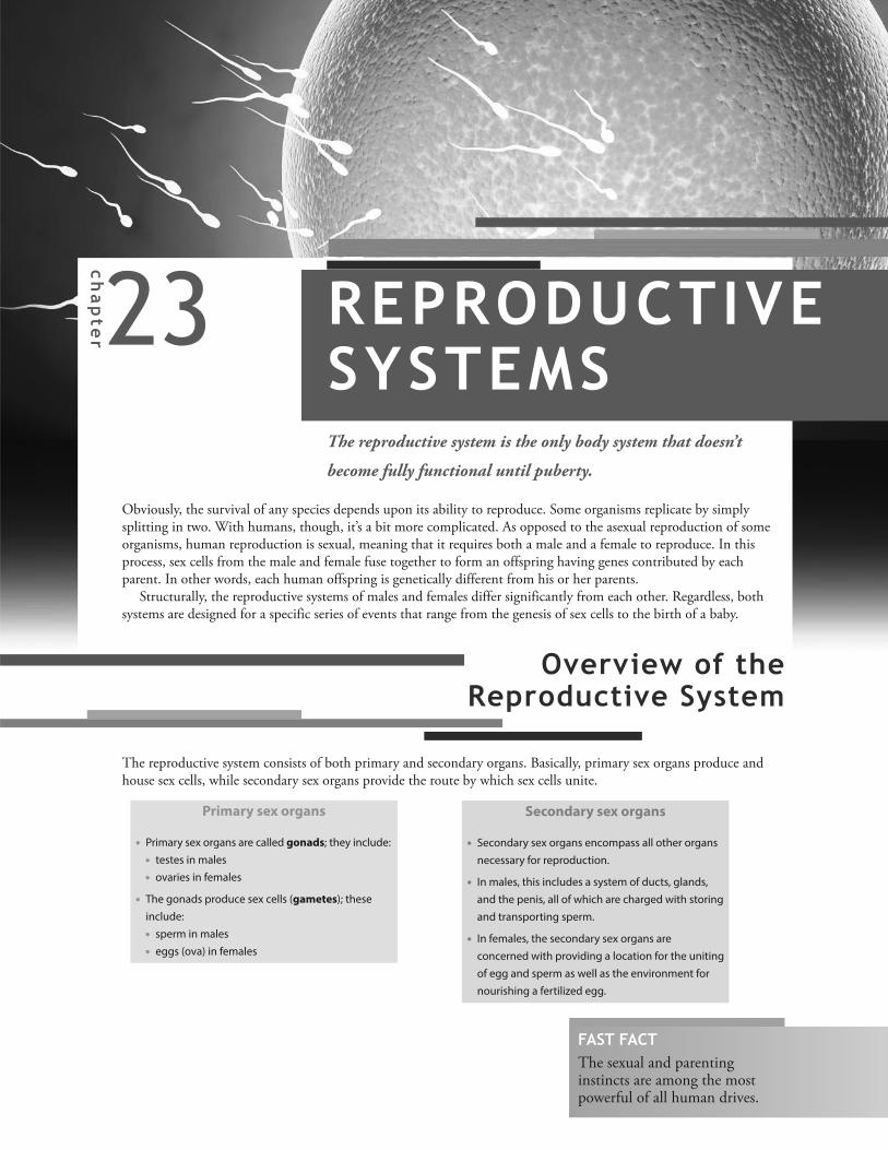
23chapter REPRODUCTIVESYSTEMSThe reproductive system is the only body system that doesn’t
become fully functional until puberty.
Obviously, the survival of any species depends upon its ability to reproduce. Some organisms replicate by simplysplitting in two. With humans, though, it’s a bit more complicated. As opposed to the asexual reproduction of someorganisms, human reproduction is sexual, meaning that it requires both a male and a female to reproduce. In thisprocess, sex cells from the male and female fuse together to form an offspring having genes contributed by eachparent. In other words, each human offspring is genetically different from his or her parents.
Structurally, the reproductive systems of males and females differ significantly from each other. Regardless, bothsystems are designed for a specific series of events that range from the genesis of sex cells to the birth of a baby.
Overview of the Reproductive System
Primary sex organs
• Primary sex organs are called gonads; they include:
• testes in males
• ovaries in females
• The gonads produce sex cells (gametes); these
include:
• sperm in males
• eggs (ova) in females
The reproductive system consists of both primary and secondary organs. Basically, primary sex organs produce andhouse sex cells, while secondary sex organs provide the route by which sex cells unite.
Secondary sex organs
• Secondary sex organs encompass all other organs
necessary for reproduction.
• In males, this includes a system of ducts, glands,
and the penis, all of which are charged with storing
and transporting sperm.
• In females, the secondary sex organs are
concerned with providing a location for the uniting
of egg and sperm as well as the environment for
nourishing a fertilized egg.
FAST FACTThe sexual and parentinginstincts are among the mostpowerful of all human drives.

PA
RT
V C
on
tinu
ity
446
The male reproductive system serves to produce, transport, and introduce mature sperm into the female reproductive tract,which is where fertilization occurs.
Testes
The penis and the scrotum (a tissue sac hanging behind the penis) are the external portionsof the male reproductive system. Inside the scrotum reside two testes, the organs thatgenerate sperm and secrete the male sex hormone testosterone.
� � � � � � � � � � � � � � � �
� � � � � � � � � � � � � � � � � �� � � � � � � � � �� � � � � � � � � �
Extending from the abdomen to each
testicle is a strand of connective tissue and
muscle called the spermatic cord; it contains
the sperm duct (vas deferens), blood and
lymphatic vessels, and nerves.
Two small, oval testes lie suspended in a
sac of tissue called the scrotum.
The median septum divides the scrotum,
isolating each testicle. This helps prevent
any infection from spreading from one
testicle to the other.
The cremaster muscle surrounds the
spermatic cord and testes. In cold weather, it
contracts to draw the testes closer to the body
for warmth. (See “The Body at Work” on this
page.)
Life lesson: UndescendedtesticleIn utero, the testes begin development near the kidneys.Then, through the course of fetal development, the testesdescend into the scrotum. A small percentage of boys,however, are born with undescended testes, a conditioncalled cryptorchidism. If the testes don’t descend on theirown during the first year of life, a surgical procedure, whichinvolves pulling the testis into the scrotum, is typically done.Alternatively, it may sometimes be corrected through aninjection of testosterone. Regardless, if left untreated, thecondition will lead to sterility or, possibly, testicular cancer.
The Body AT WORKA key reason the testes reside outside the body is
because the temperature inside the body is too
warm for sperm to develop. (The temperature
inside the scrotum is 5° F [3° C] cooler than the
temperature inside the body.) Muscles within the
scrotum help the testes maintain an ideal
temperature for sperm production. For example,
in warm temperatures, the cremaster muscle
relaxes, allowing the testes to drop further away
from the body so as to avoid becoming too warm.
In cold weather, it contracts to draw the testes
closer to the body for warmth. A layer of smooth
muscular fiber (dartos fascia) in the scrotum also
contracts when it’s cold, drawing the testes closer
to the body. This gives the scrotum a wrinkled
appearance.
Male Reproductive System

Inside the Testes
Underneath its fibrous capsule covering, the testes contain a vast length of tubules and a series of spermatic ducts.� � � � � � � � � � � �� � � � � � � � � � � � � � � � � � � �
� � � � � � � � � � � � �
� � � � � � � � � � � � � � �
� � � � � � � � � � � � � � �Fibrous tissue separates each testis into
over 200 lobules.
1 A network of vessels called the rete
testis leads away from the
seminiferous tubules; these vessels provide
a location in which sperm partially mature.
2 Efferent ductules conduct immature
sperm away from the testis to the
epididymis.
3 Sperm pass into the epididymis,
which is attached to the posterior side
of the testis. (Note that the epididymis is
outside of the testis but still inside the
scrotum.) Sperm move from the head of
the epididymis to the tail, maturing as they
go. They are then stored in the tail of the
epididymis, where they remain fertile for
40 to 60 days. After that, unless they are
ejaculated, the aging sperm disintegrate
and are reabsorbed by the epididymis.
4 Sperm leave the tail of the epididymis
and pass into the vas deferens.
� � � � � � � � �� � � �� � � � � �� � � � � � �� � � � � � � � � � � �
� � � � � � � � �5 The vas deferens travels up the
spermatic cord, through the inguinal
canal, and into the pelvic cavity. It loops
over the ureter and descends along the
posterior bladder wall.
6 As the vas deferens turns downward, it
widens into an ampulla and ends by
joining the seminal vesicle to form the
ejaculatory duct. (Remember that there are
two ejaculatory ducts: one for each testis.) The
ejaculatory ducts pass through the prostate
and empty into the urethra.
FAST FACTThe urethra serves both the urinary system (to carryurine) and the reproductive system (to carry semen).It cannot, however, carry both at the same time.
TubulesThe tubules continuouslygenerate sperm.
Spermatic ductsSperm continue to mature as theyfollow a specific path through thespermatic ducts.
Coiled within each lobule are one to three
seminiferous tubules: tiny tubes in which
sperm are produced. Several layers of cells line
the walls of the tubules, with each layer
containing germ cells in the process of
becoming sperm. (A germ cell is a cell that gives
rise to gametes.) Also contained in the wall of
the tubule are cylindrical cells called Sertoli
cells. These cells promote the development of
sperm by supplying nutrients, removing waste,
and secreting the hormone inhibin, which plays
a role in the maturation and release of sperm.
Lying between the seminiferous tubules are
clusters of interstitial cells—also called Leydig
cells—that produce testosterone.
447
CH
AP
TE
R 2
3 R
ep
rod
uctiv
e S
yste
ms

PA
RT
V C
on
tinu
ity
448
� � � � � � � � � � � � � � � � � � � � � � � � �� � � � � � � � � � � �
� � � � � � � � � �
� � � � � � �� � � � � � � � � �! � � � � � � � � � � � � � �" � � � � � � � # � � � � � � � � �� � � �$ � � � � �� � � � � � � � �
$ � � � � �� � � � � � � � �
Accessory Glands
The male reproductive system includes three sets of accessory glands: the seminal vesicles, prostate gland, and bulbourethralglands.
Located at the base of the bladder, a pair
of seminal vesicles (one for each vas
deferens) secretes a thick, yellowish fluid
into the ejaculatory duct. The fluid—which
comprises about 60% of semen—contains
fructose (an energy source for sperm
motility) as well as other substances that
nourish and ensure sperm motility.
The prostate gland sits just below the
bladder, where it encircles both the urethra
and ejaculatory duct. It secretes a thin, milky,
alkaline fluid into the urethra; besides
adding volume to semen (it comprises about
30% of the fluid portion of semen), the fluid
also enhances sperm motility.
Two pea-shaped bulbourethral glands (also
called Cowper’s glands) secrete a clear fluid
into the penile portion of the urethra during
sexual arousal. Besides serving as a lubricant
for sexual intercourse, the fluid also
neutralizes the acidity of residual urine in the
urethra, which would harm the sperm.
Life lesson: Prostate disordersThe prostate gland is about the size of a walnut in a young man. By about theage of 45, however, the gland begins to enlarge slowly. This noncancerousenlargement resulting from normal aging is called benign prostatic hyperplasia(BPH). As the prostate enlarges, it squeezes the urethra and obstructs the flow ofurine. Symptoms include difficulty urinating, slowing of the urine stream, andfrequent urination, particularly at night.
Prostate cancer, on the other hand, involves the growth of a malignant tumorwithin the prostate gland. These types of tumors usually grow slowly and,because they tend to develop outside of the gland, don’t obstruct urine flow. Asa result, they often go unnoticed. Eventually, the tumor can spread beyond theprostate gland and metastasize to surrounding tissues as well as the lungs andother organs.
Prostate cancer is the most common cancer in American men and the secondleading cause of death from cancer (after lung cancer). It is diagnosed by digitalrectal examination as well as by blood tests for prostate-specific antigen (PSA)and acid phosphatase (a prostatic enzyme). When detected and treated early,prostate cancer has a high survival rate; however, the survival rate fallsdramatically if the cancer has spread beyond the prostate gland.

449
CH
AP
TE
R 2
3 R
ep
rod
uctiv
e S
yste
ms
Penis
The purpose of the penis in the reproductive system is to deposit sperm in the female vagina.% & ' ( ) * ( + ,- . / & ) 0 ( ' % & ' ( ) * ( + , . 1 +- . / & ) 0 ( '� � � � � � �� 2 � � � � �� � � � � � �� � � � � � �
The body of the penis is called the shaft.
The slightly bulging head is called the
glans penis.
The loose skin covering the penis continues
over the glans to form a cuff called the
prepuce, or foreskin. (The foreskin is
removed by circumcision.) Sebaceous glands
in the prepuce and foreskin secrete a waxy
substance called smegma.
� � � � � � � � �� � � � � � � � � �3 � � � � �4 � � � �� � � � � �
Interior of the penis
Three cylinders of erectile tissue fill the shaft of the penis.During sexual arousal, the tissues fill with blood, causing thepenis to enlarge and become erect.
The two larger cylinders of tissue are called
the corpus cavernosa.
The smaller cylinder of tissue, called the
corpus spongiosum, encircles the urethra.
The Body AT WORKDuring the first trimester of male fetal development, the testes secrete a significant amount of testosterone. After
birth, testosterone levels continue to rise for several weeks before falling dramatically, becoming barely detectable
by age 4 to 6 months. Low levels of testosterone continue through childhood until, at about age 13, puberty begins;
this is the period in which the child’s body begins to transform into an adult capable of reproduction.
The onset of puberty is marked by the secretion of gonadotropin-releasing hormone (GnRH) by the hypothalamus.
This triggers the secretion of two gonadotropins: follicle-stimulating hormone (FSH) and luteinizing hormone (LH).
These hormones promote enlargement of the testes, which is the first sign of puberty. LH—also called interstitial
cell-stimulating hormone (ICSH) in males—prompts the interstitial cells to begin secreting testosterone. FSH primes
the spermatogenic cells to respond to testosterone, and sperm production begins.
The increased production of testosterone also stimulates the development of such secondary sex characteristics as:
• Pubic, axillary, and facial hair
• Darker and thicker skin
• Increased activity of oil and sweat glands, leading to body odor
• Increased growth along with an increase in muscle mass
• Deepening of the voice due to a larger larynx
FAST FACTPrimary sex characteristics refer to the organsdirectly involved in reproduction (such as the penisin males and the uterus in females). Secondary sexcharacteristics refer to features that distinguish eachsex but aren’t directly involved in reproduction (suchas facial hair in males and breasts in females).
PA
RT
V C
on
tinu
ity
450
� � � � � � �� � � � � � �
5 � � � � � �� � � � � � � � � � � �� � � � � �
6 78 9 8 9
6 76 7 6 78 9 8 9 8 9 8 9
Sperm
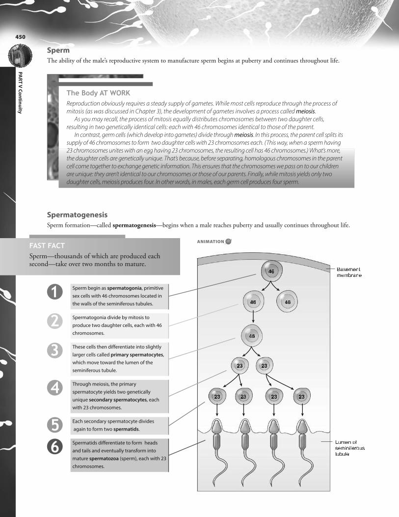
The ability of the male’s reproductive system to manufacture sperm begins at puberty and continues throughout life.
Spermatogenesis
Sperm formation—called spermatogenesis—begins when a male reaches puberty and usually continues throughout life.
FAST FACTSperm—thousands of which are produced each second—take over two months to mature.
Sperm begin as spermatogonia, primitive
sex cells with 46 chromosomes located in
the walls of the seminiferous tubules.
Spermatogonia divide by mitosis to
produce two daughter cells, each with 46
chromosomes.
These cells then differentiate into slightly
larger cells called primary spermatocytes,
which move toward the lumen of the
seminiferous tubule.
Through meiosis, the primary
spermatocyte yields two genetically
unique secondary spermatocytes, each
with 23 chromosomes.
Each secondary spermatocyte divides
again to form two spermatids.
Spermatids differentiate to form heads
and tails and eventually transform into
mature spermatozoa (sperm), each with 23
chromosomes.
1
3456
2
The Body AT WORKReproduction obviously requires a steady supply of gametes. While most cells reproduce through the process of
mitosis (as was discussed in Chapter 3), the development of gametes involves a process called meiosis.
As you may recall, the process of mitosis equally distributes chromosomes between two daughter cells,
resulting in two genetically identical cells: each with 46 chromosomes identical to those of the parent.
In contrast, germ cells (which develop into gametes) divide through meiosis. In this process, the parent cell splits its
supply of 46 chromosomes to form two daughter cells with 23 chromosomes each. (This way, when a sperm having
23 chromosomes unites with an egg having 23 chromosomes, the resulting cell has 46 chromosomes.) What’s more,
the daughter cells are genetically unique. That’s because, before separating, homologous chromosomes in the parent
cell come together to exchange genetic information. This ensures that the chromosomes we pass on to our children
are unique: they aren’t identical to our chromosomes or those of our parents. Finally, while mitosis yields only two
daughter cells, meiosis produces four. In other words, in males, each germ cell produces four sperm.
ANIMATION

451
CH
AP
TE
R 2
3 R
ep
rod
uctiv
e S
yste
ms
Life lesson: Male infertilityOver 2 million couples in the United States suffer from infertility. About half of those cases are due to maleinfertility. The most common form of male infertility is a low sperm count; even so, a number of other factors—including the size, shape, and motility of sperm—also influence male fertility. The World Health Organizationprovides a number of characteristics of a “normal” sperm sample. For example, the total volume of semen perejaculate should be at least 2 ml and contain at least 40 million sperm. Of the total spermatozoa in the ejaculate:
• At least 75% should be alive (it is normal for up to 25% to be dead)• At least 30% should have a normal shape• At least 25% should be swimming with rapid forward movement• At least 50% should be swimming forward, if only sluggishly
A sperm count lower than 20 million indicates infertility.
3 � � � � � � �4 � � � � � �: � � � � � � � � � � The head contains the nucleus, which
is packed with genetic material. Topping
the head of the sperm is a cap called an
acrosome. The acrosome contains
enzymes that help the sperm penetrate
the egg during fertilization.
The middle piece contains numerous
mitochondria that supply the sperm with
the energy it needs to migrate up the
female reproductive tract.
The tail is a flagellum whose beating,
whip-like movements propel the sperm
forward.
Spermatozoa
The mature sperm consists of a head, a middle piece, and a long, whip-like tail.
Semen
Emitted during the ejaculation that accompanies orgasm, semen is a whitish fluid containing both sperm and the fluidsecretions of the accessory glands. About 65% of the fluid volume of semen comes from the seminal vesicles, about 30%comes from the prostate gland, and about 5% comes from the bulbourethral gland. Each ejaculation expels between 2 and 5 ml of semen containing between 40 and 100 million sperm.
Two key qualities of semen include its stickiness and its alkalinity. Immediately after ejaculation, semen becomes stickyand jelly-like. This characteristic promotes fertilization by allowing the semen to stick to the walls of the vagina and cervixinstead of immediately draining out. The alkalinity of semen counteracts the acidity of the vagina; this is important becausesperm become immobile in an acidic environment.
The Body AT WORKAfter puberty, testosterone is continually secreted throughout the life of the male. Testosterone controls
spermatogenesis and supports the male sex drive. Blood levels of testosterone are controlled through a negative
feedback loop:
• High levels of testosterone inhibit secretion of GnRH by the hypothalamus. This depresses secretion of LH by the
anterior pituitary, and testosterone production declines.
• Low testosterone levels stimulate the anterior pituitary to increase secretion of LH, which triggers the interstitial cells
to step up testosterone secretion.

PA
RT
V C
on
tinu
ity
452
Male Sexual Response
The male sexual response can be divided into four phases: excitement, plateau, orgasm, and resolution.
Plateau
• The urethral sphincter contracts to prevent urine from mixing with semen.
• Heart rate, blood pressure, and respirations remain elevated.
Orgasm
• This brief, intense reaction involves the ejaculation of semen.
• Ejaculation occurs in two stages: emission and expulsion.
• In emission, the sympathetic nervous system stimulates peristalsis in the vas
deferens to propel sperm to the urethra; it also triggers the release of fluids from
the prostate gland and seminal vesicles.
• Semen in the urethra activates somatic and sympathetic reflexes that result in
the expulsion of semen.
Resolution
• Immediately following orgasm, sympathetic signals cause the arteries in the penis
to constrict, reducing blood flow.
• Muscles between the erectile tissues contract to squeeze blood out of the erectile
tissues.
• The penis becomes flaccid.
Excitement
• Visual, mental, or physical stimulation causes sexual excitement.
• Parasympathetic nerves cause the arteries in the penis to relax and fill with blood.
• As tissues within the penis become engorged with blood, the penis enlarges and
becomes rigid and erect so as to allow it to enter the female reproductive tract.
Ovaries
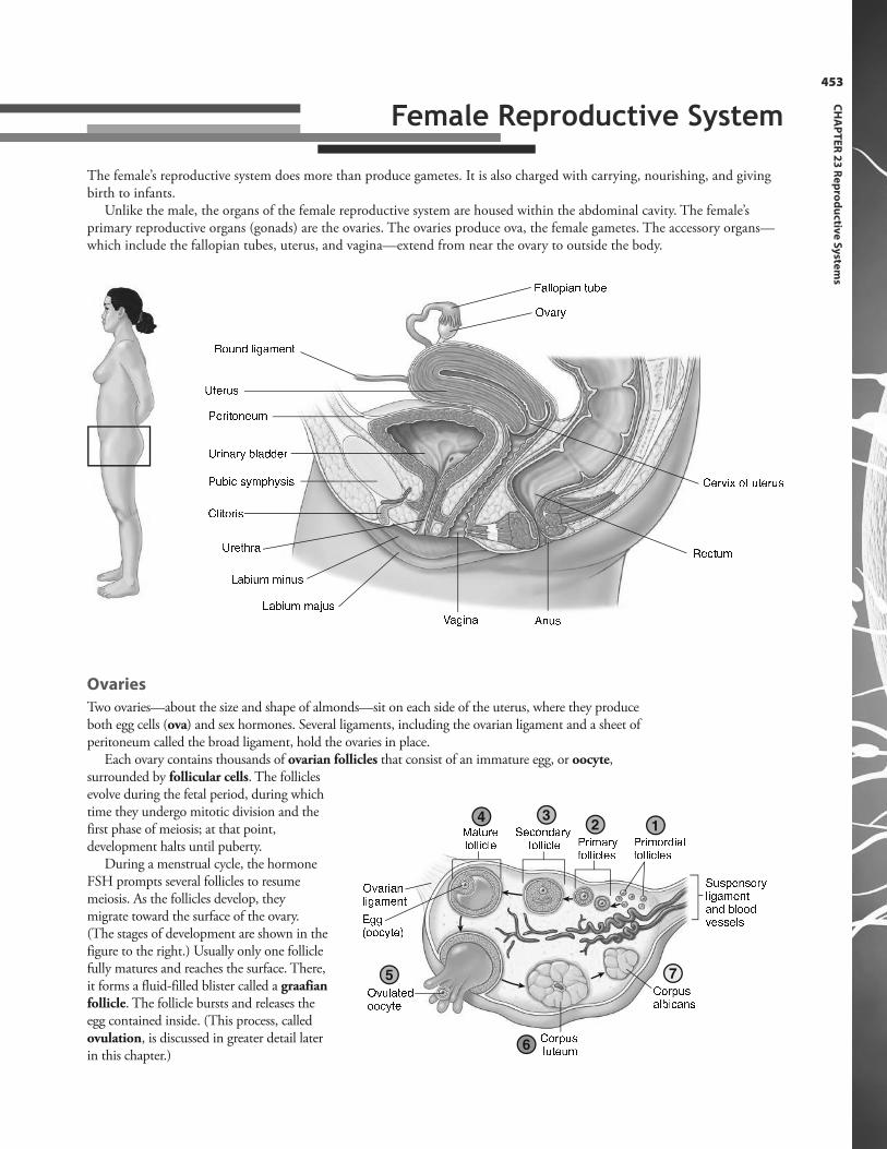
Two ovaries—about the size and shape of almonds—sit on each side of the uterus, where they produceboth egg cells (ova) and sex hormones. Several ligaments, including the ovarian ligament and a sheet ofperitoneum called the broad ligament, hold the ovaries in place.
Each ovary contains thousands of ovarian follicles that consist of an immature egg, or oocyte,surrounded by follicular cells. The folliclesevolve during the fetal period, during whichtime they undergo mitotic division and thefirst phase of meiosis; at that point,development halts until puberty.
During a menstrual cycle, the hormoneFSH prompts several follicles to resumemeiosis. As the follicles develop, theymigrate toward the surface of the ovary.(The stages of development are shown in thefigure to the right.) Usually only one folliclefully matures and reaches the surface. There,it forms a fluid-filled blister called a graafianfollicle. The follicle bursts and releases theegg contained inside. (This process, calledovulation, is discussed in greater detail laterin this chapter.)
453
CH
AP
TE
R 2
3 R
ep
rod
uctiv
e S
yste
ms
The female’s reproductive system does more than produce gametes. It is also charged with carrying, nourishing, and givingbirth to infants.
Unlike the male, the organs of the female reproductive system are housed within the abdominal cavity. The female’sprimary reproductive organs (gonads) are the ovaries. The ovaries produce ova, the female gametes. The accessory organs—which include the fallopian tubes, uterus, and vagina—extend from near the ovary to outside the body.
� � � � � � � � � � � � �� � � � � � ; � � � � � � � � � �� � � � � � � � � � � � � � �� � � � � � � � � � � �
� � � � � � � � � � �
5 � � � � � � � � � ; � � � � �5 � � � � � � � �$ � � � � � � �
< � � �$ � � � � 2 � � � � � � � �
3 � � �� � � 12
34
5
6
7
< � � � �� � � � � � � � � � � � � �� � � � � � � � � � � � � �� � � � � � � � �� � � � � � � �� � � � � � � �: � � � �� � � � � � � �� " � � � � � � # � � � � � � � � � �� � � � � � � � � � � � �� � � � � � �< � � � � � �� � � � � � $ � � � � � � � � � � �$ � � � � �� � � � � �
Female Reproductive System

PA
RT
V C
on
tinu
ity
454
� � � � � � �< � � �
� � � �� � � � � � $ � � � � � �� � � � � � � � � � � � � �: � � � � � � � � � � � � � � � � � � �
Internal Genitalia
The female reproductive system includes both internal and external genitalia. The internal genitalia include the fallopiantubes, uterus, and vagina. Because the fallopian tubes do not attach to the ovaries, the female reproductive tract is essentiallyan “open” system in which infection can spread from the reproductive tract into the peritoneal cavity.
Fallopian Tubes
The fallopian tubes (also called uterine tubes), are about 4inches (10 cm) long and extend from the ovary to the uterus.
A narrow isthmus is the portion of the
fallopian tube closest to the uterus.
The middle portion of the tube, called
the ampulla, is the usual site of egg
fertilization. Cilia line the inside of the
tube. Their beating movements,
combined with peristaltic contractions
of the tube, propel an egg toward the
uterus.
The distal funnel-shaped end of the
fallopian tube is called the
infundibulum. The fallopian tube does
not attach directly to the ovary.
Instead, finger-like projections called
fimbriae fan over the ovary.
Uterus
A muscular chamber called the uterus houses and nurturesa growing embryo. The uterus sits between the urinarybladder and the rectum, held in place by the broadligament. Usually, the uterus tilts forward over the bladder.
The curved upper portion of the uterus
is called the fundus. The upper two
corners of the uterus connect with the
fallopian tubes.
The central region of the uterus
is the body.
The inferior end is the cervix. A
passageway through the cervix,
called the cervical canal, links the
uterus to the vagina. Glands within
the cervical canal secrete thick
mucus; during ovulation, the
mucus thins to allow sperm to pass.Vagina
A muscular tube about 3 inches (8 cm) long, the vagina serves as a receptacle
for the penis and sperm, a route for the discharge of menstrual blood, and the
passageway for the birth of a baby. The smooth muscle walls of the vagina can
expand greatly, such as during childbirth.
The lower end of the vagina contains ridges (vaginal rugae) that help
stimulate the penis during intercourse and allow for expansion during
childbirth.
A fold of mucous membrane called the hymen partially covers the entrance
to the vagina. During the first intercourse, the hymen ruptures, sometimes
producing blood. However, a number of things can tear the hymen before that
time, including the use of tampons, vigorous exercise, and medical examinations.
The vagina extends slightly beyond the
cervix, creating pockets called fornices.
The Body AT WORKThe wall of the uterus has two key roles: housing and nourishing a growing fetus and expelling the fetus from the
body during delivery. The uterine wall consists of three layers that aid in those tasks:
• The outer layer—called the perimetrium—is a serous membrane.
• A thick middle layer—called the myometrium—consists of smooth muscle that contracts during labor to
expel the fetus from the uterus.
• The innermost layer—the endometrium—is where an embryo attaches. The upper two-thirds portion (called
the stratum functionalis) thickens each month in anticipation of receiving a fertilized egg. If this doesn’t occur,
this layer sloughs off, resulting in menstruation. The layer underneath—the stratum basalis—attaches the
endometrium to the myometrium. It does not slough off; rather, it helps the functionalis layer regenerate each
month.

External Genitalia
The external genitals, which include the mons pubis, labia majora (singular: labium majus), labia minora (singular: labiumminus), clitoris, and accessory glands, are collectively called the vulva.
� � � � � � �� � � � � �
� � � � � � � � � � 3 � � � � � � � � � � � � � � � � � � � � �3 � � � � � � � � � � � � � � � � � � � � � � � � � � � � � � �
The mons pubis is a mound of hair-covered
adipose tissue overlying the symphysis
pubis.
The labium majus is one of two thick folds
of skin and adipose tissue; hair grows on
the lateral surfaces of the labia majora
while the inner surfaces are hairless.
The labium minus is a thinner, hairless fold
of skin just inside each labium majus.
The area inside the labia is called the
vestibule; it contains the urethral and
vaginal openings.
The labia minora meet to form a hood of
tissue called the prepuce over the clitoris.
The clitoris is small mound of erectile
tissue that resembles a penis. Its role is
strictly sensory, providing a source of
sexual stimulation.
A pair of mucous glands, called the lesser
vestibular glands (or Skene’s glands),
open into the vestibule near the urinary
meatus, providing lubrication.
Two pea-sized glands called greater
vestibular glands (or Bartholin’s glands)
sit on either side of the vaginal opening;
their secretions help keep the vulva moist
and provide lubrication during sexual
intercourse.
Each breast contains 15 to 20 lobules
separated by fibrous tissue and adipose
tissue.
Each lobule consists of clusters of tiny,
sac-like acini that secrete milk during
lactation. Minute ducts drain the acini,
merging to form larger ducts as they travel
toward the nipple.
The ducts unite to form a single lactiferous
duct for each lobe. Before reaching the
nipple, the ducts enlarge slightly to form
lactiferous sinuses.
Each duct ends in a tiny opening on the
surface of the nipple.
A pigmented area called the areola
encircles the nipple. Numerous sebaceous
glands (that look like small bumps) dot the
surface. Sebum from these glands
lubricates the areola, helping prevent
dryness and cracking during nursing.
Suspensory ligaments help support the
breasts and also serve to attach the breasts
to the underlying pectoralis muscles.
FAST FACTThe amount of adipose tissue—not the size of the mammaryglands—determines breast size;therefore, breast size has norelationship to the amount ofmilk breasts can produce.
Breasts
Developing during puberty (as a result of stimulation by estrogen and progesterone), the breasts lie over the pectoralismajor muscle.
455
CH
AP
TE
R 2
3 R
ep
rod
uctiv
e S
yste
ms
PA
RT
V C
on
tinu
ity
456
Life lesson: Breast cancerBreast cancer affects one out of eight women and is one of the leading causes ofcancer-related death. Most breast cancers begin in the ducts and, from there, canspread to other organs by way of the lymphatic system. Symptoms of breastcancer include a lump in the breast or armpit; redness, dimpling, or puckering ofthe skin of the breast; or drainage from the nipple.
About 20% to 30% of women with breast cancer have a family history of thedisease. Scientists have recently discovered defects in the BRCA1 and BRCA2genes that increase the risk for developing breast cancer. Because many breasttumors are stimulated by estrogen, women who begin menstruating before age12, as well as those who go through menopause after age 55, have an increasedrisk for the developing breast cancer. Women who have never had children orwho had them only after age 30 also have an increased risk. Other risk factorsinclude aging, excessive alcohol use, and exposure to radiation.
FAST FACTIn 1860, most girls began tomenstruate at age 16; today, theaverage age is 12 or 13.
FAST FACTThe process through which a mature ovum isformed is called oogenesis.
Female Reproductive Cycle
Beginning in adolescence and extending until menopause, a woman’s reproductive system undergoes cyclical changes eachmonth as it prepares for the possibility of pregnancy. These changes, called the reproductive cycle, consist of two interrelatedcycles: the ovarian cycle, which centers on changes in the ovaries, and the menstrual cycle, which focuses on changes in theuterus.
Controlled by varying patterns of hormone secretion, the reproductive cycle averages 28 days in length; however, thelength of the cycle can range from 20 to 45 days, depending upon the individual. Both cycles are controlled by the cyclicalsecretion of hormones: the ovarian cycle is governed by the hormones FSH and LH, while the menstrual cycle is under theinfluence of estrogen and progesterone.
The Body AT WORKJust as in males, female puberty is triggered by rising levels of gonadotropin-releasing hormone (GnRH). GnRH
stimulates the anterior lobe of the pituitary to secrete follicle-stimulating hormone (FSH) and luteinizing hormone
(LH). FSH stimulates the development of ovarian follicles; in turn, ovarian follicles secrete estrogen and progesterone.
Estrogen is the hormone responsible for producing the feminine physical changes that occur during puberty, such as
the development of breasts; the deposition of fat beneath the skin of the hips, thighs, and buttocks; and the widening
of the pelvis.
Puberty tends to begin earlier in females than in males, at about age 9 or 10 as opposed to age 13. The first sign of
puberty in girls is breast development. This is followed by the growth of pubic and axillary hair. Finally, at about age
12 or 13, the first menstrual period (menarche) arrives, although ovulation doesn’t begin for another year. In other
words, menstruation doesn’t indicate fertility.
The Ovarian Cycle
At birth, a female’s ovaries contain about 2 million eggs, or oocytes. Each oocyte (whichis surrounded by follicular cells) reaches an early stage of meiosis before haltingdevelopment. Many of these oocytes—also called primary follicles—degenerate duringchildhood. By the time puberty arrives, only 400,000 oocytes remain. (Considering thatmost women ovulate fewer than 500 times during the course of their reproductive lives,the supply of oocytes is more than adequate.)
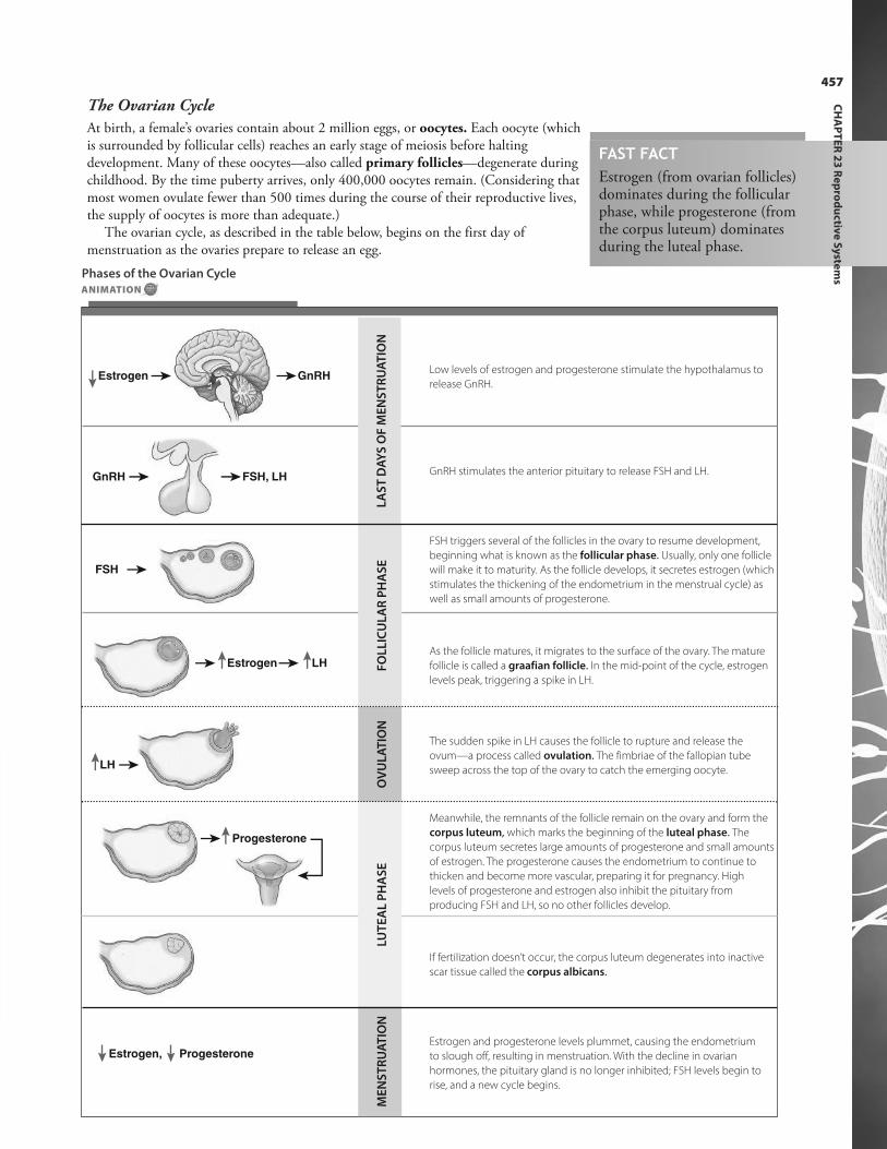
The ovarian cycle, as described in the table below, begins on the first day ofmenstruation as the ovaries prepare to release an egg.
FAST FACTEstrogen (from ovarian follicles)dominates during the follicularphase, while progesterone (fromthe corpus luteum) dominatesduring the luteal phase.
Low levels of estrogen and progesterone stimulate the hypothalamus to
release GnRH.
GnRH stimulates the anterior pituitary to release FSH and LH.
FSH triggers several of the follicles in the ovary to resume development,
beginning what is known as the follicular phase. Usually, only one follicle
will make it to maturity. As the follicle develops, it secretes estrogen (which
stimulates the thickening of the endometrium in the menstrual cycle) as
well as small amounts of progesterone.
As the follicle matures, it migrates to the surface of the ovary. The mature
follicle is called a graafian follicle. In the mid-point of the cycle, estrogen
levels peak, triggering a spike in LH.
The sudden spike in LH causes the follicle to rupture and release the
ovum—a process called ovulation. The fimbriae of the fallopian tube
sweep across the top of the ovary to catch the emerging oocyte.
Meanwhile, the remnants of the follicle remain on the ovary and form the
corpus luteum, which marks the beginning of the luteal phase. The
corpus luteum secretes large amounts of progesterone and small amounts
of estrogen. The progesterone causes the endometrium to continue to
thicken and become more vascular, preparing it for pregnancy. High
levels of progesterone and estrogen also inhibit the pituitary from
producing FSH and LH, so no other follicles develop.
If fertilization doesn’t occur, the corpus luteum degenerates into inactive
scar tissue called the corpus albicans.
Estrogen and progesterone levels plummet, causing the endometrium
to slough off, resulting in menstruation. With the decline in ovarian
hormones, the pituitary gland is no longer inhibited; FSH levels begin to
rise, and a new cycle begins.
FSH
ProgesteroneEstrogen,
Progesterone
LH
Estrogen LH
GnRH FSH, LH
Estrogen GnRHL
AS
T D
AY
S O
F M
EN
ST
RU
AT
ION
FO
LL
ICU
LA
R P
HA
SE
OV
UL
AT
ION
LUT
EA
L P
HA
SE
ME
NS
TR
UA
TIO
N
Phases of the Ovarian Cycle
ANIMATION
457
CH
AP
TE
R 2
3 R
ep
rod
uctiv
e S
yste
ms

PA
RT
V C
on
tinu
ity
458
The Menstrual Cycle
The hormones estrogen and progesterone—which are secreted by the ovaries—drive the menstrual cycle. This cycle involves thebuildup of the endometrium (which occurs through most of the ovarian cycle) followed by its breakdown and discharge. Themenstrual cycle is divided into four phases: the menstrual phase, proliferative phase, secretory phase, and premenstrual phase.
Phase Days Activity
Menstrual 1 to 5 The first day of noticeable vaginal bleeding is the first day of
the menstrual cycle. Lasting from 3 to 5 days, menstruation
occurs as the endometrium sheds its functional layer (the
stratum functionalis).
Proliferative 6 to 14 ≠estrogen
= growth of blood vessels
When menstruation ceases (about day 5 of the cycle), only
the base layer (stratum basalis) remains in the uterus. About
day 6, rising levels of estrogen (secreted by the ovaries)
stimulates the repair of the base layer as well as the growth
of blood vessels. During this stage, the endometrium
thickens to 2 to 3 mm.
OVULATION
Secretory 15 to 26 ≠progesterone
= ≠endometrial thickening
After ovulation (about day 14), increased progesterone from
the corpus luteum causes the functional layer to thicken
even more, this time as a result of secretion and fluid
accumulation. During this phase, the endometrium
develops into a nutritious bed about 5 to 6 mm thick, just
right for a fertilized ovum.
Premenstrual 26 to 28 Ø progesterone = ischemic
endometrium
If fertilization doesn’t occur, the corpus luteum atrophies
and progesterone levels plummet. Blood vessels nourishing
the endometrium spasm, interrupting blood flow. The
endometrium becomes ischemic and necrotic, causing it to
slough off the uterine wall. This forms the menstrual flow.
Phases of the Menstrual Cycle

459
CH
AP
TE
R 2
3 R
ep
rod
uctiv
e S
yste
ms
Interrelationship between the Ovarian Cycle and the Menstrual Cycle
As previously discussed, the ovarian and menstrual cycles are interrelated, with activities in both cycles occurringsimultaneously. Study the chart below to tie the activities of the two cycles together and to link each to the fluctuations inhormone levels.
Days
Menstruation
Menstruation
Proliferative phase Secretory phase Premenstrual
phase
Endom
etr
ium
Horm
ones
0 2 4 6 8 10 12 14 16 18 20 22 24 26 28 30
Estrogen
ProgesteroneOvulation
FSH
LH
Days
Follicular phase Ovulation Luteal phase
Egg
deve
lopm
ent
Horm
ones
0 2 4 6 8 10 12 14 16 18 20 22 24 26 28 30
Ovulation
Developing follicleMature follicle
Early corpus
luteum Regressing
corpus luteum Corpus
albicans
Uterine cycle
Ovarian cycle
Life lesson: MenopauseMenstruation continues from puberty until about the age of 45 or 50, when itceases. Called menopause, this stage of life is associated with declining estrogenand progesterone levels (as the remaining ovarian follicles are less responsive togonadotropins). As a result of the declining hormone levels, the uterus, vagina,and breasts atrophy. Vaginal dryness can make intercourse uncomfortable andvaginal infections more common. Symptoms of menopause vary, althoughcommon symptoms include hot flashes and mood changes.
PA
RT
V C
on
tinu
ity
460
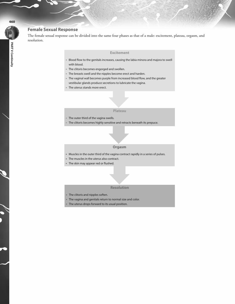
Female Sexual Response
The female sexual response can be divided into the same four phases as that of a male: excitement, plateau, orgasm, andresolution.
Plateau
• The outer third of the vagina swells.
• The clitoris becomes highly sensitive and retracts beneath its prepuce.
Orgasm
• Muscles in the outer third of the vagina contract rapidly in a series of pulses.
• The muscles in the uterus also contract.
• The skin may appear red or flushed.
Resolution
• The clitoris and nipples soften.
• The vagina and genitals return to normal size and color.
• The uterus drops forward to its usual position.
Excitement
• Blood flow to the genitals increases, causing the labia minora and majora to swell
with blood.
• The clitoris becomes engorged and swollen.
• The breasts swell and the nipples become erect and harden.
• The vaginal wall becomes purple from increased blood flow, and the greater
vestibular glands produce secretions to lubricate the vagina.
• The uterus stands more erect.
461
CH
AP
TE
R 2
3 R
ep
rod
uctiv
e S
yste
ms
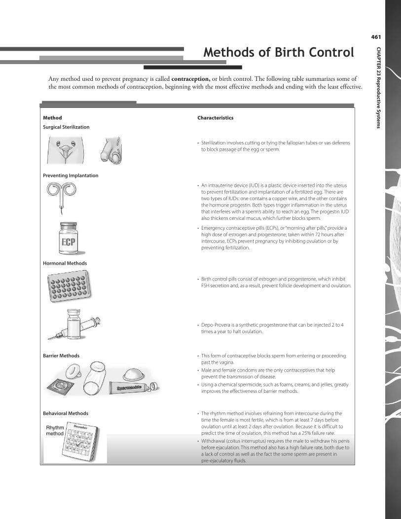
Any method used to prevent pregnancy is called contraception, or birth control. The following table summarizes some ofthe most common methods of contraception, beginning with the most effective methods and ending with the least effective.
Method Characteristics
Surgical Sterilization
• Sterilization involves cutting or tying the fallopian tubes or vas deferens
to block passage of the egg or sperm.
Preventing Implantation
• An intrauterine device (IUD) is a plastic device inserted into the uterus
to prevent fertilization and implantation of a fertilized egg. There are
two types of IUDs: one contains a copper wire, and the other contains
the hormone progestin. Both types trigger inflammation in the uterus
that interferes with a sperm’s ability to reach an egg. The progestin IUD
also thickens cervical mucus, which further blocks sperm.
• Emergency contraceptive pills (ECPs), or “morning after pills,” provide a
high dose of estrogen and progesterone; taken within 72 hours after
intercourse, ECPs prevent pregnancy by inhibiting ovulation or by
preventing fertilization.
Hormonal Methods
• Birth control pills consist of estrogen and progesterone, which inhibit
FSH secretion and, as a result, prevent follicle development and ovulation.
• Depo-Provera is a synthetic progesterone that can be injected 2 to 4
times a year to halt ovulation.
Barrier Methods • This form of contraceptive blocks sperm from entering or proceeding
past the vagina.
• Male and female condoms are the only contraceptives that help
prevent the transmission of disease.
• Using a chemical spermicide, such as foams, creams, and jellies, greatly
improves the effectiveness of barrier methods.
Behavioral Methods
12 3
4 5 687
9 10 11 12 1315
1416 17 18 19 2022
2123
24 25 26 272928
3031
DecemberRhythmmethod
• The rhythm method involves refraining from intercourse during the
time the female is most fertile, which is from at least 7 days before
ovulation until at least 2 days after ovulation. Because it is difficult to
predict the time of ovulation, this method has a 25% failure rate.
• Withdrawal (coitus interruptus) requires the male to withdraw his penis
before ejaculation. This method also has a high failure rate, both due to
a lack of control as well as the fact the some sperm are present in
pre-ejaculatory fluids.
Methods of Birth Control

PA
RT
V C
on
tinu
ity
462
Review of Key TermsAmpulla: Middle portion of thefallopian tube
Cervix: Inferior end of the uterus
Corpus albicans: Inactive scar tissuethat results when the corpus luteumdegenerates
Corpus luteum: Remnants of theovarian follicle after ovulation thatsecretes large amounts of progesteroneand small amounts of estrogen
Endometrium: Vascular mucousmembrane lining the uterus; thickenseach cycle in anticipation of receivinga fertilized egg
Epididymis: Convoluted tube restingon the side of the testes in whichsperm mature
Estrogen: Hormone secreted by theovaries that is responsible forstimulating development of femalesecondary sex characteristics; it alsoplays a role in triggering ovulation
Fallopian tubes: Tubes extending fromnear the ovary to the uterus
Gametes: Sex cells, which include thesperm in males and eggs in females
Gonad: Primary sex organs; includesthe testes in males and the ovaries infemales
Graafian follicle: A mature follicle ofthe ovary
Infundibulum: Funnel-shaped, distalend of the fallopian tube
Isthmus: Portion of the fallopian tubeclosest to the uterus
Meiosis: Process of cell divisionproducing cells (eggs or sperm) thatcontain half the number ofchromosomes found in somatic cells
Menopause: The period that marks thepermanent cessation of menstruation
Menstruation: Cyclical shedding ofuterine endometrium
Myometrium: Smooth muscle layer ofthe uterus; contracts during delivery
Oocyte: Immature egg
Oogenesis: Process whereby a matureovum is formed
Ovarian follicle: Oocyte and surroundingfollicular cells
Perimetrium: Outer serous layer ofuterine wall
Prostate gland: Gland that surroundsthe neck of the bladder and urethra inmales; secretes alkaline fluid thatforms part of semen
Scrotum: Sac of tissue surrounding thetestes
Semen: Whitish fluid containingsperm emitted during ejaculation
Seminiferous tubules: Tiny ducts inthe testes in which sperm areproduced
Spermatogenesis: Sperm formation thattakes place in the seminiferous tubulesof the testicles
Testes: Male organs that manufacturesperm and produce the male hormonetestosterone
Testosterone: Primary male sexhormone; secreted by the testes
Uterus: Muscular chamber that housesand nurtures a growing embryo andfetus
Vas deferens: Tube that carries spermout of the epididymis to theejaculatory duct
Own the InformationTo make the information in this chapter part of your
working memory, take some time to reflect on what you’ve
learned. On a separate sheet of paper, write down
everything you recall from the chapter. After you’re done,
log on to the DavisPlus website, and check out the Study
Group podcast and Study Group Questions for the chapter.
Key Topics for Chapter 23:
• Primary and secondary sex organs
• Structure and function of the testes
• Structure and function of the male accessory glands
• Structure and function of the penis
• Process of male puberty
• Formation of sperm
• Components of semen
• Male sexual response
• Structure and function of the ovaries, fallopian tubes,
uterus, and vagina
• Female external genitalia
• Structure of the female breast
• Process of female puberty
• Female reproductive cycle
• Female sexual response

Test Your Knowledge1. The first hormone secreted at the
onset of puberty in both malesand females is:a. testosterone.b. follicle-stimulating hormone.c. gonadotropin-releasing
hormone.d. progesterone.
2. Gametes are:a. primary sex organs.b. sex cells.c. immature sperm.d. immature ova.
3. Until ejaculation, sperm arestored in the:a. vas deferens.b. seminiferous tubules.c. seminal vesicle.d. epididymis.
4. Where is testosterone produced?a. Seminiferous tubulesb. Interstitial cells of the testesc. Epididymisd. Sustentacular (Sertoli) cells
5. Which organ supplies most ofthe fluid volume of semen?a. Bulbourethral glandb. Penisc. Seminal vesiclesd. Prostate
6. The surge in which hormonecauses ovulation?a. Follicle-stimulating hormoneb. Luteinizing hormonec. Estrogend. Progesterone
7. An embryo attaches to whichlayer of the uterine wall?a. Perimetriumb. Endometriumc. Myometriumd. Vestibule
8. Falling levels of which two hormones trigger menstruation?a. FSH and LHb. Estrogen and progesteronec. GnRH and FSHd. Estrogen and testosterone
9. The structure that secretes progesterone during the last halfof the ovarian cycle is the:a. corpus albicans.b. ovarian follicle.c. acini.d. corpus luteum.
10. Birth control pills prevent pregnancy by:a. preventing implantation of a
fertilized egg.b. changing the acidity of the
vagina to kill sperm.c. interfering with follicular
development and ovulation.d. blocking the passage of an egg
through the fallopian tube.
Answers: Chapter 231. Correct answer: c. Testosterone stimulates the
development of male secondary sex characteristics;however, testosterone is secreted only after thetestes have begun to develop, a result of thesecretion of gonadotropin-releasing hormone(GnRH). GnRH triggers the release of follicle-stimulating hormone (FSH) andluteinizing hormone (LH), which promotetesticular growth and, ultimately, testosteronesecretion. Progesterone is secreted by ovarianfollicles, which occurs only after GnRH stimulatesthe secretion of FSH and LH.
2. Correct answer: b. Primary sex organs, calledgonads, produce gametes. Immature sperm arecalled spermatogonia or spermatocytes. Immatureova are called primary follicles.
3. Correct answer: d. The vas deferens carries thesperm from the epididymis to the ejaculatory duct.The seminiferous tubules are the tiny ducts inwhich sperm are produced. The seminal vesiclessecrete fluid into the ejaculatory duct to help formthe fluid portion of semen.
4. Correct answer: b. Seminiferous tubules are ductsin which sperm are produced. The epididymisprovides a place for sperm to mature and remainuntil ejaculation. The sustentacular (Sertoli) cellssupply nutrients to sperm; they also secrete thehormone inhibin, which plays a role in thematuration and release of sperm.
5. Correct answer: c. The bulbourethral glandsupplies 5% of the fluid volume of semen, whilethe prostate supplies 30% (as opposed to the 65%supplied by the seminal vesicles). The penis doesnot supply any of the fluid volume of semen.
6. Correct answer: b. Follicle-stimulating hormoneprompts ovarian follicles to resume development.A peak in estrogen levels triggers the release of LH.Progesterone is secreted by the corpus luteum tomaintain the vascular endometrial lining.
7. Correct answer: b. The perimetrium is the outwardserous lining of the uterus. The myometrium is themuscular layer that contracts during the deliveryof a fetus. The vestibule is the area between thelabia that contains openings to the urethra andvagina.
8. Correct answer: b. None of the other hormonepairs influence menstruation. Testosterone isprimarily a male sex hormone.
9. Correct answer: d. The corpus albicans is inactivescar tissue left behind by the corpus luteum. Theovarian follicle develops before ovulation. Theacini are sac-like structures in the female breastthat secrete milk.
10. Correct answer: c. An IUD prevents implantationof a fertilized egg. Birth control pills do not act tochange the acidity of the vagina for the purpose ofkilling sperm. Surgical sterilization blocks thepassage of an egg through the fallopian tube.
463
CH
AP
TE
R 2
3 R
ep
rod
uctiv
e S
yste
ms
Go to http://davisplus.fadavis.com Keyword:Thompson to see all of the resources availablewith this chapter.

CHAPTER OUTLINEFertilization
Stages of Prenatal Development
Physical Changes During Pregnancy
Childbirth
Lactation
The Neonate
LEARNING OUTCOMES1. Discuss the process of fertilization, including
when and where it occurs and how the egg
prevents fertilization by more than one sperm.
2. Describe the events of the preembryonic stage
of development.
3. Summarize the process of implantation and
the changes that occur in the blastocyst.
4. Name the three germ layers and identify the
major organs and tissues arising from each.
5. Identify the four extraembryonic membranes
and describe the functions of each.
6. Describe the structure and functions of the
placenta.
7. Trace the path of the fetal circulatory system.
8. Describe the major events of fetal
development.
9. List the key physical changes that occur during
pregnancy.
10. Identify three factors thought to trigger labor.
11. Identify the three stages of labor and describe
the actions, as well as the duration, of each
stage.
12. Name the hormones that promote
development of the mammary glands for
lactation.
13. Describe the process of milk production and
milk secretion.
14. Discuss some of the changes experienced by a
neonate immediately after delivery.
24chapter PREGNANCY & HUMAN DEVELOPMENTThe human body—which contains 100 trillion cells and
thousands of organs—begins as a single cell.
For new life to begin, an egg and a sperm must meet and fuse together. The instant that occurs, the fertilized egg begins aseries of changes that, amazingly, transforms a single cell into a fully developed human being. Consider: from that one cellcome 100 trillion cells—cells that, in turn, evolve into tissues as diverse as skin, nerves, and blood, and organs as varied asthe kidneys, brain, and heart. Indeed, from one cell come not just your physical body but also your mind, your emotions,and your intellect. The process of human development, from conception until birth, is perhaps the most fascinating andmiraculous aspect of human life.
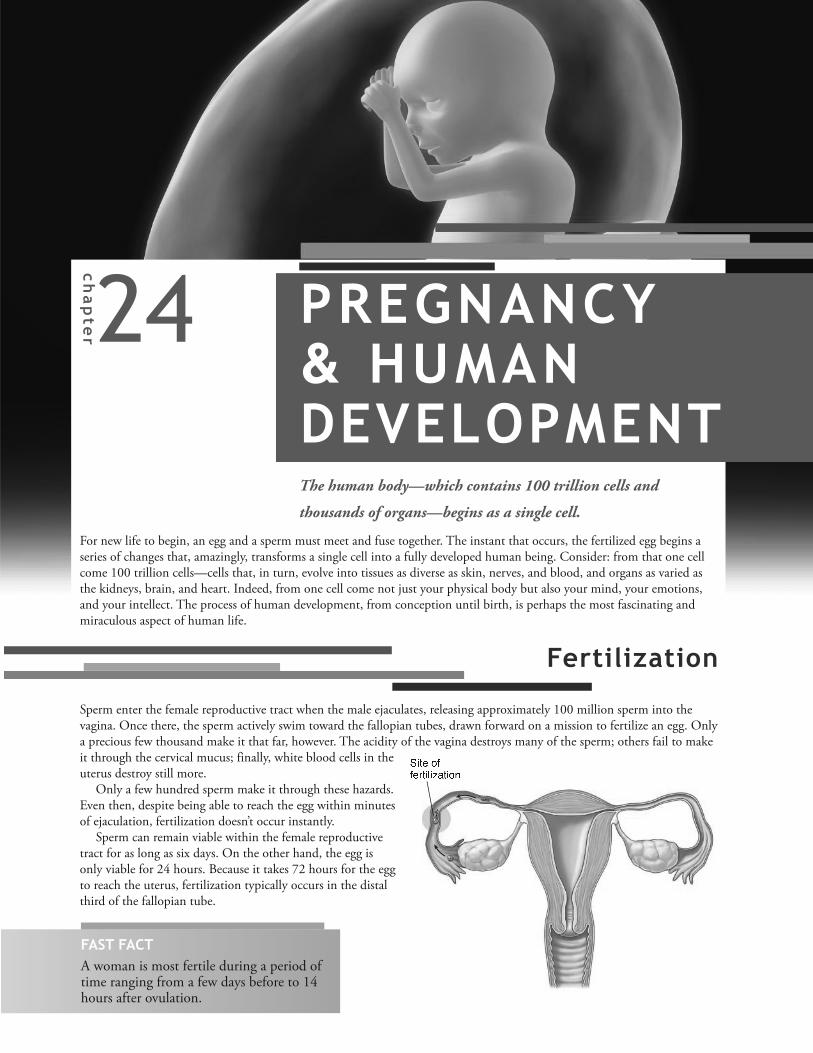
FertilizationSperm enter the female reproductive tract when the male ejaculates, releasing approximately 100 million sperm into thevagina. Once there, the sperm actively swim toward the fallopian tubes, drawn forward on a mission to fertilize an egg. Onlya precious few thousand make it that far, however. The acidity of the vagina destroys many of the sperm; others fail to makeit through the cervical mucus; finally, white blood cells in theuterus destroy still more.
Only a few hundred sperm make it through these hazards.Even then, despite being able to reach the egg within minutesof ejaculation, fertilization doesn’t occur instantly.
Sperm can remain viable within the female reproductivetract for as long as six days. On the other hand, the egg isonly viable for 24 hours. Because it takes 72 hours for the eggto reach the uterus, fertilization typically occurs in the distalthird of the fallopian tube.
= > ? @ A BB @ C ? > D > E F ? > A GFAST FACTA woman is most fertile during a period oftime ranging from a few days before to 14hours after ovulation.

PA
RT
V C
on
tinu
ity
466
Even though only one sperm actually fertilizes the egg, a team of sperm helps make fertilization possible by clearing a paththrough the layer of cells and glycoprotein membrane (the zona pellucida) encasing the ovum. The following figuredescribes this process. Keep in mind that this is a “time lapse” view of fertilization: although many sperm assist withfertilization, only one sperm actually enters the egg.
1
2
3
H D A I J @ K L M @ C NO A G F M @ D D P I > K F
Q C F G P D A L F I @ D D L
Life lesson: In vitro fertilizationCouples experiencing infertility, particularly women withblocked or damaged fallopian tubes, may choose toundergo in vitro fertilization (IVF) in an effort to conceive.To perform the procedure, a doctor retrieves eggs from thewoman’s ovary using a needle inserted through the vagina.At the same time, the man provides a semen sample. Theactive sperm are then combined with the retrieved eggs ina laboratory dish. After about 18 hours in a temperature-controlled environment, the eggs are examined. Iffertilization has occurred, the eggs are kept in an incubatorfor 2 or 3 more days to allow them to grow into the 8- or 16-cell stage. At that point, the doctor transfers thedeveloping embryos into the woman’s uterus by way of acatheter inserted through the woman’s vagina and cervix. Ifimplantation occurs, the pregnancy test is positive and thepregnancy proceeds.
It’s estimated that since 1981 (when IVF was used for thefirst time), 5 million babies have been born as a result ofthis procedure. Even so, a normal term birth occurs onlyabout 30% of the time following IVF.
The Body AT WORKPregnancy, or gestation, ranges from conception until
birth and lasts about 266 days. (Typically, gestation is
measured from the first day of the last menstrual
period, making the time until birth about 40 weeks or
280 days.)
• The duration of pregnancy is divided into
three-month periods called trimesters.
• The first trimester lasts from conception through
the first 12 weeks. (During this period of time, the
developing embryo is most susceptible to toxins,
stress, drugs, and nutritional deficiencies.)
• The second trimester ranges from week 13
through week 24. (Most of the organs are
developed during this phase.)
• The third trimester lasts from week 25 until birth.
Most infants are viable after about 35 weeks.
As hundreds of sperm swarm the egg, the
acrosomes on the sperm heads release enzymes
that break down the cells and the zona pellucida.
Due to the efforts of multiple sperm, a path
through the zona pellucida eventually results,
allowing a single sperm to penetrate. As soon as
this happens, the egg undergoes changes that
bar any other sperm from entering.
The nucleus of the sperm is released into
the ovum as its tail degenerates and falls
away. The nucleus of the sperm (which has
23 chromosomes) fuses with the nucleus of
the egg (which also has 23 chromosomes),
creating a single cell with 46 chromosomes.
The fertilized egg is now called a zygote.

467
CH
AP
TE
R 2
4 P
reg
na
ncy
& H
um
an
De
ve
lop
me
nt
Ovum
First
mitosis
Blastomere
Morula
Implantation
Blastocyst
Inner cell mass
Trophoblast
Ovary
The union of egg and sperm ignites a period of development that ends with the birth of a baby. This period of growthbefore birth is called the prenatal period. During this time, the fetus undergoes three major stages of development:
l The preembryonic stage, which begins at fertilization and lasts for 16 daysl The embryonic stage, which begins after the sixteenth day and lasts until the eighth weekl The fetal stage, which begins the eighth week and lasts until birth
Preembryonic Stage
Shortly after fertilization, the fertilized cell divides by mitosis—a process called cleavage—to produce two identicaldaughter cells. The mitotic divisions continue, with each division doubling the number of cells, until the zygote arrives atthe uterus. The following illustration portrays this sequence of events, beginning with ovulation and ending withimplantation of a fertilized egg.
The preembryonic stage
begins when fertilization
forms a zygote with 46
chromosomes.
Within 24 to 36 hours, the zygote
divides by mitosis to form
two daughter cells called
blastomeres.
The mitotic divisions, or cleavage, continue, with the cells
doubling with each division. Finally, a blackberry-like
cluster of 16 cells called a morula results. Three to four days
after fertilization, the morula enters the uterine cavity,
where it floats for two or three days.
As the morula continues to divide, a hollow cavity forms; the morula is now
called a blastocyst. The blastocyst consists of an outer layer of cells (the
trophoblast) and an inner cell mass. The trophoblast eventually forms the
placenta while the inner cell mass becomes the embryo.
About six days after ovulation, the
blastocyst attaches to the
endometrium—a process called
implantation.
1 2 3
45
Stages of Prenatal Development
ANIMATION
FAST FACTThe detection of HCG in themother’s blood or urine forms thebasis for pregnancy tests. In fact,HCG may be detectable within 8 to 10 days following fertilization.
PA
RT
V C
on
tinu
ity
468
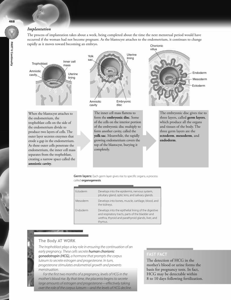
Implantation
The process of implantation takes about a week, being completed about the time the next menstrual period would haveoccurred if the woman had not become pregnant. As the blastocyst attaches to the endometrium, it continues to changerapidly as it moves toward becoming an embryo.
Uterine
lining
Inner cell
mass
Amniotic
cavity
Trophoblast
Uterine lining
Embryonicdisc
Amniotic cavity
Yolk sac
Chorionicvillus
Endoderm
Mesoderm
Ectoderm
When the blastocyst attaches tothe endometrium, thetrophoblast cells on the side ofthe endometrium divide toproduce two layers of cells. Theouter layer secretes enzymes thaterode a gap in the endometrium.As these outer cells penetrate theendometrium, the inner cell massseparates from the trophoblast,creating a narrow space called theamniotic cavity.
The inner cell mass flattens toform the embryonic disc. Someof the cells on the interior portionof the embryonic disc multiply toform another cavity, called theyolk sac. Meanwhile, the rapidlygrowing endometrium covers thetop of the blastocyst, burying itcompletely.
The embryonic disc gives rise tothree layers, called germ layers,which produce all the organsand tissues of the body. Thethree germ layers are theectoderm, mesoderm, andendoderm.
The Body AT WORKThe trophoblast plays a key role in ensuring the continuation of an
early pregnancy. These cells secrete human chorionic
gonadotropin (HCG), a hormone that prompts the corpus
luteum to secrete estrogen and progesterone. In turn,
progesterone stimulates endometrial growth and prevents
menstruation.
For the first two months of a pregnancy, levels of HCG in the
mother’s blood rise. By that time, the placenta begins to secrete
large amounts of estrogen and progesterone—effectively taking
over the role of the corpus luteum—and the levels of HCG decline.
Germ layers: Each germ layer gives rise to specific organs, a process
called organogenesis.
Ectoderm Develops into the epidermis, nervous system,
pituitary gland, optic lens, and salivary glands.
Mesoderm Develops into bones, muscle, cartilage, blood, and
the kidneys.
Endoderm Develops into the epithelial lining of the digestive
and respiratory tracts, parts of the bladder and
urethra, thyroid and parathyroid glands, liver, and
thymus.
469
CH
AP
TE
R 2
4 P
reg
na
ncy
& H
um
an
De
ve
lop
me
nt
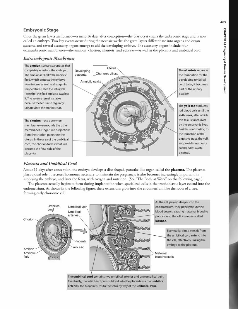
Placenta and Umbilical Cord
About 11 days after conception, the embryo develops a disc-shaped, pancake-like organ called the placenta. The placentaplays a dual role: it secretes hormones necessary to maintain the pregnancy; it also becomes increasingly important insupplying the embryo, and later the fetus, with oxygen and nutrition. (See “The Body at Work” on the following page.)
The placenta actually begins to form during implantation when specialized cells in the trophoblastic layer extend into theendometrium. As shown in the following figure, these extensions grow into the endometrium like the roots of a tree,forming early chorionic villi.
Uterus
Chorionic villusDeveloping
placenta
Amniotic cavity
Umbilical vein
Umbilical arteries
Maternal blood vessels
Placenta
Chorion
Umbilical cord
Yolk sacAmnion
Amnioticfluid
The amnion is a transparent sac that
completely envelops the embryo.
The amnion is filled with amniotic
fluid, which protects the embryo
from trauma as well as changes in
temperature. Later, the fetus will
“breathe” the fluid and also swallow
it. The volume remains stable
because the fetus also regularly
urinates into the amniotic sac.
The chorion—the outermost
membrane—surrounds the other
membranes. Finger-like projections
from the chorion penetrate the
uterus. In the area of the umbilical
cord, the chorion forms what will
become the fetal side of the
placenta.
The allantois serves as
the foundation for the
developing umbilical
cord. Later, it becomes
part of the urinary
bladder.
The yolk sac produces
red blood cells until the
sixth week, after which
this task is taken over
by the embryonic liver.
Besides contributing to
the formation of the
digestive tract, the yolk
sac provides nutrients
and handles waste
disposal.
As the villi project deeper into the
endometrium, they penetrate uterine
blood vessels, causing maternal blood to
pool around the villi in sinuses called
lacunae.
Eventually, blood vessels from
the umbilical cord extend into
the villi, effectively linking the
embryo to the placenta.
The umbilical cord contains two umbilical arteries and one umbilical vein.
Eventually, the fetal heart pumps blood into the placenta via the umbilical
arteries; the blood returns to the fetus by way of the umbilical vein.
Embryonic Stage
Once the germ layers are formed—a mere 16 days after conception—the blastocyst enters the embryonic stage and is nowcalled an embryo. Two key events occur during the next six weeks: the germ layers differentiate into organs and organsystems, and several accessory organs emerge to aid the developing embryo. The accessory organs include fourextraembryonic membranes—the amnion, chorion, allantois, and yolk sac—as well as the placenta and umbilical cord.
Extraembryonic Membranes

One
placenta
Identical twins
One egg andone sperm
Two
placentas
Fraternal twins
Two eggs andtwo sperm
Life lesson: TwinsMost twins result when two eggs are ovulated andthen fertilized by separate sperm. These twins—called dizygotic or fraternal twins—do not have thesame genetic information. They may be the same,or different, gender. Because they’re formed fromthe union of different eggs and different sperm,they are no more similar than are siblings who areborn on separate occasions. Each twin implants ona different part of the uterine wall, and eachdevelops its own placenta.
Occasionally, twins result when a fertilized eggdivides in two. In this instance, the twins are thesame sex and carry identical genetic information;they are called monozygotic or identical twins.Monozygotic twins almost always share the sameplacenta, although each develops in a separateamniotic sac.
PA
RT
V C
on
tinu
ity
470
FAST FACTAspirating and testing a sample of amniotic fluid, ortesting a tissue sample of a chorionic villus, can revealvaluable genetic information about the developing fetus.The test carries certain risks, however, including miscarriage, infection, or the leakage of amniotic fluid.
The Body AT WORKThe fetal stage begins the eighth week, and, by the twelfth week, the placenta is the fetus’ sole source of nutrition.
Although the mother’s blood furnishes the developing fetus with nutrients, maternal and fetal blood do not actually
mix. Instead, the chorionic villi are filled with fetal blood and surrounded by maternal blood. A thin layer of placental
cells separates the two blood systems.
Unfortunately, some toxins such as nicotine, alcohol, and most drugs can also cross the placenta. When they do,
they can have a devastating effect on embryonic development.
The placenta also serves an endocrine function, secreting hormones necessary for the continuation of the
pregnancy. These hormones include estrogen, progesterone, and HCG.
Umbilical vein
Umbilical artery
Maternal arteryMaternal vein
Fetal waste products move from fetal
blood in the umbilical arteries to the
maternal blood; the maternal veins
carry away the waste for disposal.
Oxygen, nutrients, and some antibodies
pass from the maternal blood—which is
pooled in the lacunae around the chorionic
villi—to fetal blood in the umbilical veins
of the placenta.

471
CH
AP
TE
R 2
4 P
reg
na
ncy
& H
um
an
De
ve
lop
me
nt
Fetal Stage
The fetal stage, which is the final stage of prenatal development, encompasses the period from the eighth week until birth.This is primarily a stage of growth, as the organs that formed during the embryonic period grow and mature.
Because the fetus depends on the placenta for oxygen and nutrients as well as for the removal of waste products, thecirculatory system of the fetus differs significantly from that of a newborn. In the fetus, neither the lungs nor the liverrequires a great deal of blood: the lungs are nonfunctioning and the liver is still immature. Therefore, the fetus’ circulatorysystem contains three shunts that allow blood to, for the most part, bypass these organs:
l The ductus venosus shunts blood around the liver.l The foramen ovale, an opening between the two atria, shunts blood directly from the right atrium to the left.l The ductus arteriosus diverts blood from right ventricle to the pulmonary artery, bypassing the lungs.
The following figure details circulation in the fetus.
1
2
3
4
5
Ductus arteriosus
Pulmonary trunk
Common
iliac artery
Ascending
aorta
Inferior
vena cava
Placenta
Fetal umbilicus
Ductus
venosus
Umbilical vein
Umbilical
cord
Umbilical arteries
Foramen
ovale
High
Mixed
Low
Oxygen contentof blood
Oxygen-rich blood enters the fetus through the
vein in the umbilical cord.
Most of the blood bypasses the liver by flowing
through the ductus venosus into the inferior
vena cava (IVC). Placental blood from the umbilical
vein then merges with fetal blood from the IVC as
it flows to the heart.
Blood flows into the right atrium; most of the
blood flows directly into the left atrium through
the foramen ovale, bypassing the lungs.
The blood that does not flow through the foramen
ovale flows into the right ventricle and then into
the pulmonary trunk. From there, the blood flows
through the ductus arteriosus and into the
descending aorta, again bypassing the lungs.
Oxygen-depleted, waste-filled blood flows
through two umbilical arteries to the placenta.
The placenta then cleanses the blood—ridding
it of carbon dioxide and waste products—
reoxygenates it, and returns it to the fetus through
the umbilical vein.
12
3
4
5
The Body AT WORKWith the neonate’s first breath, fetal circulation changes. As
soon as the lungs are called upon to supply the fetus with
oxygen, they demand a larger supply of blood. To meet this
need, the ductus arteriosus closes so that blood no longer
bypasses the lungs. Then, when blood flows into the left
atrium after circulating through the lungs, the newly arriving
blood increases the pressure in the left atrium. The increased
pressure pushes back the flaps of the foramen ovale and
closes the hole. Finally, the ductus venosus deteriorates,
eventually becoming a ligament in the liver.
That Makes SenseWhen you think of the placenta as the center of the fetus’
universe, the following makes more sense:
• Umbilical arteries pump oxygen-poor, waste-filled blood
away from the fetus and toward the placenta.
• The umbilical vein carries oxygenated blood away from
the placenta and toward the fetus.
However, much of the fetus’ blood is a blend of oxygenated
and unoxygenated blood.
!
ANIMATION

Week 12
• The face is well formed.
• The arms are long and thin.
• The sex is distinguishable.
• The liver produces bile.
• The fetus swallows amniotic fluid and produces urine.
• The eyes are well developed but the eyelids are fused
shut.
• Length: 3.54 inches (9 cm)
Week 4
• The brain, spinal cord, and heart begin to develop.
• The gastrointestinal tract begins to form.
• The heart begins to beat about day 22.
• Tiny buds that will become arms and legs are visible.
• Length: 0.25 inch (0.6 cm)
PA
RT
V C
on
tinu
ity
472
Fetal Development
During the first three months following conception, the outward appearance of the embryo changes rapidly as it developsinto a fetus. During the last six months, the organs that formed during the embryonic stage mature and become functional.The fetus also continues to grow and accumulate fat stores.
Week 8
• The embryo is now a fetus.
• Eyes, ears, nose, lips, tongue, and tooth buds take shape.
• Head is nearly as large as the rest of the body.
• Brain waves are detectable.
• The arms and legs are recognizable.
• Blood cells and major blood vessels form.
• Bone calcification begins.
• Genitals are present but gender is not distinguishable.
• Length: 1.2 inches (3 cm)
Week 16
• The scalp has hair.
• The lips begin sucking movements.
• The skeleton is visible.
• The heartbeat can be heard with a stethoscope.
• The kidneys are well formed.
• Length: 5.5 inches (14 cm)
FAST FACTBetween the fourteenth and twenty-secondweeks of pregnancy, maternal blood is oftenscreened for alpha-fetoprotein (AFP). AFP is aprotein produced by the fetal yolk sac and,later, by the fetal liver. High levels of AFP suggest certain abnormalities, such as a neuraltube defect in the developing fetus. Low levelssuggest a chromosomal abnormality, such asDown syndrome.

Week 36
• More subcutaneous fat is deposited.
• Lanugo has mostly disappeared, although it’s still
present on the upper arms and shoulders.
• Length: 18.5 inches (47 cm)
Week 20
• A fine hair called lanugo covers the body, which, in turn
is covered by a white cheese-like substance called vernix
caseosa; both these substances protect the fetus’ skin
from amniotic fluid.
• Fetal movement (quickening) can be felt.
• Nails appear on fingers and toes.
• Length: 8 inches (20 cm)
473
CH
AP
TE
R 2
4 P
reg
na
ncy
& H
um
an
De
ve
lop
me
nt
Week 24
• The fetus has a startle reflex.
• Lungs begin producing surfactant, a lipid and protein
mixture that reduces alveolar surface tension.
• Skin is wrinkled and translucent.
• The fetus gains weight rapidly.
• Length: 11.8 inches (30 cm)
Week 28
• The eyes open and close.
• The respiratory system, although immature, is capable of
gas exchange at 28 weeks.
• Testes begin to descend into the scrotum.
• The brain develops rapidly.
• Length: 14.8 inches (37.6 cm)
Week 32
• The amount of body fat increases rapidly.
• Rhythmic breathing movements begin, although lungs
are still immature.
• The bones are fully formed, although they are still soft.
• Length: 16.7 inches (42 cm)
FAST FACTExperts now define “full term” as being a two-week window starting at 39 weeks becausethose newborns tend to have the best healthoutcomes. Those born during a two-weekwindow starting at 37 weeks are called “earlyterm,” whereas those born during a two-weekwindow starting at 41 weeks are called “lateterm.”
Life lesson: Respiratorydistress syndromeBecause neonates born before 7 months lack pulmonarysurfactant, they typically develop respiratory distresssyndrome (RDS) after delivery. Surfactant serves to keep the alveoli from sticking together during exhalation.Without surfactant, the alveoli collapse every time theneonate exhales. As a result, he must work hard with everybreath, exerting considerable energy just to reinflate thealveoli.
The condition is usually treated with mechanicalventilation that provides air at a positive pressure; thishelps keep the alveoli inflated between breaths. Even so,RDS is the most common cause of neonatal death.
Weeks 39 and 40
• The fetus is considered full term.
• The average full-term infant measures approximately
20 inches (51 cm) long and weighs 7 to 71/2 lbs (3.2 to
3.4 kg).

Pregnancy challenges almost every body system as the woman’s body nourishes and carries the developing fetus. Thefollowing chart lists some of the key changes experienced by pregnant women.
PA
RT
V C
on
tinu
ity
474
FAST FACTEven during the last trimester of pregnancy,pregnant women having a normal weightneed to consume only an additional 300kcal/day to give them the recommendedweight gain of 24 lbs (11 kg).
Body System Changes
Digestive system • Nausea and vomiting (morning sickness) commonly occur during the first three
months; the cause is unknown.
• Constipation often results from decreased intestinal motility.
• Heartburn is a frequent occurrence later in the pregnancy as the enlarging uterus
presses upward on the stomach.
• The basal metabolic rate rises about 15% during the second half of the pregnancy,
and the mother’s appetite increases.
Circulatory system • The mother’s blood volume increases by 30% to 50%.
• Cardiac output increases 30% to 40% by week 27 as the uterus demands more of the
blood supply; at the same time, heart rate also increases.
• Later in the pregnancy, the uterus exerts pressure on the pelvic blood vessels,
interfering with venous return; hemorrhoids, varicose veins, and swelling in the feet
may result.
Respiratory system • Ventilation increases about 50% to meet the increased demands for oxygen caused by
the developing fetus.
• Late in the pregnancy, the enlarged uterus pushes against the diaphragm, often
causing shortness of breath.
• Increased estrogen levels cause the nasal mucosa to swell, resulting in nasal stuffiness.
Urinary system • An increase in aldosterone promotes water and salt retention by the kidneys.
• The glomerular filtration rate increases to deal with the added burden of disposing of
the fetus’ waste; this leads to a slightly elevated urine output.
• Later in the pregnancy, the enlarged uterus presses on the bladder and reduces its
capacity; this leads to increased frequency of urination.
Integumentary system • The skin of the abdomen grows and stretches to accommodate the expanding uterus;
this often leads to stretch marks or striae.
• Skin over the breasts also grows to accommodate the breasts, which enlarge in
preparation for milk production.
Uterus • The uterus enlarges dramatically during pregnancy. During the first 16 weeks of
gestation, it grows from the size of a fist until it occupies most of the pelvic cavity. As
pregnancy continues, the uterus expands until it reaches the level of the xiphoid
process and fills most of the abdominal cavity.
• The uterus increases from its nonpregnant weight of 0.1 lbs (50 g) to about 2 lbs
(900 g) by the end of pregnancy.
Physical Changes During Pregnancy
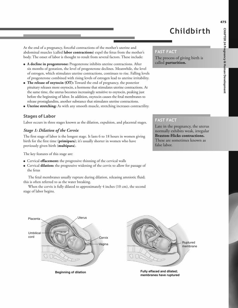
Placenta
Umbilical
cord
Uterus
Vagina
Cervix
Beginning of dilation
Ruptured
membrane
Fully effaced and dilated;
membranes have ruptured
Childbirth
At the end of a pregnancy, forceful contractions of the mother’s uterine andabdominal muscles (called labor contractions) expel the fetus from the mother’sbody. The onset of labor is thought to result from several factors. These include:
l A decline in progesterone: Progesterone inhibits uterine contractions. After six months of gestation, the level of progesterone declines. Meanwhile, the levelof estrogen, which stimulates uterine contractions, continues to rise. Falling levelsof progesterone combined with rising levels of estrogen lead to uterine irritability.
l The release of oxytocin (OT): Toward the end of pregnancy, the posterior pituitary releases more oxytocin, a hormone that stimulates uterine contractions. Atthe same time, the uterus becomes increasingly sensitive to oxytocin, peaking justbefore the beginning of labor. In addition, oxytocin causes the fetal membranes torelease prostaglandins, another substance that stimulates uterine contractions.
l Uterine stretching: As with any smooth muscle, stretching increases contractility.
Stages of Labor
Labor occurs in three stages known as the dilation, expulsion, and placental stages.
Stage 1: Dilation of the Cervix
The first stage of labor is the longest stage. It lasts 6 to 18 hours in women givingbirth for the first time (primipara); it’s usually shorter in women who havepreviously given birth (multipara).
The key features of this stage are:
l Cervical effacement: the progressive thinning of the cervical wallsl Cervical dilation: the progressive widening of the cervix to allow for passage of
the fetus
The fetal membranes usually rupture during dilation, releasing amniotic fluid;this is often referred to as the water breaking.
When the cervix is fully dilated to approximately 4 inches (10 cm), the secondstage of labor begins.
475
FAST FACTThe process of giving birth iscalled parturition.
FAST FACTLate in the pregnancy, the uterusnormally exhibits weak, irregularBraxton-Hicks contractions.These are sometimes known asfalse labor.
CH
AP
TE
R 2
4 P
reg
na
ncy
& H
um
an
De
ve
lop
me
nt
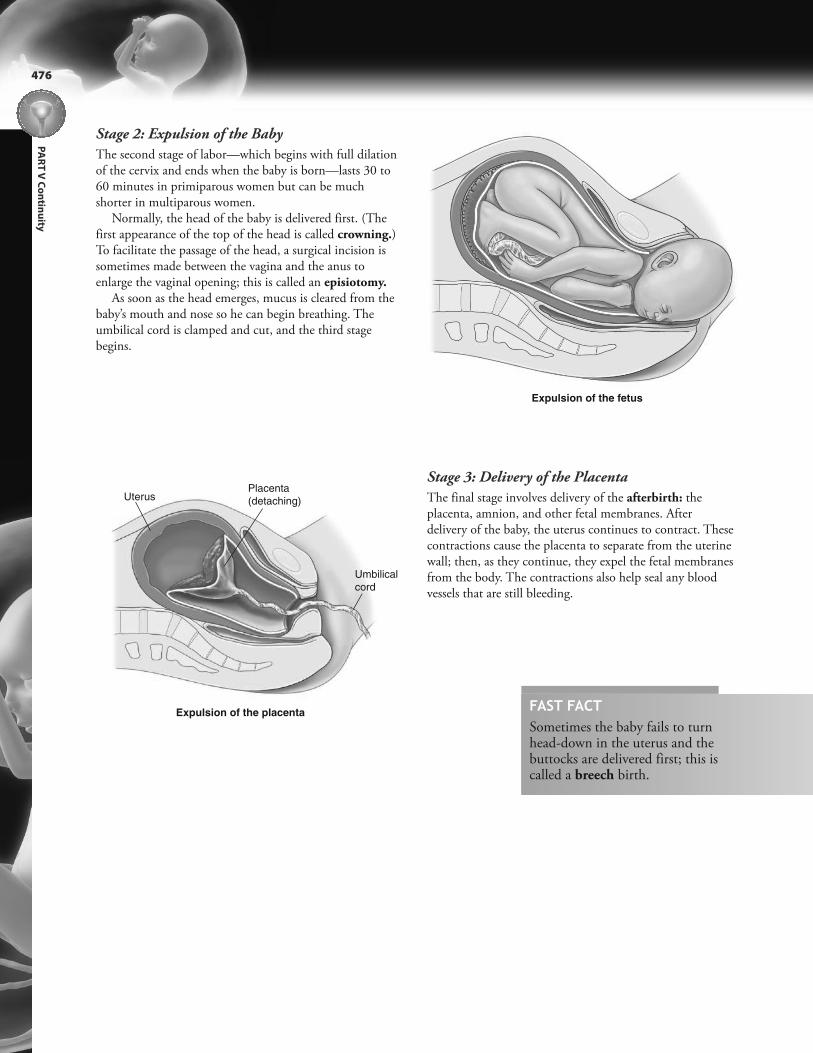
PA
RT
V C
on
tinu
ity
476
Stage 2: Expulsion of the Baby
The second stage of labor—which begins with full dilationof the cervix and ends when the baby is born—lasts 30 to60 minutes in primiparous women but can be muchshorter in multiparous women.
Normally, the head of the baby is delivered first. (Thefirst appearance of the top of the head is called crowning.)To facilitate the passage of the head, a surgical incision issometimes made between the vagina and the anus toenlarge the vaginal opening; this is called an episiotomy.
As soon as the head emerges, mucus is cleared from thebaby’s mouth and nose so he can begin breathing. Theumbilical cord is clamped and cut, and the third stagebegins.
Stage 3: Delivery of the Placenta
The final stage involves delivery of the afterbirth: theplacenta, amnion, and other fetal membranes. Afterdelivery of the baby, the uterus continues to contract. Thesecontractions cause the placenta to separate from the uterinewall; then, as they continue, they expel the fetal membranesfrom the body. The contractions also help seal any bloodvessels that are still bleeding.
Expulsion of the fetus
Expulsion of the placenta
Uterus
Umbilical
cord
Placenta
(detaching)
FAST FACTSometimes the baby fails to turnhead-down in the uterus and thebuttocks are delivered first; this iscalled a breech birth.

477
CH
AP
TE
R 2
4 P
reg
na
ncy
& H
um
an
De
ve
lop
me
nt
The production of milk depends on the hormone prolactin, while the secretion of milk through the nippledepends on the hormone oxytocin.
Anterior
pituitaryPosterior
pituitary
1
23
LactationFollowing childbirth, the mammary glands produce and secrete milk (called lactation) to nourish the newborn child (calleda neonate). The process of preparing the mammary glands, as well as the process of producing and secreting milk, dependson the actions of various hormones. Specifically, development of the structure of the mammary glands depends on estrogenand progesterone, while the hormones prolactin and oxytocin control milk production and secretion.
High levels of progesterone stimulate the
development of acini at the ends of the
ducts.
High levels of estrogen during pregnancy
stimulate the growth of ducts throughout
the mammary glands.
The anterior pituitary secretes prolactin, which initiates
the production of milk. (Every time the neonate nurses,
the mother’s prolactin levels surge; this boosts milk
production for the next feeding.)
Anterior pituitary Ô Prolactin Ô Milk production
The posterior pituitary secretes oxytocin. Oxytocin
causes the lobules in the breast to contract, forcing milk into
the ducts. (This is known as the milk let-down reflex.)
Posterior pituitary Ô Oxytocin Ô Milk secretion
Suckling by the neonate sends nerve impulses to the
anterior and posterior pituitary gland.
The Body AT WORKDuring pregnancy, high levels of estrogen block the secretion of prolactin, keeping milk production at bay. As soon as
the placenta is delivered, the levels of estrogen plummet: the anterior pituitary begins secreting prolactin and milk
production begins. A lag of two to three days occurs, however, between the birth of the baby and the secretion of
milk. In the interim, the breasts secrete a thin, yellowish fluid called colostrum. Colostrum is rich in protein and
immunoglobulins that provide the neonate with passive immunity.
The composition of breast milk changes daily and even over the course of a feeding. The milk secreted at the
beginning of a feeding (called foremilk) is thin, bluish in color, low in fat, and high in carbohydrates. The milk
secreted toward the end of a feeding (called hindmilk) is thicker, whiter, and much higher in fat.
The World Health Organization recommends that women breastfeed exclusively for the first six months of life.
Numerous studies show that breastfeeding lowers risks for sudden infant death syndrome (SIDS), infection, asthma,
eczema, dental problems, and obesity. Breastfeeding also triggers uterine contractions that help reduce postpartum
bleeding and also prompts the uterus’ return to its pre-pregnancy size.
12
3

Life lesson: Apgar scoreThe first few minutes following birth are critical inthe life of a neonate. With this in mind, a scoringsystem, called the Apgar score, is used to rate theneonate’s condition and determine the need forrespiratory support during this timeframe.
At 1 minute and 5 minutes following birth, theneonate is evaluated for heart rate, respiratoryeffort, skin color, muscle tone, and reflexes. Eachcriterion is scored 0, 1, or 2, with 0 being poor and2 being excellent. The five values are then addedtogether to give a total Apgar score. Scores 7 to 10are normal; scores 4 to 6 are fairly low; scores 3 andbelow are critically low. A low Apgar score at 1minute indicates that the neonate needs medicalattention, but it does not necessarily mean that the child will suffer from long-term problems,particularly if the 5-minute score showsimprovement.
Be aware that the Apgar score is used only todetermine the neonate’s immediate need formedical support. It is not designed to predictfuture health problems.
Immediately following birth, the neonate’s body undergoes a number of changes as it adapts to life outside the mother’sbody. These changes affect most body systems.
l Cardiovascular: Pressure changes in the heart cause the foramen ovale to shut, while pressure changes in thepulmonary artery and aorta lead to the collapse of the ductus arteriosus. (The foramen ovale seals permanentlyduring the first year of life, while the ductus arteriosus closes permanently about three months of age.)
l Respiratory: Although most neonates begin breathing spontaneously, the first few breaths require consider-able effort as they work to inflate the collapsed alveoli.
l Immune system: Neonates have weak immune systems at birth, placing them at risk for infection.l Thermoregulation: Neonates risk becoming hypothermic because their surface area, in relationship to their
size, is larger than in an adult.l Fluid balance: Neonates require a fairly high fluid intake because their immature kidneys do not concentrate
urine adequately.
PA
RT
V C
on
tinu
ity
478
The Body AT WORKHuman development continues throughout the
life span. Following the neonatal period (which
encompasses the first four weeks of life), the
developmental stages are:
• Infancy, which lasts until the end of the first
year
• Childhood, which lasts from the beginning
of the second year until puberty
• Adolescence, which lasts from puberty until
about age 19
• Adulthood
Most body systems peak in development and
efficiency during early adulthood. After that, a
gradual but certain decline occurs. This process of
degeneration is called senescence. The process of
aging affects every organ system, with each
experiencing a loss of reserve capacity, an
impaired ability to repair damage, and an
increased susceptibility to disease.
The Neonate
FAST FACTAlthough some body systems (such as thecardiovascular, respiratory, and gastrointestinalsystems) undergo significant change at birth, othersystems take years to develop. For example,myelination of the nervous system isn’t completeuntil adolescence.

Review of Key TermsAmnion: Transparent sac envelopingthe embryo and fetus; fills with amniotic fluid
Blastocyst: Cell cluster (forming at theend of preembryonic development)that implants in the endometrium
Chorion: Outermost fetal membranethat develops projections (chorionicvilli) that penetrate the uterus
Colostrum: Thin, yellowish fluid richin protein and immunoglobulins secreted by the mother’s breast for thefirst few days following delivery
Ductus arteriosus: Shunt existing between the pulmonary artery and descending aorta that is present during fetal development
Ductus venosus: Shunt bypassing thefetal liver that is present during fetaldevelopment
Ectoderm: The outer germ layer in adeveloping embryo
Effacement: Progressive thinning ofcervical walls during first stage oflabor
Embryo: Stage of development beginning 16 days after conceptionand lasting until the eighth week
Endoderm: Innermost of the threegerm layers in a developing embryo
Fertilization: The union of an egg anda sperm, which is the beginning ofhuman development
Fetus: Stage of development beginning the eighth week and lastinguntil birth
Foramen ovale: Opening between theright and left atria present during fetaldevelopment; normally closes shortlyafter birth
Gestation: Length of time from conception until birth
Human chorionic gonadotropin (HCG):
Hormone secreted during the earlypart of pregnancy that prompts thecorpus luteum to secrete estrogen andprogesterone; forms the basis for mostpregnancy tests
Lactation: The process whereby themammary glands secrete milk
Mesoderm: The middle germ layer ina developing embryo
Morula: Cluster of 16 cells resultingfrom cleavage of an ovum
Multipara: Woman who has previouslygiven birth
Placenta: Pancake-shaped accessoryorgan that supplies the fetus with oxygen and nutrients and also secretesthe hormones necessary to maintainthe pregnancy
Primipara: Woman giving birth for thefirst time
Trophoblast: Outermost layer of thedeveloping blastocyst
Umbilical cord: Cord containing twoarteries and one vein that attach thedeveloping fetus to the placenta
Zona pellucida: Gel-like membranesurrounding the ovum
Zygote: A fertilized egg
479
CH
AP
TE
R 2
4 P
reg
na
ncy
& H
um
an
De
ve
lop
me
nt
Own the InformationTo make the information in this chapter part of your
working memory, take some time to reflect on what you’ve
learned. On a separate sheet of paper, write down
everything you recall from the chapter. After you’re done,
log on to the DavisPlus website, and check out the Study
Group podcast and Study Group Questions for the chapter.
Key Topics for Chapter 24:• Process of fertilization
• Stages of prenatal development
• Implantation
• Development of germ layers
• Development of extraembryonic membranes
• Development of, and function of, the placenta and
umbilical cord
• Fetal circulation
• Milestones in fetal development
• Physical changes during pregnancy
• Childbirth
• Lactation
• Physical changes in the neonate

Answers: Chapter 241. Correct answer: d. The fertilized egg is known as an
embryo from the third until the eighth week, azygote at the time of fertilization, and a morulawhen it first enters the uterine cavity.
2. Correct answer: b. The egg is viable for 24 hours, andit takes 72 hours for the egg to reach the uterus.
3. Correct answer: a. The inner cell mass separatesfrom the trophoblast to form the amniotic cavity.The placenta begins to form during implantationwhen specialized cells in the trophoblastic layerextend into the endometrium. The inner portionof the embryotic disc multiplies to form the yolksac; however, the three germ layers are much moresignificant as these are the layers that produce allthe organs and tissues of the body.
4. Correct answer: d. The other tasks are allperformed by the placenta.
5. Correct answer: d. The placenta begins secretinglarge amounts of estrogen and progesterone afterthe first two months of pregnancy. The chorionand allantois do not play a role in estrogen andprogesterone secretion.
6. Correct answer: a. Even though the mother shouldavoid such hazards throughout the pregnancy, thedeveloping fetus is most vulnerable during the firsttrimester.
7. Correct answer: a. The ductus venosus divertsblood from the umbilical vein to the inferior venacava so as to bypass the liver. The ductus arteriosusdiverts blood from the pulmonary artery to thedescending aorta to bypass the lungs. There is noshunt from the umbilical artery to the umbilicalvein.
PA
RT
V C
on
tinu
ity
480
Test Your Knowledge1. The fertilized egg is at which
stage when it implants in theuterus?a. Embryob. Zygotec. Morulad. Blastocyst
2. How long can sperm remain viable within the female reproductive tract?a. As long as 72 hoursb. As long as 6 daysc. 24 hoursd. 14 hours
3. What is the significance of theembryotic disc?a. It gives rise to the three germ
layers.b. It develops into the amniotic
cavity.c. It develops into the placenta.d. It develops into the yolk sac.
4. Which is a function of the yolksac?a. Secrete HCGb. Supply the fetus with oxygenc. Supply the fetus with nutrientsd. Secrete progesterone
5. During the first two months ofpregnancy, what is the source ofestrogen and progesterone?a. Placentab. Chorionc. Allantoisd. Corpus luteum
6. During which stage of pregnancyis the developing fetus most vulnerable to toxins, stress, drugs,and nutritional deficiencies?a. First trimesterb. Second trimesterc. Third trimesterd. Throughout the entire
pregnancy
7. What is the function of the foramen ovale?a. Divert blood from the right to
the left atriumb. Divert blood from the
umbilical vein to the inferiorvena cava
c. Divert blood from the pulmonary artery to the descending aorta
d. Shunt blood from the umbilical artery to the umbilical vein
Go to http://davisplus.fadavis.com Keyword:Thompson to see all of the resources availablewith this chapter.
8. Fertilization normally occurs inthe:a. uterus.b. vagina.c. cervix.d. fallopian tube.
9. At which point is the product ofconception called a fetus?a. Once the germ layers are
formed
b. At the moment of fertilizationc. After the organs are formedd. When the heart begins to beat
10. Which hormone is responsiblefor the production of milk in themammary glands?a. Estrogenb. Oxytocinc. Prolactind. Progesterone
8. Correct answer: d. None of the other answers iscorrect.
9. Correct answer: c. The product of conception isknown as an embryo once the germ layers areformed; it is known as a zygote at the moment offertilization. The heart begins to beat on day 22;the product of conception isn’t known as a fetusuntil the ninth week, which is after the organs areformed (which occurs in the embryonic stage).
10. Correct answer: c. Estrogen stimulates the growthof ducts throughout the mammary glands.Oxytocin is responsible for the secretion of milk.Progesterone stimulates the development of aciniat the ends of the ducts.
481
CH
AP
TE
R 2
4 P
reg
na
ncy
& H
um
an
De
ve
lop
me
nt
CHAPTER OUTLINEChromosomes
Genes
Sex-Linked Inheritance
Genetic Disorders
LEARNING OUTCOMES1. Differentiate between heredity and genetics.
2. Explain the relationship between
chromosomes, DNA, and genes.
3. Describe how chromosomes are paired in the
human karyotype.
4. Explain how the gender of offspring is
determined.
5. Define allele and describe how allele traits are
expressed.
6. Discuss the process of sex-linked inheritance.
7. Explain autosomal dominant and autosomal
recessive inheritance, and state the
percentage chance that a disease will be
expressed.
8. Explain what occurs in nondisjunction and
identify a common disorder resulting from
nondisjunction.
9. Discuss the interaction between heredity and
the environment.
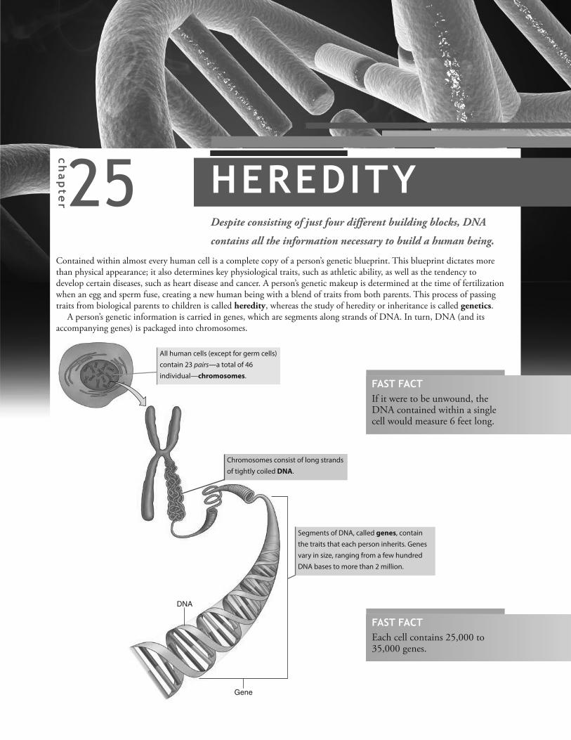
25chapter HEREDITYDespite consisting of just four different building blocks, DNA
contains all the information necessary to build a human being.
Contained within almost every human cell is a complete copy of a person’s genetic blueprint. This blueprint dictates morethan physical appearance; it also determines key physiological traits, such as athletic ability, as well as the tendency todevelop certain diseases, such as heart disease and cancer. A person’s genetic makeup is determined at the time of fertilizationwhen an egg and sperm fuse, creating a new human being with a blend of traits from both parents. This process of passingtraits from biological parents to children is called heredity, whereas the study of heredity or inheritance is called genetics.
A person’s genetic information is carried in genes, which are segments along strands of DNA. In turn, DNA (and itsaccompanying genes) is packaged into chromosomes.
DNA
Gene
All human cells (except for germ cells)
contain 23 pairs—a total of 46
individual—chromosomes.
Chromosomes consist of long strands
of tightly coiled DNA.
Segments of DNA, called genes, contain
the traits that each person inherits. Genes
vary in size, ranging from a few hundred
DNA bases to more than 2 million.
FAST FACTIf it were to be unwound, theDNA contained within a singlecell would measure 6 feet long.
FAST FACTEach cell contains 25,000 to35,000 genes.

PA
RT
V C
on
tinu
ity
484
R S
T U V W XY Z [ \ T ] T T T UT V T W T X T YT \ U ] U T U U T Z T [
Of the 23 pairs of chromosomes contained within the cell’s nucleus, 22 are matched with a similar-looking (homologous)chromosome. The following chart (called a karyotype) shows all the chromosomes, arranged in order by size and structure.
Each pair of chromosomes consists of a chromosome
inherited from the mother and a chromosome inherited from
the father. (See “The Body at Work” on this page.) These
chromosomes are called autosomes.
The members of the last pair of chromosomes are known as
the sex chromosomes. In females, both chromosomes are
relatively large and are designated by the letter X. In males,
one sex chromosome is an X chromosome and one is a
smaller chromosome designated by the letter Y.
The Body AT WORKEach gamete produced by a
female contains only an X
chromosome, whereas the
gametes produced by a male
may contain either an X or a Y
chromosome. When a sperm
with an X chromosome
fertilizes an egg, the offspring is
female (two X chromosomes).
When a sperm with a Y
chromosome fertilizes an
ovum, the offspring is male
(one X and one Y
chromosome).
^ _ ` a b _c d e b fg a b _c d e b f
h h h ih h
h i
The Body AT WORKGametes (eggs and sperm) are the only cells
that contain a single (unpaired) set of
23 chromosomes. At fertilization, the
chromosomes from the father (contained in
the sperm) align with similar chromosomes
from the mother (contained in the egg) to
create a set of 23 pairs, or 46 chromosomes.
Consequently, the fertilized egg—as well
as all the cells of the body that arise from
it—contains genetic instructions from both
the mother and the father.
Chromosomes
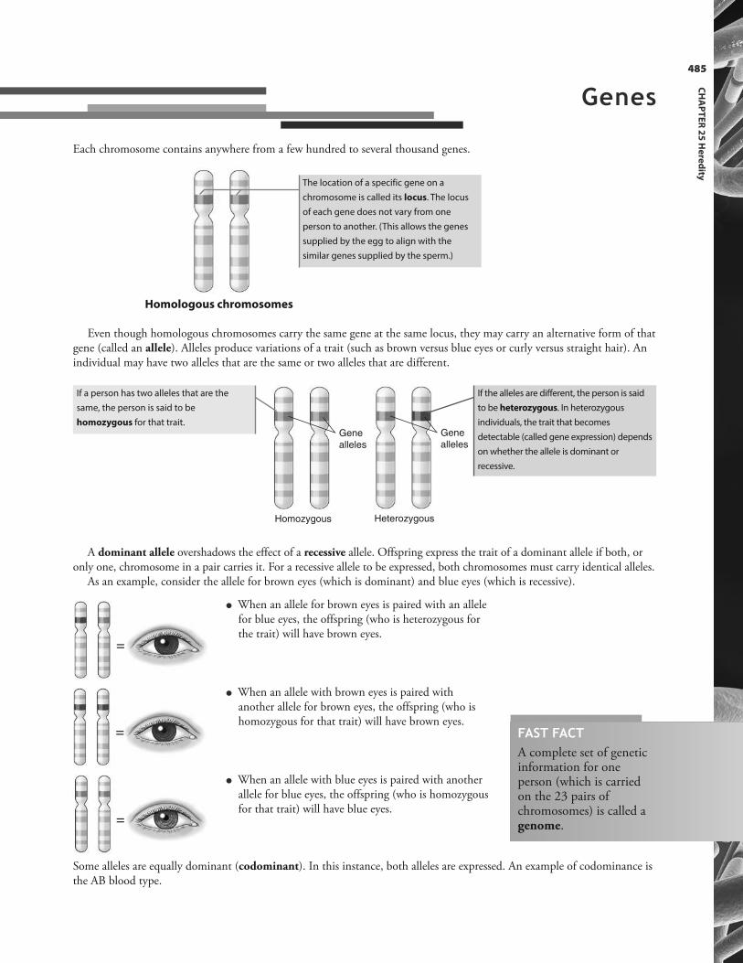
485
CH
AP
TE
R 2
5 H
ere
dity
Each chromosome contains anywhere from a few hundred to several thousand genes.
Homozygous
Gene
alleles
Heterozygous
Gene
alleles
=
=
=
The location of a specific gene on a
chromosome is called its locus. The locus
of each gene does not vary from one
person to another. (This allows the genes
supplied by the egg to align with the
similar genes supplied by the sperm.)
If a person has two alleles that are the
same, the person is said to be
homozygous for that trait.
If the alleles are different, the person is said
to be heterozygous. In heterozygous
individuals, the trait that becomes
detectable (called gene expression) depends
on whether the allele is dominant or
recessive.
Even though homologous chromosomes carry the same gene at the same locus, they may carry an alternative form of thatgene (called an allele). Alleles produce variations of a trait (such as brown versus blue eyes or curly versus straight hair). Anindividual may have two alleles that are the same or two alleles that are different.
A dominant allele overshadows the effect of a recessive allele. Offspring express the trait of a dominant allele if both, oronly one, chromosome in a pair carries it. For a recessive allele to be expressed, both chromosomes must carry identical alleles.
As an example, consider the allele for brown eyes (which is dominant) and blue eyes (which is recessive).
Some alleles are equally dominant (codominant). In this instance, both alleles are expressed. An example of codominance isthe AB blood type.
l When an allele for brown eyes is paired with an allelefor blue eyes, the offspring (who is heterozygous forthe trait) will have brown eyes.
l When an allele with brown eyes is paired withanother allele for brown eyes, the offspring (who ishomozygous for that trait) will have brown eyes.
l When an allele with blue eyes is paired with anotherallele for blue eyes, the offspring (who is homozygousfor that trait) will have blue eyes.
Homologous chromosomes
Genes
FAST FACTA complete set of geneticinformation for oneperson (which is carriedon the 23 pairs ofchromosomes) is called agenome.

PA
RT
V C
on
tinu
ity
486
Recessiveallele
Dominant allele(blocks recessive allele)
j k l m n ko oRecessive
allele
p m n ko qSome traits, called sex-linked traits, are carried on the sex chromosomes. Almost all of these traits, which are recessive, arecarried on the X chromosome—mainly because the X chromosome has much more genetic material than does the Y. Anexample of a common sex-linked condition is red-green color deficit (color blindness).
FAST FACTThe X chromosome carries hundredsof genes, most of which have nothingto do with determining gender.
If a woman inherits the allele for this condition, the allele on her
other X chromosome would overpower the recessive allele.
Consequently, she would be a carrier of the trait but would not
exhibit any symptoms.
Because a man has only one X chromosome, he
does not have a dominant matching allele to
overpower the X-linked recessive trait. As a result,
he would be color-blind.
FAST FACTThe genetic information stored at the locus of agene, even if the trait is not expressed, constitutes aperson’s genotype. The detectable, outwardmanifestation of a genotype is called a phenotype.
The Body AT WORKWhile it is generally true that genes for brown eyes are dominant over genes for green eyes, and genes for both brown
and green eyes are dominant over genes for blue eyes, the expression of eye color is not that simple. Eye color results
from the presence of melanin in the iris. The various combinations of yellow and black melanin produce the shades
of eye color ranging between brown and blue, such as green and hazel. Many different genes participate in melanin
production. If one of these genes contains a mutation, the eye color of the offspring will be affected. That’s why it’s
possible for blue-eyed parents to give birth to a child with green or brown eyes. For example, if a blue-eyed man has a
“brown-eye gene” because of a mutation, that gene would dominate the woman’s “blue-eye gene,” and the offspring
would have brown eyes.
The phenomenon whereby genes at two or more loci contribute to the expression of a single trait is called
polygenic inheritance. Skin color is another example of polygenic inheritance, as are certain diseases such as cancer,
heart disease, asthma, and even some mood disorders.
Sex-Linked Inheritance
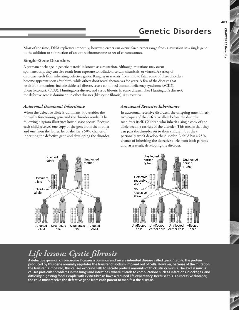
487
CH
AP
TE
R 2
5 H
ere
dity
Autosomal Recessive Inheritance
In autosomal recessive disorders, the offspring must inherittwo copies of the defective allele before the disordermanifests itself. Children who inherit a single copy of theallele become carriers of the disorder. This means that theycan pass the disorder on to their children, but theypersonally won’t develop the disorder. A child has a 25%chance of inheriting the defective allele from both parentsand, as a result, developing the disorder.r s s _ c t _ fs a t d _ u v w a s s _ c t _ f` x t d _ uy x ` e w a w ta b b _ b _z _ c _ { { e | _a b b _ b _
r s s _ c t _ fc d e b f v w a s s _ c t _ fc d e b f v w a s s _ c t _ fc d e b f r s s _ c t _ fc d e b f
} ~ � � � � � � � �� � � � � � �� � � � � � } ~ � � � � � � � �� � � � � � �� � � � � �} ~ � � � � � � � �� � � � � } ~ � � � � � � � �� � � � � � � � � � � � } ~ � � � � � � � �� � � � � � � � � � � � � � � � � � � �� � � � �
� � � � � � � � �� � � � � � � � �� � � � � �� � � � � �� � � � � � � � �� � � � � �
Most of the time, DNA replicates smoothly; however, errors can occur. Such errors range from a mutation in a single geneto the addition or subtraction of an entire chromosome or set of chromosomes.
Single-Gene Disorders
A permanent change in genetic material is known as a mutation. Although mutations may occurspontaneously, they can also result from exposure to radiation, certain chemicals, or viruses. A variety ofdisorders result from inheriting defective genes. Ranging in severity from mild to fatal, some of these disordersbecome apparent soon after birth, while others don’t reveal themselves for years. A few of the diseases thatresult from mutations include sickle cell disease, severe combined immunodeficiency syndrome (SCID),phenylketonuria (PKU), Huntington’s disease, and cystic fibrosis. In some diseases (like Huntington’s disease),the defective gene is dominant; in other diseases (like cystic fibrosis), it is recessive.
Autosomal Dominant Inheritance
When the defective allele is dominant, it overrides thenormally functioning gene and the disorder results. Thefollowing diagram illustrates how disease occurs. Becauseeach child receives one copy of the gene from the motherand one from the father, he or she has a 50% chance ofinheriting the defective gene and developing the disorder.
Life lesson: Cystic fibrosisA defective gene on chromosome 7 causes a common and severe inherited disease called cystic fibrosis. The proteinproduced by this gene normally regulates the transfer of sodium into and out of cells. However, because of the mutation,the transfer is impaired; this causes exocrine cells to secrete profuse amounts of thick, sticky mucus. The excess mucuscauses particular problems in the lungs and intestines, where it leads to complications such as infections, blockages, anddifficulty digesting food. People with cystic fibrosis have a reduced life expectancy. Because this is a recessive disorder,the child must receive the defective gene from each parent to manifest the disease.
Genetic Disorders

PA
RT
V C
on
tinu
ity
488
Chromosome Abnormalities
In these disorders, large segments of a chromosome, or even entire chromosomes, aremissing, duplicated, or otherwise altered. The most common disorders result whenhomologous chromosomes fail to separate during meiosis. This is called nondisjunction.� � � � � � � � � � � � � �
� ~ � � � � � � � � � � � � � � � � � � � � � � � � � � � � � � � � � � � � � �� � � � � � � � � � � � � � � � � � � � � � � � � � � � � � � � � � � � �� � � � � � � � � � � � ¡ � � � � � � � � � � � � � � � � � � � � � � �� � � � ~ ¢ � � � � � � � � � � � � � � � � � � � £¤ � � ~ � � � � � � � ¥ � � � � ~ � � � � � � � � � � � � � ~ � � � � � � � � � � � �� ~ � � � � � � � � � � � � � � � � � � � � � � � � � � � � � � � � � � �� � � � � � � � � � ¦ � � � � � � � § � � � ¨ © ª � ¡ � � � � � � � � � � � �� � � � � � � � � � � � � � � ~ � � � � � � � � � � � ¡ � � � ~ � � � � �¦ � � � � � � ¨ � � � � � ¨ © ª £
� � § ¨ « ¬� � � � � � � � � � � � � � � � � � � � � � ~ � � � � � � � � � � � � � � � � � � � � � � � � � � � �� � � � � � � � � � � � � � � � � ¡ � � � � � � � � � � � � � ¡ � � � ® ¯� � � � � � � � � � � � � � � £ ° � � � � � � � � � � � ~ � � � � � � � � �� � � � � � � � � � � � � � � � � ±
² � � � � � � ¥ � � � � ~ � � � � � � � � ~ � � � � � ® ¯ � � � � � � � � � � � �� � � � � � ~ � � ~ � � � � � � � � � � � � � � ³ ´ � � � � � � � � � � � £FAST FACTSeveral tests, including chorionic villus sampling,amniocentesis, and umbilical blood sampling, canidentify the presence of an extra chromosome 21 ina developing fetus.
Life lesson: Down syndromeMost pregnancies involving extra ormissing chromosomes end in miscarriage.The most survivable trisomy, and thereforethe most common, is Down syndrome ortrisomy 21. People with Down syndromehave distinctive physical features, includinga round face, flattened nose, “Oriental” foldsaround the eyes, an enlarged tongue, andshort fingers. Most have severe to profoundmental retardation and also suffer fromabnormalities of the heart and kidneys.
µ ¶· ¸ ¹ º »¼ ½ ¾ ¿ · À · · · ¸· ¹ · º · » · ¼· ¿ ¸ À ¸ · ¸ ¸ · ½ · ¾° � � � � � Á ® Â

489
CH
AP
TE
R 2
5 H
ere
dity
Nondisjunction of Sex Chromosomes
Nondisjunction can affect sex chromosomes as well as autosomal chromosomes. The effectsfrom nondisjunction of a sex chromosome typically aren’t as severe as nondisjunction of anautosomal chromosome. In fact, a boy with an extra Y chromosome or a girl with an extra X chromosome will usually have no symptoms. However, if a male inherits an extra X chromosome, or if a girl lacks an X chromosome, symptoms become apparent.
Multifactorial Disorders
Many common diseases fall into a category called multifactorial inheritance. This meansthat environmental factors have a strong influence over genetic mutation, determining theprogression of a disease or even whether the disease develops at all.
For example, heart disease tends to run in families, meaning it has a genetic link. Ofcourse, environmental factors (such as diet, exercise, and whether or not a person smokes)also influence the onset and progression of heart disease. Other examples of multifactorialdisorders include hypothyroidism, diabetes, and cancer.
� � ~ � � � à ~ � � � � ~ � � � � � ¡ � � ~� � � Ä � � � � � � � � � � � � � � � � � � � � � � � � � £Å ÆÇ ~ � � � � � � � � � � � � � � �� ¡ � Ä � � � � � � � � � � � £ Ç ~ � � � � � � � � � � � � � � �~ � Ä � � � � � � � � � � � £
² � � � � � � ¥ � � � � ~ � Á � � � � � �¡ � � � � � � È � � � � � � � � � �� � � � � � � ~ Ä Ä È �É � � ~ � � � � � � � � Á ~ � � � � � £ ² � � � � � � ¥ � � � � ~ � Á � � � � � �¡ � � � � � � Ä � � � � � � � � � �� � � � � � � ~ Ä Ç �° � ~ � � � Á ~ � � � � � £If a boy inherits an extra X
chromosome (XXY), Klinefelter
syndrome develops. This
syndrome—which usually becomes
apparent at puberty when secondary
sexual characteristics fail to
develop—is characterized by
undeveloped testes, sparse body hair,
unusually long arms and legs,
enlarged breasts, low intelligence,
and sterility.
If a girl is born with only one X
chromosome, Turner syndrome
develops. This syndrome becomes
apparent at puberty when secondary
sexual characteristics fail to develop.
Other features include a webbed
neck and short stature.
FAST FACTAt least 10% of sperm and 25% of oocytes haveextra, missing, or broken chromosomes; however,zygotes with extra or missing chromosomes don’tusually survive more than a few days.

PA
RT
V C
on
tinu
ity
490
Review of Key TermsAllele: Alternative form of a gene
Autosomes: Non-sex chromosomes
Carrier: Someone who carries a normalgene along with its recessive allele
Chromosome: Long strand of DNAfound in the cell’s nucleus
Genes: Segments of DNA that containthe traits each person inherits
Genetics: The study of heredity orinheritance
Genome: A complete set of geneticinformation for one person
Genotype: The genetic informationstored at the locus of a gene, even ifthose traits are not expressed
Heredity: The passing of traits frombiological parents to children
Heterozygous: Possessing differentalleles at a given locus
Homologous: Similar in structure,such as two similar chromosomes thatare paired together
Homozygous: Possessing similar allelesat a given locus
Karyotype: A chart showing all thechromosomes arranged in order bysize and structure
Locus: The location of a specific geneon a chromosome
Mutation: A permanent change ingenetic material
Nondisjunction: When chromosomesfail to separate during meiosis
Phenotype: The detectable, outwardmanifestation of a genotype
Polygenic inheritance: Phenomenonwhereby genes at two or more locicontribute to the expression of asingle trait
Sex chromosomes: Chromosomesdesignated by the letters X and Y thatdetermine gender
Life lesson: Human genome projectBeginning in 1990 and ending in 2003, a team of scientists studied the human genome to determine itsentire sequence of DNA and the location and identity of all genes. Just one of the many fascinatingfindings is that all humans are 99.99% genetically identical. Even so, with just the .01% variation, over 3 million base pairs differ between individuals.
But perhaps the most exciting result of the project is the implication for medicine. Knowing thesequence of someone’s genome could help doctors predict that person’s risk for disease instead of justtreating it once it develops. Other benefits include detecting a disease earlier and then fine-tuningtreatment according to that individual’s genetic makeup. New treatments, too, are evolving in whichhealthy genes are introduced into a person’s body to replace the specific genes that are defective. Thiswould allow more effective treatment of a variety of diseases and conditions, including cancer,Alzheimer’s disease, Parkinson’s disease, hemophilia, diabetes, and even high cholesterol levels.
FAST FACTIf the DNA sequence of the human genomewere to be complied in books, it would fill200 volumes having 1000 pages each.
Own the InformationTo make the information in this chapter part of your
working memory, take some time to reflect on what you’ve
learned. On a separate sheet of paper, write down
everything you recall from the chapter. After you’re done,
log on to the DavisPlus website, and check out the Study
Group podcast and Study Group Questions for the chapter.
Key Topics for Chapter 25:
• The difference between heredity and genetics
• Chromosomes and the human karyotype
• Genes
• Alleles and the expression of traits
• Sex-linked inheritance
• Single-gene genetic disorders
• Autosomal dominant and autosomal recessive inheritance
• Chromosome abnormalities
• Multifactorial disorders
491
CH
AP
TE
R 2
5 H
ere
dity
Answers: Chapter 251. Correct answer: b. Nucleic acids make up DNA.
Chromosomes contain thousands of genes.Autosomes are any chromosomes other than the sexchromosomes.
2. Correct answer: a. Although the offspring is uniquefrom either parent, he inherits half of hischromosomes from his mother and half from hisfather.
3. Correct answer: b. A male offspring results when asperm with a Y chromosome fertilizes an egg. Eggsonly have X chromosomes. A sperm with two X chromosomes would be considered abnormal.
4. Correct answer: c. A locus is the location of aspecific gene on a chromosome. A genome is acomplete set of genetic information for one person.A karyotype is a chart that shows all thechromosomes arranged in order by size andstructure.
5. Correct answer: d. A dominant allele overrides arecessive allele and is expressed. A recessive allele isexpressed when the corresponding allele is alsorecessive. A person who has two alleles that are thesame is said to be homozygous for that trait.
6. Correct answer: a. All the other answers areincorrect.
7. Correct answer: b. All the other answers areincorrect.
Test Your Knowledge1. Chromosomes consist of:
a. pairs of nucleic acids.b. long strands of tightly coiled
DNA.c. a single gene.d. autosomes.
2. The genetic information in maleoffspring is:a. inherited from both the father
and the mother.b. inherited from only the father.c. inherited from only the
mother.d. unique from that of either
parent.
3. Female offspring result when:a. a sperm with a Y chromosome
fertilizes the egg.b. a sperm with an X chromosome
fertilizes the egg.c. an egg with a Y chromosome is
fertilized by a sperm with an X chromosome.
d. a sperm with two X chromo-somes fertilizes the egg.
4. An alternative form of a gene iscalled:a. a locus.b. a genome.c. an allele.d. a karyotype.
5. Which of the following statements about alleles is correct?a. A dominant allele cancels out a
recessive allele and the trait issuppressed.
b. A recessive allele is expressedonly when the correspondingallele is absent.
c. If a person has two alleles thatare the same, the person is saidto be heterozygous.
d. An individual may have twoalleles that are the same or twoalleles that are different.
6. Which of the following statements about sex-linked inheritance is correct?a. Almost all of the sex-linked
traits are carried on the X chromosome and are recessive.
b. Almost all of the sex-linkedtraits are carried on the X chromosome and are dominant.
c. Almost all of the sex-linkedtraits are carried on the Y chromosome and are recessive.
d. Almost all of the sex linkedtraits are carried on the Y chromosome and are dominant.
7. Most of the common chromosomal abnormalities result when:a. homologous chromosomes
separate repeatedly.b. homologous chromosomes fail
to separate during meiosis.c. an egg is fertilized by more
than one sperm.d. a mutation occurs.
Go to http://davisplus.fadavis.com Keyword:Thompson to see all of the resources availablewith this chapter.


493
AA (adenine), 48
Abdominal aorta, 302
Abdominal cavity, 10
Abdominal pain, 11
Abdominal quadrants, 11
Abdominal regions, 11
Abdominal wall muscles, 146
Abdominopelvic cavity, 10
Abducens nerve, 189, 216
Abduction, 122
Abnormal spinal curvatures, 106
Abscess, 325
Acceleratory center, 286
Accessory gland, male reproductive system, 448
Accessory muscles of respiration, 344
Accessory organs, digestive system, 389
Accommodation, 219, 221
Accumulation, fluid, 377
ACE (angiotensin-converting enzyme), 362
Acetabulum, 112
Acetylcholine (ACh), 135, 136, 169, 192, 196, 198, 199, 286, 288
Acetylcholinesterase, 135
Acetyl coenzyme A (acetyl CoA), 420, 421
Achilles tendon, 151
Acid-base balance, 83, 380–383
Acid-base imbalances, 384–385
Acidic solution, 28
Acid mantle, 322
Acidosis, 384
Acids, 27, 380
amino, 31
Acinar cells, 401
Acini, 243, 455
Acne, 79
Acquired immunodeficiency syndrome (AIDS), 332
Acromegaly, 233
Acromion process, 110
Acrosome, 451
ACTH (adrenocorticotropic hormone), 233
Actin, 133, 134
Action potential, 167
Active immunity, 327
Active transport, 45–47
Active transport pump, 45, 47
Acute leukemia, 262
Acute renal failure, 368
Adaptation, 206
Addison’s disease, 242
Adduction, 122
Adductor brevis, 149
Adductor longus, 149
Adductor magnus, 149
Adductor muscles, 149
Adenine (A), 48
Adenohypophysis, 232
Adenoids, 320
Adenosine triphosphate (ATP), 23, 33, 134, 137, 140, 419, 420
Adenovirus, 436
ADH (antidiuretic hormone), 234, 310, 364, 365, 378
Adipose tissue, 60, 61, 63, 373
breast, 455
Adolescence, 478
Adrenal cortex, 241, 242
Adrenal cortical insufficiency, 242
Adrenal disorders, 242
Adrenal glands, 194, 241–242
Adrenal medulla, 194, 241, 242
Adrenergic fibers, 196
Adrenergic receptors, 197, 199
Adrenocorticotropic hormone (ACTH), 233
Aerobic exercise, 153
Aerobic processes, 419
Aerobic respiration, 140, 419, 420
Afferent arterioles, 359
Afferent division, 160
Afferent lymphatic vessels, 319
AFP (alpha-fetoprotein), 472
Afterbirth, 476
Afterload, 288
Agglutinate, 267
Agglutination, 267
Agglutinins, 267
Agglutinogen, 266
Aging
blood and, 261
blood pressure and, 311
brain and nervous system and, 199
cells and, 50
digestive system and, 407
hormones and, 242
integumentary system and, 76
menopause, 459
respiratory system and, 343
vascular system and, 311
Agranulocytes, 260, 261
AIDS (acquired immunodeficiency syndrome), 332
Airflow, 347–348
Albinism, 73
Albumin, 253
Aldosterone, 241, 242, 310, 362, 364, 378, 379
Alimentary canal, 389
Alkalosis, 384
Allantois, 469
Allele, 485
Allergens, 331
Allergic reactions, 331–332
Allergy, 331
Alopecia, 75
Alpha-(α)-adrenergic receptors, 197, 198, 199
Alpha cells, 243
Alpha-fetoprotein (AFP), 472
Index
Alveolar ducts, 340
Alveolar sacs, 340
Alveolar surface tension, 348
Alveoli, 341
Amino acids, 31, 405, 416, 422
Ammonia, 363, 422
Amnion, 469
Amniotic cavity, 468
Amniotic fluid, 470
Amphiarthroses, 119
Ampulla, 214, 454
of Vater, 400
Amygdala, 183, 210, 287
Amylase, 392, 404, 407
Anabolism, 23, 419
Anaerobic fermentation, 419
Anaerobic processes, 419
Anaerobic respiration, 140
Anal canal, 406
Analgesics, 208
Anal sphincter, 406
Anaphase, 51
Anaphylactic shock, 332
Anaphylaxis, 332
Anastomosis, 300
Anatomical dead space, 349
Anatomical terms, 7–11
Anatomy
defined, 3
early study of, 3
hierarchy in organization of, 4
study of, 3
typical, 3
Androgens, 241, 242
Anemia, 257, 259, 261
Aneurysm, 294
Angina, 281
Angiotensin, 362
Angiotensin-converting enzyme (ACE), 362
Angiotensin I, 310, 362
Angiotensin II, 310, 362
Angiotensinogen, 362
Angular gyrus, 186
Anions, 21
Ankle
bones, 114
muscles, 151
Annulus fibrosus, 107
ANP (atrial natriuretic peptide), 247, 310, 364
ANS. See Autonomic nervous system (ANS)
Antagonists, 141
Anterior cavity of eye, 218
Anterior chamber of eye, 218
Anterior cruciate ligament (ACL), 125
Anterior descending artery, 280
Anterior fontanel, 105
Anterior pituitary, 232, 477
Anterior tibial artery, 302
Anterior tibial vein, 304
Anterior (ventral), 7
Antibiotics, effect on microbiome, 431, 434, 438
Antibodies, 31, 267, 328, 330
Antibody-mediated immunity, 327, 330–331
Antibody titer, 331
Anticoagulants, 265
Anticodon, 49
Antidiuretic hormone (ADH), 234, 310, 364, 365, 378
Antigen, 266, 328
Antigen presentation, 329
Antigen-presenting cell (APC), 329
Antimicrobial proteins, 324
Anus, 406
Aorta, 275, 301
abdominal, 302
thoracic, 302
Aortic arch, 301
Aortic area, 277
Aortic body, 287
Aortic insufficiency, 277
Aortic valve, 275, 276
Apex
of heart, 273
of lung, 342
Apgar score, 478
Aphasia, 186
Apnea, 350
Apneustic center, 345
Apocrine glands, 77
Aponeurosis, 132
Appendages of the skin, 75–77
Appendectomy, 406
Appendicitis, 406
Appendicular skeleton, 97, 98
Appendix, 406
Aqueous humor, 218
Arachnoid mater, 171, 177
Arachnoid villus, 177
Arches of the foot, 114
Areola, 455
Areolar tissue, 60, 61, 63
Arrector pili muscle, 75
Arrhythmias, 283
Arteries, 293, 295, 300–303
coronary, 280–281
hepatic, 398, 399
principal, 302
pulmonary, 275
renal, 359
structure, 294
umbilical, 469, 470, 471
Arterioles, 295
Arteriovenous anastomosis, 300
Arthritis, 126, 326
Arthrology, 119
Arthroscopy, 125
Articular cartilage, 85, 120
Articulations, bone, 98, 119
Artificial active immunity, 327
Artificial passive immunity, 327
Ascending aorta, 301
Ascending colon, 406
Ascending limb, nephron, 360
Ascites, 289, 391
Aspirin, 326
Asthma, 342
Astigmatism, 220
Astrocytes, 161
Atherosclerosis, 281, 308
Ind
ex
494
495
Ind
ex
Athlete’s foot, 438
Atlas vertebrae, 108
Atmospheric pressure, 347
Atomic number, 19
Atomic weight, 19
Atoms, 4, 19
ATP (adenosine triphosphate), 23, 33, 134, 137, 140, 419, 420
Atria, 275
Atrial flutter, 283
Atrial kick, 284
Atrial natriuretic peptide (ANP), 247, 310, 364
Atrial systole, 284
Atrioventricular (AV) bundle, 282
Atrioventricular (AV) node, 282, 286
Atrioventricular (AV) valves, 276
Atrophy, 141
Attachment, skeletal muscle, 132
Auditory association area, 186
Auditory canal, 211
Auditory ossicles, 104, 211
Auditory tube, 211
Auricle (pinna), 211
Autoimmune diseases, 332
Automaticity, 282
Autonomic motor division, 160
Autonomic nervous system (ANS), 191
divisions, 193–195
effects on target organs, 196–199
somatic vs ., 192
structure, 192
visceral reflexes, 191
Autonomic tone, 193
Autosomal dominant inheritance, 487
Autosomal recessive inheritance, 487
Autosomes, 484
AV. See under Atrioventricular
Axial skeleton, 97, 98
Axillary artery, 302
Axillary lymph nodes, 319
Axillary nerve, 174
Axillary vein, 304
Axis vertebrae, 108
Axons, 64, 135, 163
autonomic nervous system and, 192
branch, 163
spinal tracts and, 172
BBacilli, 434
Bacteria, 429, 430, 433–435
identifying, 435
obesity and, 438
shapes of, 434
Bacterial flora, 407
Bacterial transplants, 434
Bacteriophages, 436
Balance, 214
Ball-and-socket joint, 121
Band cells, 262
Baroreceptors (pressoreceptors), 287, 310
Bartholin’s glands, 455
Basal cell carcinoma, 79
Basal ganglia, 183
Basal metabolic rate (BMR), 412
Basal nuclei, 183
Base
of heart, 273
of lung, 342
Basement membrane, 58
Base pairing, 48
Bases, 27, 380
Basic solution, 28
Basilar artery, 303
Basilar membrane, 212
Basilic vein, 304
Basophils, 260
B cells, 328, 330
Bed, nail, 76
Bell’s palsy, 145, 190
Belly, of muscle, 141
Benign prostatic hyperplasia (BPH), 448
Benign tumor, 52
Beta-(β)-adrenergic receptors, 197, 198
Beta blockers, 197
Beta cells, 243
Bicarbonate, 377, 384
buffer system, 381
Biceps brachii, 148
Biceps femoris, 150
Bicuspid valve, 276
Bile, 258, 265, 400
duct, 400
salts, 400
Bilirubin, 258, 400
Bipolar neurons, 162
Birth, 475–476
Birth control, 461
Bitter, 209
Bladder, urinary, 366
Blastocyst, 467, 468
Blastomeres, 467
Bleeding, control of, 263–266
Blind spot, 218
Blood, 62
cell development, 57
cell formation, 254
cells, red, 253, 254, 255–259
cells, white, 253, 254, 260–262
clotting, 263–266
components of, 253
as connective tissue, 60, 62, 63
flow and pressure, 306–307
flow through the heart, 278–279
flow through the liver, 399
gas transport and, 353
glomerular filtration and, 361–362
glucose regulation, 243, 244–246
pH, 380, 382
platelets, 253, 254, 263–266
pressure, 306, 307
production and bones, 83
Rh group, 268–269
spleen and storage of, 321
transfusions, 268
types, 266–269
viscosity, 253, 308
volume and pressure, 307
Blood-brain barrier (BBB), 161, 179
Blood clotting disorders, 266
Blood pressure, 307
B lymphocytes (B cells), 328, 330
BMR (basal metabolic rate), 412
Body
cavities, 10
fat, 63
fluids, 25
major muscles of, 152–153
nail, 76
planes, 8
regions, 9
of sternum, 109
temperature, regulation of, 424
of uterus, 454
of vertebrae, 107
Bolus of food, 392
Bonds, chemical, 20–22
peptide, 32
Bone marrow, 85, 88
red, 254
Bone(s), 60, 62, 63. See als o Joints; Skeletal system
ankle, 115
classification of, 84–85
development, 88–89
foot, 98, 115
fractures, 91–92
functions, 83
growth, 86, 89
hand, 111
long, 84, 85
lower limb, 113–114
marrow, 85, 88
pectoral girdle, 110
pelvic girdle, 112–113
remodeling, 90
short, 84
skull, 84, 101–105
surface markings, 98
thoracic cage, 109
tissue, 86–87
upper limb, 110–111
vertebral column, 106–108
vitamin D and, 74
widening and thickening of, 89
Bony labyrinth, 212
Botulism, 136
Bowman’s capsule, 360
Boyle’s law, 347
BPH (benign prostatic hyperplasia), 448
Brachial artery, 302, 307
Brachialis, 148
Brachial plexus, 174
Brachiocephalic artery, 301
Brachiocephalic vein, 304
Brachioradialis, 148
Bradycardia, 285
Brain. See als o Head
aging and, 199
autonomic nervous system and, 191–195
blood-brain barrier and, 179
breathing and, 180
cerebral cortex, 184–187
cerebral lateralization, 187
cranial nerves, 188–190
divisions of, 180–183
gastric secretion and, 396
general structures of the, 176–179
language and, 185
lesions of, 182
memory and, 187
meninges of, 177
sleep and, 186
special senses and, 186
swelling, 178, 190
swelling of, 102
tumors of, 161
urination and, 368
Brainstem, 176, 180, 210
Braxton-Hicks contractions, 475
Breastfeeding. See Lactation
Breasts, 455
cancer, 320, 456
Breathing. See Respiration; Respiratory system
Breech birth, 476
Broad-spectrum antibiotics, effect on microbiome, 431, 434, 438
Broca’s aphasia, 186
Broca’s area, 186
Bronchi, 340
Bronchial tree, 340
Bronchioles, 340
Bronzing, 73
Bruise (hematoma), 73
Buccal cavity, 392. See als o Mouth
Buccinator, 145
Buffers, 28, 381–382
Buffy coat, 253
Bulb, hair, 75
Bulbourethral glands, 448
Bundle of His, 282
Burns, 78
Bursae, 120, 124
CC (cytosine), 48
Calcaneal (Achilles) tendon, 151
Calcaneus, 114
Calcitonin, 237, 239, 240
Calcium, 288, 377, 380, 418
abnormalities, 240
ATP contraction and, 134
homeostasis, 239
parathyroid hormone and, 238–240
Calorie, 412
calorie, 412
cAMP (cyclic adenosine monophosphate), 231
Canaliculi, 87, 399
Canal of Schlemm, 218
Cancellous bone, 87
Cancer, 52
breast, 320, 456
lymph nodes and, 320
prostate, 448
skin, 79
Candida albicans , 438
Canine teeth, 393
Capacitance vessels, 296
Ind
ex
496

497
Ind
ex
Capillaries, 293, 297–299
peritubular, 359
Capillary beds, 297
Capillary exchange, 298–299
Capsid, 436
Capsomeres, 436
Capsule
bacterial, 433
joint, 120
Carbaminohemoglobin, 353
Carbohydrates, 29, 404, 414
metabolism, 419–420
Carbon, 30
Carbon dioxide, 26, 381, 383, 385
gas exchange and, 352
respiration and, 345, 346
transport, 353
Carbonic acid, 27, 381, 383
Carbonic anhydrase, 381, 383
Cardiac center, 180, 286–287
Cardiac conduction, 282–283
Cardiac cycle, 284
Cardiac muscle, 64, 131
Cardiac nerves, 286
Cardiac output (CO), 285–289
blood pressure and, 307
Cardiology, 273
Cardiovascular system. See als o Heart; Vascular system
heat generation and, 12
neonate, 478
Carina, 340
Carotene, 73
Carotid arteries, 303
Carotid body, 287
Carpal bones, 111
Carpal tunnel syndrome, 148
Carpopedal spasm, 240
Carrier, 45
Cartilage, 60, 62, 63
articular, 120
costal, 109
disorders, 62
exercise and, 126
Cartilaginous joints, 119
Catabolism, 23, 419
Catalysts, 24
Cataracts, 220
Catecholamines, 241
Categorical hemisphere, 187
Cations, 21
Cauda equina, 170
Caudate lobe, 398
Cavities
body, 10
joint, 120
CCK (cholecystokinin), 401, 413
Cecum, 406
Celiac trunk, 302
Cell body (soma), 163
Cell(s), 4
aging, 50
B, 328, 330
chief, 396
cycle, 50–51
cytoplasm and organelles, 40–42
goblet, 58, 59
growth and reproduction, 50–52
layers in epithelial tissue, 58
mast, 331
movement through membranes of, 43–47
nervous system, 161–164
nucleus, 38, 40
shape in epithelial tissue, 58
stem, 57
structure, 38–42
T, 318, 327, 328, 329, 330
variations, 37
Cellular (cell-mediated) immunity, 327, 329
Cellular respiration, 26
Cellulose, 29, 404, 414
Cell wall, 433, 435
Cementum, 393
Centers for ossification, 88
Central canal, 171
Central nervous system (CNS), 160. See als o Nervous system; Spinal cord
autonomic nervous system and, 191
neuroglia of, 161
pituitary secretions and, 235
Central obesity, 63
Central vein, 399
Centrioles, 41
Cephalic phase, 397
Cephalic vein, 304
Cerebellum, 176, 180
Cerebral cortex, 183, 345
functions of, 184–187
Cerebral lateralization, 187
Cerebrospinal fluid (CSF), 178–179
Cerebrum, 176, 182–183, 210
Cerumen, 77
Ceruminous glands, 77
Cervical canal, 454
Cervical curve, 106
Cervical lymph nodes, 319
Cervical plexus, 174
Cervical vertebrae, 106
Cervix, 454
dilation of, 475
Chambers, eye, 218
Chemical bonds, 20–22
peptide, 32
Chemical buffers, 381, 382
Chemical digestion and absorption, 390, 404–405
Chemical reactions, 24
Chemicals
atoms in, 19
basic processes of life and, 23–24
basic structures of life and, 18–22
bonds, 20–22
common elements and, 17
compounds of life and, 25–33
elements in, 18
reactions, 24
Chemoreceptors, 206, 287
Chemotaxis, 323, 325
Chemotherapy, 52
Chewing muscles, 145
Cheyne-Stokes respirations, 350

Chief cells, 396
Childbirth, 475–476
Childhood, 478
Chloride, 377, 380
Chlorine, 21
Cholecystokinin (CCK), 401, 413
Cholesterol, 30, 39
Cholinergic fibers, 196, 198
Cholinergic receptors, 197, 198
Chondrocytes, 62, 89
Chordae tendineae, 278
Chorion, 469
Chorionic villus, 470
Chorionic villus sampling, 488
Choroid, 217
Choroid plexus, 178
Chromaffin cells, 241
Chromatids, 51
Chromatin, 40
Chromosomes, 40, 48, 483, 484, 485, 486
abnormalities, 488–489
Chronic leukemia, 262
Chronic renal insufficiency, 368
Chronotropic agents, 288
Chyme, 395, 396
Chymotrypsin, 405, 407
Cilia, 42
bronchial, 341
nasal, 338
Ciliary body, 217
Ciliary muscles, 221
Circle of Willis, 303
Circular folds, of small intestine, 403
Circulation. See als o Vascular system
collateral, 281
coronary, 280–281
fetal, 471
hepatic portal, 305
liver, 398, 399
peripheral resistance and, 307, 308–310
principles of, 306–311
pulmonary, 300
renal, 359
routes, 300–305
systemic, 300, 301–305
umbilical, 469, 470, 471
venous return, 311
Circulatory system, 6
pregnancy and, 474
Circumduction, 123
Circumflex artery, 280
Citric acid cycle, 420, 421
Classification
bones, 84–85
joints, 119–126
Clavicle, 110
Cleavage, 467
Clitoris, 455
Closed reduction, 91
Clos tridium difficile, 434
Clotting, blood, 263–266
Clubbing, nail, 76
CNS. See Central nervous system (CNS)
Coagulation, 264
CO (cardiac output), 285–289
Cocci, 434
Coccyx, 106
Cochlea, 212
Cochlear duct, 212
Cochlear nerve, 212
Codominant alleles, 485
Codon, 49
Cold receptors, 208
Collagen, 31, 61
Collagenous fibers, 61
Collateral circulation, 281
Collecting duct
lymphatic system, 318
renal, 360
Colloid, 26
Colloid osmotic pressure, 299
Colon, 406
Color
blindness, 222, 486
hair, 75
skin, 73
Colostrum, 477
Columnar cells, 58, 59
Comminuted fracture, 91
Common bile duct, 400
Common hepatic duct, 400
Common iliac arteries, 301, 302
Common iliac vein, 304
Compact bone, 87
Complement, 324
Complement system, 324
Complete blood count (CBC), 262
Complete proteins, 416
Complete tetanus, 138
Complex capsid, 436
Complex carbohydrates, 414
Compound fracture, 91
Compounds, 17
defined, 25
inorganic, 25–28
of life, 25–33
organic, 29–33
Compressional strength, 86
Concentration
gradient, 43
reaction rates and, 24
Conchae, 338
Condoms, 461
Conducting arteries, 295
Conduction
heat, 423
impulse, 166–168
Conductive hearing loss, 213
Condyles, 125
Condyloid joint, 121
Cones, 217, 222
Congestive heart failure (CHF), 289, 350, 364
Conjunctiva, 215
Connective tissue, 4, 60–63
membranes, 66
osseus, 62
role of, 62
types of, 61–62, 63
Ind
ex
498

499
Ind
ex
Contact digestion, 404
Contraception, 461
Contractility, 288
Contraction, muscle, 135–140
Control center and homeostatic regulation, 12–13
Convection, 423
Convergence, 219
Coracoid process, 110
Core temperature, 423
Cornea, 217
Coronal plane, 8
Coronal suture, 102
Coronary arteries, 280–281
disease, 281
Coronary circulation, 280–281
Coronary sinus, 281
Corpus albicans, 457
Corpus callosum, 176, 183
Corpus cavernosa, 449
Corpus luteum, 457
Corpus spongiosum, 449
Cortex, brain, 176
Cortical nodules, 319
Corticospinal tracts, 172
Corticosteroids, 241
Corticotropin-releasing hormone, 233
Cortisol, 241, 242
Costal angle, 109
Costal cartilage, 109
Costal margins, 109
Coumadin, 266
Covalent bonds, 22
Cowper’s glands, 448
Coxal bone, 112
CP (creatine phosphate), 140
Cramps, heat, 424
Cranial bones, 101
Cranial cavity, 10
Cranial nerves, 188–190
disorders of, 190
mnemonic, 190
Craniosacral division, 195
Cranium, 100, 101
Creatine phosphate (CP), 140
Creatinine, 365
Cremaster muscle, 446
Cretinism, 238
Cribriform plate, 101
Cristae, 42
Cross bridge, 134
Crown, tooth, 393
Crowning, 476
Cryptorchidism, 446
CSF (cerebrospinal fluid), 178–179
Cuboid, 114
Cuboidal cells, 58, 59
Cuneiforms, 114
Curare, 136
Curvatures of the spine, 106
Cushing syndrome, 242
Cusps, 276
Cutaneous membranes, 66, 71
Cuticle, 76
Cyanosis, 73, 76
Cyclic adenosine monophosphate (cAMP), 231
Cystic duct, 400
Cystic fibrosis, 487
Cytoplasm, 38, 40–42
Cytoplasmic membrane, 433
Cytosine (C), 48
Cytoskeleton, 42
Cytotoxic T cells, 329
DDartos fascia, 446
Deafness, 213
Deamination, 422
Deciduous teeth, 393
Decomposition, 24
Decussation, 172
Deep fascia, 132
Deep inspiration, 344
Deep region, 7
Deep veins, 304
Defection, 407
Deficiency, fluid, 376
Dehydration, 375, 376
Delta cells, 243
Deltoid, 147
Dementia, 199
Dendrites, 64, 163
Dens, 108
Dense connective tissue, 61, 63
Dense fibrous tissue, 60
Dentin, 393
Deoxyribonucleic acid. See DNA (deoxyribonucleic acid)
Deoxyribose, 48
Depolarization, 166
Depo-Provera, 461
Depressions, bone, 98
Dermatitis, 79, 326
Dermatomes, 175
Dermis, 71
Descending aorta, 301
Descending colon, 406
Descending limb, nephron, 360
Descending thoracic aorta, 301
Detrusor muscle, 366
Deuterium, 20
Development
male fetal, 449
spinal cord, 170
Diabetes mellitus, 245–246, 332
heart disease and, 289
Diabetic ketoacidosis, 245
Dialysis, 368
Diapedesis, 323
Diaphragm, 146, 344
Diaphysis, 85
Diarthroses, 120
Diastole, 284
Diastolic pressure, 306, 307
Diencephalon, 176, 181
Dietary fiber, 414
Differentiation, 57
Diffusion, 43, 47
in capillary exchange, 298
facilitated, 45, 47
Digestion, 390, 393
contact, 404
Digestive system, 6
chemical digestion and absorption, 390, 404–405
enzymes, 395, 404–405, 407
esophagus, 394
gallbladder, 400
heat generation and, 12
large intestine, 406–407
liver, 398–399
mouth, 392–393
overview, 389–391
pancreas, 401
pharynx, 394
pregnancy and, 474
small intestine, 402–403
stomach, 395–397
Digestive tract, 389, 390
Dilation, cervical, 475
Diplococci, 434
Diplopia, 219
Direct attachment, 132
Directional terms, 7
Disaccharides, 29, 404, 414
Disjunction, 488
Dissociation, 21
Distal convoluted tubule, 360
Distal region, 7
Distributing arteries, 295
Diuresis, 364
Diuretics, 364
Dizygotic twins, 470
DNA (deoxyribonucleic acid), 29, 33, 40, 48, 483
bacterial, 433
cell cycle and, 50–51
in cell growth and reproduction, 50
chromosomes, 40
protein synthesis and, 49
structure, 48
viral, 436, 437
Dominant allele, 485
Dopamine, 169
Dorsal cavity, 10
Dorsal column, 172
Dorsalis pedis artery, 302
Dorsal (posterior) nerve root, 171
Dorsal region, 7
Dorsiflexion, 122
Double helix, 48
Down syndrome, 488
Dual innervation, 199
Ductus arteriosus, 471
Ductus venosus, 471
Duodenum, 396, 402
Dural sinuses, 177
Dura mater, 171, 177
Dwarfism, pituitary, 233
Dynamic equilibrium, 12
Dyspnea, 350
EEardrum, 211
Ear(s)
bones, 100
inner, 212
middle, 211
outer, 211
wax, 77
Ebola virus, 436
Eccrine glands, 77
ECF (extracellular fluid), 373, 374, 379
ECG (electrocardiogram), 283
Ectoderm, 57, 468
Ectopic focus, 283
Ectopic pacemakers, 282
Eczema, 79
Edema, 299, 377
Effacement, cervical, 475
Efferent arteriole, 359
Efferent division, 160
Efferent ductules, 447
Efferent lymphatic vessel, 319
Eggs, human, 453, 465–466
Ejaculation, 452
Ejaculatory duct, 447
Ejection fraction, 288
Elastic arteries, 295
Elastic cartilage, 62, 63
Elastic fibers, 61
Elbow, 124
Elbow fracture, 91
Electric fields, 205
Electrocardiogram (ECG), 283
Electrolytes, 21, 377
balance, 83, 377–380
Electrons, 19
valence, 20
Electron-transport chain, 420, 421
Elements, 17, 18
Embolus, 266
Embryo, 469
Embryonic disc, 468
Embryonic stage, 467, 469–470
Emergency contraceptive pills (ECPs), 461
Emission, ejaculation, 452
Emmetropia, 220
Emotions
breathing rate and, 346
heart rate and, 287
Emphysema, 344, 348
Emulsification, 405
Enamel, 393
Endocardium, 274
Endochondral ossification, 89
Endocrine system, 6, 159
adrenal glands, 241–242
glands, 58, 77
gonads, 247
heat generation and, 12
hormones, 229, 231
hypothalamus, 232–235
nervous system and, 230
overview, 230
pancreas, 243–246
parathyroid glands, 238–240
pineal gland, 236
pituitary gland, 232–235
thymus, 236
Ind
ex
500

501
Ind
ex
thyroid gland, 237–238
Endocrinology, 229
Endocytosis, 46
Endoderm, 57, 468
Endolymph, 212, 214
Endometrium, 454
Endomysium, 132
Endoplasmic reticulum (ER), 40, 41
Endosteum, 85
Endothelium, 294
Endurance (aerobic) exercise, 153
Energy, 23
for contractions, 140
units of, 412
Enteric nervous system (ENS), 390, 391
Enteroendocrine cells, 396
Envelope, viral, 436
Enzymes, 31, 395, 404–405, 407
Eosinophils, 260
Ependymal cells, 161
Epicardium, 274
Epidermis, 71
layers of, 72
Epididymis, 447
Epidural space, 171
Epigastric region, 11
Epiglottis, 339
Epimysium, 132
Epinephrine, 169, 194, 242, 288, 310
Epiphyseal fracture, 89
Epiphyseal line, 89
Epiphyseal plate, 85, 89
Epiphysis, 85
Episiotomy, 476
Epithelial membranes, 66
Epithelial tissue, 4, 58–59
EPO (erythropoietin), 259
Equilibrium
diffusion and, 43
dynamic, 12
static, 214
ER (endoplasmic reticulum), 40, 41
Erythema, 73
Erythroblastosis fetalis, 269
Erythrocytes (red blood cells), 253, 254, 255–259
Erythropoiesis, 257
Erythropoietin (EPO), 259
Esophagus, 394
Essential amino acids, 31, 416
Essential fatty acids, 415
Essential nutrients, 414
Estrogen, 241, 242, 247, 457, 458, 470, 477
body fat and, 63
bone growth and, 89
Ethmoid bone, 101
Ethmoid sinus, 104
Eumelanin, 73, 75
Eupnea, 350
Eustachian tube, 211
Evaporation, 423
Eversion, 123
Exchange, 24
Exchange vessels, 297
Excretion, 357
Exercise
atrophy and, 141
bone growth and maintenance and, 90
cartilage and, 126
lactic acid accumulation and, 140
muscles and, 141, 153
vital capacity and, 350
Exocrine glands, 58
Exocytosis, 46, 47
Exophthalmos, 238
Expiration, 344, 347
Expiratory center, 345
Expiratory reserve volume, 349
Expulsion, ejaculation, 452
Extension, 122
Extensor digitorum longus, 151
Extensors, 148
External abdominal oblique, 142, 143
External acoustic meatus, 211
External auditory meatus, 101
External carotid artery, 303
External iliac artery, 302
External iliac vein, 304
External intercostals, 146, 344
External jugular vein, 305
External oblique, 146
External rotation, 123
External sphincter, 406
External urinary meatus, 366
External urinary sphincter, 366
Extracellular fluid (ECF), 373, 374, 379
Extracellular matrix, 61
Extraembryonic membranes, 469
Extrapyramidal tracts, 172
Extrinsic eye muscles, 216
Extrinsic pathway, 264
Eyebrow, 215
Eyelashes, 215
Eyelids (palpebrae), 215
Eye(s)
accessory structures, 215
anatomy, 217–218
color, 486
extrinsic eye muscles, 216
vision and, 219–223
Eyestrain, 221
FFace bones, 100, 103
Facial expression muscles, 145
Facial nerve, 188, 195, 209
Facilitated diffusion, 45, 47
Falciform ligament, 398
Fallopian tubes, 454
False (greater) pelvis, 113
False ribs, 109
Falx cerebri, 177
Farsightedness, 220
Fascia, 61, 132
Fascicles, 132, 173
Fast pain fibers, 207
Fast-twitch (type II) fibers, 137
Fat. See Adipose tissue
Fats, dietary, 405, 415
lymphatic system and absorption of, 316
Fat-soluble vitamins, 417
Fatty acids, 30
essential, 415
Febrile, 326
Feces, 406, 407, 434
Feedback
negative, 13, 235
positive, 13
Female pelvis, 113
Female reproductive system, 6, 453–460. See als o Childbirth; Pregnancy
breasts, 455, 456
cycle, 456–459
external genitalia, 455
female sexual response and, 460
internal genitalia, 454
lactation and, 477
ovaries, 453
overview, 453
Female urethra, 367
Femoral artery, 302
Femoral nerve, 174
Femoral vein, 304
Femur, 84, 113, 126
Fermentation, anaerobic, 419
Fertilization, 465–466
Fetal stage, 467, 471–473
Fever, 326
Fibrin, 264
Fibrinolysis, 265
Fibroblasts, 65
Fibrocartilage, 62, 63
Fibrinogen, 325
Fibrosis, 65
Fibrous connective tissue, 61
Fibrous joints, 119
Fibrous pericardium, 274
Fibula, 113
Fibular collateral ligament, 125
Fibularis longus, 142
Fight or flight response, 193
Filiform papillae, 209
Filtration, 44, 47, 299
glomerular, 361–362
Fimbriae, 454
First-degree burns, 78
First gap phase, 50
First heart sound (S1), 284
First trimester, 466
Fissures, 176
Flagella, 42, 433
Flat bones, 84
Flatus, 407
Flexion, 122
Flexors, 148
Floating ribs, 109
Fluid(s). See als o Water
accumulation, 377
amniotic, 470
body, 25
compartments, 373–374
deficiency, 376
excess, 377
eye, 218
intravenous, 44
lymphatic system and, 316
neonate, 478
Foliate papillae, 209
Follicle-stimulating hormone (FSH), 233, 449, 453, 456, 457
Follicular cells, 453
Follicular phase, 457
Fontanels, 88, 105
Foot
bones, 98, 114
muscles acting on, 151
Foramen magnum, 102
Foramen ovale, 471
Foramina, 102
Forced expiration, 344
Forearm muscles, 148
Foremilk, 477
Foreskin, 449
Formed elements, 253
Fornices, 454
Fourth ventricle, 178
Fovea centralis, 217
Fractures, 91–92
epiphyseal, 89
repair, 92
Fraternal twins, 470
Frontal bone, 84, 101
Frontalis, 145
Frontal lobe, 182
lesion, 182
Frontal plane, 8
Frontal sinus, 104
Fructose, 29
FSH (follicle-stimulating hormone), 233, 449, 453, 456, 457
Fundus, 454
Fungi, 430, 438
Fungiform papillae, 209
GG (guanine), 48
Galactose, 29
Gallbladder, 400
Gallstones, 400
Gametes, 445, 484
Ganglion, 171
Gap phase, 50
Gas
exchange, 351–353
intestinal, 407
transport, 353
Gastric artery, 302
Gastric glands, 396
Gastric juice, 396
Gastric phase, 397
Gastric pits, 396
Gastrin, 401
Gastrocnemius, 151
Gastroenterology, 389
Gastrointestinal tract, microbiome and, 430
General senses, 207–208
Genes, 483, 485–486. See als o Heredity
Genetic disorders, 487–490
Genetics, 483
Ind
ex
502

503
Ind
ex
Genomes, 485
Human Genome Project, 430, 490
Genotype, 486
Germ cells, 447
Germinal centers, 319
Germ layers, 468
Gestation, 466
Gestational diabetes, 246
GH (growth hormone), 89, 233, 242
Ghrelin, 413
Gigantism, 233
Gingiva, 393
Gland cells, 37
Glands, 77
Glandular epithelium, 58
Glans penis, 449
Glaucoma, 218
Glenohumeral joint, 124
Glenoid cavity, 110
Glial cells, 64, 161
Gliding joint, 121
Gliomas, 161
Globins, 255, 258
Glomerular filtration, 361–362
Glomerular filtration rate (GFR), 361–362, 365
Glomerulus, 359, 360
Glossopharyngeal nerve, 189, 195, 209
Glottis, 339
Glucagon, 243, 244, 246
Glucocorticoids, 241
Gluconeogenesis, 241, 420
Glucose, 29, 363
regulation of blood, 243, 244–246
Gluteus maximus, 150
Gluteus medius, 150
Gluteus minimus, 150
Glycerol, 405
Glycogen, 29
Glycogenesis, 420
Glycogenolysis, 241, 420
Glycolysis, 419, 421
Glycoproteins, 39
Glycosuria, 245
GnRH (gonadotropin-releasing hormone), 233, 449, 451, 456, 457
Goblet cells, 58, 59
Goiter, simple, 238
Golgi apparatus, 41
Gonadotropin, 233
Gonadotropin-releasing hormone (GnRH), 233, 449, 451, 456, 457
Gonads, 247, 445
Graafian follicle, 453, 457
Gram negative/gram positive, 436
Gram staining, 436
Granulation tissue, 65
Granulocytes, 260
Graves’ disease, 238, 332
Gray matter, 171, 176
Greater curvature, 395
Greater omentum, 391
Greater sciatic notch, 112
Greater trochanter, 126
Greater vestibular glands, 455
Great saphenous vein, 304
Great vessels, 275
Greenstick fracture, 91
Growth hormone (GH), 89, 233, 242
Growth hormone-releasing hormone, 233
Growth plate, 85
Guanine (G), 48
Gustation, 209
Gustatory cells, 209
Gyri (gyrus), 176
G-zero phase, 50
HHair, 75
Hair follicle, 75
Hallux, 114
Hamstrings, 150
Hand, 111
muscles, 148
Hard palate, 392
Haustra, 406
Haustral contractions, 407
Haversian canal, 87
HCG (human chorionic gonadotropin), 468, 470
Head
arteries of, 303
of femur, 113
of humerus, 110
muscles of, 145
sperm, 451
veins of, 305
Hearing, 211–213
loss of, 213
Heart
blood flow through, 278–279
cardiac cycle, 284
cardiac output, 285–289
chambers and great vessels, 275
conduction, 282–283
coronary circulation, 280–281
failure, 289
overview, 273
rate, 285–287
skeleton, 276
sounds, 277
stroke volume, 288
structures of, 274–276
valves, 276
wall, 274
Heartburn, 394
Heart murmur, 277
Heart rate (HR), 285–287
Heat
cramps, 424
exhaustion, 424
generation, 12
from inflammation, 325
production and loss, 423
related disorders, 424
stroke, 424
Heimlich maneuver, 351
Helical capsid, 436
Helper T cells, 329, 330
Hematocrit, 253, 256
Hematology, 256
Hematoma, 73

Hematopoiesis, 321
Heme, 255, 258
Hemispheres, brain, 187
Hemodialysis, 368
Hemoglobin, 31, 255, 256, 353
Hemolysis, 258, 267
Hemolytic anemia, 259
Hemophilia, 266
Hemopoiesis, 254
Hemopoietic tissue, 254
Hemorrhoids, 407
Hemostasis, 263–265
Heparin, 265
Hepatic artery, 302, 398, 399
Hepatic ducts, 399, 400
Hepatic lobules, 399
Hepatic portal circulation, 305
Hepatic veins, 304
Hepatitis, 398
Hepatocytes, 399
Hepatopancreatic ampulla, 400
Hepatopancreatic sphincter, 400
Heredity, 483
bone growth and maintenance and, 90
chromosomes and, 483, 484, 485, 486
genes and, 483, 485–486
genetic disorders, 487–490
sex-linked inheritance, 486
Hering-Breuer reflex, 346
Herniated disc, 107
Herpes virus, 436
Heterozygous, 485
Hilum, 342, 358
Hinge joint, 121
Hip
joint, 126
muscles, 149–150
Hippocampus, 183, 210
Histamine, 169, 325, 331
HIV (human immunodeficiency virus), 332
Homeostasis, 12–13, 229
calcium, 239
Homeostatic regulation, 13
Homologous chromosome, 484, 485
Homozygous, 485
Homunculus, 184
Horizontal fissure, 342
Horizontal plane, 8
Hormones, 31, 229, 231
adrenal cortex, 241
adrenal gland, 242
affecting urinary system, 364
anterior pituitary, 233, 234
blood pressure and, 310
bone growth and maintenance and, 90
posterior pituitary, 234
thyroid and parathyroid glands, 240
Human chorionic gonadotropin (HCG), 468, 470
Human genome, 435
Human Genome Project, 429, 430, 490
Human immunodeficiency virus (HIV), 332
Human microbiome, 429
bacteria, 429, 430, 433–435
building, 431–432
fungi, 430, 438
viruses, 430, 436–437
Human Microbiome Project, 429, 430, 435
Humeroradial joint, 124
Humeroscapular joint, 120, 124
Humeroulnar joint, 124
Humerus, 110
Humoral (antibody-mediated) immunity, 327, 330–331
Hunger, 413
Hyaline cartilage, 62, 63
Hydrocephalus, 105, 178
Hydrochloric acid, 27
Hydrogen, 18, 377
acids and, 27
bonds, 22
pH and, 27, 28
Hydrogen ions, 27, 346, 363, 381
Hydronephrosis, 367
Hydrostatic pressure, 43, 44
Hymen, 454
Hyoid bone, 100, 104
Hypercalcemia, 240, 380
Hyperemia, 325
Hyperextension, 122
Hyperglycemia, 245
Hyperkalemia, 379, 384
Hypernatremia, 379
Hyperopia (farsightedness), 220
Hypersensitivity, 331–332
Hypertension, 307, 364
kidney damage and, 361
Hypertonic solution, 44
Hypertrophy, 141
Hyperventilation, 350
Hypervitaminosis, 417
Hypocalcemia, 240, 380
Hypodermis, 71
Hypogastric region, 11
Hypoglossal nerve, 189
Hypokalemia, 379, 384
Hyponatremia, 379
Hypophyseal portal system, 232
Hypothalamus, 181, 207, 232–235, 413, 424
Hypothermia, 424
Hypotonic solution, 44
Hypoventilation, 350
Hypovolemia, 376
Hypovolemic shock, 376
IIbuprofen, 247, 326
ICF (intracellular fluid), 373, 374
ICSH (interstitial cell-stimulating hormone), 449
Identical twins, 470
Ig (immunoglobulins), 328
Ileocecal valve, 406
Ileum, 402
Iliac arteries, 301, 302
Iliac crest, 112
Iliac region, 11
Iliopsoas, 149
Ilium, 112, 126
Imbalances, acid-base, 384–385
Immune cells, 37
Ind
ex
504

505
Ind
ex
Immune system
antibodies and, 327, 328
antimicrobial proteins and, 324
cellular, 327, 329
disorders, 331–332
fever and, 326
humoral, 327, 330–331
hypersensitivity and, 331–332
inflammation and, 325
lymphocytes and, 327–328
microbiome and, 431
natural killer cells and, 324
neonatal, 478
nonspecific, 322–326
overview, 322
phagocytosis, 323
specific, 322, 327–331
Immunity
lymphatic system and, 316
spleen and, 321
Immunodeficiency diseases, 332
Immunoglobulins (Ig), 328
Impetigo, 79
Implantation, 467–468
Impulse conduction, 166–168
Incisors, 393
Incompetent valve, 277
Incomplete proteins, 416
Incomplete tetanus, 138
Incus (anvil), 104, 211
Indigestion, 397
Indirect attachment, 132
Infancy, 478
Infants. See Neonates
Infections
hearing loss and, 213
meningitis, 177
middle ear, 211
urinary tract, 367
Inferior mesenteric artery, 302
Inferior nasal conchae, 103
Inferior oblique muscle, 216
Inferior region, 7
Inferior vena cava (IVC), 275, 304
Infertility, male, 451
Inflammation
immunity and, 325
meningitis and, 177
Influenza virus, 436
Infraspinatus, 147
Infundibulum, 232, 454
Inguinal lymph nodes, 319
Inhibin, 447
Inhibiting hormones, 232
Inhibitory center, 286
Injections, subcutaneous and intradermal, 70
Injuries
brain, 182
knee, 125
nerve fiber, 165
rotator cuff, 147
spinal cord, 173
Inner ear, 212
Innervation. See Nerve(s); Nervous system
Innominate bone, 112
Inorganic molecules, 25–28
Inotropic agents, 288
Insensible perspiration, 77
Insertion, muscle, 141
Inspiration, 344, 347
Inspiratory center, 345
Inspiratory reserve volume, 349
Insula, 182
Insulin, 242, 243, 244–246, 413
Integumentary system, 5
aging and, 76
appendages of the skin, 75–77
burns and, 78
disorders of the, 79
functions of the skin, 74
glands, 77
hair, 75
heat generation and, 12
nails, 76
pregnancy and, 474
skin color, 73
structure of the skin, 71–72
Interatrial septum, 275
Intercalated disks, 64, 131
Intercostal muscles
external, 146, 344
internal, 146, 344
Intercostal nerve, 345
Interferons, 324
Interleukin, 329
Internal carotid artery, 303
Internal elastic lamina, 295
Internal iliac artery, 302
Internal iliac vein, 304
Internal intercostals, 146, 344
Internal jugular vein, 304, 305
Internal oblique, 146
Internal rotation, 123
Internal sphincter, 406
Internal urethral sphincter, 366
Interneurons, 162
Interphase, 50
Interstitial cells, 447
Interstitial cell-stimulating hormone (ICSH), 449
Interstitial fluid, 373
Interventricular septum, 275
Intervertebral discs, 107, 119
Intestinal crypts, 403
Intestinal phase, 397
Intoxication, water, 377
Intracellular fluid (ICF), 373, 374
Intradermal injections, 72
Intramembranous ossification, 88
Intraperitoneal, 391
Intrapleural pressure, 347
Intrapulmonic pressure, 347
Intrauterine device (IUD), 461
Intravenous fluids, 44
Intrinsic eye muscles, 216, 221
Intrinsic factor, 259
Intrinsic pathway, 264
Inversion, 123
In vitro fertilization (IVF), 466
Involuntary muscle, 131
Ionic bonds, 21
Ionization, 21
Ions, 21
Iris, 217
Iron, in blood, 256
Irregular bones, 84
Irritants, respiration and, 346
Ischemia, 281
Ischial spine, 112
Ischial tuberosity, 112
Ischium, 112
Islets of Langerhans, 243
Isometric contraction, 139
Isotonic contraction, 139
Isotonic solution, 44
Isotopes, 20
Isovolumetric contraction, 284
Isovolumetric ventricular relaxation, 284
Isthmus, fallopian tube, 454
IUDs, 461
JJaundice, 73, 258
Jejunum, 402
Joints. See als o Bone(s)
ball-and-socket, 121
capsules, 120
cartilaginous, 119
cavities, 120
condyloid, 121
elbow, 124
fibrous, 119
gliding, 121
hinge, 121
hip, 126
key synovial, 124
knee, 125
movement, 122–123
pivot, 121
replacement, 125
saddle, 121
shoulder, 124
synovial, 120–124
Jugular veins, 304
Juxtaglomerular cells, 362
KKaposi sarcoma, 332
Karyotype, 484
Keratin, 31, 72
Keratinocytes, 72, 73
Ketoacidosis, 245, 421
Ketone bodies, 245
Kidneys, 357, 358–360
dialysis, 368
hypertension and, 361
neonatal, 478
stones, 367
Kinetic energy, 23
Klinefelter syndrome, 489
Knee, 125
injuries to, 125
Kupffer cells, 399
Kussmaul respiration, 350
Kyphosis, 106
LLabium majus, 455
Labium minus, 455
Labor, stages of, 475–476
Labor contractions, 475
Lacrimal apparatus, 215
Lacrimal bones, 103
Lacrimal gland, 215
Lacrimal punctum, 215
Lactase, 404, 407
Lactation, 432, 477
Lacteal, 403
Lactic acid, 140
Lactiferous duct, 455
Lactiferous sinuses, 455
Lactobacillus johns onii, 432
Lactobacillus s akei, 432
Lactose, 29
Lacunae, 87, 469
Lambdoidal suture, 102
Lamellae, 87
Laminectomy, 107
Language, 185
Lanugo, 473
Large intestine, 406–407
Large veins, 296
Laryngopharynx, 338
Larynx, 339
Lateral canthus, 215
Lateral epicondyle, 113
Lateralization, cerebral, 187
Lateral malleolus, 113
Lateral meniscus, 125
Lateral rectus, 216
Lateral region, 7
Lateral ventricle, 178
Latissimus dorsi, 147
Layers
epidermis, 72
epithelial tissue, 58
germ, 468
meningeal, 177
parietal, 274, 390
Leaflets, 276
Left bundle branch, 282
Left colic (splenic) flexure, 406
Left common carotid artery, 301, 303
Left coronary artery, 280
Left hemisphere, 187
Left hepatic duct, 399, 400
Left hypochondriac region, 11
Left iliac region, 11
Left lobe, liver, 398
Left lumbar region, 11
Left subclavian artery, 301
Left ventricle, 280
Left ventricular failure, 289
Leg muscles, 149–151
Length-tension relationship, 136
Lens, 218, 221
Leptin, 413
Lesions, brain, 182
Ind
ex
506

507
Ind
ex
Lesser curvature, 395
Lesser elements, 18
Lesser omentum, 391
Lesser sciatic notch, 112
Lesser trochanter, 126
Lesser vestibular glands, 455
Leukemia, 262
Leukocytes (white blood cells), 253, 254, 260–262
Leukocytosis, 262
Leukopenia, 262
Leydig cells, 447
LH (luteinizing hormone), 233, 449, 451, 456, 457
Ligaments, 61, 120
elbow, 124
hip, 126
knee, 125
shoulder, 124
transverse, 108
Limbic system, 183, 207
Limbs
lower, 100, 113–114, 149–151
upper, 100, 110–111, 147–148
Linea alba, 146
Lingual frenulum, 392
Lingual papillae, 392
Lingual tonsils, 320
Linolenic acid, 30
Lipase, 392, 405, 407
Lipids, 30, 415
metabolism, 421
Lips, 392
Lithotripsy, 367
Liver, 398–399
blood clotting and, 265
hepatic portal circulation, 305
lobules, 399
Locus, 485
Long bones, 84, 85
Longitudinal fissure, 176
Long-term memory, 187
Loop of Henle, 360
Loose connective tissue, 61, 63
Lordosis, 106
Lower esophageal sphincter (LES), 394
Lower limbs, 100, 113–114
muscles, 149–151
Lower respiratory tract, 337, 340–343
Lumbar curve, 106
Lumbar plexus, 174
Lumbar region, 9, 11
Lumbar vertebrae, 106
Lund-Browder chart, 78
Lungs, 342–343
capacity, 350
diseases, 342
pneumothorax and, 348
pregnancy and, 474
tissue, 57
Lunula, 76
Luteal phase, 457
Luteinizing hormone (LH), 233, 449, 451, 456, 457
Lymph, 316
nodes, 317, 319–320
Lymphatic nodules, 318
Lymphatic system, 5
collecting ducts, 318
components, 315
edema and, 299
functions, 316
lymph, 316
lymphatic vessels, 316–318
lymph nodes, 317, 319–320
spleen, 321
thymus, 318
tissues and organs, 254, 318–321
tonsils, 320
Lymphatic vessels, 316–318
Lymphedema, 320
Lymphocytes, 261, 328
B, 328
T, 328
Lymphocytic leukemia, 262
Lysosomes, 41
Lysozyme, 322
MMacronutrients, 414
Macrophages, 258, 323, 325
Macula lutea, 217
Magnesium, 418
Major calyx, 358
Major duodenal papilla, 400
Major elements, 18
Male infertility, 451
Male pattern baldness, 75
Male pelvis, 113
Male reproductive system, 6, 446–452
accessory glands, 448
male sexual response and, 452
penis, 449
semen, 451
sperm, 450
spermatogenesis, 450
spermatozoa, 451
testes, 446–447
Male urethra, 367
Malignant melanoma, 79
Malignant tumor, 52
Malleus (hammer), 104, 211
Maltase, 404, 407
Maltose, 29
Mandible, 103
Manubrium, 109
Marrow, bone, 85, 88
Masseter, 142, 145
Masseter muscle, 392
Mass movements, 407
Mast cells, 331
Mastication, 393
Mastoid process, 101
Matter, 17
Maxillae, 103
Maxillary sinus, 104
Measurements of ventilation, 349–350
Mechanical digestion, 390
Mechanoreceptors, 206, 208
Medial canthus, 215
Medial epicondyle, 113
Medial malleolus, 113
Medial meniscus, 125

Medial region, 7
Median cubital vein, 304
Median nerve, 174
Median septum, 446
Mediastinum, 273
Medications, nervous system and, 199
Medium-sized veins, 296
Medulla oblongata, 180
Medullary cavity, 85
Megakaryocytes, 263
Meiosis, 51, 450
Melanin, 75
Melanocytes, 73
Melanoma, 79
Melatonin, 236
Membrane attack complex, 324
Membrane potential, 166
Membranes, 66
Membranous labyrinth, 212
Memory, 187
loss, 199
T cells, 329
Menarche, 456
Meningeal layer, 177
Meninges
brain, 177
spinal cord, 171
Meningitis, 177
Menopause, 459
Menstrual cycle, 456, 458–459
Mesenteries, 391
Mesocolon, 391
Mesoderm, 57, 468
Metabolic acid-base imbalance, 384–385
Metabolic water, 374
Metabolism, 23, 411, 419–422. See als o Nutrition; Thyroid gland
microbiome and, 430
Metacarpal (MC) bones, 111
Metaphase, 51
Metarterioles, 295
Metastasis, 320
Metatarsals, 114
Microbiome. See Human microbiome
Microcirculation, 297
Microfilaments, 38
Microglia, 161
Micronutrients, 414
Microtubules, 38
Microvilli, 42, 403
Micturition, 368
Midbrain, 180
Middle ear, 211
infections, 211
Middle piece, sperm, 451
Midsagittal plane, 8
Milk let-down reflex, 477
Mineralocorticoids, 241
Minerals, 418
Minor calyx, 358
Mitochondria, 42
Mitosis, 50, 51
Mitotic phase, 50
Mitral area, 277
Mitral valve, 275, 276
Mixed nerves, 171, 173
Mixtures, 25
types of, 26
Mnemonic, cranial nerve, 190
Molars, 393
Molecules, 4, 20, 24
ATP, 23, 33
covalent bonds in, 22
inorganic, 25–28
polar, 22
Monocytes, 261, 323
Monosaccharides, 29, 404, 414
Monosomy, 488
Monozygomatic twins, 470
Mons pubis, 455
Morula, 467
Motor association area, 184
Motor functions of cerebral cortex, 184
Motor nerves, 173
Motor neurons, 135, 162
Motor unit, 137
Mouth, 392–393
microbiome, 430
Movement
bone and, 83
of synovial joints, 122–123
through cell membranes, 43–47
mRNA (messenger RNA), 49
Mucosa, 390, 396
Mucous cells, 396
Mucous membranes, 66
nonspecific immunity and, 322
Mucus, 66, 341
Multifactorial inheritance, 489
Multipara, 475
Multiple sclerosis, 168
Multipolar neurons, 162
Muscarinic receptors, 197, 198, 199
Muscle(s), 4. See als o Skeletal muscle
abdominal wall, 146
arrector pili, 75
cardiac, 131
cells, 37
conditioning and exercise, 153
contraction and relaxation, 135–140
extrinsic eye, 216
fibers, 64, 132, 133–134, 135, 137
foot, 151
forearm, 148
function, 141
head and neck, 145
heat generation and, 12
hip and thigh, 149–150
intrinsic eye, 216, 221
major body, 152–153
names, 144
respiratory, 344–346
shoulders and upper arm, 147
skeletal, 131
smooth, 131
structure, 132–134
superficial, 142–143
tissue, 64
tissue development, 57
Ind
ex
508

509
Ind
ex
tissue types, 64
tone, 136
trunk, 146
types of, 131
wrist and hand, 148
Muscular arteries, 295
Muscularis, 390
Muscular system, 5
heat generation and, 12
Mutations, 487
viral, 437
Myasthenia gravis, 136
Myelin, 164
Myelinated fibers, 168
Myelination, 164
Myelin sheath, 163, 164
Myeloid leukemia, 262
Myocardial infarction (MI), 281
Myocardium, 274, 280
Myofibrils, 133, 134
Myofilaments, 133
Myometrium, 454
Myopia (nearsightedness), 220
Myosin, 133, 134
Myxedema, 238
NNails, 76
Names, muscle, 144
Nasal bones, 103
Nasal cavities, 338
Nasal epithelium, 210
Nasolacrimal duct, 215
Nasopharynx, 338
Natural active immunity, 327
Natural killer (NK) cells, 324
Natural passive immunity, 327
Navicular, 114
Nearsightedness, 220
Neck
arteries of, 303
of femur, 113
muscles of, 145
of tooth, 393
veins of, 305
Necrosis, 281
Negative feedback, 13, 235
Negative nitrogen balance, 422
NE (norepinephrine), 169, 194, 196, 198, 242, 286, 310
Neonates, 478
childbirth and, 475–476
dehydration and, 376
development of microbiome and, 431
lactation and, 477
skull, 105
Nephrons, 359, 360
Nerve(s)
cells, 37
cranial, 188–190
defined, 173
facial, 188, 195, 209
fiber repair, 165
hearing loss, 213
impulse conduction, 166–168, 223
injuries to, 165
mixed, 171, 173
motor, 173
renal, 360
sensory, 173
somatic reflexes and, 175
spinal, 171, 173–175
synapses, 169
tissue, 4, 64
Nervous system, 6. See als o Brain; Spinal cord
aging and, 199
autonomic, 191–199
cells, 161–164
central, 160
comparison of endocrine and, 159
divisions, 160
endocrine system and, 230
enteric, 390, 391
heat generation and, 12
medications and, 199
neurons, 162–164
overview, 159
parasympathetic division, 193, 195, 198
peripheral, 160
sympathetic division, 193–194, 198
Nervous tissue, 64
Neural control/regulation
of blood pressure, 310
of breathing, 345
Neurilemma, 164
Neuroglia, 64, 161
Neurohypophysis, 232
Neuromuscular junction, 135
disorders of, 136
Neurons, 37, 64, 162–164
motor, 135, 162
sensory, 162
Neurotransmitters, 135, 169, 196
receptors and, 198–199
Neutrons, 19
Neutrophils, 260, 323, 325
Newborns. See Neonates
Nicotinic receptors, 197, 198, 199
Nipple, 455
Nitrogen balance, 422
Nociceptors, 206, 207
Nodes of Ranvier, 163
Nondisjunction, 488, 489
Nonessential amino acids, 31, 416
Nonessential nutrients, 414
Nonshivering thermogenesis, 424
Nonspecific immunity, 322–326
Nonsteroidal anti-inflammatory drugs (NSAIDs), 247
Nonsteroid hormones, 231
Nonstriated muscle, 131
Norepinephrine (NE), 169, 194, 196, 198, 242, 286, 310
Normal sinus rhythm, 283
Nose, 338
Nosebleeds, 338
Nostrils, microbiome of, 430
NSAIDS (nonsteroidal anti-inflammatory drugs), 247
Nuclear envelope, 40
Nuclear pores, 40

Nucleic acids, 33, 48–49
Nucleolus, 40
Nucleotides, 33, 48
Nucleus (nuclei)
atomic, 19
brain, 176
cell, 38, 40
Nucleus pulposus, 107
Number, atomic, 19
Nutrients, 411, 414–418
Nutrition. See als o Metabolism
bone growth and maintenance and, 90
calories and, 412
hunger and satiety and, 413
OObesity, microbiome and, 438
Oblique fissure, 342
Oblique muscles, 142, 143, 146
Obturator foramen, 112
Occipital bone, 101
Occipital lobe, 182
Oculomotor nerve, 189, 190, 195, 216
Odontoid process, 108
Odors, 210
Olecranon fossa, 110
Olecranon process, 110
Olfaction, 210
Olfactory association area, 186
Olfactory bulbs, 210
Olfactory nerve, 188, 210
Oligodendrocytes, 161, 164
Oliguria, 365
Oocytes, 453, 457
Oogenesis, 456
Open reduction, 91
Ophthalmology, 215
Optic chiasm, 223
Optic disc, 217, 218
Optic nerve, 189, 217
Oral cavity, 392. See als o Mouth
Orbicularis oculi, 145
Orbicularis oris, 145
Organelles, 4, 40–42
Organic compounds, 29–33
Organization of the body
anatomical terms in, 7–11
hierarchy in, 4
homeostasis and, 12–13
organ systems in, 5–6
Organ of Corti, 212, 213
Organogenesis, 468
Organs, 4
lymphatic, 318–321
Organ systems, 4, 5–6
Orgasm, 452, 460
Origin, of muscle, 141
Oropharynx, 338
Orthopedics, 92
Orthopnea, 350
Os coxae, 112
Osmolarity, 44, 374
Osmosis, 43–44
Osmotic pressure, 43
colloid, 299
Osseous tissue, 86–87. See als o Bone(s)
Ossification, 88–89, 90
endochondral, 89
Osteoarthritis, 126
Osteoblasts, 86, 88, 89
Osteoclasts, 86
Osteocytes, 62, 86
Osteology, 86
Osteomyelitis, 85
Osteon, 87
Osteonic canal, 87
Osteoporosis, 90, 242
Otitis media, 211
Otoliths, 214
OT (oxytocin), 234, 475, 477
Outer ear, 211
Output, cardiac, 285–289
Oval window, 211
Ovarian cycle, 456, 457, 459
Ovarian follicles, 453
Ovaries, 453
Ovulation, 453, 458, 465
Ovum, 453, 465–466
Oxygen, 22, 26, 337
in blood, 256
gas exchange and, 352, 353
respiration and, 346
transport, 353
Oxygenation, 353
Oxygen debt, 140
Oxyhemoglobin, 255, 353
Oxytocin (OT), 234, 475, 477
PPain, 207–208
abdominal, 11
breathing and, 346
from inflammation, 325
referred, 11, 208
sensation of, 172
spinal tracts and, 172
Pairings, chromosomes, 48
Palate, 338
Palatine bones, 103
Palatine tonsils, 320
Pallor, 73
Palmitic acid, 30
Palpebral fissure, 215
Pancreas, 243–246, 401
in digestion, 404–405
tissue, 57
Pancreatic amylase, 404
Pancreatic islets, 243
Papillae, 71, 209
Papillary muscles, 278
Parafollicular cells, 237
Paralysis, 145
Paranasal sinuses, 104
Paraplegia, 173
Parasympathetic impulses, 310
Parasympathetic nervous system, 193, 195, 198, 286
Parasympatholytics, 199
Parasympathomimetics, 199
Parathyroid glands, 238–240
Ind
ex
510
511
Ind
ex
Parathyroid hormone (PTH), 238–239, 240, 242, 364
Parietal bones, 101
Parietal cells, 396
Parietal layer, 274, 390
Parietal lobe, 182
lesion, 182
Parietal membrane, 66
Parietal pleura, 343
Parotid gland, 392
Partial pressure, 351
Parturition, 475
Passages, bone, 98
Passive immunity, 327
Passive transport, 43–45, 47
Passive ventricular filling, 284
Patella, 113
Pathogens, 430, 438
Pathologic fracture, 91
Pathophysiology, 3
Pectoral girdle, 100, 110
Pectoralis major, 147
Pelvic brim, 113
Pelvic cavity, 10
Pelvic girdle, 100, 112–113
Pelvic outlet, 113
Pelvis, 113
Penicillin, 435
Penis, 446, 449
Pepsin, 405, 407
Peptic ulcer, 397
Peptidases, 405, 407
Peptide bonds, 32
Peptide YY (PYY), 413
Peptidoglycan, 435
Pericardial cavity, 274
Pericardium, 66, 274
Perilymph, 212, 213
Perimetrium, 454
Perimysium, 132
Periodontal ligament, 393
Periosteal layer, 177
Periosteum, 85
Peripheral nervous system, 160. See als o Nervous system
neuroglia of, 161
Peripheral resistance, 308–310
Peristalsis, 394, 402
Peritoneal dialysis, 368
Peritoneum, 66, 390, 391
Peritubular capillaries, 359
Pernicious anemia, 259, 261
Peyer’s patches, 318, 402
PGE (prostaglandin E), 326
pH, 27
blood, 380, 382
buffers and, 381–382
normal body, 384
renal control of, 383
respiratory control of, 382
scale, 28
tissue, 380
urine, 365
Phagocytes, 329
Phagocytosis, 46, 47, 323
Phalanges, 111, 114
Pharyngeal tonsil, 320
Pharynx, 338, 394
Phenotype, 486
Pheomelanin, 73, 75
Phosphate, 377, 380
buffer system, 381
Phospholipids, 30, 39
Phosphorus, 418
Photopupillary reflex, 221
Photoreceptors, 206, 222
Phrenic nerve, 174
Physiological buffers, 382
Physiological dead space, 349
Physiological jaundice, 258
Physiology
defined, 3
study of, 3
Pia mater, 171, 177
Pili, 433
Pineal gland, 236
Pinna, 211
Pinocytosis, 46, 47
Pitch of sound, 212
Pituitary dwarfism, 233
Pituitary gland, 232–235
Pivot joint, 121
Placenta, 469, 470, 476
Planes, body, 8
Plantar flexion, 122
Plasma, 62, 253
Plasma membrane, 38, 39
Plasmids, 433
Plasmin, 265
Plasminogen, 265
Plasminogen activator (t-PA), 266
Platelet plug, 263
Platelets, 253, 254, 263–266
Pleura, 66, 343
Pleural cavity, 343
Pneumotaxic center, 345
Pneumothorax, 348
PNS (peripheral nervous system), 160. See als o Nervous system
neuroglia of, 161
Poisoning, 18
Polarization, 166
Polar molecule, 22
Polycythemia, 259
Polycythemia vera, 259
Polydipsia, 245, 246
Polygenic inheritance, 486
Polyhedral capsid, 436
Polymer, 48
Polypeptides, 32
Polyphagia, 246
Polysaccharides, 29, 404, 414
Polyuria, 246
Pons, 180
Popliteal artery, 302
Popliteal vein, 304
Portal of entry, 438
Portal system, 300
Portal vein, 398, 399
Positive feedback, 13
Positive nitrogen balance, 422
Postcentral gyrus, 184, 207
Posterior cavity of eye, 218

Posterior chamber of eye, 218
Posterior cruciate ligament (PCL), 125
Posterior (dorsal) horns, 171
Posterior (dorsal) region, 7
Posterior (occipital) fontanel, 105
Posterior pituitary, 232, 234, 477
Posterior tibial artery, 302
Posterior tibial vein, 304
Postganglionic neuron, 192
Postsynaptic neuron, 169
Potassium, 288, 377, 379, 384, 418
Potential energy, 23
Precapillary sphincter, 297
Precentral gyrus, 184
Preembryonic stage, 467–468
Preganglionic neuron, 192
Pregnancy. See als o Reproductive system
childbirth and, 475–476
embryonic stage, 469–470
fertilization and, 465–466
fetal stage, 471–473
lactation and, 477
physical changes during, 474
preembryonic stage, 467–468
Rh group and, 268–269
stages of prenatal development, 467–473
vaginal changes during, 432
Preload, 288
Premature ventricular contractions (PVCs), 283
Premolars, 393
Prenatal development, 467–473
Prepuce, 449, 455
Presbyopia, 220
Pressoreceptors, 287, 310
Pressure gradient, 306
Presynaptic neuron, 169
Primary auditory complex, 186
Primary bronchi, 340
Primary follicles, 457
Primary gustatory complex, 186
Primary lymphatic organs, 318
Primary motor cortex, 186
Primary response, 331
Primary sex characteristics, 449
Primary sex organs, 445
Primary somatic sensory area of brain, 184
Primary spermatocytes, 450
Primary structure, protein, 32
Primary visual cortex, 186
Prime movers, 141
Primipara, 475
Principal arteries, 302
Principal veins, 304
PR interval, 283
PRL (prolactin), 233, 477
Products, 24
Proerythroblasts, 254
Progesterone, 247, 457, 458, 470, 475, 477
Projections, sensory, 98
Prolactin-inhibiting hormone, 233
Prolactin (PRL), 233, 477
Pronation, 110, 123
Pronator muscles, 148
Prophase, 51
Proprioceptors, 206, 287
Prostaglandin E (PGE), 326
Prostaglandins, 247
Prostate gland, 448
disorders, 448
Prosthesis, 125
Proteases, 405, 407
Protein balance, 422
Proteins, 31–32, 416
antimicrobial, 324
buffer system, 381
in cell membrane, 39
digestion, 405
metabolism, 422
structure of, 32
synthesis of, 49
Proteinuria, 361
Prothrombin, 264
Prothrombin activator, 264
Protium, 20
Protons, 19
acceptors, 27
donors, 27
Protraction, 123
Proximal convoluted tubule, 360
Proximal head of radius, 110
Proximal region, 7
Pseudopods, 323
Pseudostratified columnar epithelium, 59
Psoriasis, 79
PTH (parathyroid hormone), 238–239, 240, 242, 364
Puberty, 456
Pubic symphysis, 451
Pubis, 112, 126
Pulmonary artery, 275
Pulmonary circulation, 300
Pulmonary compliance, 348
Pulmonary valve, 275, 276
Pulmonary veins, 275
Pulmonary ventilation, 344–353
gas exchange and, 351–353
gas transport and, 353
measurements, 349–350
pressure and airflow in, 347–348
respiratory muscles in, 344–346
respiratory rhythm, 350
Pulmonic area, 277
Pulp, white and red, 393
Pulp cavity, 393
Pulse pressure, 307
Pulses, 301
Pumps
skeletal muscle, 311
transport by, 45, 47
Pupil, 221
Pupillary constrictor, 221
Pupillary dilator, 221
Purkinje fibers, 282
Pus, 325
PVCs (premature ventricular contractions), 283
P wave, 283
Pyloric sphincter, 395, 396
Pylorus, 395
Pyramidal tracts, 172
Pyrexia, 326
Pyrogen, 326
Ind
ex
512

513
Ind
ex
Q
QRS complex, 283
Quadrants, abdominal, 11
Quadrate lobe, 398
Quadriceps femoris, 149
Quadriplegia, 173
Quaternary structure, protein, 32
Quickening, fetal, 473
RRabies virus, 436
Radial artery, 301, 302
Radial nerve, 174
Radial tuberosity, 110
Radial vein, 304
Radiation, 423
Radiation therapy, 20
Radioactivity, 20
Radioisotopes, 20
Radius, 110
RBC. See Red blood cells
RDS (respiratory distress syndrome), 473
Reactants, 24
Reaction cascade, 264
Reactions, chemical, 24
Receptive field, 206
Receptors, 197
neurotransmitters and, 198–199
sensory, 205–206
Recessive allele, 485
Recruitment, 139
Rectum, 406, 407
Rectus abdominis, 142, 146
Rectus femoris, 142
Red blood cells (RBC), 37, 253, 254, 255–259
breakdown, 258
count, 256
hemoglobin, 255, 256
life cycle, 257
spleen and, 321
Red bone marrow, 85, 88, 254
Redness, from inflammation, 325
Red pulp, 321
Referred pain, 11, 208
Reflex arc, 175
Reflexes
somatic, 175
visceral, 191
Refraction, 219
Refractory period, 167
Regeneration, 65
Relaxation, muscle, 135–140
Releasing hormones, 232
Remodeling, bone, 90
Renal arteries, 302, 359
Renal calculi, 367
Renal circulation, 359
Renal columns, 358
Renal compensation for acid-base imbalances, 385
Renal control of pH, 383
Renal corpuscle, 360
Renal cortex, 358
Renal dialysis, 368
Renal innervation, 360
Renal insufficiency (renal failure), 368
Renal medulla, 358
Renal papilla, 358
Renal pelvis, 358
Renal pyramids, 358
Renal tubule, 360
Renal veins, 359
Renin, 310, 362
Renin-angiotensin-aldosterone system, 362
Repair
fracture, 92
nerve fiber, 165
tissue, 65
Repetitive strain injuries, 111
Repolarization, 167
Reproduction, cellular, 50–52
Reproductive system. See als o Pregnancy
birth control and, 461
female, 6, 453–460
male, 6, 446–452
overview, 445
Residual volume, 284, 349
Resistance, 306, 307
peripheral, 308–310
Resistance exercise, 153
Resistance vessels, 295
Resorption, 90
Respiration
accessory muscles of, 344
aerobic, 140, 419, 420
anaerobic, 140
cellular, 26
factors influencing, 348
gas exchange, 351–353
muscles involved in, 146, 344–346
neural control of, 345
pressure and airflow in, 347–348
rhythm, 350
Respiratory acid-base imbalance, 384–385
Respiratory centers, 180
Respiratory compensation for acid-base imbalances, 385
Respiratory cycle, 344
Respiratory distress syndrome (RDS), 473
Respiratory membrane, 341
Respiratory pump, 311
Respiratory system, 5
control of pH, 382
lower respiratory tract, 337, 340–343
neonatal, 478
pregnancy and, 474
pulmonary ventilation, 344–353
tracts, 337
upper respiratory tract, 337, 338–339
Resting potential, 166
Rete testis, 447
Reticular activating system (RAS), 181
Reticular fibers, 61
Reticular formation, 181, 207
Reticular tissue, 60, 61, 63
Reticulocytes, 257
Retina, 217
Retinal image, formation of, 219
Retraction, 123
Retroperitoneal, 391
Reversible reactions, 24
Rheumatic fever, 332

Rheumatoid arthritis, 126
Rh group, 268–269
Rhythm, respiratory, 350
Rhythmicity, 282
Rhythm method, 461
Ribonucleic acid. See RNA (ribonucleic acid)
Ribosomes, 40, 433
Ribs, 109
Right bundle branch, 282
Right colic (hepatic) flexure, 406
Right common carotid artery, 303
Right coronary artery, 280
Right hemisphere, 187
Right hepatic duct, 399, 400
Right hypochondriac region, 11
Right iliac region, 11
Right lobe, liver, 398
Right lumbar region, 11
Right lymphatic duct, 318
Right ventricular failure, 289
Rigor mortis, 137
Ringworm, 438
RNA (ribonucleic acid), 29, 33, 48
protein synthesis and, 49
structure, 49
viral, 436, 437
Rods, 217, 222
Root
hair, 75
tooth, 393
Rotation, 123
Rotator cuff, 147
injury, 147
Rough endoplasmic reticulum, 40
Round ligament, 453
Round window, 211, 212
Rugae, 366, 395, 454
Rule of nines, 78
Ruptured disc, 107
SSaccule, 214
Sacral curve, 106
Sacral plexus, 174
Sacroiliac joint, 112, 120
Sacrum, 106
Saddle joint, 121
Sagittal suture, 102
Sagittal plane, 8
Saliva, 392
Salivary glands, 392
Saltatory conduction, 168
Salty, 209
Sarcolemma, 133
Sarcomeres, 134
Sarcoplasm, 133
Sarcoplasmic reticulum (SR), 133
Sartorius, 149
SA (sinoatrial) node, 282, 286
Satiety, 413
Saturated fats, 415
Saturated fatty acids, 30
Scapula, 110
Schwann cells, 161, 164
Sciatic nerve, 163, 174
SCID (severe combined immunodeficiency disease), 332
Sclera, 217
Scleroderma, 332
Scoliosis, 106
Scrotum, 446
Seasonal affective disorder, 236
Sebaceous glands, 77
Secondary bronchi, 340
Secondary lymphatic organs, 318
Secondary polycythemia, 259
Secondary response, 331
Secondary sex characteristics, 449
Secondary sex organs, 445
Secondary spermatocytes, 450
Secondary structure, protein, 32
Second-degree burns, 78
Second gap phase, 50
Second heart sound (S2), 284
Second trimester, 466
Secretin, 401
Segmentation, 402
Selective permeability, 39
Sella turcica, 101
Semen, 451, 452
Semicircular canals, 212, 214
Semilunar valves, 276
Semimembranosus, 150
Seminal vesicles, 447, 448
Seminiferous tubules, 447
Semitendinosus, 150
Senescence, 478
Senses
balance, 214
general, 207–208
hearing, 211–213
smell, 210
special, 186, 209–223
taste, 209
vision, 215–223
Sensorineural hearing loss, 213
Sensory functions of cerebral cortex, 184
Sensory nerves, 173
Sensory neurons, 162
Sensory perception
cerebral cortex and, 184
skin and, 74
Sensory projection, 206
Sensory receptors, 205–206
Septum, 338
Serosa, 390
Serotonin, 169
Serous membranes, 66
Serous pericardium, 274
Serratus anterior, 147
Sertoli cells, 447
Serum, 253
Set point (set point range), 12
Severe combined immunodeficiency disease (SCID), 332
Sex chromosomes, 484, 489
Sex-linked inheritance, 486
Sex steroids, 241
Sexual response
female, 460
Ind
ex
514
515
Ind
ex
male, 452
Shaft
hair, 75
penile, 449
Shape
bacteria, 434
epithelial tissue, 58
Shell temperature, 423
Shock
anaphylactic, 332
hypovolemia, 376
Short bones, 84
Short-term memory, 187
Shoulder, 124
dislocation, 124
girdle, 110
muscles of, 147
synovial joints, 122–123
Sickle cell disease, 256
SIDS (sudden infant death syndrome), 477
Sigmoid colon, 406
Simple columnar epithelium, 59
Simple cuboidal epithelium, 59
Simple epithelia, 58, 59
Simple fracture, 91
Simple goiter, 238
Simple squamous epithelium, 59
Simple sugars, 414
Single-gene disorders, 487
Sinoatrial (SA) node, 282, 286
Sinuses, 104, 319
Sinusitis, 432
Sinusoids, 298, 399
Situs inversus, 3
Skeletal muscle, 64, 131
pump, 311
structure, 132–134
Skeletal system, 5. See als o Bone(s)
adult, 99
bone surface markings, 98
examining skeletal remains and, 100
overview, 97–100
Skeleton, 83
heart, 276
Skene’s glands, 455
Skin, 66
appendages of, 75–77
burns and, 78
cancer, 79
color, 73
functions of, 74
nonspecific immunity and, 322
pregnancy and, 474
structure, 71–72
thickness, 72
Skull(s), 100, 101–105
bones associated with, 104
infant, 105
Sleep, 186
disorders, 236
Sliding-filament model, 134
Slipped disc, 107
Slow pain fibers, 207
Slow-twitch (type I) fibers, 137
Small intestine, 402–403
Smegma, 449
Smell, 210
Smooth endoplasmic reticulum, 40
Smooth endothelium, 40, 265
Smooth muscle, 64, 131
Sodium, 21, 363, 377, 378–379, 418
Sodium bicarbonate, 383
Sodium chloride, 21, 377
Sodium hydroxide, 27
Sodium-potassium pump, 45
Soft palate, 392
Soleus, 151
Solute, 26, 45
Solutions, 26
hypertonic, 44
hypotonic, 44
isotonic, 44
Solvent, 26
Soma, 64, 163
Somatic motor division, 160
Somatic nervous system, 192
Somatic reflexes, 175
Somatic sensory association area, 184
Somatostatin, 233, 243, 246
Somatotropin, 233
Sounds, heart, 277
Sour, 209
Specialty vertebrae, 108
Specific immunity, 322, 327–331
Specificity, hormone, 230
Sperm, 450, 465–466
Spermatic cord, 446
Spermatic ducts, 447
Spermatids, 450
Spermatogenesis, 450
Spermatogonia, 450
Spermatozoa, 450, 451
Spermicide, 461
Sphenoid bone, 101
Sphenoid sinus, 104
Sphincter of Oddi, 400
Spinal accessory nerve, 189
Spinal cavity, 10
Spinal cord, 170. See als o Nervous system
injury, 173
somatic reflexes and, 175
structure, 171–172
Spinal curvatures, abnormal, 106
Spinal nerves, 173–175
attachment to spinal cord, 171
Spinal tracts, 172
Spinocerebellar tract, 172
Spinoreticular tract, 207
Spinothalamic tract, 172
Spinous process, 107
Spiral fracture, 91
Spirilla, 434
Spirometer, 349
Spleen, 321
Splenectomy, 321
Splenic artery, 302
Splenic rupture, 321
Splenic vein, 305

Spongy bone, 87
Sports, muscle fibers and, 137
Squamous cells, 58, 59
carcinoma, 79
Squamous suture, 102
SR (sarcoplasmic reticulum), 133
Stapes (stirrup), 104, 211
Staphylococci, 434
Starch, 29
Starling’s law of the heart, 288
Startle reflex, 473
Static equilibrium, 214
Stem cells, 57
Stenotic valve, 277
Sternocleidomastoid, 145
Sternum, 109
Steroid hormones, 231
Steroids, 30
Sticky platelets, 263
Stimulus frequency, 138
Stimulus intensity, 139
Stomach, 395–397
Stratified epithelia, 58, 59
Stratified squamous epithelium, 59
Stratum basale, 72, 454
Stratum corneum, 72
Stratum functionalis, 454
Stratum germinativum, 72
Streptococci, 434
Stress, cortisol and, 242
Stretch, respiration and, 346
Stria, 275
Striated muscle, 64, 131
Stroke volume (SV), 285, 288
Strong acids, 27
Strong stimulus, 139
ST segment, 283
Styloid process, 101
of the radius and ulna, 110
Subarachnoid space, 171, 177
Subclavian artery, 302
Subclavian vein, 305
Subcutaneous injections, 72
Subdural space, 177
Sublingual gland, 392
Submandibular gland, 392
Submucosa, 390
Subscapularis, 147
Sucrase, 404, 407
Sucrose, 29
Sudden infant death syndrome (SIDS), 477
Sugars, simple, 414
Sulci (sulcus), 176
Superficial fascia, 132
Superficial muscles, 142–143
Superficial region, 7
Superficial veins, 304
Superior mesenteric artery, 302
Superior mesenteric vein, 305
Superior oblique muscle, 216
Superior region, 7
Superior vena cava (SVC), 275, 304
Supination, 110, 123
Supinator muscle, 148
Supraspinatus, 147
Suprasternal notch, 109
Surfactant, 341, 473
Surgical sterilization, 461
Suspension, 26
Suspensory ligaments, 455
Suture joints, 119
Suture lines, 102, 105
Sutures, 102
Swallowing, 394, 396
Sweat glands, 77
Sweet, 209
Swelling, from inflammation, 325
Sympathetic impulses, 310
Sympathetic nervous system, 193–194, 195, 198, 286
Sympatholytics, 199
Sympathomimetics, 199
Symphysis, 119
Synapses, 169
Synaptic cleft, 135
Synaptic knob, 163
Synarthroses, 119
Synergists, 141
Synovial fluid, 120
Synovial joints, 120–124
key, 124
movement of, 122–123
types of, 121
Synovial membranes, 66, 120
Synthesis, 24
Synthesis phase, 50
Systemic circulation, 300, 301–305
Systemic lupus erythematosus, 332
Systole, 284
Systolic pressure, 306, 307
TT (thymine), 48
Tachycardia, 285
Tachypnea, 350
Tail, sperm, 451
Talus, 114
Target cells, 230
Tarsal, 84
Tarsal bones, 114
Tarsal glands, 215
Tarsal plate, 215
Taste, 209
Taste cells, 209
Taste pore, 209
T cells, 318, 327, 328, 329, 330
Tectorial membranes, 212
Teeth, 393
microbiome, 430
Telophase, 51
Temperature, 208
blood and body, 253
reaction rates and, 24
regulation, 423–424
testes and, 446
Temporal bones, 101
Temporalis, 145
Temporal lobe, 182
lesion, 182
Temporomandibular joint (TMJ), 103, 120
Ind
ex
516
517
Ind
ex
TEM (transmission electron microscope), 38
Tendons, 61, 132
Tensile strength, 86
Tenting, 376
Tentorium cerebella, 177
Teres major, 143
Teres minor, 143, 147
Tertiary bronchi, 340
Tertiary structure, protein, 32
Testes, 446–447
Testosterone, 241, 247, 447, 449, 451
body fat and, 63
bone growth and, 89
Tetanus, 136
complete, 138
incomplete, 138
Tetany, 240
Thalamus, 181
Thermoreceptors, 206, 208
Thermoregulation, 423–424
neonate, 478
skin and, 74
Thick filaments, 133
Thigh, muscles acting on, 149–150
Thin filaments, 133
Third-degree burns, 78
Third trimester, 466
Third ventricle, 178
Thoracic aorta, 302
Thoracic cage, 100, 109
Thoracic cavity, 10
Thoracic curve, 106
Thoracic duct, 318
Thoracic vertebrae, 106
Thoracolumbar division, 194
Threshold, 137
Thrombin, 264
Thrombocytes. See Platelets
Thrombus, 266
Thrush, 438
Thymine (T), 48
Thymopoietin, 236
Thymosin, 236, 318
Thymus, 236, 318
Thyroid gland, 237–238, 242
cartilage, 339
colloid, 237
disorders, 238
follicles, 237
tissue, 57
Thyroid-stimulating hormone (TSH), 233, 237
Thyrotropin, 233
Thyrotropin-releasing hormone (TRH), 233
Tibia, 113
Tibial collateral ligament, 125
Tibialis anterior, 151
Tibial tuberosity, 113
Tibiofemoral joint, 125
Tic douloureux, 190
Tidal volume, 349
Tinea, 79
Tissue plasminogen activator (t-PA), 266
Tissue(s), 4, 57
bone, 61, 62
connective, 4, 60–63
development, 57
epithelial, 4, 58–59
layers of the eye, 217
layers of digestive tract, 390
lymphatic, 315, 318–321
membranes, 66
muscle, 64
nerve, 4, 64
osseous, 86–87
pH of, 380
repair, 65
T lymphocytes (T cells), 318, 327, 328, 329, 330
TMJ (temporomandibular joint), 103, 120
Tone, muscle, 136
Tongue, 392
Tonicity, 44
Tonsillitis, 320
Tonsils, 320
Torsional strength, 86
Total lung capacity, 349
Touch, 208
Trabeculae, 87, 319
Trace elements, 18
Trachea, 340
Tracts, spinal, 172
Transcellular fluid, 373
Transcription, 49
Transdermal administration, 74
Transfusion reaction, 267
Transitional epithelium, 59
Translation, 49
Transmission electron microscope (TEM), 38
Transport
active, 45–47
passive, 43–45, 47
by pumps, 45
by vesicles, 46
Transverse colon, 406
Transverse ligament, 108
Transverse plane, 8
Transverse processes, 107
Transverse (T) tubules, 133
Transversus abdominis, 146
Trapezium, 111
Trapezius, 145, 147
Treppe, 138
TRH (thyrotropin-releasing hormone), 233
Triceps brachii, 148
Tricuspid area, 277
Tricuspid valve, 275, 276
Trigeminal nerve, 189, 210
Trigeminal neuralgia, 190
Triglycerides, 30
Trigone, 366
Trimesters, 466
Trisomy, 488
Trisomy 21, 488
Tritium, 20
tRNA (transfer RNA), 49
Trochlear nerve, 189, 216
Trophoblast, 467, 468
Tropic (trophic) hormones, 233
Tropomyosin, 133
Troponin, 133
True (lesser) pelvis, 113

True ribs, 109
Trunk muscles, 146
Trypsin, 405, 407
TSH (thyroid-stimulating hormone), 233, 237
T4
(thyroxine), 237, 240
T3
(triidothyronine), 237, 240
Tubular reabsorption, 363–364
Tubular secretion, 363–364
Tubules, testicular, 447
Tumors, 52
brain, 161
Tunica externa, 294, 295, 296
Tunica intima, 294, 295, 296
Tunica media, 294, 295, 296
Tunics, 294
Turgor, 376
Turner syndrome, 489
T wave, 283
Twins, 470
Twitch, 137
Tympanic membrane, 211
Type 1 diabetes, 245
Type 2 diabetes, 245
UU (uracil)
Ulcerative colitis, 332
Ulcers, peptic, 397
Ulna, 110
Ulnar nerve, 174
Ultraviolet light, 216
Umami, 209
Umbilical arteries, 469, 470, 471
Umbilical cord, 469
Umbilical region, 11
Umbilical vein, 469, 470, 471
Undescended testicle, 446
Unipolar neurons, 162
Units of energy, 412
Universal blood donor, 268
Universal recipient, 268
Unsaturated fats, 415
Unsaturated fatty acids, 30
Upper limbs, 100, 110–111
muscles, 147–148
Upper respiratory tract, 337, 338–339
Uracil (U), 49
Ureter, 358, 366
Urethra, 366
female, 367
male, 367, 447
Urinalysis, 365
Urinary bladder, 366
Urinary system, 5
kidneys, 357, 358–360
overview of, 357
pregnancy and, 474
urination, 368
Urinary tract infections, 367
Urination, 368
Urine
composition, 365
formation, 361–364
storage and elimination of, 366–368
Urochrome, 365
Urticaria, 79
Uterine stretching, 475
Uterine tubes, 454
Uterus, 454
pregnancy and, 474
Utricle, 214
Uvula, 392
VVaccinations, 327
Vagina, 454
microbiome, 430, 431, 432
Vaginal rugae, 454
Vaginitis, 438
Vagus nerve, 188, 195, 209, 286
Valence electrons, 20
Vallate papillae, 209
Valves, heart, 275, 276
Valvular disease, 277
Valvular insufficiency, 277
Valvular stenosis, 277
Vascular spasm, 263
Vascular system, 293
arteries, 293, 295, 300–303
capillaries, 293, 297–299
circulation principles, 306–311
circulatory routes, 300–305
pregnancy and, 474
veins, 293, 296, 304–305
vessel structure, 294
Vas deferens, 447
Vasoconstriction, 308, 310
Vasodilation, 308, 310
Vasomotion, 308
Vasomotor center, 180, 310
Vasopressin, 234
Vastus intermedius, 149
Vastus lateralis, 149
Vastus medialis, 149
Veins, 293, 296, 304–305
liver, 398, 399
principal, 304
pulmonary, 275
renal, 359
umbilical, 469, 470, 471
venous return, 311
Velocity, blood, 309
Vena cava, 275, 304
Venous anastomosis, 300
Venous return, 311
Ventilation. See Pulmonary ventilation
Ventral (anterior) horns, 171
Ventral (anterior) nerve roots, 171
Ventral cavity, 10
Ventral region, 7
Ventricles
brain, 178
heart, 275
Ventricular ejection, 284
Ventricular fibrillation, 283
Venules, 296
Vernix caseosa, 473
Ind
ex
518