Embed Size (px)
Citation preview

Brain Diagnostic Testing and Advances in NeurosurgeryUNCG - Speech and Hearing Neurology
KYNDALL ALLRED ALEXA DAVIS
JULIE HURDKAYLA LOFLIN

Brain Diagnostic Testing: Overview
Watch this video to learn the basics of Brain Diagnostic Testing:

What is Brain Diagnostic Testing?
• A vital tool that helps confirm or rule out the presence of a neurological disorder or other medical conditions
• Allows physicians to simultaneously see the structure of the brain & the changes in brain activity as they occur
• The most common are: MRI & CT Scans

Types of Neuro Diagnostic Tests:• Angiography: a test used
to detect blockages
of the arteries or veins
• Biopsy: a test that
involves the removal
and examination of
tissue from the body Picture of an Angiography
Types of Neuro Diagnostic Tests:
• Brain Scan: imaging techniques used to diagnose tumors, blood vessel malformations, or hemorrhage in the brain
• Cerebral Spinal Fluid Analysis: removal of a small amount of the fluid that protects the brain and spinal cord
• Computed Tomography (CT Scan):o noninvasive, painless process used to produce rapid,
clear two-dimensional images of organs, bones, and tissues
o yields a three-dimensional image of the braino CT uses an x-ray beam that is passed through the brain

Types of Neuro Diagnostic Tests:
• Discography: suggested for patients who are considering llumar surgery, or whose lower back pain has not responded to conventional treatments
• Electroencephalography (EEG): o monitors brain activity through the skullo EEG is used to help diagnose certain seizure
disorders, brain tumors, brain damage from head injuries, inflammation of the brain and/or spinal cord, and degenerative disorders that affects the brain

Types of Neuro Diagnostic Tests:
• Electromyography (EMG): used to diagnose nerve and muscle dysfunction and spinal cord disease
• Electronystagmography (ENG): describes a group of tests used to diagnose involuntary eye movement, dizziness, and balance disorderso also evaluates some brain functions
• Positron Emission Tomography (PET): visual technique where the subject is given radioactive form of glucose

Types of Neuro Diagnostic Tests:
• Functional MRI (fMRI): a form of magnetic resonance imaging of the brain that registers blood flow to functioning areas of the brain
• Regional Cerebral Blood Flow
• Magnetoencephalograph: a technique for mapping brain activity by recording magnetic fields produced by electrical currents occurring naturally in the brain, using very sensitive magnetometers.

Types of Neuro Diagnostic Tests:
• Single Photon Emission Tomography (SPECT): integrates two technologies to view your body: combination of a CT and a radioactive material (tracer)o the tracer is what allows doctors to see how
blood flows to tissues and organs
• Fluoroscopy: type of x-ray with low dose of radiationo evaluates blood flow
• Polysonogram: a multiparametric test used in the study of sleep.

Types of Neuro Diagnostic Tests:
• Magnetic Resonance Imaging (MRI): uses computer-generated radio waves and a powerful magnetic field to produce detailed images of body structures including tissues, organs, bones, and nerves.o allows views of multiple slices of the braino most widely used diagnostic imaging technique in
neurologyo very sensitive to abnormalitieso generates cross-sectional image by using radio waves
and a strong magnetic field to detect the distribution of water molecules in living tissue

Types of Neuro Diagnostic Tests:
• Thermography: uses an infrared thermometer to detect temperature changes in your skin, usually between two extremities
• Cranial Ultrasound: o Head Ultrasound: produces images of the brain and the
cerebrospinal fluid mostly used on infants and babies
o Transcranial Doppler (TCD): evaluates both the direction and velocity of the blood flow in the major cerebral arteries of the brain. Usually used during surgical procedures to monitor
blood flow in the brain Also can be used with MRI, MRA, and CT scans
• X-ray: usually done as a first examination with additional tests following

ADVANCES IN NEUROSURGERY
WATCH THIS VIDEO TO LISTEN TO A BRIEF OVERVIEW OF IMPROVEMENTS AND ADVANCES IN NEUROSURGERY
http://abcnews.go.com/WNT/video/gabrielle-giffords-part-skull-replaced-congresswoman-takes-huge-step-towards-recovery-health-13633732
http://www.princetonbrainandspine.com/subject.php?pn=mclaughlin-brain-video1

Frameless Stereotaxy
• Mathematical discipline
• Calculates angles and distances from outside of the skull to a point within the brain
• Helps surgeons navigate safely through high risk areas of the skull and brain
• Allows neurosurgeons to know where he or she is in the brain at all times.
• Neurosurgeons can confidently undergo complex surgical approaches through the base of the skull.
http://216.31.180.7/old%20files/pages/practiceframeless.html

Telemedicine
• Internet link of surgery
• Princeton University can do video conferencing of microscopic surgeries and advise neurosurgeons of remote areas.
• Use of image guidance and optical cameras.
• Providing advanced neurosurgery to remote areas such as Tanzania, Africa
http://aviationweek.typepad.com/ares/2007/05/medicine_via_sa.html

X-Knife Radiosurgery
• For brain lesions
• Vital technology for destroying intracranial metastatic tumors and benign tumors.
• Non coplanar arcs are used to target a lesion established by CT scans.
• Allow accurate destruction and avoids surrounding structures. No immediate side effects.
http://chicagopressrelease.com/news/northwestern-memorial-expands-radiosurgery-program-to-treat-tumors-throughout-the-body
READ: Northwestern Memorial Hospital, the only academic medical center in Chicago to offer Gamma Knife radiosurgery, has enhanced its radiosurgery program by upgrading to the most sophisticated equipment available for treating cancerous tumors and lesions.

Ultrasound Guided
• Ultrasound guided chiari malformation surgery
• Ultrasound guided Chiari malformation surgery - defects of cerebellum.
• Neurosurgeons are using ultrasound to guide the operation.
• It gives the surgeon the ability to monitor the patient’s condition in real time and decrease risks to the patient.
http://medicalphysicsweb.org/cws/article/research/39624

Spinal Cord Monitoring
• Surgeons choose to monitor the a MEP, EMG and nerve action portals during surgery.
• It allows the surgeon to remove bone fragments, tumors and substances while keeping track of the nerve conditions.
• The process immediately alerts the neurosurgeon to potential complications.
http://www.pbase.com/scottnelson/spine_program

Balloon Technology
• Balloon manufactured by Kyphon http://www.kyphon.com/us/home.aspx?siteid=1
• For vertebral compression fractures.
• One or 2 small incisions are made in the back, a catheter with a balloon called
KyphX Inflatable Bone Tamp is passed into fractured vertebrae under x-ray guidance.
• After the balloon expands, plastic cement is injected into the cavity to stability the bone and relieve pain.
• http://www.nciinc.us/Technology.htm

Balloon Technology (cont.)
Illustration of the intra-aneurysmal balloon-assisted coiling technique. (a) Inflated Hyperform balloon at the entrance of the larger distal aneurysmal lobule, limiting coiling to the proximal lobule. (b) A Hyperglide balloon is inflated across the aneurysm neck as the deflated intra-aneurysmal balloon is slowly removed to prevent coil mass disturbance
http://www.surgicalneurologyint.com/article.asp?issn=2152-7806;year=2011;volume=2;issue=1;spage=59;epage=59;aulast=Orozco

Stereotactic Surgery
• Minimally invasive surgery
• Provides extreme accuracy
• There is a computerized link between a CT Scan or MRI to the equipment
• Neurosurgeons can precisely place a needle into a very small lesion or tumor with an accuracy of one millimeter
TO LEARN MORE ABOUT STEREOTACTIC SURGERY - CLICK ON THIS LINKhttp://www.nciinc.us/Technology.htm
WATCH A VIDEO GUIDE TO STEREOTACTIC SURGERYhttp://www.youtube.com/watch?v=OQ8vnsEldcw

Open Microneurosurgery
• For brain aneurysms
• Also called “clipping”
• Patients under general anesthesia, an incision is made on the head and a window is made in the bone of the skull
• The surgeon then uses a microscope to place a platinum clip on the neck of the aneurysm, isolating it from the normal vessel
• The aneurysm is typically cured and the risk of subsequent re-bleeding is minimized
• In the case of an unruptured aneurysm, most patients are out of bed within a day or two of surgery

Endovascular Surgery for Brain Aneurysms
• Minimally-invasive, endovascular method of treating aneurysms
• In the endovascular technique, the patient is placed under general anesthesia and a catheter is inserted into an artery in the groin and brought up into the blood vessels of the brain.
• Images are then taken of the vessels of the brain to identify the aneurysm.
• Next, a smaller micro-catheter is then placed in the aneurysm and the aneurysm is filled with detachable coils, causing the aneurysm to be obliterated
http://neurosurgery.med.nyu.edu/conditions-we-treat/z-conditions-guid

Hemicrainectomy
In the News: GABRIELLE GIFFORD'S SURGERYo when a piece of the skull is removed to allow for brain
swellingo the flap of bone that is removed was once a critical
protective plate for the brain
http://abcnews.go.com/WNT/video/gabrielle-giffords-part-skull-replaced-congresswoman-takes-huge-step-towards-recovery-health-13633732
WATCH THIS VIDEO:
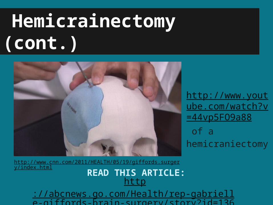
Hemicrainectomy (cont.)
http://abcnews.go.com/Health/rep-gabrielle-giffords-brain-surgery/story?id=13631297
READ THIS ARTICLE:
http://www.youtube.com/watch?v=44vp5FO9a88 of a hemicraniectomy
http://www.cnn.com/2011/HEALTH/05/19/giffords.surgery/index.html

THANK YOU
We hope that this presentation has helped to increase your knowledge on
Brain Diagnostic Testing and Advances in Neurosurgery!


![[Allred Charles]the GOUT](https://img.pdfslide.us/doc/110x75/577cc6281a28aba7119dd1e0/allred-charlesthe-gout.jpg)