Embed Size (px)
Citation preview

INFORMATION TO USERS
This manuscript has been reproduced from the microfilm master. UMI
films the text directly from the original or copy submitted. Thus, some
thesis and dissertation copies are in typewriter face, while others may
be from any type of computer printer.
The quality of this reproduction is dependent upon the' quality of the
copy submitted. Broken or indistinct print, colored or poor quality
illustrations and photographs, print bleedthrough, substandard margins,
and improper alignment can adverselyaffect reproduction.
In the unlikely event that the author did not send UMI a complete
manuscript and there are missing pages, these will be noted. Also, if
unauthorized copyrightmaterial had to be removed, a note will indicate
the deletion.
Oversize materials (e.g., maps, drawings, charts) are reproduced by
sectioning the original, beginning at the upper left-hand corner and
continuing from left to right in equal sections with small overlaps. Each
original is also photographed in one exposure and is included in
reduced form at the back of the book.
Photographs included in the original manuscript have been reproducedxerographically in this copy. Higher quality 6" x 9" black and white
photographic prints are available for any photographs or illustrations
appearing in this copy for an additional charge. Contact UMI directly
to order.
U·M·IUniversity Microturns International
A B8!' & Howel! IdorrnallOrl CorrpanvJOO North Zeeb Road Ann Arbor MI 48 106-13..\6 USA
313 761-4700 809521.0600


Order Number 9300346
Investigations on the nucleic acids coding for the sea anemonetoxins, anthopleurins A and B
Sorensson, Melinda Manaig, Ph.D.
University of Hawaii, 1992
U·M-I300 N. Zeeb Rd.Ann Arbor,MI48106


INVESTIGATIONS ON THE NUCLEIC ACIDS CODINGFOR THE SEA ANEMONE TOXINS,
ANTHOPLEURINS A AND B
A DISSERTATION SUBl\tIlTIED TO THE GRADUATE DIVISIONOFTIlE
UNIVERSITY OF HAWAIl IN PARTIAL FULFILLMENTOFTIlE REQUIREMENTS FORTIlE DEGREE OF
DOCTOR OFPHILOSOPHY
IN BIOMEDICAL SCIENCES(BIOCHEMISlRY)
August 1992
By
Melinda M. Sorensson
Dissertation Committee:
Howard F. Mower, ChairpersonMarguerite Volini
Ian GibbonsMorton MandelDavid Haymer

ACKNOWLEOOMENT
I thank Dr. Kerry T. Yasunobu for giving me the opportunity
to work with him, and for supporting me.
I thank the members of my dissertation committee for all
their valuable suggestions during the course of the work. I also
thank Dr. Steve Palumbi for serving in my committee earlier.
I thank the Founder Region Fellowship for support in
writing my dissertation.. and the American Heart Association.
Hawaii Chapter, for funding in part this research.
iii

ABSTRACT
Anthopleurin B CAP-B) is a 49 amino acid cardiotonic
polypeptide produced by the giant green sea anemone,
Anthopleura xanthogrammiea. Along with AP-B, another
polypeptide, anthopleurin A CAP-A), of the same length, and
differing only in 7 amino acids, is produced by the animal in
higher quantities. The object of this study was to investigate the
the two peptides at the nucleic acid level.
Genomic and cDNA libraries of the animal were constructed
in A. replacement vectors. The genomic library was constructed in
the Bam HI site of EMBL3, while the cDNA library was constructed
in the unique Eeo RI site of A. gtl0.
Synthetic nucleotides spanning the first 10 amino acids of
AP-B and the complement of the codons for the last 10 amino
acids of the AP-A peptides were constructed from back
translation of the peptides' sequences. The primers were. used to
initiate a polymerase chain reaction, using cDNA as the template.
A 144-147 base pair PCR product was obtained.
The PCR product was purified from the primers and used to
probe the genomic DNA and to screen both the genomic and cDNA
libraries. Positive clones were isolated from both libraries.
A 1.8 kb Bam HI genomic fragment common to 5 of the 14
genomic clones examined, and a 2.8 kb EeoRI genomic fragment
common to 3 clones, that hybridized both to the peR product and
the primers were subcloned into M13mp18.
iv

The peR product was cloned into the phagemid vector,
pBluescript KSII+. The sequence of a random clone showed some
sequence homology with the ambiguous sequence predicted for
AP-A and AP-B.
The cDNA clones were cut with two enzymes flanking the
Eco RI site of the cloning vector and a 480 base pair fragment was
subcloned into M13mp18.
v

TABLE OFCONlENTS
Page
ACKN"OWlEDGMENT iii
ABSlRACf............................................................................................................ iv
LIST OF FIGURES............................................................................................... ix
LIST OF APPENDICES....................................................................................... xiii
LIST OF ABBREVIATIONS........................ xiv
I. IN"'fR.ODUCTION............................................................................................... 1
II. REVIEW OF LffERATURB............................................... 3
A. Toxins from marine organisms
1. Mechanism of action of polypeptide toxins
from marine organisms.................................................... 4
2. Biological Importance of the peptide toxins. 7
B. Polypeptide toxins from sea anemones 8
1. Primary structure of sea anemone toxins 9
.2. Mechanism of action of -the anthopleurins 10
C. Importance of isolation and characterization of the
nucleic acid structure of the anthopleurins 20
m. OBJECfIVE OF TIlE STUDy 23
IV. MAlERIALS AND METIIODS 24
A. Biochemicals ! ••••••••••••••••••••••••••••• 24
B. BacterialStrains 25
C. Vectors 26
VI

Page
D. Media 26
E. Solutions................. 27
F. Enzymes 29
G. Isolation of Genomic DNA from Anthopleura
xanthogrammica 30
H. Genomic library construction 32
I. Isolation of RNA from Anthopleura xanthogrammica 34
J. Construction of cDNA Library 36
K. Primer Construction and Labeling..................... 38
L. Polymerase Chain Reaction using cDNA as template 41
M. Isolation of phageDNA 41
N. In situ plaque hybridizations .43
O. Subcloning of genomic fragments 44
P. Cloning of the PCR product. 44
Q. Subcloning of cDNA inserts from cDNA clones 46
R. Bacterial Transformation 46
S. DNA Sequencing 47
V. RESULTS
A~ Isolation of high molecular weight DNA 50
B. Construction of genomic library 53
C. Construction of cDNA library 58
D. Polymerase Chain Reaction 60
vii

Page
E. Screening of genomic and cDNA libraries using PCR
product as the probe 68
F. Cloning of the 2.8 kb Eco RI and the 1.8 kb Bam HI
genomic fragments 85
G. Screening of the cDNA library with the PCR product 92
H. Subcloning of cDNA insert into M13mp18 95
I. Cloning and sequencing of the PCR product 99
VI. DISCUSSION 101
VII. SUMMARYANDCONCLUSIONS 113
APPENDICES 115
REFEREN"CES 127
viii

LISTOFFIGURES
Page
Fig. 1. Agarose gel of genomic DNA isolated from Anthopleuraxanthogrammica using the method of Kaiser and Murray 51
Fig. 2. Agarose gel of genomic DNA isolated from Anthopleuraxanthogrammica using the method of Maniatis et al 52
Fig. 3. Agarose gel of aliquots of partial Sau 3A genomic DNAdigest after sucrose density gradient centrifugation 54
Fig. 4. Agarose gel of partial Sau 3A digest of A. x. DNA afterreprecipitation following sucrose density gradientcentti.fugation...............•..............................................................................56
Fig. 5. Schematic representation of lEMBL3 cloning vector 57
Fig. 6. Schematic representation of cloning vector 19t1O 58
Fig. 7. Schematic representation of Eco RI adaptor ligationsystem used in constructing the cDNA library 59
Fig. 8. Polymerase chain reaction products from differenttemplates, sized on 4 % agarose gel 61
Fig. 9. Polymerase chain reaction products sized on a 4 % gel..........63
Fig. 10. Agarose gel of polymerase chain reaction productsfrom different templates and varying primer ratios 65
ix

PageFig. 11. The product from the polymerase chain reaction after
concentration on centricon filters and after electroelutionhom a 4 % agarose gel 67
Fig. 12. Agarose gel of genomic DNA after complete digestionwith Eco RI, Bam Ill, and Hind In 69
Fig. 13. Southel'1:' blot of genomic DNA after complete digestionwith Eco RI, Bam III and Hind III, probed with the peRprod.uct••.••••••..••.•.•..•••.••••.•.•••..•.•••.••••.••.•.••.•..••.•..•••.•....•..•..•..•.•.......•.•.••.........71
Fig. 14. Autoradiogram of a filter used in in situ plaquehybridization of the amplified genomic library probedwit.b. tilePCR prod.uct....•.•..••...•...••..•.....•.•..•••..............•. I" •••••••••••••••••••••••72
Fig. 15. Agarose gel of 14 genomic clones digested with Eco RI.. ...74
Fig. 16. Southern blot of Eco RI digests of 14 genomic clonesusing peR product as probe............•..••••..••.••.•.••..•.•...•....••....•.....•........76
Fig. 17. Comparison of genomic DNA and genomic DNA andgenomic clones after digestion with Eco RI, Bam III andHilld, m 78
Fig. 18. Southern blot of genomic DNA and genomic clonesdigested with Eco RI, Bam III and Hind 111.•.........•..•..•.....•...........79
Fig. 19. Agarose gel of of 8 genomic clones digested with Bam HIandHind m 82
x

PageFig. 20. Southern blot of 8 genomic clones digested with
Bam III and Hind TIl, probed with labeled PCR product 84
Fig. 21. Agarose gel of M13mp18 subclones from genomicclone g24 with. Eco R1 85
Fig. 22. Southern blot of the Eco RI digests of M13mp18subclones from g24 using the PCR product as probe 87
Fig. 23. Agarose gel of M13mp18 Bam III digests of A.subclones 88
Fig. 24. Southern blot of Bam HI digested M13mp18 subclonesusing the peR product as probe 89
Fig. 25. Agarose gel of M13mp18 g15 subclones withBQ11I, Hl.............................................................•............................................9{)
Fig. 26. Southern blot of the M13mp18 subclones usingprimers as me probe.....•...........................••.................•.....•...................91
Fig. 27. Agarose gel of the cDNA clones digested with Eco Rl. 93
Fig. 28. Southern blot of cDNA clone digests using the PCRprocluct as probe....•.......•..•.•...••....................•.........•.................................94
Fig. 29. Agarose gel of the M13 subclones digested with Eco RI. ...96
xi

Fig. 30. Agarose gel of M13mp18 eDNA subclones withECD RI for Southern blotting 97
Fig. 31. Southern blot of M13mp18 eDNA subclone 98
xii

LISTOF APPENDICES
Page
Appendix 1. Comparison of the sequence of clone pBSI with theambiguous and unambiguous nucleotide sequences ofanthopleurins A and B 115
Appendix 2. Comparison of two hour sequence of pBSI withpBluescript KSTI+ 121
Appendix 3. Gap analysis of the consensus sequence of thepBS1 insert against the ambiguous sequence obtained bybacktranslating anthopleurin A 122
Appendix 4. Gap analysis of the consensus sequence of thepBSI insert against the ambiguous sequence obtained bybacktranslating anthopleurin B 123
Appendix 5. Comparison between the amino acid sequenceobtained by translating the consensus sequence of clonepBSI and the anthopleurin A peptide 124
Appendix 6. Comparison between the amino acid sequenceobtained by translating the consensus sequence of clonepBS1 and the anthopleurin B peptide 125
Appendix 7. Comparison between the sequences of clones pBS3,pBS7, and pBS8 with the cloning vectorpBluescript KSTI+ 126
Xlll

AMV
cDNA
dATP
dCTP
dGTP
DNA
dT
dTT
dTTP
EDTA
g
IPTG
1
Jlg
Jll
ml
mV
PeR
PEG
RNA
SDS
SM
sseTAB
LIST OF ABBREVIATIONS
Avian myeloblastosis virus
complementary or copy DNA
deoxyadenosine S' -triphospahate
deoxycytidine 5'-triphosphate
deoxyguanidine S'-triphosphate
deoxyribonucleic acid
deoxyribothymidilic acid
dithiothreitol
deoxythymidine S' -triphosphate
ethylene-diamine tetraacetic acid
grams
Isopropy1-~ -D- galactopyranoside
liter
micrograms
microliters
milliliters
millivolts
Polymerase chain reaction
polyethylene glycol
ribonucleic acid
Sodium dodecyl sulfate
Storage medium
Saline sodium citrate
Tris Acetate EDTA
xiv

lBE
TCA
1E
1EMED
X-gal
CC
Tris Borate EDTA
trichloroacetic acid
Tris-EDTA
tetramethylethylenediamine
5-bromo-4-chloro-3-indolyl-~-D-thiogalactopyranoside
degree Centigrade
xv

I. INTRODUCfION
Several species of marine organisms produce a variety of
biologically active peptides. In general, these peptides are toxic;
the most potent include brevetoxins, maitotoxin, paragracine or
the palytoxin. Aside from the fact that they pose a serious threat
to public health, these toxins and the other novel toxins have
proven to be invaluable tools in studying the molecular details of
ion channels and receptors of excitable cells because of their high
binding affinities to ion channels. However, a more exciting pos
sibility in the study of these toxins is their potential to be devel
oped into pharmacologically useful drugs. The latter possibility
entails thorough testing .before it becomes a reality and a basic
understanding of the mode of actions of these toxins needs to be
achieved. The techniques of recombinant DNA technology offers
the possibility of looking at these toxins at the molecular level.
This review will cover briefly marine organisms, with particular
emphasis on the novel polypeptides anthopleurins A (AP-A) and B
(AP-B). ·These toxins are produced by the sea anemone,
Anthopleura xanthogrammica, This study is aimed at
investigating the molecular genetics of the sea anemone toxins A
and B
The sea anemone, Anthopleura xanthogrammica (Brandt)
produces two polypeptides, Anthopleurins A (AP-A) and B (AP-B),
which are neurotoxic at high levels but exhibit a positively
inotropic effect at lower levels. Of the two, AP-A has been well
characterized. Anthopleurin A has been shown to have a positive
1

inotropic effect on the hearts of dogs (Blair et al., 1978), guinea
pigs (Hashimoto et al., 1980) and mice (Shibata et al., 1976). A
potent cardiac stimulant, AP-A can potentially be used in the
treatment of heart disease. In the study of cardiac muscle
physiology it is useful because it is more potent than either
ouabain or glucagon (Low et al., 1979), and its inotropic action is
not accompanied by chronotropic or hypertensive effects and it is
relatively hypoxia resistant. The inotropic effect of AP-A is not
inhibited by propranolol pretreatment (Shibata et al., 1976). In
addition to its inotropic activity, AP-A, binds to the voltage
sensitive sodium channel present in the membranes of nerve,
skeletal and heart cells. Detailed investigations of the excitable
membranes of giant crayfish axons revealed that AP-A binds to
an external site of the sodium channel and prolongs the
inactivation process. The site of binding of AP-A is distinct from
the tetrodotoxin binding site, where activation of the sodium
channel is effected. Hence, there are two main reasons to explore
these polypeptides: as a tool to investigate the mechanism of the
sodium channel inactivation, and as a potential substitute for the
inotropic agents currently being used in treating heart disorders.
Anthopleurin B differs from AP-A in only seven of 49 amino acid
residues. These substitutions confer on AP-B ten times higher
inotropic activity than AP-A. Because of their potential to be
developed as pharmacologically useful drugs, and because they
can be used to probe the sodium channel, it was deemed
important to study them at the molecular level.
2

II. REVIEW OF LITERATURE
A. Toxins from marine organisms
Tetrodotoxin is a heterocyclic guanidine originally isolated
from the ovaries and liver of puffer fish (Brown and Mosher,
1963), but has also been found in newt (Crone et al., 1976),
octopus (Sheumack et al., 1978), frog (Kim et al., 1975), and goby
(Noguchi and Hashimoto, 1973).
Saxitoxin, like tetrodotoxin, is a heterocyclic guanidine, pro
duced by the dinoflagellates of the genus Gonyaulax. It is found
in large concentrations in clams, mussels and other shellfish that
feed on the dinoflagellates (Caterrall, 1986).
The unarmored dinoflagellate Ptychodiscus brevis, produces
eight toxins collectively known as brevetoxins (Shimizu, 1982).
Another dinoflagellate Gambierdtscus toxicus produces three
types of toxins: ciguatoxin, scaritoxin and maitotoxin, which have
been established as the cause of ciguaterra poisoning (Legrand
and Bagnis, 1984). Although the three toxins are produced by the
same organism, much more maitotoxin is secreted than the other
two. Other marine organisms producing toxins are coelenterates:
Parazoanthus gracillis (paragracine), Palythoa (palytoxins);
marine snails of the genus Conus (conotoxins); and sea anemones
(toxins I, II, III, and IV, anthopleurins A, B and C).
3

1. Mechanism of action of peptide toxins from marine
organisms
It has been shown that tetrodotoxin at very low concentrations
(10-7 M) selectively blocks the increase in sodium permeability
caused by depolarization, but does not affect the ion permeability
of the unstimulated axon, nor the delayed increase of potassium
permeability caused by depolarization (Narahashi et al., 1964).
Saxitoxin acts similarly to tetrodotoxin, in that it blocks action
potentials without depolarization (Kao and Nishiyama, 1965). Kao
and Nishiyama (1965) first proposed that the guanidinium moi
eties of these two toxins might enter the sodium channels like
guanidine, but due to their bulkiness, they may bind tightly to the
ion channel and in effect block the passage of the Na" ions.
Henderson et ale (1974) extended the hypothesis of the previous
authors by proposing that the receptor site for the toxins was a
specific coordination site in the sodium channel. Similarly, Hille
(1975) proposed that the receptor site was the ion selectivity fil
ter, a coordination site that determines the ion selectivity of the
channel.
Studies utilizing radiolabeled tetrodotoxin and saxitoxin have
formed most of the current findings on the nature of the sodium
channel. The finding that each of the two toxins inhibit the bind
ing of the other provided strong evidence that they bind the same
receptor (Colquohoun et al., 1972; Henderson et al., 1973; Barnola
et al., 1972).
4

Brevetoxins depolarize nerve and muscle membranes in a dose
dependent manner (Huang et al., 1984; Wu et al., 1985). The ex
tent of maximum depolarization is about 40 mV, and on crayfish
giant axons the EC50 was reported to be 1.7 nM (Wu et al., 1985).
Voltage clamp experiments showed that the target of action of
brevetoxins is the sodium channel. The modified sodium channel
exhibits the following three characteristics: (1) the channel modi
fied is activated at membrane potentials ranging from -160 to
180 mY, levels at which' the sodium channel is normally closed,
(2) the channel is activated very slowly (375-127 msec in the
potential range of -80 to +10mV) compared to that of the normal
sodium channel, and (3) the channel is essentially devoid of fast
inactivation (Huang and Wu, 1985). Hence, the toxins depolarize
the membrane by removing the inactivation process after opening
the sodium channels at potential where it is normally closed.
Ciguatoxin, at very low concentrations (0.2 -1.0 nM), induces a
membrane depolarization and spontaneous action potentials in
neuroblastoma cells and in frogs' nodes of Ranvier (Legrand et al.,
1985). Ciguatoxin modifies the sodium channel in a manner simi
lar to that of the brevetoxins. Maitotoxin, on the other hand,
causes a calcium dependent contraction of skeletal and smooth
muscles. In cardiac muscles, it has a positive inotropic effect at
low concentrations (0.1 to 4 ng/ml), which is completely elimi
nated by Co 2+ or verapamil (Kobayashi et al.; 1985; Kobayashi et
al., 1986). Maitotoxin induces (1) an increased release of nore
pinephrine and dopamine from rat pheochromocytoma cells
5

(Takahashi et al.; 1982), and (2) a high uptake of Ca 2+ by some
cultured cell lines. Both effects can be inhibited by blockers of the
sodium channel (Kobayashi et al., 1983). These results suggest
that the toxin induces an increase in the permeability of the cell
membrane to calcium, which in turn triggers the release of
transmitters and muscle contraction.
Paragracine is one of the troponoid substances secreted by the
coelenterate species Parazoanthus, which are collectively known
as zooxanthins. This toxin blocks the sodium channel without
affecting the potassium channel, based on the observation that
blockers of the sodium channel had no effect on its action (Seyama
et al., 1980). Paragracine blocks the sodium channels only on the
axoplasmic side, and does so only when the sodium ions are
flowing unidirectionally in the outward direction. This blockage of
the sodium channel by brevetoxin can be alleviated if an inward
sodium current is generated (Seyama et al., 1980)..
Palytoxin, produced by the coelenterate genus Palythoa, is the
most lethal marine toxin known to date. When administered
intraperitoneally, the LD 50 for mice is 50-100 ng toxin/kg weight
(Wiles et al., 1974). Palytoxin depolarizes excitable tissues
including cardiac muscle (Alsen et al., 1982; Ito et al., 1979;
Wiedman, 1977), skeletal muscle (Ito et al., 1979; Ohizumi and
Shibata, 1980) and both myelinated and unmyelinated nerve
fibers (Muramatsu et al., 1984; Pichon, 1982). Based on the
observations that the action of palytoxin is potentiated by ATP,
and that dog erythrocytes which lack Na, K-ATPase are resistant6

to the action of palytoxin, Habermann (1983), Habermann et al. ,
(1982), and Chattwal et al. (1983) have proposed that palytoxin
interacts with the Na, K-ATPase and converts the pump into an
ion channel.
To date, there have been twelve toxins isolated from marine
snails of the genus Con us. In general, the conotoxins are
neurotoxins. Kobayashi et al. (1982) surveyed 29 species of toxins
and found that the conotoxins exhibit a variety of actions such as :
(1) blocking acetylcholine receptors, (2) blocking muscle Na
channels, (3) blocking calcium channels, (4) opening Na channels,
(5) increasing Na permeability, (6) increasing calcium influx, or (7)
contracting rabbit aorta. The above actions are specific for each
toxin. Only toxins isolated from Conus geographus (conotoxin GI,
GIl, GIllA and geographutoxin), Conus magus (conotoxin MI) and
Conus striatus (striatoxin) pose serious threats to humans.
2. Biological importance of the peptide toxins
As mentioned earlier, the most potent toxins are serious
threats to public health. For example, the catastrophic event of
the red tide in the gulf of Mexico in 1946-1947 which resulted in
tons of dead fish littering the coast of Florida was caused by
brevetoxins. Fish poisoning in the Carribean, the South Pacifc and
even in the United States , has been traced to the ciguatoxins
produced by the dinoflagellates that enter the food chain via the
fishes that feed on them.
Aside from the above major reasons, some of the toxins so far
identified have been used as molecular tools in studying the neu
7

romuscular system, because of their high binding affinity to ex
citable membranes. For example, alpha bungarotoxin has been
useful in studying the acetylcholine receptors. Similarly,
tetrodotoxin and scorpion toxins have been useful in studying the
sodium channel. In the case of the novel peptides, omega
conotoxin has been utilized to study the presynaptic terminal of
excitable membranes. Hence, the toxins from marine organisms
could provide a wealth of information on the nature and
physiology of excitable membranes (Shibata et al., 1976).
B. Polypeptide toxins from sea anemones
Sea anemones synthesize polypeptides which display a wide
variety of biological activities, such as cardiotoxicity, cardiostimu
lation, cytolysis, and proteinase inhibition (Alsen, 1983). These
polypeptides are classified according to their molecular weights.
They are: (1) class I, <3,000 ; (2) class II, 4,000-6,000; (3) class III,
6,000-7,000; and (4) class IV, > 10,000. Classes I and II act on
excitable membranes, mainly on the fast sodium channel, class III
inhibits proteinase activity, and class IV exhibits mainly cytolytic
activity. At present, there have been 23 such toxins isolated. Of
these, those belonging to class II have elicited much interest be
cause they exhibit actions such as cardiostimulation,
cardiotoxicity, and neurotoxicity in mammals (Norton et al., 1978;
Beress, 1978;1982; Schweitz et al., 1981). Included in this class
are anthopleurins A (AP-A), and B (AP-B) from Anthopleura xan
thogrammica, anthopleurin C (AP-C) from Amhopleura elegantis-
8
sima. and toxins I and II from Anemonia sulcata. The pharmaco
logical properties of these peptides such as LD 50 on mice and
crab, their binding to synaptosomes and stimulation of Na +
uptake by neuroblastoma cells have been determined by Schweitz
et al. (1981).
These peptides function as heart stimulants and are very po
tent positive inotropic agents. They increase the force of heart
contraction without affecting the heart rate or blood pressure, and
they are neurotoxins at somewhat higher concentrations (Norton
et al., 1978; Shibata et al., 1976). Their mode of action has been
attributed to binding to the gate of the fast sodium channel, thus
causing a delay in inactivation of this channel and a prolongation
of the action potential (Romey et al., 1976; Kodama et al., 1981).
The inotropic activity has also been attributed to the peptides'
binding to and affecting the slow (calcium/potassium) channel
(Ohizumi and Shibata, 1981; De Barry et al.; 1977).
1. Primary structure of the polypeptide toxins
The amino acid sequence of AP-A (Tanaka et al., 1977) and
AP-B (Reimer et al., 1985) have been determined. AP-C (Beress,
1978), toxin I (Wunderer and Eulitz, 1978) and toxin II
(Wunderer et al., 1976) primary sequences have also been
reported. All of these toxins are short peptides between 46 and
49 amino acids, with considerable sequence homology. A variety
of spectral techniques including NMR have been used to study the
secondary structure of AP-A (Ishizaki et al., 1979; Norton and
Norton, 1979), toxin I (Norton et al., 1980), and toxin II (Norton
9

et al., 1976). AP-A has been crystallized and preliminary
diffraction data has been reported (Smith et al., 1984).
Conformational studies point to a random coil secondary structure
for toxin II, while a more globular structure for that of AP-A.
Anthopleurin B, the most potent peptide heart stimulant from
the sea anemone Anthopleura xanthogrammica, behaves anoma
lously on Sephadex gel filtration columns and elutes at a position
indicating a molecular weight of about 2,400 (Reimer et al., 1985).
However, sequence determination showed that AP-B consists of a
single polypeptide chain of 49 amino acids with a molecular
weight of 5,257. The sequence of the peptide was shown to be:
Gly-Val-Pro-Cys-Leu-Cys-Asp-Ser-Asp-Gly-Pro-Arg-Pro-Arg-Gly
Asn-Thr-Leu-Ser-Gly-Ileu-Leu-Trp-Phe-Tyr-Pro-Ser-Gly-Cys
Pro-Ser-Gly-Trp-His-Asn-Cys-Lys-Ala-His-Gly-Pro-Asn-Ile-Gly
Trp-Cys-Cys-Lys-Lys. Although six carboxymethyl cysteine
residues were formed by reduction and alkylation of the
polypeptide, no cysteine residues were detected in the native
peptide indicating that there are three cystine bonds in AP-B
(Reimer et al., 1985). The positions of the disulfide bonds were
chemically determined for toxin II (Barhanin et al., 1981), but
until the present time, those cystine bonds have not been localized
for either AP-A or AP-B.
2. Mechanism of action of the anthopleurin polypeptide toxins
The cardiac stimulatory action of coelenterate toxins on rat
atria was first reported by Shibata et al., 1974, while studying the
antitumor activity of the extracts from the sea animals. Most10

notable was the finding that extracts from the sea anemone
Anthopleura xanthogrammica gave the highest percent increase in
inotropic activity of rat hearts, at concentrations as low as 119
parts per million (ppm). Norton et al. (1976) isolated the active
peptide from the Anthopleura xanthogrammica and named it
anthopleurin. The presence of two kinds of peptides from the
ethanol extract from these marine invertebrates was apparent on
the elution profile of the cation exchange column. The fraction that
eluted at VeNo (ratio of eluted volume to void volume) of 2.99
3.39 they termed AP-A, and the fraction that eluted at VeNo of
6.3 to 7.01, they called AP-B.
Shibata et al. (1976) investigated the mechanism of the posi
tive inotropic action of AP-A on the hearts of mongrel dogs, rab
bits, rats, cats and guinea pigs. They found that the cardiotonic
potency of AP-A was greater than that of glucagon and ouabain
and equal to that of isoproterenol, but without an accompanying
chronotropic effect on the isolated cardiac muscles of the animals
examined. In addition, the cardiac contractility of the heart mus
cles became more resistant to the effects of hypoxia and tempera
ture stress after pretreatment with AP-A. This latter effect was
better in AP-A treated than in ouabain treated cardiac muscles.
Since cardiac glycosides, such as digitalis and ouabain
increase the contractility of the heart muscle by inhibiting the
Na,K-ATPase pump, Shibata et al. (1976) determined whether
AP-A also induces positive inotropy by the same mechanism. The
results from studies utilizing assays for Na,K -ATPase activity and1 1

cAMP hydrolysis showed that neither the ATPase activity nor
phosphodiesterase activity was affected by AP-A. An interesting
observation was that the peptide prolonged the action potential in
guinea pig atria and ventricles, without affecting all other
electrical properties of membranes of the cardiac' muscle (Shibata
et al., 1976). Blair et ale (1978 ) studied the effect of AP-A on
cardiac dynamics in conscious dogs and found that AP-A does not
alter mean arterial pressure, mean left atrial pressure and left
ventricular end systolic diameter. They concluded that AP-A has
a direct inotropic effect on the myocardium with little or no effect
at all on the peripheral circulation. Hence AP-A is potentially a
promising substitute for the current cardiac glycosides being used
to treat heart disorders.
The other property of AP-A is its ability to prolong the action
potential in excitable membranes which contain voltage gated
sodium channels. Voltage clamp experiments showed that AP-A
prolongs the action potential on crayfish giant axons Low et al.,
(1979). Treatment of the axons with as little as 10-8 M AP-A re
sulted in membrane polarization and depolarization, and in repeti
tive firing. The prominent plateau generated on the action poten
tial lasted up to 300 milliseconds (msec), an extension of about
seven times that of the untreated controls.
Like the skeletal muscle membranes and axons from giant
crayfish, cardiac muscle cell membranes contain voltage gated
sodium channels. This voltage gated sodium channel is
responsible for the increased permeability to sodium ions12

following membrane depolarization (Ganong, 1985). The cardiac
muscle cells differ from the other two, in that its membrane also
contains a voltage gated calcium channel, which is responsible for
prolonged repolarization. Shimizu et al. (1979) reported that, upon
exposure to AP-A (3 x 10⁻⁸ g/ml) guinea pig ventricular muscle
exhibited the following characteristics: 1. prolonged action
potential, 2. increased tension and 3. no significant change in the
maximum voltage change per unit time (dV/dT). In order to
relate the mechanism of the prolongation of the action potential to
the positive inotropic effect of AP-A, Hashimoto et al. (1980)
investigated the ionic mechanism of the prolongation of action
potential in isolated guinea pig ventricular muscle. They found
that the prolongation of action potential by AP-A was
accompanied by a decreased rate of outward current. The
decreased rate of outward current could have been due to
blockage of the slow inward Ca current or to a delayed outward
potassium current, or to a delay in the closure of the sodium
channel. Hashimoto et at. (1980) found that delayed increase in
potassium permeability was not affected, since treatment of the
ventricular muscles with blockers of the K channel had no effect
on the prolonged action potential. AP-A did not alter the slow Ca
channel since treatment of rabbit ventricular muscle with
Verapamil, a blocker of the slow Ca channel, did not inhibit the
effect of AP-A (Kodama et al., 1981). In the presence of ryan
odine, an agent which is known to interfere with Ca release from
the sarcoplasmic reticulum, AP-A prolonged the action potential,
13

but failed to cause a positive inotropic effect. Hence, the increase
in contractile force is probably an outcome of the alteration of the
Ca kinetics by the prolonged action potential.
The other polypeptide which can be isolated from Anthopleura
xanthogrammica is Anthopleurin B (AP-B), which can be sepa
rated from AP-A by ion exchange chromatography. AP-B is the
more potent toxic polypeptide of the two.
On isolated, intraarterially perfused, spinal cord of the
bullfrog, Rana catesbiana, AP-B caused a marked augmentation of
the ventral and dorsal root potentials produced by the stimulation
of the dorsal root (Kudo and Shibata, 1980). It also prolonged the
excitatory post synaptic potential (EPSP), and elevated the
membrane resistance, without affecting the motoneuronal action
potentials. The excitatory action of AP-B was greater in low Ca2 +
(0.9 mM) than in high Ca2+ media (3.6 mM). Based on these
observations, Kudo and Shibata (1980) proposed that AP-B acts on
subsynaptic membranes to make it more sensttive to
neurotransmitters.
Ohizumi and Shibata (1981) tested AP-B on guinea pig's iso
lated ileum and isolated taenia caeci, in order to determine the ef
fect of AP-B on the autonomic nervous system and the smooth
muscle. In both taenia caeci and ileum, AP-B induced contraction
followed by relaxation in concentrations as low as 3xlO-9 M. The
other sea anemone toxins effected similar actions but at much
higher concentrations (10-7 M or higher). The AP-B induced re
sponses were inhibited by tetrodotoxin or low Na+ in the medium,14

suggesting that AP-B causes membrane depolarization by increas
ing the Na+ permeability across cell membranes.
Contractions induced by AP-B on both the ileum and the taenia
caeci were inhibited by muscarinic blockers, but not by nicotinic
blockers, blockers of histamine and tryptamine release, nor by
blockers of prostaglandin synthesis. This suggests that contrac
tions on both tissues induced by AP-B were mainly mediated
through endogenous acetylcholine released from cholinergic nerve
terminals (Ohizumi and Shibata, 1981). In contrast, relaxation
was different between the two tissues examined. In taenia caeci,
relaxation was inhibited by guanethidine and phentolamine.
These same agents had no effect on the relaxation induced by AP
B on the ileum. This observation suggests that in the spinal cord,
the rhythmic relaxation maybe due to the release of
noradrenaline, via the excitation of adrenergic nerves. On the
other hand, in the smooth muscles of the mammalian
gastrointestines, relaxation could be due to the excitation of
adrenergic or non-adrenergic inhibitory nerves. It would appear
that in this case, AP-B induced relaxation of the ileum was due to
excitation of the latter.
In order to further examine the mode of action of AP-B on
smooth muscles, Norton et al, (1981) tested its excitatory effect on
guinea pig vas deferens. This study was prompted by the hy
potheses concerning the nature of motor transmission in vas def
erens: the presence of both adrenergic and non-adrenergic compo
nents. Norton et al. (1981) found that at concentrations as low as
15

3xl0-9 M, AP-B caused rhythmic contractions on guinea pig vas
deferens. Except for toxin II, a peptide with considerable
sequence homology with AP-B, the other sea anemone toxins also
caused contractions but only in higher concentrations (>5xl0-8 M).
A more important finding, however, was that at concentrations
between 10-8 and 10-5 M, AP-B caused a dose dependent release
of noradrenaline in the surrounding medium. At the higher limit,
(10-5 M) AP-B caused increased release of noradrenaline 310 x
more than that of the untreated vas deferens. Both contraction
and adrenaline release induced by AP-B were inhibited by the ab
sence of Ca 2+. These results suggest that the AP-B induced con
traction is mediated by noradrenaline release from the
adrenergic nerve endings, and that the release of noradrenaline
induced by AP-B may require extracellular Ca 2+.
The heart stimulant activity of AP-B is over ten times higher
than that of AP-A (Norton et al., 1978). Therefore it is of interest
to compare the sequences of these peptide heart stimulants in
order to understand the chemical basis of their activity. There are
only 7 amino acid differences between AP-B and AP-A, 5 of which
are likely candidates for activity differences. The net charge in
crease at neutral pH from +3 to +5 between AP-A and AP-B may
be significant as has been pointed out for the activity of the other
sea anemone toxins on mammals (Schweitz et al., 1981).
Anthopleurin C from Anthopleura elegantissima and toxin II from
Anemonia sulcata differ from AP-A and AP-B in that they consist
of 47 amino acid residues. However, this shortening of the16

polypeptide chain does not appear to significantly affect the heart
stimulant activity since AP-C has about the same heart stimulant
activity as AP-A.· The following observations summarize the
amino acid sequence information between AP-B and related
toxins.
The amino acid sequence differs in 7 places between AP-A and
AP-B. These are residues 3 (P for S), 12 (R for S), 13 (P for V), 21
(I for T), 24 (F for L) 42 (N for T), and 49 (K for Q). These dif
ferences are important since AP-B is a better heart stimulant than
AP-A, even when they are produced by the same organism.
Similarly, AP-B is more potent than AP-C from Anthopleura ele
gantissima, or toxin II from Anemonia sulcata. Since Pro (residue
3) and lIe (residue 21) are also present in AP-C, the presence of
these changes in AP-B may not confer the increased potency of
AP-B. This observation leads to consideration of the remaining
amino acid changes from AP-A, in residues 12, 13, 24, 42 and 49.
The stability of a particular conformation of an anthopleurin
toxin through primary structure factors is related to its binding
and the effect of binding on the membrane receptor. Barhanin e t
al. (1981) has proposed a two step model for the activity of toxins
from sea anemone. The first step in the model is the binding of
the polypeptide to its receptor. The second step is proposed to be
a conformational transition required to bring about the activity
responsible for the cellular changes observed at the
neuromuscular junctions, namely, the delay in closure of the
sodium channel by stabilizing the open conformation. The17

possible effects of the differences in amino acid sequence between
AP-A and AP-B can be evaluated from the perspective of possible
effects on the two steps. If we know more about the polypeptides
at the nucleic acid level, for AP-B these differences could be
exaggerated in designing changes in the amino acid sequence, with
the ultimate objective of increasing the inotropic activity of the
peptide, as well as reducing its antigenicity.
In studies of toxin II, the only ionizable group shown to be
essential for both binding and activity was the single Arg residue
at position 14 (Barhanin et al., 1981). AP-B is the only toxin from
sea anemone sequenced to date, which has two Arg residues in
this portion of the polypeptide. Furthermore, this portion of the
polypeptide seems to be the site of antigenic determinant, since
there are 4 hydrophilic charged amino acid residues within an
eight amino acid stretch (residues 7-14). Hopp and Woods (1981)
used a computer program to predict the most antigenic portion of
a polypeptide and concluded that the point of highest local
average hydrophilicity is invariably located in or adjacent to the
antigenic determinant site. Since aspartic acid, glutamic acid and
arginine are given the highest hydrophilicity values, it is very
likely that in AP-B, the antigenic determinant site is within
residues 7-14.
In AP-A, both binding and activity seems to depend on Arg
14. Assuming that this is also true for AP-B, Arg-12 is a good
candidate for site directed mutagenesis. Asp-7 and Asp-9 are also
good candidates in decreasing the antigenicity of the peptide.
18

However, as will be discussed shortly, the carboxyl groups on the
side chains of these residues may be important in carrying out
AP-B's inotropic action.
It is interesting to note that four of the seven substitutions
from AP-A to AP-B involve replacement of hydroxyl groups with
residues that can not donate a hydrogen bond through their side
chains (3, Ser to Pro ; 12, Ser to Arg ; 21, Thr to lIe; and 42, Thr to
Asn). It seems likely that these changes , if significant, would af
fect the second step (conformational change) possibly by
destabilizing the inactive con:formation through the loss of
intramolecular hydrogen bonding potential.
The C-terminal substitution of positive lysine for glutamine
could be significant in binding to the receptor or possibly may af
fect the pK for Asp-7 or Asp-9 beta carboxyl groups, one of which
was found to be unusually low in toxin I, toxin II, and AP-A
(Wunderer and Eulitz, 1978; Ishizaki et al., 1979; Norton and
Norton, 1979). Two of the aspartyl carboxyl groups of AP-A has
been shown to be essential for its cardiotonic activity in rat atria.
If the pK value of one the carboxyl groups is lower because of its
proximity to Lys-48 in AP-A (Lys-46 in toxin II), then the addi
tional epsilon amino group from Lys-49 in AP-B could possibly
lower the pK even more. The pK of that carboxyl group may be
important for its activity because of its involvement in the con
formational transition of the molecule, which occurs concomitant
with the protonation of that carboxyl group (Wunderer and Eulitz,
19

1978; Ishizaki et al., 1979; Norton and Norton, 1979), at about pH
2.
There is a slight increase in the hydrophobicity of AP-B over
AP-A (34.7 % hydrophobic residues for AP-B versus 30.6 % for
AP-A). A change of Phe for Leu at position 24 would not be
expected to change the hydrophobic character of that region
substantially. However, it has been shown that for toxin II, the
two Trp residues (23 and 31) interact through pi bonding
(Ishizaki et al.; 1979; Norton and Norton, 1979). This interaction
is apparently broken during the low pH induced conformational
change. These residues are also present in AP-B (23 and 33). The
third Trp residue however (residue 45 in both AP-A and AP-B)
becomes less random upon the changes in conformation. Such
constraint maybe strengthened by the presence of an additional
aromatic residue in AP-B (Phe-24).]
c. Importance of isolation and characterization of the gene for
AP-B
Matsueda and Norton (1982) have shown that the synthesis
of AP-A is possible through the Merrifield synthesis from t-boc
amino acids. They also found that disulfides will form in the syn
thetic peptide. However, relative to native AP-A, the synthetic
peptide has only 30 % inotropic activity. Hence it is possible to
evaluate the relationship of binding and changes in amino acid
structure via chemical means. This approach to the problem,
however, requires long term commitment, in addition to large
20
expenses commonly encountered every time a natural product is
synthesized chemically. The other alternative is the use of
molecular techniques to isolate and translate the gene, confer
changes in the amino acid structure via site directed mutagenesis,
and then compare the activity of the native peptide and the
synthetic peptides. This may lead to the development of peptides
with more desirable properties.
The small size of these peptides along with the availability
of the amino acid and activity data make them particularly
attractive models for protein engineering. However, the first step
to be accomplished is the isolation and characterization of the
gene. The availability of the gene encoding AP-B will pave the
way to single and multiple amino acid changes in the peptide by
in vitro mutagenesis techniques. This technique involves the
chemical synthesis of small sections of DNA (oligonucleotides)
which are complementary to sections of the gene but, with
alterations , which will result in the synthesis of a peptide .whose
amino acid 'sequence is different from that of the native peptide.
These altered peptides when compared to the native peptide will
reveal how certain changes in the amino acid composition will
affect properties such as inotropic activity, toxicity and
antigenicity. It will also allow an increased understanding not
only of these molecules, but also of the sodium channel in its
various electrically sensitive conformations. This will certainly
provide a revolutionary approach to the neurobiology of the
sodium channel. Furthermore, generation of altered peptides with21

mor~ desirable characteristics will bring them closer towards their
use as therapeutic heart stimulants.
22

m. OBJECTIVE OF THE STUDY
Until the present time, there has been no nucleic acid
studies on the anthopleurins. If these peptides are to be utilized
for their pharmacological properties, as well as one of the tools to
probe the sodium channel, sufficient quantities of the protein
should be readily obtainable. The first step towards this goal is to
have a cloned gene that can be expressed in the laboratory.
This study was conducted to investigate the sea anemone
toxins, anthopleurins a and b at the nucleic acid level.
23

IV. MATERIALS AND METHODS
A. Biochemicals
Acrylamide, agarose, bisacrylamide, ethidium bromide,
heparin, IPTG, phenol, spermidine, TEMED, Tris, and Xgal were
purchased from Boehringer Mannheim, Indianapolis, Indiana.
Ammonium chloride, ammonium persulfate, ampicillin, EDTA,
magnesium chloride, ficoll, glucose, magnesium sulfate, maltose,
polyvinylpyrrolidone, sodium chloride, sodium pyrophosphate,
and vitamin Bl were purchased from Sigma Chemical Corporation,
St. Louis, Mo.
Acetic acid, n-butanol, calcium chloride, ethyl alcohol, hexane,
hydrochloric acid, sarcosyl, Scintiverse cocktail mix and sodium
dodecyl sulfate were purchased from Fisher Scientific.
Bacto agar, bacto tryptone, and bacto yeast extract, which are
all products of Difco laboratories were purchased from Fisher
Chemicals.
The oligo dT column was purchased from Collaborative
Research Inc, Bedford, Ma.
Nytran and Elutip D were purchased from Sleicher and Schuell,
Keene, New Hampshire.
The Eco RI adaptors and the spin columns were purchased
from Promega Corporation, Madison Wisconsin.
[a-32P]-dCTP and [a-35S]-dATP were purchased from New
England Nuclear.
24
B. Bacterial Strains
DH5a and JMI09 were gifts from Dr. Charles Romeo.
DH5a is an E. coli strain that has the genotype supE44, hsd
RI7, recAI, endAI, gyrA 96, thi-I , relAI, is a recombination
deficient suppressing strain and was used to propagate plasmids.
JMI09 is a recAI, supE44, end AI, hsd RI7, gyrA96, relAI, thi,
L1 (lac pro AB), F'[traD36 proA+B+ lacIq lacZ .d MI5] is a strain
that will support growth of vectors carrying amber mutations, and
will modify but will not restrict transfected DNA. This strain is
recombination deficient and was used to propagate M13
C600 and C600 hfI A were purchased from Promega
Corporation and were used to select AgtlO recombinants.
C600 has the genotype supE44, hsdR, thi-I, thr-I, leuB6, lacY
1, tonA2]. C600 hfl A has the genotype supE44, hsd R, thi-l , thr
1, leuB6, lacY i, tonA2I, hflAI50 [chr::Tn 10 (tetr)) . The latter
strain distinguishes between cI+ (parent A gtlO) and cI-
(recombinant). Insertion of a DNA fragment into the single Eco RI
phage repressor gene generates a cI- phage which forms plaques
with clear center when plated on C600. On the other hand,
nonrecombinants form a turbid plaque on this strain. When
plated on C600 hflA, the cI+ phage is repressed so efficiently that
plaque formation is suppressed, while recombinants form a
regular plaque.
LE392 is a Sup E44, sup F58, hsd R514, gal K2, gal T22, met B
I, trp R55, lac YI and was used to propagate the genomic library.
25

C. Vectors
Lambda EMBL3 and AgtlO were purchased from Promega
Corporation, Madison, Wisconsin.
pBluescript KS II + was a gift from Kathy Hautchins.
M13mp18 and M13mp19 were purchased from Boehringer
Mannhein Corporation, Indianapolis, Indiana
D. Media
Luria Bertani (LB) medium is composed of 109 bacto-
tryptone, 5 g bacto yeast extract, and 10 g NaCl. To make plates,
15 g bacto agar was added to the above solution. This medium
was used to grow and maintain E. coli strains C600, C600 hfl, and
DHSa.
TB medium has 109 bacto-tryptone and 5 g NaCl. This
medium was used to grow and maintain E. coli strain LE392.
YT medium is composed of 8 g tryptone, 5 g yeast extract and
2.5 g NaCI per liter of solution. This medium was used to
propagate JMI09.
M9 minimal medium is composed of 6 g NazHP04, 3 g KHzP04,
1 g ~Cl, 0.5 g NaCl, 0.5 ml of 1 M MgS04 solution, 0.05 ml of 1 M
CaCIZ solution, 2.5 ml of a 1 mg/ml vitamin Bl solution and 5 ml
of a 20 % (w/v) glucose solution. This medium was used to
maintain JMI09.
26

E. Solutions
For the isolation of DNA from the sea anemones, the
homogenization buffer contained 10 mM Tris-CI pH 7.4, 60 mM
NaCI, 10 mM EDTA, 0.15 mM spermidine and 0.5 % (v/v) Triton-X
100.
The storage buffer for DNA isolation was TE and it was
composed of 10 mM Tris-CI, pH 8.0, and 1 mM EDTA.
For routine agarose gel electrophoreresis, the running buffer
was TAE diluted 50 times. The concentrated solution of TAE has
242 g Tris base, 57.6 ml glacial acetic acid and 100 ml of 0.5 M
EDTA.
For ligation of the EMBL3 arms to genomic DNA, the ligase
buffer contained: 50 mM Tris HCI (pH 7.8), 10 mM MgCI2, 20 mM
dithiothreitol, 1 mM ATP, and 50 ug/ml BSA.
For the isolation of total RNA, the following buffers were used:
extraction buffer containing 3 M LiCI, 6 M urea, 10 mM sodium
acetate, pH 5.2, 0.2 mg/ml Heparin and 0.1 % SDS, dissolving
buffer made of 0.1 M sodium acetate, pH 5.2, and 0.1 % SDS and
wash buffer containing 4 M LiCI and 8 M Urea.
For the isolation of polyadenylated mRNA, the equilibration
buffer contained 0.01 M Tris-HCI, pH 7.5, 0.5 M NaCI, 0.5 % SDS,
and 1 mM EDTA, while the elution buffer contained 0.01 M Tris-CI,
pH 7.5,0.05 % SDS and 1 roM EDTA.
27
For the kinase catalysed reaction following the addition of Eco
RI adaptors to eDNA, the kinase buffer contained 0.7 M Tris-Cl, pH
7.6, 0.1 M MgCI2, and 50 roM dTT.
For separating the labeled primers and PCR products from the
unincorporated nucleotides using Elutip-D, the buffers used were
a high salt buffer containing 1.0 M NaCI, 20 mM Tris-CI, pH 7-3
7.5, and1.0 roM EDTA and a low salt buffer containing 0.2 M NaCI,
20 mM Tris-CI, pH 7.3-7.5, and 1.0 mM EDTA.
To store phage particles, the buffer used was SM composed of
5.8 g NaCI, 2 g MgS04 .7H20, 50 ml 1 M Tris-CI, pH 7.5, and 5 ml
2% gelatin per liter of solution.
For packaging A, the dilution buffer contained 20 mM Tris-HCI,
pH 7.4, 100 mM NaCI, and 10 mM MgS04.
For the isolation of plasmids and M13, the lysis buffer
contained 50 mM glucose, 10 mM EDTA, 25 roM Tris-CI (pH8.0),
and 4 mg/ml lysozyme chloride. The potassium acetate solution
which is 3M with respect to K+, 5M with respect to aceteate, pH
4.8 was prepared by adding 60 ml of 5 M potassium acetate 11.5
ml of glacial acetic acid and 28.5 ml of water.
For Southern blots, the prehybridization buffer contained 42 %
formamide, Denhardt's solution at 5 times its original
concentration, 200 Itg/ml sheared, heated salmon sperm DNA, SSC
at 5 times its original concentration, 0.15· % sodium pyrophosphate
and 0.1% SDS. Denhardt's solution contained 5 g of Ficoll, 5 g of
polyvinylpyrrolidone and 5 g of BSA per 500 ml water. SSC
contained 150 mM NaCI, and 15 mM sodium citrate, pH 7.2.
28

For the sequencing reactions using sequenase, the
concentrated buffer containing 200 mM Tris-Cl, pH 7.5; 100 mM
MgCI2; and 250 mM NaCI was used. The labeling mix concentrate
contained 7.5 JIM each of dGTP, dCTP, and dTTP, and the dideoxy
mixes contained the following : for ddG, 8 JIM each dGTP, dATP,
dCTP, dTTP, 50 mM NaCI and 8 JIM ddGTP; for ddA : 80 JIM each
of the dGTP, dATP, dCTP, dTTP, 50 mM NaCI and 8 JIM ddATP, for
ddC: 80 JIM each of dATP, dCTP, dGTP, dTTP, 50 mM NaCI, 8 JIM
ddCTP, for ddT 80 JIM each of dATP, dCTP, dGTP, dTTP, 50 JIM
NaCI and 8 JIM ddTTP.
The running buffer for sequencing gels was a solution of TBE
diluted 5 times. The concentrated solution has 54 g Tris base,
27.5 g boric acid and 20 ml of 0.05 M EDTA per liter of water)
For the PCR reactions, the concentrated PCR buffer contained
670 mM Tris, pH 8.0, 30 mM MgCl2 and 160 mM ammonium
sulfate.
F. Enzymes
All restriction enzymes, as well as exonuclease, S1 nuclease,
RNAse A, DNAse I, proteinase K, calf intestinal phosphatase and T4
DNA ligase were purchased from Boehringer Mannheim
Biochemicals, Indianapolis,Indiana. Taq Polymerase and
Sequenase were purchased from Perkin Elmer Cetus Corporation.
29

G. Isolation of genomic DNA from Anthopleura xanthogrammica
To isolate genomic DNA from A. x, two methods were used.
The first method used in isolating high molecular weight DNA
was that of Kaiser and Murray (1985), with minor modifications.
Live sea anemones each weighing 100-120 g (fresh weight) were
washed in tap water to remove sea debris and cut up into one
inch cubes. The cubes were placed into a large Waring blender
containing liquid nitrogen and ground to a fine powder. The
powder was placed immediately in 250 ml Nalgene centrifuge
bottles and frozen at -70°C. Ten g of the powder was transferred
to a 15 ml Wheaton homogenizer in an ice bath and homogenized
gently with 10 ml of homogenization buffer. The homogenate was
filtered through two layers of sterile cheese cloth. The filtrate
was transferred to a 30 ml Corex tube and centrifuged for 7
minutes at 5,921 x g at 4°C. The supernatant was discarded and
the nuclear pellet rehomogenized in 20 ml of the same buffer.
The homogenate was centrifuged as previously. The supernatant
was discarded and the nuclear pellet was resuspended in 16.5 ml
homogenization buffer. This homogenate was transferred to a
preweighed 50 ml Falcon tube. Sarkosyl was added to a final
concentration of 2 %, and was mixed with the solution by gentle
inversion. The tube was incubated at 50°C for 1 hour. Because
the animal can harbor two different kinds of intracellular
symbionts (Zoochlorellae), the lysate was examined under the
microscope to be certain that none of the algal cell walls were
disrupted. After incubation, the tube was weighed and the weight
30

weight of the lysate determined by difference. Cesium chloride
was added to the tube (1.25 gIg lysate) and the tube was inverted
gently to dissolve the CsCl crystals. When the CsCl was completely
dissolved, the solution was divided into two 13.5 mI Beckman
tubes. Four hundred J..lI of Ethidium bromide (10 mg/ml) was
added to each tube before sealing. The tubes were centrifuged for
40 hours at 144,794 x g, at 18°C in an L8-70 Beckman
Ultracentrifuge.
The genomic DNA was visualized in the tubes by using a hand
held uv lamp and was aspirated through a 16 gauge needle. To
remove the ethidium bromide from the DNA, the sample was ex
tracted exhaustively with n-butanol. The DNA was allowed to
precipitate overnight at -20°C with an equal volume of deionized
water and 2 volumes of ethanol. The pellet obtained was dis
solved in TE, extracted twice with phenol-chloroform, twice with
chloroform, and precipitated in ethanol by adding 5M ammonium
sulfate until the final concentration of 0.25 M was reached. .
The method reported by Maniatis et al. (1982) was also used
to isolate sea anemone DNA except the homogenization buffer was
that used by Kaiser and Murray (1985), and minor modifications
were also added. Forty four g of a powdered preparation of the
homogenized animal was suspended in 80 ml of a solution
containing 0.5 M EDTA and 0.5 % Sarcosyl. Twenty mg of pro
teinase K was added and the suspension was incubated at 50°C
for 3.5 hrs. After incubation, the proteinase K digest was filtered
through two layers of sterile cheesecloth. The tissue remaining in
31

the cheesecloth was removed and homogenized and the
homogenate was filtered. The combined filtrate was centrifuged
for 20 minutes at 12,085 x g and at 4°C. The pellet obtained was
rehomogenized in homogenization buffer. The final volume was
determined by pouring in a graduated cylinder. Cesium chloride
was added at a concentration of 1 g/ml homogenate. The final
density of the solution was 1.6547. The solution was subdivided
into 13.5 ml Beckman tubes, and to each was added 400 J.11 of
ethidium bromide (10 mg/ml). The tubes were centrifuged in
the Beckman L8-70 ultracentrifuge for 65 hrs, at 183,254 x g and
at 20°C in a 50 Ti rotor. The DNA band was visualized with a hand
held uv lamp and the DNA band removed from the tube with the
.use of a 16 gauge needle. Ethidium bromide was removed from
the DNA by extensive butanol extraction. The DNA in solution was
precipitated with ethanol. The pellet obtained was redissolved in
TE and dialyzed extensively in TE at 4°C, with four changes of
buffer. Routinely, DNA was visualized in agarose gels elec
trophoresed with TAB buffer diluted 50 times.
H. Genomic Library Construction
The genomic library of Anthopleura xanthogrammica was
constructed according to the method of Maniatis et ale (1982).
High molecular weight DNA isolated from Anthopleura xan
thogrammica was partially digested with Sau 3A to generate 17
20 kb fragments. In order to optimize the number of 17-20 kb
fragment for insertion into the vector the digest was centrifuged
32

in a sucrose gradient (5-20 %) for 18 h at 58,423 x g at 15°C using
the swinging bucket rotor (SW28) in the Beckman L8-70
ultracentrifuge. At the end of the centrifugation, the tubes were
removed gently from the rotors and a 50 J.lI capillary pipette at
tached to a plastic tubing was inserted gently into the 30 ml tube,
until the tip was about 3 mm from the bottom. A 5 ml syringe
was used to suction the fluid until a steady flow was obtained. 1.0
ml aliquots were collected until all the gradient was removed
from the tube. Eight J.1.1 of these aliquots were added with 2 J.lI
of stop dye and sized on a 0.7 % agarose gel. The tubes containing
the 17-20 kb fragments were pooled and the DNA from these
samples precipitated with ethanol.
The fraction consisting mainly of 17-20 kb was inserted into a
A vector EMBL3 which had been cut with Bam HI and treated with
calf intestinal phosphatase. The ligation reaction mixture
consisted of the following : 3.6 J.1.g Anthopleura xanthogrammica
DNA, 3 ug EMBL3 vector, 3 J.lI ligase buffer and 5 units of T4 DNA
ligase, and sterile double distilled water in a final volume of 30
J.ll. The ligation mixture was incubated at room temperature for
4 hours. As a control, an aliquot (10 J.lI) of the ligation mixture
was taken out before the enzyme was added. Two J.lI of the
ligation mixture containing 300 ng DNA were packaged by adding
2 J.lI sonicated extract and 12.5 J.lI freeze thaw lysate and
incubating at room temperature for 2 hours. After the ligation
reaction, 150 J.1.1 A. dilution buffer was added to the packaging
mixture. The mixture was extracted with 25 J.lI CHCI3, and
33

centrifuged for 3 minutes at 2000 x g at room temperature. The
supernatant was transferred to a fresh tube, and stored in the
dark at 4. °C. To test for background, unligated controls were
treated similarly. The library was titered in E. coli strain LE392.
The titer of the library was determined as the difference in
plaque counts between samples that received the enzyme and
those that did not.
I. Isolation of RNA from Anthopleura xanthogrammica
For RNA isolations, all glasswares were baked at 450 OF for 6
hours before use.
To construct the eDNA library, total RNA was first isolated as
in the method of [Haymer et al., 1990] and poly-adenylated mRNA
was isolated on an oligo dT column as in the method of Maniatis et
al, (1982).
A 2 g sample of finely ground Anthopleura xanthogrammica
stored at -70°C was homogenized in 10 ml of extraction buffer.
Homogenization was done manually at O°C, in order to prevent
shearing and degradation of RNA. The pestle and the homogenizer
were rinsed with another 10 ml of extraction buffer. The
homogenates and washes were pooled into a 30 ml Corex tube and
kept at -20°C overnight. After precipitation, the homogenate was
centrifuged at 10,000 x g for 15 minutes at 4°C. The supernatant
was drawn off carefully, to prevent disturbance of the pellet. Ten
ml of wash buffer was added to the pellet. The pellet was broken
gently with the tip of a 5 ml pipette. The suspension was34

vortexed briefly, and then centrifuged for 15 minutes at 10,000 x
g at 4°C. The supernatant was decanted and the pellet was
dissolved in 3 ml dissolving buffer. The solution was vortexed
briefly and then shaken gently at room temperature, until the
pellet was completely dissolved. To remove the remaining pro
teins from the sample, the solution was extracted with 5 ml of
phenol saturated with TE, and 5 ml of a solution of chloroform
isoamyl alcohol (24:1 v/v). Phenol-chloroform extraction was
done by allowing the mixtures to shake gently in a capped tube, at
room temperature. The mixture was centrifuged at 10,000 x g for
5 minutes. The aqueous phase and the interface were both
retained and extracted with the chloroform isoamyl alcohol
solution. From the chloroform extract, the aqueous phase was
transferred to a fresh Corex tube and precipitated by addition of
1/20 th volume of a 5 M sodium acetate solution pH 5.2, and 2.5
volumes of 95 % ethanol. Precipitation was allowed to proceed at
-20°C overnight. The precipitate was recovered by centrifugation
at 10,000 x g for 20 minutes at 4°C. This pellet contained total
RNA. The pellet was washed twice with ice cold 70. % ethanol, and
allowed to air dry.
Total RNA was resuspended in 1-2 ml equilibration buffer
The solution was applied to an oligo-dT column. The column was
washed with 10 bed volumes (20 ml) of the equilibration buffer.
The eluates from the column were collected in 1.5 ml fractions
and the absorbance determined at 260 nm in a Beckman Model 25
Spectrophotometer. Poly-adenylated mRNA was eluted from the
35
column with 6 ml of elution buffer. The sample collected was
ethanol precipitated as above. The poly-adenylated mRNA pellet
was dissolved in sterile double distilled water, and stored at -70
°C until use.
J. Construction of the cDNA library
The poly-adenylated mRNA was retrotranscribed using AMV
reverse transcriptase as in the method of Gubler and Hofman
(1983), and the second strand was synthesized using the Klenow
fragment of DNA polymerase 1. For first strand synthesis, the
reaction mixture contained : 2.4 J.lg poly-adenylated mRNA, 1 J.lI
RNAse inhibitor, 10 J.lM each of dATP, dCTP, dGTP and dTIP, 600
ng oligo (dTh5 primer and reverse transcriptase buffer. The
mixture was incubated at 42°C for 60 minutes. In order to
determine the efficiency of reverse transcription, a reaction with
a 32P-dCTP was also done. The percent dCTP incorporated was
calculated by taking measurements of radioactive counts at 0 and
1 hour incubation times after precipitation with 5% TCA and 20
mM sodium pyrophosphate. To determine the efficiency of second
strand synthesis, radioactive dCTP was included in an unlabeled
sample of first strand eDNA. The reaction mixture for the
synthesis of the second strand contained all of the first strand
synthesis components, and also RNAse H, to degrade the RNA
template in the DNAIRNA hybrid. Second strand synthesis was
accomplished using E. coli DNA polymerase 1, and incubation at 12
°C for 60 minutes. In order to fill up recessed ends, T4 DNA
36

polymerase was added after the second strand synthesis, and the
reaction mixture was incubated for 10 minutes at 37°C. The
reaction was terminated by addition of 10 IJ.I of 0.2 M EDTA
solution, pH 7.2, and 2 IJ.I of a 10 % (w/v) sarkosyl solution. The
reaction mixture was extracted once with phenol/chloroform, once
with chloroform and then precipitated by addition of ethanol and
sodium acetate, and storing at -70°C for 30 minutes.
The resulting double stranded, blunt-ended cDNA was ligated
to Eco Rl adaptors (Promega Corporation). The adaptor is a du
plex DNA molecule with one blunt end which ligates to the cDNA
and one sticky end which is an Eco Rl site. The ligation reaction
contained: 1 IJ.g of eDNA, 30 mM Tris-CI, pH 7.8, 10 roM MgCI2, 10
mM dithithreitol, 0.05 mM ATP, 1 Jlg/ml BSA, 10 picomoles of
Eco-R1 adaptors and 7.5 units of T-4 DNA ligase in a total volume
of 30 Ill. Ligation of adaptors to cDNA was allowed to proceed at
15°C for 18 hours. The ligase was inactivated by heating the
mixture for 10 minutes at 70 °C. Immediately after ligation, the
cDNA ligated to adaptors was subjected to a kinase catalyzed
reaction in order to phosphorylate it for attachment into Eco Rl
restricted and dephosphorylated A. gtl0 arms. Four IJ.I of kinase
buffer was added to the 30 III reaction mixture. The excess
adaptors were removed from the reaction mixture by the Spin
column procedure ( Maniatis et al., 1982). The eluted eDNA
containing adaptors was inserted into ~ gtl0, using a molar ratio
of 1:0.5 A. arm to insert.
37

Since A.gtlO is a vector that can accommodate from 0-7.6 kb
cDNA, the packaged phages were titered on two bacterial strains
that distinguish between recombinant and non-recombinant
phages. The bacterial strains used were E. coli C600 and E coli
C600 hflA.
K. Primer Construction and Labeling
Synthetic oligomers were constructed using Applied
Biosystems 380 B DNA synthesizer, and were either gifts from Dr.
Earl Davie of the University of Washington, Seattle Washington, or
synthesized by Dr. Neil Reimer of the University of Hawaii.
Three sets of primers were used in the study. The first set of
primers consisted of two 30-mer overlapping nucleotide oligomers
chosen from the backtranslation of the segment of the peptide
that is least degenerate. This consists of amino acids 33 to 49 for
APA:
33
CCAGGCfGGTAGCCAACCAC(A,G)AC(A,G)TI(T,C)GT(T,C)
H2N-Trp-His-Asn-Cys-Lys-Ala-His-Gly-Pro-Thr-Ile-Gly-Trp-Cys-Cys-Lys-Gln-
TGGCA(T,C)AA(T,C)TG(T,C)AA(A,G)GCTCACGGTCCGACC
49
The second set of synthetic nucleotides was constructed from
the backtranslation of the first 10 amino acids of the peptides us
ing the codons of highest frequency in the sea urchin.
5' GGA-GTC-CCA-TGC-CI'C-TGC-GAC-AGC-GAC-GGA-3'
38
Gly-Val-Pro-Cys-Leu-Cys-Asp-Ser-Asp-Gly-
3' GT-ACG-GAG-ACG-CI'G-TCG-CfG-eCf-S'
The third set of nucleotides were constructed to be used as the
primers for the polymerase chain reaction, and thus consisted of
all the possible codons for the first and last 10 amino acids of the
peptides.
The first primer (KY-1) consisted of all the possible codons for
the first 10 amino acids of AP-B when peptide was backtrans
lated.
(KY-1) S'GGN-GTN-CCN-TG(C,T)-CfN-TG(C,T)-GA(C,T)-AGN-CA(C,T)-GG-3'
Gly-Val-Pro-Cys-Leu-Cys-Asp-Ser-Asp-Gly-
The second primer (KY-2) was constructed from backtranslat
ing the last 10 amino acids of the AP-A peptide and then obtain
ing its complement .
Gly-Pro-Thr- Ile-Gly-Trp-Cys-Cys-Lys-Gln
GGN-CCN-ACN-AT(T,C)-GGN-TGG-TG(T,C)-TG(T,C)-AA(A,G)-CA(A,G)
(KY-2) S'(C,T)TG-(C,T)TT-(A,G)CA-CCA-NCC-(A,T,G(AT-NGT-NGG-NCC-3'
The nuc1eotides were labeled using the random priming kit
from Boehringer Mannheim, according to the standard random
primed DNA labeling reaction as described by Feinberg and
Vogel stein (1983). Two hundred fifty to 500 ng template DNA
was denatured by heating for 2 minutes at 95°C. The denatured
DNA dissolved in sterile double distilled water was incorporated
into a 20 J1.1 reaction volume containing:Tris-CI pH 7.2, (50
mmol/l), dithiothreitol (0.1 mmol/l), MgC12 (10 mmol/l), bovine
39

serum albumin (200 Ilg/ml), random hexanucleotides (3.1
mg/ml), dATP (25 umol/l), dGTP (25 umol/l), dTTP (25 umol/l),
ex 32p-dCTP (50 IlCi, or 5 III of 3000 Ci/mmol labeled dCTP) and
Klenow enzyme (2 units). The reaction was allowed to proceed for
at least 30 minutes and then stopped by addition of 2 III of 0.2
mol/l EDTA.
In order to separate the labeled DNA fragments from the unin
corporated nucleotides, a column of Elutip-D was used. After
stopping the reaction 480 III sterile water was added to the
reaction mixture. The solution was mixed thoroughly by repeated
aspiration and discharge via a 16 gauge needle attached to a 5 ml
disposable syringe. Two III were aliquoted into 10 ml of
Scintiverse scintillation cocktail mix (Fisher Chemicals). The
solution was then passed in Elutip previously hydrated with a
high salt and. After applying the sample to the column, the
column was washed with 5 ml of a low salt buffer, and the DNA
was eluted from with 1 ml high salt buffer. Two III of the eluate
was aliquoted into 10 ml Scintiverse cocktail mix, and the samples
were counted in a Beckman LS8100 scintillation counter.
The percent incorporation was determined as the ratio of the
the counts before and after passing the samples through the col
umn. Typically 4xl07-2xl08 cpm/ Ilg of DNA was obtained.
40

L. Polymerase Chain Reaction
Since initial results using oligonucleotide primers alone yielded
equivocal results in the screening of both genomic and eDNA
clones, two highly degenerate oligonucleotides were constructed
for use as primers in the polymerase chain reaction. The oligonu
cleotide sequences were constructed by back translation of the
first and the last 10 amino acids of AP-B peptide. Ten to 20 ng of
either genomic DNA or eDNA was used as template. The reaction
vessel contained 500 ng of the two primers (approximately
equivalent to 50 picomoles each), 0.2 mM each of dATP,
dCTP,dGTP and dTTP, and 10 J.lI PCR buffer and 0.125 units of Taq
polymerase, in a total volume of 100 J.ll. To prevent evaporation,
a drop of mineral oil was placed on top of the solution. The tubes
were incubated in a Perkin Elmer Cetus Thermal cycler set to 50
cycles of 1 minute at 94°C, 2 minutes at 50 °C and 2 minutes at
72°C, for denaturation of template, annealing of template and
primers, and primer extension, respectively.
M. Isolation of Phage DNA
Several different phage preparation procedures were used.
The method described by Chrisholm (1989) gave the highest
yields. The method as used by Chrisholm (1989) was for large
scale preparation of phage DNA; in the present work the
procedure has been scaled down for 5 ml cultures. A multiplicity
of infection (MOl) of 10 phages per 1000 bacteria was used in all41

isolations. A 5 ml aliquot of LB medium was inoculated with the
appropriate host, such that the bacteria has been diluted 1:100.
Phage stocks maintained in SM and chloroform were then added.
The tubes were shaken at 38°C for 12 to 15 hours, and then 13.5
III of chloroform was added. The tubes were vortexed and RNAse
A and DNAse I were each added to a final concentration of 50
Ilg/ml. The tubes were incubated at 37°C for 30 minutes.
Sodium chloride was added to a final concentration of 1 M. After
mixing, the solution was centrifuged at 5,921 x g for 20 minutes
and at 4°C. The supernatant containing the phage particles was
saved, and the pellet containing bacterial debris was discarded.
The supernatant was treated with 1.5 ml of 40 % polyethylene
,glycol (PEG), and stored on ice for 1 hour. The phage was then
pelleted with the PEG by spinning at 12,085 x g for 20 minutes at
4 "C. The phage-PEG pellet was resuspended in 1 ml of SM
medium. The solution was divided into two-1.5 ml microfuge
tubes and extracted with an equal volume of chloroform twice to
remove the PEG. The upper aqueous layer obtained after
chloroform extraction was transferred to a fresh tube and added
with 2.7 III 0.5 M EDTA, 10 Jl,l of a 10 % (w/v) SDS, and 1.25 III of
a 20 mg/ml stock of proteinase K. The tubes were incubated at
65°C for 30 minutes. After incubation, the proteinase K digest was
subjected to extraction once with phenol, extraction with
phenol:chloroform twice and extraction with chloroform 3 times,
and then ethanol precipitated using sodium acetate as the salt.
42

N. In situ Plaque Hybridizations and Southern Blots
Recombinant phages containing the cDNA library was plated
with E coli strain C600 hflA while phages containing the genomic
library was plated with E coli strain LE392.
When the plaques were about 0.2 mm, the plates were trans
ferred to 4 °C for an hour. The plaques formed were allowed to
transfer to Nytran membranes for 2 minutes for the first filter
and for 5 minutes for the duplicate filter. The phage protein coat
was removed by denaturation in 1.5 M Tris, 0.5 M NaOH for 2
minutes, and then neutralized in 1.5 M NaCI, 0.5 M Tris for 5
minutes. After neutralization, the filters were allowed to soak for
2 minutes in twice concentrated Saline sodium citrate solution
(SSC). The filters were air dried for 15 minutes and then
transferred to 60°C oven for 20 minutes.
The dry filters were then transferred to 11" x 12" heat seal
able pouches (Kapak Co., Minneapolis, Minn), and wetted with
prehybridization buffer. The bag was incubated in a water bath
at 42°C, for 6 hrs. After prehybridization, all the solution was
drained from the bag and replaced with a similar solution except
for the presence of the labeled nucleotide in the hybridization
mix. Labeled PCR product or primers containing 4 x 10⁷ cpm/u.g
DNA to 2 x 10⁸ cpm/J.Lg DNA were used. Hybridization was
allowed to proceed at 42°C for 36 hours. The filters were
washed initially with 6x sse at 37°C, then 2X SSC, at 42°C, for
first screening, and then at higher stringency during plaque
purification steps.43

o. Subcloning of the Genomic Fragments
For further investigations the fragments from the genomic
clones that hybridized with the PCR product were subcloned into
M13mp18 (Yanisch-Perron et al.,1984) as follows: 100 ng (5.2
picomoles) of MI3mp18 digested with Eco Rl were ligated with
0.4 J1g Eco Rl digest each of clones #24 and 29 or a ratio of 1:2
moles insert to vector ratio.
Similarly, 833 ng Bam HI digested M13mp18 were ligated
with 3 J1g Bam HI digest of clone # 15. Ligations were done at 0
°c overnight, using T4-DNa ligase. Recombinants were selected on
H medium plates and H top agar supplemented with 100 J11 of
100 mmol/l solution of IPTG and 50 J11 of a 2 % (w/v) solution of
X-gal in dimethyl formamide.
P. Cloning of the PCR Product
The PCR product was cloned into pBluescript KS II (+) for se
quencing. The PCR products were first pooled from the tubes and
ethanol precipitated. The contaminating mineral oil from the re
action mixture was removed first extracting the solution twice
with 500 J1l hexane, and twice with chloroform before addition of
the salt and ethanol. After ethanol precipitation, the PCR product
was electroeluted from 4 % agarose gels. Alternatively, the bands
of interest was visualized after electrophoresis and were cut out
from the gels and recovered by the phenol freeze extraction
method as reported by Bewsey et al. (1991). The PCR product44

was blunt ended with the Klenow fragment of DNA polymerase 1
before ligating with Sma 1 digested phagemid vector. Since the
recovered peR products were very low in concentration, the
pooled products from 10 peR reaction tubes were ligated to 2 J.1g
Sma 1 digested vector, after subjecting the restricted vector to an
alkaline phosphatase catalyzed reaction. The recombinant
phagemids were selected on LB plates to which 100 J.Lg/ml
ampicillin, IPTG and X-gal were added as above.
45
Q. Subcloning of the cDNA Inserts from cDNA Clones
Since it was not possible to release the cDNA inserts from A
gtlO using Eco R1, an alternative approach to characterizing the
putative clones was used. It consisted of double digesting the
cDNA clones with Bgl II and Hind III, two restriction sites flanking
the Bco RI site in the vector A gt 10, and ligating the digest with
M13mp18 digested with Bam HI and Hind III.
A 200 ng aliquot of the M13 vectors Ml3mp18 and
M13mpl9 were double digested with Bgi II and Hind III. The
double digests were phenol extracted and reprecipitated, and then
treated with alkaline phosphatase. The DNA was then ligated to
double digests of the cDNA clones (approximately 500 ng of each).
The ligation reaction mixtures used to transform E. coli DH5a and
the transformed bacteria were plated in H medium modified with
ampicillin, X-gal and IPTG. The plates were incubated at 37°C
overnight and white colonies appearing on the plates· were
isolated.
R. Bacterial Transformation
E. coli strains LE392, C600, C600 hfl and DH5a were made
competent for transformation as in the method of Mandel and
Higa (1970). For DH5a, five ml of LB medium was inoculated with
a single bacterial colony and grown for 5 hours at 37°C. For
LE392, the cells were grown in 5 ml TB medium supplemented
with 50 IJ.I of 1 M MgS04 and 50 IJ.I of 20 % maltose, and the
46

incubation was done overnight at 35°C. For C600 and C600 hfl,
the cells were grown in LB medium supplemented with 50 JlI 1 M
MgS04. The cells were transferred to 7 ml falcon tubes, chilled on
ice for 5 minutes and centrifuged at 4000 x g for 5 minutes, at 4
°C. The supernatant was discarded and the bacterial pellet was
resuspended in 2.5 ml sterile solution of 50 mM CaCI2. The
resuspended bacterial cells were stored on ice for 15 minutes, and
again centrifuged as above. The pellet was resuspended in 300 JlI
of 50 mM CaCl2 and stored at 4 °C overnight before use.
S. DNA Sequencing
Sequencing of both the PCR clones and M13 subclones was
done using the Sequenase kit from U.S. Biochemical Corporation,
Cleveland, Ohio.
The following steps were carried out for DNA sequencing.
Approximately 1-2 ug of single stranded M13 DNA dissolved in 7
JlI of water was mixed with 1 JlI of M13 universal primer
(5'GTAAAACGACGCCAGT-3', 0.5 pmol/ul), and 2 JlI of
concentrated sequenase buffer. The annealing reaction mixture
was heated to 65°C for 3 minutes and then allowed to cool slowly
to room temperature. For double stranded DNA sequencing of the
pBluescript KSII + clones, the template was first denatured by
adding 1/10th volume of 2M NaOH, 2mM EDTA and then incubat
ing at 37°C for 5 minutes. The mixture was neutralized by adding
0.4 volumes of 3 M sodium acetate, pH 5.2, and then precipitated
with 4 volumes of 95 % ethanol. Precipitation was done at -70°C
47

for 30 minutes. The DNA was pelleted by centrifugation at 12,000
x g 10 minutes. The pellet was washed in 70 % ethanol and then
dried in vacuo. The dried pellet was redissolved in 7 ul water.
To the annealed template and primer (10 JlI total volume), the
following were added: 1 JlI DTT (O.lM), 2 JlI labeling mix diluted
5 times, 0.5 JlI of [ex _35 S] dATP (10 JlCiIJlI) , and 2 JlI of
sequenase buffer diluted 8 times. The solution was mixed
thoroughly while avoiding bubble formation. The extension
reaction was allowed to proceed at room temperature for 5
minutes.
To terminate the reaction, 3.5 JlI of the reaction mixture was
aliquoted into prewarmed termination reaction tubes labeled G, A,
T, and C, containing 2.5 JlI of the dideoxy termination mixes.
termination reaction was allowed to proceed for 5 more minutes
at 37°C, and then stopped by adding 4 JlI of stop solution (95 %
formamide, 20 mM EDTA 0.05% bromophenol blue, 0.05% xylene
cyanol).
The reaction vessels were heated to 95°C for 5 minutes and
then immersed on ice prior to loading into 6 % acrylamide se
quencing gels in a 27 em x 55 em Biorad Sequencing apparatus.
The gels were electrophoresed at constant power of 55 watts from
Biorad power source 3000 XI.
Acrylamide sequencing gels were prepared and casted as fol
lows: A 40 % solution of acrylamide-bisacrylamide was prepared
by adding 38 g acrylamide and 2 g bisacrylamide to double
distilled water. After dissolving the crystals, the volume was
48

adjusted to 100 ml. The solution was filtered through a #4
Whatman filter paper. Nine ml of the 40 % acrylamide solution
was added' with 25.2 g urea. The solution was stirred at room
temperature and the volume adjusted to 60 ml with distilled
water. The bottom of the sequencing gel sandwich was sealed by
adding 240 J.LI of 25 % ammonium persulfate and 240 JlI TEMED
to initiate and catalyse the polymerization, and the solution was
poured into the casting tray, with the apparatus in vertical
position. Polymerization was observed within 5 minutes. After
sealing the bottom of the gel sandwich, the apparatus was brought
to an angle of about 450. Another 60 ml of the 6 % acrylamide gel
was prepared, and added with 120 J.11 25 % ammonium persulfate
and 80 JlI TEMED. This solution was applied between the plates
with the use of a 50 ml syringe. The acrylamide gel was allowed
to polymerize for at least 2 hours.
The gels were electrophoresed on a TBE buffer for about an
hour at 55 watts constant power to bring the temperature- up to
55°C before the samples were loaded.
Immediately after electrophoresis, the gel was washed on a 10
% methanol-l0 % acetic acid solution for 10 minutes, and then
rinsed twice with distilled water. The gels were dried at 80°C for
2 hours and Fuji X-ray film was exposed on the gel overnight.
After exposure, the films were soaked on Kodak X-ray developer
for 5 minutes, washed in water for 1 minute and soaked in Kodak
X-ray fixer for 1 minute, before washing extensively with water.
49

V. RESULTS
The object of the study was to investigate the molecular
genetics of the cardiotonic polypeptides anthopleurins a and b,
produced by the sea anemone, Anthopleura xanthogrammica.
In order to begin the study, high molecular weight DNA
from the animal was needed.
A. Isolation of high molecular weight DNA
Since there has been no previous reports on isolation of DNA
from this animal, two methods were used. The two methods were
those of Kaiser and Murray (1985), and a modification of the
method reported by Maniatis et al. (1982).
The DNA obtained from the two methods was
electrophoresed on 0.5 % agarose gel and the patterns are shown
in Figs. 1 and 2. Fig. 1 shows that the DNA obtained using the
method of Kaiser and Murray (1985) was highly sheared, and that
the distribution of the fragments of DNA ranged from less than 43
kb to less than 3.5 kb. On the other hand, the DNA' obtained using
the method of Maniatis et al. (1982) had a higher proportion
molecules whose size is greater than 43 kb as shown in Fig. 2.
50
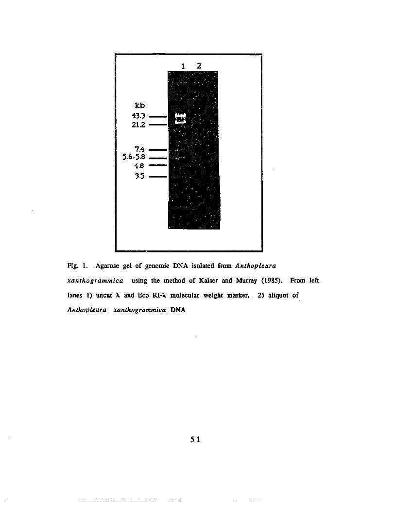
1 2
kb43.321.2-
7.45.6,5.8 _
4.8
3.5-
Fig. 1. Agarose gel of genomic DNA isolated from Anthopleura
xantho grammica using the method of Kaiser and Murray (1985). From left
lanes 1) uncut A and Eco RI-A. molecular weight marker, 2) aliquot of
Anthopleura xanthogrammica DNA
51

kb4'3.321.2
7.45.8,5.6
4.8
'3.5
1 2 4 5 6 7 8
Fig. 2. Agarose gel of genomic DNA isolated from Anthop leu r a
xanthogrammica using the method of Maniatis et al., (1982) 1) uncut A. and
Eco RI-A.·molecular weight marker. 2-8) aliquots from different tubes.
52
B. Construction of the Genomic Library
Since the DNA obtained by the method of Maniatis et al
(1982) which employed proteinase K digestion and several phenol
extractions had a greater percentage of high molecular weight
DNA, it was used to construct the genomic library of the animal.
The DNA was partially digested with Sau 3A and subjected to
sucrose density gradient centrifugation. Aliquots of the two
gradients were electrophoresed on 0.5 % agarose gel. Fig. 3 shows
that the DNA fragments were distributed along the gradient
according to mass, and tubes 4 and 5 from the first gradient tube,
and 5 and 6 from the second gradient tube contained the
fragments that are closest to 20 kb in size.
53

1 2 3 4 5 6 7 8 9 10 11 12 13 14 15 16
43.3 ---21.2
7.45.8 5.6 10.-_
4.83.5
Fig. 3. Agarose gel of aliquots of partial Sau 3A genomic DNA digest after
sucrose density gradient centrifugation. Lanes 1-7) aliquots from 1
gradient tube. 8) A. -Eco RI molecular weight marker, 9-15) aliquots from a
similar gradient. Each well was loaded with a 8 ~ aliquot from a 1. 0 ml
sample.
S4

The gradient fraction consisting mainly of the 17-20 kb
fragments were pooled and reprecipitated and again
electrophoresed on a 0.5 % gel for sizing. Fig. 4 shows that the
partial digest that had not been subjected to sucrose gradient
centrifugation ranged in size from> 43.3 kb to < 3.5 kb, whereas
the pooled fractions consisted mainly of the desired 17-20 kb
fragment size.
55

1 2 3
kb
43.'3 -21.2 --
7.45.8,5.6 -
4.8--3.5
Fig. 4. Agarose gel of partial Sau 3A digest of Anthopleura xanthogrammica
DNA after reprecipitation following sucrose density centrifugation. Lane
1) Eco RI- A. + uncut A. molecular weight marker, 2) aliquot of parial Sau 3A
digest of Anthopleura xanthogrammica before sucrose density gradient
centrifugation, 3) aliquot of partial Sau 3A digest after sucrose gradient
centrifugation and reprecipitation.
56

The pooled fractions consisting mainly of the 17-20 kb
fragments were inserted into the Bam III site of l replacement
vector EMBL3 as shown in Fig. 5.
lelt irm (20kll) central stulter (lCklll "Ohl irm (9kbl
SCblmltlcII EMIU IlmHI clonlnl tlCtlr.
Fig. S. Schematic representation of A EMBL3 cloning vector (Frischauf e t
al.• 1983)
There were 543,000 clones in the unamplified library.
Based on an average insert of 17 kb and an extrapolated value of
8x10⁸. base pairs for invertebrates, the library consisted of a total
fragment size greater than 10 times the size of the genome of the
animal. The genomic library was amplified and titered on E. coli
strain LE392.
57

C. Construction of the eDNA Library
To construct the cDNA library, total RNA was isolated from
fresh tissues. The poly-adenylated mRNA was separated from
ribosomal RNA and tRNA by affinity chromatography, using an
oligo dT column.
About 0.6 mg total RNA was isolated from 1 g fresh tissue,
and from this total RNA, 18 flg poly-adenylated mRNA was
isolated from oligo dT column. From 2.4 flg poly-adenylated
mRNA, 4.66 ug double stranded cDNA was obtained. The double
stranded cDNA was ligated into Eco RI adaptors and inserted into
the single Eco RI site of A. gtlO as shown in Fig. 6.
0'S'C -CaI ~
::: EaI til~ lD
b)
~=i == -
.2x-= III- E
~CI)
Fig. 6. Schematic representation of A. cloning vector A. gt 10 (Murray et al,
1977)
58

The Eco RI adaptor ligation system that was used is shown
in Fig. 7......--------------------.
~~~?J,leo ... Ada9lor
170 C '0"""poIynuc:leOloGe klllaM
37C 3Omon
, .r-I__--:~__IL-~ L-....J.p
••eIG' a'''' L., p .s=J~__.=:..:::.:.___[~.,C;: ••Cl0' •• m
1~,eo veclOt .,ms
l~ONA~
,~ C ~IV to _n9'"
I I__DL-__= __-L...L.-__
RecombInant
Fig. 7. Schematic representation of Eco-RI adaptor ligation system used in
constructing the cDNA library
From 1 IJ.g of cDNA ligated to 3 IJ.g of the vector, 325,000
cDNA clones were obtained.
59

D. Polymerase Chain Reaction
Screening of either the genomic or the cDNA library with
two sets of synthetic oligomeric probes constructed from the back
translation of AP-B yielded inconsistent results. Plaques isolated
using these four primers did not show signals upon subsequent
purification steps. The failure to detect clones harboring the
message for the peptide was probably due to the lack of sufficient
data to create an oligomer that most closely resembles the nucleic
acids coding for the amino acids.
Since screening of the libraries with the synthetic probes did
not result in the isolation of clones that harbor the AP-A or AP-B
gene, the polymerase chain reaction was deemed an alternative
approach.
Two sets of highly degenerate oligonucleotide primers were
constructed based on the backtranslation coding for the two
peptides. These primers were used to amplify the message using
genomic and cDNA as templates. Fig. 8 shows an agarose gel when
the products of - the polymerase chain reaction were
electrophoresed on a 4% agarose gel. Three distinct bands were
present in aliquots of reactions that received cDNA as the
template as shown in lanes I, 2, 14, and 15. These bands are also
present in aliquots of reactions which received genomic DNA as
template as shown in lanes 3, 4, 11, 12 and 13. In contrast, the
aliquot from the control reaction which received the primers alone
show only two bands, the larger probably representing the
dimerization product of the two primers.
60

1 2 3 4 5 6 1 8 9 10 11 12 13 14 15 16 11
Fig. 8. Agarose gel of polymerase chain reaction products from different
templates. From left lanes 1 and 2. cDNA as template. 3 and 4. genomic DNA
as template. lanes 5-8. cDNA as template. lane 9. primers alone. lane 10.
eDNA as template. lanes 11-13. genomic DNA as template. lanes 14 'and 15.
eDNA as template. Each lane was loaded with 25 IJl aliquot from a total peR
reaction tube of 100 J.L1.
61
To ascertain whether the product obtained from the
poymerase chain reaction is the fragment which is of interest,
aliquots of the tubes that showed a third band were run on
another 4% agarose gel for sizing. In order to be certain that the
larger band is not an artifact, nor a polymerization product of the
primers alone, double the enzyme concentration was added to the
control reactions receiving only the primers as in the previous
reactions. The tubes were. again loaded to the thermal cycler for
. another round of chain reaction.
Fig. 9 shows that a 144-147 base pair product was obtained
from the polymerase chain reaction, and that the control reactions
did not show another band after 2 rounds of chain reaction.
62
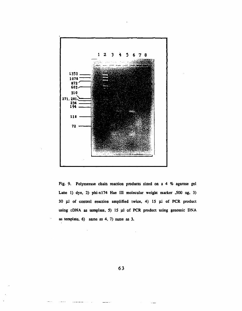
12'345678
1353_······1078 ----.;872r--603---
31~
271. 281-===234 ----194 --
118 --
72--
Fig. 9. Polymerase chain reaction products sized on a 4 % agarose gel
Lane 1) dye. 2) phi-xl74 Hae III molecular weight marker .500 ng, 3)
30 J.LI of control reaction amplified twice. 4) 15 ILl of PCR product
using cDNA as template. 5) 15 ILl of PCR product using genomic DNA
as template, 6) same as 4, 7) same as 3.
63

Since the size of the product of the polymerase chain
reaction corresponded to the expected size of DNA for the message
of the anthopleurin peptides, the next step was to sequence the
product. In order to sequence the product without subcloning,
either primer should be able to catalyze a polymerase chain
reaction and generate the correctly sized product when present in
excess. The principle is to use each primer to enrich for one
strand, and then use the other primer for the sequencing reaction.
In addition, if the message for the peptides anthopleurins a
and be are present in the message isolated, such primers should
be able to catalyze the second strand synthesis of cDNA. Thus,
another set of polymerase chain reaction was initiated, using
varying ratios of the primers and either mRNA or cDNA as
template.
Fig. 10 shows that the primers are effective in generating
the desired product from the cDNA template as shown in lanes 7
13, for as little as a 10:1 ratio of each. Further, either primer can
be used to prime the second strand synthesis of cDNA as shown in
lanes 4 and 6.
64

1 2.:i 4 5 6 1 8 9 10 11 12 13 14 15 16 11
Fig. 10. Agarose gel of polymerase chain reaction products from
different templates and varying primer ratios. From left, lane 1)
both primers without a template, 2) KY-1 alone; 3) KY-2 alone; 4)
mRNA as template, 1:10 -KY-1:KY-2 ratio; 5) mRNA template 1:1 ratio
of KY-1:KY-2; 6) mRNA template. 10:1 KY-1:KY-2 ratio; 7) cDNA
template, 1:1 ratio of KY-l:KY-2; 8) eDNA template 10: 1 ratio KY
1:KY-2; 9) eDNA template 1: 10 ratio KY-l:KY-2 ; 10) cDNA template
1.25:1 KY-l:KY-2 ratio 11) same as 8; 12) same as 9; 13) same as 8; 14)
o template. 10:1 ratio KY-1:KY-2; 15) 0 template 1:1 ratio KY-1:KY-2;
16) KY- alone 17) KY·2 alone
65

Since previous attempts to isolate genomic or eDNA clones
harboring the message for the anthopleurin peptides using
synthetic oligomeric probes yielded negative results, and since the
product from the polymerase chain reaction yielded the correctly
sized fragment, it was deemed possible to use the product from
the peR to screen the libraries.
The product from the polymerase chain reaction was first
separated from the remaining primers after the reaction by
concentration on Centricon filters. The concentrated product was
then electroeluted from a 4 % gel. Fig. 11 shows that although the
correctly sized band was prominent, the concentrated product has
a large range of fragment sizes, whereas the electroeluted product
consists mainly of the desired fragment.
66

bp1'35'3-1078-872-603 __
'310271.2811~~~
2'34 ~19411872---
Fig. 11. The product from the polymerase chain reaction after
concentration on centricon filters and after electroelution from a 4 %
agarose gel. Lane 1) phi-X 174 Hae III molecular weight marker. 2) aliquot
of product after concentration in centricon filter. 3) electroeluted peR
product.
67

E. Screening of genomic and cDNA libraries with PCR product
To determine whether the product can be used to screen the
libraries, the PCR product was first used to probe genomic DNA
digested with different restriction enzymes. Genomic DNA was
digested with 6 base cutters Eco RI, Bam HI and Hind III, and the
digests were electrophoresed on a 1% agarose gel overnight. Fig.
12 shows the complete digest of the Anthopleura xanthogrammica
DNA as seen on the agarose gel. The range of the fragments
generated was from 23 kb to less than 125 base pairs.
68

23.121.2"-Q.4---,7.4 __6.1-
5.6, ').8 j ', '4.8
3.5 2.32.0,,"--
0.56'=:= '.125~,
Fig. 12. Agarose gel of Genomic DNA isolated from Anthopleura
xanthogrammica after complete digestion with Eco RI, Bam HI and
Hind III. Lanes 1 and 2, genomic DNA digested with Hind III, lanes 4
and 5 genomic DNA digested with Eco RI ; lanes 7 and 8 genomic DNA
digested with Bam HI; lane 10, Eco- RI A., lane 12, Hind III A.. Each
lane was loaded with either 5.6 J.l.g genomic digest or 500 ng each of
the molecular weight marker.
69

The DNA from the gel was allowed to transfer to a nylon
membrane, and probed with the product from the polymerase
chain reaction.
Fig. 13 shows the Southern blot of genomic DNA digested
with three restriction enzymes and probed with the labeled
product from the polymerase chain reaction.
The Bco RI digest showed 4 prominent bands corresponding
to 8, 6.6, 4.1 and 3.6 kb. The Bam HI digest showed bands
corresponding to 8 and 6 kb, and the Hind III digest showed 7.4
and 6.6 kb bands.
The presence of the bands that hybridized tightly with the
radiolabeled probe indicates that the amplified product from the
cDNA has sequence homology with genes present in the DNA of
the animal. Furthermore, the presence of several bands in each of
the digest suggests one of two things, that the gene is present in
more than one copy, or that the digestion has not gone to
completion. Alternatively, the labeled peR product could be
binding to genomic DNA sequences that have some sequence
similarity with the anthopleurin peptide messages.
70
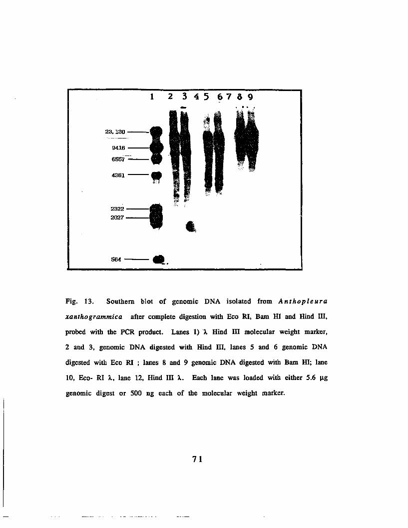
1 2 3 4 5 67 3 9-23.130
I------
9416
6557
4361
"2322 I2027 -564 ~.
Fig. 13. Southern blot of genomic DNA isolated from Anthopleura
xanthogrammica after complete digestion with Bco RI, Bam HI and Hind III,
probed with the peR product. Lanes 1) 'A Hind III molecular weight marker,
2 and 3, genomic DNA digested with Hind III, lanes 5 and 6 genomic DNA
digested with Eco RI ; lanes 8 and 9 genomic DNA digested with Bam HI; lane
10, Eco- RI A, lane 12, Hind III A. Each lane was loaded with either 5.6 J.1g
genomic digest or 500 ng each of the molecular weight marker.
71

Using the radiolabeled product from the polymerase chain
reaction as the probe, the amplified genomic library was screened
for clones· harboring the gene for the anthopleurin peptides.
Fig. 14 shows an autoradiogram of one of filters used In
screemng the library. The presence of many positive clones
probably indicates amplification of similar clones. Alternatively,
it could also reflect the heterogeneity of the peR products. Thirty
one genomic clones were isolated from the genomic library.
Clones were confirmed positive if the filters showed an exact
match upon alignment with the master plate.
Fig 14. Autoradiogram of a filter used in in situ plaque hybridization of
the amplified genomic library probed with the labeled peR products
72

Fourteen of the 31 genomic clones isolated were
investigated further. The phages were grown in E. coli strain
LE392 and the DNA isolated. The clones were digested with Eco
RI, and the digests were electrophoresed on 1% agarose gel for
Southern blot. Fig. 15 shows that instead of distinct bands, a
smear was obtained, the smear probably representing
contamination by bacterial DNA. However, since the probe to be
used for Southern blots was obtained by labeling purified
products from the polymerase chain reaction and should not at all
hybridize with bacterial DNA, the DNA from the digests were
transferred to nylon membranes for Southern blotting.
73

21.2 ----I
';'.4~;;;;;;;j5.8,5.6~
4.8;-__-1
3.51.35
1.08 ----I0.87 ----I0.60----1,
0.31---
Fig. 15. Agarose gel 14 genomic clones digested with Eco RI. Lanes 1) A-
Hind III + phi- x174 molecular weight markers. 1 I1g. 2) gl, 3) g2. 4) g3, 5)
gll. 6) g13. 7) glS. 8) g17. 9) g18. 10) g19. 11) g22. 12) g23, 13) g24. 14)
g27. 15) g29
74

When probed with the PCR product, clones # 24 and 29
showed a 2.8 kb Eco RI band hybridizing strongly with the labeled
PCR product as shown in lanes 2 and 4 of Fig. 16. Since the
autoradiograms were developed after only an overnight exposure
of the film, the data strongly suggests that the clones 24 and 29
harbor the gene for the anthopleurin peptides. The presence of a
bigger band that corresponds to the vector probably indicates
some molecules of the 2.8 kb fragment trapped in the vector
fragment.
75

- ...."",i
.-_ I ...,-I"~:r;o
t
1 2 3 4 5 6 1 8 9 101112131415.~...
0.31---
21.7 --....
7.52 --5.83 ---
3.48 ----'
1.35 --
1.08 --0.87--0.60---
Fig. 16. Southern blot of Eco RI digests of 14 genomic clones using peR
product as probe. Lanes 1) A-Hind III + phi X-174, 2) 129, 3) g27, 4) g24, 5)
g23, 6) g22, 7) 19, 8) g18, 9) g17, 10) gIS, 11) g13, 12) gIl, 13) g3. 14) g2,
IS) gl.
76
Because previous digestion of the genomic DNA with Eco RI
did not show a 2.8 kb band, another set of digestion of the
genomic DNA with the same set of restriction enzymes was done.
Similarly, A. clones g24 and g29 were subjected to restriction
enzyme digestion and were electrophoresed alongside of the
genomic DNA digests, in order to verify the result of the previous
Southern blot. Fig. 17 shows that A. clones were releasing the
inserts. The Bam HI digest pattern of the two clones appear
similar as shown in lanes 8 and 9, but the Hind III digests as
shown in lanes 11 and 12 were different suggesting that the two
clones are distinct from one another.
77

1 2 3 4 B; f. ? 8 9 10 11 12 1314 15
23.1 '"""=:::::::121.2 9.4---17.4===16.7r;:.
5.6, ';.8....---44.8 __---t
4.43.52.3
2.0
0.56 ----I
Fig. 17. Comparison of genomic DNA and genomic clones digested
with Eco RI, Bam HI and Hind III electrophoresed on a 1% agarose
gel. Lane 1) Beo RI-)"; 2) Hind 111-),,; 3) g24 Beo RI digest, 4) g29 Beo
Rl-digest, 5) genomic DNA Eco Rl-digest, 6) Eco RI-Bam HI double
digest of clone 24, 7)genomic DNA Bam HI- digest, 8) g24 Bam HI
digest, 9) g29 Bam HI digest, 10) genomic DNA Hind III digest, 11)
g24 Hind III-digest, 12) g29 Hind III digest, 13) gl Eco RI digest; 14)
g3 Eco RI digest. Ten J1g of the genomic digest and 6J1g of the
genomic clone digests were loaded in each well.
78

When the different digests of the two clones were probed
with the radiolabeled peR product, the autoradiogram shown in
Fig. 18 was obtained.
1 2 3 4 5 Ii 7 8 9 10 11 12 13
23.130---'
9416 -
6557 ---I4361--- 1
2322--2027---
e',6..,. ~ ""e 610 014 G8 .24& ~s GH ,a1JII;; Xli 3=
.....
-564-~
Fig. 18. Southern blot of genomic DNA and genomic clones digested with
Eco RI. Bam HI and Hind III, using labeled peR product as the probe. Lane
1) Eco RI-~; 2) Hind III-~; 3) g24 digested with Eco RI; 4) g29 digested with
Eco RI; 5) genomic DNA digested with Eco RI; 6) Eco RI-Bam HI double digest
of clone 24; 7)genomic DNA Bam HI digest; 8) g24 Bam m digest; 9) g29 Bam
HI digest; 10) genomic DNA Hind III digest; 11) g24 Hind III digest; 12 g29
Hind III digest; 13) gl Eco RI digest; 14) g3 Eco RI digest.
79

Genomic clones # 24 and # 29 were digested with Eco RI,
Bam HI and Hind III, and redigestion of the genomic DNA was also
done. When reprobed with the labeled PCR product, the
autoradiogram of genomic DNA digested with Eco RI showed 4
strong bands corresponding to 4.1, 4.0, 3.6 and 2.8 kb. The 6.6 kb
band previously present (Fig. 15 ) was more faint, suggesting that
it was being split into the 2.8 and 4 kb bands. In comparison with
the previous digestion (Fig. 13), the strong 8 kb band and a more
faint 5.8 and 4.4 kb bands are present in the genomic DNA
digested with Bam HI. On the other hand, digestion of the
genomic DNA with Hind III gave a strong 7.4 kb band, and in
place of the 6.6 kb band, 4 fainter bands corresponding to 5.0,
4.4, 3.8 and 3.4 kb again suggesting that the previous digestion
had not gone to completion. Hybridization of the 2 clones digested
with Eco RI with the labeled PCR product yielded a strong 2.8 kb
band, whereas digestion with Bam HI gave different results. In
both clones, a 5.8 kb Bam HI fragment was present, consistent
with the presence of a faint 5.8 kb Bam HI fragment on the Bam
HI restricted genomic DNA. In addition to the strong 5.8 kb band
present in clone # 24, there is also a fainter signal corresponding
to 4.4 kb, indicating that the clones are different from each other
and that the signals obtained represent similar inserts that are
homologous to the PCR product.
Double digestion of clone #24, first with Eco RI and then
with Bam HI resulted in a decrease of the 2.8 kb signal and the
80

appearance of a 1.8 kb band, suggesting that the 1.8 kb fragment
is nested within the 2.8 kb Eco RI fragment.
In order to determine which other clones contain an insert
that hybridizes to the peR product, 8 genomic clones were
digested with Bam HI and Hind III and subjected to agarose gel
electrophoresis. The results are shown in Fig 19.
81

1 2 3 4 5 6 7 8 9 10 J1 12 13 14 :15
Fig. 19. Agarose gel of 8 genomic clones digested with Bam HI and
Hind III. Lane 1) ).. -Hind III marker; 2) gl Bam HI- digest. 3) gl,
Hind III-digest. 4) gil. Bam m digesu, 5) gl I, Hind III digest. 6) g15.
Bam HI digest; 7) g15. Hind III digest. 8) g16. Bam HI digest. 9) g16.
Hind III digest. 10) g18. Bam m digest. 11) g18. Hind III digest. 12)
g19. Bam HI digest. 13) g19. Hind III digest. 14) g24 Hind III digest.
15) g29. Hind III digest
82
The agarose gel patterns (Fig. 19) show that genomic clones
#1, 11, 15, 16, 18, 19, 24 and 29 have inserts which average 17
kb. Hybridization with radiolabeled PCR product (Fig. 20 ) shows
a 8kb, a 5.8 kb and a 1.8 kb Bam HI fragments which hybridize
strongly with the labeled PCR product. These fragments were
common to 5 of the 8 clones, and these fragments probably
contain the whole gene or a part of it for the anthopleurin
peptides. The absence of the 1.8 kb fragment from the genomic
DNA restricted with Bam. HI (Fig. 18) can be explained by the
complete lack of a 4.4 kb Bam HI fragment from the clones. This
observation suggests that there is a Sau 3A site within the 4.4. kb
fragment that was cut, and was inserted into the vector.
83

1
23.130 --
9416-
6557 --
4361---
2322 •2027--- •
564 ~
2 3.. 5 6 1 8 9 10 11 12 13 14 15
,....- -
Fig. 20. Southern blot of 8 genomic clones digested with Bam HI and Hind
III, probed with labeled peR product. Lane 1) A -Hind III marker; 2) g 1,
Bam HI digest; 3) g 1, Hind III digest; 4) g 11, Bam HI digest; S) g 11, Hind III
digest; 6) gIS, Bam HI digest; 7) gIS, Hind III digest; 8) g 16. Bam III digest;
9) g 16, Hind III digest; 10) g 18, Bam HI digest; 11) g 18, Hind III digest, 12)
g 19. Bam HI digest; 13) g 19. Hind III digest. 14) g 24 Hind III digest, 15) g
29, Hind III digest
84

F. Cloning of the 2. 8 kb Eco RI and 1.8 kb Bam Hl fragments
The 2.8 kb Eco RI fragment present in both genomic clones
g24 and' g29 were subcloned in the Eco RI site of M13mp18.
When the subclones were digested with Eco RI, some of the
subclones showed 10 and 4.4 kb inserts as shown in lanes 12, 13,
14, and 15 of Fig. 21, while the rest had a 4 and 2.8 kb inserts.
kb
23.1 _--:9.4~
6.6 - ·Ji!~~:~~~J;;ti".~#~~.tc'iJ}',i4.'3 -- .
2.3 ;::::::=/
2.0
Fig. 21 . Agarose gel of Eco RI digest of the M13mp18 subclones from
g24. Lanes 1) A.-Hind .uI molecular weight marker, 2-15) subclones 1-14.
85

When the digests were blotted to nylon membranes and
probed with the PCR product, the autoradiogram shown in Fig. 22
was obtained. The PCR product bound to the 4 kb and 2.8 kb Eco
RI fragments, but in addition, the product also bound to the M13
vector from where these fragments were released. If the PCR
product in fact binds to the M13 vector by itself, all the vector
lanes should show hybridization with the probe. However, this is
true only of subclones 1 ·to 7 (lanes 2-8), which harbored the 4 kb
and 2.8 kb bands. Therefore, the binding of the labeled PCR
product to the vector only represents some 2.8 kb fragments tha
got trapped with the vector. Alternatively, such binding could be
due to some undigested M13 subclones, that being supercoiled,
comigrated with the linear vector band.
86

1 2 3 4 5 6 7 8 9 10 11 12 13 14 15
tb
23.1
9.4
6.6
4.4
2.3
2.0
0.54
1
~ ~
c::>~ c:::::> cC:>c;;J
Fig. 22. Southern blot of &0 RI digest of M13 mp 18 subclones from g24.
Lane 1) A-Hind III molecular weight marker. 2-15) subclones 1-14
87

Since a 1.8 kb Bam HI fragment was present in 5 of the 14
genomic clones examined and since this 1.8 kb fragment seems to
be nested within the 2.8 kb Eco RI fragment, the 1.8 kb fragment
was subcloned into MI3 mpl8.
Fig. 23 shows the agarose gel pattern when the MI3mpl8
subclones were digested with Bam HI. Subclones I, 4, and 5
which are subclones from g16, and 8 which is a subclone of gl8
(lanes 2, 5, 6 and 9 respectively) had the expected 1.8 kb Bam HI
fragments.
1 2 3 4 5 6 7 8 9 10 1112 J314 15
kb
21.1-9.46.6
4.1
2.1 ;:===2.0
Fig. 23. Agarose gel pattern of M13mp18 subclones from g16 and g18. From
left, lane 1) Hind III -i.. molecular weight marker. 2-15) subclones 1-14
88
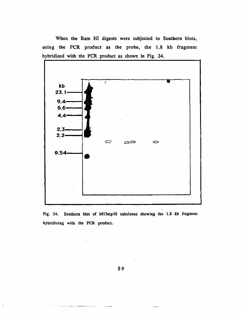
When the Bam HI digests were subjected to Southern blots,
using the PCR product as the probe, the 1.8 kb fragment
hybridized with the PCR product as shown in Fig. 24.
Icb23.1----1
9.4---16.6---t
4.4---4
2.3---12.2---1
0.54,----1 •
•
Fig. 24. Southern blot of MI3mp18 subclones showing the 1.8 kb fragment
hybridizing with the peR product.
89

The 1.8 kb fragment present in 5 of the genomic clones was
already shown to hybridize with the peR product. To further prove
that this fragment contains the gene or part of it coding for the
anthopleurin peptides is its binding to the primers by themselves.
Hence, another set of subclones from genomic clone 15 were
digested with Bam HI. Fig. 25 shows that of the 14 subclones, there
were only 2 (lanes 5 and 7) that did not contain the desired 1.8 kb
fragment.
kb
23.1--
9.4--
6.7---
4.3----
2.
123456789101112131415
Fig. 25. Agarose gel pattern of 14 M13mp18 subclones from g15 .
From left, lane I, Hind III I -molecular weight marker; lanes 2-15)
M13mp18 subclones 1-14
90

When subjected to Southern blot, using the labeled primers as
the probe, the 1.8 kb Bam HI fragment from the subclones
hybridized with the primers. Further, the labeled primers did not
at all bind to the M13mp18 vector.
1 2 3 4 5 6 7 8 9101112 131415
23.1
9.4
6.6
4.4
2.3
2.0
•
c::;)~ c:::::> c::> OC?c:>O
Fig. 26. Southern blot of the M13mp18 subclones probed with the labeled
primers.
91

G. Screening of the cDNA clones with the peR product
The peR product was also used to screen the unamplified
cDNA library, and 7 positive clones were identified.
Digestion of the cDNA clones with Eco RI resulted in the
splitting of the cDNA subclones into 2 large fragments as shown in
Fig. 27.
This could be due to one of three possibilities; that one or
both of the Eco RI sites flanking the insert has been destroyed, or
that the insert has an Eco RI site that the enzyme preferentially
digested, or that there are two adjacent Eco RI sites in the insert,
reieasing a very small fragment that migrated very fast in the 1 %
agarose gel used.
92

1.353 '---..1.08 ;==t0.87
0.60 ---f~~~~~~~~
~\~""Jr.,p..Fig. 27. Agarose gel of eDNA elones~with Eco RI. Lanes 1) phi X174-
Hae III molecular weight marker, 2) blank, 3) cl, 4) e2, 5) c3, 6) eS,
7) e6. 8) c17.
When the digest was subjected to Southern blot, both
fragments hybridized with the PCR product .as shown in Fig. 28,
indicating that in both fragments, there are sequences homologous
to the. PCR product.
93

1 2 3 .. 5 6....
Fig. 28. Southern blot of the cDNA clones digested with Eco RI. using
the peR product as probe. From left. lanes 1) c1; 2) c2; 3) c3; 4) c5; 5)
c6; 6)c17.
94
H. Subcloning of the cDNA inserts from A. gtlO into M13mp18 and
M13mpl9
Since attempts to release the insert from the cDNA clones
using Eco RI proved difficult, another approach to sequencing the
inserts was done. This involved digesting the cDNA clones with
restriction enzymes flanking the Eco RI site, and subcloning the
digests in M13 cloning vectors M13mp18 and M13mp19. The two
enzymes used were Bgi II and Hind III.
In order to be certain mat none of the A. vector arms will not
be subcloned, the M13 vectors were digested with Bam HI and
Hind III and subjected to phospatase treatment before ligation.
Of the 5 cDNA clones double digested with Bgi II and Hind
III and ligated with the M13 vectors digested with Bam HI and
Hind III, one M13 isolate showed an insert. Upon subsequent
digestion of this clone with Eco RI, the apparent the vector
released fragments that are respectively 0.9 kb, 0.8 kb,. 0.48 kb
and 0.3 kb, as shown in Fig. 29, indicating that the cDNA insert
was about 2.5 kb.
95

1 2 3 4 5 6? 8 9 10 11 12 13 14 15 16 11
Fig. 29. Agarose gel of the MI3mpI8-cDNA subclones digested with Eco-RI.
From left, 1) molecular weight marker, 2) uncut c3-18, 3) aliquot of PCR
product after concentration in Centricon filter, 4) aliquot of PCR product
after phenol freeze extraction 5) c3-18 Eco RI digest, 6) c6-19 Eco RI digest,
7) c5-19 Eco RI digest, 8) cl-18 Eco RI digest
96

leb
2.18-1.77--1.23__1.03--
0.650.52---1
0.45:,,---.,0.39'
0.22..0.23
Fig. 30. Agarose gel of M13mp18 cDNA subclone and peR product pBS
subclones with Eco RI
97

..... 2.18 ---'1.77 -----::
1.23 ---'
1.03
;!?65---~
]0-52 --........;,··;0.45--.-;
;0.39
0.23.0.22
Fig. 31. Southern blot of M13mp18-cDNA subclones, using KY-l and
KY-2 as the probe.
98

I. Cloning and sequencing of the PCR product
Since direct sequencing of the PCR product using the primers
yielded negative results, another approach was used.
The PCR product was cloned into the phagemid vector
pBluescript KS II+, and the recombinants were selected in LB plates
modified with ampicillin, Xgal and IPTG. In these plates only those
bacteria that harbor the phagemid will grow, and and those
bacteria harboring the phagemids whose b galactosidase gene has
been interrupted will be colorless.
Four white colonies were randomly isolated from the plates
and sequenced. To determine the length of time necessary to
electrophorese the sequencing gels, the first clone pBS1 was
electrophoresed for two hours initially. The sequence showed that
this electrophoresis time was sufficient to read close to the priming
site
Clone pBS #1 which was sequenced using 2, 3 and 4 hour
electrophoretic runs showed the sequence of the vector close to the
priming site (Appendix 2), as well as a 237 base pair sequence
(Appendix 1), that is not at all homologous to the vector. When this
237 base pair sequence was subjected to sequence analysis and
compared to the ambiguous sequence of the peptides AP-A and AP
B, there is some sequence similarity with the ambiguous sequences
of both toxins (Appendix 3 and 4). The data showed that the
consensus sequence of the clone has a 57 % similarity and 22 %
identity with that reported for the ambiguous sequence of AP-A
(Appendix 3) and a 53 % similarity with that reported for AP-B.
99
However upon translation into all 6 reading frames , the consensus
sequence for clone pBS! showed only a 24 % identity over a 25
amino acid' overlap with the peptide anthopleurin A (Appendix 5)
and 67 % identity over a 6 amino acid overlap with anthopleurin B
(appendix 6)
When the 3 other clones pBS3, pBS? and pBS8 were
sequenced, they all showed sequence homology with the vector
(Appendix 7).
100

VI. DISCUSSION
The toxins anthopleurins a and b are of interest because they
bind to the region of the voltage gated sodium channel responsible
for its closing. In addition, the toxins could have a therapeutic
value in the treatment of heart disorders. In order to utilize these
peptides to probe the sodium channel, and in order to further
explore their value as therapeutic heart stimulants, the peptides
should be readily available. The first step in obtaining large
amounts of the toxins is: cloning of the genes that code for them.
Since there had been no nucleic acid studies done previously on
the anthopleurin peptides, this study was undertaken to gain
information on the nucleic acids coding for anthopleurins a and b.
Southern blotting of the genomic DNA with labeled PCR
products. The genomic digest of the DNA of the sea anemone,
Anthopleura xanthogrammica, was probed with the labeled PCR
product. Strong bands were obtained (Fig. 13) indicating that it is
indeed the animal that is producing the peptides and not the algal
symbionts.
Genomic and cDNA Libraries. High molecular weight DNA was
first isolated using two methods, and the modified method of
Maniatis et al (1982) yielded DNA which was suitable for genomic
library construction. Since there has been no published protocols
for such isolation, the method reported here can be used to isolate
DNA from other sea anemones. The method which employed
proteinase K digestion and several phenol extractions was
101

probably the crucial factor In releasing the chromosomal DNA
without too much shearing.
The genomic library of Anthopleura xanthogrammica was
constructed in the Bam Hl site of the A cloning vector EMBL3. The
primary library consisted of 543,000 clones. Based on the
extrapolated value for an invertebrate genome of 8.1 xl08 base
pairs (Britten and Davidson, 1971) and an average insert size of
17 kb, the library consisted of DNA fragments totalling more than
10 times the putative size of the animal's genome, and hence even
a single copy gene will be represented in this library. In addition
to it's value in the study of the anthopleurins, this library can be
used in studies of the other toxins whose amino acid structure is
similar to those of the anthopleurins, among them the 47 amino
acid toxin II from Anemonia sulcata.
The E. coli host strain LE392 was chosen for titering the A
EMBL3 because it lacks the E. coli K restriction system and will
not modify inserted DNA. It is a rec+ strain and thus will allow
recombination of the phages. It is also a permissive host and
allows both parental and recombinant phages to grow, but since
the vector arms were predigested and treated with alkaline
phosphatase, it was not necessary to select against parental
phages (Huynh et al, 1985).
The cDNA library was constructed in the unique Eco RI site of
the cl gene in A gtlO. Insertion at this site causes inactivation of
the A repressor. Consequently, the recombinant phages are lytic
and produce clear plaques, whereas parental phages are lysogenic
102

and produce turbid plaques. When grown in a permissive host
like C600, both parental and recombinant phages grow, but when
grown in a restrictive host like C600 hfl (high frequency lysogeny)
as was used here the lytic phage grows in preference to the
lysogenic phage.
The unamplified cDNA library consisted of 325,000 clones,
which translates to a transformation efficiency of 3.25 xl05
plaque forming units (pfu) Illg of the cDNA. Since A. gtlO
packages from 0-7 kb of DNA, it was not possible to determine the
number of messages cloned. However, since the library was
titered in the non-permissive host C600 hfl, the clones must
represent a large spectrum of messages that the animal expresses
at the time of RNA extraction. Furthermore, both anthopleurins a
and b can be extracted at any time in the life cycle of the animal,
thus indicating that the peptides are constitutively produced.
These facts taken together made it highly probable that the cDNA
library contains the messages for both the peptides.
Screening of the amplified genomic library and the cDNA using
the first two sets of synthetic oligonucleotides constructed from
backtranslation of the anthopleurin B peptide yielded only
spurious spots, probably because of a lack of information as to the
codon- usage for the amino acids comprising the peptides.
Consequently, two degenerate primers spanning the first and last
10 amino acids of the peptides were constructed to determine
whether the message could be amplified via the polymerase chain
reaction (Saiki et al, 1988) using cDNA as the template.
103

Polymerase Chain Reaction. The polymerase chain reaction is a
highly sensitive method to amplify DNA, as long as two primers
that will bind the two strands of the DNA of interest can be
obtained. The method was first used by Saiki et al. (1986) in
amplifying human ~ globin gene. The key component of the
reaction is the Taq polymerase, an enzyme isolated from Thermus
aquaticus, which is not heat denatured up to temperatures of
95°C. The optimum temperature for activity of the enzyme is
70°C. This characteristic allows for a continuous cycle consisting
of denaturation of the DNA sample at 94°C, annealing of template
DNA and primer at 50°C and extension of the primer at 70°C.
Of the two oligonucleotides that were constructed for use as
primers for the PCR reaction, KY-1 is a mixture of 29-mers
constructed by backtranslating the first 10 amino acids of the AP
B peptide while KY-2 is a mixture of 30-mers constructed by
obtaining the complement of the backtranslated amino acids 40
49 of AP-A. Both primers represent all the possible codons for
the constituent amino acids. As a consequence, KY-1 is comprised
of a combination of 16,384 possible sequences, while KY-2 is
comprised of 12,288 possible sequences. Hence it was expected
that both the AP-B and AP-A messages would be isolated, and in
addition, messages related to the two peptides.
Using the two degenerate oligonucleotides as primers for the
PCR reaction, a 144-147 base pair peR product was obtained (Fig.
9). This size is that expected as both AP-A and AP-B are 49
amino acid peptides, and the uninterrupted message should be a
104

sequence of 147 base pairs. It is interesting to note that even
when genomic DNA was used as the template, a similarly sized
product was obtained (Fig. 8), suggesting that either or both of the
peptides are encoded for by a single exon.
The PCR product was separated from the primers by
concentration on a Centricon filter, labeled by random priming
and used to probe the genomic DNA, and to probe the genomic
DNA and to screen both the genomic and cDNA libraries.
Screening of the genomic library with the product from the
polymerase chain reaction. When complete digests of the genomic
DNA were probed with the PCR product, strong signals were
obtained, indicating that the genomic DNA of Anthopleura
xanthogrammica contains sequences that are similar to that of the
amplified product (Fig. 13).
The Eco RI'digest of the genomic DNA showed 4 prominent
bands that hybridized tightly with the radiolabeled PCR product.
The bands corresponed to 8, 6.6, 4.1 and 3.6 kb, The Bam HI
digest showed bands corresponding to 8 and 6 kb bands, while
the Hind III digest showed 7.4 kb and 6.6 kb bands.
Upon screening the genomic and cDNA libraries with the PCR
product, 31 genomic clones and 7 cDNA clones were isolated.
When reprobed with the PCR product, these clones consistently
showed strong signals indicating that they contained sequences
that are similar to those of the PCR product.
105
The genomic and cDNA clones were examined for the
presence of sequences that could contain all or part of the gene for
the anthopleurin peptides.
Previous digestion of the genomic DNA with Eco RI (Fig. 13)
showed 4 prominent bands corresponding to 8, 6.6, 4.1 and 3.6
kb. Subsequent digestion of the genomic DNA with E co RI as
shown in Fig. 19 shows that the 6.6 kb band that was previously
present became more faint, and a 4.0 and 2.8 kb fragments
appeared, indicating that 'the 6.6 kb fragment was being split into
the 4.0 and 2.8 kb fragments, and that each fragment contained
sequences that are homologous to the sequence of the PCR
product, indicating that the gene was being split.
Of the 14 genomic clones examined 3 clones contained a 2.8
kb Eco RI fragment that hybridized strongly with the PCR product
(Fig. 18) and 5 contained a 1.8 kb Bam HI fragment that also gave
a strong signal with the labeled probe (Fig. 20).
These fragments were subsequently subcloned into M13 and
probed with· the primers used in the PCR reaction, and found to
hybridize also with the primers alone (Fig. 22 and 26), suggesting
that there are sequences in the clones that are similar to those of
the primers. These clones could contain part or all of the gene for
the peptides of interest. Fig. 23 and 24 show that when the
recombinant M13mp18 were subjected to restriction enzyme
digestion, the 2.8 kb fragment was released, and and it also
hybridized with the PCR product (Fig. 24)
106

Digestion of genomic clone #24 first with Eco RI and then with
Bam HI (Fig. 18) showed that there was a marked reduction of the
2.8 kb signal and a consequent appearance of a strong 1.8 kb
band, indicating that there is at least one Bam HI site nested
within the 2.8 kb E co RI fragment, and that this fragment contains
the sequences of interest, since the remaining 1 kb fragment did
not show a signal with the labeled probe.
Since the1.8 kb Bam HI fragment appears to be nested within
the 2.8 kb Eco RI fragment, and since the 1.8 kb fragment seems
to be common to the 5 clones, it was subcloned into M13mp18 for
future sequencing.
M13 was used as a subcloning vehicle mainly for ease of
generating a single-stranded template for sequencing. M13 is a
single-stranded filamentous phage which replicates inside the
host, first by forming a double stranded circular DNA termed the
replicative form (RF). When about 200 copies of RF are formed
inside the host, it starts to package and extrude the" single
stranded form of DNA. The reason for this is that once the host
has made about 200 copies of the RF, the protein product of gene
V accumulates at high levels and binds the + strand of the phage
DNA, thus preventing further formation of RF. The + strand
assembles with coat proteins to' form a single stranded mature
phage.
This DNA is extruded into the medium, and can therefore be
collected without lysing the host. The RF form can be obtained by
alkaline lysis of the host. This property of M13 allows it to be
107

used as a cloning vector for sequencing. The RF can be digested
with restriction enzymes. The DNA of interest can then be ligated
with the RF form, if the insert was generated using the same
enzyme.
The M13 derived vectors contain the 5' regulatory region
coding for the first 145 amino acids of the E. coli lac operon
(Messing 1977). These sequences code for the ex peptide of the
enzyme 13 galactosidase. The polylinker cloning site is so arranged
that the correct reading frame is maintained. When expression of
this gene is induced, for example by IPTG, in a host which lacks a
functional ex peptide but produces the wild type Cl) peptide, the two
polypeptides assemble to form a functional enzyme, and galactose
is utilized as a carbon source.
The insertion of the DNA of interest disrupts the 13
galactosidase gene, and when bacteria transfected with the phage
are plated in a medium containing X-gal (5-bromo-4 chloro-3
indolyl-Bvgalactostde), a chromogenic dye, the plaques generated
by phages containing an insert will appear colorless. In contrast,
those phages which do not have an insert will .appear blue as
complementation between the host and the phage will allow the 13
galactosidase gene to be expressed, and a blue product of the
reaction is formed. This allows for a simple screening procedure.
The white colonies can then be screened for the right sized
insert easily, and upon finding the putative recombinants of
interest, sequenced.
108

Since the single stranded form is extruded into the medium, it
allows for an easy method of obtaining template for sequencing.
The peR product was also used to screen the cDNA library, and
seven cDNA clones were isolated. Since the cDNA was cloned in
the single Eco RI site of the cloning vector A. gtlO, the clones were
digested with Eco RI in an attempt to release the insert.
When the seven cDNA clones were digested with Eco RI to
release the insert, there were only two bands that could be
obtained, both of which hybridized strongly with the labeled peR
product. The two fragments released were very large, which
probably corresponded to the vector sizes. This observation
suggests several explanations: that the insert could not be
released because the E co RI sites were destroyed when the
adaptor was ligated into the blunt ended cDNA; and that there is
an EC 0 RI site within the insert that is cleaved leaving part of the
insert with the arms of the vector A. gtlO. Another possibility is
that there is more than one E C0 RI site in the insert, and that the
fragment released was too small in comparison with the. arms, to
be seen in gels. If the latter were true, the released small
fragment could not be detected in the Southern blots because it
would migrate very fast in the 1.0 % gels used for the Southern
blots. Indeed such resistance to restriction enzyme digestion after
cloning into the E C 0 RI site of both A. gtl0 and the expression
vector A.gt 11 have been previously reported (Helms et al, 1985;
Ziai et al, 1988).
109

Since it was not feasible to sequence the cDNA clones directly,
another approach was attempted, this time involving double
digestion' of the cDNA clones with two enzymes flanking the E co RI
site, and subcloning the digest in an M13 vector. Using this
method one subclone was isolated containing a 480 basepair E c 0
RI fragment that hybridized with the primers (Fig. 31), suggesting
that this subclone harbors part of the message for either AP-A or
AP-B. Similarly, sequencing the peR product directly was
attempted but yielded negative results presumably because the
primers used were too degenerate. As noted previously, KY -1,
the mixture of 30 -mers of synthetic oligonucleotides coding for
the first 10 amino acids of the anthopleurin peptides represents
16,384 possible sequences, while KY-2 represents 12,288 possible
sequences. This means that in order to compensate for the
degeneracy of the primers, the primer concentration should be
raised, however, doing so could effectively inhibit the enzyme as
the primers could chelate the MgCI2.
Other problems with sequencing the PCR is that small linear
molecules rapidly reanneal, preventing annealing of the desired
primer with the template. In addition, depending on the base
composition of the DNA, small linear DNA molecules may exhibit
regions 'of strong secondary structures that impede the progress of
the polymerase along the template. The problem of formation of
the secondary structure can be alleviated by raising the reaction
temperature but polymerases such as AMV reverse transcriptase,
110

the Klenow fragment of DNA polymerase I, and T7 DNA
polymerase are rapidly deactivated at elevated temperatures.
Since the PCR product could not be sequenced directly the
product was cloned into pBluescript KS 11+ and 4 white colonies
were chosen randomly and sequenced.
A sample of Clone pBS #1 was electrophoresed at three
different lenghts of time. The two hour sequence showed regions
that are close to the priming site (Appendix 2), right at the region
of the Sma I cloning site, and was therefore used as the basis for
electrophoresing the samples of other PCR subclones.
The three other clones that were sequenced pBS3, pBS7, and
pBS8 showed homology with each other, as well as to the vector
(Appendix 7). All clones showed homology with the vector at
regions different from the Sma 1 cloning site as it was in pBS1.
This indicates that the color selection was not perfect. It is known
that the absence of a single nucleotide in II galactosidase gene is
enough to offset color selection.
A possible explanation could be the presence of nuclease
activity in the calf intestinal phosphatase used to dephosphorylate
the vector (David Haymer, personal communication) which could
have degraded the vector. This is in fact evident in the gaps
shown in the comparisons.
There was no region of overlap when clone pBS 1 was
sequenced at 3 different lengths of times were compared to each
other which suggests that clone pBSl was a composite of more
than one peR product which ligated to each other. This is possible
111
since individual comparisons of the three l-hour electrophoresis
runs showed that each of the three has sequence homology with
the anthopleurin peptides (Appendix 1). This being the case, the
three sequences were aligned to each other to give maximum
overlap. The results showed that there was correspondence with
the anthopleurin peptides at amino acids 1, 5, 10, 11, 12, 19, 22,
24 and 25. The sequence that was cloned does not translate to the
original peptide since the PCR product that was generated by
using very degenerate primers is a heterogeneous mixture, and
the clone that was sequenced was not the correct one.
When a comparison of the 2, 3 and 4 hour sequences of pBSI
were done against the ambiguous and unambiguous sequences of
the backtranslation of the two pep tides using the codon for
prokaryotes, drosophila and yeast, the data obtained showed that
glycine is preferentially coded for by GGC, cysteine by TGA,
proline by CCT, leucine by CTC, and arginine by CGA. Such
information can be used in designing a more specific primer for
screening . the libraries of the sea anemone Ant hop 1e u r a
xanthogrammica or, in designing a better primer for amplifying
cDNA from the animal.
112

VIT. SUMMARY ANDCONCLUSIONS
Genomic and cDNA libraries of the sea anemone,
Anthopleura xanthogrammica were constructed in lambda
replacement vectors, in order to investigate the 49 amino acid
peptide toxins, anthopleurins a and b at the nucleic acid level.
The libraries were screened, first with synthetic nucleotides, and
then with a PCR product.
However, the use of primers in screening the cDNA and
genomic libraries of the sea anemone Ant hop leu r a
xanthog rammica was not effective in isolating clones that harbor
the message for the peptides anthopleurins a and b.
The use of degenerate primers in priming both the cDNA
and genomic DNA for the polymerase chain reaction generates the
correctly sized peR product. The degeneracy of the primers used
effectively eliminated their further use in directly sequencing the
PCR product.
The 144-147 base pair PCR product was used in isolating the
putative cDNA clones that may harbor the anthopleurin peptide
messages. A 480 base pair cDNA fragment that hybridized with
the primers was isolated from one of the cDNA clones, and
subcloned into M13mp18 vector.
The PCR product was also used to isolate genomic clones that
may harbor the gene for the peptide toxins.
113

A 2.8 kb Eco RI and a 1.8 kb Bam H1 genomic DNA
fragments that hybridized with the PCR product and the primers
were subcloned into M13mp18 vector.
Cloning and sequencing the PCR product generated offers an
alternative way of characterizing the message for the small
peptides anthopleurins a and b. However, the use of degenerate
primers leads to the generation of multiple heterogeneous peR
products, some of which have limited homology to the desired
sequence.
The data obtained from sequencing the PCR products can be
used to design less degenerate primers, to prime eDNA.
114

APPENDIX 1. Comparison of the sequence of the clone pBS! with the ambiguous andunambigous sequences of the AP-A and AP·B sequences. N=A.T.G. or C;Y=T or C; S=G or C; H=Tor C;M=C or A;W=T or A or C; R=A or G.
Amino Ap-A Ap-b pBSl pBSl pBSl Ap-B Ap-B Ap-B Ap-A ApA Ap·AAcid Amb Amb 2hr 3hr 4hr Prok Dros Yeast Prok Yeast Dros
GLY GLY GLYG G GG G GN N C
GLY GLY GLY GLY GLYG G G G GG G G G GT C T T T
GLYG +G +C +
2
VALGTN
VAL OLUG GT AN A
VALGTT
VALGTG
VALGTT
VALGTT
VALGTT
VALGTG
+
+
3
SERWSN
PRO LEU TYRW T TS T AN G T
PRO PRO PRO SER SERC C C T TC C C C CG C ACT
SERT +CC +
4
CYS CiST TG GY Y
OLU ALAG GA CG T
CYS CYS CYS CYS CYST T T T TG G G G GC C T C T
CYSTGC +
5
LEU LEUY YT TN N
LEU. OLUC GT AC G
LEUCTG
LEU LEU LEUC C CT T TG G G
LEUCTG
LEUC +T +G +
6
CYS CiST TG GY Y
InSCAC
STOPTGA
CYS CYST TG GC C
CYS CYST TG GT C
CYSTGT
CYST +G +C +
7
ASP ASP ARG 1YRG GeTA A G AY yeT
ASP ASPG GA AC C
ASP ASP ASPG G GA A AC C C
ASPGA +C +
8
SER SER GLY PROW W G CSSG CN N T C
SER SER SER SERT T T TC C C CC C T C
115
SER SERT TC C +T C +

Amino Ap-A Ap-b pBSI pBSI pBSI Ap-B Ap-B Ap-B Ap-A ApAAp-AAcid Amb Amb 2hr 3hr 4hr Prok Dros Yeast Prok Yeast Dros------------.------------------------------.----------------------------------------------
ASP ASP GLY GLY ASP ASP ASP ASP ASP ASPG G G G G G G G G G +
9 A A G G A A A A A AY Y C T C C C C C C +
------------.-------------------.---------------.-.--.-------------------------------.----GLY GLY GLY TYR GLY GLY GLY GLY GLY GLY
G G G T G G G G G G10 G G G A G G G G G G +
N N A C T C T T T C +.---------------------------.-----------------.----.--------------------------------.----
PRO PRO ARG PRO PRO PRO PRO PRO PRO PROC C C C C C C C C C +
11 C C G C C C C C C C +N N C T G C A G A C +
----------------------------------------------------------.-------------------------------SER. ARG SER ARG ARG ARG ARG SER. SER. SER.W M T C C C A T T T +
12 S G C G G G G C C C +N N T A T C A C T C +
------------------------------------------------------------------------------------------VAL PRO GLU GLY ILE PRO PRO PRO VAL VAL VALG C A G A C C C G G G +
13 T C G G T C C C T T T +N N A T C G C A T T G +
------.--------------------------------------------------- ..--------------.----._---------ARG ARG ILE eLY LEU ARG ARG ARG ARG ARG ARG
M M A G T C C A C A C +14 G G T G T G G G G G G +
N N A A G T C A T A C +------------------------------------------------------------------------------------------
GLY GLY VAL ARG ILE eLY (LY GLY GLY eLY eLYG G G C A G G G G G G +
15 G G T G T G G G G G G +N N G A C T C T T T C +
------------------------------------------------------------------------------------------ASN ASN eLY ARG THR ASN ASN ASN ASN ASN ASNA A G C A A A A A A A +
16 A A A G C A A A A A A +Y Y T A C C C C C C C +.
------------------------------------------------------------------------------------------THR THR PRO PRO SER THR THR THR THR THR THR
A A C C T A A A A A A17 C C C C C C C C C C G +
N N C G A C C C C C C +------------------------------------------------------------------------------------------
- 116

Amino Ap-A Ap-b pBSl pBSl pBSl Ap-B Ap-B Ap-B Ap-A ApA Ap-AAcid Amb Amb 2hr 3hr 4hr Prok Dros Yeast Prok Yeast Dros._----------------------------------------------------------------------------------------
LEU LEU PRO TIIR GLY LEU LEU LEU LEU LEU LEUY Y: C A G C C T C T C +
18 T T C C G T T T T T TN N G G G G G G G G G +
------------------------------------------------------------------------------------------SER SER VAL SER ALA SER SER SER SER SER SERW W G A G T T T T T T
19 S S T G C C C C C C C +N N A C C C C T C T C +
----.-----------------------.----.--------------------------------------------------------OLY OLY PRO OLU ASP GLY GLY GLY GLY CLY GLY
G G C G G G G G G G G +20 G G C A A G G G G G G
N N C G C T C T T T C +--------------------------------------------------.---------------------------------------
THR. ll.E TYR ARG VAL us ll.E ILE THR. THR. THRA A T C G G G G G G G +
21 C T A G T G G G G G G +N H C A C T C T T T C +
----------------.-----------.-------------------------.----._-----------------------------LEU LEU VAL GLY LEU LEU LEU LEU LEU LEU LEUY Y G G C C C T C T A +
22 T T T G T T T T T T C +N N T A T G G G G G C
---------------------------------.-------.---------------------------_._---- ........ -_ .. _-TRP TRP LEU ASP LYS TRP TRP TRP TRP TRP TRP
T T T G A T T T T T T +23 G G T A A G G G G G G
G G G T G G G G G G G +-------._._-_._----------_._---_._-_ .... __ ._----------_._-.. _-_._------------~~_ ... --_.---
LEU PHE LEU LEU LEU PHE PRE PHE LEU LEU LEUY T C C C T T T C T A +
24 T T T T T T T T T T C +N Y C T A C C C G G C +
----------_.---------------------_.-_.----------------_._---------------------------_.-._-TYR TYR ARG ASP TYR TYR TYR TYR TYR TYR TYR
T T C G T T T T T T G +25 A A G A A A A A A A C +
Y Y T T T C C C C C T +------------------------------------------------------------------------------------------
PRO PRO LEU IDS SER PRO PRO PRO PRO PRO PROC C T C T C C C C C C +
26 C C T A C C C C C C C +N N A C G G C A G A C +
----_._-----------_._--------------------.-------------------------_._--------------------
117

Amino Ap-A Ap-b pBS! pBS! pBS! Ap-B Ap-B Ap-B Ap-A ApA Ap-AAcid Amb Amb 2hr 3hr 4hr Prok Dros Yeast Prok Yeast Dros--------------------------.---------------------------------------------------------------
SER SER VAL LEU ASN 'SER SER SER SER SER SERW W G C A T T T T T T +
27 S S T T A C C C C C CN N C A T C C T C T C +
GLY GLY SER THR CYS GLY GLY GLY GLY GLY GLYG G T A T G G G G G G +
28 G G C C G G G G G G G +N N G G C T C T T T C +
--------------------------.--------_.-----------------------------------------------------CYS CYS GLY GLY 1YR CYS CYS CYS CYS CYS CYS
T T G G T T T T T T T +29 G G G G A G G G G G G +
Y Y C C T C C T C T C +------------------------------------------------------ - - - - - - - - - - - - - - - - - - - - - - - - - - - - - - - - - - ~ -
PRO PRO LEU LEU GLY PRO PRO PRO PRO PRO PROC C C C G C C C C C C +
30 C C T T G C C C C C CN N T C C G C A G A C +
------------------------------------------------------------------------------------------SER SER ASN ARG THR SER SER SER SER SER SERW W A C A T T T T T T
. 3! S S A G C C C C C C C +N N T A A C C T C T C +
----------------------------------------------------------.--_._----------.---------------GLY GLY SER THR CYS GLY eLY (LY eLY eLY eLY
G G T A T G G G G G G32 G G C C G G G G G G G +
N N G G C T C T T T C +------------------------------------------------------------------------------------------
TRP TRP LEU ILE MEl' TRP TRP TRP TRP TRP TRPT T C A A T T T T T T
33 G G T T T G G G G G GG G A A G G G G G G G +
------------------------------------------------------------------------------------------IDS IDS ALA Gl.N GLU HIS HIS HIS IDS IDS IDS
C C G C G C C C C C C +34 A A C A A A A A A A A +
Y Y C G G C C C C C C +------------------------------------------------------------------------------------------
ASN ASN ARG LEU LEU ASN ASN ASN ASN ASN ASNA A C C C A A A A A A
35 A A G T T A A A A A AY Y C T C C C C C C C +
------------------------------------------------------------------------------------------
118

Amino Ap-A Ap-b pBSl pBSl pBSl Ap-B Ap-B Ap-B Ap-A ApA Ap-AAcid Amb Amb 2hr 3hr 4hr Prok Dros Yeast Prok Yeast Dros------.-----------------------------------------------------------------------------.-----
CYS as PHE GLU ASP CYS CYS CYS CYS CYS CYST T T G G T T T T T T +
36 G G T A A G G G G G GY Y C G T C C T C T C +
------------------------------------------------------------------------------------------
LYS LYS GLY ARG LYS LYS LYS LYS LYS LYSA A G A A A A A A A +
37 A A G G A A A A A AR R G A A G G G G G +
------------------------------------------------------------------------------------------ALA ALA LEU LEU ALA ALA ALA ALA ALA ALAG G C C G G G G G G
38 C C T T C C C C C CN N C A T C T T T C +
------------.--.---.-----.-.--------------------------------------------------------------HIS HIS ASN STOP HIS HIS HIS HIS HIS HIS
C C A T C C C C C C39 A A A A A A A A A A +
Y Y T A C C C C C C +------------.------.----------------------------------------------------------------------
GLY GLY HIS VAL GLY (LY (LY CLY (LY CLYG G C G G G G G G G +
40 G G A T G G G G G GN N T T T C T T T C +
------------------------------------------------------.---------------_._-----------._----PRO PRO ARG ALA PRO PRO PRO PRO PRO PRO
C C C G C C C C C C +41 C C G C C C C C C C +
N N G A G C A G A C +--------------------------_._---------------------------------------------------_.--------
ASN THR TYR ll..E 1HR THR THR ASN ASN ASNA A T A A A A A A A +
42 C A A T A A A C C C +N Y T A C C C C T C +
------------------------------------------------------ ..----------------------------------ILE ILE SER ASP ILE ILE ILE ILE ILE ILE
A A A G A A A A A A +43 T T G A T T T T T T
H H C C C C T C C C +------------------------------------------------------------------------------------------
GLY GLY CYS TYR eLY eLY (LY eLY <LY (LY
G G T T G G G G G G44 G G G A G G G G G G +
N N C T T C T T T C +------------------------------------------------------------------------------------------
119

Amino Ap-A Ap-b pBSl pBSl pBSl Ap-B Ap-B Ap-B Ap-A ApA Ap-AAcid Amb Amb 2hr 3hr 4hr Prok Dros Yeast Prok Yeast Dros
----------------------------~-------------------------- - - - - - - - - - - - - - - - - - - - - - - - - - - - - - - - - - -
TRP TRP PRE GLY TRP TRP TRP TRP TRP TRPT T T 0 T T T T T T +
45 0 G T 0 G G G G G G +G G T G G 0 G G 0 +
----------------------------------------------------.-------------------------------------CYS CYS PRO CYS CYS CYS CYS CYS CYS
T T C T T T T T T46 0 G C G G G G G G
Y Y T C C T C T C +------------------------------------------------------------------------------------------
CYS CYS VAL CYS CYS CYS CYS CYS CYST T G T T T T T T +
47 G G T G G G G 0 G +Y Y G C C T C T C +
-------------.--------------------------------------------------------------------------- ..LYS LYS STOP LYS LYS LYS LYS LYS LYS
A A T A A A A A A48 A A G A A A A A A
R R A A 0 0 0 0 G +------------------------------------------------------------------------------------------
OLN LYS ASN LYS LYS LYS OLN OLN GLNC A A A A A C C C +
49 A A A A A A A A A +R R T A 0 G G A G
----------------------.-----------------------.------------------ ...................... _.-
120

SCORES Inltl: 164 Initn: 295 Opt: 21298.2% Identity In 57 bp oYe~lap
10 20 30Pbsl.S GGGCGAATTGG~GCTCCACCGCGGTGGCGG
I I I I I I I I I I I I I I I I I I I I I I I I I I I I I IBlues. GGCCAGTGAATTGTAATACGACTCACTATAGGGCGAATTGGAGCTCCACCGCGGTGGCGG
620 630 640 650 660 670
40 50 60 70 80Pbsl.S CCGCTCTAGAA-TAGTGGATCCCCCGGTACCCTACGTTTTGCTCCGTTTAGTCGAGGGCC
11111111111 111111111111111Blues. CCGCTCTAGAACTAGTGGATCCCCCGGGCTGCAGGAATTCGATATCAAGCTTATCGATAC
. 680 690 700 710 720 730
Appendix 2. Comparison of two hr sequence of pBS1 withpBluescript KSII+
121

GAP of: Blue.Consensus check: 2409 from: 1 to: 212
to: Apa.Amb check: 4637 from: 1 to: 147
BACKTRANSLATE of: : Naxa check: 3375 from: 1 to: 49PliNAXA - Anthopleurin A - GIant green sea anemoneC;Specles: Anthopleura xanthogrammica (gIant green sea anemone)CiAccession: A01794R;Tanaka, M., Hanlu, M., Yasunobu, K.T., and Norton, T.R.Biochemistry 16, 204-208, 1977 (Complete sequence) •••
Symbol comparison table: Gencoredisk:[Gcgcore.Data.RundataJNwsgapdna.CrnpCompChecl<: 6876
Gap Weight: 5.000 Average Match: 1.000Length Weight: 0.300 Average Mismatch: 0.000
Quality: 78.4 Length: 212Ratio: 0.533 Gaps: 1
Percent Similarity: 57.143 Percent Identity: 22.449
Blue.Consensus x Apa.Amb June 3, 1992 09:27
. . .1 GATCTTGATCACCTCAGGGGCCGACGTCCTTAAGCTATATTCGAATAGCT 50
II: ::::1 ::1: I: I::: : I:1 •.•.••.••••.••••• GGNGTNWSNTGYYTNTGYGAYWSNGAYGGNCCN 33. . . .
51 ATGGCACATGCATGGAGCTCGATATCAACGTTATCGATACCGATCTCGCT 100::1:::1: I::::: :11::1:111 :1:1:::
34 WSNGTNMGNGGNAAYACNYTNWSNGGNACNYTNTGGYT ••NTAYCCNWSN 81. . .101 AGCTCGACTAGGGCCGTACCAGCTTGTCCTTACCCTTTAGTGAGGGTTAA 150
I : I 1: : : :,: I : I I.: : I : : : I : I:82 GGNTGYCCNWSNGGNTGGCAYAAYTGYAARGCNCAYGGNCCNACNATHGG 131
.. . . . .151 TTTGCGATCCCTTGAGGTCCAATCATGGTCATAGCTGTTTTCCTGTGAAA 200
: I I I I: I :132 NTGGTGYTGYAARCAR................... . . . • •• . • . • . •• .• 147
Appendix 3. Gap analysis of the consensus sequence of the pBS1insert against the ambiguous sequence obtained bybacktranslating anthopleurin A.
122

GAP of: Blue.Consensus check: 2409 from: 1 to: 212
to: Apb.Amb check: 3012 from: I' to: 147
BACKTRANSLATE of: : Naxab check: 2355 from: 1 to: 49Pl;NAXAB - Anthopleurln B - GIant green sea anemoneC;Species: Anthopleura xanthogrammica (giant green sea anemone)C;Accession: A01795R;Reimer, N.S., Yasunobu, C.L., Yasunobu, I.T., and Norton. T.R.J. Bioi. Chem. 260. 8690-8693, 1985 (Sequence) •••
Symbol comparison table: Gencoredisk:[Gcgcore.Data.RundataJNwsgapdna.cmpCompCheck: 6876
Gap Weight: 5.000 Average Match: 1.000Length Weight: 0.300 . Average MIsmatch: 0.000
Quality: 78.0 Length: 212Ratio: 0.531 Gaps: ,.. a
Percent Simi Iar Ity: 53.061 Percent Identity: 23.129
Blue.Consensus x Apb.Amb June 3. 1992 09:33
· . . . .1 GATCTTGATCACCTCAGGGGCCGACGTCCTTAAGCTATATTCGAATAGCT 50
I :1 ••••••••• 0••••••••••••••••••••••••••••••••••••• • GGN 3
· . . . .51 ATGGCACATGCATGGAGCTCGATATCAACGTTATCGATACCGATCTCGCT 100
I: I: :11 :::: : I: : I::: :1: 1::14 GTNCCNTGYYTNTGYGAYWSNGAYGGNCCNMGNCCNMGNGGNAAYACNYT 53
· . . .101 AGCTCGACTAGGGCCGTACCAGCTTGTCCTTACCCTTTAGTGAGGGTTAA 150
:::1:1: 11:11: :1:::1:11 I54 NWSNGGNATHYTNTGGTTYTAYCCNWSNGGNTGYCCNWSNGGNTGGCAYA 103.
151 TTTGCGATCCCTTGAGGTCCAATCATGGTCATAGCTGTTTTCCTGTGAAA 200: I I: I I': I: I: I I : : I I : I : I I :
104 AYTGYAARGCNCAYGGNCCNAAYATHGGNTGGTGYTGYAARAAR .••••• 147
Appendix 4. Gap analysis of the consensus sequence of the pBS1insert against the ambiguous sequence obtained bybacktranslating anthopleurin B.
123

C;Species: Anthopleura xanthogrammica (giant green sea anemone)C;Accession: A01794R;Tanaka, M., Haniu, M., Yasunobu, K.T., and Norton, T.R.BiochemIstry 16. 204-208, 1977 (Complete sequence)R;Yasunobu, K.T.unpublished results, cited by Norton, T.R., Fed. Proc. 40,21-25, 1981 •..
SCORES Frame: (2) Inlt1: 18 lnltn: 18 Opt: 2624.0% identity in 25 aa overlap
10 20 30 40Naxa GVSCLCDSDGPSVRGNTLSGTLWLYPSGCPSGWHNCKAHGPTIGWC
I : : :1 :::1:::: I :11Blue.C ILITSGADVLKLYSNSYGTCMELDINVI DTDLASSTRAVP-ACP'iPLVRVNLRSLEVQSW
10 20 30 40 50
Appendix 5. Comparison between the amino acid sequenceobtained by translating the consensus sequence of clone pBS1 andthe anthopleurin A peptide.
124
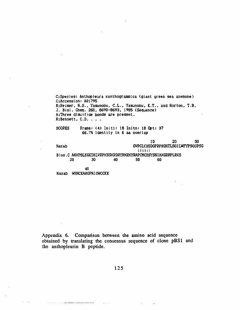
C;Species: Anthopleura xanthogrammica (giant green sea anemone)C;Accession: A01795R;Reimer, N.S., Yasunobu. C.L., Yasunobu, I.T., and Norton, T.R.J. Bioi. Chern. 260, 8690-8693, 1985 (Sequence)A;Three disulfide bonds are present.R;Bennett, C.D•.••
SCORES Frame: (4) Inlt1: 18 Initn: 18 Opt: 3766.7% identity 1n 6 aa overlap
10 20 30Naxab GVPCLCDSDGPRPRGNTLSGILWFYPSGCPSG
\I : I : ,Blue.C ANXPSLKGKDKLVRPXSSXRDRYRXRXYRAPCMCHSYSNIAXGRRPLRXS
~ ~ ~ ~ ~
40Naxab WHNCIAHGPNIGWCCIK
Appendix 6. Comparison between the amino acid sequenceobtained by translating the consensus sequence of clone pBSl andthe anthopleurin B peptide.
125

SCORES
blueSC~lpt clone 3
Inltl. 211 Inltn. 218 Opt. 26591.~ Identity In 83 bP oYe~l.p
670 680 690 700 710 720BIun. GCGGCCGCTCT1lGMCT1lG'l'GGATCCCCCGGGCTGCAGGMTTCGATATCAAGCTT1lTCG
11111111111111111111111111111Pbe3. S GGGTGCAGGAATTCGATilTCAAGCTT1lTCG
10 20 30
730 740 7SG 760 770 780Blues. 1lT1lCCGTCG1lCCTCG1l~CGGT1lCCCAGCTTTT-GTTCCCTTT1lGTGAGGGTT
1111111 1111111 1111111111111111111111 1I1111I11I1Pbe3.S 1lT1lCCGT1l1lACCTCGIl-CGGGGGCCCGGT1lCCCAGCTTTT1lCTTCCCTTT1lGT1lT""CCT
40 50 60 70 80
SCORES Inltl' 176 Jnltn' 176 Opt. 27414.7% Identity In III bP ov.~lap
650 660 . 670 680 690 700B1u.s. ""TTGG1lGCTCCIlCCGCGGTGGCGGCCGCTCT1lG1l1lCT1lGTGGATCCCCCGGGCTGCAGG
111111111111111 111111111111Pbs7 •S CT1lG1l1lCT1lGTGGAT--TCCGGGCTGCAGG
10 20
710 720 730 740 750 760BIu... A1lTTCG1lT1lTC1l1lGCTTATCGAT1lCCGTCGACCTCGIlGGGGGGGCCCGGT1lCCCIlGCTTT
1111111111111111111111111111111 11111 I III 11111111 1 IIPbs7.S ""TTCGAT1lTC1l1lGCTT1lTCGAT1lCCGTCGA-CTCGIlCGTGGG--CCGGT1lCC--1lCGTT
30 40 50 60 70 80
770 780 790 800 810
Blues. TGTTCCCTTT1lGTGAGGGTT""TTTCGAGCTTGGCGT""TCATGGTCAT1lGCTG'MTCCTII 11111111111111
Pbe7 •S TG----CCTT1lGTGAGGGTTATCGCGCG90 100
SCORES Jnltl: 165 Jnltn. 165 Opt. 19682.4% Identity In 85 bP ov.~I.p
660 670 680 690 700 710aIu.s. CTCCIlCCGCGGTGGCGGCCGCTCTAGMCTAGTGGATCCCCCGGGCTGCAGG1l1lTTCGllT
1111111 11111111 111111111111Pbs8. S 1lGTGGAT--CCCGGGCTACAGGAATTCGAT
10 20
720 730 740 750 760 770Blues. 1lTCAAGCTT1lTCGIlT1lCCGTCG--ACCTCGIlGGGGGGGCCCGGT1lCCCAGCT'lTI'GTTCC
1111111111111111111111 111111 III I III 1111111 IPbS8 •S 1lTCAAGCTTATCGATACCGTCGCTAGCTCGACT1lGGG---CCGTil-CCIlGCTTGTCCTTil
30 40 50 60 70 eo
710 790 800 810 820 830
Appendix 7. Comparison between the sequences of clones pBS3,pBS7, and pBSS with the cloning vector pBluescript KSII+.
126

RFFERENCES
Alsen, C.' (1983) Biological significance of peptides from
Anemonia sulcata. Fed. Proc. Fed. Am. Soc. Exp. BioI. 42,
101-108
Alsen, C., Beress, L., and Tessaraux, 1. (1978) Toxicities of sea
anemone (Anemonia sulcata) polypetides in mammals.
Toxicon v 16, 561-566
Bahraoui, E. M., EI ayeb, M., Granier, C., Beress, L., and Rochat, H.
(1989) Specificity of antibodies to sea anemone toxin III and
immunogenicity of the pharmacological site of anemone and
scorpion toxins. Eur. J. Bioch. 180, 55-60
Barhanin, J. (1981) Structure function relationships of sea
anemone toxin II from Anemonia sulcata. J. Bioi. Chern. 256,
5764.:5769
Bamola, F., Villegas, R., and Camejo, G. (1973) Tetrodotoxin
receptors in plasma membranes isolated from lobster nerve
fibre. Biochim. Biophys. Acta 298, 84-94
Benton, D. and Davis, R. (1977) Screening A. gt recombinant
clones by hybridization to plaques in situ. Science. 196,
180-182
127

Beress, L. (1978). In Drugs and Food from the Sea: Myth or
Reality? (P. A. Kaul and C. J. Sindermann, eds) pp 59-72.
Oklahoma Press, Norman, Oklahoma
Beress, L. (1982) Biologically active compounds from
coelenterates. Pure Appl. Chem. 534, 1981-1994
Bergman, c., Dubois, J. M., Rojas, E., and Rathmeyer, W. (1976)
Decreased rate of sodium conductance in the node of Ranvier
induced by a polypeptide toxin from sea anemone. Biochim.
Biophys. Acta. 455, 173-184
Bernheimer, A. W., and Avigad, L. (1976) Properties of a toxin
from sea anemone Stoichactis helianthus including specific
binding to sphingomyelin. Proc. Nat. Acad. Sci. (USA) 73,
467-471
Bewsey. K., Johnson, M. E., and Huff, J. P. (1991) "Rapid isolation
and purification of DNA from agarose gels: The phenol-freeze
fracture method. Biotechniques 10, 724-725
Blair, R. W., Peterson, D. F., and Bishop, V. S. (1978) The effects of
anthopleurin A on cardiac dynamics in conscious dogs. J.
Pharmacol. Exp. Ther. 207, 271-276
128
Britten, R. J. and Davidson
46, 111-
(1971) Quarterly Review of Biology
Brown, M. and Mosher, H. (1963) Tarichatoxin: Isolation and
purification. Science 140, 295-296
Caterall, W. A. (1978) Sea anemone toxin and scorpion toxins
share a common receptor site associated with the action
potential sodium ionophore. J. Bioi. Chern. 253, 7393-7396
Caterall, W. A. (1980) Neurotoxins that act on voltage sensitive
sodium channels in excitable membranes. Ann. Rev.
Pharmacol. 20, 15-43
Caterall, W. A. (1986) Molecular properties of voltage sensitive
sodium channels. Ann. Rev. Biochem. 55, 953-985
Cha, I., Christ, W., Fujioka, H. and Kishi, Y. (1982)
Stereochemistry of palytoxin 4. Complete structure. J. Am.
Chern. Soc. 104, 7369-7371
Chien, A., Edgar, B., and Trela, I. M. (1976) Deoxyribonucleic acid
polymerase from the extreme thermophile Thermus
aquaticus. J. Bacteriol127, 1550-1557.
129
Chirgwin, J. M.• Przybyla, R. Macdonald. R. J., and Rutter, W. J.
(1918) Isolation of Biologically active ribonucelic acid from
sources enriched in ribonuclease. Biochemistry 18, 5294
5299
Chrisholm, D. (1989). Biotechniques 7:21-23
Clark, J. M. (1988) Novel non nucleotide addition reactions
catalyzed by prokaryotic and eukaryotic DNA polymerases.
Nucleic Acid Research 16,9677
Compton, T. (1990) Degenerate primers for DNA amplification.
In M. A. Innis, D. H. Gelfand, J. J. Sninsky and T. J. White (Eds.)
peR Protocols:A guide to Methods and Applications, pp 39-45.
Academic Press N.Y.
Conti, F., Hille, B., Neumcke, B., Nonner, W., and Stampfli. R.
(1976) Measurement of the conductance of the sodium
channel from current fluctuation at the node of Ranvier. J.
Physiol. 262, 699-727
Conti, F., Hille, B., Neumcke, B., Nonner, W., and Stampfli, R.
(1976) Conductance of the sodium channel in myelinated
nerve fibres with modified sodium inactivation. J. Physiol.
(London) 262, 729-742
130
Cook, J. S., and Brake, E. T. (1978) Uptake and fate of ouabain
bound to HeLa cells. In Cell Membrane Receptors for Drugs
and Hormones pp 343-351. (R.W. Straub, and L. Bolis, eds.)
Raven press, N.Y.
Crone, R., Leake, B., Jarvis, M., and Freeman, S. (1976) On the
nature of maculotoxin. Toxicon 14, 4213-426
Crowe, J. S. , Cooper, H. J., Smith. M. A., Sims, M. J., Parker, D., and
Gewert, D. (1991) Improved cloning of polymerase chain
reaction (PCR) products after proteinase K digestion. Nucleic
Acid Research 19, 184
Culquohoun, D., Henderson, R., and Ritchie, M. (1972) The
binding of labeled tetrodotoxin to nonmyelinated nerve fibres.
J. Physiol. London 227, 95-126
Debarry., J., Fosset, M., and Lazdunski, M. (1977) Molecular
mechanism of the cardiotoxic action of a polypeptide
neurotoxin from sea anemone on cultured embryonic cardiac
cells. Biochem. 16, 3850-3855
131

Deguchi, T., Urakawa, N., Takamatsu, S. (1976) Some
pharmacological properties of palythoa toxin isolated from the
zoanthid Palythoa tuberculosa. In Animal. Plant; and
Microbial Toxins, V. 2. pp. 379-394 (A. Ohsaka, K. Hayashi,
and Y. Sewai, eds.) Plenum Press, N.Y.
Dehaseth, P. L., Goldman, R. A., Cech, C. L., and Caruthers, M. H.
(1982) In Genetic Engineering V.4 (J.Setlow, and A.
Hollaender, eds.) Plenum Press, N.Y. 1-16
Feinberg. A. P., and Vogelstein, B. (1983) A technique for
radiolabeling DNA restriction endonuclease fragments to high
specific radioactivity. Analytical Biochem 132, 6-13
Fleming, W. W. (1980) The electrogenic pump in smooth muscle:
Physiologic and pharmacologic significance. Ann. Rev...
Pharmacol. Toxicol. 20, 129-149
Gooley, P. R. Beress, L., and Norton, R. S. (1984) IH Nuclear
magnetic resonance spectroscopic study of the polypeptide
toxin from Anemonia sulcata. Biochem, 23, 2144-2152
132

Goulian, M., Lucas, Z.J., and Kornberg, A. (1968) Enzymatic
synthesis of deoxyribonucleic acid.XXV. Purification and
properties of deoxyribonucleic acid polymerase induced by
infection with phage T-4 . J. Bioi. Chern. 243, 627-638
Hashimoto, K., Rikuo, 0., Hashimoto, K., Inui, J., and Miura, Y.
(1980) The ionic mechanism of prolongation of action
potential duration of cardiac ventricular muscle by
anthopleurin-A and its relationship to inotropic effect. J.
Pharmacol. Exp. Ther. 215, 479-485
Haymer, D., Anleitner, J. E., He, M., Thanaphum, S., Saul, S., Ivy, J.,
Houtchens, K., and Arcangeli, L., (1990) Actin Genes in the
Mediterranean fruit fly, Ceratitis capitata Genetics 125 #1,
155-160
Haymerle, H., Herz, J., Bressan, G. M., Frank R., and Stanley, K.
(1986) Nucleic Acids Research 14, 8615
Helms. C., Graham, M. Y., Dutchik, J. E. and Olson, M. V. (1985) A
new method of purifying A. DNA from phage lysates. DNA 4,
39-49
Hemsley, A. , Arnheim, N., Toney, M. D., Cortopassi, G., and Galas,
D. J. (1989) A simple method for site directed mutagenesis
133

using the polymerase chain reaction. Nucleic Acid Res. 17,
6545-6551
Henderson, R., Ritchie, J. and Stricartz, G. (1973) The binding of
labeled saxitoxin to the sodium channel in nerve membranes.
J. Physiol. London 235, 783-804
Henderson, R., Ritchie, J. and Stricartz, G. (1974) Evidence that
tetrodotoxin and saxitoxin act at a metal binding site in the
sodium channels in the nerve membrane. Proc. Nat. Acad. Sci.
USA 71, 3936-3940
Henikoff, S. (1987) Unidirectional digestion with Exonuclease
III in DNA sequence analysis. Methods in Enzymology 155,
156-165
Hille, B. (1975) Ion selectivity, saturation, and block in sodium
channels. A four barrier model. J. Gen. Physiol. 66, 635-650
Hopp, P. T., and Woods, K. R. (1981) Prediction of protein
antigenic determinants from amino acid sequences. Proc. Nat.
Acad. Sci. (USA) 78, 3824-3828
Huang, J. M., and Wu, C. (1985) Mechanism of the toxic action of
T17 brevetoxin from Ptychodiscus brevis on nerve
134
membranes. In Toxic Dinoflagellates pp. 351-356, (D.M.
Anderson, A.W. White, and D.G. Baden, eds.) Elsevier, N.Y.
Huang, J. M., Wu, C., and Baden, D. (1984) Depolarizing action of a
redtide dinoflagellate brevetoxin on axonal membranes. J.
Pharmacol. Exp. Ther. 229, 615-621
Huang, M. N., and High K. A. (1990) Efficient subcloning of DNA
fragments amplified by crude oligonucleotides. Biotechniques
9, 710
Huynh, T., Young, R. A., and Davis, R. (1985) in DNA Cloning VI
(D. Glover, ed) IRL Press Ltd. 56-110
Ishizaki, H., Mackay, R. H., Norton, T. R., Yasunobu, K. T., Lee, J.,
and Tu, A. T. (1979) Conformational studies of peptide heart
stimulant anthopleurin A. J. Bioi. Chern. 254, 9651~9656
Ito, K., Karaki, H. , and Urakawa, N. (1977) The mode of
contractile action of palytoxin on vascular smooth muscle.
Eur. J. Pharmacol, 46, 9-14
Javed, A., and Joshi, S. (1990) Targeted DNA sequencing: Rapid
Identification of clones by sequencing DNA using mixed
oligonucleotide probes as primers. Biotechniques 9, 28-32
135
Kaiser, K and Murray, N. (1985) in : DNA cloning techniques, A
practical approach (D. M. Glover, ed). R. L. Press, Oxford p38.
Karaki, R., Nagase, R., Ohizumi, Y., Satake, N., and Shibata, S.
(1988) Palytoxin-induced contraction and release of
endogenous noradrenaline in rat tail artery. Br. J. Pharmacol.
95, 183-188
Kaufman, D. L., and Evans, G. A. (1990) Restriction
endonuclease cleavage at the termini of PCR products. Bio
Techniques 9, 304-306
Kim, Y., Brown, G., Mosher, R., and Furhman, F. (1975)
Tetrodotoxin: Occurence in Atelopoid frogs of Costa Rica.
Science 184, 151-152
Kobayashi, M., Kondo, S., Yasumoto, T., and Ohizumi, Y. (1986)
Cardiotoxic effects of maitotoxin, a principal toxin of seafood
poisoning, on guinea pig and rat cardiac muscle. J. Pharmacol.
Exp. Ther. 238, 1077-1083
Kobayashi, M., Ohizumi, Y., and Yasumoto, T. (1985) The
mechanism of action of maitotoxin in relation to Ca2 +
movement in guinea pig and rat cardiac muscle. Br. J.
Pharmacol. 86, 385-391
136

Kodama, I., Shibata, S., Toyama, J., and Yamada, K. (1981)
Electrochemical effects of anthopleurin A (AP-A) on rabbit
ventricular muscle: Influence of driving frequency, calcium
antagonists, tetrodotoxin, lidocaine, and ryanodine. Br. J.
Pharmacol. 74, 29-37
Kudo, Y., and Shibata, S. (1980) The potent excitatory effect of a
novel polypeptide, anthopleurin B, isolated from a sea
anemone (Anthopleura xanthogrammica) on the frog spinal
cord. J. Pharmacol. Exp. Ter. 214, 443-448
Legrand, A. M., and Bagnis, R. (1984) Mode of action of ciguatera
toxins. In Seafood and Toxins pp.217-233 (E.P. Ragelis, ed.)
Washington, D.C., Am. Chern. Soc.
Legrand, A.M., Benoit, E., and Dubois, J. (1985 Electrophysiological
studies of the effects of ciguatoxin in the frog myelinated
nerve fibre. In Toxic Dinoflagellates pp.381-3'82, (D.M.
Anderson, A.W. White, and D.O. Baden, eds.) Elsevier, N.Y.
Liu, Z., and Schwartz, L. M. (1992) An efficient method for blunt
end ligation of peR products. Bio Techniques 12, 28-30
137

Low, P.A., Wu, C.H., and Narahashi, T. (1979) The effect of
anthopleurin A on crayfish giant axon. J. Pharmacol. Exp.
Ther. 210, 417-421
Mandel, M. and Higa, A. (1970) Calcium dependent bacteriophage
DNA infection. J. Mol. Bioi. 53: 154-
Maniatis, T., Fritsch, E., and Sambrook, J. (1982) Molecular
cloning: A laboratory manual. Cold Spring Harbor Laboratory,
N.Y.
Matsueda, G., and Norton, T. R. (1982) Chemical synthesis of
anthopleurin A, a polypeptide from sea anemone with
positive inotropic activity. Fed. Proc. 41, 1183-1187
Matteucchi, M. D., and Caruthers, M. H. (1980) Synthesis of
oligodeoxypyrimidines on solid support. Tetrahedron Letters
21, 719
Messing, J. (1983) New M13 vectors for cloning. Methods in
Enzymology 101, 20-78
Muramatsu, l., Uemura, D., Fujiwara, M., and Narahashi, T.
(1984) Characteristics of palytoxin-induced depolarization in
squid axons. J,. Pharmacol. Exp. Ther. 201, 148-155
138

Newcomb, R., Yasunobu, K. T., Seriguchi, D., and Norton, T. R.
(198) In Frontiers in Protein Chemistry (T. Y. Liu, K. T.
Yasunobu, and G. Mamiya, eds.) Elsevier, North Holland Press.
pp.539-550
Noguchi, T. and Hashimoto, Y. (1973) Isolation of tetrodotoxin
from a goby Gobius criniger. Toxicon 11, 305-307
Norton, R. S., and Norton, T. R. (1979) Natural abundance 13C
nuclear magnetic resonance study of toxin I from Anthopleura
xanthogrammica. J. Bioi. Chem. 254, 10220-10226
Norton, R. S., Norton, T. R., Sleigh, R. W. and Bishop, D. B. (1982)
Interaction of the polypeptide heart stimulant anthopleurin A
with H+, Ca 2+ and membrane lipids. Arch. Biochem. Biophys.
213, 87-97
Norton, R. S., Zwick, J., and Beress, L. (1980) Natural abundance
13C nuclear magnetic resonance study of toxin II from
Anemonia sulcata. Eur. J. Biochem. 113, 75-83
Norton", T. R., Kashiwagi, M., and Shibata, S. (1978) In Drugs and
Food from the Sea: Myth or Reality.? (P. A. Kaul and C. J.
Sindermann, eds.) pp. 59-72, Oklahoma Press, Norman,
Oklahoma.
139

Norton, T. R., Ohizumi, Y., and Shibata, S. (1981) Excitatory effect
of a new polypeptide (anthopleurin B) from sea anemone on
the guinea pig vas deferens. Br. J. Pharmacol. 74, 23-28
Norton, T. R., Shibata, S., Kashiwagi, M., and Bentley, J. (1976)
Isolation and characterization of the cardiotonic polypeptide
anthopleurin A from the sea anemone Anthopleura
xanthogrammica. J. Pharmacol. Sci. 54, 1368-1374
Ohizumi, Y., and Shibata, S. (1981) Possible mechanisms of the
dual action of the new polypeptide (anthopleurin B) from sea
anemone in the isolated ileum and taenia caeci of the guinea
pig. Br. J. Pharmacol. 72, 239-244
Ohizumi, Y., Kobayashi, M., Nakamura, H., and Kobayashi, J. (1988)
The mechanism of inotropic action of striatoxin, a novel
polypeptide from a marine snail, in isolated cardiac muscle.
Br. J. Pharmacol. 95, 867-875
Ohizumi, Y., Nakamura, H., Kobayashi, J., and Caterall, W.A. (1986)
Specific inhibition of 3H saxitoxin binding to skeletal muscle
sodium channels by geographutoxin II, a polypeptide channel
blocker. J. Bioi. Chem. 261, 6149.;6152
Okayama, H., and Berg, P. (1982) High efficiency cloning of full
length eDNA. Mol. Cell. Bioi. 2, 161-166
140

Olivera, B. M., McIntosh, J. M., Cruz, L. J., Luque, F. A. and Gray, W.
R. (1984) Purification and sequence of a presynaptic peptide
from Conus geographus venom. Biochem. 23, 5087-5090
Pichon, Y. (1982) Effects of palytoxin on sodium and potassium
permeabilities in unmyelinated axons. Toxicon 20, 41-47
Reimer, N. S. Yasunobu, C. L., Yasunobu, K. T., and Norton, T. R.
(1985) Amino acid sequence of the Anthopleura
xanthogrammica heart stimulant, anthopleurin B. J. Bioi.
Chem. 260, 8690-8693
Romey, G., Abita, J. P., Schweitz, H., Wunderer, G., and Lazdunski,
M. (1976) Sea anemone toxin: A tool to study molecular
mechanisms of nerve conduction and excitation-secretion
coupling. Proc. Nat. Acad. Sci. (USA) 73, .4055-4059 ..
Rosenberg, S. M., Stahl, M. M., Kobayashi I and Stahl, F. W. (1985)
Gene 38, 165-175
Saiki, R. K., Gelfand, D. H., Stoffe, S., Scharf, S. J., Higuchi, R., Hom,
G. T., Mullis, K. B., and Erlich, H. A. (1988) Primer directed
amplification of DNA with a thermostable DNA polymerase.
Science 239, 487-491
141

Sambrook, I., Fritsch, E. F., and Maniatis, J. (1989) Enzymes used
in molecular cloning p 5.40-~.41. In Molecular Cloning: A
laboratory Manual 2nd Edition. Cold Spring Harbor
Laboratory, Cold Spring Harbor, New York
Sanger, F., Nicklen, S. and Coulson, A.R. (1977) DNA sequencing
with chain terminating inhibitors. Proc. Nat. Acad. Sci. (USA)
74, 5463-5466
Scharf, S. J. (1990) Cloning with PCR. In M. A. Innis, D. H. Gelfand,
I.J. Sninsky and T. J. White (Eds.) peR Protocols:A guide to
Methods and Applications, pp 84-91. Academic Press N.Y.
Scharf, S. J., Hom, G. T., and Erlich, H. A. (1986) Direct cloning
and sequence analysis of enzymatically amplified genomic
sequences. Science 233, 1076-1078
Schweitz, H., Concent, J. P., Barhanin, I., Frelin, C., Linden, B.,
Hugues, M., and Lazdunski, M. (1981) Purification and
pharmacological properties of eight sea anemone toxins from
Anemonia sulcata, Anthopleura xanthoerammica. Stoichactis
eteanteus, and Actinondendron plumosum. Biochem. 20,
5245-5252
142

Seino, A., Kobayashi, M., Momose, K., Yasumoto, T., and Ohizumi, Y.
(1988) The mode of inotropic action of ciguatoxin on guinea
pig cardiac muscle. Br. J. Pharmacol. 95, 876-882
Seyama, 1., Wu, C., and Narahashi, T. (1980) Current dependent
block of nerve membrane sodium channels by paragracine.
Biophys. J. 29, 531-538
Sheumack, D., Howden, M., Spence, I., and Quinn, R. (1978) A
neurotoxin from the venom glands of the octopus
Hapalochlaena maculosa identified as tetrodotoxin. Science.
199, 188-189
Shibata, S., Norton, T. R., Izumi, T., Matsuo, T., and Katsuki, W.
(1976) A polypeptide (AP-A) from sea anemone Anthopleura
xanthogrammica with potent positive inotropic action. J.
Pharmacol. Exp. Ther. 199, 298-309
Shimizu, Y. (1982) Recent progress in marine toxin research.
Pure Appl. Chem.. 54, 1973-1980
Silhavy, T. J. Berman, M. L., and Enquist L. W. (1984)
Experiments with Gene fusions, Cold Spring Harbor
Laboratory, Cold Spring Harbor, N. Y.
143

Smith, C. D., Delucas, L., Eallck, S. E., Schweitz, H., Lazdunski, M.,
and Bigg, C. E. (1984) Crystallization and preliminary x-ray
investigation of a protein from the sea anemone Anthopleura
xanthogrammica. J. Bioi. Chern. 259, 8010-8011
Smythies, J. R., Benington, F., Bradley, R. J., Bridgers, W. F., Morin,
R. D., and Romine, W. O. Jr. (1975) The molecular structure of
the receptor ionophore complex at the neuro-muscular
junction. J. Theor. Bioi. 51, 111-126
Southern, E. (1975) Detection of specific sequences among DNA
fragments by gel elctrophoresis. J. Mol. Bioi. 98, 503-510
Stengelin, S., Rathmeyer, W., Wunderer, G., Beress, "L., and Hucho,
F. (1981) Radioactive labeling of toxin II from Anemonia
sulcata. Anal. Biochem. 113, 277-285
Takahashi, M., Ohizumi, Y., and Yasumoto, T. (1982) Maitotoxin, a
Ca2+ activator candidate. J. Bioi. Chern. 257, 7287-7289
Takahashi, M., Tatsumi, M., Ohizumi, Y., and Yasumoto, T. (1983)
Ca2+ channel activating function of maitotoxin, the most
potent marine toxin known, in clonal rat pheochromocytoma
cells. J. Bioi. Chern. 258, 10944"-109849
144

Tanaka, M., Haniu, M., Yasunobu, K. T., and Norton, T. R. (1977)
Amino acid sequence of the Anthopleufa" xanthQ~fammica
heart stimulant anthopleurin A. Biochem. 16, 204-208
Vincent, J. P., Balema, M., Barhanin, J., Fosset, M., and Lazdunski,
M. (1980) Binding of sea anemone toxin to receptor sites
associated with gating system of sodium channel in synaptic
nerve endings in vitro. Proc. Nat. Acad. Sci. (USA). 77, 1646
1650
Weidmann, w. (1977) Effects of palytoxin of mechanical and
electrical activity of dog and rabbit heart. Experentia 33,
1487-1488
Wu, C. H., and Narahashi, T. (1988) Mechanism of action of novel
marine neurotoxins on ion channels. Ann. Rev. Pharmacol.
Toxicol. 28, 141-162
Wu, C., Huang, J., Vogel, S., and Luke, V. (1985)' Actions of
Ptychodiscus brevis toxins on nerve and muscle membranes.
Toxicon 23, 481-487
Wunderer, G., and Eulitz, M. (1978) Amino acid sequence of
toxin I from Anemonia sulcata. Eur. J. Biochem. 89, 11-12
145

Yanisch-Perron, C., Vieira, J. and Messing, J. (1985) Improved
M13 phage cloning vectors and host strains: Nucleotide
sequences of the M13mp18 and pUC19 vectors. Gene 33,
103-119
Ziai, M. R., Giordano, A., Armandola, E., and Ferrone, S. (1988)
Purification by ammonium sulfate precipitation of
bacteriophage A. gtl l. Anal. Bioch. 171, 192-196
146


![(Actinoporins) in Sea Anemones - Semantic Scholar...Among Cnidaria, actinoporin-like toxins have been described in only one non-actiniarian species, Hydra magnipapillata [18]. The](https://img.pdfslide.us/doc/110x75/60300c33e367e33be01b37ff/actinoporins-in-sea-anemones-semantic-scholar-among-cnidaria-actinoporin-like.jpg)