Embed Size (px)
Citation preview
Arch. Dis. Childh., 1968, 43, 382.
Umbilical Artery Catheterization in the NewbornJ. M. GUPTA*t, N. R. C. ROBERTONtt, and J. S. WIGGLESWORTH
From the Nuffield Neonatal Research Unit, Institute of Child Health, Hammersmith Hospital, London
Satisfactory care of the sick premature newbornbaby requires investigations of blood gas tensions(Dahlenburg et al., 1968) and of various biochemicalparameters, such as blood sugar, often at frequentintervals during the first few hours of life. Bloodsamples for biochemistry can be obtained from heelpricks or by venepuncture. Heel pricks are painful,may become infected, and the number which canbe carried out is limited. Repeated venepuncturein the newborn carries its own not inconsiderablehazards (McKay, 1966; Asnes and Arendar, 1966).Blood can be obtained from umbilical venouscatheters, but here air embolus is a hazard, as isportal venous thrombosis, though this is rare withgreater attention to sterile techniques (Tizard, 1962);unless the umbilical venous catheter passes throughthe ductus venosus into the inferior vena cava ittends to wedge in a branch of the portal sinus andneeds periodic manipulation before blood can bewithdrawn.To obtain specimens for blood gas analyses direct
arterial puncture or arterialized capillary blood canbe used. To obtain blood by the latter route foraccurate and repeated estimations requires con-siderable skill, and with the peripheral vasocon-striction often found in ill babies, the results maybe inaccurate. In both cases the child is trauma-tized, cries, and may therefore lower his PaCO2.A haematoma may form round an arterial punctureand equilibrate with a high oxygen tension presentin the skin if the child is being nursed in a highoxygen concentration, and give a falsely high PaO2if it is aspirated. This especially applies to temporalartery puncture. It is again not practicable toobtain repeated samples by these routes. For thesereasons sampling of arterial blood through anumbilical artery catheter is preferable for bothbiochemical and blood gas determinations.
Before arterial catheterization could be considered
Received November 16, 1967.* Present address: Department of Paediatrics, University of
Khartoum.t Requests for reprints should be addressed to N.R.C.R.t Percy J. Neate Research Fellows of the Clothworkers Company.
the sterility of the cord stump had to be investigated.We report the results of the initial study of methodsfor maintaining cord sterility, and then the techniqueof arterial catheterization and the complicationsencountered in the years 1963-66.
Material and MethodsTo investigate umbilical stump sterility a pilot study
was carried out in normal babies. At birth and dailythereafter hexachlorophene soap was applied to theumbilical cord of all babies. Alternate babies had theircords sprayed with neomycin, polymyxin, and bacitracinpowder (Polybactrin) at birth and daily thereafter.Swabs for culture were taken routinely immediatelybefore the daily application of Polybactrin and/orhexachlorophene, for the first 5 days of life.
Table I shows a significant improvement in the cordsterility using Polybactrin. Table II shows theorganisms cultured. Of the 5 positive cultures in the
TABLE IEffect of Phisohex and Phisohex+Polybactrin on
Umbilical Swab Cultures
No. of ~I No. withCleanser No. of No. of Swabs Positive
Babieslorophen .. 112CulturesHexachlorophene I11 55 22Hexachlorophene and
Polybactrin 11 52 5
X2 = 9-98; 0-01 >p>0 001 for one degree of freedom.
TABLE IIOrganisms Cultured from Positive Umbilical Swabs
Cleanser
Organism HexachloropheneHexachlorophene and
Polybactrin
Mixed growth .. 1Staphylococcus albus .. 3 2Micrococci .. .. 3Coliforms .. .. 15 2Streptococcus faecalis . 1
382
on 12 June 2018 by guest. Protected by copyright.
http://adc.bmj.com
/A
rch Dis C
hild: first published as 10.1136/adc.43.229.382 on 1 June 1968. Dow
nloaded from
Umbilical Artery Catheterization in the NewbornPolybactrin sprayed group, 2 were positive in the first 2days of life in the same baby, and it was subsequentlyfound that this child had not been sprayed with Polybac-trin immediately after birth. The later swabs were
negative. This policy of routine spraying of the cordshas not only prevented local infection but has alsoeliminated staphylococcal infection as a hazard in our
newborn babies. As the cord stump remains dry andsterile it takes much longer (up to 3 weeks) to separateby dry instead of wet gangrene.
Technique
The technique used for inserting umbilical arterycatheters is simple. It is usually possible to insert thecatheter for the first time up to 48-72 hours of age,
though they have been passed for the first time at 10days. Only if the umbilical cord stump is sterile (andtherefore clean) are the umbilical artery orifices easilyidentifiable after 72 hours. The size of the baby is notcritical; catheters have been passed in babies under400 g. and for the first time at 7 days in a 920 g. baby.The catheterization is carried out with full sterile
precautions within the incubator. Babies requiring morethan 40% 02 are better placed in a Gairdner (Warleyand Gairdner, 1962) box at this stage, as it is difficult tomaintain the oxygen concentration within the incubatorhigher than this with the portholes open. The cordstump and surrounding skin are cleaned with a 2-5%solution of iodine in spirit. It is important to ensure thatall the iodine is cleaned off to prevent iodine bums,particularly in the very immature baby.The cord is cut 0-5-1-0 cm. from the skin-umbilical
cord junction, and the vessels are usually obvious onthe cut surface. The umbilical arteries may still bepatent in the first few hours of life and it is wise to tie a
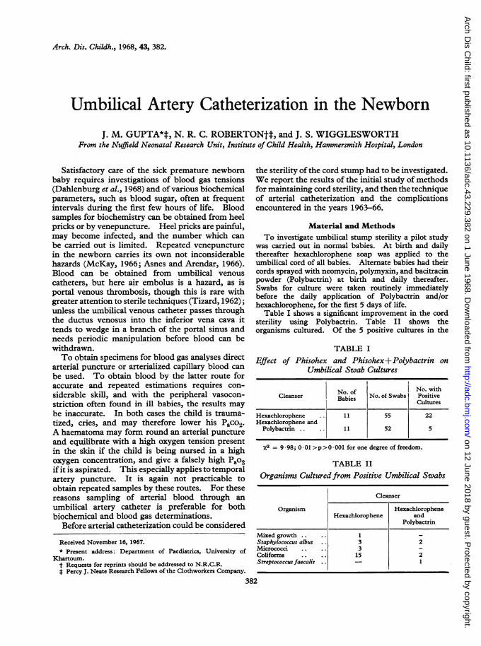
cord ligature loosely round the base ofthe cord to preventa brisk arterial haemorrhage and a slow ooze from theumbilical vein. The orifice of the artery is then teasedopen with a special dilator (Fig. 1). Great care shouldbe taken at this stage as it is very easy to make a falsepassage in the adventitia of the artery or in the jellyaround it, a mistake that is difficult to correct. A K32FG 5 polyvinyl feeding tube of outside diameter 2 mm.(Pharmaseal Inc.) is then inserted. The catheterlumen is previously filled with normal salinecontaining 10 u. heparin/ml. The syringe containingheparinized saline is attached to the open end of thecatheter. Once within the artery orifice the cathetercan usually be slid gently in. Obstruction may occur
TABLE IIIDistances at Which Catheters Must be Inserted to
Reach Diaphragm
Body Weight (g.) Distance (cm.)
1000 8-101000-1500 10-111500-2000 11-122000-2500 12-13
2500 14-15
at a distance of 1-2 cm. where the vessels turn suddenlydownwards. This can usually be overcome by pullingthe stump upwards towards the baby's head. It iseasy at this stage to create a false passage by forcing thecatheter through the arterial wall. A second site ofobstruction is at a distance of 5-6 cm., due presumablyto spasm and kinking of the artery at its origin from theiliac vessels. Gentle sustained pressure with thecatheter will usually result in the obstruction beingpassed. At a distance of 6-10 cm. blood can easily bewithdrawn. It is aimed to push the catheter to thelevel of the diaphragm. Table III shows the average
distance the catheter has to be pushed in to reach thissite in babies of various sizes.The catheter is held in place by a purse-string suture of
chromic catgut around the umbilical cord and a doublespiral arrangement tied tightly around it. It is sub-sequently kept patent, by keeping it full of heparinizedsaline (10 u./ml.). The catheter volume is 0-2 ml., andevery time a sample is withdrawn the catheter is flushedthrough with 0 3 ml. heparinized saline, and a bung isinserted in the end held firmly in place with adhesivetape. It is not necessary to keep a constant infusiongoing, nor is it necessary to flush the catheter throughat set intervals; this will only result in overheparinizingthe baby.
In most cases where we insert arterial catheters weinsert an umbilical venous catheter, which is of particularvalue for giving alkali in the respiratory distress syndrome(RDS) (Gupta, Dahlenburg, and Davis, 1967). It isusually easier to insert the arterial catheter first, unlessthe slow ooze from the vein is troublesome as thevenous catheter hinders manipulation at the umbilicus.
In all cases once the catheters are sutured in place,Polybactrin spray is applied and the stump covered witha sterile dressing. Thereafter the cord is sprayed dailywith Polybactrin until the catheter is removed. It is
0 I 2 3 4 5cm.
FIG. 1.-Umbilical artery dilator (designed by Professor L. B. Strang).
383
on 12 June 2018 by guest. Protected by copyright.
http://adc.bmj.com
/A
rch Dis C
hild: first published as 10.1136/adc.43.229.382 on 1 June 1968. Dow
nloaded from
Gupta, Roberton, and Wigglesworthimportant to have the catheters easily identified lest thearterial one be confused with the venous one and viceversa. If there is any doubt about the localization ofthe catheter either a pressure tracing or a plain x-raycan be taken.To remove the catheter the hard dried scab of Whar-
ton's jelly is cut away down to fresh tissue. A newpurse-string suture is then inserted around the stumpand the catheters gently withdrawn. It is importantto have the purse-string in situ in freshened tissue beforethe catheters are withdrawn, otherwise, on pulling ittight, the arterial lumen will not be occluded and abrisk haemorrhage will result. The catheters are usuallynecessary for the first 24-48 hours after insertion inRDS or in babies needing metabolic monitoring.However, if the clinical state of the baby warrants it,and particularly if there is a persisting need to knowblood gas tensions on a baby receiving more than 40%02, catheters have been left in for considerably longer.The longest they have been left in place is 20 days, andseveral babies have exceeded 8 days without ill effect.
ResultsIn four years 381 attempts have been made to
insert arterial catheters with 46 failures (12%)(Table IV). However, as we became more skilfulour failure rate dropped. Most cases of failedinsertion were due to obstruction at one of the two
TABLE IVNumber of Catheterizations Attempted and their
Outcome
Year No. No. Failed No. Compli- TotalInserted cations*
1963 38 11 (22) 4 (10) 491964 94 18 (16) 10 (11) 1121965 73 4 (5) 7 (10) 771966 130 13 (9) 13 (10) 143
335 46 (12) 34 (10) 381
Percentages are given in parentheses.* Calculated as a % of successful catheterizations.
TABLE VReasons for Inserting Umbilical Artery Catheter
Disease No.
Respiratory distress syndrome .161Metabolic (including small-for-dates, hypogly-
caemia, infant of diabetic mothers, and fits) 46Very small babies (<32 wk.; <1500 g.) .. 45Birth asphyxia 30Congenital malformation .20Haematology problems (haemorrhagic and
haemolytic disease of the newborn) 15Others. .. . 18
TABLE VIComplications of Umbilical Arterial Catheterization
Clncl ThrombosisYear Haemorrhage Obstruction at
Obstruction Necropsy
1963 .. .. 4 - -
1964 .. .. 3 6 11965 .. .. 1 5 11966 .. .. 5 6 3*
Total .. .. 13 17 5
Total with arterial occlusion = 21.* Includes a baby who had clinical signs of thrombosis.
levels mentioned above, or to creating a falsepassage. It is interesting to note that our only 5attempts to catheterize a single artery have allfailed.
Arterial catheterization was carried out in babieswho needed frequent blood sampling. This wasparticularly so in babies with respiratory illness(Table V). Other indications were infants of diabe-tic mothers, small-for-dates babies at risk fromhypoglycaemia, and in haemolytic disease wherethey can be used as a route for exchange transfusion.
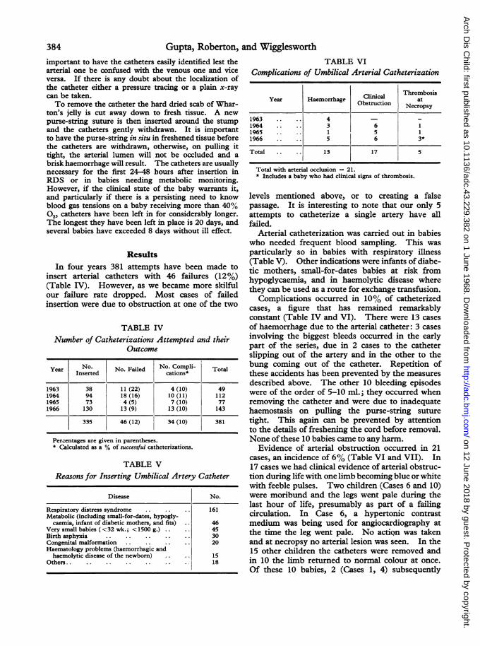
Complications occurred in 10% of catheterizedcases, a figure that has remained remarkablyconstant (Table IV and VI). There were 13 casesof haemorrhage due to the arterial catheter: 3 casesinvolving the biggest bleeds occurred in the earlypart of the series, due in 2 cases to the catheterslipping out of the artery and in the other to thebung coming out of the catheter. Repetition ofthese accidents has been prevented by the measuresdescribed above. The other 10 bleeding episodeswere of the order of 5-10 ml.; they occurred whenremoving the catheter and were due to inadequatehaemostasis on pulling the purse-string suturetight. This again can be prevented by attentionto the details of freshening the cord before removal.None of these 10 babies came to any harm.
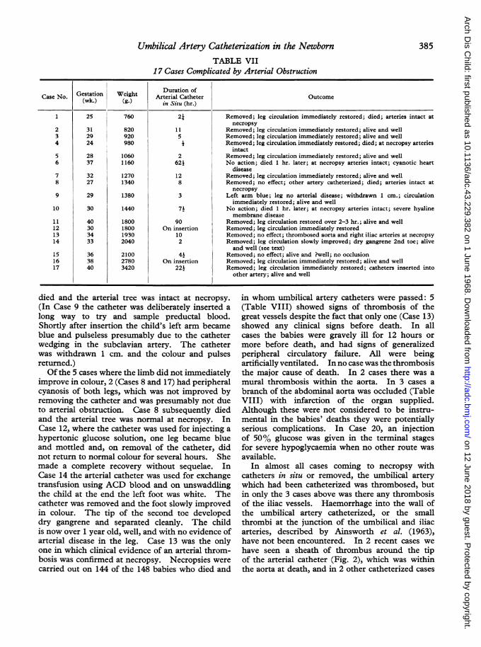
Evidence of arterial obstruction occurred in 21cases, an incidence of 6% (Table VI and VII). In17 cases we had clinical evidence of arterial obstruc-tion during life with one limb becoming blue or whitewith feeble pulses. Two children (Cases 6 and 10)were moribund and the legs went pale during thelast hour of life, presumably as part of a failingcirculation. In Case 6, a hypertonic contrastmedium was being used for angiocardiography atthe time the leg went pale. No action was takenand at necropsy no arterial lesion was seen. In the15 other children the catheters were removed andin 10 the limb returned to normal colour at once.Of these 10 babies, 2 (Cases 1, 4) subsequently
384
on 12 June 2018 by guest. Protected by copyright.
http://adc.bmj.com
/A
rch Dis C
hild: first published as 10.1136/adc.43.229.382 on 1 June 1968. Dow
nloaded from
Umbilical Artery Catheterization in the NewbornTABLE VII
385
17 Cases Complicated by Arterial Obstruction
Duration ofCase No. Gestation Weight Arterial Catheter Outcome(wk.) (g.) in Situ (hr.)
1 25 760 21 Removed; leg circulation immediately restored; died; arteries intact atnecropsy
2 31 820 11 Removed; leg circulation immediately restored; alive and well3 29 920 5 Removed; leg circulation immediately restored; alive and well4 24 980 i Removed; leg circulation immediately restored; died; at necropsy arteries
intact5 28 1060 2 Removed; leg circulation immediately restored; alive and well6 37 1160 621 No action; died 1 hr. later; at necropsy arteries intact; cyanotic heart
disease7 32 1270 12 Removed; leg circulation immediately restored; alive and well8 27 1340 8 Removed; no effect; other artery catheterized; died; arteries intact at
necropsy9 29 1380 3 Left arm blue; leg no arterial disease; withdrawn 1 cm.; circulation
immediately restored; alive and well10 30 1440 71 No action; died 1 hr. later; at necropsy arteries intact; severe hyaline
membrane disease11 40 1800 90 Removed; leg circulation restored over 2-3 hr.; alive and well12 30 1800 On insertion Removed; leg circulation immediately restored13 34 1930 10 Removed; no effect; thrombosed aorta and right iliac arteries at necropsy14 33 2040 2 Removed; leg circulation slowly improved; dry gangrene 2nd toe; alive
and well (see text)15 36 2100 41 Removed; no effect; alive and ?well; no occlusion16 38 2780 On insertion Removed; leg circulation immediately restored; alive and well17 40 3420 22i Removed; leg circulation immediately restored; catheters inserted into
other artery; alive and well
died and the arterial tree was intact at necropsy.(In Case 9 the catheter was deliberately inserted along way to try and sample preductal blood.Shortly after insertion the child's left arm becameblue and pulseless presumably due to the catheterwedging in the subclavian artery. The catheterwas withdrawn 1 cm. and the colour and pulsesreturned.)Of the 5 cases where the limb did not immediately
improve in colour, 2 (Cases 8 and 17) had peripheralcyanosis of both legs, which was not improved byremoving the catheter and was presumably not dueto arterial obstruction. Case 8 subsequently diedand the arterial tree was normal at necropsy. InCase 12, where the catheter was used for injecting ahypertonic glucose solution, one leg became blueand mottled and, on removal of the catheter, didnot return to normal colour for several hours. Shemade a complete recovery without sequelae. InCase 14 the arterial catheter was used for exchangetransfusion using ACD blood and on unswaddlingthe child at the end the left foot was white. Thecatheter was removed and the foot slowly improvedin colour. The tip of the second toe developeddry gangrene and separated cleanly. The childis now over 1 year old, well, and with no evidence ofarterial disease in the leg. Case 13 was the onlyone in which clinical evidence of an arterial throm-bosis was confirmed at necropsy. Necropsies werecarried out on 144 of the 148 babies who died and
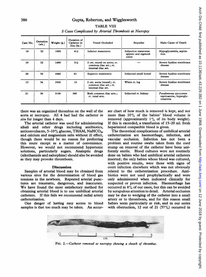
in whom umbilical artery catheters were passed: 5(Table VIII) showed signs of thrombosis of thegreat vessels despite the fact that only one (Case 13)showed any clinical signs before death. In allcases the babies were gravely ill for 12 hours ormore before death, and had signs of generalizedperipheral circulatory failure. All were beingartificially ventilated. In no case was the thrombosisthe major cause of death. In 2 cases there was amural thrombosis within the aorta. In 3 cases abranch of the abdominal aorta was occluded (TableVIII) with infarction of the organ supplied.Although these were not considered to be instru-mental in the babies' deaths they were potentiallyserious complications. In Case 20, an injectionof 50% glucose was given in the terminal stagesfor severe hypoglycaemia when no other route wasavailable.
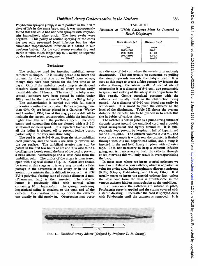
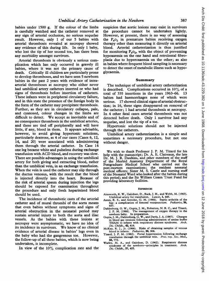
In almost all cases coming to necropsy withcatheters in situ or removed, the umbilical arterywhich had been catheterized was thrombosed, butin only the 3 cases above was there any thrombosisof the iliac vessels. Haemorrhage into the wall ofthe umbilical artery catheterized, or the smallthrombi at the junction of the umbilical and iliacarteries, described by Ainsworth et al. (1963),have not been encountered. In 2 recent cases wehave seen a sheath of thrombus around the tipof the arterial catheter (Fig. 2), which was withinthe aorta at death, and in 2 other catheterized cases
on 12 June 2018 by guest. Protected by copyright.
http://adc.bmj.com
/A
rch Dis C
hild: first published as 10.1136/adc.43.229.382 on 1 June 1968. Dow
nloaded from
386 Gupta, Roberton, and WigglesworthTABLE VIII
5 Cases Complicated by Arterial Thrombosis at Necropsy
Gestation Duration ofCase No. (wk.) Weight (g.) Catheter in Vessel Occluded Sequelae Main Cause of Death
Situ (hr.)
18 32 1000 41 Inferior mesenteric Infarction transverse Hypoglycaemia, aspira-splenic and sigmoid tioncolon
19 32 1490 31k 2 cm. mural on aorta; rt. _ Severe hyaline membranecommon iliac art.; rt. diseaseinternal iliac art.
20 32 1640 61 Superior mesenteric Infarcted small bowel Severe hyaline membranedisease
13 34 1930 10 2 cm. aorta (mural); rt. White rt. leg Severe hyaline membranecommon iliac art.; rt. diseaseinternal iliac art.- -
21 38 3100 340 Both common iliac arts.; Infarcted rt. kidney Pseudomonas pyocyaneart. renal arts. septicaemia; hypergly-
cinaemia
there was an organized thrombus on the wall of theaorta at necropsy. All 4 had had the catheter insitu for longer than 4 days.The arterial catheter was used for administering
alkali and other drugs including antibiotics,anticonvulsaats, 5-10% glucose, THAM, NaHCO3,and calcium and magnesium salts without ill effect,though there would be no reason for preferringthis route except as a matter of convenience.However, we would not recommend hypertonicsolutions, particularly sugars. Analeptic drugs(nikethamide and nalorphine) should also be avoidedas they may provoke thrombosis.
DiscussionSamples of arterial blood may be obtained from
various sites for the determination of blood gastensions in the newborn. Repeated arterial punc-tures are traumatic, dangerous, and inaccurate.We have found the most satisfactory method forobtaining arterial blood is to use umbilical arterialcatheters. If this fails we recommend radial arterycatheterization.One danger of having easy access to blood
samples is that too much may be taken. An accur-
ate chart of how much is removed is kept, and notmore than 10% of the babies' blood volume isremoved (approximately 1% of its body weight).If this is exceeded, a transfusion of 15-20 ml. freshheparinized compatible blood is given.The theoretical complications of umbilical arterial
catheterization are haemorrhage, infection, andvascular occlusion. Infection has not been aproblem and routine swabs taken from the cordstump on removal of the catheter have been uni-formly sterile. Blood cultures were not routinelydone on babies who had umbilical arterial cathetersinserted; the only babies whose blood was cultured,with positive results, were those with signs ofovert infection elsewhere which was not obviouslyrelated to the catheterization procedure. Anti-biotics were not used prophylactically and wereonly administered when indicated clinically forsuspected or proven infection. Haemorrhage hasoccurred in 4% of our cases, but this can be avoidedby scrupulous attention to detail. Arterial occlusionmay be due to wedging of the catheter into a smallartery or to thrombosis, and for this reason smallbabies seem particularly at risk, and in our serieswith obstruction, 12 out of 21 (57%) occurred in
I & i J g-
cm.FIG. 2.-Catheter removed at necropsy showing a sheath of thrombus.
on 12 June 2018 by guest. Protected by copyright.
http://adc.bmj.com
/A
rch Dis C
hild: first published as 10.1136/adc.43.229.382 on 1 June 1968. Dow
nloaded from
Umbilical Artery Catheterization in the Newborn 387
babies under 1500 g. If the colour of the limbsis carefully watched and the catheter removed atany sign of arterial occlusion, no serious sequelaeresult. However, only 1 of our 5 babies witharterial thrombosis revealed at necropsy showedany evidence of this during life. In only 1 baby,who lost the tip of her second toe, has there beenany morbidity amongst survivors.
Arterial thrombosis is obviously a serious com-plication which has only occurred in gravely illbabies, where it was not the primary cause ofdeath. Critically ill children are particularly proneto develop thromboses, and we have seen 5 newbornbabies in the past 2 years with evidence of intra-arterial thromboses at necropsy who either neverhad umbilical artery catheters inserted or who hadsigns of thrombosis before insertion of catheters.These infants were in peripheral circulatory failure,and in this state the presence of the foreign body inthe form of the catheter may precipitate thrombosis.Further, as they are in circulatory collapse, pale,and cyanosed, colour changes in the limbs aredifficult to detect. We accept as inevitable and ofno consequence thrombosis in the umbilical arteries,and these are tied off peripherally and will havelittle, if any, blood in them. It appears advisable,however, to avoid giving hypertonic solutions,particularly dextrose, as 4 of our cases (Cases 6, 12,14, and 20) were associated with administeringthem through the arterial catheter. In Case 14one leg became white and pulseless during exchangetransfusion with ACD blood, and recovery was slow.There are possible advantages in using the umbilicalartery for both giving and extracting blood, ratherthan the umbilical vein, in an exchange transfusion.When the vein is used the catheter may slip throughthe ductus venosus, with the result that the bloodis injected directly into the heart. Because ofthe risk of arterial spasm during injection the legsshould be exposed for examination throughoutthe procedure and only fresh heparinized bloodshould be used.The incidence of thrombotic casts of the arterial
catheter and of mural thrombi of the aorta meansthat even babies without symptoms and signs ofarterial obstruction in the neonatal period maysustain arterial injury to both the aorta and iliacvessels. As the babies with these lesions atnecropsy were asymptomatic, we have no idea ofits incidence in survivors. We know of no clinicalevidence of arterial disease in babies' legs even inthe baby who had the gangrenous toe. However,the follow-up of all these babies, which is now beingundertaken, is incomplete.
In view of the 10% complication rate and the
suspicion that aortic lesions may exist in survivorsthe procedure cannot be undertaken lightly.However, at present, there is no way of assessingthe Pao2 in premature babies receiving oxygentherapy other than measuring it directly on arterialblood. Arterial catheterization is thus justifiedfor monitoring Pao2, with the object of preventinghypoxaemia on the one hand and retrolental fibro-plasia due to hyperoxaemia on the other; as alsoin babies where frequent blood sampling is necessaryto detect the onset ofmetabolic illness such as hypo-glycaemia.
SummaryThe technique of umbilical artery catheterization
is described. Complications occurred in 10% of atotal of 335 insertions in the years 1963-66. 13babies had haemorrhages none of which wereserious. 17 showed clinical signs ofarterial obstruc-tion; in 16, these signs disappeared on removal ofthe catheter; 1 had arterial thrombosis at necropsy.In 4 other fatal cases arterial thrombosis was notdetected before death. Only 1 survivor had anysequelae, and lost the tip of a toe.
Hypertonic solutions should not be injectedthrough the catheters.
Umbilical artery catheterization is a simple andsometimes a necessary procedure, but not onewithout danger.
We wish to thank Professor J. P. M. Tizard for hishelp with the manuscript; Dr. A. E. Claireaux, the lateDr. M. J. R. Dawkins, and other members of the staffof the Morbid Anatomy Department of the RoyalPostgraduate Medical School who carried out thepost-mortem examinations; the resident neonatalmedical officers; Sister M. S. Castle and nursing staffof the Neonatal Ward who looked after the babies duringthis period; and the Sir William Coxen Trust Fund forproviding laboratory facilities.
REFERENCES
Ainsworth, R. W., Gairdner, D., Rack, J. H., and Webb, M. (1963).Umbilical artery for transfusion. Lancet, 1, 445.
Asnes, R. S., and Arendar, G. M. (1966). Septic arthritis of thehip: a complication of femoral venepuncture. Pediatrics, 38,837.
Dahlenburg, G. W., Gupta, J. M., Roberton, N. R. C., and Tizard,J. P. M. (1968). The management of oxygen therapy in thenewborn baby. In preparation.
Gupta, J. M., Dahlenburg, G. W., and Davis, J. A. (1967). Changesin blood gas tensions following administration of amine bufferTHAM to infants with respiratory distress syndrome. Arch.Dis. Childh., 42, 416.
McKay, R. J., Jr. (1966). Risks of obtaining samples of venousblood in infants. Pediatrics, 38, 906.
Tizard, J. P. M. (1962). Portal hypertension following exchangetransfusion through the umbilical vein. Proc. roy. Soc. Med.,55, 772.
Warley, M. A., and Gairdner, D. (1962). Respiratory distresssyndrome of the newborn-principles in treatment. Arch.Dis. Childh., 37, 455.
on 12 June 2018 by guest. Protected by copyright.
http://adc.bmj.com
/A
rch Dis C
hild: first published as 10.1136/adc.43.229.382 on 1 June 1968. Dow
nloaded from