Embed Size (px)
Citation preview
2894
ISSN 2286-4822
www.euacademic.org
EUROPEAN ACADEMIC RESEARCH
Vol. II, Issue 2/ May 2014
Impact Factor: 3.1 (UIF)
DRJI Value: 5.9 (B+)
Ultrastructure of Spleen in the Freshwater Fish,
Tilapia mossambica (Peters)
MEENAKSHI SUNDARESAN1 Department of Zoology
D. G. Ruparel College, Senapati Bapat Marg, Mahim
Mumbai, Maharashtra
India
Abstract:
Spleen is an important centre of blood cell production and
destruction. Fishes, being the first vertebrates to have spleen, a well
organized structure as is evident in higher forms is not expected. In
fish spleen is externally covered by a capsule which is lined by a layer
of cells. Spleen shows presence of reticulocytes cells below the capsule
but no distinct trabeculae within. Interior of spleen is represented by
red and white pulp, veins, sinusoids and arteries. The red and while
pulp regions appear intermixed. Melanomacrophage centres (MMC)
are physiological features found in fish spleen and may contain four
types of pigments namely, melanin, lipofuscin, ceroid and
haemosiderin. Not much work has been done to study the cellular
details of the spleen at the electron microscopic level. The present work
has been carried out to identify the different cells (endothelial cells,
reticulocytes, megakaryocytes, macrophages and cells belonging to the
erythrocytic and leucocytic series) found in the ultrastructure of spleen
of Tilapia mossambica (Peters).
Key words: Histology, ultrastructure, spleen, Tilapia mossambica,
1 Dr. Meenakshi Sundaresan is Associate Professor in Zoology in D.G. Ruparel
College, Mumbai, India. She has been in academics for the past 30 years and
her research interests are cell biology and toxicology. She has done extensive
research on the effect of cadmium on fish and has published five research
papers.

Meenakshi Sundaresan- Ultrastructure of Spleen in the Freshwater Fish, Tilapia
mossambica (Peters)
EUROPEAN ACADEMIC RESEARCH - Vol. II, Issue 2 / May 2014
2895
haemopoietic tissue.
Introduction:
Spleen of vertebrates is concerned with the formation, storage
and destruction of blood corpuscles. In lower vertebrates, spleen
is the only place where haemopoietic tissues assume the
condition of discrete organs (Romer, 1963; Aguius and Roberts
2003). It is also concerned with the defense mechanism against
diseases. So the gland is expected to counter the ill effects of the
pollutants too. The structure of spleen of fish has been
described by Bozidar Kurtovic et al. (2008) in the European sea
bass. There are descriptions of normal histology for some select
species (Yasutake and Wales 1983; Rocha and Monteiro, 1999).
But however there has been no systematic review of the spleen
histology at the cellular level. As fish have no lymph nodes, the
spleen alone plays an essential role in antigen trapping (Press,
1998). Melanomacrophage centres (MMC) are physiological
features in fish spleen and kidney (Aguis and Roberts, 2003).
They are believed to be functional equivalents of the germinal
centers of the spleen and lymph nodes in mammals (Ellis,
1980). MMC may contain four types of pigments- melanin,
lipofuscin ceroid and haemosiderin (Aguis and Agbede, 1984;
Couillard et al 1999). Wolke et al (1985) first suggested MMC as
potential monitors of fish health. Sundaresan (2014) and
Montero et al (1999) found that stressful situations can result
in increased numbers of spleenic and kidney MMC. This study
throws light on the histological structure of spleen through
light and electron microscopy.
Material and Methods:
Live Tilapia fish were obtained from Masunda Lake in Thane
district in Maharashtra, India and were kept for a fortnight for

Meenakshi Sundaresan- Ultrastructure of Spleen in the Freshwater Fish, Tilapia
mossambica (Peters)
EUROPEAN ACADEMIC RESEARCH - Vol. II, Issue 2 / May 2014
2896
laboratory acclimatization. The spleen of ten fish samples were
fixed in 3% glutaraldehyde for 30 minutes at 40 C and processed
for electron microscopy. Ultra thin sections were cut on the
LKB ultramichrotome and picked up on G-200 copper grids.
They were stained for 1 hour with uranyl acetate and counter
stained with lead citrate. Grids were scanned under a Ziess EM
109 electron microscope and JEM Joel 100 ‘S’ Japan make
electron microscope. The haematology of fish reveals variations
in the blood cell types. Hence, determination of cell types was
carried out through blood smears by Giemsa staining (Gurr,
1956) and the blood cells were observed within blood capillaries
and sinuses. Stained sections of gill filaments and their electron
micrographs were also used for the same.
Observations and Discussion:
Histology of spleen - Light Microscopy:
Spleen is covered externally by a thin capsule which consists of
a single layer of cells (occasionally double layered). The
capsular cells are elongated and appear similar to reticulocytes.
The cells are however larger and the cytoplasm is full of
eosinophilic granules. The capsule therefore appears as a pink
coloured lining over the outer extremity in sections stained with
H/E. Nuclei are elongated and stained blue. There are no
distinct trabeculae extending interiorly as are seen in spleen of
mammals; however several of reticulocytes are seen attached to
the inner surface of the capsule.

Meenakshi Sundaresan- Ultrastructure of Spleen in the Freshwater Fish, Tilapia
mossambica (Peters)
EUROPEAN ACADEMIC RESEARCH - Vol. II, Issue 2 / May 2014
2897
Fig. 1. LS of Spleen – Stain H/E Fig. 2. LS of Spleen - Stain H/E
Key
E- erythrocytes, cp – capsule, R – reticulocyte, hmpt – haemopoietic tissue,
cc – central capillary, rp – red pulp, wp – white pulp
The peripheral portion of spleen which is situated immediately
below the capsule is represented exclusively by reticulocytes
while the interior of the spleen is represented by red and white
pulp, veins, sinusoids and arteries (Fig. 1, 2).
The arrangement of red and white pulp is not in an
orderly manner as is observed in the spleen of higher
vertebrates. The two types of tissues therefore are intermixed;
however the presence of the two tissues can be realized by the
differential colour when the sections are stained. In H/E stained
sections white pulp appears light pink while red pulp appears
brownish red. Spleens being concerned with the destruction of
blood cells, haemosiderin deposits are seen at various places.
Such sites are seen as brown pigmented regions.
Arteries:
Splenic arteries wherever present are cut in sections. Such
arteries can be easily differentiated by the presence of
endothelial linings. The endothelial cells are stained deeply.
Sinusoids:
These are the blood filled spaces containing blood and blood

Meenakshi Sundaresan- Ultrastructure of Spleen in the Freshwater Fish, Tilapia
mossambica (Peters)
EUROPEAN ACADEMIC RESEARCH - Vol. II, Issue 2 / May 2014
2898
cells. They are of varying sizes and do not have any distinct
walls but are lined by reticulocytes.
Spleen being concerned with the destruction of blood
cells, several of deformed erythrocytic cells can be seen. Besides
red cells several of macrophages are also seen. These cells can
be distinguished easily form the presence of phagocytic vesicles
most of which are filled with phagocytosed blood cells. The
vesicles appear dark owing to the presence of haemosiderin
within (Fig. 3).
Splenic corpuscles (white pulp):
Spleen of fishes is not as well organized as those of higher
vertebrates, hence the differentiation of red and white pulp was
not distinctly observed as in higher forms. In section of spleen,
most of the area is occupied by reticulocytes and is comparable
to the red pulp of higher vertebrate spleen. White pulp region
represented by several of small spherical corpuscles have been
designated different terms by different authors and the terms
such as ‘ellipsoids’ (Roberts, 1978; Gaikwad, 1981; Awari, 1985;
Gaikwad and Rege, 1990; Awari, 1991), ‘white pulp’ (Guzdar,
1966; Daterao, 1989) and ‘splenic corpuscles’ (Mariano and
Fiore, 1982) are used. In the present work, the term ‘splenic
corpuscles’ has been used.
Each splenic corpuscle consists of a centrally lodged
splenic artery termed as ‘sheathed artery’. The vessel is lined
by endothelial cells. Various stages of leucocytic cells are seen
around the central artery arranged in a concentric manner.
Splenic corpuscles are short and have uniform size with a
diameter of about 25-30 microns. In H/E stained sections
splenic corpuscles are somewhat difficult to locate; however, in
Giemsa stained sections they are easily noticeable.
Reticulocytes: These cells are represented throughout
the splenic tissue. The cells usually have two, at times three or
four cytoplasmic processes extending from the central body. The
Meenakshi Sundaresan- Ultrastructure of Spleen in the Freshwater Fish, Tilapia
mossambica (Peters)
EUROPEAN ACADEMIC RESEARCH - Vol. II, Issue 2 / May 2014
2899
central regions is spherical or oval and appear dark, situated
within this is a roughly spherical nucleus. The cytoplasm is
densely filled with coarse granules which impart dark
colouration to the cells in stained H/E sections. Owing to the
densely filled cytoplasmic granules, it becomes difficult to
distinguish the nucleus from the rest of the cytoplasm. The
ratio between the nucleus to cytoplasm is 1:1.2 to 1.5. The
cytoplasmic processes which extend outward are also full of
cytoplasmic granules that impart dark colouration to the
region.
The cytoplasmic processes of these cells form a network.
Haemopoietic regions are located at various sites within the
network of reticulocytes. Haemopoietic tissue regions are
however much more at the central region of the spleen. The
central region therefore appears dark in stained sections. The
whole of the interior of the spleen being represented by the
closely packed reticulocytes, in light microscopic studies it
becomes very difficult to identify the various types of white
blood cells and their structural details. The erythrocytes,
wherever present can be detected to a certain extent owing to
their oval shape and large size.
Red Pulp:
In the red pulp regions, cells that are observed are mostly the
erythrocytic series, granulocytes and megakaryocytes.
Electron microscopy:
Observation of electron micrographs of spleen reveals the
presence of several sinusoids, red pulp and the white pulp. The
details of various cells that are observed within the spleen are
as under:
(1) Endothelial Cells:
These are highly irregular in shape and they line the blood
vessels. The cytoplasm is drawn out into two or three processes.
Meenakshi Sundaresan- Ultrastructure of Spleen in the Freshwater Fish, Tilapia
mossambica (Peters)
EUROPEAN ACADEMIC RESEARCH - Vol. II, Issue 2 / May 2014
2900
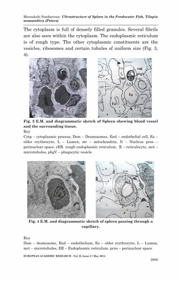
The cytoplasm is full of densely filled granules. Several fibrils
are also seen within the cytoplasm. The endoplasmic reticulum
is of rough type. The other cytoplasmic constituents are the
vesicles, ribosomes and certain tubules of uniform size (Fig. 3,
4).
Fig. 3 E.M. and diagrammatic sketch of Spleen showing blood vessel
and the surrounding tissue.
Key
Cytp – cytoplasmic process, Dsm – Desmosomes, End – endothelial cell, Eo –
older erythrocyte, L – Lumen, mt – mitochondria, N – Nucleus prns –
perinuclear space, rER- rough endoplasmic reticulum, R – reticulocyte, mct –
microtubules, phgV – phagocytic vesicle
Fig. 4 E.M. and diagrammatic sketch of spleen passing through a
capillary.
Key
Dsm – desmosome, End – endothelium, Eo – older erythrocyte, L – Lumen,
mct – microtubules, ER – Endoplasmic reticulum, prns – perinuclear space
phgV

Meenakshi Sundaresan- Ultrastructure of Spleen in the Freshwater Fish, Tilapia
mossambica (Peters)
EUROPEAN ACADEMIC RESEARCH - Vol. II, Issue 2 / May 2014
2901
The cell nucleus is spherical or irregularly oval and has more or
less uniformly distributed chromatin material. The cell nuclei
with the surrounding cytoplasm are often extended into the
lumen of the capillary (Fig 3).
The endothelial cells are little difficult to locate for the
reason that the cytoplasmic processes are too long and
irregular. In any electron micrograph, therefore only a part of
cytoplasmic process may be represented. These cytoplasmic
processes can be detected by the presence of characteristic
desmosomes between the two neighbouring cells.
(2) Reticulocytes:
These are the elongated cells that taper at the two extremities
of the cells. The cells are characterized by the presence of a
large prominent nucleus. The cytoplasm is highly granular and
has clusters of fibrous material. Such clusters are held parallel
to the plasma membrane. Mitochondria are few and are lodged
around the nucleus.
Each cell has a large nucleus that tapers at the two
ends. The chromatin is of heterochromatin type (Fig 5, 6).
Fig 5 EM and diagrammatic sketch of spleen showing various types of
cells.
Key
E1 – erthrocytic stage, Eo – older erythrocyte, Enp – endothelial cell process,
L – lymphocyte, N – neutrophil, Mgk – Megakaryocyte, M – Monocyte, R –

Meenakshi Sundaresan- Ultrastructure of Spleen in the Freshwater Fish, Tilapia
mossambica (Peters)
EUROPEAN ACADEMIC RESEARCH - Vol. II, Issue 2 / May 2014
2902
Reticulocyte, Mc - Macrophage
Fig 6 EM of spleen showing single Reticulocyte enlarged.
Key
Mt – mitochondria, rER – rough Endoplasmic Reticulum, R – reticulocyte
(3) Megakaryocytes:
These are the large cells and each is characterized by the
presence of a large nucleus. Young cells are oval while fully
formed ones may assume spherical or irregular shape. The
cytoplasm is densely filled with fine granules. Mitochondria are
small and spherical in shape.
Nucleus of the cells is oval or round depending upon the
shape of the cell. The chromatin is spread over more or less
evenly Fig. 5).
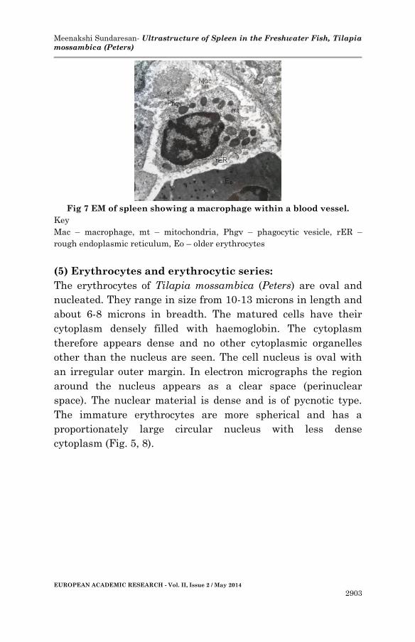
(4) Macrophages:
These cells have irregular boundaries. The cell nucleus is
comparatively small and is usually eccentric in position. The
cytoplasm is full of granules and a network of endoplasmic
reticulum is seen. The mitochondria are oval in shape. The cells
have a large number of lysosomes and phagosomes because of
which, they can be easily be recognized. The cells are often seen
with devoured RBCs or other cell particles. The cells have
several other vesicles and vacuoles too (Fig 7).
Meenakshi Sundaresan- Ultrastructure of Spleen in the Freshwater Fish, Tilapia
mossambica (Peters)
EUROPEAN ACADEMIC RESEARCH - Vol. II, Issue 2 / May 2014
2903
Fig 7 EM of spleen showing a macrophage within a blood vessel.
Key
Mac – macrophage, mt – mitochondria, Phgv – phagocytic vesicle, rER –
rough endoplasmic reticulum, Eo – older erythrocytes
(5) Erythrocytes and erythrocytic series:
The erythrocytes of Tilapia mossambica (Peters) are oval and
nucleated. They range in size from 10-13 microns in length and
about 6-8 microns in breadth. The matured cells have their
cytoplasm densely filled with haemoglobin. The cytoplasm
therefore appears dense and no other cytoplasmic organelles
other than the nucleus are seen. The cell nucleus is oval with
an irregular outer margin. In electron micrographs the region
around the nucleus appears as a clear space (perinuclear
space). The nuclear material is dense and is of pycnotic type.
The immature erythrocytes are more spherical and has a
proportionately large circular nucleus with less dense
cytoplasm (Fig. 5, 8).
Meenakshi Sundaresan- Ultrastructure of Spleen in the Freshwater Fish, Tilapia
mossambica (Peters)
EUROPEAN ACADEMIC RESEARCH - Vol. II, Issue 2 / May 2014
2904
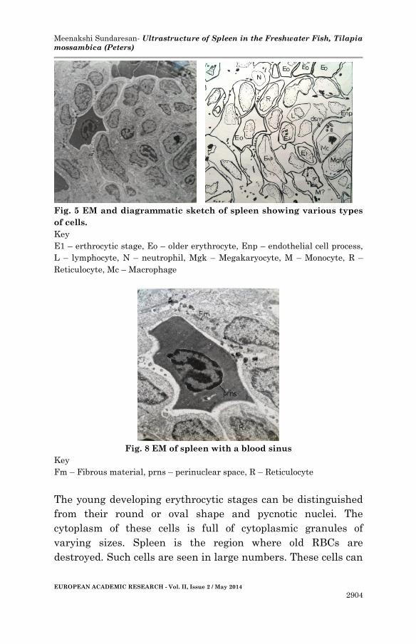
Fig. 5 EM and diagrammatic sketch of spleen showing various types
of cells.
Key
E1 – erthrocytic stage, Eo – older erythrocyte, Enp – endothelial cell process,
L – lymphocyte, N – neutrophil, Mgk – Megakaryocyte, M – Monocyte, R –
Reticulocyte, Mc – Macrophage
Fig. 8 EM of spleen with a blood sinus
Key
Fm – Fibrous material, prns – perinuclear space, R – Reticulocyte
The young developing erythrocytic stages can be distinguished
from their round or oval shape and pycnotic nuclei. The
cytoplasm of these cells is full of cytoplasmic granules of
varying sizes. Spleen is the region where old RBCs are
destroyed. Such cells are seen in large numbers. These cells can

Meenakshi Sundaresan- Ultrastructure of Spleen in the Freshwater Fish, Tilapia
mossambica (Peters)
EUROPEAN ACADEMIC RESEARCH - Vol. II, Issue 2 / May 2014
2905
easily be distinguished from other cells of erythrocytic series by
their highly irregular shape.
Leucocytic series: Leucocytes are represented by neutrophils
and lymphocytes.
Neutrophils: These are roughly irregular cells with clear
cytoplasm. The presence of clear cytoplasm helps to recognize
cells. The cytoplasmic granules are fine and are uniformly
distributed. The cell nucleus is large and spherical. The
chromatin material is uniformly distributed (Fig. 5).
Lymphocytes: These cells are comparatively small and have
an irregular shape. The nucleus is large and spherical.
Cytoplasm is restricted to peripheral extremity. (Fig. 5).
There is no unananimity pertaining to the types of fish
leucocytes. While many have reported the presence of all
leucocytic cell types in fishes, Yokote (1982) and Pai Vinaya
(1993) puts forth the leucocytic cells of fishes to be of only two
types viz. the neutrophils and the lymphocytes. In Tilapia
mossambica, the blood smears show presence of erythrocytes
(mature, immature and abnormal), smudged cells, lymphocytes,
neutrophils and thrombocytes. These cells were also clearly
seen in the electron micrographs of gill lamellae. (Sundaresan
et al 2009).
Acknowledgements
Author is grateful to Dr. S. V. Shanbhag for providing valuable
guidance and encouragement for this research. I am also grateful to
Dr. Gayathri N. for critical review of the paper. Laboratory facility
provided by the Principal of Ramnarain Ruia College, Matunga is also
appreciated.
Meenakshi Sundaresan- Ultrastructure of Spleen in the Freshwater Fish, Tilapia
mossambica (Peters)
EUROPEAN ACADEMIC RESEARCH - Vol. II, Issue 2 / May 2014
2906
REFERENCES
Aguis, C. and S. A. Agbede. 1984. “An electron microscopical
study on the genesis of lipofuscin, melanin and
haemosiderin in the haemopoietic tissues of fish.”
Journal of fish biology 24: 471-488.
Aqius, C. and R. J. Roberts. 2003. “Melanomacrophage centers
and their role in fish pathology.” J. Fish Dis. 26: 499-
509.
Awari Subhash B. 1985. Toxic effects of cadmium on edible fish
Ambassis ranga (Hamilton buchanan) M.Sc. Thesis
University of Bombay.
Awari Subhash B. 1991. Toxicity and Depuration studies of
cadmium on Ambassis ranga (Cuvier valenciennes) with
reference to variations in environmental and some
biochemical parameters. Ph.D. Thesis. University of
Mumbai.
Basim, M. Jasim. 2008. “Effects of prolonged exposure to
cadmium on the haematopoietic organs in grass carp
(Ctenopharyngodon idella, Cyprinidae).” Bas. J. Vet.,
Res. 7(2): 108-120.
Bozidar, Kurtovic, Emin Teskeredzic and Zlatica Teskeredzic.
2008. “Histological comparison of spleen and kidney
tissues from farmed and wild European Sea bass
(Dicentrarchus labrax L.)” Acta Adrait. 49(20): 147-154.
Couillard, C. M., P. J. Williams, S. C. Courtenay and G. P.
Rawn. 1999. “Histopathological evaluation of Atlantic
tomcod (Microgadus tomcod) collected at estuarine sites
receiving pulp and paper mill effluent.” Aquatic
toxicology 44: 263-278.
Daterao, M. S. 1989. Effect of certain heavy metal salts on the
physiology of the fish Tilapia mossambica (Peters) alias
Saratherodon mossambicus (Trevawas) Ph.D. Thesis,
University of Mumbai.
Ellis, A. E. 1980. “Antigen trapping in the spleen and kidney of

Meenakshi Sundaresan- Ultrastructure of Spleen in the Freshwater Fish, Tilapia
mossambica (Peters)
EUROPEAN ACADEMIC RESEARCH - Vol. II, Issue 2 / May 2014
2907
the plaice (Pleuronectes platessa L.)” J. Fish Dis. 3: 413-
426.
Gaikwad, S. A. and Rege, M. S. 1990. “Effects of chronic
exposure to pesticides, Thiodon 35 EC and Phenyl
mercuric acetate on the various tissues of Tilapia
mossambica (Peters).” Environ. Concern and Tissue
injury. Vol. IV: 179-192.
Gaikwad, S. A. 1981. Toxicity studies with Thiodon35 EC and
Phenyl mercuric acetate on Tilapia mossambica (Peters)
Ph.D. Thesis, University of Mumbai.
Gurr, E. 1956. A practical manual of medical and biological
staining techniques. London: Leonard Hill (Books) Ltd.
Guzdar, G. S. 1966. Haemopoesis in certain fishes. M.Sc. Thesis,
University of Mumbai.
Mariano, S. H. di Fiore. 1982. Atlas of Human histology. 5th
Edition. Philadelphia: Lea and Febiger.
Montero, D., V. S. Blazer, J. Socorro, M. S. Izquierdo, and L.
Tort. 1999. “Dietary and culture influences on
macrophage aggregate parameters in gilthead seabream
(Sparus aurata) juveniles.” Aquaculture 179: 523-534.
Oguri, M. 1985. “Pigment granules in the renal interstitial
tissue of marine teleosts.” Bulletin of the Japanese
Society of Scientific Fisheries 51(9): 1447-1449.
Pai, Vinaya. 1988. On haematological aspects of two puffer
fishes Tetradon oblongus (Bl,), Tetradon lunaris (Bl.
Schn.). M. Phil Thesis submitted to University of
Bombay.
Press, C. M. 1998. “Immunology of fishes.” In Handbook of
vertebrate immunology, edited by P. P. Pastoret, P.
Griegel, H. Bazin and A. Govaerts, 3-62. San Diego:
Academic Press.
Roberts, Ronald. 1978. Fish Pathology. Cassel Ltd. 35 Red Lion
Square, London WCIR, ISG.
Roberts, R. J., 2001. Fish Pathology. 3rd edition. Philadelphia:
W. B. Saunders, 367 pp.
Meenakshi Sundaresan- Ultrastructure of Spleen in the Freshwater Fish, Tilapia
mossambica (Peters)
EUROPEAN ACADEMIC RESEARCH - Vol. II, Issue 2 / May 2014
2908
Rocha, E. and A. F. Monteiro. 1999. “Histology and Cytology of
Fish Liver.” A review In Ichthyology; Research Advances,
edited by D. N. Saksena, 321-344. Science Publishers.
Romer, Alfred Shrewood. 1955. The Vertebrate Body.
Philadelphia and London: W. B. Saunders Company.
Ruparelia, S. G., Verma Yogendra, Kashyap S. K. and
Chatterjee B. B. 1986. “A new approach for the use of
standard fish in the toxicological study.” Environmental
Biology, Coastal ecosystem. 89-92: (c) The academy of
Environmental Biology, India.
Sundaresan, Meenakshi and Shanbhag, S.V. 2009.
“Ultrastructural details of the inter lamaller region in
the gill of Tilapia mossambica (Peters).” J. Aqua. Biol.,
24(2): 160 – 168
Sundaresan, Meenakshi. 2014. “Cadmium induced histological
& histochemical changes in the spleen of freshwater fish,
Tilapia mossambica (Peters).” Indian Streams Research
Journal 4(3).
Wolke, R. E. G., C. J. George and V. S. Blazer. 1985.
“Pigmented macrophage accumulation (MMC, PMB):
possible monitors of fish health.” In NOAA Technical
Report NMFS. 25, edited by W. J. Hargis, 93-97.
Washington D. C.
Yasutake, W. T. and J. H. Wales. 1983. “Microscopic anatomy of
Salmonids: an atlas.” In U. S. Fish and Wildlife
Resource Publication, edited by W.T. Yasutake and J.H.
Wales, 150.
Yokote, M. 1982. An Atlas of fish histology; Chapter IV – Blood,
pp. 64-72. Tokyo: Kodansha Ltd.