Embed Size (px)
Citation preview

Jane —Ultrastructure of endosperm development in Arundo formosana 69Bot. Bull. Acad. Sin. (2004) 45: 69-85
Ultrastructure of endosperm development in Arundo formosanaHack. (Poaceae) from differentiation to maturity
Wann-Neng Jane
Electron Microscopy Lab, Institute of Botany, Academia Sinica, Taipei, Taiwan
(Received January 7, 2003; Accepted August 27, 2003)
Abstract. Endosperm development of Arundo formosana Hack. is examined ultrastructurally and histochemicallyfrom differentiation to seed maturity. The differentiated endosperm contains four major cell types: the cells of theembryo surrounding region, transfer cells, aleurone layer, and starchy endosperm. After cellularization, cells of theembryo surrounding region and transfer cells contain dense cytoplasm and large quantities of rough endoplasmic reticu-lum (RER) and many dictyosomes. In the transfer cells, the wall ingrowths are present on the walls adjacent to thenucellus. The embryo surrounding region is distributed on the ventral side and surrounds the suspensor. After leafprimordium initiation, most cells of the embryo surrounding region have degenerated, but the outermost layer of theventral side becomes part of the aleurone layer. The transfer cells differentiate into outer and inner cells. The outertransfer cells contain electron-dense cytoplasm, many oil bodies, large quantities of RER, and dictyosomes. Thesecells appear PAS-positive, especially in the thickening walls. The inner transfer cells have electron-transparentcytoplasm, some small vacuoles, organelles, and oil bodies. The outer transfer cells are finally compressed anddegenerated. The inner cells contain PAS-positive thickening walls, many oil bodies, protein bodies, and RER atmaturity. The starchy endosperm cells gradually accumulate reserves (mainly proteins and starch) during development,and are filled with amyloplasts and protein bodies at maturity. The protein bodies are of two types that originate inthe RER and are then stored in the cisternal lumen of RER, and vacuoles, respectively. The outermost layer of theendosperm becomes the aleurone layer, except for the transfer cell region. The contents of the aleurone cells areinitially similar to other starchy endosperm cells, and are finally filled with aleurone grains surrounded by many smalllipid bodies. The aleurone grains are initiated from ER and accumulate in vacuoles.
Keywords: Aleurone grain; Aleurone layer; Arundo formosana Hack.; Embryo surrounding region; Endospermdevelopment; Protein body; Starchy endosperm; Transfer cell.
Introduction
A general model for the development of cereal en-dosperm (Olsen et al., 1995; Olsen et al., 1998; Olsen etal., 1999) recognizes four major stages: syncytial,cellularization, differentiation, and maturation. Aftercellularization, the endosperm differentiates into two celltypes in Triticum (Smart and O’Brien, 1983) and Zea (Schelet al., 1984), one with dense cytoplasm and the other withlarge vacuoles. Smart and O’Brien (1983) termed the formermodified endosperm and the later general endosperm.Large quantities of RER are characteristic of the modifiedendosperm. The wal l ingrowths occur in theplacentochalazal region of Zea (Schel et al., 1984), but notin that of Triticum (Smart and O’Brien, 1983). Beyond theabsorption and transport function of the endosperm, a highdegree of synthesis also takes place in the modified en-dosperm (Schel et al., 1984).
According to the mechanisms of nuclear endospermcellularization and development in cereals, Olsen et al.
*Corresponding author. Tel: 886-2-27899590 ext.110; Fax: 886-2-27827954; E-mail: [email protected]
(1999) and Olsen (2001) suggested that the differentiatedcereal endosperm is composed of four cell-types: the em-bryo surrounding region, transfer cells, the starchyendosperm, and the aleurone layer. The underlying geneticprograms for cell fate specification probably originated asindependent genetic programs (Olsen, 2001).
In a previous paper, I described the embryo develop-ment of Arundo in detail (Jane, 1999). In this study, I willobserve endosperm development from differentiation tomaturation and discuss the relationship of endosperm andembryo development.
Material and Methods
Grains (caryopses) of Arundo formosana Hack., an en-demic grass in Taiwan, were collected from Pingshi andWulai of Taipei county. The voucher specimens, identifi-cation number 229386-229391, are deposited in the Her-barium of Department of Botany, National TaiwanUniversity. Whole grains were fixed in 2.5% glutaraldehydein 0.1 M sodium phosphate buffer, pH 7.2 at 4°C overnight.After three 20 min buffer rinses, the grains were postfixedin 1% OsO4 in the same buffer for 4 h at room tempera-ture and then rinsed in three 20 min changes of buffer. The

70 Botanical Bulletin of Academia Sinica, Vol. 45, 2004
grains were dehydrated in an acetone series, embedded inSpurr’s resin (Spurr, 1969), and sectioned with a LeicaUltracut E ultramicrotome. Semi-thin sections (1 µm) forlight microscopy were placed on slides and stained with0.1% toluidine blue for 1 min at 60°C on a hot plate. His-tochemical tests included 0.3% Sudan Black B in 70% al-cohol for lipids (Bronner, 1975), periodic acid-Schiff (PAS)reaction for polysaccharides (Gahan, 1984), and 1%Coomassie Brilliant Blue in 7% acetic acid for proteins(Gahan, 1984). Thin sections on grids were stained withuranyl acetate and lead citrate (Reynolds, 1963) and stud-ied with Philips CM 100 or JEM 1200 EX II transmissionelectron microscope at 80 KV.
Results
After cellularization, most of the endosperm cells areoccupied by large vacuoles except for those surroundingthe embryo and in the placentochalazal region. The en-dosperm surrounding the embryo and distributed in theplacentochalazal region contains abundant cellular con-tents classified into embryo surrounding region and trans-fer cell region, respectively (Figure 1A). The endospermwith large vacuoles is starchy endosperm. Followingdevelopment, the embryo surrounding region and thetransfer cell region gradually increase in cell number andcell layer (Figure 1B). The cells of the embryo surround-ing region (Figure 1C-D) and the transfer cells (Figure 2A-B) display dense cytoplasm, large nuclei, an abundance ofER, and many dictyosomes. In these cells, the mitochon-dria have well-developed cristae. The plastids are devoidof starch grains, and the dictyosomes display many smallvesicles (Figure 1D, 2C). In the transfer cells, the wall in-growths are present on the walls adjacent to the nucellus(Figure 2C) and the tangential walls. The degree of wallingrowths increases from lower cells (Figure 2A) to uppercells (Figure 2B). In the starchy endosperm cells, most ofthe cell lumen is occupied by vacuoles, and most of theorganelles are distributed in the perinuclear region (Figure2D).
In subsequent development, the embryo surroundingregion and the transfer cell region appear differentiated inthe cell constitution, and the cells in the middle region ofthe starchy endosperm begin to accumulate starch. Whenan embryo has differentiated into embryo proper andsuspensor, the embryo surrounding region with an abun-dance of cell contents is mainly distributed on the ventralside and in the surrounding suspensor (Figure 3A). Theendosperm cells surrounding the embryo proper are com-pressed and broken, and some regions appear PAS-posi-tive (Figure 3B). Two parts of the transfer cell region canbe distinguished: the outermost layer and the inner cells(Figure 3A). The transfer cells of the outermost layer ap-pear PAS-positive, especially the thickening walls (Figure3C), and contain many lipid bodies (Figure 3E) and an abun-dance of proteins (Figure 3G). Compared to the outermostlayer, the inner transfer cells contain less polysaccharideand proteins (Figure 3C-G), but have some lipid bodies(Figure 3E). The starchy endosperm accumulates some
storage reserves, including starch grains (Figure 3D), lipidbodies (Figure 3F), and protein bodies (Figure 3H). Thestarch grains are stored in plastids forming amyloplasts,and the protein bodies are formed in cytoplasm and vacu-oles (Figure 3H). The accumulation of the storage reservesis mainly in the upper half of the starchy endosperm(Figure 3A). The first reserve formed is starch, followedby lipids, and finally proteins. The outermost layer of theendoperm, except for the transfer cell region, is aleuroneinitials. The starch grains and protein bodies are seldomfound in aleurone initials (Figure 3D), in which some lipidbodies are present (Figure 3F). In general, the aleurone ini-tials in the dorsal side are larger than those in the ventralside (Figure 3A). The aleurone initials undergo periclinaldivision, and the outermost cells become aleurone cells,and the inner cells become starchy endosperm cells (Figure3D).
Large quantities of RER and dictyosomes are charac-teristic of cells in the embryo surrounding region (Figure4A) and in the outermost layer of the transfer cell region(Figure 4B). In these cells, RER is usually arranged paral-lel with the cell wall. The cells in the outermost layer ofthe transfer cell region have electron-dense cytoplasm(Figure 4B). Many lipid bodies, some plastids and mito-chondria are also present in theses cells (Figure 4B). Theinner cells of the transfer cell region have electron-trans-parent cytoplasm containing fewer RER, dictyosomes,plastids, mitochondria, lipid bodies, and more small vacu-oles (Figure 4C) than the outermost cells. The upper halfof the starchy endosperm contains many storage reserves,especially starch grains. In these cells, the protein bodiesare initiated in the cisterae of RER (Figure 4D).
When the embryo undergoes coleoptile initiation, thealeurone layer is apparent in the outermost layer of theendosperm, except for the transfer cell region (Figure 5A).The vacuolation of the cells of the embryo surroundingregion increases, especially in the ventral region. Somecells of the embryo surrounding region in the dorsal re-gion still contain large quantities of RER (Figure 5C). Thecytoplasm of the outer cells of the transfer cell region ap-pear denser (Figure 5A) than in the previous stage. Theouter cells of the transfer cell region contain electron-densecontents, many lipid bodies, and thickening walls (Figure5D). In these cells, the organelles are difficult todistinguish. In the inner cells of the transfer cell region,some plastids, mitochondria, dictyosomes, ER, proteinbodies, and many vacuoles are present in the cytoplasm(Figure 5B).
In the starchy endosperm cells, each nucleus is sur-rounded by many large amyloplasts and protein bodies(Figure 6A). The protein bodies have two types: Type I(PB1) is enlarged in the cisteral lumen of RER, and type II(PB2) is accumulated in vacuoles (Figure 6B). The ventralaleurone cells (Figure 6C) are smaller than the dorsal ones(Figure 6D). The aleurone cells contain some organellesand lipid bodies, but fewer starch grains (Figure 6C, 6D).
In subsequent development, most cells of the embryosurrounding region are degenerated, but the outermost

Jane —Ultrastructure of endosperm development in Arundo formosana 71
Figure 1. Longitudinal section of developing caryopses. A, The cells of the embryo surrounding region ( ) and the transfer cellregion ( ) are rich in cell contents. The cells of the starchy endosperm are occupied by large vacuoles; B, The embryo surroundingregion ( ), the transfer cell region ( ) and the starchy endosperm increase in cell number during development; C, The cells of theembryo surrounding region in the dorsal region contain many organelles, including large nucleus, plastids, mitochondria, small vacuoles,ER, and dictyosomes; D, The cell of the embryo surrounding region in the ventral region showing cell contents. The plastids containdense matrix, and the mitochondria have well-developed cristae. The dictyosomes are active in producing vesicles. AP: Antipodalcell; C: Chromosome; Ch: Chalaza; D: Dictyosome; Em: Embryo; ER: Endoplasmic reticulum; M: Mitochondrion; N: Nucleus; NC:Nucellus; P: Plastid; RER: Rough endoplasmic reticulum; SE: Starchy endosperm; V: Vacuole.
72 Botanical Bulletin of Academia Sinica, Vol. 45, 2004
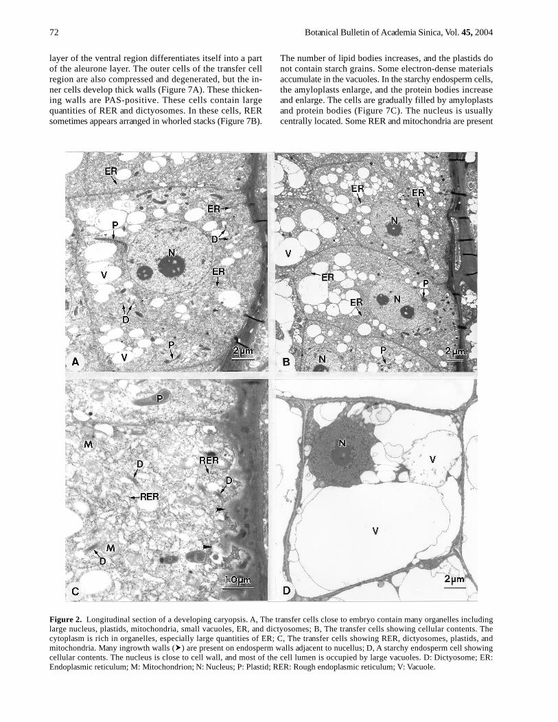
Figure 2. Longitudinal section of a developing caryopsis. A, The transfer cells close to embryo contain many organelles includinglarge nucleus, plastids, mitochondria, small vacuoles, ER, and dictyosomes; B, The transfer cells showing cellular contents. Thecytoplasm is rich in organelles, especially large quantities of ER; C, The transfer cells showing RER, dictyosomes, plastids, andmitochondria. Many ingrowth walls ( ) are present on endosperm walls adjacent to nucellus; D, A starchy endosperm cell showingcellular contents. The nucleus is close to cell wall, and most of the cell lumen is occupied by large vacuoles. D: Dictyosome; ER:Endoplasmic reticulum; M: Mitochondrion; N: Nucleus; P: Plastid; RER: Rough endoplasmic reticulum; V: Vacuole.
layer of the ventral region differentiates itself into a partof the aleurone layer. The outer cells of the transfer cellregion are also compressed and degenerated, but the in-ner cells develop thick walls (Figure 7A). These thicken-ing walls are PAS-positive. These cells contain largequantities of RER and dictyosomes. In these cells, RERsometimes appears arranged in whorled stacks (Figure 7B).
The number of lipid bodies increases, and the plastids donot contain starch grains. Some electron-dense materialsaccumulate in the vacuoles. In the starchy endosperm cells,the amyloplasts enlarge, and the protein bodies increaseand enlarge. The cells are gradually filled by amyloplastsand protein bodies (Figure 7C). The nucleus is usuallycentrally located. Some RER and mitochondria are present
Jane —Ultrastructure of endosperm development in Arundo formosana 73
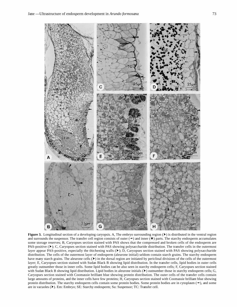
Figure 3. Longitudinal section of a developing caryopsis. A, The embryo surrounding region ( ) is distributed in the ventral regionand surrounds the suspensor. The transfer cell region consists of outer ( ) and inner ( ) parts. The starchy endosperm accumulatessome storage reserves; B, Caryopses section stained with PAS shows that the compressed and broken cells of the endosperm arePAS-positive ( ); C, Caryopses section stained with PAS showing polysaccharide distribution. The transfer cells in the outermostlayer appear PAS-positive, especially the thichening walls ( ); D, Caryopses section stained with PAS showing polysaccharidedistribution. The cells of the outermost layer of endosperm (aleurone initial) seldom contain starch grains. The starchy endospermhave many starch grains. The aleurone cells ( ) in the dorsal region are initiated by periclinal divisions of the cells of the outermostlayer; E, Caryopses section stained with Sudan Black B showing lipid distribution. In the transfer cells, lipid bodies in outer cellsgreatly outnumber those in inner cells. Some lipid bodies can be also seen in starchy endosperm cells; F, Caryopses section stainedwith Sudan Black B showing lipid distribution. Lipid bodies in aleurone initials ( ) outnumber those in starchy endosperm cells; G,Caryopses section stained with Coomassie brilliant blue showing protein distribution. The outer cells of the transfer cells containlarge amounts of proteins, and the inner cells have few proteins; H, Caryopses section stained with Coomassie brilliant blue showingprotein distribution. The starchy endosperm cells contain some protein bodies. Some protein bodies are in cytoplasm ( ), and someare in vacuoles ( ). Em: Embryo; SE: Starchy endosperm; Su: Suspensor; TC: Transfer cell.
74 Botanical Bulletin of Academia Sinica, Vol. 45, 2004
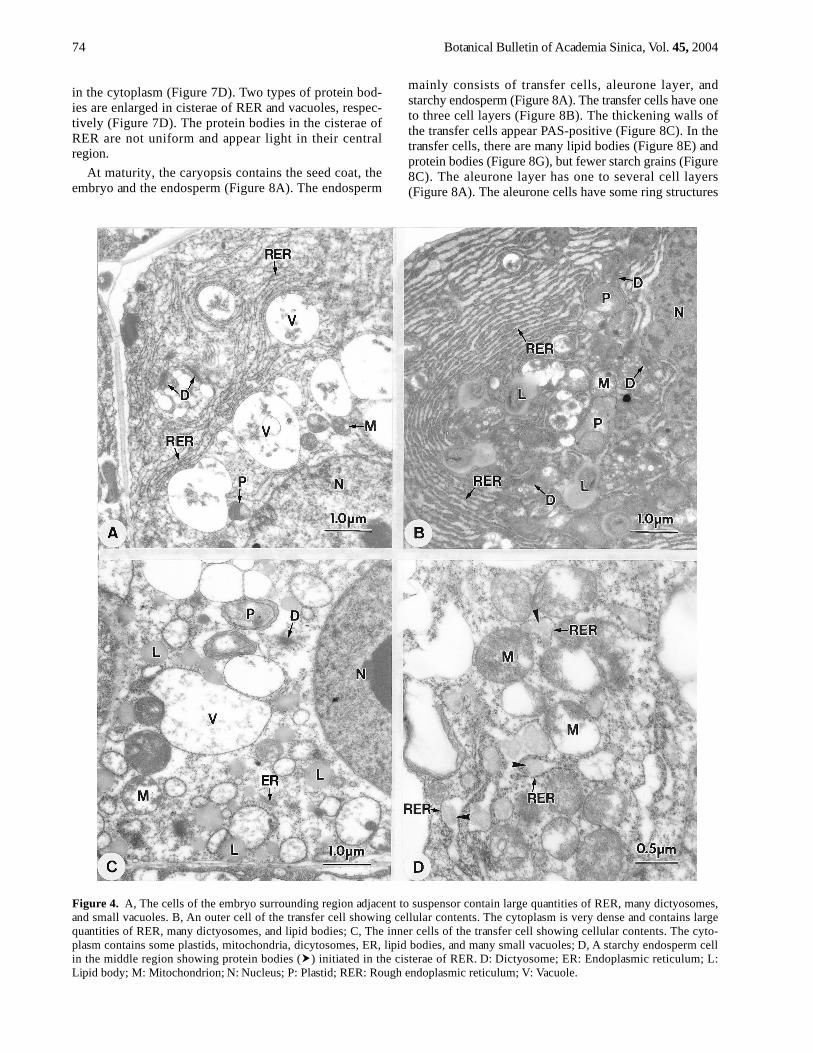
Figure 4. A, The cells of the embryo surrounding region adjacent to suspensor contain large quantities of RER, many dictyosomes,and small vacuoles. B, An outer cell of the transfer cell showing cellular contents. The cytoplasm is very dense and contains largequantities of RER, many dictyosomes, and lipid bodies; C, The inner cells of the transfer cell showing cellular contents. The cyto-plasm contains some plastids, mitochondria, dicytosomes, ER, lipid bodies, and many small vacuoles; D, A starchy endosperm cellin the middle region showing protein bodies ( ) initiated in the cisterae of RER. D: Dictyosome; ER: Endoplasmic reticulum; L:Lipid body; M: Mitochondrion; N: Nucleus; P: Plastid; RER: Rough endoplasmic reticulum; V: Vacuole.
in the cytoplasm (Figure 7D). Two types of protein bod-ies are enlarged in cisterae of RER and vacuoles, respec-tively (Figure 7D). The protein bodies in the cisterae ofRER are not uniform and appear light in their centralregion.
At maturity, the caryopsis contains the seed coat, theembryo and the endosperm (Figure 8A). The endosperm
mainly consists of transfer cells, aleurone layer, andstarchy endosperm (Figure 8A). The transfer cells have oneto three cell layers (Figure 8B). The thickening walls ofthe transfer cells appear PAS-positive (Figure 8C). In thetransfer cells, there are many lipid bodies (Figure 8E) andprotein bodies (Figure 8G), but fewer starch grains (Figure8C). The aleurone layer has one to several cell layers(Figure 8A). The aleurone cells have some ring structures

Jane —Ultrastructure of endosperm development in Arundo formosana 75
Figure 5. Longitudinal section of a developing caryopsis during coleoptile initiation. A, The vacuolation of the embryo surroundingregion increases, especially on ventral side. The outer cells of the transfer cell region ( ) contain dense cytoplasm and the inner cells( ) is high vacuolation. The aleurone cells ( ) of the ventral region are initiated by periclinal divisions of the cells in the outermostlayer of the endosperm. The starchy endosperm accumulates more storage reserves; B, The inner cells of the transfer cells showingcellular contents. Many vacuoles, some plastids, mitochondria, dictyosomes, ER, and lipid bodies are present in the cells. Someprotein bodies can be found in the cistae of ER. The cell walls thicken ( ); C, The cells of the embryo surrounding region showingcellular contents. Large quantities of RER are present in cytoplasm; D, The outer cells of the transfer cells showing cellular contents.The cytoplasm with many lipid bodies is very electron-dense, and the thick walls ( ) are electron-lucent. AL: Aleurone layer; Ch:Chalaza; D: Dictyosome; Em: Embryo; ER: Endoplasmic reticulum; L: Lipid body; M: Mitochondrion; N: Nucleus; P: Plastid; RER:Rough endoplasmic reticulum; SE: Starchy endosperm; Su: Suspensor; V: Vacuole.
76 Botanical Bulletin of Academia Sinica, Vol. 45, 2004
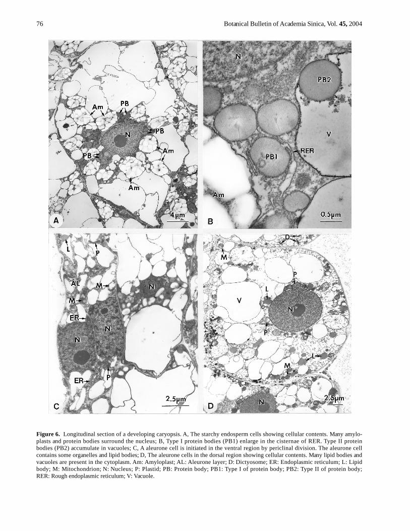
Figure 6. Longitudinal section of a developing caryopsis. A, The starchy endosperm cells showing cellular contents. Many amylo-plasts and protein bodies surround the nucleus; B, Type I protein bodies (PB1) enlarge in the cisternae of RER. Type II proteinbodies (PB2) accumulate in vacuoles; C, A aleurone cell is initiated in the ventral region by periclinal division. The aleurone cellcontains some organelles and lipid bodies; D, The aleurone cells in the dorsal region showing cellular contents. Many lipid bodies andvacuoles are present in the cytoplasm. Am: Amyloplast; AL: Aleurone layer; D: Dictyosome; ER: Endoplasmic reticulum; L: Lipidbody; M: Mitochondrion; N: Nucleus; P: Plastid; PB: Protein body; PB1: Type I of protein body; PB2: Type II of protein body;RER: Rough endoplasmic reticulum; V: Vacuole.
Jane —Ultrastructure of endosperm development in Arundo formosana 77
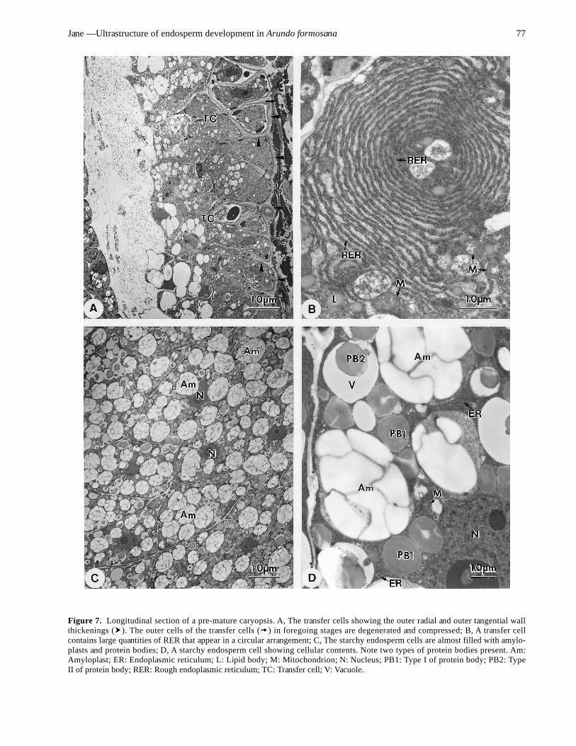
Figure 7. Longitudinal section of a pre-mature caryopsis. A, The transfer cells showing the outer radial and outer tangential wallthickenings ( ). The outer cells of the transfer cells ( ) in foregoing stages are degenerated and compressed; B, A transfer cellcontains large quantities of RER that appear in a circular arrangement; C, The starchy endosperm cells are almost filled with amylo-plasts and protein bodies; D, A starchy endosperm cell showing cellular contents. Note two types of protein bodies present. Am:Amyloplast; ER: Endoplasmic reticulum; L: Lipid body; M: Mitochondrion; N: Nucleus; PB1: Type I of protein body; PB2: TypeII of protein body; RER: Rough endoplasmic reticulum; TC: Transfer cell; V: Vacuole.
78 Botanical Bulletin of Academia Sinica, Vol. 45, 2004
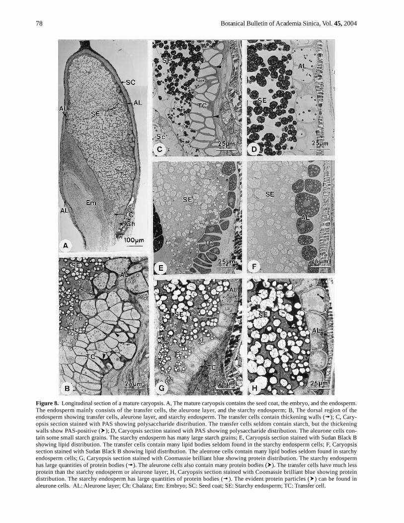
Figure 8. Longitudinal section of a mature caryopsis. A, The mature caryopsis contains the seed coat, the embryo, and the endosperm.The endosperm mainly consists of the transfer cells, the aleurone layer, and the starchy endosperm; B, The dorsal region of theendosperm showing transfer cells, aleurone layer, and starchy endosperm. The transfer cells contain thickening walls ( ); C, Cary-opsis section stained with PAS showing polysaccharide distribution. The transfer cells seldom contain starch, but the thickeningwalls show PAS-positive ( ); D, Caryopsis section stained with PAS showing polysaccharide distribution. The aleurone cells con-tain some small starch grains. The starchy endosperm has many large starch grains; E, Caryopsis section stained with Sudan Black Bshowing lipid distribution. The transfer cells contain many lipid bodies seldom found in the starchy endosperm cells; F, Caryopsissection stained with Sudan Black B showing lipid distribution. The aleurone cells contain many lipid bodies seldom found in starchyendosperm cells; G, Caryopsis section stained with Coomassie brilliant blue showing protein distribution. The starchy endospermhas large quantities of protein bodies ( ). The aleurone cells also contain many protein bodies ( ). The transfer cells have much lessprotein than the starchy endosperm or aleurone layer; H, Caryopsis section stained with Coomassie brilliant blue showing proteindistribution. The starchy endosperm has large quantities of protein bodies ( ). The evident protein particles ( ) can be found inaleurone cells. AL: Aleurone layer; Ch: Chalaza; Em: Embryo; SC: Seed coat; SE: Starchy endosperm; TC: Transfer cell.
Jane —Ultrastructure of endosperm development in Arundo formosana 79
that are aleurone grains (Figure 8B). The aleurone cells con-tain abundant lipid bodies (Figure 8F), protein bodies(Figure 8H), and some starch grains (Figure 8D). Thestarchy endosperm is filled with storage reserves, includ-ing starch grains (Figure 8C-D) and protein bodies (Figure8G-H), but fewer lipid bodies (Figure 8E-F).
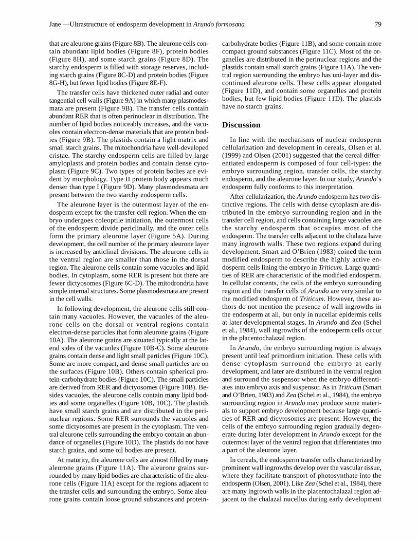
The transfer cells have thickened outer radial and outertangential cell walls (Figure 9A) in which many plasmodes-mata are present (Figure 9B). The transfer cells containabundant RER that is often perinuclear in distribution. Thenumber of lipid bodies noticeably increases, and the vacu-oles contain electron-dense materials that are protein bod-ies (Figure 9B). The plastids contain a light matrix andsmall starch grains. The mitochondria have well-developedcristae. The starchy endosperm cells are filled by largeamyloplasts and protein bodies and contain dense cyto-plasm (Figure 9C). Two types of protein bodies are evi-dent by morphology. Type II protein body appears muchdenser than type I (Figure 9D). Many plasmodesmata arepresent between the two starchy endosperm cells.
The aleurone layer is the outermost layer of the en-dosperm except for the transfer cell region. When the em-bryo undergoes coleoptile initiation, the outermost cellsof the endosperm divide periclinally, and the outer cellsform the primary aleurone layer (Figure 5A). Duringdevelopment, the cell number of the primary aleurone layeris increased by anticlinal divisions. The aleurone cells inthe ventral region are smaller than those in the dorsalregion. The aleurone cells contain some vacuoles and lipidbodies. In cytoplasm, some RER is present but there arefewer dictyosomes (Figure 6C-D). The mitodrondria havesimple internal structures. Some plasmodesmata are presentin the cell walls.
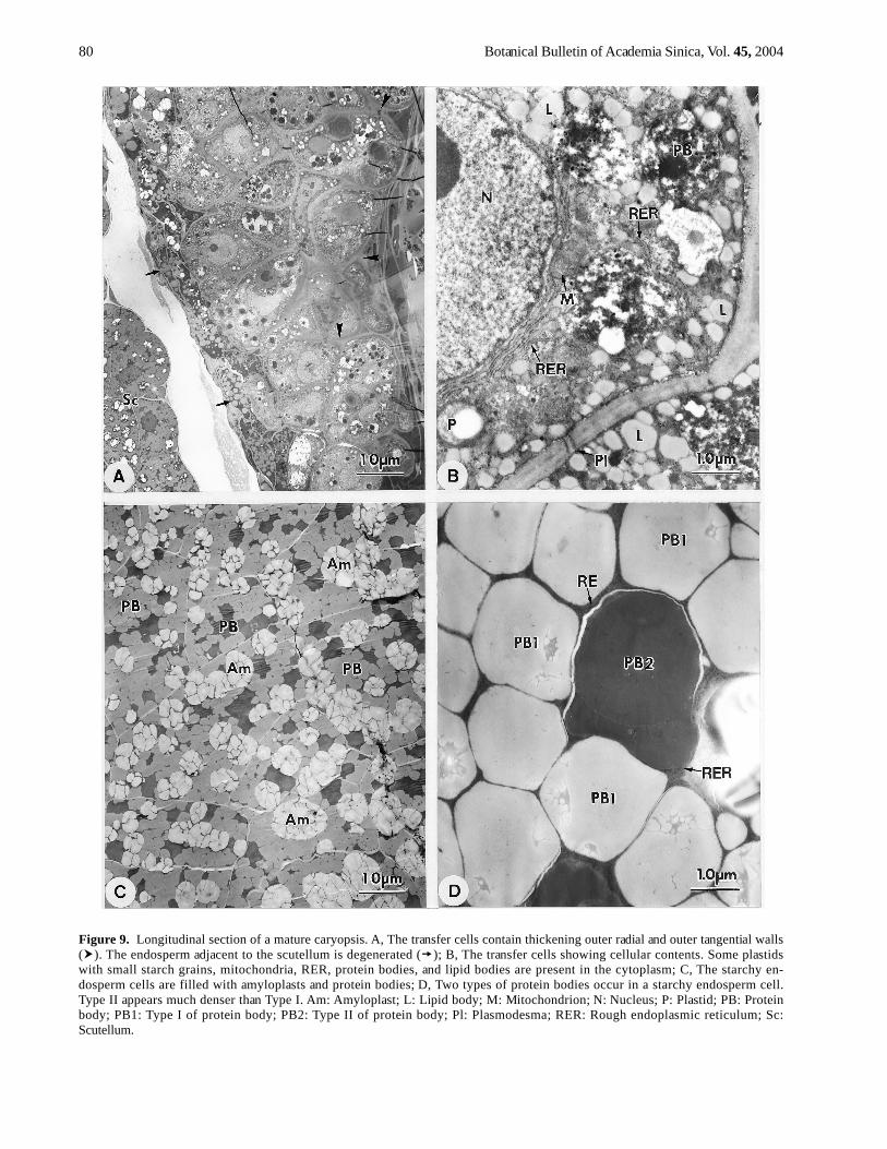
In following development, the aleurone cells still con-tain many vacuoles. However, the vacuoles of the aleu-rone cells on the dorsal or ventral regions containelectron-dense particles that form aleurone grains (Figure10A). The aleurone grains are situated typically at the lat-eral sides of the vacuoles (Figure 10B-C). Some aleuronegrains contain dense and light small particles (Figure 10C).Some are more compact, and dense small particles are onthe surfaces (Figure 10B). Others contain spherical pro-tein-carbohydrate bodies (Figure 10C). The small particlesare derived from RER and dictyosomes (Figure 10B). Be-sides vacuoles, the aleurone cells contain many lipid bod-ies and some organelles (Figure 10B, 10C). The plastidshave small starch grains and are distributed in the peri-nuclear regions. Some RER surrounds the vacuoles andsome dictyosomes are present in the cytoplasm. The ven-tral aleurone cells surrounding the embryo contain an abun-dance of organelles (Figure 10D). The plastids do not havestarch grains, and some oil bodies are present.
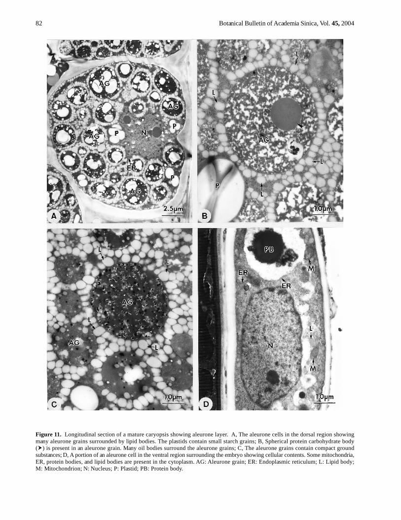
At maturity, the aleurone cells are almost filled by manyaleurone grains (Figure 11A). The aleurone grains sur-rounded by many lipid bodies are characteristic of the aleu-rone cells (Figure 11A) except for the regions adjacent tothe transfer cells and surrounding the embryo. Some aleu-rone grains contain loose ground substances and protein-
carbohydrate bodies (Figure 11B), and some contain morecompact ground substances (Figure 11C). Most of the or-ganelles are distributed in the perinuclear regions and theplastids contain small starch grains (Figure 11A). The ven-tral region surrounding the embryo has uni-layer and dis-continued aleurone cells. These cells appear elongated(Figure 11D), and contain some organelles and proteinbodies, but few lipid bodies (Figure 11D). The plastidshave no starch grains.
Discussion
In line with the mechanisms of nuclear endospermcellularization and development in cereals, Olsen et al.(1999) and Olsen (2001) suggested that the cereal differ-entiated endosperm is composed of four cell-types: theembryo surrounding region, transfer cells, the starchyendosperm, and the aleurone layer. In our study, Arundo’sendosperm fully conforms to this interpretation.
After cellularization, the Arundo endosperm has two dis-tinctive regions. The cells with dense cytoplasm are dis-tributed in the embryo surrounding region and in thetransfer cell region, and cells containing large vacuoles arethe starchy endosperm that occupies most of theendosperm. The transfer cells adjacent to the chalaza havemany ingrowth walls. These two regions expand duringdevelopment. Smart and O’Brien (1983) coined the termmodified endosperm to describe the highly active en-dosperm cells lining the embryo in Triticum. Large quanti-ties of RER are characteristic of the modified endosperm.In cellular contents, the cells of the embryo surroundingregion and the transfer cells of Arundo are very similar tothe modified endosperm of Triticum. However, these au-thors do not mention the presence of wall ingrowths inthe endosperm at all, but only in nucellar epidermis cellsat later developmental stages. In Arundo and Zea (Schelet al., 1984), wall ingrowths of the endosperm cells occurin the placentochalazal region.
In Arundo, the embryo surrounding region is alwayspresent until leaf primordium initiation. These cells withdense cytoplasm surround the embryo at earlydevelopment, and later are distributed in the ventral regionand surround the suspensor when the embryo differenti-ates into embryo axis and suspensor. As in Triticum (Smartand O’Brien, 1983) and Zea (Schel et al., 1984), the embryosurrounding region in Arundo may produce some materi-als to support embryo development because large quanti-ties of RER and dicytosomes are present. However, thecells of the embryo surrounding region gradually degen-erate during later development in Arundo except for theoutermost layer of the ventral region that differentiates intoa part of the aleurone layer.
In cereals, the endosperm transfer cells characterized byprominent wall ingrowths develop over the vascular tissue,where they facilitate transport of photosynthate into theendosperm (Olsen, 2001). Like Zea (Schel et al., 1984), thereare many ingrowth walls in the placentochalazal region ad-jacent to the chalazal nucellus during early development
80 Botanical Bulletin of Academia Sinica, Vol. 45, 2004
Figure 9. Longitudinal section of a mature caryopsis. A, The transfer cells contain thickening outer radial and outer tangential walls( ). The endosperm adjacent to the scutellum is degenerated ( ); B, The transfer cells showing cellular contents. Some plastidswith small starch grains, mitochondria, RER, protein bodies, and lipid bodies are present in the cytoplasm; C, The starchy en-dosperm cells are filled with amyloplasts and protein bodies; D, Two types of protein bodies occur in a starchy endosperm cell.Type II appears much denser than Type I. Am: Amyloplast; L: Lipid body; M: Mitochondrion; N: Nucleus; P: Plastid; PB: Proteinbody; PB1: Type I of protein body; PB2: Type II of protein body; Pl: Plasmodesma; RER: Rough endoplasmic reticulum; Sc:Scutellum.
Jane —Ultrastructure of endosperm development in Arundo formosana 81
Figure 10. Longitudinal section of a pre-mature caryopsis showing aleurone layer. A, The aleurone cells in the ventral regionshowing cellular contents. Many organelles and lipid bodies are present in the cytoplasm. The plastids contain small starch grains,and the aleurone grains are formed in vacuoles ( ); B, A portion of the aleurone cells in the ventral region showing cellular contents.Some vesicles ( ) can be seen in the periphery of vacuoles. Many electron dense particles ( ) are present in vacuoles and accumu-late together; C, A portion of an aleurone cell in the dorsal region showing cellular contents. Spherical particles ( ) are present in thealeurone grains; D, An aleurone cell in the ventral region surrounding the embryo showing cellular contents. Some plastids,mitochondria, dictyosomes, ER, and lipid bodies are present in the cytoplasm. AG: Aleurone grain; D: Dictyosome; ER: Endoplas-mic reticulum; L: Lipid body; M: Mitochondrion; N: Nucleus; P: Plastid; Pl: Plasmodesma; RER: Rough endoplasmic reticulum.
82 Botanical Bulletin of Academia Sinica, Vol. 45, 2004
Figure 11. Longitudinal section of a mature caryopsis showing aleurone layer. A, The aleurone cells in the dorsal region showingmany aleurone grains surrounded by lipid bodies. The plastids contain small starch grains; B, Spherical protein carbohydrate body( ) is present in an aleurone grain. Many oil bodies surround the aleurone grains; C, The aleurone grains contain compact groundsubstances; D, A portion of an aleurone cell in the ventral region surrounding the embryo showing cellular contents. Some mitochondria,ER, protein bodies, and lipid bodies are present in the cytoplasm. AG: Aleurone grain; ER: Endoplasmic reticulum; L: Lipid body;M: Mitochondrion; N: Nucleus; P: Plastid; PB: Protein body.

Jane —Ultrastructure of endosperm development in Arundo formosana 83
of Arundo’s endosperm. In Zea (Schel et al., 1984), two tothree cell layers of endosperm cells have wall ingrowthsin a gradient decreasing toward the interior of theendosperm. Only one cell layer is present in Arundo. Schelet al. (1984) suggested that the transfer cells transport nu-trients to the embryo beside secreting materials in Zea.
In subsequent development, the transfer cells of Arundogradually differentiate into two cell types: the outer cellswith dense cytoplasm and the inner cells with many smallvacuoles. The transfer cells differentiate into two differ-ent regions with various functions never described in otherPoaceae. In Arundo, the outer cells of the transfer cell re-gion contain very dense cytoplasm, in which many lipidsand proteins are present, as well as large quantities ofRER. During development, the ingrowth walls in the outerportion of the transfer cell region are covered by thicken-ing cell walls that appear PAS-positive. The inner cellshave many small vacuoles and some organelles. The outertransfer cells gradually degenerate during development.The inner transfer cells gradually accumulate lipid bodiesand protein bodies in cytoplasm and develop thickeningcell walls that appear PAS-positive. RER and dictyosomesare more developed compared to the foregoing stage. Atmaturity, the cellular contents of the Arundo transfer cellsare very similar to the groove aleurone layer of Triticum(Morrison et al., 1978). In Arundo, the transfer cells havetwo suspected functions at different stages. In the earlyembryo stage, the transfer cells synthesize and transportnutrients to support embryo development. In the later em-bryo stage, the transfer cells can accumulate nutrients andmay become a part of the aleurone layer.
During early development, the Arundo starchy en-dosperm occupies most of the endosperm and proliferatesby cell division. Like other angiosperms (Bhatnagar andSawhney, 1980), the Arundo starchy endosperm begins toaccumulate storage reserves at the later globular embryostage. The accumulation and distribution of the storagereserves—including starch, lipids, and proteins—arevariable. Starch first appears in the middle region and ac-cumulates toward the two poles. Most lipid bodies are dis-tributed in the peripheral region. Proteins appear late andtheir accumulation is similar to starch.
During development of Arundo’s starchy endosperm,the storage reserves are further from the embryo than otherregions. The endosperm surrounding the embryo isdegenerated, especially the elongated regions of thescutellum. This condition can also be found in Triticum(Smart and O’Brien, 1983). The degenerated region may re-sult from compression by the developing scutellum. Thecellular contents of the degenerated region appear PAS-positive. In early development of Solanum seed (Briggs,1993a, b), a special region called the zone of separationand secretion (ZSS) is distinguishable in the endosperm.This region expands during development, and these cellscontain a large number of lipid bodies. The ZSS facilitatesthe growth of the embryo through the rest of theendosperm.
In Poaceae, a main focus of endosperm study is the for-mation of protein bodies, which seem to have variousorigin. In general, the protein bodies of Poaceae starchyendosperm have two forms: One is initiated and enlargedin the cistae of RER, and dictyosomes may be involved intheir enlargement; the other is initiated from RER or/anddictyosomes, and is accumulated and enlarged in vacuoles.In Arundo, two types of protein bodies in the starchy en-dosperm are initiated in the cisterae of RER, enlarge in thecisteral lumen of RER, and are stored in the vacuoles. Oryzahas three types of protein bodies (Bechtel and Juliano,1980). The large, spherical protein bodies are initiated inthe cisterae of RER and distributed in the central regionof the endosperm (Harris and Juliano, 1977; Bechtel andPomeranz, 1978; Bechtel and Juliano, 1980). Two othertypes, the crystalline and small-spherical protein bodies,are initiated in the dictyosomes and cistae of RER,respectively. These two types of protein bodies are onlydistributed in the subaleurone region. In Arundo’s gen-eral endosperm, the dictyosomes are seldom present andmay not be involved in the initiation and enlargement ofthe protein bodies. In Zea (Khoo and Wolf, 1970; Larkinsand Hurkman, 1978), the protein bodies are initiated in thecistae of RER and enlarge, and dictyosomes are also in-volved in protein body enlargement by producing vesicles.Researchers do not agree about the initiation and enlarge-ment of Triticum protein bodies. Some have suggestedthat RER is directly involved in it (Campbell et al., 1981).Some have suggested that only dictyosomes are involved(Bechtel et al., 1982; Parker and Hawes, 1982; Kim et al.,1988). Still others have suggested RER and dictyosomesare all involved.
Like most Poaceae, the outermost layer of the Arundoendosperm develops into the aleurone layer that coversthe endosperm, excluding the transfer cell region. In theendosperm of all cereals, immediately after completion ofendosperm cellularization, aleurone cell differentiation isinitiated in the proximal part of the endosperm above thematernal vascular bundle (Olsen et al., 1998). The aleuronegrains are characteristic of the aleurone cells. In Poaceae,the structure and contents of the aleurone grains showvariation. Some only contain protein matrix, such as inOryza (Bechtel and Pomeranz, 1977), Zea (Kyle and Styles,1977), Triticum (Morrison et al., 1978), Pennisetum and Sor-ghum (Zelezank and Varriano-Marston, 1982); some con-tain globoids besides matrix, such as in Setaria (Rost andLersten, 1970) and Hordeum (Jacobsen et al., 1971; Bethke,et al., 1998); some contain protein-carbohydrate bodies(also called protein crystalloids) besides matrix, such asin Hordeum (Jacobsen et al., 1971; Bethke et al., 1998) andTriticum (Morrison et al., 1975). In Arundo, the aleuronegrains do not contain globoids, but some contain protein-carbohydrate bodies. Like most Poaceae, the aleuronegrains of Arundo are surrounded by many small lipid bod-ies that form many ring structures. The aleurone cells ofsome grasses, such as Setaria (Rost and Lersten, 1970),Echinochloa (Zee and O’Brien, 1971), Pennisetum (Fussell

84 Botanical Bulletin of Academia Sinica, Vol. 45, 2004
and Dwarte, 1980) and Zea (Schel et al., 1984), contain in-growth walls adjacent to the chalaza called aleurone trans-fer cells.
Besides Poaceae, the aleurone layers can also be foundin seeds of several families, e.g. in Brassicaceae (Corner,1976). However, the initiation of the aleurone layer appearsdifferent. In Brassica (Groot and van Caeseele, 1993), thealeurone layer is developed from the outermost layer ofthe endosperm. However, in Sinapis (Bergfeld andSchopfer, 1986), the aleurone layer is initiated from the in-ner integument and distributed in the seed coat, whichwould make it “perisperm.” The aleurone cells are alsopresent on the seed coat of Glycine (Yaklich et al., 1992).
In this study, I describe in detail the development ofthe four parts of Arundo endosperm. This study confirmsOlsen’s interpretation (2001), but the development ofArundo endosperm shows some differences from othergrasses. The most special structure is the transfer cells thatdifferentiate to two regions and have different supposedfunctions. The function of the transfer cells in early de-velopment is secretion and transport based on abundantRER and ingrowth walls; the function in later developmentis secretion and storage because of abundant RER andreserves.
Literature Cited
Bechtel, D.B., R.L. Grains, and Y. Pomeranz. 1982. Early stagesin wheat endosperm formation and protein body initiation.Ann. Bot. 50: 507-518.
Bechtel, D.B. and B.O. Juliano. 1980. Formation of protein bodiesin the endosperm of rice (Oryza sativa L.): A reinvestigation.Ann. Bot. 45: 503-509.
Bechtel, D.B. and Y. Pomeranz. 1977. Ultrastructure of the ma-ture ungerminated rice (Oryza sativa) caryopsis. The cary-opsis caot and aleurone cells. Amer. J. Bot. 64: 966-973.
Bechtel, D.B. and Y. Pomeranz. 1978. Ultrastructure of the ma-ture ungerminated rice (Oryza sativa) caryopsis. The germ.Amer. J. Bot. 65: 75-85.
Bergfeld, R. and P. Schopfer. 1986. Differentiation of a func-tional aleurone layer within the seed coat of Sinapsis albaL. Ann. Bot. 57: 25-33.
Bethke, P.C., S.J. Swanson, S. Hillmer, and R.L. Rost. 1998.From storage compartment to lytic organelles: the metamor-phosis of the aleurone protein storage vacuole. Ann. Bot.82: 399-411.
Bhatnagar, S.P. and V. Sawhney. 1980. Endosperm- Itsmorphology, ultrastructure, and histochemistry. Intl. Rev.Cytol. 73: 55-102.
Briggs, C.L. 1993a. Endosperm development in Solanum nigrumL. Formatiom and distribution of lipid bodies. Ann. Bot.72: 295-301.
Briggs, C.L. 1993b. Endosperm development in Solanum nigrumL. Formation of the zone of separation and secretion. Ann.Bot. 72: 303-313.
Bronner, R. 1975. Simultaneous demonstration of lipids andstarch in plant tissues. Stain Technol. 50: 1-4.
Campbell, W.P., J.W. Lee, T.P. O‘Brien, and M.G. Smart. 1981.Endosperm morphology and protein body formation in de-
veloping wheat grains. Aust. J. Plant Physiol. 8: 5-19.
Corner, E.J.H. 1976. The Seeds of Dicotyledrons. Vols 1 and 2.pp. 311 and 552. Cambridge University Press, Cambridge.
Fussel, L.K. and D.M. Dwarte. 1980. Structural changes of thegrains associated with black region formation in Pennisetumamericanum. J. Exp. Bot. 31: 645-654.
Gahan, P.B. 1984. Plant histochemistry and cytochemistry: anintroduction. Academic Press Inc.
Groot, E.P. and L.A. van Caeseele. 1993. The development ofthe aleurone in canloa (Brassica napus). Can. J. Bot. 71:1193-1201.
Harris, N. and B.O. Juliano. 1977. Ultrastructure of endospermprotein bodies in developing rice grains differing in proteincontents. Ann. Bot. 41: 1-5.
Jacobsen, J.V., R.B. Knox, and N.A. Pyliotis. 1971. The struc-ture and composition of aleurone grains in the barley aleu-rone layer. Planta 101: 189-209.
Jane, W.N. 1999. Ultrastructure of embryo development inArundo formosana Hack. (Poaceae). Int. J. Plant Sci. 160:46-63.
Khoo, U. and M.J. Wolf. 1970. Origin and development of pro-tein granules in maize endosperm. Amer. J. Bot. 57: 1042-1050.
Kim, W.T., V.R. Franceschi, H.B. Krishnan, and T.W. Okita.1988. Formation of wheat protein bodies: involvement ofthe golgi apparatus in gliadin transport. Planta 176: 173-182.
Kyle, D.J. and E.D. Styles. 1977. Development of aleurone andsubaleurone in maize. Planta 137: 185-193.
Larkins, B.A. and W.J. Hurkman. 1978. Synthesis and deposi-tion of zein in protein bodies of maize endosperm. PlantPhysiol. 62: 256-263.
Morrison, I.N., J. Kuo, and T.P. O‘Brien. 1975. Histochemis-try and fine structure of developing wheat aleurone cells.Planta 123: 105-116.
Morrison, I.N., T.P. O’Brien and J. Kuo. 1978. Initialcellularization and differentiation of the aleurone cells in theventral region of the developing wheat grain. Planta 140:19-30.
Olsen, O.-A. 2001. Endosperm development: cellularization andcell fate specification. Annu. Rev. Plant Physiol. Plant Mol.Biol. 52: 233-267.
Olsen, O.-A., R.C. Brown, and B.E. Lemmon. 1995. Pattern andprocess of wall formation in developing endosperm..BioEssays 17: 803-812.
Olsen, O.-A., B.E. Lemmon, and R.C. Brown. 1998. A modelfor aleurone cell development. Trends Plant Sci. 3: 168-169.
Olsen, O.-A., C. Linnestad, and S.E. Nichols. 1999. Develop-mental biology of the cereal endosperm. Trends Plant Sci.4: 253-257.
Parker, M.L. and C.R. Hawes. 1982. The golgi apparatus in de-veloping endosperm of wheat (Triticum aestivum L.). Planta154: 277-283.
Reynolds, E.S. 1963. The use of lead citrate at high pH as anelectron-opaque stain in electron microscopy. J. Cell Biol.55: 541-552.
Rost, T.L. and N.R. Lersten. 1970. Transfer aleurone cells inSetaria lutescens (Gramineae). Protoplasma 71: 403-408.
Schel, J.H.N., H. Kieft, and A.A.M. van Lammeren. 1984. In-teractions between embryo and endosperm during early de-

Jane —Ultrastructure of endosperm development in Arundo formosana 85
velopmental stages of maize caryopses (Zea mays). Can. J.Bot. 62: 2842-2853.
Smart, M.G. and T.P. O’Brien. 1983. The development of thewheat embryo in relation to the neighbouring tissues.Protoplasma 114: 1-13.
Spurr, A.R. 1969. A low-viscosity epoxy resin embedding me-dium for electron microscopy. J Ultrastruct. Rev. 26: 31-43.
Yaklich, R.W., E.L. Vigil, E.F. Erbe, and W.P. Wergin. 1992. The
Arundo formosana Hack.
fine structure of aleurone cells in the soybean seed coat.Protoplasma 167: 108-119.
Zee, S.Y. and T.P. O’Brien. 1971. Aleurone transfer cells andother structural features of the spikelets of millet. Aust. J.Biol. Sci. 24: 391-395.
Zeleznak, K. and E. Varriano-Marston. 1982. Pearl millet(Pennisetum americanum (L.) Leeke) and grain sorghum(Sorghum bicolor (L.) Moench) ultrastructure. Amer. J.Bot. 69: 1306-1313.


![Graduate in Biochemistry [Habilitações Académicas] Arundo](https://img.pdfslide.us/doc/110x75/6273980f6259f336d05fad07/graduate-in-biochemistry-habilitaes-acadmicas-arundo-.jpg)