Embed Size (px)
Citation preview
Ultrastructure and Composition of the Nannochloropsis gaditana CellWall
Matthew J. Scholz,a Taylor L. Weiss,b Robert E. Jinkerson,a Jia Jing,c Robyn Roth,d Ursula Goodenough,b Matthew C. Posewitz,a
Henri G. Gerkene
Department of Chemistry and Geochemistry, Colorado School of Mines, Golden, Colorado, USAa; Department of Biology, Washington University, St. Louis, Missouri, USAb;CAS Key Laboratory of Biofuels, Shandong Key Laboratory of Energy Genetics and BioEnergy Genome Center, Qindao Institute of BioEnergy and Bioprocess Technology,Chinese Academy of Sciences, Qingdao, Shandong, Chinac; Department of Cell Biology and Physiology, Washington University School of Medicine, St. Louis, Missouri,USAd; Arizona Center for Algal Technology and Innovation, Arizona State University, Mesa, Arizona, USAe
Marine algae of the genus Nannochloropsis are promising producers of biofuel precursors and nutraceuticals and are also har-vested commercially for aquaculture feed. We have used quick-freeze, deep-etch electron microscopy, Fourier transform infra-red spectroscopy, and carbohydrate analyses to characterize the architecture of the Nannochloropsis gaditana (strain CCMP526) cell wall, whose recalcitrance presents a significant barrier to biocommodity extraction. The data indicate a bilayer struc-ture consisting of a cellulosic inner wall (�75% of the mass balance) protected by an outer hydrophobic algaenan layer. Cellulasetreatment of walls purified after cell lysis generates highly enriched algaenan preparations without using the harsh chemicaltreatments typically used in algaenan isolation and characterization. Nannochloropsis algaenan was determined to compriselong, straight-chain, saturated aliphatics with ether cross-links, which closely resembles the cutan of vascular plants. Chemicalidentification of >85% of the isolated cell wall mass is detailed, and genome analysis is used to identify candidate biosyntheticenzymes.
The genus Nannochloropsis comprises at least six photoau-totrophic algal species in the Eustigmatophyceae stramenopile
lineage that are found in fresh, brackish, and ocean waters (1).Nannochloropsis cells reproduce asexually, dividing to yield twodaughter cells that then shed their mother cell wall (2, 3). SeveralNannochloropsis species have been studied as candidate produc-tion strains in large-scale biofuel facilities because of their hardyoutdoor growth profiles and high lipid yields (4–9). They are alsoproducers of valuable pigments (10) and nutritive oils (11, 12) andare commonly used as an aquaculture feed (13).
Algae are frequently grown in large outdoor ponds until beingharvested, dewatered, and extracted for biocommodities. The ef-ficacy of each of these steps— growth, harvesting, dewatering, andextraction— depends upon the composition and architecture ofthe cell wall. The wall creates a buffer between the external envi-ronment and the living protoplast, protecting the cell from en-vironmental pressures. The outer surface of the wall interactswith flocculants (14), and its rigidity helps determine the vis-coelastic parameters that characterize algal slurry bulk flow(15). Finally, the cell wall erects mass transfer barriers againstdewatering and extraction and may itself contain extractablecommodities (16, 17).
Despite the importance of algal cell wall properties in biotech-nological applications, little structural information is available forthe majority of species. The Chlamydomonas reinhardtii cell wall isthe most extensively characterized and appears to be constructedentirely from a suite of hydroxyproline-rich glycoproteins ar-ranged in six distinct layers (18–20). However, algal cell walls dis-play great diversity, varying in molecular components, intra- andintermolecular linkages, and overall structure (21). Wall constit-uents may include carbohydrates (22), proteins (23, 24), lipids(25, 26), carotenoids (27), tannins (28), and even lignin (29, 30).Much remains to be learned regarding how these constituentscross-link into the networks that form discrete layers around the
cell and how they reconfigure in response to physiological andenvironmental cues.
Among the most extensively studied polymers of the algal cellwall are polysaccharides. These include cellulose (31), chitin-/chi-tosan-like molecules (32), hemicelluloses (33), pectins (34), fu-cans (35), alginates (24), ulvans (36), carrageenans (37), and li-chenins (38). The polysaccharides in marine algae are frequentlysulfated (22).
The composition and architecture of Nannochloropsis cell wallshave been assessed in several studies. Brown reported that thepolysaccharides of Nannochloropsis oculata contained �68% glu-cose along with about 4 to 8% each rhamnose, mannose, ribose,xylose, fucose, and galactose (39). Recently, Vieler et al. character-ized the neutral carbohydrates in the alcohol-insoluble residue(AIR) of Nannochloropsis oceanica (strain CCMP 1779) cell ex-tracts (40). This residue, enriched for cell wall material, washydrolyzed with trifluoroacetic acid (TFA) followed by Saemanhydrolysis. The authors observed that �9% of the AIR was carbo-hydrate, 90% of which was glucose, �3% mannose, and the resttraces of rhamnose, fucose, arabinose, xylose, and galactose.Treatment of the residue with endoglucanase II (EGII), a hydro-lyzing enzyme specific for �-1,4-linked glucans, liberated 85% ofthe glucose, while laminarinase, an enzyme that hydrolyzes �-1,3-glucans, liberated 20%. Bioinformatic analysis of the CCMP 1779
Received 30 July 2014 Accepted 14 September 2014
Published ahead of print 19 September 2014
Address correspondence to Henri G. Gerken, [email protected].
Supplemental material for this article may be found at http://dx.doi.org/10.1128/EC.00183-14.
Copyright © 2014, American Society for Microbiology. All Rights Reserved.
doi:10.1128/EC.00183-14
1450 ec.asm.org Eukaryotic Cell p. 1450 –1464 November 2014 Volume 13 Number 11
on June 20, 2018 by guesthttp://ec.asm
.org/D
ownloaded from
genome yielded two proteins annotated as cellulose synthases,similar to those found in cyanobacteria, and nine proteins that theauthors describe as highly similar to plant endoglucanases.
Nannochloropsis cell walls also contain algaenans, a term thatlikely encompasses several lipid-related species (41, 42). Algaen-ans are highly resistant to alkali/acid hydrolysis and aqueous/or-ganic solubilization, and their biochemical characterization hasbeen considered tentative since isolation procedures may haveinduced chemical alterations (43, 44). Published studies indicatethat Nannochloropsis algaenan comprises long-chain aliphatic hy-drocarbons that are subject to ether cross-linking reactions (41), adescription that also applies to the cutan of several species ofdrought-resistant plants (45). The biosynthetic pathways thatproduce algaenans and cutans are not presently known.
In this study, �86% of the isolated cell wall material of N.gaditana has been positively identified. A new method for isolatingNannochloropsis algaenans was developed, allowing an analysis ofnative algaenan structure, and this material was characterized byattenuated total reflectance Fourier transform infrared spectros-copy (ATR-FTIR). Furthermore, quick-freeze, deep-etch electronmicroscopy (QFDE-EM) was used to visualize native and isolatedwall components.
MATERIALS AND METHODSCell culture. Nannochloropsis strain CCMP 526 was from the NationalCenter for Marine Algae and Microbiota (formerly CCMP). CCMP 526was grown at 23°C in f/2 medium (125) with 1.0 g/liter nitrate in a 2-footby 2-foot flat-panel photobioreactor with a 40-mm width (14-liter capac-ity). One percent CO2 was bubbled through the cultures, and 70 �molm�2 s�1 of photosynthetically active radiation (PAR) from cool whitefluorescent light illuminated one side. The initial cell density was 4 � 107
cells per ml, and cells were harvested on day 8, during mid-linear phase (4g/liter). For comparative purposes, CCMP 526 was also grown semicon-tinuously at 23°C in Roux flasks in Tris-buffered (pH 7.9) artificialseawater (ASW [0.125 M NaCl, 7 mM KNO3, 0.44 mM KH2PO4, 0.24mM NaHCO3, 0.018 mM FeCl3·6H2O, 0.049 mM MnCl2·4H2O, 1.7 mMCaCl2·2H2O, 13.4 mM MgSO4·7H2O, 13.8 mM MgCl2·6H2O, 0.1 mMNa2EDTA, 0.42 �M CoCl2·6H2O, 0.14 �M Na2MoO4·2H2O, 0.89 �MZnSO4·7H2O, 0.4 �M CuSO4·5H2O]). These cultures were bubbled withsupplemental CO2 (1%) in air; 600 �mol m�2 s�1 of 680-nm light wasprovided by an LED light bank on one side and 150 �mol m�2 s�1 PARfrom cool white fluorescent light was supplied to the other side. Cellsgrown under the conditions outlined above have been designated “f/2preparations” or “ASW preparations” throughout the text.
Preparation of cell walls. Cells pellets were harvested by centrifuga-tion at 4,000 � g for 10 min. Shed cell walls, derived from wall sheddingduring cell division and from dead cells, formed a red-orange layer on topof the green cell pellet and were carefully removed by pipette. The greenwhole-cell pellet was resuspended in 50 ml of deionized water (dH2O) andrepelleted a total of 3 times to remove traces of medium.
Whole cells were then lyophilized and resuspended to �10% solids indH2O. To every 5 ml of suspended solids, 15 �l of protease inhibitorcocktail (product number P9599; Sigma, St. Louis, MO) and 1 �l of 2,000U/ml DNase I (NEB, Ipswich, MA) were added. Cells were drawn into the30-ml chamber of a French press pressure cell (Thermo, Waltham, MA).Seven passes through the French press at a working pressure of 18,000lb/in2 were required for nearly complete cell lysis, although a small per-centage of the cells (�5%) remained intact even after 7 passages. Afterevery 2 passages, cells were centrifuged at 10,000 � g for 20 min to separatethe soluble lysate, which was discarded, from residual cells and cell walls.The latter were resuspended in dH2O as before, the DNase and proteaseinhibitor cocktail were replenished, and the next passages were per-formed. Cell walls were separated from residual whole cells by multiple
centrifugations at 5,000 � g, each time removing the walls from the greenpellet at the bottom of the tube until no green pellet was observed. Thewalls isolated in this manner are referred to hereinafter as “pressed” cellwalls.
Pressed cell walls were separated from residual debris and shed cellwalls by layering them upon sucrose gradients of 20, 30, 40, and 60% andcentrifuging for 30 to 60 min at 10,000 � g. Shed cell walls and otherdebris typically remain in the upper strata and were separated frompressed cell walls, which migrated to the bottom of the tube, stuck to theside of the tube, or floated in the 60% sucrose layer. Pressed walls werethen washed three times by resuspending the pellet in 50 ml of water andcentrifuging as before. After the final wash, the recovered walls were ly-ophilized.
Every 20 mg of lyophilized cell walls was extracted successively with 10ml of 80% ethanol, n-hexane–acetone (1:1, vol/vol), and n-hexane for 15min in a sonicating water bath (model number 91957; Harbor FreightTools, Calabasas, CA) to remove any loosely associated proteins, lipids,carotenoids, and other soluble matter. Additional washes with n-hexanewere performed until the color of the residual material was white or off-white. The organic supernatants from these washes were collected into apreweighed glass beaker and evaporated. The dry residue was weighed.Light microscopy visualizations after these extractions were used to verifythat the insoluble material remaining after extraction was morphologi-cally consistent with purified cell walls. The extracted cell walls were ly-ophilized in preweighed glass vials and weighed. In the sections that fol-low, “isolated” or “pressed” cell walls refers to walls purified and subjectedto organic extraction as described above.
Enzymatic degradation of walls. For enzymatic digestions of isolatedcell walls, approximately 10 mg of walls were used in each 1-ml digestionmixture. Cellulase (product number C0615 [cellulase 1]), chitinase(product number C8241), chitosanase (product number C9830, 25.9U/ml), lysozyme (product number L6876), lyticase (product numberL4025), protease 2 (product number P5147), and sulfatase (product num-ber S1629, 3.37 mg/ml) were purchased from Sigma, while cellulase Ono-zuka R10 (catalog number 16419.02 [cellulase 2]) was purchased fromServa (Heidelberg, Germany). Stock concentrations of enzymes were 20mg/ml unless otherwise noted, and enzymes were dissolved in 20 mMsodium phosphate buffer (pH 7) for all digestions. Triplicate sampleswere digested at room temperature for 24 h on a shaker table. The finalenzyme concentrations were 1 mg/ml, and 50 �g/ml of ampicillin wasadded to each reaction mixture. For one set of controls, enzymes were heatdenatured for at least 10 min at 100°C before being added to the cell wallsuspensions. As a second set of controls, active enzymes were incubated inreaction mixtures without cell walls. After digestion, the reaction mixtureswere centrifuged at 10,000 � g for 5 min. Supernatants were collected, andresidual material was washed twice by resuspension in 1 ml of dH2O,followed by centrifugation and supernatant removal. The residual mate-rial was then lyophilized and weighed.
Acid hydrolysis of cell walls. Cell walls isolated from f/2 medium-grown cells were digested in 72% sulfuric acid as described in an analysisprotocol from the National Renewable Energy Laboratory (46).
Characterization of monosaccharides. Supernatants from enzymedigests were assayed for released carbohydrates. For each sample, 490 �l ofsupernatant was saved for direct analysis by high-performance anion-exchange chromatography (ion chromatography [IC]) with pulsed am-perometric detection (HPAEC-PAD) and 10 �l was used for anthroneassays. To the other half of the sample, an equal volume of 4 N TFA wasadded, and the sample was hydrolyzed to monosaccharides by incubationat 100°C for 6 h. The samples were then evaporated at 65°C under nitro-gen. Samples were immediately resuspended in water for analysis.
Monosaccharides were analyzed by HPAEC-PAD on an ICS 5000equipped with a 2- by 250-mm Carbopac PA-1 analytical column and a 2-by 50-mm Aminotrap guard column (Dionex Corporation, Sunnyvale,CA). The PAD detector used a carbohydrate-certified disposable goldelectrode (Thermo) with the gold standard PAD waveform. Neutral sug-
Nannochloropsis gaditana Cell Wall
November 2014 Volume 13 Number 11 ec.asm.org 1451
on June 20, 2018 by guesthttp://ec.asm
.org/D
ownloaded from
ars and amino sugars were eluted at 31°C and 0.25 ml/min with a 1 mNKOH eluent produced from an eluent generator. Uronic acids were ana-lyzed separately at 40°C and 0.25 ml/min using 100 mN NaOH for 5 min,followed by a 60 to 300 mM gradient of sodium acetate over 10 min toelute the uronic acids. The mobile-phase solutions were degassed and keptunder a bed of helium. Individual standards were used to measure theretention times of each sugar and determine carbohydrate concentra-tions. All major peaks in the IC traces were identified; however, a few verysmall peaks remained unidentified. Data collection and analysis weredone using Chromeleon 7.1.0 software (Dionex).
To validate the IC results, aliquots of 10 �l of supernatants from cel-lulase digests (cellulase 1) of cell walls and wall-free controls were broughtup to 100 �l in dH2O for anthrone quantitation of carbohydrate content(47). A stock solution containing 2 g/liter anthrone reagent in 26.6 Nsulfuric acid was prepared, as was a set of 100-�l glucose standards rang-ing in concentration from 100 to 400 �g/ml. To 100 �l of sample, 900 �lof the anthrone stock solution was added. The samples were placed inboiling water for 12 min and then transferred to an ice bath. Three hun-dred-microliter aliquots of each were transferred to individual wells of a96-well plate, and the absorbance at 625 nm was determined using a platereader (Synergy 2; Biotek, Winooski, VT). The absorbance values wereregressed against glucose standards (R2 � 0.999), and sample carbohy-drate concentrations were inferred from the resulting regression equa-tions.
Glycosyl linkage analysis. Glycosyl linkage analysis was performed bythe Complex Carbohydrate Research Center at the University of Georgia.Samples were permethylated, depolymerized, reduced, and acetylated.The resultant partially methylated alditol acetates (PMAAs) were analyzedby gas chromatography-mass spectrometry (GC-MS) as described previ-ously (48).
Amino acid analysis. Amino acid analysis was conducted by the Mo-lecular Structure Facility at the University of California at Davis. Cell wallswere hydrolyzed for 48 h in 6 N HCl–1% phenol at 110°C. Samples (�2.5mg) were analyzed for amino acids and related compounds using a lith-ium citrate buffer system, a Beckman 6300 amino acid analyzer (Beck-man, Brea, CA), and an amino acid standard (product number A9906;Sigma).
Fourier transform infrared spectroscopy characterization. Isolatedwalls were progressively treated with cellulase 1 and then protease in so-dium phosphate buffer at pH 7.0 for 18 h at room temperature beforetreatment with 6 N HCl for 18 h at 100°C. Four dH2O washes followedafter each step, and aliquots from each step were preserved. For ATR-FTIR analysis, �1-mg amounts of lyophilized samples of cell walls andalgaenan preparations were loaded onto the germanium crystal of a Nexus470 FTIR spectrophotometer Smart Performer platform (Nicolet, Madi-son, WI). Spectra were collected using OMNIC software (Nicolet) butwere also further analyzed using LabSpec 6 software (Horiba Scientific,Edison, NJ). All spectra were collected across the 4,000 to 600 cm�1 rangeat a 1 cm�1 spectral resolution. The spectra presented for each sample arethe averages of between 200 and 400 spectra. All data were collected atWashington University at St. Louis in the Jens Environmental Molecularand Nanoscale Analysis Laboratory.
Quick-freeze deep-etch electron microscopy. Samples were rinsedtwice in dH2O by pellet centrifugation, pipetted onto a cushioning mate-rial, and dropped onto a liquid helium-cooled copper block; the frozenmaterial was transferred to liquid nitrogen and fractured, etched at80°C for 2 min, and platinum replicated using Pt-C rotary replicationas described previously (49). The replicas were examined with a JEOLelectron microscope, model JEM 1400, equipped with an AMTV601digital camera.
Elemental analysis. Screw-cap Teflon vials (VWR, Randor, PA) werewashed thoroughly with dH2O and then pretreated by soaking in trace-metal-grade 6 N nitric acid for 18 h at 100°C. The nitric acid in each vialwas replaced with 10 ml of fresh 6 N nitric acid to which 10 to 25 mg ofsample was added. A blank with nitric acid but no sample was also pre-
pared as a control. Vials were heated for 18 h at 100°C, and then the nitricacid was evaporated using a hot plate. The insides of the Teflon vials wereeach washed with 20 ml of dH2O, which was then filtered and used forelemental analysis by inductively coupled-plasma atomic emission spec-troscopy (ICP-AES) (Optima 5300 DV; PerkinElmer, Waltham, MA).Cadmium was run as an internal standard and used to correct the valuesobtained during the analysis. The system uses a nonbaffled cyclonic spraychamber with a type A Meinhard nebulizer. The rates of argon gas flowwere 16 liters/min for the plasma, 0.65 liters/min for the nebulizer, and 0.5liters/min for the auxiliary flow. A custom-made multispectral fitting filewas used to correct for spectral interferences between elements. Argon gaswas used to purge the spectrometer and introduction system. Dried com-pressed air was used as the shear gas. Standards were prepared from theAccuTrace reference standard (AccuStandard, New Haven, CT), fromanalytical standards QCS-7-M and QCS-1 (High Purity Standards,Charleston, SC), and from EM standard ICP-126-5 (Ultra Scientific,North Kingstown, RI). The standards for metals and nonmetals wereCCV-1A and -B, respectively (High Purity Standards). Elemental concen-trations observed in the blank were subtracted from sample values.
Bioinformatics. Cell wall-related protein sequences were obtainedfrom the Cell Wall Navigator database (50). Additional cell wall-relatedprotein sequences were extracted from the CAZy database (www.cazy.org) (51). These protein sequences were compared against protein mod-els from the CCMP 526 genome (NCBI BioProject accession numberPRJNA73791), genome assembly, and transcriptome shotgun assembly(NCBI BioProject accession number PRJNA157493) using BLAST (52).Combined, these searches yielded a list of protein and nucleotide se-quences that were curated to identify putative cell wall-related proteins.Matching protein sequences (E value, �1 � 10�20) were blasted againstnonredundant protein sequences in the NCBI database using the DELTA-BLAST algorithm. Matching nucleotide sequences (E value, �10�10)were blasted (blastx) against the same database. In both cases, a match wasdeclared when a protein matched a protein domain with known cell wall-related activity with an E value of less than 10�20 (typically less than10�100). The resulting list of protein models was mapped back to thegenome to prevent redundancy.
All protein alignments were conducted with MUSCLE (53). A phylo-genetic tree of cellulose synthases (CESAs) and cellulose synthase-like(CSL) proteins was inferred by using the maximum-likelihood methodbased on the Poisson correction model (see Fig. 1) (54). The bootstrapconsensus tree inferred from 100 replicates is taken to represent the evo-lutionary history of the taxa analyzed (55). Branches corresponding topartitions reproduced in less than 10% of bootstrap replicates are col-lapsed. The percentage of replicate trees in which the associated taxa clus-tered together in the bootstrap test (100 replicates) is shown next to thebranches (55). Initial trees for the heuristic search were obtained by ap-plying the neighbor-joining method to a matrix of pairwise distances es-timated using a JTT (Jones, Taylor, Thornton) model. The analysis in-volved 71 CESA and CSL sequences from a wide variety of organisms,including plants (56), cyanobacteria (57), bacteria (58), stramenopiles(24), red algae, green algae, and fungi. The accession numbers for thesesequences can be found in Table S1 in the supplemental material. Allambiguous positions were removed for each sequence pair. There were atotal of 2,826 positions in the final data set. Evolutionary analyses wereconducted in MEGA5 (59).
All CCMP 526 accession numbers are available at the National Centerfor Biotechnology Information website (http://www.ncbi.nlm.nih.gov/)and are part of the genome BioProject with accession numberPRJNA73791 or the transcriptome shotgun assembly project with acces-sion number PRJNA157493.
RESULTSThe N. gaditana cell wall biomass is primarily cellulose. In pre-liminary experiments, we ascertained that intact N. gaditana cellswere insensitive to incubation in exogenous cellulase, as deter-
Scholz et al.
1452 ec.asm.org Eukaryotic Cell
on June 20, 2018 by guesthttp://ec.asm
.org/D
ownloaded from
mined by their retention of full resistance to chlorophyll releasewith subsequent exposure to 1% Triton X-100. Given publishedreports that the outer cell wall layer is composed of algaenan (2),which is expected to block enzyme access, we conducted our sub-sequent analyses using preparations of isolated cell walls(“pressed” walls; see Materials and Methods).
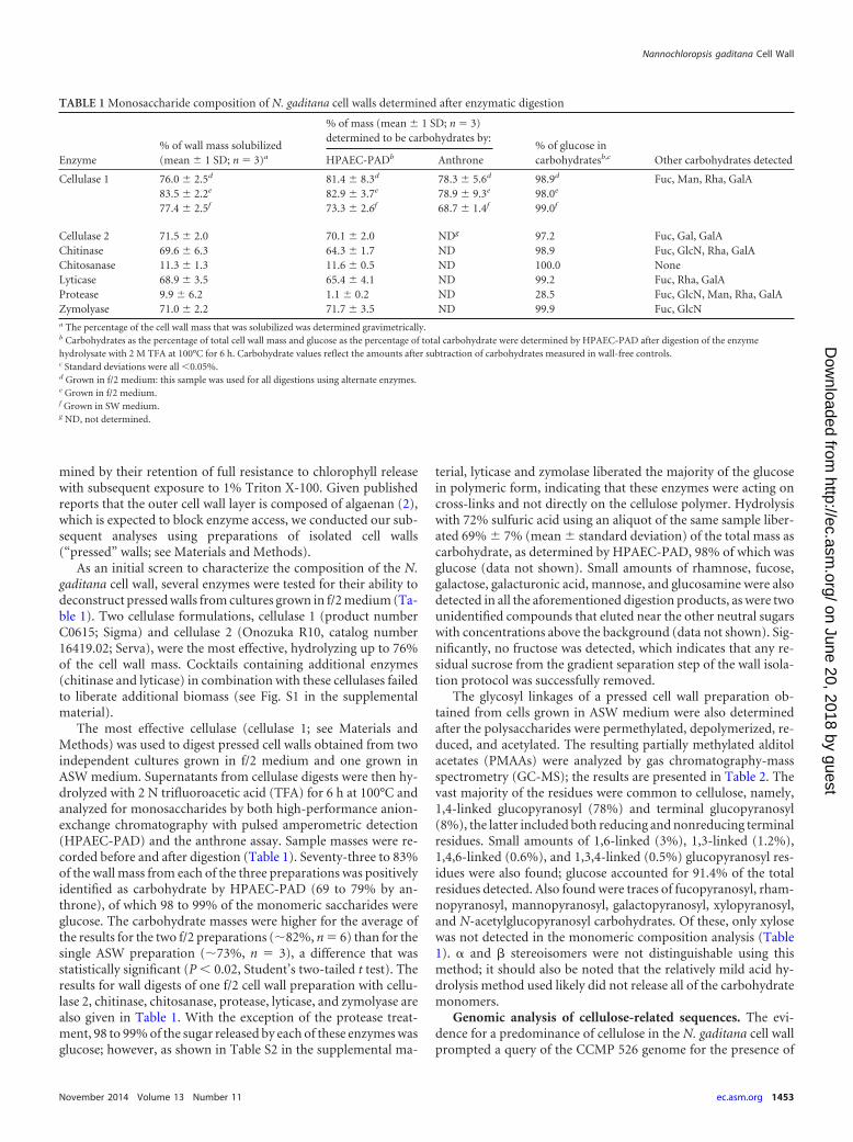
As an initial screen to characterize the composition of the N.gaditana cell wall, several enzymes were tested for their ability todeconstruct pressed walls from cultures grown in f/2 medium (Ta-ble 1). Two cellulase formulations, cellulase 1 (product numberC0615; Sigma) and cellulase 2 (Onozuka R10, catalog number16419.02; Serva), were the most effective, hydrolyzing up to 76%of the cell wall mass. Cocktails containing additional enzymes(chitinase and lyticase) in combination with these cellulases failedto liberate additional biomass (see Fig. S1 in the supplementalmaterial).
The most effective cellulase (cellulase 1; see Materials andMethods) was used to digest pressed cell walls obtained from twoindependent cultures grown in f/2 medium and one grown inASW medium. Supernatants from cellulase digests were then hy-drolyzed with 2 N trifluoroacetic acid (TFA) for 6 h at 100°C andanalyzed for monosaccharides by both high-performance anion-exchange chromatography with pulsed amperometric detection(HPAEC-PAD) and the anthrone assay. Sample masses were re-corded before and after digestion (Table 1). Seventy-three to 83%of the wall mass from each of the three preparations was positivelyidentified as carbohydrate by HPAEC-PAD (69 to 79% by an-throne), of which 98 to 99% of the monomeric saccharides wereglucose. The carbohydrate masses were higher for the average ofthe results for the two f/2 preparations (�82%, n 6) than for thesingle ASW preparation (�73%, n 3), a difference that wasstatistically significant (P � 0.02, Student’s two-tailed t test). Theresults for wall digests of one f/2 cell wall preparation with cellu-lase 2, chitinase, chitosanase, protease, lyticase, and zymolyase arealso given in Table 1. With the exception of the protease treat-ment, 98 to 99% of the sugar released by each of these enzymes wasglucose; however, as shown in Table S2 in the supplemental ma-
terial, lyticase and zymolase liberated the majority of the glucosein polymeric form, indicating that these enzymes were acting oncross-links and not directly on the cellulose polymer. Hydrolysiswith 72% sulfuric acid using an aliquot of the same sample liber-ated 69% 7% (mean standard deviation) of the total mass ascarbohydrate, as determined by HPAEC-PAD, 98% of which wasglucose (data not shown). Small amounts of rhamnose, fucose,galactose, galacturonic acid, mannose, and glucosamine were alsodetected in all the aforementioned digestion products, as were twounidentified compounds that eluted near the other neutral sugarswith concentrations above the background (data not shown). Sig-nificantly, no fructose was detected, which indicates that any re-sidual sucrose from the gradient separation step of the wall isola-tion protocol was successfully removed.
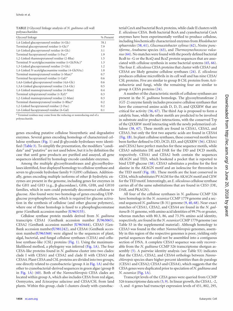
The glycosyl linkages of a pressed cell wall preparation ob-tained from cells grown in ASW medium were also determinedafter the polysaccharides were permethylated, depolymerized, re-duced, and acetylated. The resulting partially methylated alditolacetates (PMAAs) were analyzed by gas chromatography-massspectrometry (GC-MS); the results are presented in Table 2. Thevast majority of the residues were common to cellulose, namely,1,4-linked glucopyranosyl (78%) and terminal glucopyranosyl(8%), the latter included both reducing and nonreducing terminalresidues. Small amounts of 1,6-linked (3%), 1,3-linked (1.2%),1,4,6-linked (0.6%), and 1,3,4-linked (0.5%) glucopyranosyl res-idues were also found; glucose accounted for 91.4% of the totalresidues detected. Also found were traces of fucopyranosyl, rham-nopyranosyl, mannopyranosyl, galactopyranosyl, xylopyranosyl,and N-acetylglucopyranosyl carbohydrates. Of these, only xylosewas not detected in the monomeric composition analysis (Table1). � and � stereoisomers were not distinguishable using thismethod; it should also be noted that the relatively mild acid hy-drolysis method used likely did not release all of the carbohydratemonomers.
Genomic analysis of cellulose-related sequences. The evi-dence for a predominance of cellulose in the N. gaditana cell wallprompted a query of the CCMP 526 genome for the presence of
TABLE 1 Monosaccharide composition of N. gaditana cell walls determined after enzymatic digestion
Enzyme% of wall mass solubilized(mean 1 SD; n 3)a
% of mass (mean 1 SD; n 3)determined to be carbohydrates by:
% of glucose incarbohydratesb,c Other carbohydrates detectedHPAEC-PADb Anthrone
Cellulase 1 76.0 2.5d 81.4 8.3d 78.3 5.6d 98.9d Fuc, Man, Rha, GalA83.5 2.2e 82.9 3.7e 78.9 9.3e 98.0e
77.4 2.5f 73.3 2.6f 68.7 1.4f 99.0f
Cellulase 2 71.5 2.0 70.1 2.0 NDg 97.2 Fuc, Gal, GalAChitinase 69.6 6.3 64.3 1.7 ND 98.9 Fuc, GlcN, Rha, GalAChitosanase 11.3 1.3 11.6 0.5 ND 100.0 NoneLyticase 68.9 3.5 65.4 4.1 ND 99.2 Fuc, Rha, GalAProtease 9.9 6.2 1.1 0.2 ND 28.5 Fuc, GlcN, Man, Rha, GalAZymolyase 71.0 2.2 71.7 3.5 ND 99.9 Fuc, GlcNa The percentage of the cell wall mass that was solubilized was determined gravimetrically.b Carbohydrates as the percentage of total cell wall mass and glucose as the percentage of total carbohydrate were determined by HPAEC-PAD after digestion of the enzymehydrolysate with 2 M TFA at 100°C for 6 h. Carbohydrate values reflect the amounts after subtraction of carbohydrates measured in wall-free controls.c Standard deviations were all �0.05%.d Grown in f/2 medium: this sample was used for all digestions using alternate enzymes.e Grown in f/2 medium.f Grown in SW medium.g ND, not determined.
Nannochloropsis gaditana Cell Wall
November 2014 Volume 13 Number 11 ec.asm.org 1453
on June 20, 2018 by guesthttp://ec.asm
.org/D
ownloaded from
genes encoding putative cellulose biosynthetic and degradativeenzymes. Several genes encoding homologs of characterized cel-lulose synthases (Fig. 1) and �-glycoside hydrolases were identi-fied (Table 3). To simplify the presentation, the modifiers “candi-date” and “putative” are not used below, but it is by definition thecase that until gene products are purified and assayed, all genesequences identified by homology encode candidate enzymes.
Among the multiple glycosyltransferases and glycosylhydro-lases identified, four displayed homology to cellulose synthase andseven to glycoside hydrolase family 9 (GH9) cellulases. Addition-ally, genes encoding multiple isoforms of other �-hydrolytic en-zymes are present in the genome, including genes for enzymes inthe GH1 and GH3 (e.g., �-glucosidase), GH6, GH8, and GH10families, which in sum could potentially deconstruct cellulose toglucose. Also found were two homologs of genes encoding UDP-glucose pyrophosphorylase, which is required for glucose activa-tion in the synthesis of cellulose (and other glucose polymers),where one of these homologs is fused to a phosphoglucomutasegene (GenBank accession number JU963155).
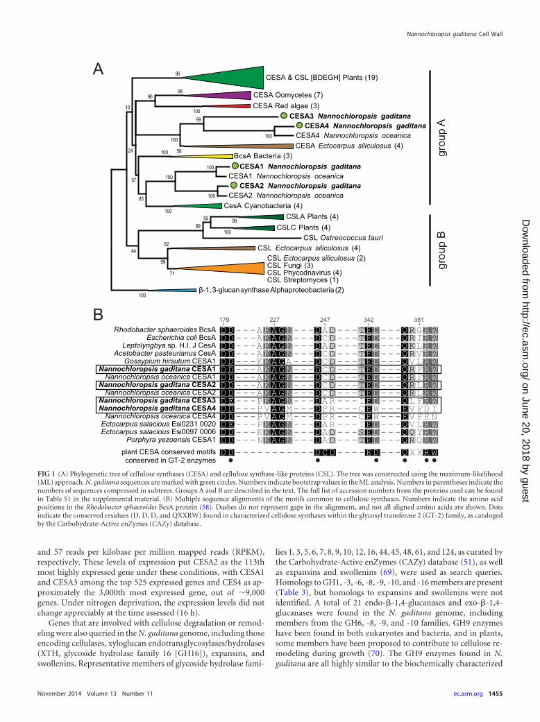
Cellulose synthase protein models derived from N. gaditanatranscripts CESA1 (GenBank accession number JU963065),CESA2 (GenBank accession number JU963444), CESA3 (Gen-Bank accession numberJU981242), and CESA4 (GenBank acces-sion numberJU963368) were aligned to the sequences of plant,algal, bacterial, and fungal cellulose synthases (CESA) and cellu-lose synthase-like (CSL) proteins (Fig. 1). Using the maximum-likelihood method, a phylogeny was inferred (Fig. 1A). The fourCESA-like proteins found in N. gaditana cluster into two clades:clade I with CESA1 and CESA2 and clade II with CESA3 andCESA4. Plant CESA and CSL proteins are divided into two groups,one directly related to cyanobacteria (group A in Fig. 1A) and theother to cyanobacterial-derived sequences in green algae (group Bin Fig. 1A) (60). Both of the Nannochloropsis CESA clades arelocated within group A, which also includes CESA from red algae,Oomycetes, and Ectocarpus salacious and CESA/CSL from landplants. Within this group, clade I clusters closely with cyanobac-
terial CesA and bacterial BcsA proteins, while clade II clusters withE. siliculosus CESA. Both bacterial BcsA and cyanobacterial CesAenzymes have been experimentally verified to produce cellulose,including biochemically characterized enzymes from Rhodobactersphaeroides (58, 61), Gluconacetobacter xylinus (62), Nostoc punc-tiforme, Anabaena species (63), and Thermosynechococcus vulca-nus (64). No matches were found with the poorly defined bacterialBcsB to -G or the BcsQ and BcsZ protein sequences that are asso-ciated with cellulose synthesis in some bacterial systems (65, 66).The four E. siliculosus CESA proteins that cluster with CESA3 andCESA4 are likely genuine cellulose synthases (24). E. siliculosusproduces cellulose microfibrils in its cell wall and has nine CESA/CSL proteins. Five are similar to group B CSL proteins from Acti-nobacteria and fungi, while the remaining four are similar togroup A CESA proteins (24).
A number of the characteristic motifs of cellulose synthases arepresent in the N. gaditana homologs. The glycosyltransferase 2(GT-2) enzyme family includes processive cellulose synthases thathave the conserved amino acids D, D, D, and QXXRW that arecritical for activity (56, 67). The third Asp is proposed to form acatalytic base, while the other motifs are predicted to be involvedin substrate and/or product interactions, with the conserved Trpof the QXXRW motif interacting with the newly polymerized cel-lulose (58, 67). These motifs are found in CESA1, CESA2, andCESA3, but only the first two aspartic acids are found in CESA4(Fig. 1B). In plant cellulose synthases, these conserved motifs havebeen further defined to DD, DCD, ED, and QXXRW (56). CESA1and CESA2 have perfect matches for these sequence motifs, whileCESA3 substitutes DE and DAR for the DD and DCD motifs,respectively. CESA1 and CESA2 both contain the sequencesAKAGN and TED, which bookend a pocket that is reported tobind UDP-glucose (58). CESA3 substitutes a proline for the firstalanine in the AKAGN motif and an isoleucine for threonine inthe TED motif (Fig. 1B). These motifs are the least conserved inCES4, which substitutes PVAGM for the AKAGN motif and CEWfor the TED motif. Interestingly, an E. siliculosus cellulose synthasecarries all of the same substitutions that are found in CES3 (DE,DAR, and PKAGN).
All four of the cellulose synthases in N. gaditana CCMP 526have homologs in the N. oceanica CCMP 1779 genome and a sec-ond sequenced N. gaditana (B-31) genome (9, 40, 68). Near-exactmatches of CESA1, CESA2, and CESA4 are found in the N. gad-itana B-31 genome, with amino acid identities of 99.7% or greater,whereas matches with 80.3, 86, and 73.3% amino acid identity,respectively, are found in the N. oceanica CCMP 1779 genome (seeTable S3 in the supplemental material). Although homology toCESA3 was found in the other Nannochloropsis genomes, assem-bly in this region of the respective genomes is poor, yielding onlypartial sequences that could not be assembled into a contiguoussection of DNA. A complete CESA3 sequence was only recover-able from the N. gaditana CCMP 526 transcriptome shotgun as-sembly (5). A pairwise identity analysis (see Table S3) indicatesthat the CESA1, CESA2, and CESA4 orthologs between Nanno-chloropsis species share higher percent identities than do paralogs(CESA1 and CESA2; CESA3 and CESA4), which suggests that theCESA genes were duplicated prior to speciation of N. gaditana andN. oceanica (Fig. 1A).
Transcript levels of the CESA genes were queried from CCMP526 transcriptome data sets (5, 9). In linear growth, the CESA1, -2,-3, and -4 genes had transcript expression levels of 451, 802, 295,
TABLE 2 Glycosyl linkage analysis of N. gaditana cell wallpolysaccharides
Glycosyl linkage % Present
1,4-Linked glucopyranosyl residue (4-Glc) 78.1Terminal glucopyranosyl residue (t-Glc)a 7.91,6-Linked glucopyranosyl residue (6-Glc) 3.1Terminal fucopyranosyl residue (t-Fuc)a 1.91,2-Linked rhamnopyranosyl residue (2-Rha) 1.5Terminal N-acetylglucosamine residue (t-GlcNAc)a 1.31,3-Linked glucopyranosyl residue (3-Glc) 1.21,4-Linked N-acetylglucosamine residue (4-GlcNAc) 1.0Terminal mannopyranosyl residue (t-Man)a 0.7Terminal fucopyranosyl residue (t-Gal)a 0.61,4,6-Linked glucopyranosyl residue (4,6-Glc) 0.61,3,4-Linked glucopyranosyl residue (3,4-Glc) 0.51,4-Linked mannopyranosyl residue (4-Man) 0.4Terminal xylopyranosyl residue (t-Xyl)a 0.31,2-Linked mannopyranosyl residue (2-Man) 0.3Terminal rhamnopyranosyl residue (t-Rha)a 0.21,2-Linked fucopyranosyl residue (2-Fuc) 0.21,6-Linked fucopyranosyl residue (6-Gal) 0.1a Terminal residues may come from the reducing or nonreducing end of apolysaccharide.
Scholz et al.
1454 ec.asm.org Eukaryotic Cell
on June 20, 2018 by guesthttp://ec.asm
.org/D
ownloaded from
and 57 reads per kilobase per million mapped reads (RPKM),respectively. These levels of expression put CESA2 as the 113thmost highly expressed gene under these conditions, with CESA1and CESA3 among the top 525 expressed genes and CES4 as ap-proximately the 3,000th most expressed gene, out of �9,000genes. Under nitrogen deprivation, the expression levels did notchange appreciably at the time assessed (16 h).
Genes that are involved with cellulose degradation or remod-eling were also queried in the N. gaditana genome, including thoseencoding cellulases, xyloglucan endotransglycosylases/hydrolases(XTH, glycoside hydrolase family 16 [GH16]), expansins, andswollenins. Representative members of glycoside hydrolase fami-
lies 1, 3, 5, 6, 7, 8, 9, 10, 12, 16, 44, 45, 48, 61, and 124, as curated bythe Carbohydrate-Active enZymes (CAZy) database (51), as wellas expansins and swollenins (69), were used as search queries.Homologs to GH1, -3, -6, -8, -9, -10, and -16 members are present(Table 3), but homologs to expansins and swollenins were notidentified. A total of 21 endo-�-1,4-glucanases and exo-�-1,4-glucanases were found in the N. gaditana genome, includingmembers from the GH6, -8, -9, and -10 families. GH9 enzymeshave been found in both eukaryotes and bacteria, and in plants,some members have been proposed to contribute to cellulose re-modeling during growth (70). The GH9 enzymes found in N.gaditana are all highly similar to the biochemically characterized
CESA & CSL [BDEGH] Plants (19)
CESA Oomycetes (7)CESA Red algae (3)
CESA3 Nannochloropsis gaditanaCESA4 Nannochloropsis gaditana
CESA4 Nannochloropsis oceanicaCESA Ectocarpus siliculosus (4)
BcsA Bacteria (3)CESA1 Nannochloropsis gaditana
CESA1 Nannochloropsis oceanicaCESA2 Nannochloropsis gaditana
CESA2 Nannochloropsis oceanicaCesA Cyanobacteria (4)
CSLA Plants (4)CSLC Plants (4)
CSL Ostreococcus tauriCSL Ectocarpus siliculosus (4)
β-1, 3-glucan synthase Alphaproteobacteria (2)
100
100
100
100
100
100
100
99100
99
83
96
9696
100
5999
58
100
82
57
71
99
44
24
10
CSL Ectocarpus siliculosus (2)CSL Fungi (3)CSL Phycodnavirus (4)CSL Streptomyces (1)
Rhodobacter sphaeroides BcsAEscherichia coli BcsA
Leptolyngbya sp. H.I. J CesAAcetobacter pasteurianus CesA
Gossypium hirsutum CESA1Nannochloropsis gaditana CESA1
Nannochloropsis oceanica CESA1Nannochloropsis gaditana CESA2
Nannochloropsis oceanica CESA2Nannochloropsis gaditana CESA3Nannochloropsis gaditana CESA4
Nannochloropsis oceanica CESA4Ectocarpus salacious Esi0231 0020Ectocarpus salacious Esi0097 0006
Porphyra yezoensis CESA1
179 227 247 342 381
plant CESA conserved motifsconserved in GT-2 enzymes
FIG 1 (A) Phylogenetic tree of cellulose synthases (CESA) and cellulose synthase-like proteins (CSL). The tree was constructed using the maximum-likelihood(ML) approach. N. gaditana sequences are marked with green circles. Numbers indicate bootstrap values in the ML analysis. Numbers in parentheses indicate thenumbers of sequences compressed in subtrees. Groups A and B are described in the text. The full list of accession numbers from the proteins used can be foundin Table S1 in the supplemental material. (B) Multiple sequence alignments of the motifs common to cellulose synthases. Numbers indicate the amino acidpositions in the Rhodobacter sphaeroides BcsA protein (58). Dashes do not represent gaps in the alignment, and not all aligned amino acids are shown. Dotsindicate the conserved residues (D, D, D, and QXXRW) found in characterized cellulose synthases within the glycosyl transferase 2 (GT-2) family, as catalogedby the Carbohydrate-Active enZymes (CAZy) database.
Nannochloropsis gaditana Cell Wall
November 2014 Volume 13 Number 11 ec.asm.org 1455
on June 20, 2018 by guesthttp://ec.asm
.org/D
ownloaded from
cellulase of Clostridium thermocellum, which has activity againstboth cellulose and lichenin but little against laminarin or xylan(71, 72). Alignment of the putative CCMP 526 cellulases with theC. thermocellum cellulase indicates that the proteins share signa-ture motifs (see Fig. S2 in the supplemental material). These in-clude the DLXGGXXDAGD, HRR, and DXXXXXXXXE motifsthat contain amino acids necessary for catalytic activity (73, 74).Our analysis also indicates that regions with the sequences LFXEXQRXG and WRXD are conserved among the C. thermocellumand CCMP 526 cellulases. Interestingly, one GH6 family cellobio-hydrolase II (CBHII)-like enzyme was found in the N. gaditanagenome. Typically, this family of enzymes is found in fungi andother cellulolytic organisms, where the enzyme acts processively
from the nonreducing ends of cellulose chains to generate cello-biose (75). A GH6 homolog is also found in the stramenopileAureococcus anophagefferens but appears to be absent in oomy-cetes, which include plant pathogens. Additionally, another pro-tein with possible fungal origin, a fungal-type cellulose-bindingdomain (CBM1), was also found in N. gaditana. A search forplant-type xyloglucan endotransglycosylases/hydrolases (GH16)yielded one match in the transcriptome assembly. Three GH1 andfour GH3 enzymes were also present. The enzymes in these fam-ilies have �-glucosidase, �-galactosidase, or �-mannosidase activ-ity (51). In the case of �-glucosidases, these enzymes hydrolyze theexocellulase product, typically cellobiose, into individual mono-saccharides (76), so these enzymes may also play a role in N. gad-itana cellulose reorganization/metabolism.
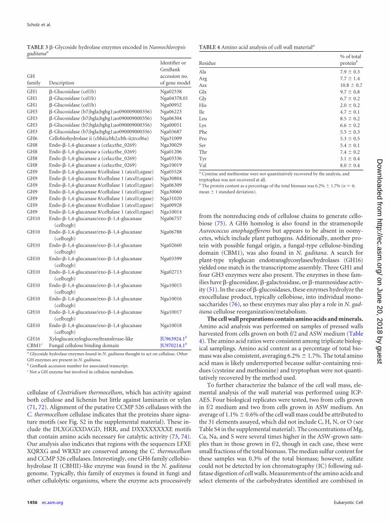
The cell wall preparations contain amino acids and minerals.Amino acid analysis was performed on samples of pressed wallsharvested from cells grown on both f/2 and ASW medium (Table4). The amino acid ratios were consistent among triplicate biolog-ical samplings. Amino acid content as a percentage of total bio-mass was also consistent, averaging 6.2% 1.7%. The total aminoacid mass is likely underreported because sulfur-containing resi-dues (cysteine and methionine) and tryptophan were not quanti-tatively recovered by the method used.
To further characterize the balance of the cell wall mass, ele-mental analysis of the wall material was performed using ICP-AES. Four biological replicates were tested, two from cells grownin f/2 medium and two from cells grown in ASW medium. Anaverage of 1.1% 0.6% of the cell wall mass could be attributed tothe 31 elements assayed, which did not include C, H, N, or O (seeTable S4 in the supplemental material). The concentrations of Mg,Ca, Na, and S were several times higher in the ASW-grown sam-ples than in those grown in f/2, though in each case, these weresmall fractions of the total biomass. The median sulfur content forthese samples was 0.3% of the total biomass; however, sulfatecould not be detected by ion chromatography (IC) following sul-fatase digestion of cell walls. Measurements of the amino acids andselect elements of the carbohydrates identified are combined in
TABLE 3 �-Glycoside hydrolase enzymes encoded in Nannochloropsisgaditanaa
GHfamily Description
Identifier orGenBankaccession no.of gene model
GH1 �-Glucosidase (cel1b) Nga02538GH1 �-Glucosidase (cel1b) Nga04378.01GH1 �-Glucosidase (cel1b) Nga00952GH3 �-Glucosidase (b7;bgla;bghg1;ao090009000356) Nga06223GH3 �-Glucosidase (b7;bgla;bghg1;ao090009000356) Nga06304GH3 �-Glucosidase (b7;bgla;bghg1;ao090009000356) Nga00051GH3 �-Glucosidase (b7;bgla;bghg1;ao090009000356) Nga03687GH6 Cellobiohydrolase ii (cbhii;cbh2;cbh-ii;trcel6a) Nga31009GH8 Endo-�-1,4-glucanase a (cela;cthe_0269) Nga30029GH8 Endo-�-1,4-glucanase a (cela;cthe_0269) Nga01206GH8 Endo-�-1,4-glucanase a (cela;cthe_0269) Nga03336GH8 Endo-�-1,4-glucanase a (cela;cthe_0269) Nga10019GH9 Endo-�-1,4-glucanase 8/cellulase 1 (atcel1;egase) Nga05528GH9 Endo-�-1,4-glucanase 8/cellulase 1 (atcel1;egase) Nga30804GH9 Endo-�-1,4-glucanase 8/cellulase 1 (atcel1;egase) Nga06309GH9 Endo-�-1,4-glucanase 8/cellulase 1 (atcel1;egase) Nga30060GH9 Endo-�-1,4-glucanase 8/cellulase 1 (atcel1;egase) Nga31020GH9 Endo-�-1,4-glucanase 8/cellulase 1 (atcel1;egase) Nga00928GH9 Endo-�-1,4-glucanase 8/cellulase 1 (atcel1;egase) Nga10014GH10 Endo-�-1,4-glucanase/exo-�-1,4-glucanase
(celb;egb)Nga06757
GH10 Endo-�-1,4-glucanase/exo-�-1,4-glucanase(celb;egb)
Nga06788
GH10 Endo-�-1,4-glucanase/exo-�-1,4-glucanase(celb;egb)
Nga02660
GH10 Endo-�-1,4-glucanase/exo-�-1,4-glucanase(celb;egb)
Nga03399
GH10 Endo-�-1,4-glucanase/exo-�-1,4-glucanase(celb;egb)
Nga02713
GH10 Endo-�-1,4-glucanase/exo-�-1,4-glucanase(celb;egb)
Nga10015
GH10 Endo-�-1,4-glucanase/exo-�-1,4-glucanase(celb;egb)
Nga10016
GH10 Endo-�-1,4-glucanase/exo-�-1,4-glucanase(celb;egb)
Nga10017
GH10 Endo-�-1,4-glucanase/exo-�-1,4-glucanase(celb;egb)
Nga10018
GH16 Xyloglucan:xyloglucosyltransferase-like JU963924.1b
CBM1c Fungal cellulose binding domain JU970214.1b
a Glycoside hydrolase enzymes found in N. gaditana thought to act on cellulose. OtherGH enzymes are present in N. gaditana.b GenBank accession number for associated transcript.c Not a GH enzyme but involved in cellulose metabolism.
TABLE 4 Amino acid analysis of cell wall materiala
Residue% of totalproteinb
Ala 7.9 0.3Arg 7.7 1.4Asx 10.8 0.7Glx 9.7 0.8Gly 6.7 0.2His 2.0 0.2Ile 4.7 0.1Leu 8.5 0.2Lys 6.6 0.2Phe 5.5 0.3Pro 5.3 0.5Ser 5.4 0.1Thr 7.4 0.2Tyr 3.1 0.4Val 8.0 0.4a Cysteine and methionine were not quantitatively recovered by the analysis, andtryptophan was not recovered at all.b The protein content as a percentage of the total biomass was 6.2% 1.7% (n 4;mean 1 standard deviation).
Scholz et al.
1456 ec.asm.org Eukaryotic Cell
on June 20, 2018 by guesthttp://ec.asm
.org/D
ownloaded from
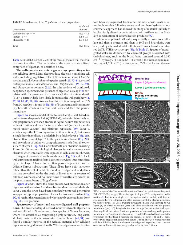
Table 5. In total, 86.5% 7.2% of the mass of the cell wall materialhas been identified. The remainder of the mass balance is likelycomprised of algaenan, as described below.
The wall comprises an outer algaenan layer protecting an in-ner cellulose layer. Many algae produce algaenan-containing cellwalls, including vegetative cells of Scenedesmus, some Chlorellaspecies, and all Nannochloropsis species tested (25, 77–81), cysts ofChlamydomonas, Haematococcus, and Polytomella (49, 82–89),and Botryococcus colonies (126). In thin sections of osmicated,dehydrated specimens, the presence of algaenan usually (43) cor-relates with the presence of a layer called the trilaminar sheath(TLS), a narrow dark-light-dark domain at the wall periphery (2,77, 80, 81, 83, 88, 90). An excellent thin-section image of the TLSfrom N. occulata is found in Fig. 3H of Murakami and Hashimoto(2), beneath which is a second wall layer and then the plasmamembrane.
Figure 2A shows a model of the Nannochloropsis wall based onquick-freeze deep-etch EM (QFDE-EM), wherein living cells orwall preparations are snap-frozen at liquid-helium temperatureswithout fixation and then fractured and etched (surface ice subli-mated under vacuum) and platinum replicated (49). Layer 1,which adopts the TLS configuration in thin section (2) but formsa single layer in replicas, is underlain by a thicker layer 2 (Fig. 2B).Struts often connect layer 2 to the plasma membrane (Fig. 2C),and extensions of unknown composition protrude from the outersurface of layer 1 (Fig. 2C). Consistent with our observations usingTriton X-100, no morphological changes in wall structure wereobserved when intact cells were exposed to cellulases (not shown).
Images of pressed cell walls are shown in Fig. 2D and E. Eachwall curves in on itself to form a concentric whorl interconnectedby struts. Layer 2 has a fluffy, often porous appearance with adelicate fibrous substructure. These fibers have a far narrowercaliber than the cellulose fibrils found in red algae and land plantsthat are assembled under the aegis of linear rows or rosettes ofcellulose synthases, and no linear rows or rosettes are evident inthe plasma membrane of N. gaditana.
Figure 2F and G show this same pressed wall preparation afterdigestion with cellulase 1 as described in Materials and Methods.Layer 2 and the struts have been completely removed, generatingan apparently pure preparation of layer 1 whose outer surface (Fig.2G, o) retains the extensions and whose newly exposed inner layer(Fig. 2G, i) is granular.
Spectroscopy of intact and enzyme-digested wall prepara-tions. The presence of lipid-derived, nonhydrolyzable algaenan iswell established in N. salina and other species of Nannochloropsis,where it is described as comprising highly saturated, long-chainaliphatic material that is cross-linked by ether bonds (41, 91). Wefound a similar material in the residual material after cellulasedigestion of N. gaditana cell walls. Whereas algaeanan has hereto-
fore been distinguished from other biomass constituents as aninsoluble residue following severe acid and base hydrolysis, ourenzymatic approach has allowed the study of material unlikely tobe chemically altered or contaminated with artifacts such as Mail-lard condensation or caramelization products (92).
Aliquots of pressed cell walls, sequentially exposed to a cellu-lase and then a protease and then to HCl acid hydrolysis, wereanalyzed by attenuated total reflectance Fourier transform infra-red (ATR-FTIR) spectroscopy (Fig. 3; Table 6). Spectra of nondi-gested walls are dominated by chemical groups associated withcarbohydrates, such as the broad band centered around 3,380cm�1 (hydroxyl, H-bonded, O-H stretch), the intense band max-imizing at 1,029 cm�1 (hydroxyl/ether, C-O stretch), and the nar-
TABLE 5 Mass balance of the N. gaditana cell wall preparations
Component% of totalbiomass
Carbohydrate (n 3) 79.2 4.9Protein (n 4) 6.2 1.7Mineral (n 4) 1.1 0.6
Total 86.5 7.2
FIG 2 (A) Model of the Nannochloropsis wall based on quick-freeze deep-etchEM (QFDE-EM) images. The native layer 1 adopts a TLS configuration in thinsection (2) but forms a single layer in replicas and is overlain with fibrousextensions. Layer 2 is thicker and often associates with the plasma membranevia narrow struts. (B) Cross fracture through the native wall showing its twolayers (1, 2), distal extensions (ext), and close association with the plasmamembrane (pm). (C) Tangential fracture through the two native wall layersshowing distal extensions (ext) and proximal struts attaching to the plasmamembrane (pm). mito, mitochondrion. (D and E) Pressed cell walls, with theprominent fibrillar layer 2 masking the presence of layer 1. (F and G) Samepressed wall preparation as shown in panels D and E after 24 h of incubation incellulase. Fibrillar layer 2 and struts were digested, exposing layer 1, whoseouter (o) and inner (i) faces are granular. All scale bars, 250 nm.
Nannochloropsis gaditana Cell Wall
November 2014 Volume 13 Number 11 ec.asm.org 1457
on June 20, 2018 by guesthttp://ec.asm
.org/D
ownloaded from
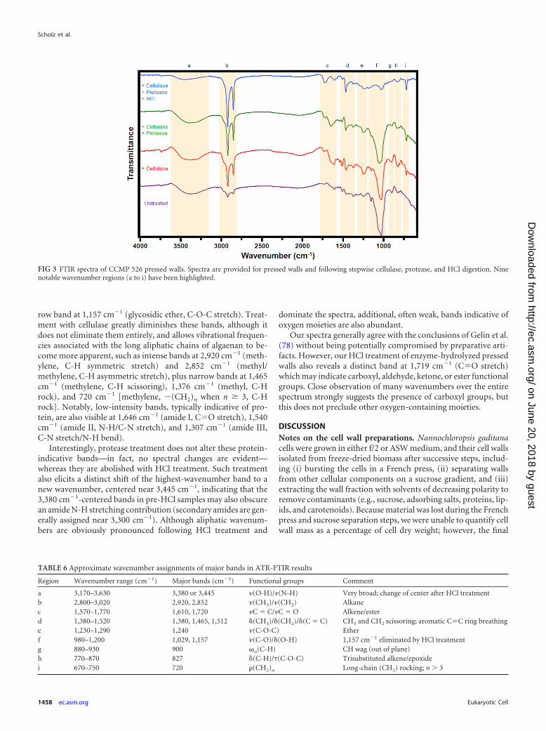
row band at 1,157 cm�1 (glycosidic ether, C-O-C stretch). Treat-ment with cellulase greatly diminishes these bands, although itdoes not eliminate them entirely, and allows vibrational frequen-cies associated with the long aliphatic chains of algaenan to be-come more apparent, such as intense bands at 2,920 cm�1 (meth-ylene, C-H symmetric stretch) and 2,852 cm�1 (methyl/methylene, C-H asymmetric stretch), plus narrow bands at 1,465cm�1 (methylene, C-H scissoring), 1,376 cm�1 (methyl, C-Hrock), and 720 cm�1 [methylene, �(CH2)n when n � 3, C-Hrock]. Notably, low-intensity bands, typically indicative of pro-tein, are also visible at 1,646 cm�1 (amide I, CO stretch), 1,540cm�1 (amide II, N-H/C-N stretch), and 1,307 cm�1 (amide III,C-N stretch/N-H bend).
Interestingly, protease treatment does not alter these protein-indicative bands—in fact, no spectral changes are evident—whereas they are abolished with HCl treatment. Such treatmentalso elicits a distinct shift of the highest-wavenumber band to anew wavenumber, centered near 3,445 cm�1, indicating that the3,380 cm�1-centered bands in pre-HCl samples may also obscurean amide N-H stretching contribution (secondary amides are gen-erally assigned near 3,300 cm�1). Although aliphatic wavenum-bers are obviously pronounced following HCl treatment and
dominate the spectra, additional, often weak, bands indicative ofoxygen moieties are also abundant.
Our spectra generally agree with the conclusions of Gelin et al.(78) without being potentially compromised by preparative arti-facts. However, our HCl treatment of enzyme-hydrolyzed pressedwalls also reveals a distinct band at 1,719 cm�1 (CO stretch)which may indicate carboxyl, aldehyde, ketone, or ester functionalgroups. Close observation of many wavenumbers over the entirespectrum strongly suggests the presence of carboxyl groups, butthis does not preclude other oxygen-containing moieties.
DISCUSSIONNotes on the cell wall preparations. Nannochloropsis gaditanacells were grown in either f/2 or ASW medium, and their cell wallsisolated from freeze-dried biomass after successive steps, includ-ing (i) bursting the cells in a French press, (ii) separating wallsfrom other cellular components on a sucrose gradient, and (iii)extracting the wall fraction with solvents of decreasing polarity toremove contaminants (e.g., sucrose, adsorbing salts, proteins, lip-ids, and carotenoids). Because material was lost during the Frenchpress and sucrose separation steps, we were unable to quantify cellwall mass as a percentage of cell dry weight; however, the final
FIG 3 FTIR spectra of CCMP 526 pressed walls. Spectra are provided for pressed walls and following stepwise cellulase, protease, and HCl digestion. Ninenotable wavenumber regions (a to i) have been highlighted.
TABLE 6 Approximate wavenumber assignments of major bands in ATR-FTIR results
Region Wavenumber range (cm�1) Major bands (cm�1) Functional groups Comment
a 3,170–3,630 3,380 or 3,445 �(O-H)/�(N-H) Very broad; change of center after HCl treatmentb 2,800–3,020 2,920, 2,852 �(CH3)/�(CH2) Alkanec 1,570–1,770 1,610, 1,720 �C C/�C O Alkene/esterd 1,380–1,520 1,380, 1,465, 1,512 (CH3)/ (CH2)/ (C C) CH3 and CH2 scissoring; aromatic CC ring breathinge 1,230–1,290 1,240 �(C-O-C) Etherf 980–1,200 1,029, 1,157 �(C-O)/ (O-H) 1,157 cm�1 eliminated by HCl treatmentg 880–930 900 �o(C-H) CH wag (out of plane)h 770–870 827 (C-H)/�(C-O-C) Trisubstituted alkene/epoxidei 670–750 720 �(CH2)n Long-chain (CH2) rocking; n � 3
Scholz et al.
1458 ec.asm.org Eukaryotic Cell
on June 20, 2018 by guesthttp://ec.asm
.org/D
ownloaded from
yields (after sample losses) were �2% of the cell dry weight. Uponinspection with electron microscopy, the wall preparations ap-peared highly homogeneous and uncontaminated by other mate-rials (Fig. 2D and E).
The pressed wall preparation is a variation of that published byTakeda (93), with the use of a sucrose rather than a Percoll gradi-ent being dictated by concerns that residual Percoll might compli-cate glycosyl linkage analysis. No trace of sucrose or fructose couldbe found in the resulting preparations when digested and analyzedby IC. The Takeda method also includes an �-amylase digestion toremove starch, but this step was deemed unnecessary because (i)Nannochloropsis species are not believed to produce starch, as isalso the case for other stramenopiles (94), (ii) no starch grains areevident by QFDE-EM of intact cells, and (iii) genomic evidence ofstarch biosynthetic enzymes (i.e., starch synthase and isoamylase)is lacking (5, 9, 40, 68, 93, 95–97). Furthermore, the low abun-dance of 1,3-linked glucose detected, combined with the absenceof detectable mannitol, suggested minimal contamination withlaminarin, a storage product common in related brown algae (98).It should be noted that the �-1,3-linked storage polysaccharidepresumed to be laminarin has not been verified in Nannochlorop-sis, nor has chrysolaminarin (99) or mycolaminarin (100), whichare found in more distantly related diatoms and oomycetes, re-spectively.
We are aware that the use of chemical treatments to removeadventitious material may inadvertently remove constituent wallmolecules as well. That said, during the organic extraction steps,only �5% of the sucrose gradient-separated cell wall mass wasremoved (data not shown).
Cellulose in the N. gaditana cell wall. The bulk (�75%) of N.gaditana cell wall preparations was determined to be cellulose,which we show by QFDE-EM to form the inner layer of a bilayeredwall. Monosaccharide quantification after both chemical and en-zymatic digestion of the cell wall preparations demonstrates thatglucose is by far the dominant carbohydrate (Table 1). Linkageanalysis confirmed the predominance of 1,4-linked glucose (Table2); however, as this methodology uses relatively mild hydrolysisconditions, only a fraction of all carbohydrate linkages was deter-mined. The pressed wall material was highly susceptible to cellu-lase digestion, as evidenced by both HPAEC-PAD and EM data(Table 1; Fig. 2F and G). The lyticase and zymolyase enzymesliberated little free glucose, but TFA digestion of the enzyme hy-drolysates produced predominantly glucose (see Table S2 in thesupplemental material); we therefore suggest that they are notacting directly on the major wall polysaccharide but, rather, act oncritical cross-links to release the wall polymers. All of these treat-ments liberated glucose and only traces of other saccharides, con-sistent with a predominately cellulosic material.
Cellulose synthesis is performed in the plasma membrane bythe action of cellulose synthase complexes, also called terminalcomplexes (101–103). The primary substrate for cellulose syn-thase, UDP-glucose, is synthesized either anabolically from glu-cose by UDP-glucose pyrophosphorylase or catabolically from su-crose by sucrose synthase (102). We have identified in silicohomologs to three cellulose synthases containing core amino acidscentral to activity, two UDP-glucose pyrophosphorylases, andseven cellulases, demonstrating that N. gaditana at least putativelypossesses the metabolic machinery for cellulose synthesis and ca-tabolism. A search for sucrose synthase sequences derived from 18plant species yielded no homologues in the two N. gaditana ge-
nomes and one N. oceanica genome queried, suggesting that Nan-nochloropsis utilizes the anabolic pathway to UDP-glucose forma-tion.
Cellulose synthase terminal complexes assemble in geometri-cal configurations, often as linear rows or hexagonal rosettes, andcellulose morphology is directly related to these configurations(103). Commonly found in land plants and some algae, rosettestend to produce the crystalline cellulose I� allomorph; for exam-ple, the rosette terminal complexes of Micrasterias and Spyrogyraare thought to correlate with the perpendicular orientation of thecellulose I� fibrils (104). Linear terminal complexes have alsobeen characterized in algae, such as Valonia, Oocystis, and Boerges-neia, and are associated with cellulose I� production (103, 105–107). The configuration of Nannochloropsis terminal complexesand the degree of cellulose crystallinity are currently unknown.CESA3 is similar to a cellulose synthase from the brown alga E.siliculosus that forms long, linear terminal complexes (108). Nosuch configurations have been visualized in the Nannochloropsiscell membrane in our QFDE-EM studies. CESA1 and CESA2 arehighly similar to the bacterial BcsA enzymes, which in the case ofEscherichia coli have been suggested to produce amorphous cellu-lose (65).
Processive �-glycosyltransferases contain the characteristicmotifs D, D, D, and QXXRW located in two protein domains, A(D, D) and B (D, QXXRW). Both domain A and B are conservedin processive cellulase enzymes, while nonprocessive enzymestypically lack domain B (109). CESA1, -2, and -3 have both the Aand B domain, whereas CESA4 is lacking key residues that coor-dinate UDP (DCD, R in QXXRW), act as the catalytic base (ED),and stabilize the acceptor glucan (W in QXXRW), indicating thatdespite homology, CESA4 likely does not encode a genuine cellu-lose synthase.
Three of the E. siliceous cellulose synthases are highly expressedin female gametes just after fertilization, which correlates withrapid primary cell wall biogenesis (110). These three CESA genesare members of the E. siliceous CESA clade that forms a clusterwith Nannochloropsis CESA3 (Fig. 1A, group A). Despite somedifferences in the E. siliceous enzyme motifs relative to the se-quences of prototypical plant cellulose synthases (Fig. 1B), itseems likely that these enzymes are responsible for producing themicrofibrils of cellulose found in the cell wall of E. siliceous. Nan-nochloropsis CESA3 shares these distinctive motifs with the E. sili-ceous CESA enzymes, which include DE instead of DD and DARinstead of DCD.
There is a surprising lack of diversity in the monosaccharidecomposition of the cell wall preparations, although other saccha-rides and uronic acids, including 1,2-linked and terminal rham-nose and fucose residues, 1,4-linked and terminal mannose andN-acetylglucosamine residues, and 1,6-linked and terminal galac-tose residues (Table 2), were present in trace amounts. Treatmentwith cellulase 2 yielded 2.7% of the polysaccharide content as ga-lactose (data not shown), but in all other enzymatic treatments,the masses of nonglucose monosaccharides and uronic acids wereless than 1% of the sample mass (Table 1).
Cell wall amino acids. Amino acid analysis indicated that�6% of the cell wall mass is comprised of amino acids. The indi-vidual amino acid ratios were highly reproducible between runs,and cell wall preparations were rigorously washed with both polarand nonpolar solvents, suggesting that the amino acids were notadventitiously associated with cell walls but, rather, represented
Nannochloropsis gaditana Cell Wall
November 2014 Volume 13 Number 11 ec.asm.org 1459
on June 20, 2018 by guesthttp://ec.asm
.org/D
ownloaded from
an integral cell wall constituent. Attempts to isolate purified pro-teins from cell wall preparations incubated with denaturing buf-fers failed to reveal well resolved protein bands using SDS gelelectrophoresis. This may be due to the proteins being embeddedwithin the cell wall and unable to diffuse from this matrix or toproteins being covalently linked to insoluble algaenan (see below)or a cell wall carbohydrate. Additionally, the current cell wallpreparations yield relatively small amounts of biomass, and theproteins may be below the limit of detection, particularly if there isheterogeneity due to differing degrees of posttranslational modi-fication (i.e., glycosylation). BLAST searches for homologs ofother algal cell wall proteins (e.g., hydroxyproline-rich glycopro-teins from C. reinhardtii) failed to reveal any significant matches.Despite our inability to identify cell wall proteins, the amino acidanalysis suggests the presence of tightly associated proteins (per-haps covalently linked to other wall constituents or encapsulatedwithin the cell wall). Additional investigation is required to ascer-tain whether the amino acids correspond to specific polypeptidesencoded in the genome or whether they are assembled by nonri-bosomal pathways (111, 112) that are core components of the cellwall.
We compared the cell wall composition of N. gaditana cellsgrown in ASW and f/2 media. While subtle differences are notedwith respect to some cell wall components, including minerals andminor carbohydrates, the main component of the cell walls (glu-cose) does not change drastically in response to varying the com-position of the medium, demonstrating the dominant structuralrole of cellulose in the N. gaditana wall. It remains to be deter-mined whether stresses like nutrient limitation and high light mayaffect the composition of Nannochloropsis cell walls.
The algaenan outer wall. The FTIR and EM data presented inthis paper clearly demonstrate that the outer wall of N. gaditanacontains algaenan (Fig. 2 and 3), which we propose to be primarilyresponsible for the wall’s recalcitrance to breakage. The term al-gaenan encompasses disparate types of enzymatically and chemi-cally resistant aliphatic material that may derive from distinct bio-chemical pathways in different organisms. The algaenan ofNannochloropsis salina and an unspecified Nannochloropsis hasbeen proposed to comprise straight-chain (�C30), highly satu-rated aliphatic compounds joined by ether bonds at terminal andone or two midchain positions (41). There is some debate aboutthe structure of algaenan in Tetraedron minimum and Scenedes-mus communis, but it appears to consist of very-long-chain (up toC120) monomeric (di)carboxylic acids and not the ester- andether-linked polymers that were originally proposed (43, 113).Meanwhile, the algaenan found in the colonial matrix of threeraces of Botryococcus braunii comprises polyacetals that are eithercross-linked by terpene epoxides (B and L race) or not (A race)(114, 115).
The composition of algaenan has been ascertained using di-verse methodologies that have the potential to affect structuraldeterminations (43, 44). To date, isolation has depended on severehydrolysis reactions at high temperature to degrade any reactivepolysaccharides, lipids, and proteins (41, 44, 113). Such harshtreatments have invited the criticism that the fundamental al-gaenan composition may be altered or contaminated with by-products, such as Maillard condensation products (44). In thepresent study, a 24-h digestion of pressed cell walls from N. gad-itana at room temperature, first with cellulase and then protease,left behind what appeared to be a homogeneous preparation of
algaenan-containing outer cell walls (Fig. 2F and G), albeit withsome remaining carbohydrate and protein signatures (Fig. 3).This enzymatic approach permitted the analysis of a highly en-riched algaenan preparation without exposure to high tempera-ture or strongly oxidizing/reducing conditions. Subsequent acidhydrolysis reduced, if not eliminated, the carbohydrate and pro-tein signatures in the FTIR but may have created artifacts (dis-cussed below). However, comparison of the FTIR spectra of thematerial before and after acid hydrolysis afforded us the opportu-nity to separate artifactual features from genuine ones.
The results after cellulase treatment demonstrate the removalof carbohydrate (and potentially protein) by the sizable reductionof related FTIR bands, such as those associated with glycosidicethers (1,157 cm�1) and amides (1,646 cm�1). Saponification isoften performed to remove ester-linked fatty acids from crudealgaenan (41, 44, 113); however, saponification followed by acid-catalyzed methyl esterification (fatty acid methyl ester [FAME]analysis) on a subset of algaenan preparations did not yield detect-able fatty acid methyl esters by GC-flame ionization detectionanalysis (data not shown). Since there was a clear lack of esterfunctionalities in our material, as demonstrated by the FTIR dataand FAME analysis, this obviated the need for saponification insubsequent sample analysis. The lack of esters is perhaps attribut-able to the preparatory steps employed, which may have yielded amaterial less contaminated (i.e., by membranes) than in previouswork.
Our FTIR data generally support the proposed Nannochlorop-sis algaenan model of Gelin et al. (78) by confirming the predom-inance of long-chain methylenic stretches with ether linkages andfew sites of unsaturation. However, there are notable differencesbetween our two sets of results. First, our data suggest that alcoholmoieties are present and that some of these may be midchain. Thelatter conclusion is drawn from comparing the FTIR spectra be-fore and after acid hydrolysis of the enzymatically isolated al-gaenan. After acid hydrolysis, alcohol-related bands at 3,380cm�1and 1,052 cm�1 are diminished and trans alkenes becomemore apparent with the band at 965 cm�1. The disappearance ofthe alcohol bands can be attributed, at least in part, to removal ofresidual carbohydrates, but the emergence of trans unsaturationintimates the dehydration of secondary alcohols to form alkenes.Alternatively, the removal of carbohydrate –OH bands may havesimply unmasked preexisting carbon double bonds. Alcoholgroups were either not observed or were of low abundance in thealgaenan of other studies, but sites of unsaturation were observed(41, 43, 113). Sites of unsaturation may be derived from dehydra-tion reactions, since the algaenan in those studies was isolated onlyafter severe acid hydrolysis. In addition to finding alcohol groupsin the algaenan material, we observed indications of carboxyland/or aldehyde functionalities after acid hydrolysis, neither ofwhich was identified in the prior study of Nannochloropsis al-gaenan, although the former was found in the algaenans of fresh-water algae (41, 43).
Nannochloropsis algaenans appear to be much the same biopo-lymer as the cutan found in drought-resistant land plants, such asAgave and Clivia (45, 113, 116–118). Isolation of cutan from plantcuticular material is performed in much the same way as classicalalgaenan isolation, namely, through solvent extraction and theapplication of strong reducing/oxidizing reagents to remove car-bohydrates, proteins, and free and ester-bound lipids (45, 116–119). As with the characterization of algaenan, structural determi-
Scholz et al.
1460 ec.asm.org Eukaryotic Cell
on June 20, 2018 by guesthttp://ec.asm
.org/D
ownloaded from
nations of cutan are uncertain because of study-to-study variationin isolation procedures and the potential for artifact generationintroduced by aggressive isolation approaches (45, 118, 120).However, it appears that this material comprises long-chain(�C30) alkanes and alkenes joined by ether linkages and is, thus, atleast an analogue of Nannochloropsis algaenan (45, 121).
Given the structural similarities between cutan and Nanno-chloropsis algaenan, it is tempting to speculate that similar biosyn-thetic machinery produces the two compounds. Radiolabeled li-noleic acid (C18:2) fed to Clivia miniata is incorporated into thealiphatic residue (i.e., cutan-rich material) of its leaves (118). In-triguingly, Nannochloropsis produces C18:1, C18:2, and C18:3 fattyacids, predominantly C18:1, and its algaenan contains long-chainaliphatics with substitutions at the �18 position (9, 41, 122). Anumber of possible algaenan precursors functionalized or unsat-urated at what would be the �18 position (the nomenclaturechanges according to functionalization) have been found in Nan-nochloropsis, including C32:1 alcohols, C32 and C32:1 diols, C32 hy-droxy fatty acids, C32 dihydroxy fatty acids, and C32 hydoxy ke-tones (41, 123, 124). This suggests that a C18 fatty acid may eitherbe elongated or condensed with a similar fatty molecule to pro-duce C28-C34 algaenan constituents, perhaps by the action ofpolyketide synthase(s).
Six polyketide synthases have been identified in silico in theCCMP 526 genome. Although the functions of these enzymes arenot yet known, we consider these promising targets for futureinterrogation in algaenan biosynthesis. Two lack dehydratase andenoyl reductase modules, and three have fatty acyl-reductase do-mains, consistent with the synthesis of predominately methylenicmonomers with alcohols/aldehydes for cross-linking. The FTIRspectra of algaenan indicate few CH3 moieties relative to CH2
moieties, suggesting that a predominately terpenoid pathway toalgaenan biosynthesis is unlikely. Although extensive work re-mains to characterize the composition and biosynthetic routes toalgaenan and its cross-linking, the methods for algaenan purifica-tion described here and the availability of multiple Nannochlorop-sis genomes will greatly facilitate such efforts.
ACKNOWLEDGMENTS
This material is based upon work supported by the Sustainable Algal Bio-fuels Consortium funded at CSM by the state of Colorado Energy Col-laboratory in support of award DE-EE0003372 from the U.S. Departmentof Energy, Bioenergy Technology Office. H.G.G. was funded by the NewAmerican University Fellowship at Arizona State University. Additionalsupport was provided to M.J.S. and M.C.P. by the Air Force Office ofScientific Research (grant FA9550-11-1-0211). U.G. was funded by con-tract DE-EE0003046 awarded to the National Alliance for Advanced Bio-fuels and Bioproducts (NAABB) from the U.S. Department of Energy andby grant SC0006873 from the DOE Office of Biological and Environmen-tal Research.
REFERENCES1. Andersen RA, Brett RW, Potter D, Sexton JP. 1998. Phylogeny of the
Eustigmatophyceae based upon 18S rDNA, with emphasis on Nannochlo-ropsis. Protist 149:61–74. http://dx.doi.org/10.1016/S1434-4610(98)70010-0.
2. Murakami R, Hashimoto H. 2009. Unusual nuclear division in Nanno-chloropsis oculata (Eustigmatophyceae, Heterokonta) which may en-sure faithful transmission of secondary plastids. Protist 160:41– 49. http://dx.doi.org/10.1016/j.protis.2008.09.002.
3. Rodolfi L, Zittelli GC, Barsanti L, Rosati G, Tredici MR. 2003. Growthmedium recycling in Nannochloropsis sp mass cultivation. Biomol. Eng.20:243–248. http://dx.doi.org/10.1016/S1389-0344(03)00063-7.
4. Bondioli P, Della Bella L, Rivolta G, Chini Zittelli G, Bassi N, Rodolfi
L, Casini D, Prussi M, Chiaramonti D, Tredici MR. 2012. Oil produc-tion by the marine microalgae Nannochloropsis sp. F&M-M24 and Tet-raselmis suecica F&M-M33. Bioresour. Technol. 114:567–572. http://dx.doi.org/10.1016/j.biortech.2012.02.123.
5. Jinkerson RE, Radakovits R, Posewitz MC. 2013. Genomic insightsfrom the oleaginous model alga Nannochloropsis gaditana. Bioengi-neered 4:37– 43. http://dx.doi.org/10.4161/bioe.21880.
6. Moazami N, Ashori A, Ranjbar R, Tangestani M, Eghtesadi R, NejadAS. 2012. Large-scale biodiesel production using microalgae biomass ofNannochloropsis. Biomass Bioenerg. 39:449 – 453. http://dx.doi.org/10.1016/j.biombioe.2012.01.046.
7. Quinn JC, Yates T, Douglas N, Weyer K, Butler J, Bradley TH,Lammers PJ. 2012. Nannochloropsis production metrics in a scalableoutdoor photobioreactor for commercial applications. Bioresour. Tech-nol. 117:164 –171. http://dx.doi.org/10.1016/j.biortech.2012.04.073.
8. Radakovits R, Jinkerson RE, Darzins A, Posewitz MC. 2010. Geneticengineering of algae for enhanced biofuel production. Eukaryot. Cell9:486 –501. http://dx.doi.org/10.1128/EC.00364-09.
9. Radakovits R, Jinkerson RE, Fuerstenberg SI, Tae H, Settlage RE,Boore JL, Posewitz MC. 2012. Draft genome sequence and genetictransformation of the oleaginous alga Nannochloropsis gaditana. Nat.Comm. 3:686. http://dx.doi.org/10.1038/ncomms1688.
10. Lubián L, Montero O, Moreno-Garrido I, Huertas IE, Sobrino C,González-del Valle M, Parés G. 2000. Nannochloropsis (Eustigmatophy-ceae) as source of commercially valuable pigments. J. Appl. Phycol. 12:249 –255. http://dx.doi.org/10.1023/A:1008170915932.
11. Krienitz L, Wirth M. 2006. The high content of polyunsaturated fattyacids in Nannochloropsis limnetica (Eustigmatophyceae) and its implica-tion for food web interactions, freshwater aquaculture and biotechnol-ogy. Limnologica 36:204 –210. http://dx.doi.org/10.1016/j.limno.2006.05.002.
12. Sukenik A. 1991. Ecophysiological considerations in the optimization ofeicosapentaenoic acid production by Nannochloropsis sp. (Eustigmato-phyceae). Bioresour. Technol. 35:263–269. http://dx.doi.org/10.1016/0960-8524(91)90123-2.
13. Ferreira M, Coutinho P, Seixas P, Fábregas J, Otero A. 2009. Enrichingrotifers with “premium” microalgae. Nannochloropsis gaditana. Mar.Biotechnol. (N. Y.) 11:585–595. http://dx.doi.org/10.1007/s10126-008-9174-x.
14. Cheng Y-S, Zheng Y, Labavitch JM, VanderGheynst JS. 2011. Theimpact of cell wall carbohydrate composition on the chitosan floccula-tion of Chlorella. Process Biochem. 46:1927–1933. http://dx.doi.org/10.1016/j.procbio.2011.06.021.
15. Adesanya VO, Vadillo DC, Mackley MR. 2012. The rheological char-acterization of algae suspensions for the production of biofuels. J. Rheol.(N. Y. N. Y.) 56:925–939. http://dx.doi.org/10.1122/1.4717494.
16. Eggers R, Sievers U, Stein W. 1985. High pressure extraction of oilseed. J. Am. Oil Chem. Soc. 62:1222–1230. http://dx.doi.org/10.1007/BF02541832.
17. Sowbhagya H, Sushma SB, Rastogi N, Naidu MM. 2013. Effect ofpretreatments on extraction of pigment from marigold flower. J.Food Sci. Technol. 50:122–128. http://dx.doi.org/10.1007/s13197-011-0313-4.
18. Imam SH, Buchanan MJ, Shin HC, Snell WJ. 1985. The Chlamydomo-nas cell wall: characterization of the wall framework. J. Cell Biol. 101:1599 –1607. http://dx.doi.org/10.1083/jcb.101.4.1599.
19. Goodenough UW, Heuser JE. 1985. The Chlamydomonas cell wall andits constituent glycoproteins analyzed by the quick-freeze, deep-etchtechnique. J. Cell Biol. 101:1550 –1568. http://dx.doi.org/10.1083/jcb.101.4.1550.
20. Lee J-H, Waffenschmidt S, Small L, Goodenough U. 2007. Between-species analysis of short-repeat modules in cell wall and sex-related hy-droxyproline-rich glycoproteins of Chlamydomonas. Plant Phys. 144:1813–1826. http://dx.doi.org/10.1104/pp.107.100891.
21. Work VH, Bentley FK, Scholz MJ, D’Adamo S, Gu HY, Vogler BW,Franks DT, Stanish LF, Jinkerson RE, Posewitz MC. 2013. Biocom-modities from photosynthetic microorganisms. Environ. Prog. Sustain.Energy 32:989 –1001. http://dx.doi.org/10.1002/ep.11849.
22. Kloareg B, Quatrano RS. 1988. Structure of the cell walls of marine algaeand ecophysiological functions of the matrix polysaccharides. Oceanogr.Mar. Biol. Annu. Rev. 26:259 –315.
23. Wang S-B, Hu Q, Sommerfeld M, Chen F. 2004. Cell wall proteomics
Nannochloropsis gaditana Cell Wall
November 2014 Volume 13 Number 11 ec.asm.org 1461
on June 20, 2018 by guesthttp://ec.asm
.org/D
ownloaded from
of the green alga Haematococcus pluvialis (Chlorophyceae). Proteomics4:692–708. http://dx.doi.org/10.1002/pmic.200300634.
24. Michel G, Tonon T, Scornet D, Cock JM, Kloareg B. 2010. The cell wallpolysaccharide metabolism of the brown alga Ectocarpus siliculosus. In-sights into the evolution of extracellular matrix polysaccharides in Eu-karyotes. New Phytol. 188:82–97. http://dx.doi.org/10.1111/j.1469-8137.2010.03374.x.
25. Gelin F, Volkman JK, Largeau C, Derenne S, Damste JSS, De LeeuwJW. 1999. Distribution of aliphatic, nonhydrolyzable biopolymers inmarine microalgae. Org Geochem. 30:147–159. http://dx.doi.org/10.1016/S0146-6380(98)00206-X.
26. Kodner RB, Surnmons RE, Knoll AH. 2009. Phylogenetic investigationof the aliphatic, non-hydrolyzable biopolymer algaenan, with a focus ongreen algae. Org. Geochem. 40:854 – 862. http://dx.doi.org/10.1016/j.orggeochem.2009.05.003.
27. Burczyk J, Szkawran H, Zontek I, Czygan FC. 1981. Carotenoids in theouter cell wall layer of Scenedesmus (Chlorophyceae). Planta 151:247–250. http://dx.doi.org/10.1007/BF00395176.
28. Koivikko R, Loponen J, Honkanen T, Jormalainen V. 2005. Contentsof soluble, cell wall-bound AND exuded phlorotannins in the brown algaFucus vesiculosus, with implications on their ecological functions. J.Chem. Ecol. 31:195–212. http://dx.doi.org/10.1007/s10886-005-0984-2.
29. Martone PT, Estevez JM, Lu FC, Ruel K, Denny MW, Somerville C,Ralph J. 2009. Discovery of lignin in seaweed reveals convergent evolu-tion of cell-wall architecture. Curr. Biol. 19:169 –175. http://dx.doi.org/10.1016/j.cub.2008.12.031.
30. Sorensen I, Pettolino FA, Bacic A, Ralph J, Lu FC, O’Neill MA, Fei ZZ,Rose JKC, Domozych DS, Willats WGT. 2011. The charophycean greenalgae provide insights into the early origins of plant cell walls. Plant J.68:201–211. http://dx.doi.org/10.1111/j.1365-313X.2011.04686.x.
31. Tamura H, Mine I, Okuda K. 1996. Cellulose-synthesizing terminalcomplexes and microfibril structure in the brown alga Sphacelariarigidula (Sphacelariales, Phaeophyceae). Phycol. Res. 44:63– 68. http://dx.doi.org/10.1111/j.1440-1835.1996.tb00039.x.
32. Kapaun E, Reisser W. 1995. A chitin-like glycan in the cell wall of aChlorella sp. (Chlorococcales, Chlorophyceae). Planta 197:577–582.
33. Domozych D, Stewart K, Mattox K. 1980. The comparative aspects ofcell wall chemistry in the green algae (chlorophyta). J. Mol. Evol. 15:1–12. http://dx.doi.org/10.1007/BF01732578.
34. Domozych DS, Serfis A, Kiemle SN, Gretz MR. 2007. The structure andbiochemistry of charophycean cell walls. I. Pectins of Penium margarita-ceum. Protoplasma 230:99 –115. http://dx.doi.org/10.1007/s00709-006-0197-8.
35. Berteau O, Mulloy B. 2003. Sulfated fucans, fresh perspectives: struc-tures, functions, and biological properties of sulfated fucans and an over-view of enzymes active toward this class of polysaccharide. Glycobiology13:29R– 40R. http://dx.doi.org/10.1093/glycob/cwg058.
36. Lahaye M, Robic A. 2007. Structure and functional properties of ulvan,a polysaccharide from green seaweeds. Biomacromolecules 8:1765–1774. http://dx.doi.org/10.1021/bm061185q.
37. Michel G, Helbert W, Kahn R, Dideberg O, Kloareg B. 2003. Thestructural bases of the processive degradation of �-carrageenan, a maincell wall polysaccharide of red algae. J. Mol. Biol. 334:421– 433. http://dx.doi.org/10.1016/j.jmb.2003.09.056.
38. Ford CW, Percival E. 1965. Paper 551. Polysaccharides synthesised byMonodus subterraneus. Part II. The cell wall glucan. J. Chem. Soc. (Re-sumed) 0:3014 –3016. http://dx.doi.org/10.1039/JR9650003014.
39. Brown MR. 1991. The amino-acid and sugar composition of 16 speciesof microalgae used in mariculture. J. Exp. Mar. Biol. Ecol. 145:79 –99.http://dx.doi.org/10.1016/0022-0981(91)90007-J.
40. Vieler A, Wu G, Tsai CH, Bullard B, Cornish AJ, Harvey C, Reca IB,Thornburg C, Achawanantakun R, Buehl CJ, Campbell MS, CavalierD, Childs KL, Clark TJ, Deshpande R, Erickson E, Armenia FergusonA, Handee W, Kong Q, Li X, Liu B, Lundback S, Peng C, Roston RL,Sanjaya Simpson JP, Terbush A, Warakanont J, Zauner S, Farre EM,Hegg EL, Jiang N, Kuo MH, Lu Y, Niyogi KK, Ohlrogge J, OsteryoungKW, Shachar-Hill Y, Sears BB, Sun Y, Takahashi H, Yandell M, ShiuSH, Benning C. 2012. Genome, functional gene annotation, and nucleartransformation of the heterokont oleaginous alga Nannochloropsis oce-anica CCMP1779. PLoS Genet. 8:e1003064. http://dx.doi.org/10.1371/journal.pgen.1003064.
41. Gelin F, Volkman JK, deLeeuw JW, Damste JSS. 1997. Mid-chainhydroxy long-chain fatty acids in microalgae from the genus Nannochlo-
ropsis. Phytochemistry 45:641– 646. http://dx.doi.org/10.1016/S0031-9422(97)00068-X.
42. Jeffree CE. 2007. The fine structure of the plant cuticle, p 11–125. InRiederer M, M̈uller C (ed), Annual plant reviews, vol 23. Blackwell Pub-lishing Ltd., Oxford, United Kingdom.
43. Allard B, Rager MN, Templier J. 2002. Occurrence of high molecularweight lipids (C80�) in the trilaminar outer cell walls of some freshwatermicroalgae. A reappraisal of algaenan structure. Org. Geochem. 33:789 –801. http://dx.doi.org/10.1016/S0146-6380(02)00029-3.
44. Allard B, Templier J, Largeau C. 1998. An improved method for theisolation of artifact-free algaenans from microalgae. Org. Geochem. 28:543–548. http://dx.doi.org/10.1016/S0146-6380(98)00012-6.
45. Boom A, Damste JSS, de Leeuw JW. 2005. Cutan, a common aliphaticbiopolymer in cuticles of drought-adapted plants. Org. Geochem. 36:595– 601. http://dx.doi.org/10.1016/j.orggeochem.2004.10.017.
46. Sluiter AD, Hames BR, Ruiz RO, Scarlata C, Sluiter JB, TempletonDW, Crocker D. 2008. Determination of structural carbohydrates andlignin in biomass: laboratory analytical procedure (LAP). National Re-newable Energy Laboratory, Golden, CO.
47. Morris DL. 1948. Quantitative determination of carbohydrates withDreywood’s anthrone reagent. Science 107:254 –255. http://dx.doi.org/10.1126/science.107.2775.254.
48. York WS, Darvill AG, McNeil M, Stevenson TT, Albersheim P. 1986.Isolation and characterization of plant cell walls and cell wall compo-nents. Methods Enzymol. 118:3– 40. http://dx.doi.org/10.1016/0076-6879(86)18062-1.
49. Heuser JE. 2011. The origins and evolution of freeze-etch electron mi-croscopy. J. Electron Microsc. (Tokyo) 60:S3–S29. http://dx.doi.org/10.1093/jmicro/dfr044.
50. Girke T, Lauricha J, Tran H, Keegstra K, Raikhel N. 2004. The CellWall Navigator Database. A systems-based approach to organism-unrestricted mining of protein families involved in cell wall metabolism.Plant Physiol. 136:3003–3008. http://dx.doi.org/10.1104/pp.104.049965.
51. Cantarel BL, Coutinho PM, Rancurel C, Bernard T, Lombard V,Henrissat B. 2009. The Carbohydrate-Active enZymes database (CAZy):an expert resource for glycogenomics. Nucleic Acids Res. 37:D233–D238. http://dx.doi.org/10.1093/nar/gkn663.
52. Altschul SF, Madden TL, Schaffer AA, Zhang J, Zhang Z, Miller W,Lipman DJ. 1997. Gapped BLAST and PSI-BLAST: a new generation ofprotein database search programs. Nucleic Acids Res. 25:3389 –3402.http://dx.doi.org/10.1093/nar/25.17.3389.
53. Edgar RC. 2004. MUSCLE: multiple sequence alignment with high ac-curacy and high throughput. Nucleic Acids Res. 32:1792–1797. http://dx.doi.org/10.1093/nar/gkh340.
54. Zuckercandl E, Pauling L. 1965. Evolutionary divergence and conver-gence in proteins, p 97–166. In Bryson V, Vogel HJ (ed), Evolving genesand proteins. Academic Press, New York, NY.
55. Felsenstein J. 1985. Confidence-limits on phylogenies—an approachusing the bootstrap. Evolution 39:783–791. http://dx.doi.org/10.2307/2408678.
56. Sethaphong L, Haigler CH, Kubicki JD, Zimmer J, Bonetta D, DeBoltS, Yingling YG. 2013. Tertiary model of a plant cellulose synthase. Proc.Natl. Acad. Sci. U. S. A. 110:7512–7517. http://dx.doi.org/10.1073/pnas.1301027110.
57. Paul R, Jinkerson RE, Buss K, Steel J, Mohr R, Hess WR, Chen M,Fromme P. 2014. Draft genome sequence of the filamentous cyanobac-terium Leptolyngbya sp. strain Heron Island J, exhibiting chromatic ac-climation. Genome Announc. 2(1):e01166 –13. http://dx.doi.org/10.1128/genomeA.01166-13.
58. Morgan JL, Strumillo J, Zimmer J. 2013. Crystallographic snapshot ofcellulose synthesis and membrane translocation. Nature 493:181–186.http://dx.doi.org/10.1038/nature11744.
59. Tamura K, Peterson D, Peterson N, Stecher G, Nei M, Kumar S. 2011.MEGA5: molecular evolutionary genetics analysis using maximum like-lihood, evolutionary distance, and maximum parsimony methods. Mol.Biol. Evol. 28:2731–2739. http://dx.doi.org/10.1093/molbev/msr121.
60. Yin Y, Huang J, Xu Y. 2009. The cellulose synthase superfamily in fullysequenced plants and algae. BMC Plant Biol. 9:99. http://dx.doi.org/10.1186/1471-2229-9-99.
61. Omadjela O, Narahari A, Strumillo J, Melida H, Mazur O, Bulone V,Zimmer J. 2013. BcsA and BcsB form the catalytically active core ofbacterial cellulose synthase sufficient for in vitro cellulose synthesis. Proc.
Scholz et al.
1462 ec.asm.org Eukaryotic Cell
on June 20, 2018 by guesthttp://ec.asm
.org/D
ownloaded from
Natl. Acad. Sci. U. S. A. 110:17856 –17861. http://dx.doi.org/10.1073/pnas.1314063110.
62. Nobles D, Brown RM. 2004. The pivotal role of cyanobacteria in the evo-lution of cellulose synthases and cellulose synthase-like proteins. Cellulose11:437–448. http://dx.doi.org/10.1023/B:CELL.0000046339.48003.0e.
63. Nobles DR, Romanovicz DK, Brown RM, Jr. 2001. Cellulose in cyano-bacteria. Origin of vascular plant cellulose synthase? Plant Physiol. 127:529 –542. http://dx.doi.org/10.1104/pp.010557.
64. Kawano Y, Saotome T, Ochiai Y, Katayama M, Narikawa R, IkeuchiM. 2011. Cellulose accumulation and a cellulose synthase gene are re-sponsible for cell aggregation in the cyanobacterium Thermosynechococ-cus vulcanus RKN. Plant Cell Physiol. 52:957–966. http://dx.doi.org/10.1093/pcp/pcr047.
65. Le Quere B, Ghigo JM. 2009. BcsQ is an essential component of theEscherichia coli cellulose biosynthesis apparatus that localizes at the bac-terial cell pole. Mol. Microbiol. 72:724 –740. http://dx.doi.org/10.1111/j.1365-2958.2009.06678.x.
66. Zogaj X, Nimtz M, Rohde M, Bokranz W, Romling U. 2001. Themulticellular morphotypes of Salmonella typhimurium and Escherichiacoli produce cellulose as the second component of the extracellular ma-trix. Mol. Microbiol. 39:1452–1463. http://dx.doi.org/10.1046/j.1365-2958.2001.02337.x.
67. Saxena IM, Brown RM. 1997. Identification of cellulose synthase(s) inhigher plants: sequence analysis of processive beta-glycosyltransferaseswith the common motif ’D, D, D35Q(R,Q)XRW’. Cellulose 4:33– 49.http://dx.doi.org/10.1023/A:1018411101036.
68. Corteggiani Carpinelli E, Telatin A, Vitulo N, Forcato C, D’Angelo M,Schiavon R, Vezzi A, Giacometti GM, Morosinotto T, Valle G. 2014.Chromosome scale genome assembly and transcriptome profiling ofNannochloropsis gaditana in nitrogen depletion. Mol. Plant. 7:323–335.http://dx.doi.org/10.1093/mp/sst120.
69. Saloheimo M, Paloheimo M, Hakola S, Pere J, Swanson B, NyyssonenE, Bhatia A, Ward M, Penttila M. 2002. Swollenin, a Trichoderma reeseiprotein with sequence similarity to the plant expansins, exhibits disrup-tion activity on cellulosic materials. Eur. J. Biochem. 269:4202– 4211.http://dx.doi.org/10.1046/j.1432-1033.2002.03095.x.
70. Cosgrove DJ. 2005. Growth of the plant cell wall. Nat. Rev. Mol. Cell.Biol. 6:850 – 861. http://dx.doi.org/10.1038/nrm1746.
71. Hazlewood GP, Davidson K, Laurie JI, Huskisson NS, Gilbert HJ.1993. Gene sequence and properties of Cell, a family E endoglucanasefrom Clostridium thermocellum. J. Gen. Microbiol. 139:307–316. http://dx.doi.org/10.1099/00221287-139-2-307.
72. Gilad R, Rabinovich L, Yaron S, Bayer EA, Lamed R, Gilbert HJ,Shoham Y. 2003. CelI, a noncellulosomal family 9 enzyme from Clos-tridium thermocellum, is a processive endoglucanase that degrades crys-talline cellulose. J. Bacteriol. 185:391–398. http://dx.doi.org/10.1128/JB.185.2.391-398.2003.
73. Nishida Y, Suzuki K, Kumagai Y, Tanaka H, Inoue A, Ojima T. 2007.Isolation and primary structure of a cellulase from the Japanese sea ur-chin Strongylocentrotus nudus. Biochimie 89:1002–1011. http://dx.doi.org/10.1016/j.biochi.2007.03.015.
74. Zhou W, Irwin DC, Escovar-Kousen J, Wilson DB. 2004. Kineticstudies of Thermobifida fusca Cel9A active site mutant enzymes. Bio-chemistry 43:9655–9663. http://dx.doi.org/10.1021/bi049394n.
75. Koivula A, Ruohonen L, Wohlfahrt G, Reinikainen T, Teeri TT, PiensK, Claeyssens M, Weber M, Vasella A, Becker D, Sinnott ML, Zou JY,Kleywegt GJ, Szardenings M, Stahlberg J, Jones TA. 2002. The activesite of cellobiohydrolase Cel6A from Trichoderma reesei: the roles of as-partic acids D221 and D175. J. Am. Chem. Soc. 124:10015–10024. http://dx.doi.org/10.1021/ja012659q.
76. Jeng WY, Wang NC, Lin MH, Lin CT, Liaw YC, Chang WJ, Liu CI,Liang PH, Wang AH. 2011. Structural and functional analysis of threebeta-glucosidases from bacterium Clostridium cellulovorans, fungusTrichoderma reesei and termite Neotermes koshunensis. J. Struct. Biol.173:46 –56. http://dx.doi.org/10.1016/j.jsb.2010.07.008.
77. Allard B, Templier J. 2000. Comparison of neutral lipid profile of vari-ous trilaminar outer cell wall (TLS)-containing microalgae with empha-sis on algaenan occurrence. Phytochemistry 54:369 –380. http://dx.doi.org/10.1016/S0031-9422(00)00135-7.
78. Gelin F, Boogers I, Noordeloos AAM, Damste JSS, Riegman R, DeLeeuw JW. 1997. Resistant biomacromolecules in marine microalgae ofthe classes eustigmatophyceae and chlorophyceae: geochemical implica-
tions. Org. Geochem. 26:659 – 675. http://dx.doi.org/10.1016/S0146-6380(97)00035-1.
79. Grossi V, Blokker P, Damste JSS. 2001. Anaerobic biodegradation oflipids of the marine microalga Nannochloropsis salina. Org. Geochem.32:795– 808. http://dx.doi.org/10.1016/S0146-6380(01)00040-7.
80. Pickett-Heaps JD, Staehelin LA. 1975. The ultrastructure of Scenedes-mus (Chlorophyceae). II. Cell division and colony formation. J. Phycol.11:186 –202. http://dx.doi.org/10.1111/j.1529-8817.1975.tb02766.x.
81. Staehelin LA, Pickett-Heaps JD. 1975. The ultrastructure of Scenedes-mus (Chlorophyceae). I. Species with the “reticulate” or “arty” type ofornamental layer. J. Phycol. 11:163–185. http://dx.doi.org/10.1111/j.1529-8817.1975.tb02765.x.
82. Blokker P, Schouten S, de Leeuw JW, Damste JSS, van den Ende H.1999. Molecular structure of the resistant biopolymer in zygospore cellwalls of Chlamydomonas monoica. Planta 207:539 –543. http://dx.doi.org/10.1007/s004250050515.
83. Damiani MC, Leonardi PI, Pieroni OI, Caceres EJ. 2006. Ultrastruc-ture of the cyst wall of Haematococcus pluvialis (Chlorophyceae): walldevelopment and behaviour during cyst germination. Phycologia 45:616 – 623. http://dx.doi.org/10.2216/05-27.1.
84. Daniel P, Henley J, VanWinkle-Swift K. 2007. Altered zygospore wallultrastructure correlates with reduced abiotic stress resistance in a mu-tant strain of Chlamydomonas monoica (Chlorophyta). J. Phycol. 43:112–119. http://dx.doi.org/10.1111/j.1529-8817.2006.00313.x.
85. Grief C, O’Neill MA, Shaw PJ. 1987. The zygote cell wall of Chlamy-domonas reinhardtii: a structural, chemical and immunological ap-proach. Planta 170:433– 445. http://dx.doi.org/10.1007/BF00402977.
86. Malmberg AE, VanWinkle-Swift KP. 2001. Zygospore germination inChlamydomonas monoica (Chlorophyta): timing and pattern of second-ary zygospore wall degradation in relation to cytoplasmic events. J. Phy-col. 37:86 –94. http://dx.doi.org/10.1046/j.1529-8817.2001.037001086.x.
87. Montsant A, Zarka A, Boussiba S. 2001. Presence of a nonhydrolyzablebiopolymer in the cell wall of vegetative cells and astaxanthin-rich cystsof Haematococcus pluvialis (Chlorophyceae). Mar. Biotechnol. (N. Y.)3:515–521. http://dx.doi.org/10.1007/s1012601-0051-0.
88. VanWinkle-Swift KP, Rickoll WL. 1997. The zygospore wall of Chla-mydomonas monoica (Chlorophyceae): morphogenesis and evidence forthe presence of sporopollenin. J. Phycol. 33:655– 665. http://dx.doi.org/10.1111/j.0022-3646.1997.00655.x.
89. Wang SB, Chen F, Sommerfeld M, Hu Q. 2005. Isolation and pro-teomic analysis of cell wall-deficient Haematococcus pluvialis mutants.Proteomics 5:4839 – 4851. http://dx.doi.org/10.1002/pmic.200400092.
90. Brown DL, Leppard GG, Massalski A. 1976. Fine structure of encyst-ment of the quadriflagellate alga, Polytomella agilis. Protoplasma 90:139 –154. http://dx.doi.org/10.1007/BF01276484.
91. Gelin F, Boogers I, Noordeloos AAM, Damste JSS, Hatcher PG, deLeeuwJW. 1996. Novel, resistant microalgal polyethers: an important sink of or-ganic carbon in the marine environment? Geochim. Cosmochim. Acta 60:1275–1280. http://dx.doi.org/10.1016/0016-7037(96)00038-5.
92. Allard B, Templier J, Largeau C. 1997. Artifactual origin of mycobac-terial bacteran. Formation of melanoidin-like artifact macromolecularmaterial during the usual isolation process. Org. Geochem. 26:691–703.
93. Takeda H. 1991. Sugar composition of the cell wall and the taxonomy ofChlorella (Chlorophyceae). J. Phycol. 27:224 –232. http://dx.doi.org/10.1111/j.0022-3646.1991.00224.x.
94. Michel G, Tonon T, Scornet D, Cock JM, Kloareg B. 2010. Central andstorage carbon metabolism of the brown alga Ectocarpus siliculosus:insights into the origin and evolution of storage carbohydrates in Eu-karyotes. New Phytol. 188:67– 81. http://dx.doi.org/10.1111/j.1469-8137.2010.03345.x.
95. Dong HP, Williams E, Wang DZ, Xie ZX, Hsia RC, Jenck A, HaldenR, Li J, Chen F, Place AR. 2013. Responses of Nannochloropsis oceanicaIMET1 to long-term nitrogen starvation and recovery. Plant Physiol.162:1110 –1126. http://dx.doi.org/10.1104/pp.113.214320.
96. Tomaselli L. 2007. The microalgal cell, p 1–19. In Richmond A (ed),Handbook of microalgal culture: biotechnology and applied phycology.Blackwell Publishing Ltd., Oxford, United Kingdom.
97. Wang D, Ning K, Li J, Hu J, Han D, Wang H, Zeng X, Jing X, ZhouQ, Su X, Chang X, Wang A, Wang W, Jia J, Wei L, Xin Y, Qiao Y,Huang R, Chen J, Han B, Yoon K, Hill RT, Zohar Y, Chen F, Hu Q,Xu J. 2014. Nannochloropsis genomes reveal evolution of microalgaloleaginous traits. PLoS Genet. 10:e1004094. http://dx.doi.org/10.1371/journal.pgen.1004094.
Nannochloropsis gaditana Cell Wall
November 2014 Volume 13 Number 11 ec.asm.org 1463
on June 20, 2018 by guesthttp://ec.asm
.org/D
ownloaded from
98. Percival EGV, Ross AG. 1951. Paper 156. The constitution of laminarin.Part II. The soluble laminarin of Laminaria digitata. J. Chem. Soc. (Re-sumed) 0:720 –726. http://dx.doi.org/10.1039/JR9510000720.
99. Beattie A, Hirst EL, Percival E. 1961. Studies on the metabolism of theChrysophyceae. Comparative structural investigations on leucosin(chrysolaminarin) separated from diatoms and laminarin from thebrown algae. Biochem. J. 79:531–537.
100. Wang MC, Bartnicki-Garcia S. 1974. Mycolaminarans: storage(1¡3)-�-D-glucans from the cytoplasm of the fungus phytophthorapalmivora. Carbohydr. Res. 37:331–338. http://dx.doi.org/10.1016/S0008-6215(00)82922-5.
101. Carpita NC. 2011. Update on mechanisms of plant cell wall biosynthesis:how plants make cellulose and other (1-�4)-beta-D-glycans. PlantPhysiol. 155:171–184. http://dx.doi.org/10.1104/pp.110.163360.
102. Endler A, Persson S. 2011. Cellulose synthases and synthesis in Arabi-dopsis. Mol. Plant. 4:199 –211. http://dx.doi.org/10.1093/mp/ssq079.
103. Tsekos I. 1999. The sites of cellulose synthesis in algae: Diversity andevolution of cellulose-synthesizing enzyme complexes. J. Phycol. 35:635– 655. http://dx.doi.org/10.1046/j.1529-8817.1999.3540635.x.
104. Kim N-H, Herth W, Vuong R, Chanzy H. 1996. The cellulose system inthe cell wall of Micrasterias. J. Struct. Biol. 117:195–203. http://dx.doi.org/10.1006/jsbi.1996.0083.
105. Brown RM, Jr, Montezinos D. 1976. Cellulose microfibrils: visualiza-tion of biosynthetic and orienting complexes in association with theplasma membrane. Proc. Natl. Acad. Sci. U. S. A. 73:143–147. http://dx.doi.org/10.1073/pnas.73.1.143.
106. Itoh T, Brown RM, Jr. 1984. The assembly of cellulose microfibrils inValonia macrophysa Kutz. Planta 160:372–381. http://dx.doi.org/10.1007/BF00393419.
107. Kudlicka K, Wardrop A, Itoh T, Brown RM. 1987. Further evidencefrom sectioned material in support of the existence of a linear terminalcomplex in cellulose synthesis. Protoplasma 136:96 –103. http://dx.doi.org/10.1007/BF01276358.
108. Katsaros C, Reiss H-D, Schnepf E. 1996. Freeze-fracture studies inbrown algae: putative cellulose-synthesizing complexes on the plasmamembrane. Eur. J. Phycol. 31:41– 48. http://dx.doi.org/10.1080/09670269600651171.
109. Saxena IM, Brown RM, Fevre M, Geremia RA, Henrissat B. 1995.Multidomain architecture of beta-glycosyl transferases: implications formechanism of action. J. Bacteriol. 177:1419 –1424.
110. Lipinska AP, D’Hondt S, Van Damme EJ, De Clerck O. 2013. Uncov-ering the genetic basis for early isogamete differentiation: a case study ofEctocarpus siliculosus. BMC Genomics 14:909. http://dx.doi.org/10.1186/1471-2164-14-909.
111. Freeman R, Geier H, Weigel KM, Do J, Ford TE, Cangelosi GA. 2006.Roles for cell wall glycopeptidolipid in surface adherence and planktonicdispersal of Mycobacterium avium. Appl. Environ. Microbiol. 72:7554 –7558. http://dx.doi.org/10.1128/AEM.01633-06.
112. Hansen FT, Droce A, Sorensen JL, Fojan P, Giese H, Sondergaard TE.2012. Overexpression of NRPS4 leads to increased surface hydrophobic-ity in Fusarium graminearum. Fungal Biol. 116:855– 862. http://dx.doi.org/10.1016/j.funbio.2012.04.014.
113. Blokker P, Schouten S, Van den Ende H, De Leeuw JW, Hatcher PG,Damste JSS. 1998. Chemical structure of algaenans from the fresh wateralgae Tetraedron minimum, Scenedesmus communis and Pediastrum bo-ryanum. Org. Geochem. 29:1453–1468. http://dx.doi.org/10.1016/S0146-6380(98)00111-9.
114. Metzger P, Rager M-N, Largeau C. 2007. Polyacetals based on polym-ethylsqualene diols, precursors of algaenan in Botryococcus braunii raceB. Org. Geochem. 38:566 –581. http://dx.doi.org/10.1016/j.orggeochem.2006.12.003.
115. Metzger P, Rager MN, Fosse C. 2008. Braunicetals: acetals fromcondensation of macrocyclic aldehydes and terpene diols in Botryo-coccus braunii. Phytochemistry 69:2380 –2386. http://dx.doi.org/10.1016/j.phytochem.2008.06.004.
116. Gupta NS, Collinson ME, Briggs DEG, Evershed RP, Pancost RD.2006. Reinvestigation of the occurrence of cutan in plants: implicationsfor the leaf fossil record. Paleobiology 32:432– 449. http://dx.doi.org/10.1666/05038.1.
117. Nip M, Tegelaar EW, Deleeuw JW, Schenck PA, Holloway PJ. 1986. Anew non-saponifiable highly aliphatic and resistant bio-polymer in plantcuticles— evidence from pyrolysis and C-13-NMR analysis of present-day and fossil plants. Naturwissenschaften 73:579 –585. http://dx.doi.org/10.1007/BF00368768.
118. Villena JF, Dominguez E, Stewart D, Heredia A. 1999. Characterizationand biosynthesis of non-degradable polymers in plant cuticles. Planta208:181–187. http://dx.doi.org/10.1007/s004250050548.
119. Schouten S, Moerkerken P, Gelin F, Baas M, de Leeuw JW, DamsteJSS. 1998. Structural characterization of aliphatic, non-hydrolyzablebiopolymers in freshwater algae and a leaf cuticle using ruthenium te-troxide degradation. Phytochemistry 49:987–993. http://dx.doi.org/10.1016/S0031-9422(97)00942-4.
120. McKinney DE, Bortiatynski JM, Carson DM, Clifford DJ, DeLeeuwJW, Hatcher PG. 1996. Tetramethylammonium hydroxide (TMAH)thermochemolysis of the aliphatic biopolymer cutan: insights into thechemical structure. Org. Geochem. 24:641– 650. http://dx.doi.org/10.1016/0146-6380(96)00055-1.
121. Schmidt HW, Schonherr J. 1982. Development of plant cuticles: occur-rence and role of non-ester bonds in cutin of Clivia miniata Reg. leaves.Planta 156:380 –384. http://dx.doi.org/10.1007/BF00397478.
122. MacDougall KM, McNichol J, McGinn PJ, O’Leary SJ, Melanson JE.2011. Triacylglycerol profiling of microalgae strains for biofuel feedstockby liquid chromatography-high-resolution mass spectrometry. Anal.Bioanal. Chem. 401:2609 –2616. http://dx.doi.org/10.1007/s00216-011-5376-6.
123. Mejanelle L, Sanchez-Gargallo A, Bentaleb I, Grimalt JO. 2003. Longchain n-alkyl diols, hydroxy ketones and sterols in a marine eustigmato-phyte, Nannochloropsis gaditana, and in Brachionus plicatilis feeding onthe algae. Org. Geochem. 34:527–538. http://dx.doi.org/10.1016/S0146-6380(02)00246-2.
124. Volkman JK, Barrett SM, Dunstan GA, Jeffrey SW. 1992. C-30-C-32alkyl diols and unsaturated alcohols in microalgae of the class Eustig-matophyceae. Org. Geochem. 18:131–138. http://dx.doi.org/10.1016/0146-6380(92)90150-V.
125. Guillard RRL. 1975. Culture of phytoplankton for feeding marine inver-tebrates, p 29 – 60. In Smith WL, Chanley MH (ed), Culture of marineinvertebrate animals. Plenum Press, New York, NY.
126. Weiss TL, Roth R, Goodson C, Vitha S, Black I, Azadi P, Rusch J,Holzenburg A, Devarenne TP, Goodenough U. 2012. Colony organi-zation in the green alga Botryococcus braunii (race B) is specified by acomplex extracellular matrix. Eukaryot. Cell 11:1424 –1440. http://dx.doi.org/10.1128/EC.00184-12.
Scholz et al.
1464 ec.asm.org Eukaryotic Cell
on June 20, 2018 by guesthttp://ec.asm
.org/D
ownloaded from