Embed Size (px)
Citation preview
African Journal of Biotechnology Vol. 11(28), pp. 7270-7285, 5 April, 2012 Available online at http://www.academicjournals.org/AJB DOI: 10.5897/AJB11.3341 ISSN 1684–5315 © 2012 Academic Journals
Full Length Research Paper
Ultrastructural study of spermatogenic stages in the protandrous sparid fish Diplodus cervinus cervinus (Lowe, 1838) from the South Eastern Mediterranean
coast
Nevine M. Abou Shabana
Aquaculture Division, Spawning Laboratory, National Institute of Oceanography and Fisheries, Alexandria, Egypt. E-mail: [email protected]. Tel: +20145579066. Fax: +2034801499 or +2034801174.
Accepted 26 January, 2012
The current study was designed to study the ultrastructure of the spermatogenic stages of the protandrous hermaphrodite sparid Diplodus cervinus cervinus. Although, it is a useful tool to enhance understanding of germ cells differentiation in this economic species, none of the available references paid attention to the studied species. The testis of the studied specie is tubular in shape and the germ cells are arranged in cysts or clusters within the seminiferous lobules. Spermatogenesis occurs in several places along the length of each lobule and induced by the action of the somatic steroidogenic secretory cells which are known as Leydig cells. Such cells contained four main morphological structural characteristics a vesicular nucleus, ovoid and elongated mitochondria with tubular cristae, a number of smooth endoplasmic reticula, and a considerable amount lipid droplets in the cytoplasm. Spermatogenic cyst displays round shaped cells with large nuclei containing clumps of heterogenic dense chromatin and reduced cytoplasm known as primary spermatogonia. They undergo a series of mitotic divisions to reach the secondary spermatogonia stage; such cells irreversibly divide meiotically to form primary and secondary spermatocytes. Spermatids are seen in different stages of spermiogenesis, as indicated by the degree of chromatin condensation. They underwent a shape remodeling and a size reduction during spermiogenesis as the morphology of the spermatid nucleus gradually changed and several mitochondria and centrosomes moved to a position just behind the nucleus of the spermatid. Spermatozoan ultrastructure showed that it is anacrosomal primitive teleostean type (type I) with round head, the posterior part of the nucleus is indented a nuclear fossa which displays a bell-shaped in longitudinal section and circular in transverse section containing centeriolar complex, part of the basal body and cytoplasm. The chromatin is heterogeneously granular, high electron-dense containing tightly packed fibers. The mid piece is short containing mitochondrial ring which consists of spherical or ovoid mitochondria. The axoneme consists of nine double outer tubules and single microtubular constructions. In conclusion, the testis structure could be described as the unrestricted spermatogonial testicular type. More attention should be rewarded to the female of this species in order to give a clear view about the process of oogenesis. Key words: Spermatogenesis, Sparid, protandrous, hermaphrodite, anacrosomal, primitive teleostean spermatozoa.
INTRODUCTION Diplodus cervinus cervinus is a member of family Sparidae commonly known as zebra sea bream. Sparids
(porgies or sea breams) is considered as one of the largest percoidei families, predominantly marine and dis-
tributed in the Atlantic, Indian and Pacific Oceans (Nelson, 2006). The family includes 33 genera with about 115 species (Nelson, 2006) and 6 subfamilies: the Boopsinae, Denticinae, Diplodinae, Dagellinae, Pagrinae and Sparinae (Smith and Smith, 1986). Zebra sea bream occurs in the eastern Atlantic coast from the Bay of Biscay to the Cape Verde Island and from Angola to South Africa, as well as around Madeira and Canary Islands, and in the warmer zones of the Mediterranean Sea (Bauchot and Hureau, 1990). The species D. cervinus (Lowe, 1838) consists of three subspecies (Pajuelo et al., 2003), D. cervinus omanensis (Bauchot and Bianchi, 1984), D. cervinus cervinus (Lowe, 1838) and D. cervinus hottentotus (Smith, 1844), which are distributed in the Oman waters, eastern Atlantic and Mediterranean Sea, and Indian Ocean, respectively (De la Paz, 1975). This family is considered of high economic importance concerning extensive farming (Gow, 1994; Nelson, 1994; Kato et al., 2001; Taddei et al., 2001; GorshKov et al., 2002 and Gow et al., 2004). Sparids are characterized by a rudimentary type of hermaphroditism with a low level of protandry and a multiple spawning character (Pajuelo et al., 2008). Fine structural work on spermatogenesis and spermatozoa continues not only to enhance understanding of the germ cell differentiation but also to provide fresh insights into the relationships between various teleosts groups, especially at or above the family level (Cinquetti and Dramis, 2003; Medina et al., 2003; Gwo et al., 2003, 2004, 2005, 2006).
Spermatogenesis process is associated by steroidogenic action of Leydig and Sertoli cells (Ee-Yung, 2008). Although Leydig and Sertoli cells have been identified in the testes of teleost fishes, the functions and activities of the Leydig cells and Sertoli cells with the germ cell developmental stages during spermatogenesis have not yet been clarified (Pudney, 1993, 1996). Sertoli cells show high phagocytic activity in the sea bream testis and it is likely that they are the only cell type involved in the phagocytosis of degenerating germ cells in this species (Elena et al., 2005). Spermatozoa ultrastructure has been studied in several groups of fishes (Jamieson, 1991, 2009) and the usefulness of this data in the identification of the phylogenetic relationships has been recognized. In particular these studies have shown that the organization of the spermatic organelles is very conservative in the member of the same family or subfamily (Gusmao-Pompiani et al., 2005; Maicchiolo et al., 2010); therefore, ultrastructural characters of spermatozoa can be well combined with usual morphologic characters, in phylogenetic analysis.
Scarce information is present concerning D. cervinus cervinus in the north western Mediterranean region (Pajuelo et al., 2008) and eastern Mediterranean as well. None of the available references studied the spermatogenesis in D. cervinus cervinus in respect to
Shabana 7271 steroid secreting cells using ultrastructure techniques, although it is a useful tool to enhance understanding of germ cells differentiation.
The objective of the current study was to examine the ultrastructure of spermatogenic stages and sperma-tozoan of Zebra seabream belonging to family Sparidae and to compare it with those available on other fish groups to assess the phylogenetic relationship with other teleosts. MATERIALS AND METHODS Transmission electron microscope Mature male specimens of D. cervinus cervinus of total length 15 to 22 cm were obtained from fishermen, captured by hooks from deep seas of Kayet Bay shore in Alexandria from January to June 2007. Samples of testes and semen were collected after sacrifice of fish then fixed in 4% glutraldehyde in 0.1 M Phosphate Buffer pH 7.4 at 4°C for 2 to 3 h, post fixed in 1% Osmium tetroxide for one hour after washing in 5% Sucrose in 0.5 M Sodium cacodylate buffer (pH 7.5) for one hour, three changes .Tissues were dehydrated in ascending ethanol series and embedded in epoxy resin.
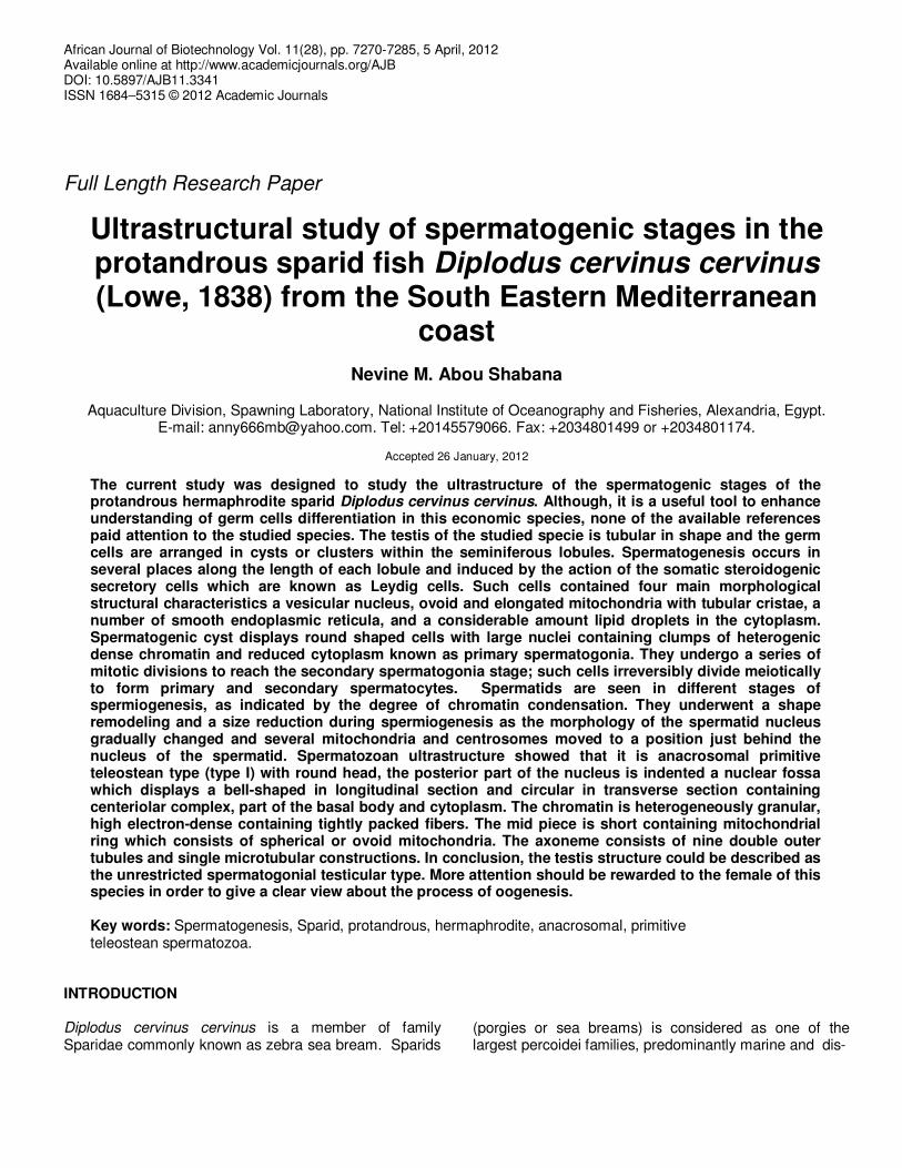
Semi-thin sections (1 µm) were cut using a LKB ultramicrotome with a glass knife and stained with toluidine blue. When appropriate regions were found, ultrathin sections were subsequently made and stained with drops of 2% uranyl acetate followed by lead citrate for 30 min. Then, these sections were examined and photographed using Leica digital camera. Ultra thin sections were cut on Joel Jum 7 ultra microtome and were stained in 5% aqueous Uranyl acetate for half an hour followed by Lead citrate for 5 min (Reynolds, 1963) and examined in a JEM – X10 transmission electron microscope at 80 KV. Scanning electron microscope For the preparations of scanning microscopy, milt was collected by stripping males by gentle abdominal massage then immediately fixed 4% glutraldehyde buffered to pH 7.2 with sodium cacodylate then filtered on 0.22 micromole Millipore filter then they were dehydrated in a graded series of ethyl alcohol ,critical point dried using CO2 and gold coating. RESULTS Light microscopy structure of testicular tissue The testis of D. cervinus cervinus is tubular in shape and the germ cells are arranged in cysts or clusters within the seminiferous lobules. Spermatogenesis occurs in several places along the length of each lobule and the testis structure could be described as the unrestricted spermatogonial testicular type (Figure 1). Spermatogonia are found near the periphery along the length of the lobule, while spermatocytes, spermatids and spermato-zoa are found toward the interior. Sertoli cells are found surrounding the germ cells. In the testicular structure,
7272 Afr. J. Biotechnol.
Figure 1. A photo-micrograph of a semithin section in Diplodus cervinus cervinus testis showing the spermatogenic stages 1ry spermatogonia (Spg1),2ry spermatogonia(Spg2),1ry spermatocyte (Spc1), 2ry spermatocyte (Spg2), spermatid (Sd), spermatozoa (Sz) and sertoli (Ser) and leydig (Ly) cells (Toluidine blue)X1000.
Leydig and Sertoli cells which appeared near germ cells in germinal cysts were involved in spermatogenesis. Clusters of Leydig cells in the interlobular space were easily distinguishable from the connective tissue composing the walls of the seminiferous lobules. They are located in the interstitium, which is surrounded by seminiferous lobules. The ultrastructures and activity of Leydig cells, which varied with the different stages of germ cell development, were observed within the inter-stitium. In particular, well developed Leydig cells were found during the period of active meiotic division and before spermiation. Morphological changes and activities of Sertoli cells showed different characteristics with germ cell developmental stages. After spermiation Sertoli cells were involved in the formation of several phagosomes that originated from degenerating spermatids and whole sperm cells by phagocytosis. Electron microscopic observation of germ cell development during spermatogenesis Based on morphological characteristics and the develop-ment of germ cells in the seminiferous lobules of the testis, spermatogenic stages are classified into 4 successive stages: a) spermatogonia, b) spermatocytes,
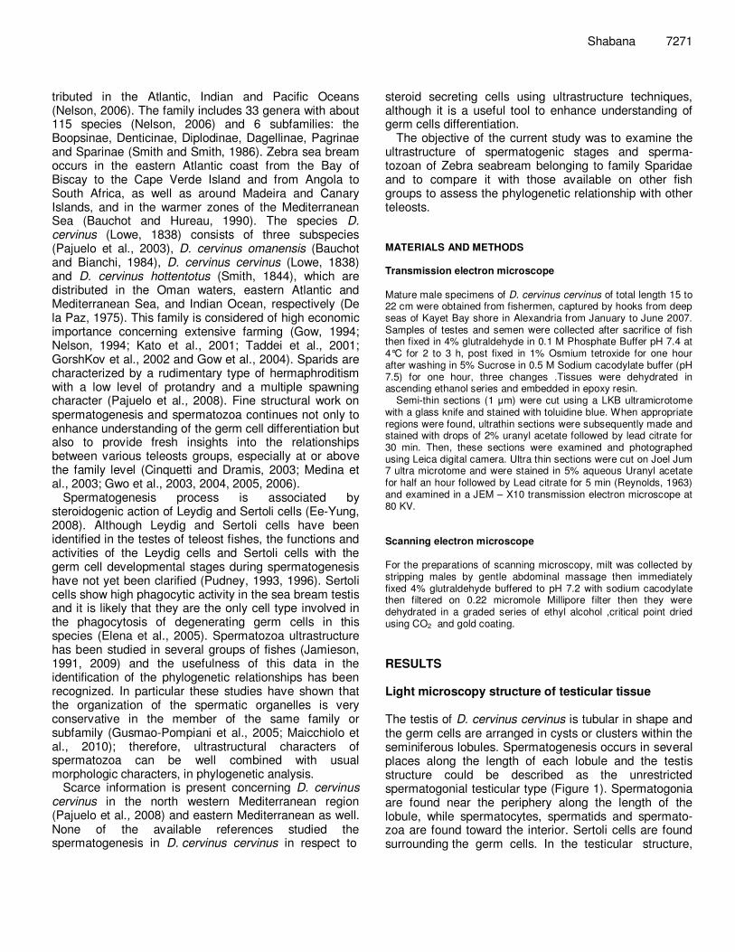
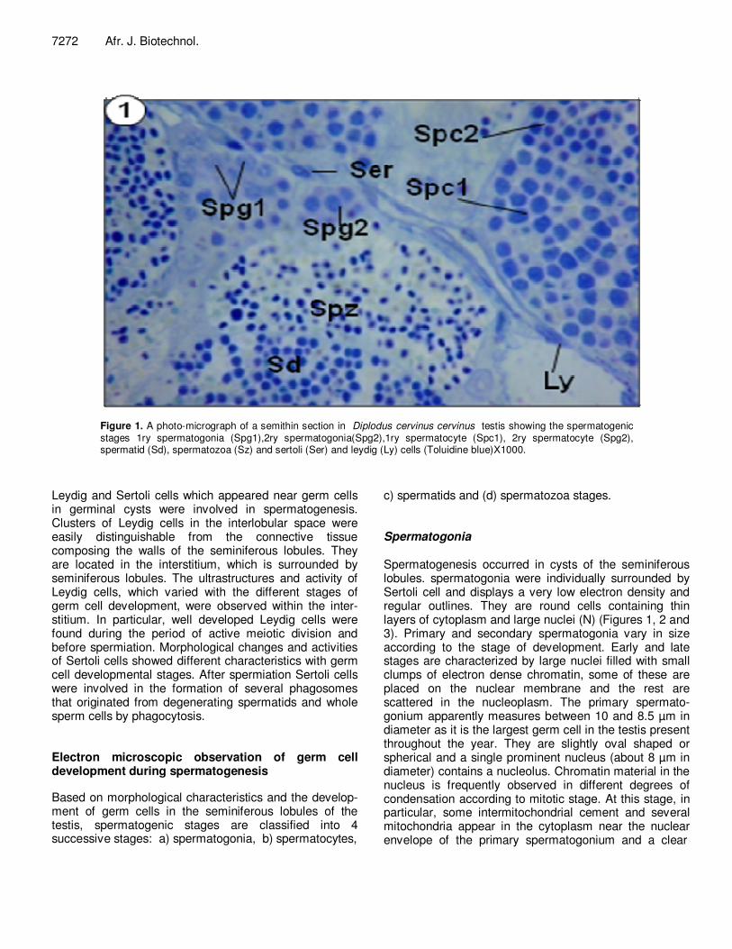
c) spermatids and (d) spermatozoa stages. Spermatogonia Spermatogenesis occurred in cysts of the seminiferous lobules. spermatogonia were individually surrounded by Sertoli cell and displays a very low electron density and regular outlines. They are round cells containing thin layers of cytoplasm and large nuclei (N) (Figures 1, 2 and 3). Primary and secondary spermatogonia vary in size according to the stage of development. Early and late stages are characterized by large nuclei filled with small clumps of electron dense chromatin, some of these are placed on the nuclear membrane and the rest are scattered in the nucleoplasm. The primary spermato-gonium apparently measures between 10 and 8.5 µm in diameter as it is the largest germ cell in the testis present throughout the year. They are slightly oval shaped or spherical and a single prominent nucleus (about 8 µm in diameter) contains a nucleolus. Chromatin material in the nucleus is frequently observed in different degrees of condensation according to mitotic stage. At this stage, in particular, some intermitochondrial cement and several mitochondria appear in the cytoplasm near the nuclear envelope of the primary spermatogonium and a clear
Shabana 7273
Pn
n
N
MM
Spg1
M
2
Cr
Figure 2. An electron micrograph showing primary spermatogonia in Diplodus cervinus cervinus displaying a large heterogenic nucleus (N), accumulation of small amount of electron dense chromatin (Cr), small nucleolus (n), mitochondrion (M) and a perinuclear nuage (Pn).X10,000.
Figure 3. An electron micrograph showing developing secondary spermatogonia in Diplodus cervinus cervinus displaying a large heterogenic nucleus (N), more accumulation of chromatin (Cr), small nucleolus (n), mitochondrion (M) and a perinuclear nuage (Pn).X10,000.
7274 Afr. J. Biotechnol.
Spc1
Ly
Sd2
4 M
Ly
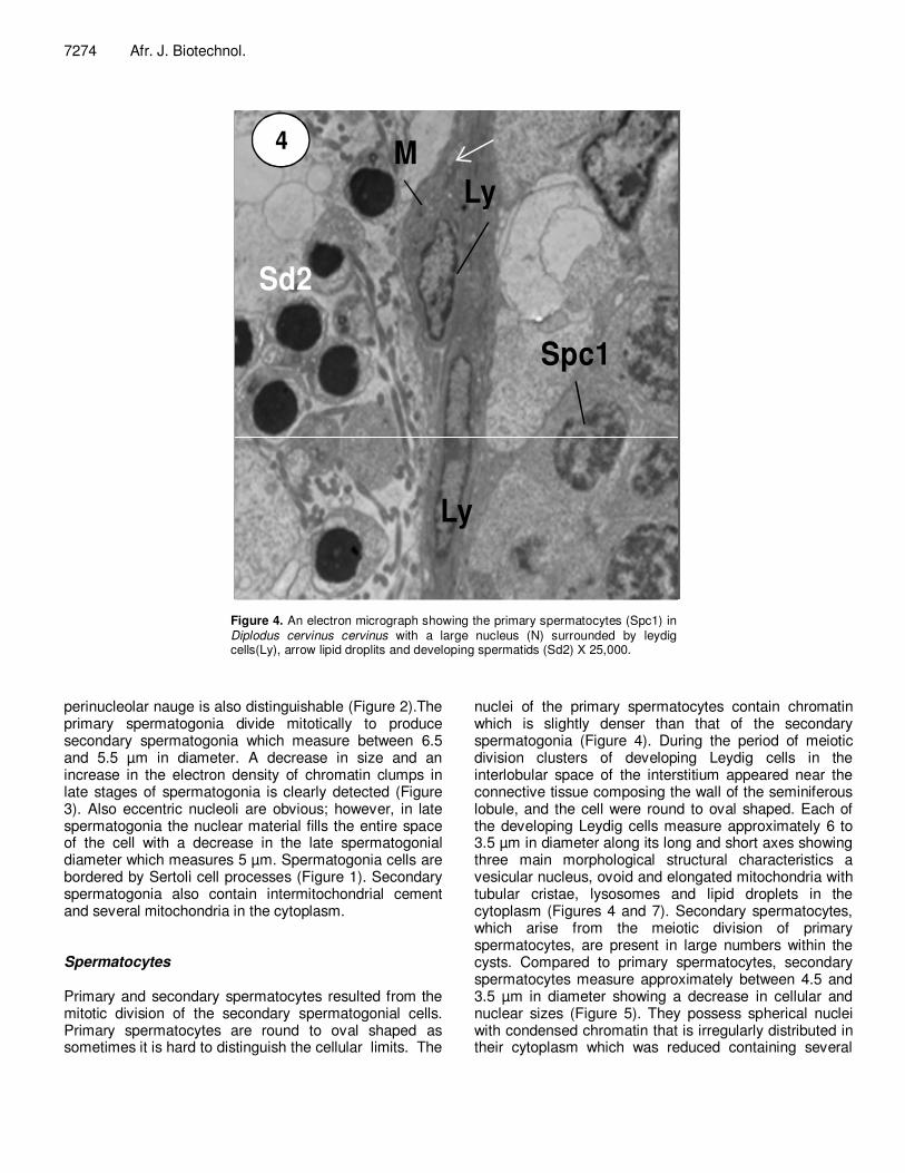
Figure 4. An electron micrograph showing the primary spermatocytes (Spc1) in Diplodus cervinus cervinus with a large nucleus (N) surrounded by leydig cells(Ly), arrow lipid droplits and developing spermatids (Sd2) X 25,000.
perinucleolar nauge is also distinguishable (Figure 2).The primary spermatogonia divide mitotically to produce secondary spermatogonia which measure between 6.5 and 5.5 µm in diameter. A decrease in size and an increase in the electron density of chromatin clumps in late stages of spermatogonia is clearly detected (Figure 3). Also eccentric nucleoli are obvious; however, in late spermatogonia the nuclear material fills the entire space of the cell with a decrease in the late spermatogonial diameter which measures 5 µm. Spermatogonia cells are bordered by Sertoli cell processes (Figure 1). Secondary spermatogonia also contain intermitochondrial cement and several mitochondria in the cytoplasm. Spermatocytes Primary and secondary spermatocytes resulted from the mitotic division of the secondary spermatogonial cells. Primary spermatocytes are round to oval shaped as sometimes it is hard to distinguish the cellular limits. The
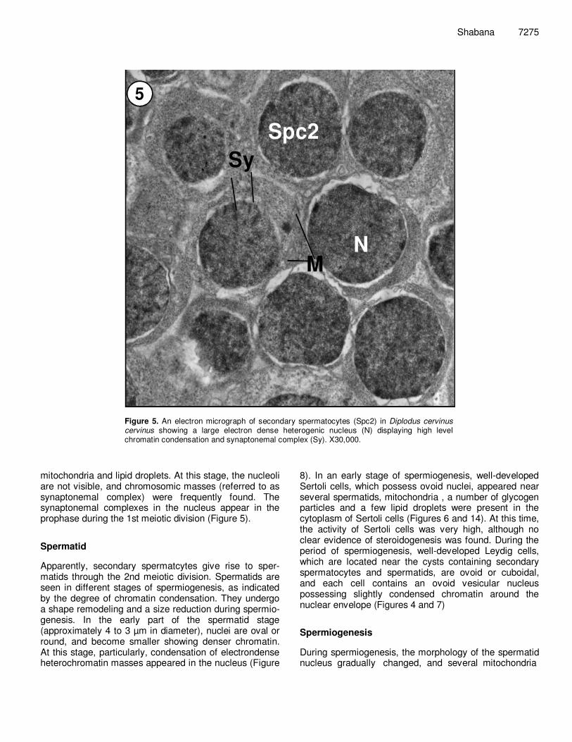
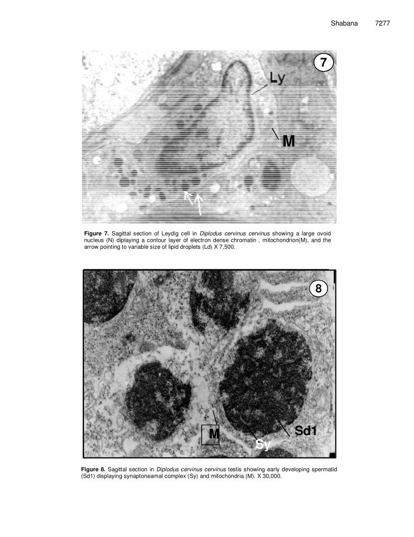
nuclei of the primary spermatocytes contain chromatin which is slightly denser than that of the secondary spermatogonia (Figure 4). During the period of meiotic division clusters of developing Leydig cells in the interlobular space of the interstitium appeared near the connective tissue composing the wall of the seminiferous lobule, and the cell were round to oval shaped. Each of the developing Leydig cells measure approximately 6 to 3.5 µm in diameter along its long and short axes showing three main morphological structural characteristics a vesicular nucleus, ovoid and elongated mitochondria with tubular cristae, lysosomes and lipid droplets in the cytoplasm (Figures 4 and 7). Secondary spermatocytes, which arise from the meiotic division of primary spermatocytes, are present in large numbers within the cysts. Compared to primary spermatocytes, secondary spermatocytes measure approximately between 4.5 and 3.5 µm in diameter showing a decrease in cellular and nuclear sizes (Figure 5). They possess spherical nuclei with condensed chromatin that is irregularly distributed in their cytoplasm which was reduced containing several
Shabana 7275
MN
5
Spc2
Sy
Figure 5. An electron micrograph of secondary spermatocytes (Spc2) in Diplodus cervinus cervinus showing a large electron dense heterogenic nucleus (N) displaying high level chromatin condensation and synaptonemal complex (Sy). X30,000.
mitochondria and lipid droplets. At this stage, the nucleoli are not visible, and chromosomic masses (referred to as synaptonemal complex) were frequently found. The synaptonemal complexes in the nucleus appear in the prophase during the 1st meiotic division (Figure 5). Spermatid Apparently, secondary spermatcytes give rise to sper-matids through the 2nd meiotic division. Spermatids are seen in different stages of spermiogenesis, as indicated by the degree of chromatin condensation. They undergo a shape remodeling and a size reduction during spermio-genesis. In the early part of the spermatid stage (approximately 4 to 3 µm in diameter), nuclei are oval or round, and become smaller showing denser chromatin. At this stage, particularly, condensation of electrondense heterochromatin masses appeared in the nucleus (Figure
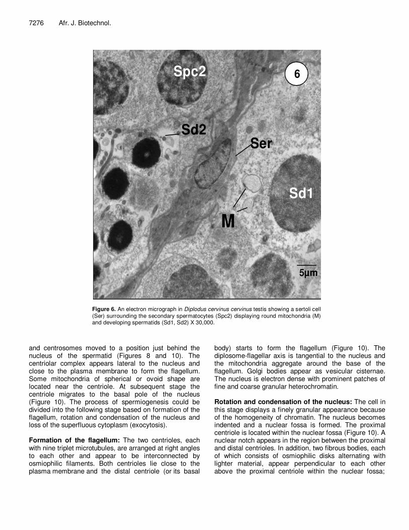
8). In an early stage of spermiogenesis, well-developed Sertoli cells, which possess ovoid nuclei, appeared near several spermatids, mitochondria , a number of glycogen particles and a few lipid droplets were present in the cytoplasm of Sertoli cells (Figures 6 and 14). At this time, the activity of Sertoli cells was very high, although no clear evidence of steroidogenesis was found. During the period of spermiogenesis, well-developed Leydig cells, which are located near the cysts containing secondary spermatocytes and spermatids, are ovoid or cuboidal, and each cell contains an ovoid vesicular nucleus possessing slightly condensed chromatin around the nuclear envelope (Figures 4 and 7) Spermiogenesis During spermiogenesis, the morphology of the spermatid nucleus gradually changed, and several mitochondria
7276 Afr. J. Biotechnol.
5µm
Spc2
Sd1
SerSd2
6
M
Figure 6. An electron micrograph in Diplodus cervinus cervinus testis showing a sertoli cell (Ser) surrounding the secondary spermatocytes (Spc2) displaying round mitochondria (M) and developing spermatids (Sd1, Sd2) X 30,000.
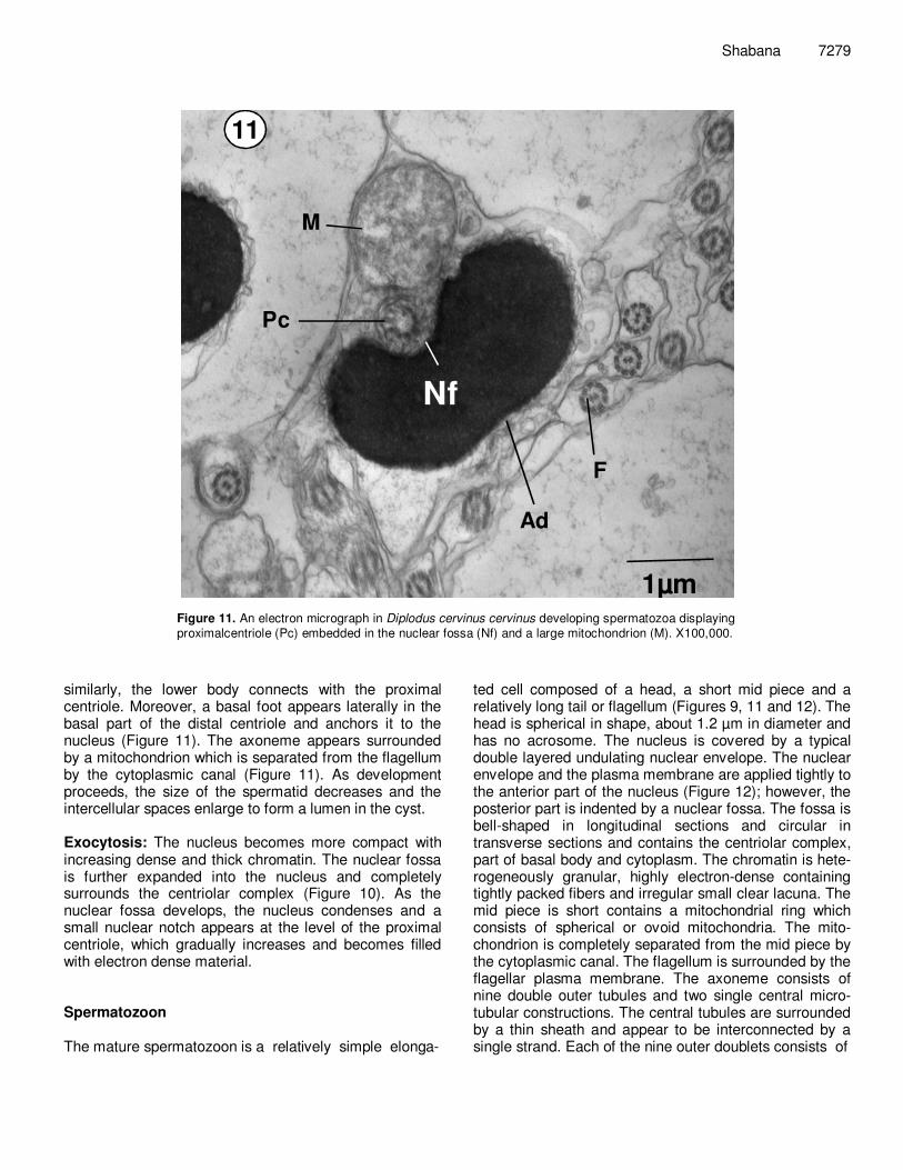
and centrosomes moved to a position just behind the nucleus of the spermatid (Figures 8 and 10). The centriolar complex appears lateral to the nucleus and close to the plasma membrane to form the flagellum. Some mitochondria of spherical or ovoid shape are located near the centriole. At subsequent stage the centriole migrates to the basal pole of the nucleus (Figure 10). The process of spermiogenesis could be divided into the following stage based on formation of the flagellum, rotation and condensation of the nucleus and loss of the superfluous cytoplasm (exocytosis). Formation of the flagellum: The two centrioles, each with nine triplet microtubules, are arranged at right angles to each other and appear to be interconnected by osmiophilic filaments. Both centrioles lie close to the plasma membrane and the distal centriole (or its basal
body) starts to form the flagellum (Figure 10). The diplosome-flagellar axis is tangential to the nucleus and the mitochondria aggregate around the base of the flagellum. Golgi bodies appear as vesicular cisternae. The nucleus is electron dense with prominent patches of fine and coarse granular heterochromatin. Rotation and condensation of the nucleus: The cell in this stage displays a finely granular appearance because of the homogeneity of chromatin. The nucleus becomes indented and a nuclear fossa is formed. The proximal centriole is located within the nuclear fossa (Figure 10). A nuclear notch appears in the region between the proximal and distal centrioles. In addition, two fibrous bodies, each of which consists of osmiophilic disks alternating with lighter material, appear perpendicular to each other above the proximal centriole within the nuclear fossa;
Shabana 7277
M
7
Figure 7. Sagittal section of Leydig cell in Diplodus cervinus cervinus showing a large ovoid nucleus (N) diplaying a contour layer of electron dense chromatin , mitochondrion(M), and the arrow pointing to variable size of lipid droplets (Ld) X 7,500.
MMSy
Sd1
8
Figure 8. Sagittal section in Diplodus cervinus cervinus testis showing early developing spermatid (Sd1) displaying synaptoneamal complex (Sy) and mitochondria (M). X 30,000.
7278 Afr. J. Biotechnol.
Ad
Ad
Mp
H
F
9
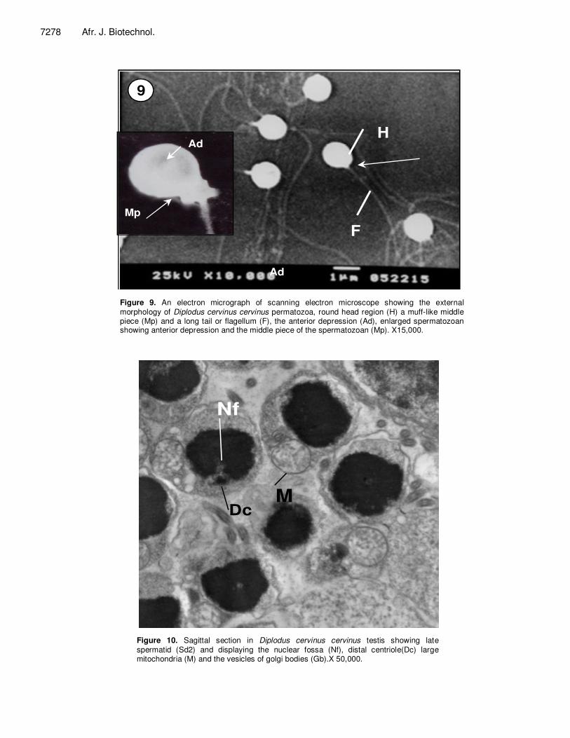
Figure 9. An electron micrograph of scanning electron microscope showing the external morphology of Diplodus cervinus cervinus permatozoa, round head region (H) a muff-like middle piece (Mp) and a long tail or flagellum (F), the anterior depression (Ad), enlarged spermatozoan showing anterior depression and the middle piece of the spermatozoan (Mp). X15,000.
5µm
Gb
10
MC.C
MDc
Nf
Figure 10. Sagittal section in Diplodus cervinus cervinus testis showing late spermatid (Sd2) and displaying the nuclear fossa (Nf), distal centriole(Dc) large mitochondria (M) and the vesicles of golgi bodies (Gb).X 50,000.
Shabana 7279
1µm
11
M
Pc
Ad
F
Nf
Figure 11. An electron micrograph in Diplodus cervinus cervinus developing spermatozoa displaying proximalcentriole (Pc) embedded in the nuclear fossa (Nf) and a large mitochondrion (M). X100,000.
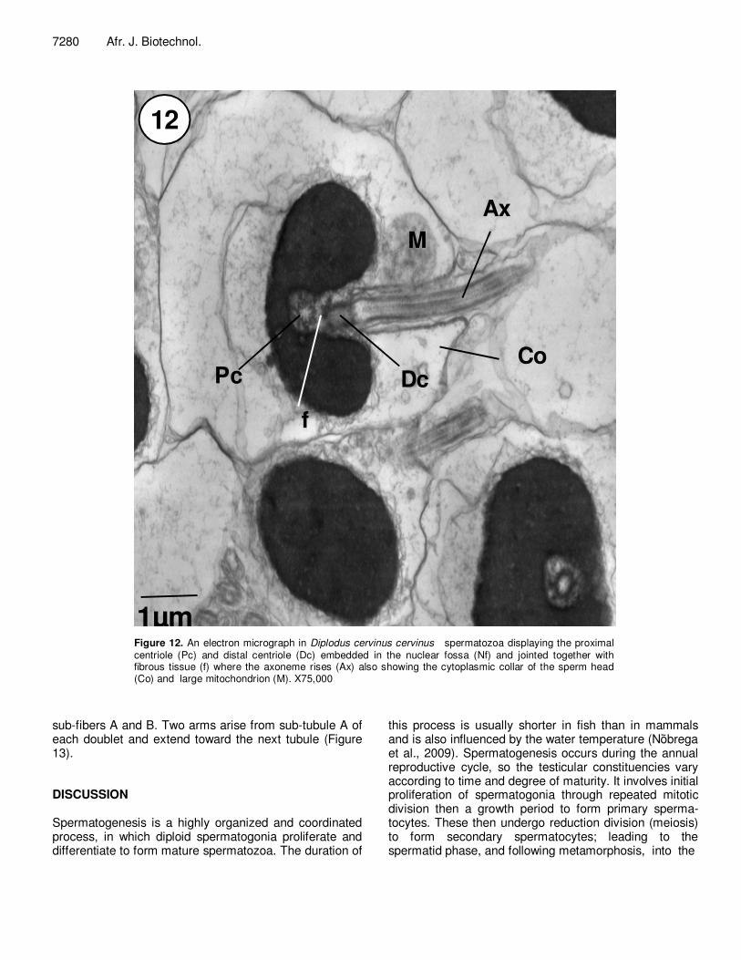
similarly, the lower body connects with the proximal centriole. Moreover, a basal foot appears laterally in the basal part of the distal centriole and anchors it to the nucleus (Figure 11). The axoneme appears surrounded by a mitochondrion which is separated from the flagellum by the cytoplasmic canal (Figure 11). As development proceeds, the size of the spermatid decreases and the intercellular spaces enlarge to form a lumen in the cyst. Exocytosis: The nucleus becomes more compact with increasing dense and thick chromatin. The nuclear fossa is further expanded into the nucleus and completely surrounds the centriolar complex (Figure 10). As the nuclear fossa develops, the nucleus condenses and a small nuclear notch appears at the level of the proximal centriole, which gradually increases and becomes filled with electron dense material. Spermatozoon The mature spermatozoon is a relatively simple elonga-
ted cell composed of a head, a short mid piece and a relatively long tail or flagellum (Figures 9, 11 and 12). The head is spherical in shape, about 1.2 µm in diameter and has no acrosome. The nucleus is covered by a typical double layered undulating nuclear envelope. The nuclear envelope and the plasma membrane are applied tightly to the anterior part of the nucleus (Figure 12); however, the posterior part is indented by a nuclear fossa. The fossa is bell-shaped in longitudinal sections and circular in transverse sections and contains the centriolar complex, part of basal body and cytoplasm. The chromatin is hete-rogeneously granular, highly electron-dense containing tightly packed fibers and irregular small clear lacuna. The mid piece is short contains a mitochondrial ring which consists of spherical or ovoid mitochondria. The mito-chondrion is completely separated from the mid piece by the cytoplasmic canal. The flagellum is surrounded by the flagellar plasma membrane. The axoneme consists of nine double outer tubules and two single central micro-tubular constructions. The central tubules are surrounded by a thin sheath and appear to be interconnected by a single strand. Each of the nine outer doublets consists of
7280 Afr. J. Biotechnol.
1µm
M
Dc
f
Pc
Ax
Co
12
Figure 12. An electron micrograph in Diplodus cervinus cervinus spermatozoa displaying the proximal centriole (Pc) and distal centriole (Dc) embedded in the nuclear fossa (Nf) and jointed together with fibrous tissue (f) where the axoneme rises (Ax) also showing the cytoplasmic collar of the sperm head (Co) and large mitochondrion (M). X75,000
sub-fibers A and B. Two arms arise from sub-tubule A of each doublet and extend toward the next tubule (Figure 13). DISCUSSION Spermatogenesis is a highly organized and coordinated process, in which diploid spermatogonia proliferate and differentiate to form mature spermatozoa. The duration of
this process is usually shorter in fish than in mammals and is also influenced by the water temperature (Nõَbrega et al., 2009). Spermatogenesis occurs during the annual reproductive cycle, so the testicular constituencies vary according to time and degree of maturity. It involves initial proliferation of spermatogonia through repeated mitotic division then a growth period to form primary sperma-tocytes. These then undergo reduction division (meiosis) to form secondary spermatocytes; leading to the spermatid phase, and following metamorphosis, into the
Shabana 7281
M
Pc
Dc
Co
F
Ad
Ax
9+2
13
Figure 13. An electron micrograph of transmission electron microscope showing Diplodus cervinus cervinus mature spermatozoan displaying the deeply embedded centriolar complex into the nucleus ,the proximal and the distal centrioles (Pc, Ac), the anterior depression (Ad) ,the cytoplasmic collar( Co) and an enlarged region of axonem showing the 9+2 configuration of axonemal doublets.X25,000 and X50,000.
motile spermatozoa. The interstitium between lobules consists of interstitial cells, fibroblasts, blood and lymph vessels. The lobular component contains two cell types: germ cells and distinct somatic cells lining the periphery of the lobule. The terminology used to describe these somatic cells has been the source of much debate (Coward et al., 2002). In some species, the lobule boundary cells are considered more likely to be homo-logous to Sertoli cells as cells are found in close proximity to spermatids/developing sperm and possess various structures that indicate a phagocytic role (Billard, 1970; Billard et al., 1972; Grier, 1975, 1981)
The present results show that Sertoli cells exist on the borders of cysts containing the primary spermatogonia. It is a spindle shape cell with large nucleus; the later has clumps of electron dense chromatin arranged on the nuclear membrane and some in the nuleoplasm. The
cytoplasm of Sertoli cells of D. cervinus cervinus contain a huge amount of lysosomes confirming the phagocytic function which has been described before. The present results conform to those prementioned concerning the function of Sertoli which said to play an important role in phagocytosis of degenerating and residual sperm cells (Grier, 1993; Loir et al., 1995; Cinquetti and Dramis, 2003). Moreover, Sertoli secrete fluid that generates the tubular lumen, and they phagocytise apoptotic germ cells, residual bodies discarded by spermatids during spermio-genesis, and residual sperm (Schulz et al., 2010). Also, Mattei and Mattei, (1982) mentioned another function of these somatic cells, that they are the storage site of steroids if they do not synthesize it.
The second type of somatic interstitial cells which are known as Leydig cells are formed between seminiferous tubules of the investigated fish, it has large irregular
7282 Afr. J. Biotechnol.
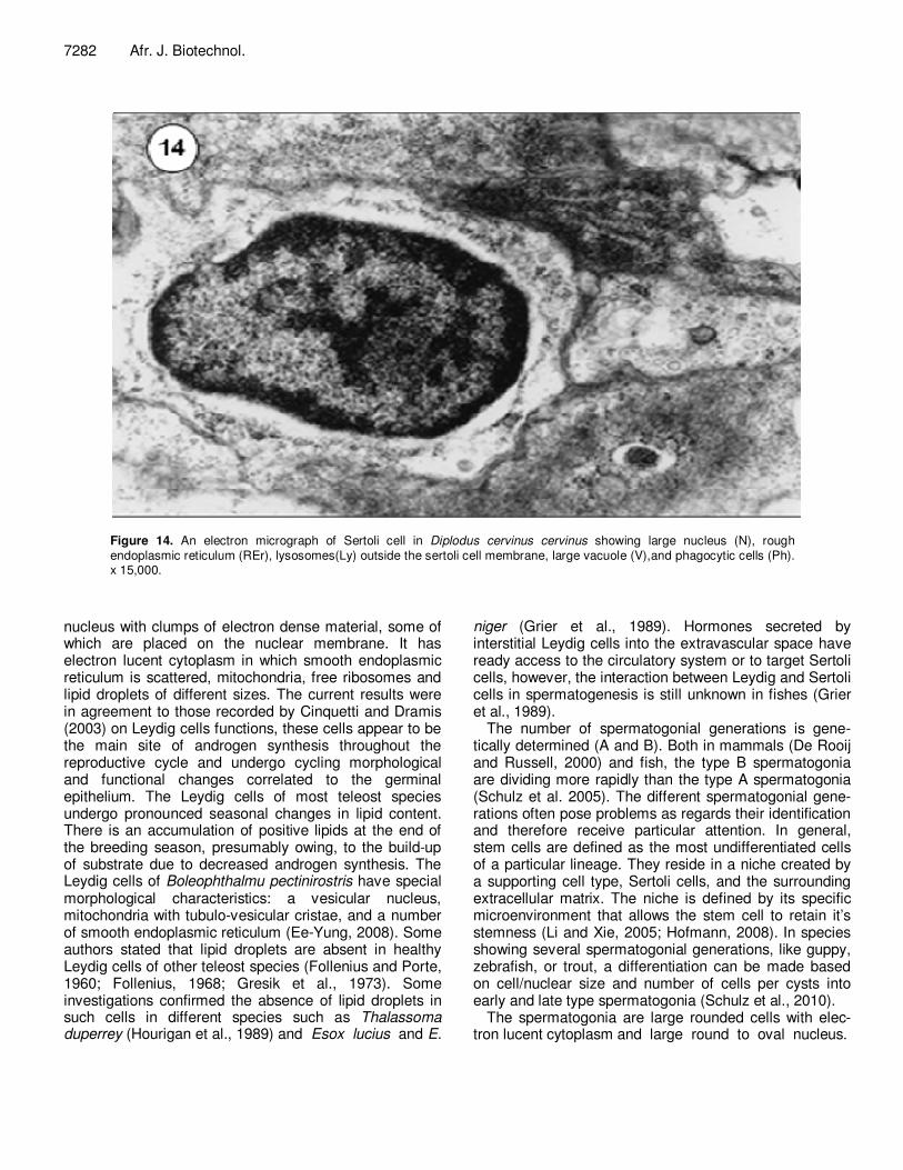
Figure 14. An electron micrograph of Sertoli cell in Diplodus cervinus cervinus showing large nucleus (N), rough endoplasmic reticulum (REr), lysosomes(Ly) outside the sertoli cell membrane, large vacuole (V),and phagocytic cells (Ph). x 15,000.
nucleus with clumps of electron dense material, some of which are placed on the nuclear membrane. It has electron lucent cytoplasm in which smooth endoplasmic reticulum is scattered, mitochondria, free ribosomes and lipid droplets of different sizes. The current results were in agreement to those recorded by Cinquetti and Dramis (2003) on Leydig cells functions, these cells appear to be the main site of androgen synthesis throughout the reproductive cycle and undergo cycling morphological and functional changes correlated to the germinal epithelium. The Leydig cells of most teleost species undergo pronounced seasonal changes in lipid content. There is an accumulation of positive lipids at the end of the breeding season, presumably owing, to the build-up of substrate due to decreased androgen synthesis. The Leydig cells of Boleophthalmu pectinirostris have special morphological characteristics: a vesicular nucleus, mitochondria with tubulo-vesicular cristae, and a number of smooth endoplasmic reticulum (Ee-Yung, 2008). Some authors stated that lipid droplets are absent in healthy Leydig cells of other teleost species (Follenius and Porte, 1960; Follenius, 1968; Gresik et al., 1973). Some investigations confirmed the absence of lipid droplets in such cells in different species such as Thalassoma duperrey (Hourigan et al., 1989) and Esox lucius and E.
niger (Grier et al., 1989). Hormones secreted by interstitial Leydig cells into the extravascular space have ready access to the circulatory system or to target Sertoli cells, however, the interaction between Leydig and Sertoli cells in spermatogenesis is still unknown in fishes (Grier et al., 1989).
The number of spermatogonial generations is gene-tically determined (A and B). Both in mammals (De Rooij and Russell, 2000) and fish, the type B spermatogonia are dividing more rapidly than the type A spermatogonia (Schulz et al. 2005). The different spermatogonial gene-rations often pose problems as regards their identification and therefore receive particular attention. In general, stem cells are defined as the most undifferentiated cells of a particular lineage. They reside in a niche created by a supporting cell type, Sertoli cells, and the surrounding extracellular matrix. The niche is defined by its specific microenvironment that allows the stem cell to retain it’s stemness (Li and Xie, 2005; Hofmann, 2008). In species showing several spermatogonial generations, like guppy, zebrafish, or trout, a differentiation can be made based on cell/nuclear size and number of cells per cysts into early and late type spermatogonia (Schulz et al., 2010).
The spermatogonia are large rounded cells with elec- tron lucent cytoplasm and large round to oval nucleus.
Primary spermatogonia are larger in size than secondary spermatogonia. Distinctive changes are observed in D. cervinus cervinus spermatogonia to develop into spermatic cysts especially in nuclei that illustrate electron lucent nucleoplasm with few clumps of electron dense chromatin. An increase in the amount of this electron dense chromatin is noticed in the late stages than in the early ones filling the entire nucleus and gaining a heterogeneous appearance. Perinuclear nuage is always seen near to the nuclear pore and associated with the germ cells. The origin and function of the perinuclear nuage in the male germ cell is not clear (Huang et al., 2002). Some authors assumed that this dense material is composed of ribonucleoproteins and mRNA species showing a long half-life time, including vasa or piwi mRNA, specific products of the germ cell lineage (Knaut et al., 2000; Houwing et al., 2007).
After final mitosis of spermatogonia (B), meiotic division started to take place to produce the primary spermato-cyte subsequently followed by the secondary spermato-cytes. Diplodus cervinus cervinus spermatocytes are characterized by large nuclei with higher electron density and the appearance of synaptonemal complex displaying diplosomal structure. Grier (1992) reported that Sertoli cells, primary spermatogonia, and the subsequent stages of germ cell development reside in spermatocysts. They are always sequestered from interstitial tissues by a basement membrane. Sertoli cells rest upon the basement membrane and their process form the borders of spermatocysts. The current results coincide with the findings on B. pectinirostris (Ee-Yung, 2008). Spermio-genesis consists of series of morphological changes that lead to the differentiation of spermatids into spermatozoa. The changes include nuclear condensation, elimination of organelles and cytoplasm, flagellum formation, and the rearrangement of cellular organelles along the sperma-tozoon cytoplasm (Jamieson, 1991). The spermatid of D. cervinus cervinus is non acrosomal with a flagellum originating perpendicular to the centriolar complex and deeply embedded into the nucleus. The current findings were in agreement to those recorded in family Scoloplacidae (Spadella et al, 2006). The nucleus has a condensed granulated chromatin, the condensation of chromatin in the nuclei of spermatids develops to maturity in a definite pattern, and it always starts adjacent to the developing flagellum. There are three types of spermiogenesis in fish, (type I, II, and III) have been described (Quagio-Grassiotto and Oliveira, 2008) based on the orientation of the flagellum to the nucleus, and on whether or not a nuclear rotation occurs. Type I is characterized by a perpendicular flagellum in relation to the nucleus with nuclear rotation; in type II, the flagellum develops parallel to the nucleus without nuclear rotation, and in type III, the flagellum is central without nuclear rotation (Mattei, 1970; Quagio-Grassiotto and Oliveira,
Shabana 7283 2008). These patterns are reflected in the spermatozoa structure, and highly conserved within taxonomic units, and are, therefore, a powerful tool for phylogenetic analyses in fish (Jamieson, 1991; Quagio-Grassiotto and Oliveira, 2008). So the present result obeys type I spermatid, which possesses a perpendicular central deeply embedded flagellum with nuclear orientation.
Stages of spermatogenesis in Diplodus cervinus c. were based on the modifications occurred in the nucleus and spermatid before spermatozoan formation. The distri-bution and organization of the cytoplasmic organelles and the implantation of the spermatid fossa of Diplodus cervinus c. happens according to the pattern described by Thiaw et al. (1988) and Zaki et al. (2005). A decrease in the volume is found in D. cervinus c. in stages of spermatocytes and spermatogonia. The centriole in D. cervinus c. is embedded in the nucleus; the proximal and distal centrioles are linked by electron dense filament to form the nuclear fossa, and the cytoplasmic collar attached to the proximal part of the flagellum. Sprando and Russell (1988) proposed three ways to reduce the cytoplasmic size in spermatid: 1- formation of residual bodies to be phagocytosed by Sertoli cells, 2- formation of tubular complexes followed by disintegration, 3- dehydration then chromatin condensation. The elimination of the residual bodies in D. cervinus c. could be of the first type. Same believe was recorded by Zaki et al. (2005), while Huang et al. (2002) mentioned that the elimination in his study is of the second type although all these authors were examining Sparid fishes. Spermto-zoan of D. cervinus c. has the characteristic morphology of teleosts. It is devoid of an acrosome as the rest of the Sparids and other bony fishes (Billard, 1970; Bacetti et al., 1984; Gow et al., 1993). Ginsburg (1968) reported that the absence of an acrosome could be attributed to the presence of micropyle in fish eggs. The head of sperm of D. cervinus c. is round shape as reported before in other species, in turbot (Suqnet et al., 1993), Oreochromis sp (Bern and Avtation, 1990), grey mullets (Brusle, 1982) and cyprinids (Bacetti et al.,1989), Sparidae (Gow et al., 2005). On the other hand, oval shaped head was reported in other species such as guppy (Billard, 1970), eel (Billard and Ginoburg, 1973), Stanoperca sp. (Matos et al., 2002), Mullidae and Siganidae (Gow et al., 2004) and Blue Spart, Clupeidae (Gow et al., 2006).
The nucleus of the spermatozoan in D. cervinus c. has an anterior and a posterior nuclear depressions which was reported before in the Rainbow and becook trout , guppy ,Tilapia, turbout, Sparus aurata ,Diplodus vulgaris and Lithognathus mormyrus by Billard (1983a, b, 1970), Bern and Avtalion (1990), Suquet et al. (1993) and Boops boops by Zaki et al. (2005).
On both sides of D. cervinus cervinus axial nuclear fossa, electron dense microfibrils exist connecting the
7284 Afr. J. Biotechnol. distal centriole to the nucleus of the sperm head. Martinez-Soler et al. (2007) have reported that in Sepia officinalis the perinuclear microtubule system represents an element responsible for rigidity and shape of the sperm nucleus and has a role in nucleoplasmic transport. A short or reduced middle piece is found in D. cervinus cervinus spermatozoan, a feature which is common in teleosts with external fertilization (Nicander and SjoÈ den, 1971; Zaki et al., 2005), while in internally fertilized fish the spermatozoan has a longer middle piece (Matti, 1969).
Depending on the orientation of the centriolar complex to the nuclear fossa, Matti (1991) had classified teleostean sperm into two types I and II. In type I the centriolar complex lies inside the nuclear fossa, while type II it lies outside the nuclear fossa. D. cervinus cervinus centriolar complex lies deeply inside the nuclear fossa (type I) as pre-mentioned in other sparid fishes (Gow et al., 2004; Zaki et al., 2005); turbout (Suquet et al., 1993) and Gymnotiforms (França et al., 2007). However, Gusmăo et al. (2005) reported that the sperms of the Sciaenidae family and other Percoidei are of type II nucleus. D. cervinus cervinus has one big mitochondrion lies beneath the head of the sperm as reported in other Sparids as a characteristic feature for this family (Gow et al., 2004; Zaki et al., 2005). The flagellum of D. cervinus cervinus has the common microtubular structure 9 + 2 which is observed in many teleosts, turbouts (Matti, 1969; Suquet et al., 1993; Gow et al., 2004; Zaki et al., 2005).
From the present study and the previous ones on Sparids, the information provided on sperms morphology and the differences between species can be used to define the taxononomic position. Although the ultrastructural characterization of the sperm is not so useful in determining the phylogenetic relationships as in different groups of other animals (Jamieson, 1991; Matti, 1991). Conclusion and recommendation In order to learn more about this species, more attention should be rewarded to the study of oogenesis process. Also it is highly recommended to concern this economic fish species in aquaculture field and to establish an induced spawning protocol for fish farming as the majority of the sparids had been already farmed successfully. REFERENCES Baccetti B, Burrini AG, Collodel G (1989). Morphogenesis of
decapitated and decaudated sperm defect in two brothers. Gamete Res. 23: 181-188.
Bauchot ML, Bianchi G (1984).*Diplodus cervinus omanensis*, nouvelle sous-espèce de *Diplodus cervinus* (Lowe, 1841), capturée en mer d'Arabie (Pisces, Perciformes, Sparidae). Historie naturelle des
poissons. v. 8 (no. 3): 103-105. Bauchot ML, Hureau JC (1990). Sparidae. In: Check-list of the fishes of
the Eastern Tropical Atlantic, Clofeta II. Quero JC, Hureau JC, Karrer C, Post A, Saldanha L (Eds), UNESCO, Paris, pp. 790-812.
Bern O, Avtalion RR (1990). Some morphological aspects of fertilization in tilapias. J. Fish. Biol. 36: 375-381.
Billard R, Ginsburg AS (1973). La spermiogenèse et le spermatozoïded’ Anguilla anguilla L. Étude ultrastructurale. Ann. Biol. Anim. Biochem. Biophys. 13: 523-534.
Billard R (1983a). Spermiogenesis in rainbow trout (Salmo gairdneri). Cell. Tissue Res. 233: 265-284.
Billard R (1983b). Ultrastructure of trout spermatozoa: Changes after dilution and deep freezing. Cell Tissue Res. 233: 205-218.
Billard R (1970). La spermatogenèse. Étudeultrastructurale. Annales de Biologie Animale, Biochemicie, Biophysique, 10: 493-510.
Billard R, Jalabert B, Breton B (1972). Les cellules de Sertoli des poisons téléostéens. I. Etude ultrastructural. Ann. Biol. Anim. Biochim. Biophys. 12: 19-32.
Bruslé S (1982). Contribution à la connaissance de la sexualité de poissons téléostéens and Bony Fishes), Part A. Sci. Pub. Enfield NH, USA. 788.
Cinquetti R, Dramis L (2003). Histological, histochemical, enzyme histochemical and ultrastructural investigations of the testis of Padogobius martensi between annual breeding seasons. J. Fish. Biol. 63: 1402-1442.
Coward K, Bromage NR, Hibbitt O, Parrington J (2002). Gamete physiology, fertilization and egg activation in teleost fish Reviews in Fish Biol. Fish. 12: 33-58.
De la Paz R (1975). Systématique et phylogènese des Sparidae dugenere Diplodus Raf. (Pisces, Teleostei). Trav. Doc. ORSTOM 45: 1-96.
De Rooij DG, Russell LD (2000). All you wanted to know about spermatogonia but were afraid to ask. J. Androl. 21: 776-798.
Ee-Yung C (2008). Ultrastructure of germ cells, the Leydig cells, and Sertoli cells during spermatogenesis in Boleophthalmus pectinirostris(Teleostei, Perciformes, Gobiidae). Tissue Cell. 40: 195-205.
Elena C, Victoriano M, Jose´ M, Alfonsa G (2005). An Overview of Cell Renewal in the Testis Throughout the Reproductive Cycle of a Seasonal Breeding Teleost, the Gilthead Seabream (Sparus aurata L.) Biol. Rep. 72: 593-601.
Follenius E (1968). Cytologie et cytophysiologie des cellules interstitielles de I’Epinoche: Gasterosteus acleatus L. Etude au microscope electronique. Gen. Comp. Endocrinol. 11: 198-219.
Follenius E, Porte A (1960). Cytologie fine des cellules inter stitielles dutesticule du poisson Lebistes reticulates R. Experientia, 16: 190-192.
Franca GF, Oliveira C, Quagio-Grassiotto I (2007). Ultrastructure of spermiogenesis and spermatozoa of Gymnotus cf.anguillaris and Brachyhypopomus cf. pinnicaudatus (Teleostei: Gymnotiformes). Tissue Cell. 39: 131-139.
Gorshkov S, Gorshkov G, Hadani A, Gordin H, Knibb W (2002). Chromosome set manipulations and hybridization experiments in giltheadseabream (Sparus aurata) II. Assessment of diploid and triploidhybrids between gilthead seabream and red seabream (Pagrus major).J. Appl. Ichthyol. 18: 106-112.
Gresik EW, Quirk JG, Hamiltonm JB (1973). A fine structural and histochemical study of the Leydig cell in the testis of the teleost, Oryzias latipes (Cyprinidontiformes). Gen. Comp. Endocrinol. 20: 86-98.
Grier HJ (1993). Comparative organization of Sertoli cells including the Sertoli cell barrier. In Russell LD, and Griswold MD (eds.), The Sertoli cell, Cache River Press, Clearwater, Florida. pp. 704-730.
Grier HJ (1992). Chordate testis: the extracellular matrix hypothesis. J. Exp. Zool. 256: 151-160.
Grier HJ, Van den Hurk R, Billard R (1989). Cytological identification of cell types in the testis of Esox lucius and E. Niger. Cell Tissue Res. 257: 491-496.
Grier HJ (1975). Aspects of germinal cyst and sperm development in
Poecilia latipinna (Teleostei: poecilidae). J. Morphol. 146: 229-250. Grier HJ (1981). Cellular organization of the testis and spermatogenesis
in fishes. Am. Zool. 21: 345-357. Gusm˜ao-Pompiani P, Oliveira C, Quagio-Grassiotto I (2005).
Spermatozoa ultrastructure in Sciaenidae and Polynemidae (Teleostei: Perciformes) with some consideration on Percoideispermatozoa ultrastructure. Tissue Cell. 37: 177-191.
Gwo JC, Chiua JY, Lin CY, Sub Y, Yub SL (2005). Spermatozoal ultrastructure of four Sparidae fishes: Acanthopagrusberda, Acanthopagrus australis, Lagodon rhomboids and Archosargus probatocephus. Tissue Cell. 37: 109-115.
Gwo JC, Gwo HH, Chang SL (1993). The ultrastructure of the spermatozoon of the teleost fish Acanthopagrus schlegeli (Perciformes, Sparidae). J. Morphol. 216: 29-33.
Gwo JC (1994). Cryopreservation of yellowfin seabream (Acanthopagruslatus) spermatozoa (Teleost, Perciformes, Sparidae). Theriogenology, 41: 989-1004.
Gwo JC, Lin CY, Yang WL, Choud YC (2006). Ultrastructure of the sperm of blue sprat, Spratelloides gracilis; Teleostei, Clupeiformes, Clupeidae. Tissue Cell 38: 285-291.
Gwo JC, Yang WT, Kuo MC, Takemura A, Chengc HY (2004). Spermatozoal ultrastructures of two marine perciform teleost fishes, thegoatfish, Paraupeneus spilurus (Mullidae) and the rabbitfish, Siganus fuscescens (Siganidae) from Taiwan. Tissue Cell 36: 63-69.
Hofmann MC (2008). Gdnf signaling pathways within the mammalian spermatogonial stem cell niche. Mol. Cell. Endocrinol. 288: 95-103.
Hourigan TF, Nakamuru MN, Nagahama Y, Yamauchi K, Gau EG (1989). Histology, ultrastructure, and in vitro steoidigenesis of the testis of two male phenotypes of the protogyneous fish, Thalassoma duperrey (Labridae). Gen. Comp. Endocrinol. 83: 193-217.
Houwing S, Kamminga LM, Berezikov E, Cronembold D, Girard A, Van den Elst H, Filippov DV, Blaser H, Raz E, Moens CB, Plasterk RH, Hannon GJ, Draper BW, Ketting RF (2007). A role for Piwi and piRNAs in germ cell maintenance and transposon silencing in Zebrafish. Cell, 129(1): 69-82.
Huang J, Mong O, Ching-ong C (2002).The morphology of gonadal tissue and male germ cells in the Protandrous blackporgy, Acanthopagrus schlegeli. Zool. Stud. 41(2): 216-227.
Jamieson BGM (1991). Fish Evolution and Systematics: Evidence from Spermatozoa. Cambridge Univ. Press, Cambridge.
Jamieson BGM (2009). Reproductive Biology and Phylogeny of Fishes (Agnathans and Bony Fishes), Part A. Sci. Pub. Enfield NH, USA. p. 788.
Knaut H, Pelegri F, Bohmann K, Schwarz H, Nusslein-Volhard C (2000). Zebrafish vasa RNA but not its protein is a component of the germ plasm and segregates asymmetrically before germline specification. J. Cell Biol. 149: 875-888.
Li L, Xie T (2005). Stem cell niche: structure and function. Annu. Rev. Cell Dev. Biol. 21: 605-631.
Loir M, Sourdaine P, Mendis-Handagama SM Jegou B (1995). Cell-cell interactions in the testes of teleosts and elasmo- branchs. Microsc. Res. Tech. 6: 533-552.
Maricchioloa G, Lauràb R, Genovesea L, Guerrerab MC, Micalea V, Mugliab U (2010). Fine structure of spermatozoa in the blackspot sea bream Pagellus bogaraveo (Brünnich, 1768) with some considerations about the centriolar complex. .Tissue Cell 42: 88-96.
Mart´ınez-Soler F, Kurtz K, Chiva M (2007). Sperm nucleomorphogenesis in the cephalopod Sepia officinalis. Tissue Cell, 39: 99-108.
Mattei X (1970). Spermiogene´se compare´ des poisson. In: Baccetti, B. (Ed.), Comparative Spermatology. Academic Press, New York, pp. 57–72.
Mattei X, Mattei C, Marchand B, Kit DLT, (1982). Ultrastructure des cellules de Sertoli d'un poisson téléostéen: Abudefdul marginatus. J. Ultras. Res., 81: 333-340.
Medina A, Megina C, Abascal FJ, Calzada A (2003). The sperm ultrastructure of Merluccius merluccius (Teleostei, Gadiformes): phylogenetic considerations. Acta Zool. 84: 131-137.
Nelson JS (1994). Fishes of the World, 3rd ed. Wiley, New York.
Shabana 7285 Nelson JS (2006). Fishes of the World, 4th edition John Wiley and
Sons, Inc., Hoboken, NJ, p. 601. Nicander L, SjoÈ den I (1971). An electron microscopical study on the
acrosomal complex and its role in fertilization in the river lamprey, Lampetra fluviatilis. J. Submicrosc. Cytol. 3: 309-317.
Nõbَrega RH, Batlouni SR, França LR (2009). An overview of functional and stereological evaluation of spermatogenesis and germ cell transplantation in fish. Fish Physiol. Biochem. 35: 197-206.
Pajuelo JG, José ML, Rosa D (2003). Age estimation and growth of the zebra seabream Diplodus cervinus cervinus (Lowe, 1838) on the Canary Islands shelf (Central-east Atlantic). Fish Res. 62: 97-103.
Pajuelo JG, José ML, Rosa D (2008). Gonadal development and spawning cycle in the digynic hermaphrodite sharpsnout seabream Diplodus puntazzo (Sparidae) off the Canary Islands, northwest of Afr. J. Appl. Ichthyol. 24: 168-176.
Pudney I (1993). Comparative cytology of the non-mammalian vertebrateSertoli cell. In: Russell LD, Griswold MD (Eds.). The Sertoli Cell, Cache River Press, Clearwater, FL, pp. 611-658.
Pudney I (1996). Comparative cytology of the Leydig cell. In: Payne AH, Hardy MP, Russell LD (Eds.). The Leydig Cell, Cache River Press, pp. 97-142.
Quagio-Grassiotto I, Oliveira C (2008). Sperm ultrastructure and a new type of spermiogenesis in two species of Pimelodidae, with a comparative review of sperm ultrastructure in siluriformes (Teleostei: Ostariophysi). J. Comp. Zool. 247: 55-66.
Reynolds ES (1963). The use of lead citrate at high pH as an electron-opaque stain in electron microscopy, J. Cell. Biol. 17: 208-212.
Schulz RW, De França LR, Lareyre JJ, Le Gac F, Chiarini-Garcia H, Nobrega RH, Miura T (2010). Spermatogenesis in fish. Gen. Comp. Endocrinol. 1: 165(3): 390-411.
Schulz RW, Menting S, Bogerd J, Franca LR, Vilela DAR, Godinho HP (2005).Sertoli cell proliferation in the adult testis: evidence from two fish speciesbelonging to different orders. Biol. Reprod. 73: 891-898.
Smith JLB, Smith MM (1986). Family Sparidae. In: Smith, M.M., Heemstra PC (Eds.). Smith’s Sea Fishes. Macmillan, Johannesburg, South Africa, 183: p. 1050.
Spadella MA, Oliveira C, Quagio-Grassiotto I (2006). Spermiogenesis and introsperm ultrastructure of Scoloplaxdistolothrix Ostariophysi:Siluriformes:Scoloplacidae). Acta Zool. 87(4): 341-348.
Sprando RL, Russell LD (1988). Spermiogenesis in blue gill (Lepomismacrochirus): a study of cytoplasmicevents including cell volume changes and cytoplasmic elimination. J. Morphol. 198: 165-177.
Suquet M, Dorange G, Omnes MH, Normant Y, Le Roux A, Fauvel C (1993). Composition of the seminal fluid and ultrastructure of the spermatozoon of turbot (Scophthalmus maximus). J. Fish Biol. 42: 509-516.
Taddei AR, Barbato F, Abelli L, Canese S, Moretti F, Rana KJ, Fausto AM, Mazzini M (2001). Is cryopreservation a homogeneous process? Ultrastructure and motility of untreated, prefreezing, and post thawed spermatozoa of Diplodus puntazzo (Cetti). Cryobiology, 42: 244-255.
Thiaw OT, Mattei X, Romand R (1988). Process of cytoplasmatic elimination during spermiogenesis in two cyprinodontidae (Teleostean fishes). J. Ultrastruct. Molec. Struc. Res. 101: 19-192.
Zaki MI, Negm RK, El-agamy A, Awad GS (2005). Ultrastructure of male germ cells and character of spermatozoa in boops boops (Family Sparidae) in Alexandria coast, Egypt. Egyp. J. Aquat Res. 31(2): 293-313.