Embed Size (px)
Citation preview

Radiology
Ultrasound training recommendations for medical and surgical specialties Second edition Board of the Faculty of Clinical Radiology The Royal College of Radiologists

Contents Foreword 3
1 Introduction 4
2 Aims and principles 5
3 Training recommendations 8
3.1 Theoretical training 8
3.2 Practical training 8
4 Continuing professional development 10
References 11
Appendix 1. Urological ultrasound 12
Appendix 2. Gynaecological ultrasound 18
Appendix 3. Gastrointestinal ultrasound 24
Appendix 4. Vascular ultrasound 29
Appendix 5. Breast ultrasound 33
Appendix 6. Thoracic ultrasound 37
Appendix 7. Cranial ultrasound in infants 40
Appendix 8. Focused emergency ultrasound 44
Appendix 9. Ultrasound training for critical/intensive care 48
Appendix 10. Musculoskeletal ultrasound 52
Appendix 11. Recommended theory syllabus 58

3
Foreword In 2005, when The Royal College of Radiologists (RCR) produced the initial document on Ultrasound Training Recommendations for Medical and Surgical Specialties, it recognised ultrasound as an evolving technology with wide application throughout medical and surgical practice.
Since 2005, there has been continuing growth in the availability of ultrasound and with it an acknowledgement that it can play an ever-increasing role in the diagnosis and management of patients.
It is, therefore, timely that the RCR updates this valuable guidance resource as part of its commitment to the development of high-quality imaging across the gamut of modalities. The original document, Ultrasound Training Recommendations for Medical and Surgical Specialties, has now been withdrawn.
The RCR wishes to ensure that access to high-quality ultrasound imaging continues to improve. For this to happen, ultrasound must be provided by properly trained and committed practitioners using appropriate quality ultrasound equipment.
In the UK, radiologists and sonographic practitioners have traditionally provided such a service from centralised departments. However, there is a growing need to provide ultrasound in other settings; for instance, in the community or by the patient’s bedside in critical care.
By adopting the recommendations in this document, those involved in ultrasound training can ensure that wherever ultrasound is performed the patient receives the best possible service.
I would like to thank Rani Thind for her work in leading the revision of this advice and the following members of the working party for their contributions and advice:
Dr Paul Dubbins, Clinical Radiologist Dr Fergus Gleeson, Clinical Radiologist Dr Alan Sprigg, Clinical Radiologist Dr Paul Sidhu, Clinical Radiologist Dr Richard McWilliams, Clinical Radiologist Dr Mike Weston, Clinical Radiologist Dr Marcus Harbord, Gastroenterologist
Thanks are also due to the following for their expert advice on the content: Dr Simon Jackson, Professor Andy Evans, Dr Charles Wakeley, Dr Eugene McNally, Dr Phil Cook, Dr Roger Bury and Dr Peter Wylie.
Dr Pete Cavanagh Vice President and Dean, Faculty of Clinical Radiology The Royal College of Radiologists

4
1 Introduction 1.1 High-quality ultrasound services are provided by properly trained and committed practitioners using
appropriate quality ultrasound equipment. In the UK, radiologists and sonographic practitioners have traditionally provided such a service from centralised departments of clinical radiology where equipment and manpower can be used cost-effectively.
1.2 Departments of clinical radiology may have difficulty responding to these demands primarily because of the national shortages of radiologists and sonographic practitioners. It is therefore essential that alternative methods of service delivery are considered. These may include the involvement of other professional groups in addition to greater investment in clinical radiology departments.
1.3 Medical specialists other than radiologists are increasingly wishing to undertake ultrasound examinations on patients referred to them for their clinical opinion as a direct extension of their clinical examination. This may take place in the outpatient department, on the wards, and in the assessment of emergency patients.
Clinicians are also using ultrasound to assist in practical procedures such as central line insertion. A separate document is aimed at providing guidance for focused ultrasound training.1
This should read some European and UK boards have incorporated ultrasound experience into clinical training and accreditation.
1.4 Radiologists have the skills, experience and commitment to provide guidelines for training of medical non-radiologists and hence influence the quality of service provided for the better. The RCR believes that this approach, of interdisciplinary co-operation, serves best the interests of patients.
1.5 Training of medical non-radiologists should be adequately funded and planned so that there is minimal adverse impact on the service provided to patients and the ability of clinical radiology departments to train clinical radiologists and sonographer practitioners.
1.6 This document makes recommendations for ultrasound training in the following areas:
Urological ultrasound Gynaecological ultrasound Gastrointestinal ultrasound Vascular ultrasound Breast ultrasound Thoracic ultrasound Cranial ultrasound in infants Focused emergency ultrasound Intensive care ultrasound Musculoskeletal ultrasound.
5
2 Aims and principles 2.1 The medical use of ultrasound remains highly operator-dependent in spite of advances in technology, and
the interests of the patient are best served by the provision of an ultrasound service which offers the maximum clinical benefit and optimal use of resources; that is, with appropriately trained personnel using equipment of appropriate quality.
2.2 All those who provide an ultrasound service are ethically and legally vulnerable if they have not been adequately trained. National Health Service (NHS) trusts and health boards in the UK, which provide professional indemnity to practitioners, are unlikely to be able to mount any defence to an action brought against an untrained practitioner. Similarly, the professional defence organisations are unlikely to be successful in mounting a defence against a claim for negligence should an error of diagnosis be made by an untrained practitioner of ultrasound. Advisory guidelines for training in ultrasound provided by the RCR will establish the principles to allow appropriate bodies to provide professional indemnity by setting out training and continuing professional development (CPD) requirements. Trusts, health boards, purchasing commissioners and patients should be aware of the requirements for training.
2.3 An appropriate level of training in ultrasound is one that allows for the provision of a safe and effective ultrasound service. This may be a purely diagnostic, predominantly interventional, or a clinically focused service. Departments of clinical radiology would normally provide all of these services, but it would be expected that other medical practitioners would deliver only those aspects of ultrasound particularly relevant to their clinical practice. Nonetheless, the training for medical non-radiologists should be to the same standard as those for radiologists, albeit restricted to the relevant and particular area of their clinical expertise. Whereas radiological training provides for the practice of ultrasound across a broad range of medical and surgical specialties, NHS trusts, health boards, purchasing commissioners and patients should be aware of the differences in the comparative depth and breadth of training, and hence ultrasound skills, between trained radiologists and trained medical non-radiologists.
2.4 The RCR has worked closely for many years with the Royal College of Obstetricians and Gynaecologists (RCOG) to ensure adequate training in obstetric ultrasound and obstetric ultrasound is not covered in this publication. It is also recognised that the RCOG has its own training module for ultrasound imaging in the management of gynaecological conditions and a number of radiologists act as preceptors for this. However, a syllabus for gynaecological ultrasound has been included as there are other groups (such as general practitioners) who might wish to train in this area.
2.5 The European Federation of Societies for Ultrasound in Medicine and Biology (EFSUMB) has proposed minimal training requirements for the practice of medical ultrasound in Europe.2 These are supported by the RCR and the British Medical Ultrasound Society.
2.6 Three levels of minimum training requirements are proposed in this document.
2.6.1 Level 1
Practice at this level would usually require the following abilities:
To perform common examinations safely and accurately To recognise and differentiate normal anatomy and pathology To diagnose common abnormalities within certain organ systems To recognise when a referral for a second opinion is indicated To understand the relationship between ultrasound imaging and other diagnostic imaging
techniques.
Within most medical specialties, the training required for this level of practice would be gained during conventional postgraduate specialist training programmes. In the UK, this level of training would equate to the end of basic training in ultrasound of radiology specialist registrars (SpRs) in year 3/4 of training. It would also be equivalent to a holder of, for example, the RCOG special skills training module in gynaecological ultrasound imaging.
2.6.2 Level 2
Practice at this level would usually require most or all of the following abilities:
To accept and manage referrals from Level 1 practitioners

6
To recognise and correctly diagnose almost all conditions within the relevant organ system and to have sufficient understanding of ultrasound depiction of pathology to optimise the referral of the patient if the condition falls outside of the practitioner’s skills
To perform common non-complex ultrasound-guided invasive procedures To teach ultrasound to trainees and Level 1 practitioners To conduct some research in ultrasound
The training required for this level of practice would be gained during a period of subspecialty training which may either be within or after the completion of a specialist training programme. This would equate to the level of training in radiology at the time of acquiring the Certification of Completion of Training (CCT), assuming that part of the fifth year of subspecialty training had involved ultrasound.
2.6.3 Level 3
This is an advanced level of practice, which includes some or all of the following abilities:
To accept tertiary referrals from Level 1 and Level 2 practitioners To perform specialised ultrasound examinations To perform advanced ultrasound-guided invasive procedures To conduct substantial research in ultrasound To teach ultrasound at all levels To be aware of and to pursue developments in ultrasound.
In the UK, this would equate to a consultant radiologist with a subspecialty practice which includes a significant commitment to ultrasound.
2.7 The boundaries between the three levels are difficult to define precisely and the above should only be regarded as a guide to different levels of competence and experience. In the detailed syllabuses attached to this document in Appendices 1–9 an attempt is made to indicate more specifically the type of experience required for each level of training. Training in musculoskeletal ultrasound does not lend itself easily to ‘levels’ of training and instead a ‘modular’ approach is recommended (Appendix 10).
2.8 The training of medical non-radiologists should foster relationships between radiological and non-radiological medical practitioners so that mutual support continues beyond the initial training period. Ideally a radiologist would continue to act as a mentor for a medical non-radiologist undertaking ultrasound after their training is completed. In addition, regular multidisciplinary meetings should continue to ensure an integrated approach to any further imaging that may be required.
2.9 A system for recording the results of any ultrasound examination in the patient’s record is mandatory. The permanent recording of images, where appropriate, is also mandatory for the purposes of correlative imaging, future comparison and audit. The preferred option is through the hospital radiology information system (RIS)/picture archive and communications systems (PACS) equipment, enabling other clinicians to access the images and report.
2.10 Knowledge of the appropriate use and integration of other imaging techniques, as well as the clinical and economic impact of ultrasound on the demand for other imaging, should be acquired.
2.11 The requirement to deliver training for medical non-radiologists must acknowledge the time commitment of the trainer and trainee, the provision of funding, the content and practicability of the syllabus and the availability of trainers and training courses. It is essential that there should be minimal adverse effects on trainees in radiology and sonography. It must be recognised that training requires additional time, space and equipment. Training should be properly costed and funded.
2.12 Training should be related to the specialist requirements of the trainee; that is, training should be modular. Within any one level of training, it may be appropriate for a trainee to become proficient in some but not all of the individual modules and only undertake ultrasound practice in this/these areas.
2.13 Training should be given in departments which have a multidisciplinary (medical, surgical, radiological and so on) philosophy, an adequate throughput of work, a radiologist or Level 2/3 sonographer practitioner with experience and an interest in training in the module required, appropriate equipment and an active audit process. The role of sonographer practitioners in delivering some or all of this modular training should be formally recognised and agreed.
2.14 Regular appraisal should take place during the training period. It must be recognised that not all trainees have the aptitude to undertake ultrasound scanning and that, some, despite undergoing training, may not acquire the appropriate skills ever to practise independently. At the end of a period of training, a ‘competency assessment’ form should be completed for each trainee, which will determine the area or

7
areas in which they can practise independently (see Section 3). The responsibility to be adequately trained and to maintain those skills lies with the individual practising ultrasound. An assessment of competence is a reflection on the position at the time the assessment is undertaken and no more. If sonographic practitioners are involved with competence assessment then they should be fully supported in this respect by a responsible radiologist experienced in ultrasound or another ultrasound Level 2/3 trained medical practitioner.
2.15 Following training, regular and relevant continuing professional development (CPD) should be undertaken and documented. It is the responsibility of the trainee to ensure that their practical skills are maintained by ensuring that regular ultrasound sessions are undertaken and that there is an adequate range of pathology seen in their ultrasound practice.

8
3 Training recommendations Training should consist of both theoretical and practical syllabuses.
3.1 Theoretical training
3.1.1 Preliminary theoretical training should cover the physics of ultrasound, levels and sophistication of equipment, image recording, reporting, artefacts and the relevance of other imaging modalities to ultrasound. This element of training may be best delivered by linking with some of the excellent courses run by university departments accredited by the Consortium for the Accreditation of Sonographic Education (CASE).
The Radiology-integrated Training Initiative(R- ITI), a free resource, includes eight sections on theoretical principles.
3.1.2 The theoretical syllabus is set out in Appendix 11.
3.2 Practical training
3.2.1 A syllabus for each area of ultrasound specialisation structured into the three levels of training has been developed, incorporating theoretical training on anatomy and pathology and a practical syllabus listing conditions which should be included in the experience of the trainee (Appendices 1–9). A modular anatomical approach is recommended for musculoskeletal ultrasound (for example, a trainee may become proficient in shoulder ultrasound alone), as set out in Appendix 10. In other areas of ultrasound specialisation, in appropriate circumstances, a limited anatomical or modular approach may also be acceptable if full competence in that area is demonstrated and future clinical practice is confined to that area alone.
3.2.2 Practical experience should be gained under the guidance of a named supervisor trained in ultrasound within a training department. In the context of advice from the RCR, this would normally be in a department of clinical radiology. There may be some areas of ultrasound practice which are not covered by these modules such as intra-operative ultrasound and transcranial Doppler ultrasound. Where required, training modules based on similar principles should be developed for any area of ultrasound practice not covered in this publication.
3.2.3 The syllabuses set out in Appendices 1–10 include a competency assessment sheet for training. This should be completed during the course of training as it will help to determine in which areas(s) the trainee can practise independently (see Section 2.14).
3.2.4 The requirements for the different levels of training are as follows:
3.2.4.1 Level 1
Different trainees will acquire the necessary skills at different rates and the endpoint of the training programme should be judged by an assessment of practical competence.
Examinations should encompass the full range of pathological conditions listed in the syllabuses.
A logbook listing the number and type of examinations undertaken by the trainee themselves should be kept.
An illustrated logbook of specific normal and abnormal findings may be appropriate for some syllabuses.
Training should usually be supervised by a Level 2/3 practitioner. In certain circumstances it may be appropriate to delegate some or most of this supervision to a Level 1 practitioner with at least two years’ experience of Level 1 practice.
3.2.4.2 Level 2
This usually requires at least one year of experience at Level 1, with the equivalent of at least one session per week.
A significant further number of examinations should have been undertaken in order to encompass the full range of conditions and procedures encountered in each module.

9
A logbook listing the numbers and types of examinations undertaken by the trainee should be maintained.
An illustrated logbook of specific normal and abnormal findings may be appropriate for some syllabuses.
Supervision of training should be undertaken by someone who has achieved at least Level 2 competence and has had at least two years’ experience at that Level.
3.2.4.3 Level 3
This requires practitioners to spend a significant part of their time undertaking ultrasound examinations, teaching, research and development.
They will have spent a continuous period of sub-specialist training in which ultrasound will have been a significant component.
They will be able to perform specialised examinations at the leading edge of ultrasound practice such as the use of intravascular ultrasound contrast agents and the performance of advanced ultrasound-guided invasive procedures.
3.2.5 The syllabuses for each area of ultrasound specialisation are outlined in Appendices 1–10.

10
4 Continuing professional development 4.1 The minimum amount of ongoing experience in ultrasound as outlined in each syllabus should be
maintained.
4.2 CPD should be undertaken which incorporates elements of ultrasound practice. This should be included in the annual appraisal and revalidation process.
4.3 Regular audit of the individual’s ultrasound practice should be undertaken to demonstrate that the indications, performance and diagnostic quality of the service are all satisfactory. The audit process should be independent and the format should be in line with RCR recommendations. Evidence of audit should be available to commissioners of service if required. Audit templates can be submitted via the RCR’s Clinical Radiology AuditLive.
4.4 The individual should keep up to date with the relevant literature.
4.5 The individual should attend regular multidisciplinary meetings and have an ultrasound mentor.
Approved by the Board of the Faculty of Clinical Radiology: 15 June 2012

11
References 1. The Royal College of Radiologists. Focused ultrasound training standards. London: The Royal College of
Radiologists, 2012.
2. European Federation of Societies for Ultrasound in Medicine and Biology (EFSUMB). Minimum training requirements for the practice of medical ultrasound in Europe. www.efsumb.org/guidelines/2009-04-14apx1.pdf (last accessed 23/11/12)

12
Appendix 1. Urological ultrasound This curriculum is intended for clinicians who perform diagnostic urological ultrasound and ultrasound-guided urological intervention. At least Level 1 should be obtained by anyone performing unsupervised diagnostic imaging.
Focused training and practice
There are frequent situations arising in clinical practice where rapid bedside assessment using focused ultrasound techniques can help with the assessment of, and treatment planning for, patients. In this situation, rapid ultrasound assessment by a competent non-radiological clinician may be more appropriate than waiting for a formal ultrasound list during normal working hours. Some clinicians may wish to focus on only one skill such as deciding whether the collecting systems/ureters are dilated or not, placing a suprapubic catheter, transrectal ultrasound (TRUS) biopsies and so on. These unitary skills may aid the clinician’s practice and greatly improve patient pathways.
Each clinician will have their own requirement for focused training and to accommodate their training requirements, a targeted curriculum and syllabus should be created by local trainers, drawing on appropriate elements of the knowledge base and competencies to be acquired from Levels 1–2, depending on the level of practice expected. An example syllabus is included in the focused ultrasound document; for example, suprapubic catheterisation.1
It is essential that all ultrasound examinations that may have any influence on patient management are performed by individuals who are competent to provide an accurate examination and assessment and that the images and a formal report are recorded on a RIS/PACS system.
Level 1. Knowledge base
Physics and technology, ultrasound techniques and administration (see Appendix 11) Sectional and ultrasonic anatomy
– Kidneys – Ureters – Other retroperitoneal structures (adrenals, aorta, inferior vena cava [IVC]) – Bladder – Seminal vesicles – Prostate – Scrotal contents – Other pelvic structures (uterus, ovaries, lymph nodes, vessels, bowel)
Pathology in relation to ultrasound – Kidneys: congenital anomalies, cysts, tumours (benign and malignant), stones, collecting system dilatation,
renal and peri-renal abscesses, trauma, diffuse renal diseases – Ureters: dilatation, obstruction – Bladder: tumours, diverticula, wall thickening, calculi, volume estimation – Prostate:, infection, hyperplasia, tumours – Scrotal contents: testicular tumours, cysts, torsion, hydrocele, inflammatory problems, trauma
Level 1. Training and practice
Practical training should involve at least one ultrasound list per week over a period of 3–6 months, with approximately 5–10 examinations performed by the trainee (under supervision) per session.
A minimum of 250 examinations should be undertaken. However, different trainees will acquire the necessary skills at different rates, and the endpoint of the training programme should be judged by an assessment of competencies.
Examinations should encompass the full range of pathological conditions listed below.
A logbook listing the types of examinations undertaken should be kept.
Training should be supervised either by someone who has obtained at least Level 2 competence in urological ultrasound or by a Level 1 practitioner with at least two years’ experience of Level 1 practice.
Trainees should attend an appropriate theoretical course and should read appropriate textbooks and literature.
During the course of training, the competency assessment sheet should be completed as this will determine in which area or areas the trainee can practise independently.

13
Level 1. Competencies to be acquired
Kidneys
To be able to:
Perform a thorough ultrasound examination of the kidneys in different planes Recognise normal renal ultrasonic anatomy and common normal variants Measure renal length and assess variation from normality Recognise and assess the degree of collecting system dilatation Recognise and diagnose simple cysts Recognise complex cysts and refer for appropriate further investigation Recognise renal tumours and refer for appropriate further investigation Recognise and diagnose renal stones Recognise peri-renal abnormalities and refer for appropriate further investigation Recognise abnormalities which need referral for scanning by a more experienced Ultrasonologist and/or further investigation
Bladder
To be able to:
Perform a thorough ultrasound examination of the bladder in different planes Recognise normal ultrasonic anatomy of the bladder and common normal variants Measure bladder volume Recognise and diagnose bladder diverticula Recognise and assess bladder tumours Recognise bladder calculi Use colour Doppler to assess ureteric jets Recognise abnormalities which need referral to a more experienced ultrasonologist and/or for further
investigation
Scrotum
To be able to:
Perform a thorough ultrasound examination of the scrotal contents in different planes Recognise normal ultrasonic anatomy of the testes and epididymi and common normal variants Recognise and diagnose epididymal cysts Recognise and diagnose varicoceles Use Doppler to help differentiate torsion/inflammatory problems Recognise and assess intra-scrotal and intra-testicular calcifications Recognise and assess testicular tumours Recognise inflammatory changes in testes and epididymides Recognise abnormalities which need referral to a more experienced ultrasonologist and/or for further
investigation
Prostate
To be able to:
Recognise normal ultrasonic anatomy and common normal variants Perform transrectal ultrasound Measure prostatic volume Identify abnormal focal lesions Recognise abnormalities which need referral to a more experienced ultrasonologist and/or for further
investigation
Other
To be able to recognise and, where appropriate, refer for further investigation: – Normal aorta and aortic aneurysm – Normal liver and liver masses – Normal uterus and ovaries and gynaecological masses
To be able to use ultrasound in the assessment of patients presenting with: – Haematuria – Loin pain/renal colic

14
– Loin mass – Lower urinary tract symptoms – Recurrent urinary tract infections – Suprapubic mass – Palpable masses in the scrotum – Scrotal pain
Level 2. Knowledge base
Physics and technology – In-depth knowledge and understanding of the physics of ultrasound – In-depth knowledge and understanding of the technology of ultrasound equipment
Ultrasound techniques – The advanced use of Doppler ultrasound, including spectral, colour and power Doppler – The use of ultrasound for guiding interventional procedures – Further applications of transabdominal ultrasound – Further application of endocavity ultrasound (eg, transvaginal and transrectal ultrasound) – Intraoperative ultrasound
Sectional and ultrasonic anatomy – The normal renal and pelvic vasculature, including an understanding of the Doppler signals obtained from
these vessels – More detailed knowledge of structures outside the urinary tract in the abdomen and pelvis – Ultrasound anatomy of the penis and female genital organs
Level 2. Training and practice
Practical training should involve at least one year of experience at Level 1 with a minimum of one session per week.
A further 600 examinations should have been undertaken in order to encompass the full range of conditions and procedures referred to below.
A logbook listing all examinations undertaken should be kept.
Supervision of training should be undertaken by someone who has achieved at least Level 2 competence in urological ultrasound, has had at least two years’ experience at that Level, and who would normally be of consultant status.
A Level 2 practitioner will be able to accept referrals from Level 1 practitioners.
Level 2. Competencies to be acquired
Competencies will have been gained during training for Level 1 practice, and refined during a period of clinical practice.
Kidneys, bladder, prostate, scrotal contents
To be able to:
Recognise all pathology affecting the urinary tract and provide an accurate diagnosis in almost all cases Recognise abnormalities which are out with their experience and refer on appropriately Perform ultrasound-guided invasive procedures, including cyst aspiration, abscess drainage, renal biopsy,
percutaneous nephrostomy , suprapubic bladder catheter insertion and transrectal prostate biopsies Perform Doppler ultrasound studies relevant to the urinary tract Recognise abnormalities elsewhere in the abdomen and pelvis which need referral for scanning by another
ultrasonologist and/or further investigation
Level 3. Training and practice
A Level 3 practitioner is likely to spend the majority of their time undertaking urological ultrasound, teaching, research and development and will be an ‘expert’ in this area.
They will have spent a continuous period of specialist training in urological ultrasound.
They will perform specialised examinations at the leading edge of ultrasound practice.
15
They will accept tertiary referrals from Level 1 and Level 2 practitioners and will perform specialised examinations (eg, the use of intravascular ultrasound agents in evaluating possible malignancy) as well as performing advanced ultrasound-guided invasive procedures.
Maintenance of skills: all levels
Having been assessed as competent to practise, there will be a need for CPD and maintenance of practical skills.
A specialist registrar will need to continue to perform ultrasound scans throughout the remainder of their training programme. Such further ultrasound practice may be intermittent, but no more than three months should elapse without trainees using their ultrasound skills, and at least 100 examinations should be performed per year.
A medical practitioner performing Level 1 ultrasound should continue to perform at least 250 ultrasound examinations per year on a regular basis, should have regular meetings with radiological colleagues and should have a named radiologist as an ‘ultrasound mentor’.
Practitioners should:
Include ultrasound in their ongoing CPD which should be included in annual appraisal and revalidation Audit their practice Participate in multidisciplinary meetings Keep up to date with relevant literature.
Approved by the British Association of Urological Surgeons.

Appendix 1. Urological ultrasound training competency assessment sheet Core knowledge base — Level 1 Trainee: Trainer: Trainer signature Date Trainer signature Date Physics and technology Administration Practical instrumentation/use of ultrasound controls Sectional and ultrasonic anatomy Ultrasound techniques Pathology in relation to ultrasound Use of the local RIS and PACS system Competencies/skills to be acquired — Level 1 To be competent to perform/diagnose the following: Trainer signature Date Trainer signature Date Kidneys Scrotum Ultrasound examination in different planes Ultrasound examination in different planes Ultrasonic anatomy and common normal variants Ultrasonic anatomy and common normal variants Renal length and variation from normality Epididymal cysts Collecting system dilatation Varicoceles Simple cysts Intrascrotal and intratesticular calcifications Complex cysts Tumours Tumours Inflammatory changes in testes and epididymides Stones Use Doppler to help differentiate torsion/inflammatory
problems
Peri-renal abnormalities Other To be able to recognise:
Bladder – Normal aorta and aortic aneurysm Ultrasound examination in different planes – Normal liver and liver masses Ultrasonic anatomy and common normal variants – Normal uterus and ovaries and gynaecological
masses
Bladder volume Use ultrasound in the assessment of patients presenting with:
Diverticula – Haematuria Tumours – Loin pain/renal colic Calculi – Loin mass Use colour Doppler to assess ureteric jets – Lower urinary tract symptoms Prostate – Recurrent urinary tract infection Ultrasonic anatomy and common normal variants – Suprapubic mass Transrectal ultrasound – Palpable scrotal masses Prostatic volume – Scrotal pain Abnormal focal lesions Know when to refer to a more expert ultrasonologist Core knowledge base — Level 2 Trainee Trainer Trainer signature Date Trainer signature Date Physics and technology Sectional and ultrasonic anatomy Ultrasound techniques

17
Competencies/skills to be acquired — Level 2 To be competent to perform/recognise the following: Pathology affecting the urinary tract and provide an accurate diagnosis in almost all cases
Doppler ultrasound studies relevant to the urinary tract
Abnormalities which are out with personal experience and refer on appropriately
Abnormalities elsewhere in the abdomen and pelvis which need referral for scanning by another ultrasonologist and/or further investigation
Ultrasound-guided invasive procedures, including cyst aspiration, abscess drainage, renal biopsy, percutaneous nephrostomy trans-rectal prostate biopsies and suprapubic bladder catheter insertion
Appendix 2. Gynaecological ultrasound This curriculum is intended for clinicians who perform diagnostic ultrasound and ultrasound-guided intervention. At least Level 1 should be obtained by anyone performing unsupervised diagnostic imaging.
The aim is to provide high-quality diagnostic ultrasound for females.
Members of the RCR and RCOG who are in training will fulfil the requirements of these Colleges.
The training outlined below is provided for the benefits of safe practice for medical practitioners who wish to provide diagnostic ultrasound for females such as GPs.
Level 1. Knowledge base
Physics and technology, ultrasound techniques and administration and report writing (see Appendix 11) The techniques of transabdominal and transvaginal scanning are essential. A full understanding of the issues relating to the performance of intimate examinations and the importance of
informed consent for the procedures is emphasised. Sectional and ultrasonic anatomy
– Uterus (including physiological changes with age and cycle) – Ovaries (including physiological changes with age and menstrual cycle) – Cervix and vagina – First trimester gestation appearances – Bladder and urethra – Associated structures; omentum and peritoneal fluid
Pathology in relation to ultrasound – Uterus: fibroids, adenomyosis, intrauterine contraceptive devices (IUCDs), endometrial hyperplasia, polyps
and tumours – Ovaries: cysts and their complications, endometrioma, tumours, inflammation and infection, polycystic and
hyperstimulated ovaries, torsion – Fallopian tubes: hydro/pyo-salpinges – Cervix and vagina: congenital lesions, cysts, tumour, retained foreign bodies – First trimester: location, viability, biometry, to include fetal number and chorionicity, ectopic pregnancy;
signs of non-viability; haemorrhage; retained products of conception – Bladder and urethra: volume estimation diverticula, wall thickening, calculi, tumours, peri-urethral cysts and
abscesses – Other pelvic pathology to recognise deviation from normal eg free fluid or masses
Level 1. Training and practice
Practical training should involve at least 30 ultrasound sessions within a period of six months with approximately 3–8 examinations performed by the trainee (under supervision) per session. However, different trainees will acquire the necessary skills at different rates and the endpoint of the training programme should be judged by an assessment of competencies to perform and report an ultrasound examination.
Examinations should ideally encompass the full range of pathological conditions listed below.
For some practitioners where Level 1 competencies in only one area of practice the training and competency assessment in these areas only can be obtained (eg, in the assessment of early pregnancy clinics or postmenopausal patients).
A logbook listing the type of examinations undertaken should be kept.
Training should be supervised either by someone who has obtained at least Level 2 competence in gynaecological ultrasound, or by a Level 1 practitioner with at least two years’ experience of Level 1 practice.
Trainees should attend an appropriate theoretical course and should read appropriate textbooks and literature.
During the course of training, the competency assessment sheet should be completed as this will determine in which area or areas the trainee can practise independently.
19
Level 1. Competencies to be acquired
Early pregnancy
To be able to:
Define pregnancy locality by ultrasound signs Recognise signs of viability/non-viability Recognise normal appearances as gestation advances Recognise signs of ectopic pregnancy Identify multiple pregnancy and chorionicity Date pregnancy by crown rump length (CRL) Recognise signs of haemorrhage Recognise signs of retained products of conception Understand the role of ultrasound in the setting of early pregnancy clinical pathways and laboratory findings Understand the terms and recognise the ultrasound findings in pregnancies of unknown location and
pregnancy of uncertain viability Write a structured report of the ultrasound findings
Abnormal vaginal bleeding
To be able to:
Recognise normal and abnormal endometrial thickness Recognise the features of endometrial polyps/carcinoma Recognise the features of atrophic endometrium Recognise features of fibroids and their localisation Recognise appearances of IUCDs and their localisation Recognise when further investigation is required and what to ask for Understand the need for further referral and clinical pathways Write a structured report of the ultrasound findings
Pelvic pain
To be able to:
Recognise the features of ovarian cyst torsion, rupture or haemorrhage Recognise the features of endometrioma Recognise appearances of hydrosalpinges Recognise the features of pelvic inflammatory disease Recognise non-gynaecological causes of pelvic pain Understand the need for further referral and clinical pathways Write a structured report of the ultrasound findings
Pelvic mass
To be able to:
Recognise typical appearances of uterine and ovarian masses Recognise features suggesting benign or malignant pathology Variations from normal suggesting non-gynaecological causes of a pelvic mass Understand the need for further referral and clinical pathways Write a structured report of the ultrasound findings
Reproductive medicine
To be able to:
Recognise features of the endometrium at different stages of the menstrual cycle Recognise features of the ovary at different stages of the menstrual cycle Recognise features of a stimulated and hyperstimulated ovary Recognise features of a polycystic ovary Understand the need for further referral and clinical pathways Write a structured report of the ultrasound findings
Bladder
To be able to:
20
Perform an ultrasound examination of the bladder in different planes Recognise normal anatomy of the bladder and common normal variants Measure bladder volume Recognise and diagnose bladder diverticula Recognise bladder tumours Recognise bladder calculi Recognise variations from normal/abnormalities which need referral for scanning by a more experienced
ultrasonologist and/or further investigation Understand the need for further referral and clinical pathways Write a structured report of the ultrasound findings
Practitioners should:
Include ultrasound in their ongoing CPD Audit their practice
Level 2. Knowledge base
Physics and technology – In-depth knowledge and understanding of the physics of ultrasound – In-depth knowledge and understanding of the technology of ultrasound equipment
Ultrasound techniques – The advanced use of Doppler ultrasound, including spectral, colour and power Doppler – The use of ultrasound for guiding interventional procedures – Further applications of transabdominal ultrasound – Further applications of transvaginal ultrasound: saline infusion hysterography (SIH), HyCoSy
Sectional and ultrasonic anatomy – The normal pelvic and gynaecological organ vasculature, including an understanding of the Doppler
signals obtained from these vessels – More detailed knowledge of structures outside the female reproductive tract in the pelvis
Level 2. Training and practice
Practical training should include at least one year of experience at Level 1 with a minimum of the equivalent of one session per week.
A further 600 examinations should have been undertaken in order to encompass the full range of conditions and procedures referred to below.
A logbook listing all examinations undertaken should be kept.
Supervision of training should be undertaken by someone who has achieved at least Level 2 competence in gynaecological ultrasound, has had at least two years’ experience at that level, and who would normally be of consultant status.
A Level 2 practitioner will be able to accept referrals from Level 1 practitioners.
Level 2. Competencies to be acquired
Competencies will have been gained during training for Level 1 practice, and refined during a period of clinical practice.
Female reproductive tract
To be able to:
Recognise and correctly diagnose almost all pathology affecting the female genital tract Perform Doppler ultrasound studies relevant to the uterus and ovaries Recognise abnormalities elsewhere in the and pelvis which need referral for scanning by another
ultrasonologist and/or further investigation
In addition specifically to be able to recognise and evaluate:
Causes of an abnormal Doppler waveform Changes associated with precocious puberty, thelarche, adrenarche and virilisation Congenital anomalies Features of lymph nodes in the inguinal and iliac chains Bartholin’s cysts, abscesses and periurethral lesions

21
Features of haematocolpos Features of adenomyosis Non-ovarian endometriosis Non-gynaecological causes of pelvic pain and how to diagnose appendicitis, inflammatory bowel disease,
bowel cancer, hernias, aneurysms and bladder disease Different types of complex ovarian masses The principles of oocyte collection by transvaginal ultrasound-guided aspiration of follicles
Practitioners should:
Include ultrasound in their ongoing CPD Audit their practice.
Level 3. Training and practice
A Level 3 practitioner is likely to spend the majority of their time undertaking gynaecological ultrasound, teaching, research and development.
They will have spent a continuous period of specialist training in gynaecological ultrasound.
They will accept tertiary referrals from Level 1 and 2 practitioners.
They will perform specialised examinations at the leading edge of ultrasound practice.
Maintenance of skills: all levels
Having been assessed as competent to practise, there will be a need for CPD and maintenance of practical skills.
Once trained and assessment of competencies confirmed, the practitioner will need to continue to perform ultrasound throughout the remainder of their training programme. Such further ultrasound practice may be intermittent, but no more than three months should elapse without the trainee using their scanning skills, and competency with evidence of sufficient scanning to maintain these competencies.
All practitioners should work in a team in order to maintain competencies.
Practitioners should:
Include ultrasound in their ongoing CPD which should be included in annual appraisal and revalidation Audit their practice Participate in multidisciplinary meetings Keep up to date with relevant literature.
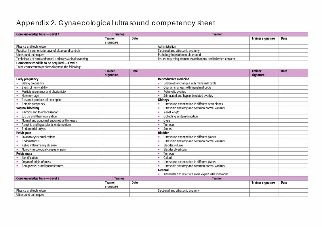
Appendix 2. Gynaecological ultrasound competency sheet Core knowledge base — Level 1 Trainee: Trainer: Trainer
signature Date Trainer signature Date
Physics and technology Administration Practical instrumentation/use of ultrasound controls Sectional and ultrasonic anatomy Ultrasound techniques Pathology in relation to ultrasound Techniques of transabdominal and transvaginal scanning Issues regarding intimate examinations and informed consent Competencies/skills to be acquired — Level 1 To be competent to perform/diagnose the following: Trainer
signature Date Trainer
signature Date
Early pregnancy Reproductive medicine Dating pregnancy Endometrial changes with menstrual cycle Signs of non-viability Ovarian changes with menstrual cycle Multiple pregnancy and chorionicity Polycystic ovaries Haemorrhage Stimulated and hyperstimulated ovaries Retained products of conception Kidneys Ectopic pregnancy Ultrasound examination in different scan planes Vaginal bleeding Ultrasonic anatomy and common normal variants Fibroids and their localisation Renal length IUCDs and their localisation Collecting system dilatation Normal and abnormal endometrial thickness Cysts Atrophic and hyperplastic endometrium Tumours Endometrial polyps Stones Pelvic pain Bladder Ovarian cyst complications Ultrasound examination in different planes Endometriosis Ultrasonic anatomy and common normal variants Pelvic inflammatory disease Bladder volume Non-gynaecological causes of pain Bladder diverticula Pelvic mass Tumours Identification Calculi Organ of origin of mass Ultrasound examination in different planes Benign versus malignant features Ultrasonic anatomy and common normal variants General Know when to refer to a more expert ultrasonologist Core knowledge base — Level 2 Trainee Trainer Trainer
signature Date Trainer signature Date
Physics and technology Sectional and ultrasonic anatomy Ultrasound techniques

23
Competencies/skills to be acquired — Level 2 To be competent to perform/recognise/diagnose/evaluate the following: Trainer
signature Date Trainer signature Date
Female reproductive tract Almost all pathology affecting the female genitourinary tract Bartholin’s cysts, abscesses and periurethral lesions Ultrasound-guided invasive procedures, including ascitic
drainage, omental biopsy, pelvic mass biopsy (transabdominal or transvaginal), lymph node aspiration, SIH and HyCoSy
Features of haematocolpos
Doppler ultrasound studies relevant to the uterus and ovaries Features of adenomyosis Abnormalities elsewhere in the abdomen and pelvis which
need Non-ovarian endometriosis
Referral for scanning by another ultrasonologist and/or further investigation
Non-gynaecological causes of pelvic pain and how to diagnose appendicitis, inflammatory bowel disease, bowel cancer, hernias, aneurysms and bladder disease
Stage ovarian and uterine tumours Different types of complex ovarian masses Other Malignant disease of the omentum, peritoneum and the rest of the
abdomen
Causes of an abnormal Doppler waveform Features of pleural effusions Changes associated with precocious puberty, thelarche,
adrenarche and virilisation Common sites and features of tumours that metastasise to the
pelvis
Congenital anomalies The principles of oocyte collection by transvaginal ultrasound-guided aspiration of follicles
Features of lymph nodes in the inguinal and iliac chains

Appendix 3. Gastrointestinal ultrasound This curriculum is intended for clinicians who perform diagnostic gastrointestinal ultrasound and ultrasound-guided intervention. At least Level 1 should be obtained by anyone performing unsupervised diagnostic imaging.
Focused training and practice
There are frequent situations arising in clinical practice where rapid bedside assessment using focused ultrasound techniques can help with the assessment of, and treatment planning for, patients. In this situation, rapid ultrasound assessment by a competent non-radiological clinician may be more appropriate than waiting for a formal ultrasound list during normal working hours. Some clinicians may wish to focus on only one skill such as drainage of ascites and ultrasound-guided liver biopsies. These unitary skills may aid the clinician’s practice and greatly improve patient pathways.
Each clinician will have their own requirement for focused training and to accommodate their training requirements, a targeted curriculum and syllabus should be created by local trainers, drawing on appropriate elements of the knowledge base and competencies to be acquired from Levels 1–2, depending on the level of practice expected. An example syllabus is included in the focused ultrasound document; for example, drainage of ascites and ultrasound-guided liver biopsies.1
It is essential that all ultrasound examinations that may have any influence on patient management are performed by individuals who are competent to provide an accurate examination and assessment and that the images and a formal report are recorded on a RIS/PACS system.
Level 1. Knowledge base
Physics and technology, ultrasound techniques and administration (see Appendix 11) Sectional and ultrasonic anatomy
– Liver – Gallbladder – Bile ducts – Pancreas – Spleen – Kidneys – Other structures (uterus, ovaries, lymph nodes, vessels, bowel)
Pathology in relation to ultrasound – Liver: cysts, benign and malignant tumours, metastatic disease, fatty change, cirrhosis – Biliary system: gallbladder stones, acute and chronic cholecystitis, gallbladder tumours, bile duct
obstruction including level of obstruction, intrahepatic duct gas and stones – Pancreas: pancreatitis, duct stones, duct dilatation, pancreatic tumours – Portal venous system and spleen: splenic enlargement, portal venous distention, varices, thrombosis,
ascites – Other structures: renal masses and urinary tract obstruction (hydronephrosis), ovarian and uterine masses
including cysts, tumours, fibroids and unexpected pregnancy
Level 1. Training and practice
Practical training should involve at least one ultrasound list per week over 3–6 months with approximately 5–10 examinations performed by the trainee (under supervision) per session. A minimum of 250 examinations should be undertaken. However, different trainees will acquire the necessary skills at different rates and the endpoint of the training programme should be judged by an assessment of competencies.
Examinations should encompass the full range of pathological conditions listed below.
A logbook listing the types of examinations undertaken should be kept.
Training should be supervised either by someone who has obtained at least Level 2 competence in gastrointestinal ultrasound or by a Level 1 practitioner with at least two years’ experience of Level 1 practice.
Trainees should attend an appropriate theoretical course and should read appropriate textbooks and literature.
During the course of training, the competency assessment sheet should be completed as this will determine in which area or areas the trainee can practise independently.

25
Level 1. Competencies to be acquired
Liver
To be able to:
Perform a thorough ultrasound examination of the liver in different scan planes Recognise normal hepatic anatomy and variants Recognise normal and abnormal liver texture, such as fatty change, cirrhosis, atrophy and hypertrophy Recognise focal lesions, particularly cysts and benign lesions, and be able to detect possible malignant lesions
requiring further investigation Recognise normal hepatic and portal venous anatomy within the liver Recognise abnormalities of the hepatic and portal venous system, eg, thrombosis Perform ultrasound-controlled biopsy for the evaluation of parenchymal liver disease
Biliary system
To be able to:
Perform a thorough evaluation of the biliary system Recognise normal ultrasonic anatomy of the biliary system and its frequent normal variants Recognise abnormalities of the gallbladder wall Recognise gallbladder stones Recognise features of acute and chronic cholecystitis Assess bile duct dilatation at intrahepatic and extrahepatic levels and determine level of obstruction
Pancreas
To be able to:
Perform a thorough examination of the pancreas recognising normal anatomy Recognise the limitations of pancreatic ultrasound because of bowel gas Recognise solid and cystic tumours within the head and body of the pancreas Recognise pancreatic duct dilatation and pancreatic duct stones Recognise the features of acute and chronic pancreatitis and their complications
Portal venous system and spleen
To be able to:
Evaluate the size of the spleen and recognise focal lesions and evidence of trauma Evaluate the portal vein and its diameter and the presence of portal venous thrombosis Recognise cavernous transformation of the portal vein and varices
Bowel
To be able to:
Recognise normal stomach, small and large bowel Recognise focal intestinal abnormality and understand the principles of further investigation Recognise small bowel obstruction
Other
To be able to:
Recognise abdominal aortic aneurysm Recognise hydronephrosis Recognise normal kidneys, uterus and ovaries Recognise renal and gynaecological masses
Level 2. Knowledge base
Sectional and ultrasonic anatomy
Detailed understanding of intestinal, mesenteric, peritoneal, omental, vascular and retroperitoneal anatomy
Pathology in relationship to ultrasound
26
An understanding of disease processes which affect the peritoneal cavity, its mesenteries, ligaments and compartments
An understanding of the pathways of spread of intraperitoneal and retroperitoneal disease
Level 2. Training and practice
Practical training should involve at least one year of experience at Level 1 with a minimum of one session per week.
A further 500 examinations should have been undertaken in order to encompass the full range of conditions and procedures listed below.
A logbook listing all examinations undertaken should be kept.
Supervision of training should be undertaken by someone who has achieved Level 2 competence in gastrointestinal ultrasound, has had at least two years’ experience at that level and who would normally be of consultant status.
A Level 2 practitioner will be able to accept referrals from Level 1 practitioners.
Level 2. Competencies to be acquired
Competencies will have been gained during training for Level 1 practice and refined during a period of clinical practice.
Perform a comprehensive ultrasound examination of all of the solid organs within the abdomen. Be able to evaluate the small bowel for focal or diffuse disease. Be able to evaluate the bowel in inflammatory bowel disease. Be able to evaluate the large bowel for the presence of diverticular disease, appendicitis and their
complications, tumours and obstruction. Be able to evaluate the peritoneal cavity, its mesenteries, compartments and the omentum for the presence of
infective or malignant disease. Be able to undertake ultrasound-guided drainage of peritoneal fluid collections. Be able to evaluate the hepatic and portal venous systems using spectral, colour and power Doppler
ultrasound. Be able to undertake ultrasound-guided biopsy of focal liver lesions.
Level 3. Training and practice
A Level 3 practitioner is likely to spend the majority of their time undertaking gastrointestinal ultrasound, teaching, research and development and will be an ‘expert’ in this area.
They will accept tertiary referrals from Level 1 and 2 practitioners and will perform specialised examinations (eg, the use of intravascular ultrasound agents in evaluating focal liver lesions) as well as performing advanced ultrasound-guided invasive procedures.
Maintenance of skills: all levels
Having been assessed as competent to practise, there will be a need for CPD and maintenance of practical skills.
A specialist registrar will need to continue to perform ultrasound throughout the remainder of their training programme. Such further ultrasound practice may be intermittent, but no more than three months should elapse without the trainee using their scanning skills, and at least 100 scans should be performed per year.
A medical practitioner performing ultrasound at Level 1 should continue to perform at least 250 examinations per year on a regular basis, should have regular meetings with radiological colleagues and should have a named radiologist as an ‘ultrasound mentor’.
Practitioners should:
Include ultrasound in their ongoing CPD which should be included in annual appraisal and revalidation Audit their practice Participate in multidisciplinary meetings Keep up to date with relevant literature.
27
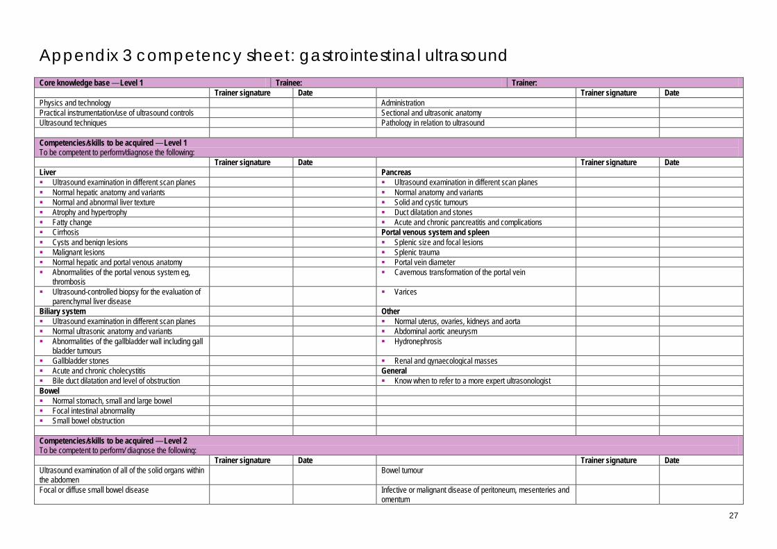
Appendix 3 competency sheet: gastrointestinal ultrasound Core knowledge base — Level 1 Trainee: Trainer: Trainer signature Date Trainer signature Date Physics and technology Administration Practical instrumentation/use of ultrasound controls Sectional and ultrasonic anatomy Ultrasound techniques Pathology in relation to ultrasound Competencies/skills to be acquired — Level 1 To be competent to perform/diagnose the following: Trainer signature Date Trainer signature Date Liver Pancreas Ultrasound examination in different scan planes Ultrasound examination in different scan planes Normal hepatic anatomy and variants Normal anatomy and variants Normal and abnormal liver texture Solid and cystic tumours Atrophy and hypertrophy Duct dilatation and stones Fatty change Acute and chronic pancreatitis and complications Cirrhosis Portal venous system and spleen Cysts and benign lesions Splenic size and focal lesions Malignant lesions Splenic trauma Normal hepatic and portal venous anatomy Portal vein diameter Abnormalities of the portal venous system eg,
thrombosis Cavernous transformation of the portal vein
Ultrasound-controlled biopsy for the evaluation of parenchymal liver disease
Varices
Biliary system Other Ultrasound examination in different scan planes Normal uterus, ovaries, kidneys and aorta Normal ultrasonic anatomy and variants Abdominal aortic aneurysm Abnormalities of the gallbladder wall including gall
bladder tumours Hydronephrosis
Gallbladder stones Renal and gynaecological masses Acute and chronic cholecystitis General Bile duct dilatation and level of obstruction Know when to refer to a more expert ultrasonologist Bowel Normal stomach, small and large bowel Focal intestinal abnormality Small bowel obstruction Competencies/skills to be acquired — Level 2 To be competent to perform/ diagnose the following: Trainer signature Date Trainer signature Date Ultrasound examination of all of the solid organs within the abdomen
Bowel tumour
Focal or diffuse small bowel disease Infective or malignant disease of peritoneum, mesenteries and omentum

28
Inflammatory bowel disease Ultrasound-guided drainage of peritoneal fluid collections Diverticular disease, appendicitis and their complications
The hepatic and portal venous systems using spectral, colour and power Doppler ultrasound
Bowel obstruction Ultrasound-guided biopsy of focal liver lesions

29
Appendix 4. Vascular ultrasound This curriculum is intended for clinicians who perform diagnostic vascular ultrasound and ultrasound-guided intervention. At least Level 1 should be obtained by anyone performing unsupervised diagnostic imaging.
Focused training and practice
There are frequent situations arising in clinical practice where rapid bedside assessment using focused ultrasound techniques can help with the assessment of, and treatment planning for, patients. In this situation, rapid ultrasound assessment by a competent non-radiological clinician may be more appropriate than waiting for a formal ultrasound list during normal working hours. Some clinicians may wish to focus on only one skill such as to provide ultrasound-guided vascular cannulation. These unitary skills may aid the clinician’s practice and greatly improve patient pathways.
Each clinician will have their own requirement for focused training and in order to accommodate their training requirements, a targeted curriculum and syllabus should be created by local trainers, drawing on appropriate elements of the knowledge base and competencies to be acquired from Levels 1–2, depending on the level of practice expected. An example syllabus is included in the focused ultrasound document; for example, ultrasound-guided vascular cannulation.1
It is essential that ALL ultrasound examinations that may have any influence on patient management are performed by individuals who are competent to provide an accurate examination and assessment and that the images and a formal report are recorded on a RIS/PACS system.
Level 1. Knowledge base
Physics and technology, ultrasound techniques and administration (see Appendix 11) To have full working knowledge of the principles, techniques, instrumentation and practical working knowledge
of real-time and Doppler ultrasound, and equipment controls. This includes colour flow and power Doppler, colour and pulsed wave, scale, gain, filter, angle correction, electronic steering, invert, sample gating, power output, colour amplitude, velocity measurement, spectral changes and all other parameters required to perform a complete diagnostic vascular duplex study.
Sectional and ultrasonic anatomy – Peripheral extremity arteries – Peripheral extremity veins – Abdominal vessels – Extracranial vessels – Common normal variants
Pathology in relation to ultrasound – Peripheral extremity arteries: patency, occlusion, stenosis, aneurysmal dilatation – Peripheral extremity veins: patency, occlusion, deep venous thrombosis, reflux and incompetence – Abdominal vessels: patency, occlusion, aneurysmal dilatation of aorta – Extracranial vessels: patency, occlusion, stenosis – Appearances and sequelae of common surgical or radiological interventions including angioplasty,
stenting, grafts, miller vein cuffs, dissections, and neointimal hyperplasia
Level 1. Training and practice
Practical training should involve at least two ultrasound lists per week over a period of no less than three months and up to six months, with approximately four to six examinations performed by the trainee under supervision per session.
A minimum of 100 examinations should be undertaken if this is the first practical training module undertaken.
Examinations should encompass the full range of pathological conditions listed below.
A logbook listing the types of examinations undertaken should be kept.
Training should be supervised either by someone who has obtained at least Level 2 competence in vascular ultrasound or by a Level 1 practitioner with at least two years’ experience of Level 1 practice. This will usually mean that training is carried out in dedicated vascular duplex sessions supervised by an accredited vascular scientist/technologist, specialist sonographer or radiologist.
Trainees should attend an appropriate theoretical course and should read appropriate textbooks and literature.
30
During the course of training, the competency assessment sheet should be completed as this will determine in which area or areas the trainee can practise independently.
Level 1. Competencies to be acquired
To be able to perform continuous wave hand-held Doppler and segmental pressures (ABPI)
Lower extremity peripheral arteries and grafts
To be able to:
Perform a complete ultrasound examination of the common femoral to popliteal arteries Recognise and assess patency, occlusion, stenosis and aneurysmal dilatation, and measure approximate
extent of abnormality Diagnose > 50% stenosis (a doubling of peak systolic velocity [PSV] with pulsed Doppler over adjacent
segments) Recognise common surgical interventions, arteriovenous (AV) fistulas and pseudoaneurysm formation
Peripheral veins
Lower extremity deep veins To be able to: – Perform a complete ultrasound examination of femoral to popliteal deep veins – Perform compression and augmentation – Recognise acute above-knee deep venous thrombosis – Recognise, diagnose and locate reflux
Lower extremity superficial veins To be able to: – Identify the saphenofemoral and saphenopopliteal junctions – Recognise and locate clinically relevant venous reflux, incompetence and perforators – Perform vein mapping and marking
Abdominal vessels To be able to: – Recognise and locate patency and occlusion of the abdominal aorta – Recognise and size aneurysmal dilatation of the abdominal aorta
Extracranial vessels To be able to: – Recognise and locate patency, occlusion, plaque and stenoses in the carotid vessels
Level 2. Knowledge base
Peripheral arteries and grafts Peripheral deep and superficial veins Transcranial Doppler ultrasound:
– Ultrasonic anatomy, common normal variants and principles and practice of the technique – Clinical indications and ultrasonic findings in common clinically relevant abnormalities
Level 2. Training and practice
Practical training should include at least one year of experience at Level 1 with continuous ongoing regular ultrasound sessions.
A logbook of all examinations undertaken should be kept.
Supervision of training should be undertaken by someone who has achieved at least Level 1 competence in vascular ultrasound and has had at least two years’ experience at that level.
Level 2. Competencies to be acquired
Competencies will have been gained during training for Level 1 practice and refined during a period of clinical practice
To be able to:
Perform a complete ultrasound scan and identify all abnormalities detailed in Level 1 in the upper and lower extremities, from iliac to infrapopliteal and subclavian to radial and ulnar arteries and veins

31
Extracranial vessels
To be able to:
Recognise and diagnose patency, occlusion, stenosis, reverse flow and steal in the carotid and vertebral vessels
Grade degrees of carotid stenosis and plaque type in accordance with local criteria and standards
Abdominal vessels
To be able to:
Recognise common normal variants, aneurysmal dilatation, patency, stenosis and occlusion of the major abdominal and iliac vessels, including the mesenteric and renal vessels
Level 3. Training and practice
A Level 3 practitioner is likely to spend the majority of their time undertaking vascular ultrasound.
They will accept tertiary referrals from Level 1 and 2 practitioners.
They should have the capability to utilise developing technologies and ultrasound techniques, develop research and teaching skills and the performance of specialised examinations including the use of non-invasive physiological studies, contrast agents, intravascular or intra-operative ultrasound and ultrasound-guided invasive procedures.
Maintenance of skills: all levels
Having been assessed as competent to practise, there will be a need for CPD and maintenance of practical skills.
A trainee should continue to perform ultrasound scans during the remainder of their training programme, ideally one session weekly and at least 50 examinations per year.
A similar minimum ongoing commitment should be required from a trained practitioner. It is recognised that due to training or clinical circumstances such further ultrasound practice may be intermittent. If a significant period has elapsed after the use of such skills, and a period of re-training is required, it should be agreed and documented with the practitioner, local trainers and assessors.
Practitioners should:
Include ultrasound in their ongoing CPD which should be included in annual appraisal and revalidation Audit their practice Participate in multidisciplinary meetings Keep up to date with relevant literature.
The Royal College of Radiologists is grateful to the Vascular Surgical Society of Great Britain and Ireland and the Society of Vascular Technologists who contributed to and approved this section of the document.
32
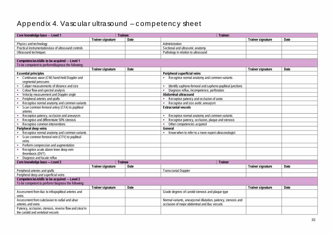
Appendix 4. Vascular ultrasound – competency sheet Core knowledge base — Level 1 Trainee: Trainer: Trainer signature Date Trainer signature Date Physics and technology Administration Practical instrumentation/use of ultrasound controls Sectional and ultrasonic anatomy Ultrasound techniques Pathology in relation to ultrasound Competencies/skills to be acquired — Level 1 To be competent to perform/diagnose the following: Trainer signature Date Trainer signature Date Essential principles Peripheral superficial veins Continuous wave (CW) hand-held Doppler and
segmental pressures Recognise normal anatomy and common variants
Caliper measurements of distance and size Identify sapheno-femoral and sapheno-popliteal junctions Colour flow and spectral analysis Diagnose reflux, incompetence, perforators Velocity measurement and Doppler angle Abdominal ultrasound Peripheral arteries and grafts Recognise patency and occlusion of aorta Recognise normal anatomy and common variants Recognise and size aortic aneurysm Scan common femoral artery (CFA) to popliteal
arteries Extracranial vessels
Recognise patency, occlusion and aneurysm Recognise normal anatomy and common variants Recognise and differentiate 50% stenosis Recognise patency, occlusion, plaque and stenosis Recognise common interventions Other competencies acquired Peripheral deep veins General Recognise normal anatomy and common variants Know when to refer to a more expert ultrasonologist Scan common femoral vein (CFV) to popliteal
veins
Perform compression and augmentation Recognise acute above knee deep vein
thrombosis (DVT)
Diagnose and locate reflux Core knowledge base — Level 2 Trainee Trainer Trainer signature Date Trainer signature Date Peripheral arteries and grafts Transcranial Doppler Peripheral deep and superficial veins Competencies/skills to be acquired — Level 2 To be competent to perform/ diagnose the following: Trainer signature Date Trainer signature Date Assessment from iliac to infrapopliteal arteries and veins
Grade degrees of carotid stenosis and plaque type
Assessment from subclavian to radial and ulnar arteries and veins
Normal variants, aneurysmal dilatation, patency, stenosis and occlusion of major abdominal and iliac vessels
Patency, occlusion, stenosis, reverse flow and steal in the carotid and vertebral vessels
33
Appendix 5. Breast ultrasound This curriculum is intended for clinicians who perform diagnostic breast ultrasound and ultrasound-guided breast intervention. At least Level 1 should be obtained by anyone performing unsupervised diagnostic breast imaging.
Level 1. Knowledge base
Physics and technology, ultrasound techniques and administration (see Appendix 11) Sectional and ultrasound anatomy
– Normal anatomy of female and male breast, and patterns of disease spread – Changes in ultrasound appearances associated with age, pregnancy and lactation, hormonal status,
medication Pathology in relation to ultrasound
– Benign conditions to include: cyst, fibroadenoma, lipoma, fat necrosis, hamartoma, papillary lesions, radial scar/complex sclerosing lesion, gynaecomastia
– Indeterminate conditions to include: atypical ductal and lobular hyperplasia – Malignancy to include: ductal, lobular, inflammatory and other carcinomas – Normal and abnormal appearances of axillary lymph nodes – Inflammatory breast conditions to include: infection, abscess formation and periductal sepsis – Iatrogenic appearances to include: breast implants, early and late postoperative changes, seroma,
haematoma, radiotherapy changes, fat necrosis, scarring Triple assessment
– Local/national guidelines – Clinical findings associated with normal, benign and malignant changes – Mammographic interpretation, correlation and co-localisation with ultrasound findings
Level 1. Training and practice
Practical training should involve at least one ultrasound session per week over a period of no less than six months and no more than one year.
A minimum of 100 examinations should be undertaken and a record of these kept.
A logbook of 50 cases should be kept which should record details of the indications for the procedure, the interpretation and a final report. These cases should be supported by correlation with clinical examination and other imaging findings, needle biopsy results and surgical histology where appropriate.
The numbers suggested will vary depending how quickly competencies are achieved.
Examinations should include an appropriate range of normal and abnormal cases including palpable and impalpable lesions which should encompass the full range of conditions listed above. They should also include patients presenting to symptomatic clinics, screening assessment clinics and postoperative surgical clinics.
Training should be supervised either by someone who has obtained at least Level 2 competence in breast ultrasound or by a Level 1 practitioner with at least two years’ experience of Level 1 practice.
Trainees should be working in line with National Occupational Standards.3 The practical experience should ideally be undertaken in conjunction with attendance on a recognised postgraduate course and trainees should read appropriate textbooks and literature.
During the course of training, the competency assessment sheet should be completed as this will determine in which area or areas the trainee can practise independently.
Level 1. Competencies to be acquired
An understanding of the strengths, weaknesses and limitations of breast ultrasound An understanding of the indications for and the importance of ultrasound in the triple assessment process An awareness of the interdependency and significance of mammographic and ultrasound appearances The ability to perform a thorough ultrasound examination of the breast and axilla, to recognise normal anatomy
and physiological variation as detailed above and to confidently exclude the presence of a sonographic lesion within the breast
The recognition of established criteria for lesion characterisation Identification and discrimination between clearly normal and suspicious axillary lymph nodes The ability to write a detailed report of the ultrasound findings with grading, differential diagnosis, conclusion
and recommendation for further management

34
The recognition of personal limitations and ability to ask for more expert advice if required
Level 2. Knowledge base
Breast imaging
Knowledge of a range of other imaging studies relevant to breast imaging and their role; eg, breast MR, PET-CT, tomosynthesis, contrast-enhanced mammography, elastography
An understanding of principles of evaluation of tumour response to treatment
Pathology in relation to ultrasound
More detailed understanding of breast disease
Level 2. Training and practice
Practical training should involve at least one year of experience at Level 1. Competencies should be further developed during a period of clinical practice, which will involve at least one session per week with at least ten examinations per week for at least three months.
Training for interventional techniques should include observation initially followed by performance of the examination and/or procedure under close supervision. When competence has been acquired, procedures may be undertaken alone but with support close to hand.
A logbook of diagnostic and interventional procedures performed should be kept with pathological correlation.
Supervision of training should be undertaken by someone who has achieved Level 2 competence in breast ultrasound and has had at least two years’ experience at that level and who would normally be of consultant status.
The Level 2 practitioner should be competent to accept referrals from Level 1 practitioners.
Level 2. Competencies to be acquired
Cyst aspiration: initially to perform a minimum of ten guided cyst aspirations of which at least five should be of cysts smaller than 2 cm.
Aspiration of cysts of less than 1 cm diameter. Guided core biopsies of breast: perform a minimum of ten* guided core biopsies with pathological correlation of
core biopsy histology and final pathology (if available). Guided fine needle aspiration biopsy (FNAB): perform a minimum of ten* FNABs of solid lesions, with
pathological correlation of cytology result and final pathology (if available). Guided core biopsy or FNAB of suspicious axillary lymph nodes, in accordance with local guidelines: perform a
minimum of ten* guided biopsies with correlation of histology/cytology result and final pathology (if available). Perform guided abscess aspiration and drainage. Perform preoperative guided localisations using skin marking and wire insertion techniques with
mammographic corroboration as appropriate. Perform guided marker insertion before neo-adjuvant chemotherapy. Evaluation of tumour response to neo-adjuvant treatment. Evaluation of integrity of breast prostheses. Ability to accept referrals from Level 1 practitioners.
* The numbers suggested may vary according to the practice of individual breast units and the speed with which the trainee achieves competence
Level 3. Knowledge base
A more detailed understanding of breast imaging and pathology as detailed above including understanding of population screening, risk and prognostic factors as applied to breast cancer.
Awareness of developments in breast ultrasound including elastography, Doppler and the use of intravascular contrast agents.
Level 3. Training and practice
A Level 3 practitioner is likely to spend the majority of their time undertaking either all aspects of ultrasound, teaching, research and development or be a specialist breast radiologist and will be an ‘expert’ in this area.
They will accept tertiary referrals from Level 1 and 2 practitioners and will perform specialised examinations as well as performing advanced ultrasound-guided invasive procedures such as vacuum-assisted breast biopsy.

35
Maintenance of skills: all levels
Having been assessed as competent to practise, there will be a need for CPD and maintenance of practical skills. A specialist registrar will need to continue to perform breast ultrasound throughout the remainder of the training programme. Such further ultrasound practice may be intermittent, but no more than three months should elapse without the trainee using their scanning skills.
In medical practice, a clinician scanning at Level 1 should perform at least one ultrasound session per week and a minimum of 150 examinations per year and have regular meetings with radiological colleagues and should have a named radiologist as an ‘ultrasound mentor’.
Practitioners should:
Include ultrasound in their ongoing CPD which should be included in annual appraisal and revalidation Audit their practice Participate in multidisciplinary meetings Keep up to date with relevant literature.
Approved by the British Society of Breast Radiology

36
Appendix 5. Breast ultrasound training – competency assessment sheet Core knowledge base — Level 1 Trainee: Trainer: Trainer signature Date Trainer signature Date Physics and technology Administration Practical instrumentation/use of ultrasound controls Sectional and ultrasonic anatomy Ultrasound techniques Pathology in relation to ultrasound Competencies/skills to be acquired — Level 1 To be competent to perform/diagnose the following: Trainer signature Date Trainer signature Date Understands strengths, weaknesses and limitations of breast ultrasound
Aware of relationship between mammographic and ultrasound appearances
Recognises established criteria for lesion characterisation
Able to write detailed report with grading, differential diagnosis, conclusion and recommendation for further management
Understands indications for & importance of ultrasound in triple assessment
Can perform thorough ultrasound examination of breast and axilla, recognise normal anatomy and physiological variation and confidently exclude presence of a sonographic lesion
Identifies and discriminates between clearly normal and suspicious axillary lymph nodes
Knows when to ask for more expert advice
Competencies/skills to be acquired — Level 2 To be competent to perform/ diagnose the following: Trainer signature Date Trainer signature Date Cyst aspiration: initially to perform a minimum of 10 guided cyst aspirations of which at least five should be of cysts smaller than 2 cm
Perform ultrasound-guided breast abscess aspiration
Aspirate cysts smaller than 1 cm in diameter Perform ultrasound-guided localisations preoperatively, using both skin marking and wire insertion techniques
Perform a minimum of 10 FNABs of solid lesions with pathological correlation. If FNA is not standard procedure within the unit then the student should be familiar with the technique and be aware of its uses and limitations
Perform ultrasound-guided marker or coil insertion prior to neo-adjuvant chemotherapy
Perform a minimum of 10 guided core biopsies with pathological correlation
Accept referrals from Level 1 practitioners
37
Appendix 6. Thoracic ultrasound The curriculum is intended for clinicians who perform diagnostic and therapeutic ultrasound. At least Level 1 should be obtained by anyone performing thoracic scans unsupervised.
Focused training and practice
There are frequent situations arising in clinical practice where rapid bedside assessment using focused ultrasound techniques can help with the assessment of, and treatment planning for, patients. In this situation, rapid ultrasound assessment by a competent non-radiological clinician may be more appropriate than waiting for a formal ultrasound list during normal working hours. Some clinicians may wish to focus on only one skill such as ultrasound-guided drainage of pleural effusions. These unitary skills may aid the clinician’s practise and greatly improve patient pathways.
Each clinician will have their own requirement for focused training and in order to accommodate their training requirements, a targeted curriculum and syllabus should be created by local trainers, drawing on appropriate elements of the knowledge base and competencies to be acquired from Levels 1–2, depending on the level of practice expected. An example syllabus is included in the focused ultrasound document for example, ultrasound-guided drainage of pleural effusions.1
It is essential that ALL ultrasound examinations that may have any influence on patient management are performed by individuals who are competent to provide an accurate examination and assessment and that the images and a formal report are recorded on a RIS/PACS system.
Level 1. Knowledge base
Physics and technology, ultrasound techniques and administration (see Appendix 11) Sectional and ultrasonic anatomy
– Right and left hemidiaphragms – Heart – Liver and spleen – Rib and intercostal spaces
Pathology in relation to ultrasound Pleural effusion Pleural thickening Consolidated lung Paralysed hemidiaphragm Pericardial effusion
Level 1. Training and practice
Practical training should include:
At least one session per week over a period of no less than three months, with approximately five scans per session performed by the trainee (under supervision of an experienced practitioner)
The numbers suggested will vary depending how quickly competencies are achieved.
Examinations should encompass the full range of pathological conditions listed above.
A logbook listing the types of examinations and procedures undertaken should be kept.
Training should be supervised either by someone who has obtained at least Level 2 competence in thoracic ultrasound or by a Level 1 practitioner with at least two years’ experience of Level 1 practice.
Trainees should attend an appropriate theoretical course and should read appropriate textbooks and literature.
During the course of training, the competency assessment sheet should be completed as this will determine in which area or areas the trainee can practise independently.
Level 1. Competencies to be acquired
Recognition of normal anatomy of pleura and diaphragm. Identification of the heart, liver and spleen. Pleural effusion recognition, including the different echogenic patterns. Pleural thickening and its differentiation from fluid using colour flow Doppler if appropriate. Consolidated lung and its differentiation from effusion.
38
Estimation of depth of effusion and its measurement. Guided thoracocentesis and drain placement.
Level 2. Knowledge base
Sectional and ultrasonic anatomy
A full understanding of thoracic and diaphragmatic anatomy
Pathology in relation to ultrasound
More detailed understanding of pleural disease
Level 2. Training and practice
Practical training should involve at least one year of experience at Level 1 with a minimum of two examinations performed per week.
A further 100 examinations should have been undertaken in order to encompass the full range of conditions and procedures referred to above in addition to those listed below.
A logbook of all examinations undertaken should be kept.
Supervision of training should be by someone who has achieved at least Level 2 competence in thoracic ultrasound, has had at least two years’ experience at that level, and who would normally be of consultant status.
A Level 2 practitioner will be able to accept referrals from Level 1 practitioners.
Level 2. Competencies to be acquired
Competencies will have been gained during training for Level 1 practice, and refined during a period of practice.
Ultrasound techniques
The advanced use of Doppler ultrasound, including spectral, colour and power Doppler The use of ultrasound for guiding interventional procedures including lung and supraclavicular nodal biopsy
Level 3. Knowledge base
It is important that physicians correctly interpret CXRs and CT scans used in the decision-making process when managing patients.
Level 3. Training and practice
A Level 3 practitioner is likely to spend the majority of their time undertaking either all aspects of ultrasound, teaching, research and development or be a specialist thoracic radiologist and will be an ‘expert’ in this area.
They will accept tertiary referrals from Level 1 and 2 practitioners and will perform specialised examinations as well as performing advanced ultrasound-guided invasive procedures.
Maintenance of skills: all levels
Having been assessed as competent to practise, there will be a need for CPD and maintenance of practical skills. A chest physician specialist registrar will need to continue to perform ultrasound scans throughout the remainder of the training programme. Such further ultrasound practice may be intermittent, but no more than three months should elapse without the trainee using their scanning skills.
In medical practice, a chest physician scanning at Level 1 should perform at least 20 ultrasound examinations per year and have regular meetings with radiological colleagues and should have a named radiologist as an ‘ultrasound mentor’.
Practitioners should:
Include ultrasound in their ongoing CPD which should be included in annual appraisal and revalidation Audit their practice Participate in multidisciplinary meetings Keep up to date with relevant literature.

39
Appendix 6. Thoracic ultrasound training – competency assessment sheet Core knowledge base — Level 1 Trainee: Trainer: Trainer signature Date Trainer signature Date Physics and technology Administration Practical instrumentation/use of ultrasound controls Sectional and ultrasonic anatomy Ultrasound techniques Pathology in relation to ultrasound Competencies/skills to be acquired — Level 1 To be competent to perform/diagnose the following: Trainer signature Date Trainer signature Date Normal anatomy of pleura and diaphragm Consolidated lung and its differentiation from effusion Identification of heart, liver and spleen Estimation of depth of effusion and its measurement Pleural effusion and different echogenic patterns Know when to refer to a more expert ultrasonologist Pleural thickening and its differentiation from fluid

40
Appendix 7. Cranial ultrasound in infants This syllabus is designed to include the whole of cranial ultrasound imaging in infants, not just the ultrasound skills needed on a neonatal intensive care unit.
It does not include spinal ultrasound imaging.
Level 1. Knowledge base
Physics and technology, ultrasound techniques and administration (see Appendix 11) Issues of parental information and consent. Sectional and ultrasonic anatomy of the brain:
– Sagittal anatomy – Coronal anatomy – Basic transaxial and near-field scanning
Pathology in relation to ultrasound: – Intracranial haemorrhage – Hypoxic ischaemic change (in full-term and premature infants) – Ventricular dilatation – Common congenital malformations
Level 1. Training and practice
Practical training should involve at least one session per week over a period of no less than three months, with approximately five scans per session performed by the trainee (under supervision of an experienced practitioner).
The practitioner should be compliant with local procedures for infection control for scanning, especially on the neonatal unit.
By the end of Level 1 training, the trainee should be able to perform and record images from a basic scan demonstrating normal anatomical structures. They should recognise all common pathologies that are seen on a neonatal intensive care unit.
Different trainees will acquire the necessary skills at different rates. The endpoint of the training program should be judged by assessment of competencies, rather than by numbers of scans performed alone. As a guide, a minimum of 100 scans should be undertaken if this is the first practical training module undertaken. For those who are already competent at ultrasound in other body areas, then a reduced number of scans may be needed to achieve competency.
Examinations should encompass all pathological conditions listed below.
A logbook listing the types of examinations undertaken should be kept.
Training should be supervised either by a practitioner who has obtained at least Level 2 competence in cranial ultrasound, or by a Level 1 practitioner with at least two years’ experience of Level 1 practice.
Trainees should attend an appropriate theoretical course (including physics of US and instrumentation) and should read appropriate textbooks and literature.
During the course of training, the competency assessment sheet should be completed as this will determine in which area or areas the trainee can practise independently.
Level 1. Competencies to be acquired
To be able to:
Perform a thorough ultrasound examination of the brain in different scan planes Recognise normal anatomy and common normal variants Recognise varying presentations of pathological processes with gestational and postnatal age Measure ventricular size and assess variation from normality Assess obstructive hydrocephalus and monitor progression Recognise and describe intracranial haemorrhage and assess its extent Recognise common congenital malformations and refer for appropriate further investigation Recognise when other imaging modalities are more appropriate than ultrasound (eg, MRI or CT) Recognise abnormalities which need referral for scanning by a more experienced practitioner and/or further
investigation

41
To be able to use ultrasound in both the routine and urgent assessment of patients presenting with:
Prematurity Fits/apnoea/collapse Meningitis Hydrocephalus Asphyxia (full term and premature) Other congenital abnormalities
Level 2. Training and practice
Practical training should involve at least one year of experience at Level 1 with a minimum of one session per week.
A significant number of further examinations should have been undertaken sufficient to encompass the full range of conditions and procedures referred to below.
A logbook of all examinations undertaken should be kept.
Supervision of training should be by a practitioner who has achieved at least Level 2 competence in cranial ultrasound, has had at least two years’ experience at that level, and who would normally be of consultant or independent practitioner status.
A Level 2 practitioner will be able to accept referrals from Level 1 practitioners.
Level 2. Knowledge base
Sectional and ultrasonic anatomy
The basic use of Doppler ultrasound, including spectral, colour and power Doppler Further applications of cranial ultrasound Use of transaxial ultrasound Posterior fossa imaging Assessment of cerebral perfusion
Pathology in relation to ultrasound
Understand of the role of ultrasound in the context of cerebral asphyxia, abnormal head circumference, congenital abnormalities, non-accidental injury and systemic disease and the role of alternative imaging modalities.
Level 2. Competencies to be acquired
Competencies will have been gained during training for Level 1 practice, and refined during a period of practice.
Vascular studies in asphyxia. Role of ultrasound in the assessment of abnormal head circumference. Recognition of most identifiable congenital brain malformations. Further assessment of antenatally suspected anomalies. Identification and location of surface collections. Appearances of non-accidental injury and the limitations of ultrasound assessment. Intraoperative ultrasound as appropriate. Infants with systemic disease (eg, postoperative or paediatric intensive care).
Level 3. Training and practice
A Level 3 practitioner is likely to spend a significant proportion of his or her time undertaking cranial ultrasound, teaching, research and development and will be an ‘expert’ in this area.
They will accept tertiary referrals from Level 1 and 2 practitioners and will perform specialised examinations.
Maintenance of skills: all levels
Having been assessed as competent to practice at Level 1, there will be a need for maintenance of practical skills, by continuing to perform regular ultrasound and update skills. Such further ultrasound practice may be intermittent, but no more than three months should elapse without the trainee using their ultrasound skills, and sufficient examinations should be performed per year to maintain competency.

42
In independent practice, a medical practitioner scanning at Level 1 should continue to scan regularly should have regular meetings with imaging colleagues and should have a designated ultrasound practitioner of Level 2 experience or above designated as their mentor.
Practitioners should:
Include ultrasound in their ongoing CPD Audit their practice Participate in multidisciplinary meetings Keep up to date with relevant literature.

43
Appendix 7. Cranial ultrasound training – competency assessment sheet Core knowledge base — Level 1 Trainee: Trainer: Trainer signature Date Trainer signature Date Physics and technology Administration Practical instrumentation/use of ultrasound controls Sectional and ultrasonic anatomy Ultrasound techniques Pathology in relation to ultrasound Infection control Competencies/skills to be acquired — Level 1 To be competent to perform/diagnose the following: Trainer signature Date Trainer signature Date Normal US anatomy of the neonatal brain/variants Image orientation and documentation US assessment of intracranial haemorrhage US assessment of neonatal asphyxia US assessment of ventricular dilatation/large head US assessment of congenital malformations US assessment of the premature infant’s brain Know when to refer to a more expert ultrasonologist US assessment of the acutely unwell neonate

44
Appendix 8. Focused emergency ultrasound This curriculum is intended for clinicians who perform diagnostic ultrasound and ultrasound-guided intervention. At least Level 1 should be obtained by anyone performing unsupervised diagnostic imaging.
Level 1. Knowledge base
Physics and technology, ultrasound techniques and administration (see Appendix 11) Sectional and ultrasonic anatomy
– Kidneys – Liver – Spleen – Retroperitoneal structures (aorta, inferior vena cava [IVC]) – Vessels: internal jugular veins, carotid arteries, femoral veins and arteries
Pathology in relation to ultrasound – Kidneys: trauma/free fluid – Liver and spleen: trauma/free fluid – Retroperitoneal: presence or absence of abdominal aortic aneurysm (AAA) – Vessels: vascular access – Cardiac scan: trauma/pericardial tamponade, pericardial effusions, asystole
Level 1. Training and practice
Practical training should involve regular emergency department or radiology department ultrasound, with approximately five examinations performed by the trainee (under supervision) per week.
Approximately 50 examinations should be undertaken if this is the first practical training module. A clinician working in emergency medicine will need to devote sufficient time to gain Level 1 competence. This may be as much as one session per week but is unlikely to be in dedicated blocks of time. However, different trainees will acquire the necessary skills at different rates, and the key endpoint of the training programme should be judged by an assessment of competencies.
Examinations should concentrate on the core clinical indications of trauma, aortic aneurysm and vascular access where there are benefits of an early focused ultrasound scan in the emergency department or acute assessment area.
A logbook listing the types of examinations undertaken should be kept.
An additional pictorial record containing an illustrated description of ten cases in which the trainee has been personally involved may be collected and is a useful confirmation of experience when moving between departments.
Training should be supervised either by someone who has obtained at least Level 2 competence in focused emergency ultrasound, or by a Level 1 practitioner with at least one year’s experience of Level 1 practice.
Trainees should attend an appropriate theoretical course and should be familiar with the published literature on focused emergency ultrasound.
During the course of training, the competency assessment sheet should be completed as this will determine in which area or areas the trainee can practise independently.
Level 1. Competencies to be acquired
To be able to:
Recognise normal anatomy Use focused ultrasound to assist in bedside emergency department decisions Recognise the limitations of a scan and be able to explain these limitations to patients/carers Recognise patients requiring formal specialist sonographic assessment Incorporate ultrasound findings with the rest of the clinical assessment
To be able to use ultrasound in the context assessment of:
Focused assessment by sonography for trauma (FAST) AAA screening/detection in symptomatic patients Peri-arrest scenario for pulseless electrical activity (PEA)/tamponade/effusion

45
Vascular access Pleural and pericardial fluid
Level 2. Knowledge base
Sectional and ultrasonic anatomy – Kidneys – Liver – Proximal leg veins – Heart – Musculoskeletal system – Pelvic structures (uterus, ovaries, bowel)
Pathology in relation to ultrasound – Kidneys: cysts, calyceal dilatation, renal calculi, trauma (free fluid) – Liver: cysts, trauma (free fluid) – Vessels: proximal deep venous thrombosis – Cardiac: reduced ventricular function, tamponade, effusion – Retroperitoneum: aortic aneurysm, fluid collection – Musculoskeletal: fractures, soft tissue collections, foreign body detection
Level 2. Training and practice
Practical training should involve at least one year of experience at Level 1 with an average of three to five scans/week.
A further 150–200 examinations should have been undertaken in order to encompass most of the conditions and procedures listed. A practitioner may develop competencies in some but not all of these areas.
A logbook of all examinations undertaken should be kept.
A further pictorial logbook should also be kept detailing ten cases examined by the trainee which may be useful as a record when moving between departments.
Supervision of training should be undertaken by someone who has achieved at least Level 2 competence in focused emergency ultrasound and who has had at least two years’ experience at this level.
A Level 2 practitioner will be able to accept referrals from a Level 1 practitioner.
Level 2. Competencies to be acquired
Competencies will have been gained during training for Level 1 practice, and refined during a period of clinical practice.
Recognise and correctly utilise ultrasound within the emergency department and be able to perform at least three of the following: – Ultrasound-guided invasive procedures (chest drain insertion, suprapubic aspiration/ catheterisation, fluid
collection drainage) – An initial assessment for patients with loin pain/haematuria – A focused assessment of a patient presenting with hypotension – Proximal DVT assessment – Detection of foreign bodies and fluid collections within soft tissues – Early assessment of symptomatic women in the first trimester of pregnancy post-trauma including
assessment of foetal cardiac activity – Emergency department obstetric presentations
Level 3. Training and practice
A Level 3 practitioner is likely to spend a significant proportion of their time undertaking emergency ultrasound, teaching, research and development and will be an ‘expert’ in this area.
They will accept referrals from Level 1 and 2 practitioners and will perform the spectrum of examinations indicated above. This will include developing the role for innovative ultrasound practice within emergency care.
Maintenance of skills: all levels
Having been assessed as competent to practise, there will be a need for CPD and maintenance of practical skills.

46
An emergency trainee will need to continue to perform ultrasound scans throughout the remainder of the training programme and into their consultant appointment. Such further ultrasound practice may be intermittent, but no more than three months should elapse without the trainee using his scanning skills, and at least 50 scans should be performed per year.
All practitioners should have regular meetings within the department to ensure appropriate focused emergency ultrasound use. The department lead for ultrasound practice will have regular contact with radiological colleagues and should have a named radiologist as an ‘ultrasound mentor’.
Practitioners should:
Include ultrasound in their ongoing CPD which should be included in annual appraisal and revalidation Audit their practice Participate in multidisciplinary meetings Keep up to date with relevant literature
Approved by the Faculty of Accident and Emergency Medicine
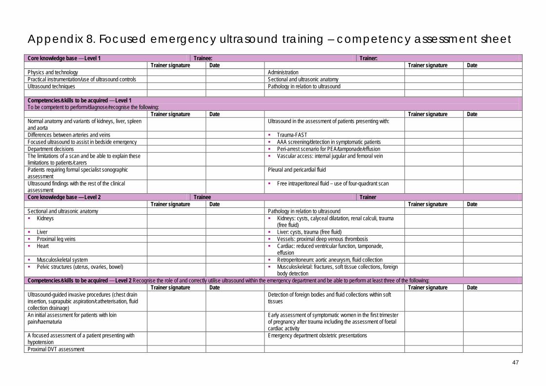
47
Appendix 8. Focused emergency ultrasound training – competency assessment sheet Core knowledge base — Level 1 Trainee: Trainer: Trainer signature Date Trainer signature Date Physics and technology Administration Practical instrumentation/use of ultrasound controls Sectional and ultrasonic anatomy Ultrasound techniques Pathology in relation to ultrasound Competencies/skills to be acquired — Level 1 To be competent to perform/diagnose/recognise the following: Trainer signature Date Trainer signature Date Normal anatomy and variants of kidneys, liver, spleen and aorta
Ultrasound in the assessment of patients presenting with:
Differences between arteries and veins Trauma-FAST Focused ultrasound to assist in bedside emergency AAA screening/detection in symptomatic patients Department decisions Peri-arrest scenario for PEA/tamponade/effusion The limitations of a scan and be able to explain these limitations to patients/carers
Vascular access: internal jugular and femoral vein
Patients requiring formal specialist sonographic assessment
Pleural and pericardial fluid
Ultrasound findings with the rest of the clinical assessment
Free intraperitoneal fluid – use of four-quadrant scan
Core knowledge base — Level 2 Trainee Trainer Trainer signature Date Trainer signature Date Sectional and ultrasonic anatomy Pathology in relation to ultrasound Kidneys Kidneys: cysts, calyceal dilatation, renal calculi, trauma
(free fluid)
Liver Liver: cysts, trauma (free fluid) Proximal leg veins Vessels: proximal deep venous thrombosis Heart Cardiac: reduced ventricular function, tamponade,
effusion
Musculoskeletal system Retroperitoneum: aortic aneurysm, fluid collection Pelvic structures (uterus, ovaries, bowel) Musculoskeletal: fractures, soft tissue collections, foreign
body detection
Competencies/skills to be acquired — Level 2 Recognise the role of and correctly utilise ultrasound within the emergency department and be able to perform at least three of the following: Trainer signature Date Trainer signature Date Ultrasound-guided invasive procedures (chest drain insertion, suprapubic aspiration/catheterisation, fluid collection drainage)
Detection of foreign bodies and fluid collections within soft tissues
An initial assessment for patients with loin pain/haematuria
Early assessment of symptomatic women in the first trimester of pregnancy after trauma including the assessment of foetal cardiac activity
A focused assessment of a patient presenting with hypotension
Emergency department obstetric presentations
Proximal DVT assessment

48
Appendix 9. Ultrasound training for critical/intensive care This curriculum is intended for clinicians who perform diagnostic ultrasound and ultrasound-guided intervention. At least Level 1 should be obtained by anyone performing unsupervised diagnostic imaging.
Level 1. Knowledge base
Physics and technology, ultrasound techniques and administration (see Appendix 11) Sectional and ultrasonic anatomy
– Thorax – Abdomen, retroperitoneal and peritoneal spaces – Abdominal aorta – Urinary tract – Peripheral vascular anatomy – Peripheral neurological anatomy
Pathology in relation to ultrasound – Thorax: pleural disease, patterns and causes of pulmonary consolidation, pericardial disease. – Abdomen: fluid collections, gastrointestinal ileus, acute urinary tract pathology, aortic aneurysms – Vascular: venous disease, eg, thrombosis
Basic knowledge of Doppler ultrasound principles
Level 1. Training and practice
Practical training should involve carrying out regular ultrasound examinations in the critical care unit or radiology department, with approximately 5–10 examinations performed by the trainee (under supervision) per week.
A minimum number of examinations in each specific area of interest should be undertaken if this is the first practical training module undertaken; for example, 25 pleural effusions, 25 vascular line insertions, 25 abnormal abdominal scans. However, different trainees will acquire the necessary skills at different rates and the endpoint of the training programme should be judged by an assessment of competencies.
Examinations should encompass the full range of pathological conditions and practical procedures listed below.
A logbook listing the types of examinations undertaken should be kept.
An additional logbook containing an illustrated description of ten cases in which the trainee has been personally involved may be a useful adjunct particularly if moving between different units.
Training should be supervised either by someone who has obtained at least Level 2 competence in critical care ultrasound themselves, or by a Level 1 practitioner with at least two years’ experience of Level 1 practice.
Trainees should attend an appropriate theoretical course and should be familiar with the published literature.
During the course of training, the competency assessment sheet should be completed as this will determine in which area or areas the trainee can practise independently.
Level 1. Competencies to be acquired
General
Recognise normal anatomy Use ultrasound to assist in critical care decisions and interventional procedures Recognise limitations of technique and own knowledge base Recognise patients requiring formal specialist sonographic assessment
Thorax
Pleural disease – Identify and assess pleural effusions: transudate, exudates, volume estimation – Use ultrasound to guide pleural aspiration/drainage
Pulmonary – Identify pulmonary consolidation – Distinguish abnormal lung from pleural diseases
Pericardial – identify significant pericardial effusions prior to formal echocardiographic assessment

49
Abdomen
Fluid collections – Identify and assess abdominal and pelvic fluid collections – Differentiate subphrenic from pleural fluid
Distension – Identify cause of abdominal distension, eg, fluid, small bowel ileus, bladder distension, masses
Urinary tract
Identify cause of acute renal impairment, specifically hydronephrosis Identify significant bladder distension
Vascular
Identify and measure abdominal aortic aneurysm Recognise abdominal features secondary to cardiac failure
Peripheral vascular
Differentiate between arteries and veins Assess veins for vascular access
– Identify suitable neck veins and their patency – Use ultrasound to guide line insertions
Peripheral neurology
Identify paths of appropriate peripheral nerves, eg, brachial plexus Use ultrasound to guide regional anaesthesia
Level 2. Knowledge base
Physics and technology – In-depth knowledge and understanding of physics of ultrasound – In-depth knowledge and understanding of the technology of ultrasound equipment
Ultrasound techniques – Further applications of thoracic, abdominal, vascular and cardiac ultrasound
Sectional and ultrasonic anatomy – More detailed recognition and understanding of normal variants with ultrasound
Pathology related to ultrasound
Level 2. Training and practice
Practical training should include at least one year of experience at Level 1 with a minimum of one session or equivalent per week.
A further 300 examinations should have been undertaken in order to encompass the full range of conditions and procedures.
A logbook listing all examinations undertaken should be kept.
A further illustrated logbook should be kept detailing 20 cases examined by the trainee which may be useful to document further progression of training.
Supervision of training should be undertaken by someone who has achieved at least Level 2 competence in critical care ultrasound, has had at least two years’ experience at that level and would normally be of consultant status.
A Level 2 practitioner will be able to accept referral from Level 1 practitioners.
Level 2. Competencies to be acquired
Competencies will have been gained during training for Level 1 practice, and refined during a period of clinical practice.
Develop further ultrasound skills such as echocardiography, transoesophageal echocardiography and perioperative monitoring.

50
Level 3. Training and practice
A Level 3 practitioner is likely to spend the majority of their time undertaking ultrasound examinations, teaching, research and development, and will be regarded as an ‘expert’ in this area.
They will accept referrals from Level 1 and 2 practitioners and will be actively involved in developing innovative ultrasound applications within critical care.
They will have spent a continuous period of specialist training in critical care ultrasound.
They will be able to mentor and supervise Level 1 and 2 practitioners.
They will be aware of and pursue developments in critical care ultrasound including Doppler and the use of intravascular contrast agents.
Maintenance of skills: all levels
Having been assessed as competent to practise, there will be a need for CPD and maintenance of practical skills.
A trainee should continue to practise ultrasound throughout their training with no more than one month elapsing without the trainee using their ultrasound scanning skills.
In medical practice, a critical care physician performing ultrasound at Level 1 should continue to perform at least 100 examinations per year, should have regular meetings with radiological colleagues and should have a named radiologist or experienced sonographic practitioner as an ‘ultrasound mentor’.
Practitioners should:
Include ultrasound in their ongoing CPD which should be included in annual appraisal and revalidation Audit their practice Participate in multidisciplinary team meetings Keep up to date with relevant literature.
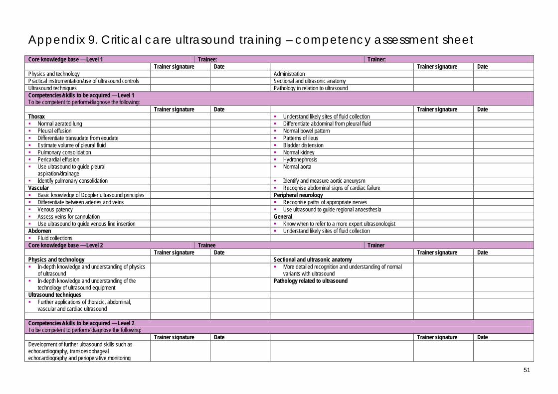
51
Appendix 9. Critical care ultrasound training – competency assessment sheet Core knowledge base — Level 1 Trainee: Trainer: Trainer signature Date Trainer signature Date Physics and technology Administration Practical instrumentation/use of ultrasound controls Sectional and ultrasonic anatomy Ultrasound techniques Pathology in relation to ultrasound Competencies/skills to be acquired — Level 1 To be competent to perform/diagnose the following: Trainer signature Date Trainer signature Date Thorax Understand likely sites of fluid collection Normal aerated lung Differentiate abdominal from pleural fluid Pleural effusion Normal bowel pattern Differentiate transudate from exudate Patterns of ileus Estimate volume of pleural fluid Bladder distension Pulmonary consolidation Normal kidney Pericardial effusion Hydronephrosis Use ultrasound to guide pleural
aspiration/drainage Normal aorta
Identify pulmonary consolidation Identify and measure aortic aneurysm Vascular Recognise abdominal signs of cardiac failure Basic knowledge of Doppler ultrasound principles Peripheral neurology Differentiate between arteries and veins Recognise paths of appropriate nerves Venous patency Use ultrasound to guide regional anaesthesia Assess veins for cannulation General Use ultrasound to guide venous line insertion Know when to refer to a more expert ultrasonologist Abdomen Understand likely sites of fluid collection Fluid collections Core knowledge base — Level 2 Trainee Trainer Trainer signature Date Trainer signature Date Physics and technology Sectional and ultrasonic anatomy In-depth knowledge and understanding of physics
of ultrasound More detailed recognition and understanding of normal
variants with ultrasound
In-depth knowledge and understanding of the technology of ultrasound equipment
Pathology related to ultrasound
Ultrasound techniques Further applications of thoracic, abdominal,
vascular and cardiac ultrasound
Competencies/skills to be acquired — Level 2 To be competent to perform/ diagnose the following: Trainer signature Date Trainer signature Date Development of further ultrasound skills such as echocardiography, transoesophageal echocardiography and perioperative monitoring

52
Appendix 10. Musculoskeletal ultrasound This curriculum is intended for clinicians who perform diagnostic ultrasound and ultrasound-guided intervention. At least Level 1 should be obtained by anyone performing unsupervised diagnostic imaging.
Musculoskeletal ultrasound comprises a wide range of examinations and pathologies. It is unlikely that any practitioner will cover the full range. A modular approach that meets the needs of an individual’s clinical practice is therefore likely to be appropriate and ‘levels’ of competence are not stipulated in this section.
Knowledge base
Physics and technology, ultrasound techniques and administration (see Appendix 11) Sectional and ultrasonic anatomy
– Shoulder – Elbow – Wrist/hand – Groin – Hip – Thigh – Knee – Lower leg – Ankle/foot – Other (eg, infant spine, brachial plexus)
Pathology in relation to ultrasound – Shoulder: rotator cuff tendinosis, partial and complete thickness tears and calcification; joint and bursal
effusion; long head of biceps tendinosis, rupture and subluxation; paralabral cyst – Elbow: tendinosis of common flexor/extensor origins; tendinosis and rupture of triceps and distal biceps
tendons; effusion; loose bodies; ulnar nerve entrapment; bursae – Wrist/hand: tendon tears and tenosynovitis; carpal tunnel syndrome; space-occupying lesions; pulley
injuries; arthropathy – Groin: hernias; tendinosis and tendon tears – Hip: developmental dysplasia of the hip (DDH); effusion (children, adults, prosthetic joints); bursae;
snapping hip; tendinosis and tendon tears – Thigh: muscle contusion and tears; tendinosis and tendon tears – Knee: effusion; bursae (including ruptured baker’s cyst); meniscal cysts; collateral ligament sprains;
patellar tendon tendinosis and rupture – Lower leg: muscle contusion and tears, muscle herniae – Ankle/foot: tendinosis and tears of Achilles, posterior tibial and peroneal tendons; tenosynovitis; ankle
effusion and loose bodies; plantar fasciitis; Morton’s neuroma; other space occupying lesions; arthropathy – Soft tissue masses – Brachial plexus: injuries and tumours – Spine: dysraphism and tethered cord in neonates
Training and practice
Practical training should involve at least one ultrasound list per week for 3–6 months with approximately ten examinations performed by the trainee under supervision per session.
A minimum of 250 examinations should be undertaken. However, trainees acquire the necessary skills at different rates and some examinations (eg, shoulder ultrasound) take considerably longer than others for trainees to acquire proficiency. The end point of the training programme should be judged by an assessment of competencies.
A logbook listing the types and numbers of examinations undertaken should be kept.
Training should be supervised by an experienced musculoskeletal sonologist.
Trainees should attend an appropriate theoretical course and should read appropriate textbooks and literature.
During the course of training, the competency assessment sheet should be completed as this will determine in which area or areas the trainee can practise independently.
53
Competencies to be acquired
Other diagnostic techniques
Be aware of alternative diagnostic methods including clinical examination and imaging techniques Recognise comparative accuracy of alternative techniques Recognise when to proceed to other imaging examinations following ultrasound examination
Shoulder
To be able to:
Perform a thorough ultrasound examination of the shoulder in different planes Recognise normal ultrasonic anatomy and common normal variants Recognise and be aware of difficulties in distinguishing accurately between tendinosis/ partial thickness/
complete thickness tears of the rotator cuff Recognise rotator cuff calcification Recognise tendinosis, rupture and subluxation of the long head of biceps tendon Recognise effusions of the shoulder joint and subdeltoid bursa Recognise paralabral cyst Recognise abnormalities which need referral to a more experienced ultrasonologist and/or for further
investigation
Elbow
To be able to:
Perform a thorough ultrasound examination of the elbow in different planes Recognise normal ultrasonic anatomy and common normal variants Recognise tendinosis of the common flexor/extensor origins Recognise tendinosis/partial/complete rupture of the triceps and distal biceps tendons Recognise joint effusions and loose bodies Recognise ulnar nerve entrapment Recognise olecranon bursitis Recognise abnormalities which need referral to a more experienced ultrasonologist and/or for further
investigation
Wrist/hand
To be able to:
Perform a thorough ultrasound examination in different planes Recognise normal ultrasonic anatomy and common normal variants including accessory muscles Recognise de Quervain’s tenosynovitis Recognise effusions in other tendons sheaths Recognise tendonitis, partial and complete tendon tears and identify retracted tendon Recognise features of carpal tunnel syndrome and entrapment in Guyon’s canal Recognise ganglia and distinguish them from solid space occupying lesions Recognise inflammatory arthropathy Recognise pulley injuries Recognise foreign bodies (FB) and FB reactions Recognise ligament injuries Recognise abnormalities which need referral to a more experienced ultrasonologist and/or for further
investigation
Groin
To be able to:
Perform a thorough ultrasound examination in different planes Recognise normal ultrasonic anatomy and common normal variants Recognise inguinal and femoral hernias Recognise strains of the rectus abdominis and adductor muscles Recognise abnormalities which need referral for scanning by a more experienced ultrasonologist and/or further
investigation
54
Hip
To be able to:
Perform a thorough ultrasound examination in different planes Recognise normal ultrasonic anatomy and common normal variants Assess the neonatal hip and recognise DDH Recognise joint effusions in children, adults and prosthetic joints Recognise iliopsoas bursa and distinguish from solid masses Recognise causes of snapping hip such as iliopsoas, tensor fascia lata and rectus femoris tendons Recognise tendinosis and tendon tears eg, of hamstrings, glutei Recognise abnormalities which need referral to a more experienced ultrasonologist and/or for further
investigation
Thigh
To be able to:
Perform a thorough ultrasound examination in different planes Recognise normal ultrasonic anatomy and common normal variants Recognise contusions and tears of the quadriceps and hamstring muscles Recognise tendinosis and partial and complete tears of the hamstring and quadriceps tendons Recognise abnormalities which need referral to a more experienced ultrasonologist and/or for further
investigation
Knee
To be able to:
Perform a thorough ultrasound examination in different planes Recognise normal ultrasonic anatomy and common normal variants Recognise joint effusion Recognise ganglia and bursae, including ruptured Baker’s cyst Recognise meniscal cysts and associated meniscal tears Recognise sprains of collateral ligaments Recognise tendinosis and tears of patellar tendon Recognise quadriceps tendon Recognise abnormalities which need referral to a more experienced ultrasonologist and/or for further
investigation
Lower leg
To be able to:
Perform a thorough ultrasound examination of the lower leg in different planes Recognise normal ultrasonic anatomy and common normal variants Recognise muscle contusions and tears Recognise muscle herniae
Ankle/foot
To be able to:
Perform a thorough ultrasound examination in different planes Recognise normal ultrasonic anatomy and common normal variants including accessory muscles Recognise tendinosis and tears of Achilles, posterior tibial and peroneal tendons Recognise joint effusions and loose bodies Recognise plantar fasciitis Recognise Morton’s neuroma Recognise ganglia and distinguish them from solid space occupying lesions Recognise arthropathy Recognise ankle ligament injuries Recognise foreign bodies and foreign body reactions Recognise abnormalities which need referral to a more experienced ultrasonologist and/or for further
investigation

55
Soft tissue masses
To be able to:
recognise solid, cystic and indeterminate masses recognise abnormalities which need referral to a more experienced ultrasonologist and/or for further
investigation
Brachial plexus
To be able to:
Recognise brachial plexus injuries and tumours Recognise abnormalities which need referral to a more experienced ultrasonologist and/or for further
investigation
Spine
To be able to:
Recognise spinal dysraphism and tethered cord in neonates Recognise abnormalities which need referral to a more experienced ultrasonologist and/or for further
investigation
Technique
To be able to:
Perform Doppler ultrasound examinations relevant to the MSK system, including spectral, colour and power Doppler
Interventional
To be able to:
Perform ultrasound-guided invasive procedures including joint and cyst aspiration and injection; abscess drainage; biopsy of soft tissue masses and synovium; therapeutic injections
Maintenance of skills
Having been assessed as competent to practise, there will be a need for CPD and maintenance of practical skills.
A specialist registrar will need to continue to perform ultrasound scans throughout the remainder of their training programme.
Such further ultrasound practice may be intermittent but no more than three months should elapse without the trainee using their ultrasound skills and at least 100 examinations should be performed per year.
A medical practitioner should perform at least 200 examinations per year on a regular basis, should have regular meetings with radiological colleagues and should have a named radiologist as an ‘ultrasound mentor’.
Practitioners should:
Include ultrasound in their ongoing CPD which should be included in annual appraisal and revalidation Audit their practice Participate in multidisciplinary meetings Keep up to date with relevant literature.
Approved by the British Society of Skeletal Radiology

56
Appendix 10. Musculoskeletal ultrasound training – competency assessment sheet Core knowledge base — Level 1 Trainee: Trainer: Trainer signature Date Trainer signature Date Physics and technology Administration Practical instrumentation/use of ultrasound controls Sectional and ultrasonic anatomy Ultrasound techniques Pathology in relation to ultrasound Competencies/skills to be acquired — Level 1 To be competent to perform/diagnose the following: Trainer signature Date Trainer signature Date Shoulder Wrist/hand cont Ultrasound examination in different planes Pulley injuries Normal anatomy and variants Ligament injuries Rotator cuff tendinosis, tears and calcification Foreign bodies and reactive changes Tendinosis, rupture and subluxation of long head of
biceps tendon Ligament injuries
Effusions of shoulder joint and subdeltoid bursa Aspiration and therapeutic injections of joints and tendon sheaths Paralabral cyst Groin Aspiration of joint or bursal effusion Ultrasound examination in different planes Aspiration of focus of calcification Normal anatomy and variants Therapeutic/diagnostic injection in bursa and joint Inguinal and femoral hernias Elbow Strains of rectus abdominis and adductor muscles Ultrasound examination in different planes Therapeutic injections Normal anatomy and variants Hip Tendinosis or rupture of common flexor/extensor
origins or biceps/triceps tendons Ultrasound examination in different planes
Joint effusion and loose bodies Normal anatomy and variants at different ages Ulnar nerve entrapment DDH Olecranon bursitis Joint effusions Aspiration of joint and bursal effusion Iliopsoas bursa Therapeutic/diagnostic injection of joint Causes of snapping hip Wrist/hand Tendinosis and tears of glutei and hamstrings Ultrasound examination in different planes Joint aspiration and therapeutic injections Normal anatomy and variants Bursal aspiration and therapeutic injection de Quervain’s tenosynovitis Thigh Effusions in tendon sheaths Ultrasound examination in different planes Tendinosis and tendon ruptures Normal anatomy and variants Carpal tunnel syndrome and ulnar nerve entrapment Contusions and tears of the quadriceps and hamstring Ganglia and distinguish them from solid space-
occupying lesions Muscles and tendons
Inflammatory arthropathy Aspiration of thigh haematoma

57
Competencies/skills to be acquired — Level 1 To be competent to perform/diagnose the following: Trainer signature Date Trainer signature Date Knee Soft tissue masses Ultrasound examination in different planes Solid, cystic and indeterminate masses Normal anatomy and variants Need for appropriate additional imaging examinations Joint effusion Appropriate aspiration and biopsy Baker’s cyst and ruptured Baker’s cyst Brachial plexus Ganglia/other bursae/meniscal cyst Normal anatomy and variants Sprains of collateral ligaments Injuries and tumours Tendinosis and tears of patellar tendon Spine Lower leg Normal neonatal anatomy Ultrasound examination in different planes Spinal dysraphism and tethered cord Normal anatomy and variants Technique Muscle contusions and tears Doppler ultrasound examinations relevant to MSK system Muscle herniae Interventional Ankle/foot Joint and cyst aspiration and injection Ultrasound examination in different planes Abscess aspiration and drainage Normal anatomy and variants Biopsy of soft tissue masses Tendinosis and tears of tendon Achilles, posterior tibial
and peroneal tendons Synovial biopsy
Joint effusions and loose bodies Therapeutic injections Plantar fasciitis General Morton’s neuroma Know when to refer to a more expert ultrasonologist Ganglia Arthropathy Ankle ligament injuries Foreign bodies Joint aspiration and therapeutic injection Injection of plantar fasciitis and Morton’s neuroma

58
Appendix 11. Recommended theory syllabus This basic theoretical training is a prerequisite to any practical training in ultrasound.
Physics and instrumentation
The basic components of an ultrasound system Types of transducer and the production of ultrasound, with an emphasis on operator-controlled variables Use of ultrasound controls An understanding of the frequencies used in medical ultrasound and the effect on image quality and
penetration The interaction of ultrasound with tissue including biological effects The safety of ultrasound and of ultrasound contrast agents The basic principles of real-time and Doppler ultrasound including colour flow and power Doppler The recognition and explanation of common artefacts Image recording systems
Ultrasound techniques
Patient information and preparation Indications for examinations Relevance of ultrasound to other imaging modalities The influence of ultrasound results on the need for other imaging Scanning techniques including the use of spectral Doppler and colour Doppler
Administration
Image and report recording Image and report storing and filing (PACS/RIS) Image reporting Medico-legal aspects – outlining the responsibility to practise within specific levels of competence and the
requirements for training Consent The value and role of departmental protocols The resource implications of ultrasound use

The Royal College of Radiologists 38 Portland Place London W1B 1JQ
Tel +44 (0)20 7636 4432 | Fax +44 (0)20 7323 3100 | Email [email protected] | www.rcr.ac.uk |
A Charity registered with the Charity Commission No. 211540
Citation details:
The Royal College of Radiologists. Ultrasound training recommendations for medical and surgical specialties, Second edition. London: The Royal College of Radiologists, 2012.
Ref No. BFCR(12)17 © The Royal College of Radiologists, December 2012 For permission to reproduce any of the content contained herein, please email: [email protected]
This material has been produced by The Royal College of Radiologists (RCR) for use internally within the specialties of clinical oncology and clinical radiology in the United Kingdom. It is provided for use by appropriately qualified professionals, and the making of any decision regarding the applicability and suitability of the material in any particular circumstance is subject to the user’s professional judgement.
While every reasonable care has been taken to ensure the accuracy of the material, RCR cannot accept any responsibility for any action taken, or not taken, on the basis of it. As publisher, RCR shall not be liable to any person for any loss or damage, which may arise from the use of any of the material. The RCR does not exclude or limit liability for death or personal injury to the extent only that the same arises as a result of the negligence of RCR, its employees, Officers, members and Fellows, or any other person contributing to the formulation of the material.