Embed Size (px)
Citation preview

2/18/2013
1
Ultrasound Soft Markers
Obstetrics Progress Lecture
April 5, 2013
Sheri Jenkins, MD
Associate Professor
UAB Maternal-Fetal Medicine
Educational Objectives
• To define and examine various ultrasound
soft markers
• To determine how to manage soft markers
detected in low-risk patients
• To examine use of the genetic sonogram in
patients at high risk for fetal aneuploidy
Disclosures
• I have no financial or other disclosures
regarding the information presented
2/18/2013
2
Aneuploidy
• An abnormal number of chromosomes
• Up to 0.5% of neonates
• Detection is a major goal of prenatal
screening programs
Aneuploidy
• Most common in live-births:
– Trisomy 21 - 1/730
– Monosomy X – 1/2,500
– Trisomy 18 – 1/5,500
– Trisomy 13 – 1/10,000
Aneuploidy
• Prenatal screening tests
– First trimester test
– Quadruple marker test
– Sequential or integrated tests
– Non-invasive prenatal testing
– Ultrasound screening

2/18/2013
3
Aneuploidy
• Ultrasound screening
– Structural malformations
– Growth restriction
– Soft markers
Soft Markers
• US findings of uncertain significance
• Often considered normal variants
– Seen in 11-17% of normal fetuses
• Increase risk for aneuploidy
– Prevalence is higher in aneuploid fetuses
• Transient & have no clinical sequelae
Breathnach, Am J Med Genet Part C 2007
First Trimester
Soft Markers
2/18/2013
4
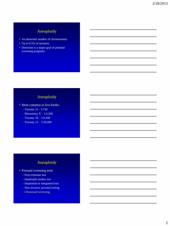
Case
• 30 yo woman presents at 12 weeks’
gestation
• Dating scan incidentally shows an increased
nuchal translucency
• How do you counsel her?
• What additional testing is offered?
Soft Markers
• First trimester
– Nuchal translucency
– Nasal bone
– Doppler studies
www.centrus.com.br
Nuchal Translucency
2/18/2013
5
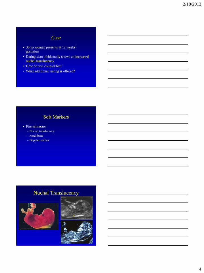
Nuchal Translucency
• NT normally increases with GA
• Abnormal is > 95th% for CRL
• Increased NT at 10-14 weeks
– Most reliable & widely used T21 marker
Nicolaides K. BJOG 1994
35 45 55 65 75 85
Crown-rump length (mm)
0.0
1.0
2.0
3.0
4.0
5.0
6.0
7.0
8.0
Nu
ch
al tr
an
slu
ce
nc
y (
mm
)
Trisomy 21
Nicolaides, AJOG 2004
Nuchal Translucency
Malone, NEJM 2005
• FASTER Trial
– Prospective study at 15 U.S. centers
– 38,167 singletons; 117 with T21
– Compared 1st & 2nd trimester
aneuploidy screening
– Increased NT detected 70% of T21
NT Screening
2/18/2013
6
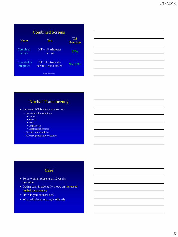
Combined Screens
Name Test T21
Detection
Combined
screen
NT + 1st trimester
serum 87%
Sequential or
integrated
NT + 1st trimester
serum + quad screen 95-96%
Malone, NEJM 2005
Nuchal Translucency
• Increased NT is also a marker for:
– Structural abnormalities
• Cardiac
• Skeletal
• Renal
• Omphalocele
• Diaphragmatic hernia
– Genetic abnormalities
– Adverse pregnancy outcome
Case
• 30 yo woman presents at 12 weeks’
gestation
• Dating scan incidentally shows an increased
nuchal translucency
• How do you counsel her?
• What additional testing is offered?
2/18/2013
7
Case
• Management:
– Refer to prenatal screening center
– Genetic counseling
– Offer invasive prenatal diagnosis
• Can consider non-invasive prenatal testing
• Serum screening not recommended
– Targeted scan & fetal echocardiogram
FetalMedicineFoundation RadioGraphics
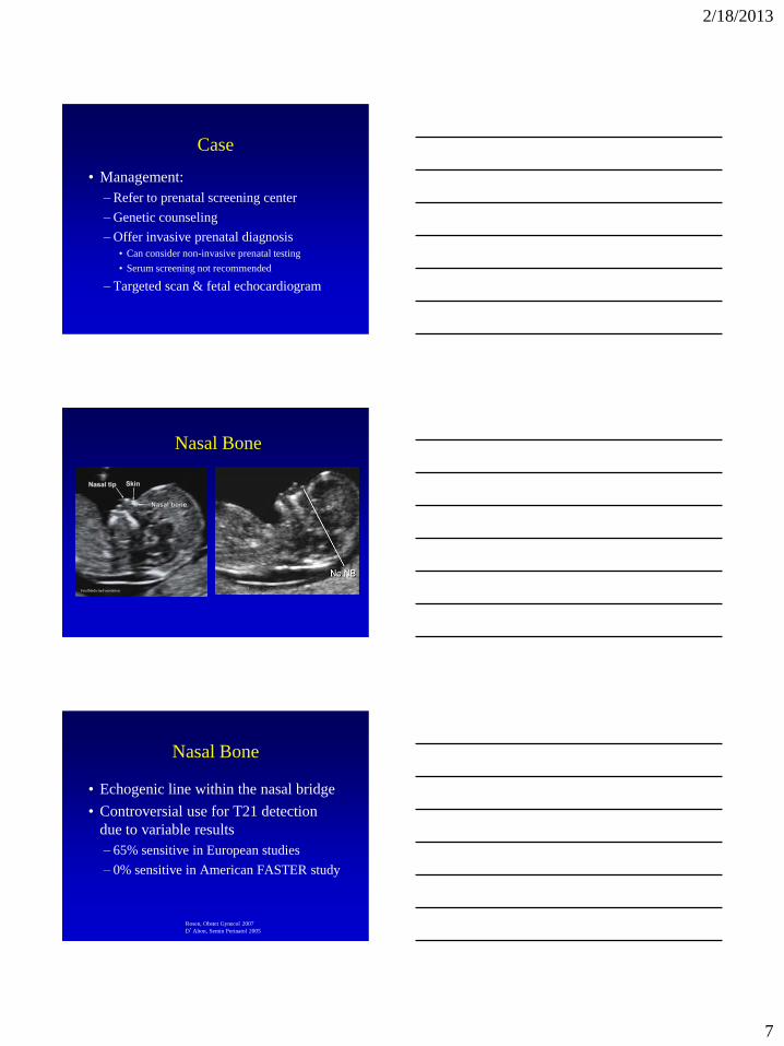
Nasal Bone
Nasal Bone
• Echogenic line within the nasal bridge
• Controversial use for T21 detection
due to variable results
– 65% sensitive in European studies
– 0% sensitive in American FASTER study
Rosen, Obstet Gynecol 2007
D’Alton, Semin Perinatol 2005

2/18/2013
8
Nasal Bone
• Important factors:
– Training & experience of operator
– Ethnic variation
– Gestational age
– Aneuploidy risk of population
• Best when combined with NT & serum
Doppler Studies
• Not used routinely but have been
associated with aneuploidy
– Ductus venosus reversed a-wave
– Tricuspid regurgitation
– Umbilical artery REDV
Second Trimester
Soft Markers

2/18/2013
9
Case
• 25 yo woman presents at 18 weeks’
gestation
• Anatomy survey shows an echogenic
intracardiac focus
• How do you counsel her?
• What additional testing is offered?
• What about same finding in a 35 yo?
• 30% have structural malformations
– Congenital heart disease
– Cystic hygroma
– Bowel atresia
• 20% have isolated soft markers
• 50% may not be detectable by US
Trisomy 21
Trisomy 21
• Soft markers
– Nuchal fold
– Nasal bone
– Echogenic focus
– Echogenic bowel
– Shortened long bones
– Pyelectasis
– Ventriculomegaly
2/18/2013
10
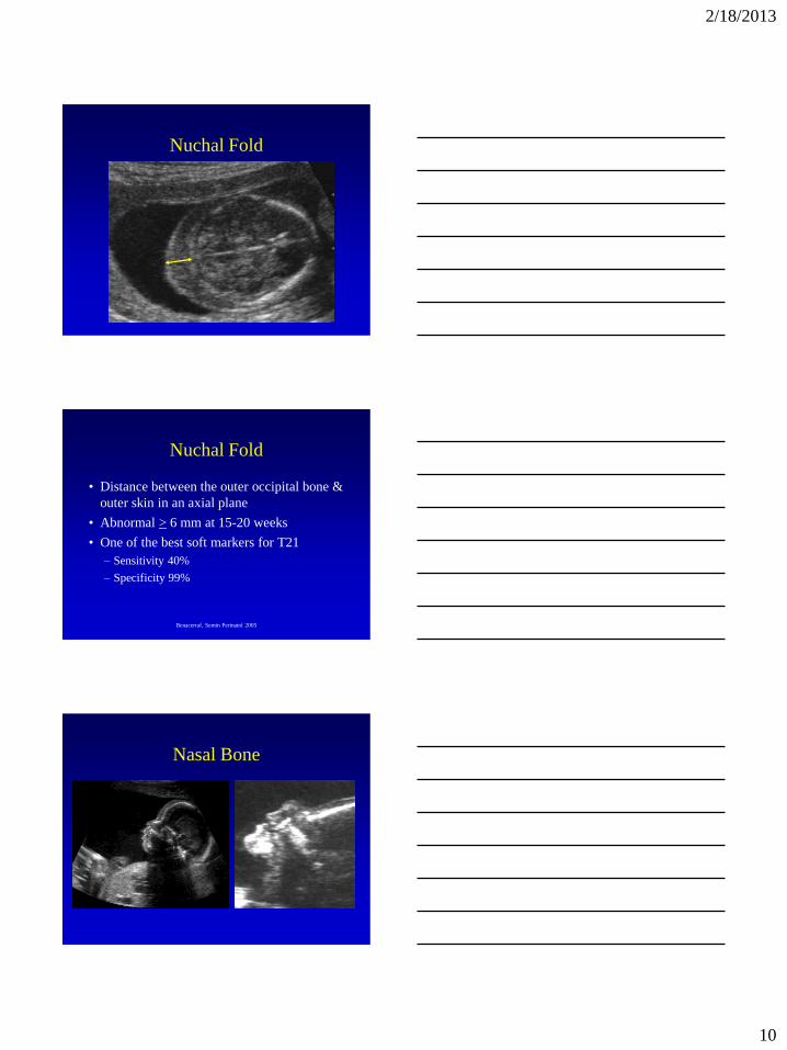
Nuchal Fold
Nuchal Fold
• Distance between the outer occipital bone &
outer skin in an axial plane
• Abnormal > 6 mm at 15-20 weeks
• One of the best soft markers for T21
– Sensitivity 40%
– Specificity 99%
Benacerraf, Semin Perinatol 2005
Nasal Bone
2/18/2013
11
Nasal Bone
• Hypoplasia
– Defined by MoM, ratio to BPD or < 2.5 mm
• Using absence or hypoplasia for T21:
– 78% sensitive
– 99% specific
• Absent in 0.3-1% normals
Cusick, Ultrasound Obstet Gynecol
2007
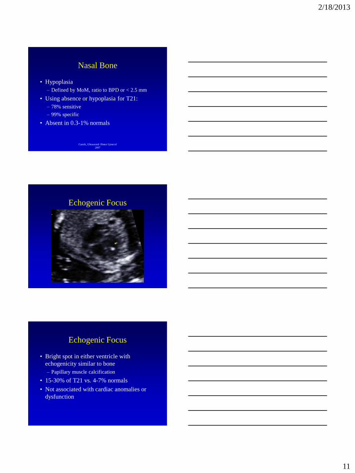
Echogenic Focus
Echogenic Focus
• Bright spot in either ventricle with
echogenicity similar to bone
– Papillary muscle calcification
• 15-30% of T21 vs. 4-7% normals
• Not associated with cardiac anomalies or
dysfunction
2/18/2013
12
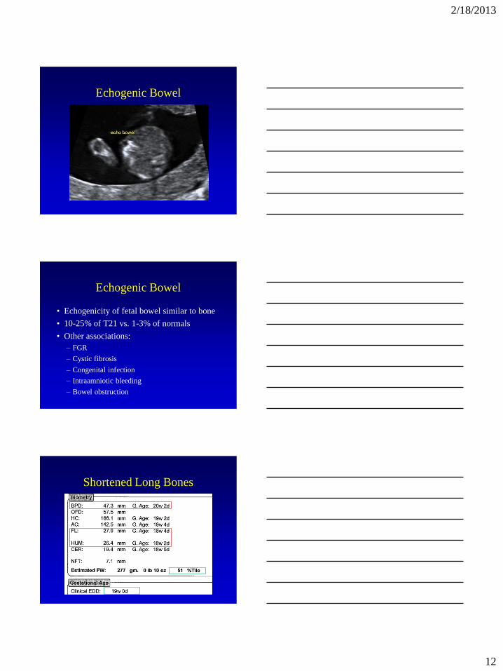
Echogenic Bowel
Echogenic Bowel
• Echogenicity of fetal bowel similar to bone
• 10-25% of T21 vs. 1-3% of normals
• Other associations:
– FGR
– Cystic fibrosis
– Congenital infection
– Intraamniotic bleeding
– Bowel obstruction
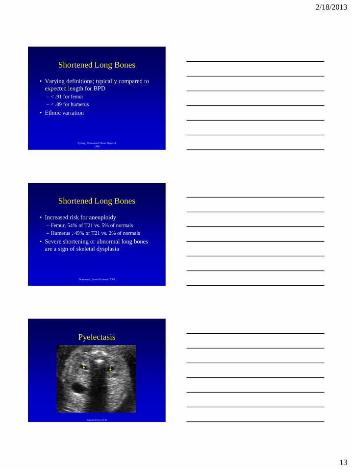
Shortened Long Bones
2/18/2013
13
Shortened Long Bones
• Varying definitions; typically compared to
expected length for BPD
– < .91 for femur
– < .89 for humerus
• Ethnic variation
Nyberg, Ultrasound Obstet Gynecol
1998
• Increased risk for aneuploidy
– Femur, 54% of T21 vs. 5% of normals
– Humerus , 49% of T21 vs. 2% of normals
• Severe shortening or abnormal long bones
are a sign of skeletal dysplasia
Shortened Long Bones
Benacerraf, Semin Perinatol 2005
www.centrus.com.br
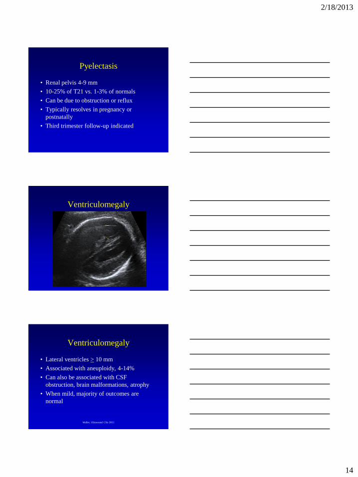
Pyelectasis
2/18/2013
14
Pyelectasis
• Renal pelvis 4-9 mm
• 10-25% of T21 vs. 1-3% of normals
• Can be due to obstruction or reflux
• Typically resolves in pregnancy or
postnatally
• Third trimester follow-up indicated
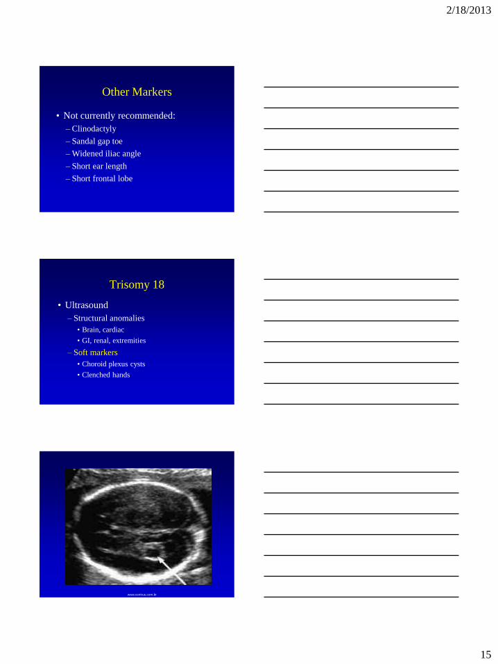
Ventriculomegaly
Ventriculomegaly
• Lateral ventricles > 10 mm
• Associated with aneuploidy, 4-14%
• Can also be associated with CSF
obstruction, brain malformations, atrophy
• When mild, majority of outcomes are
normal
Waller, Ultrasound Clin 2011
2/18/2013
15
Other Markers
• Not currently recommended:
– Clinodactyly
– Sandal gap toe
– Widened iliac angle
– Short ear length
– Short frontal lobe
• Ultrasound
– Structural anomalies
• Brain, cardiac
• GI, renal, extremities
– Soft markers
• Choroid plexus cysts
• Clenched hands
Trisomy 18
www.centrus.com.br
2/18/2013
16
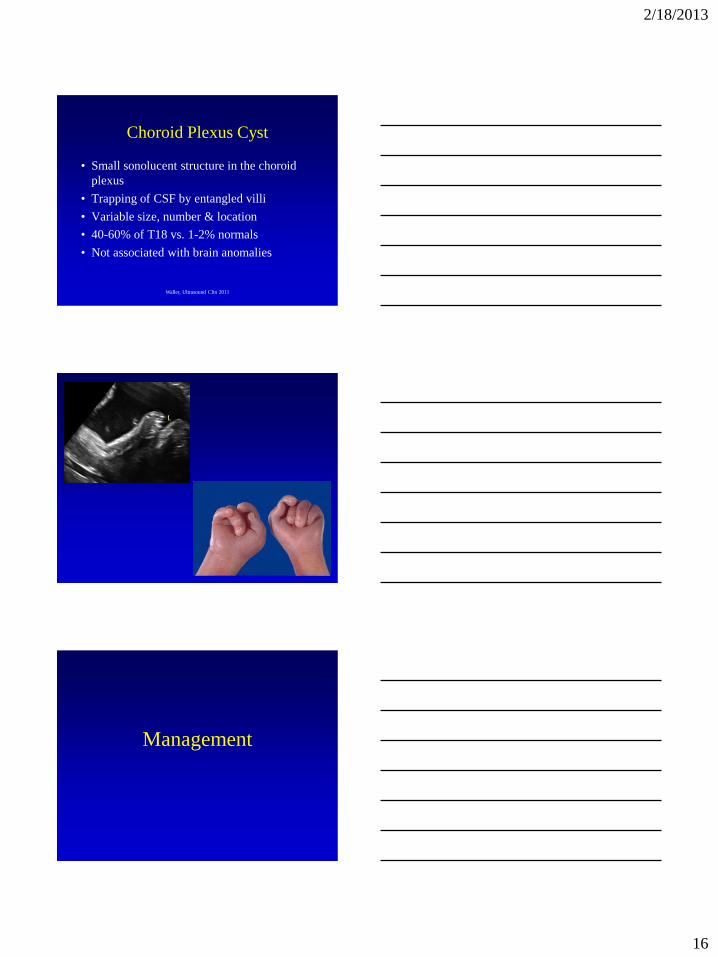
Choroid Plexus Cyst
• Small sonolucent structure in the choroid
plexus
• Trapping of CSF by entangled villi
• Variable size, number & location
• 40-60% of T18 vs. 1-2% normals
• Not associated with brain anomalies
Waller, Ultrasound Clin 2011
Management

2/18/2013
17
Soft Markers
• Soft marker screening is not indicated
on basic US exam
• Aneuploidy screening best
accomplished with serum +/- NT
– High detection rates
– Low false-positive rates
Soft Markers
• If a soft marker is detected:
– Detailed anatomic survey indicated
– Correlate the finding with baseline risk
for aneuploidy
• Age
• Serum screen
• Family history
• Low-risk patients
– Consider invasive prenatal diagnosis for:
• Nuchal thickening > 6 mm
• A major structural anomaly
• More than one soft marker
Soft Markers
2/18/2013
18
Genetic Sonogram
• High-risk patients
– US can supplement serum screening
• Genetic sonogram = targeted US for
structural anomalies & soft markers
• Used in most prenatal screening programs
• T21 sensitivity, 59-87%
Breathnach, Am J Med Gen Part C 2007
• Normal US
– Reduces T21 risk
– May allow avoidance of diagnostic testing
– Reduce loss of normal fetuses from amnio
• Abnormal US
– Lower false-negative serum screens
– Improve detection of affected fetuses
Genetic Sonogram
• Limitations
– Higher false-positive rates than other screening
methods, 10%
– May reduce detection of T21 if US normal
– Markers subjective, dependent on GA &
habitus
Genetic Sonogram
Breathnach, Am J Med Gen Part C 2007
Smith-Bindman, Prenat Diagn 2007
2/18/2013
19
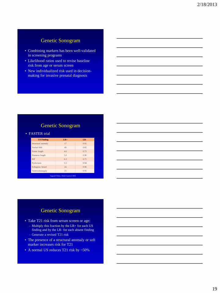
Genetic Sonogram
• Combining markers has been well-validated
in screening programs
• Likelihood ratios used to revise baseline
risk from age or serum screen
• New individualized risk used in decision-
making for invasive prenatal diagnosis
Genetic Sonogram
• FASTER trial
US Finding LR+ LR-
Structural anomaly 17 0.92
Nuchal fold 49 0.82
Femur length 4.6 0.73
Humerus length 5.0 0.90
EIF 6.3 0.75
Pyelectasis 5.5 0.94
Echogenic bowel 24 0.96
Ventriculomegaly 25 0.95
Aagaard-Tillery, Obstet Gynecol 2009.
Genetic Sonogram
• Take T21 risk from serum screen or age:
– Multiply this fraction by the LR+ for each US
finding and by the LR- for each absent finding
– Generate a revised T21 risk
• The presence of a structural anomaly or soft
marker increases risk for T21
• A normal US reduces T21 risk by ~50%

2/18/2013
20
Genetic Sonogram
• Performs best for T21 detection after a quad
screen
– Increases sensitivity from 81 to 90%
• Not as helpful after sequential screening
– Increases sensitivity from 97 to 98%
Aagaard-Tillery, Obstet Gynecol
2009.
Case
• 25 yo woman presents at 18 weeks’
gestation
• Anatomy survey shows an echogenic
intracardiac focus
• How do you counsel her?
• What additional testing is offered?
• What about same finding in a 35 yo?
Case
• Low-risk patient
– Detailed anatomic survey
– Offer a quad screen
• High-risk patient
– Targeted ultrasound & genetic counseling
– Consider invasive prenatal diagnosis or non-
invasive prenatal testing
2/18/2013
21
Conclusions
Conclusions
• Aneuploidy is associated with structural defects, soft markers & FGR that may be detected by US
• Soft markers increase the risk for aneuploidy but are most often seen in normal fetuses
• The best soft markers for T21 are nuchal & nasal bone assessments
Conclusions
• Soft markers should not be used in isolation in low-risk patients
• A genetic sonogram should only be used in high-risk patients at a prenatal screening center
– Performance is best when used to modify age or quad screen risk

2/18/2013
22
The End!