Embed Size (px)
Citation preview
Alexandria Journal of Medicine (2014) 50, 25–29
Alexandria University Faculty of Medicine
Alexandria Journal of Medicine
www.sciencedirect.com
Ultrasound screening for developmental dysplasia
of the hip and its socioeconomic impact: Experience
of tertiary care health level
Khaled Aly Matrawy *, Mohamed Ragab Nouh
Department of Radiodiagnosis, Medical Research Institute, Alexandria University, Egypt
Department of Radiodiagnosis, Faculty of Medicine, Alexandria University, Egypt
Received 29 November 2012; accepted 24 April 2013Available online 1 July 2013
*
E
Pe
M
20
ht
KEYWORDS
Hip dysplasia;
Screening;
Ultrasonography
Corresponding author. Tel.-mail address: kmatrawy@g
er review under responsibilit
edicine.
Production an
90-5068 ª 2014 Alexandria
tp://dx.doi.org/10.1016/j.ajm
: +20 12mail.com
y of Ale
d hostin
Universit
e.2013.04
Abstract Objective: The purpose of this study was to investigate the usefulness of screening ultra-
sound to detect developmental dysplasia of the hip in infants with risk factors and to assess its
socioeconomic impact.
Patients and methods: This is a retrospective study. In the duration of 30 months, all infants born
at Hospitals in our region were examined clinically for hip dislocation. All those with clinically dis-
located hips were excluded and referred for follow up to a pediatric orthopedic surgeon. After refer-
ral to our tertiary care center, a 6 week hip ultrasound scan was performed for those infants with
stable hips on examination, having risk factors for dysplastic hips including positive family history,
breech presentation and inconclusive clinical findings.
We used an ultrasound technique that combines the two known methods (Graf’s technique and
Harcke’s method).
Results: 3540 Hip ultrasound scans were performed, of those scanned 12 (0.33%) were found to
have dislocated hips and 98 (2.8%) to have different grades of dysplastic hips. Among these twelve
patients; six of them had a first degree relative with congenital dislocation of hips, three had breech
presentation at birth and three had inconclusive clinical findings. Those with dysplastic hips were
followed up by serial ultrasound examinations but did not require active intervention.
Conclusion: Screening ultrasound is a useful tool for detection of hip dislocation and dysplasia
especially among the population of infants at increased risk of developmental dysplasia of the
3377159; fax: +20 32466656.(K.A. Matrawy).
xandria University Faculty of
g by Elsevier
y Faculty of Medicine. Production and hosting by Elsevier B.V. All rights reserved.
.006
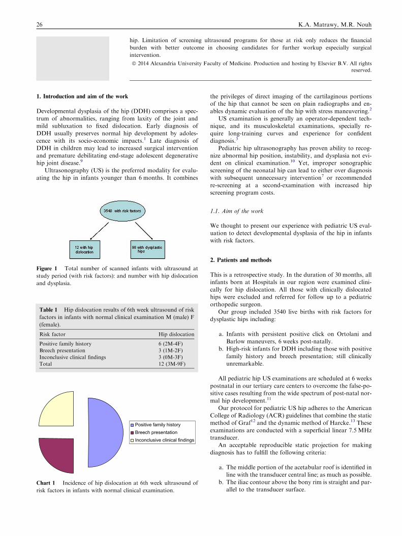
Chart 1 Incidence of hip dislocatio
risk factors in infants with normal cli
Table 1 Hip dislocation results of 6
factors in infants with normal clinica
(female).
Risk factor
Positive family history
Breech presentation
Inconclusive clinical findings
Total
Figure 1 Total number of scanned
study period (with risk factors): and
and dysplasia.
26 K.A. Matrawy, M.R. Nouh
hip. Limitation of screening ultrasound programs for those at risk only reduces the financial
burden with better outcome in choosing candidates for further workup especially surgical
intervention.
ª 2014 Alexandria University Faculty of Medicine. Production and hosting by Elsevier B.V. All rights
reserved.
1. Introduction and aim of the work
Developmental dysplasia of the hip (DDH) comprises a spec-trum of abnormalities, ranging from laxity of the joint andmild subluxation to fixed dislocation. Early diagnosis of
DDH usually preserves normal hip development by adoles-cence with its socio-economic impacts.1 Late diagnosis ofDDH in children may lead to increased surgical intervention
and premature debilitating end-stage adolescent degenerativehip joint disease.9
Ultrasonography (US) is the preferred modality for evalu-
ating the hip in infants younger than 6 months. It combines
Positive family historyBreech presentationInconclusive clinical findings
n at 6th week ultrasound of
nical examination.
th week ultrasound of risk
l examination M (male) F
Hip dislocation
6 (2M-4F)
3 (1M-2F)
3 (0M-3F)
12 (3M-9F)
infants with ultrasound at
number with hip dislocation
the privileges of direct imaging of the cartilaginous portionsof the hip that cannot be seen on plain radiographs and en-
ables dynamic evaluation of the hip with stress maneuvering.2
US examination is generally an operator-dependent tech-nique, and its musculoskeletal examinations, specially re-
quire long-training curves and experience for confidentdiagnosis.3
Pediatric hip ultrasonography has proven ability to recog-
nize abnormal hip position, instability, and dysplasia not evi-dent on clinical examination.10 Yet, improper sonographicscreening of the neonatal hip can lead to either over diagnosiswith subsequent unnecessary intervention7 or recommended
re-screening at a second-examination with increased hipscreening program costs.
1.1. Aim of the work
We thought to present our experience with pediatric US eval-
uation to detect developmental dysplasia of the hip in infantswith risk factors.
2. Patients and methods
This is a retrospective study. In the duration of 30 months, allinfants born at Hospitals in our region were examined clini-
cally for hip dislocation. All those with clinically dislocatedhips were excluded and referred for follow up to a pediatricorthopedic surgeon.
Our group included 3540 live births with risk factors fordysplastic hips including:
a. Infants with persistent positive click on Ortolani andBarlow maneuvers, 6 weeks post-natally.
b. High-risk infants for DDH including those with positive
family history and breech presentation; still clinicallyunremarkable.
All pediatric hip US examinations are scheduled at 6 weeks
postnatal in our tertiary care centers to overcome the false-po-sitive cases resulting from the wide spectrum of post-natal nor-mal hip development.11
Our protocol for pediatric US hip adheres to the AmericanCollege of Radiology (ACR) guidelines that combine the staticmethod of Graf12 and the dynamic method of Harcke.13 These
examinations are conducted with a superficial linear 7.5 MHztransducer.
An acceptable reproducible static projection for makingdiagnosis has to fulfill the following criteria:
a. The middle portion of the acetabular roof is identified inline with the transducer central line; as much as possible.
b. The iliac contour above the bony rim is straight and par-allel to the transducer surface.
Ultrasound screening for developmental dysplasia of the hip 27
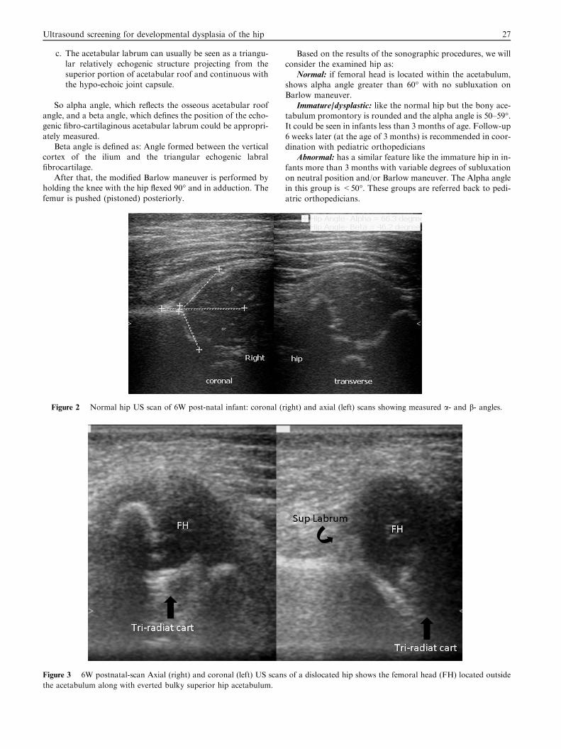
c. The acetabular labrum can usually be seen as a triangu-
lar relatively echogenic structure projecting from thesuperior portion of acetabular roof and continuous withthe hypo-echoic joint capsule.
So alpha angle, which reflects the osseous acetabular roofangle, and a beta angle, which defines the position of the echo-genic fibro-cartilaginous acetabular labrum could be appropri-
ately measured.Beta angle is defined as: Angle formed between the vertical
cortex of the ilium and the triangular echogenic labral
fibrocartilage.After that, the modified Barlow maneuver is performed by
holding the knee with the hip flexed 90� and in adduction. The
femur is pushed (pistoned) posteriorly.
Figure 2 Normal hip US scan of 6W post-natal infant: coronal (r
Figure 3 6W postnatal-scan Axial (right) and coronal (left) US scan
the acetabulum along with everted bulky superior hip acetabulum.
Based on the results of the sonographic procedures, we will
consider the examined hip as:Normal: if femoral head is located within the acetabulum,
shows alpha angle greater than 60� with no subluxation onBarlow maneuver.
Immature/dysplastic: like the normal hip but the bony ace-tabulum promontory is rounded and the alpha angle is 50–59�.It could be seen in infants less than 3 months of age. Follow-up
6 weeks later (at the age of 3 months) is recommended in coor-dination with pediatric orthopedicians
Abnormal: has a similar feature like the immature hip in in-
fants more than 3 months with variable degrees of subluxationon neutral position and/or Barlow maneuver. The Alpha anglein this group is <50�. These groups are referred back to pedi-
atric orthopedicians.
ight) and axial (left) scans showing measured a- and b- angles.
s of a dislocated hip shows the femoral head (FH) located outside
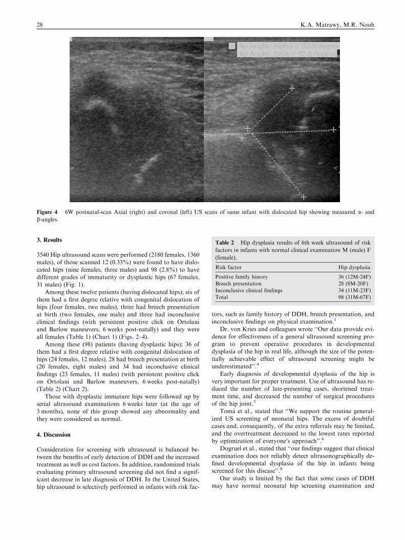
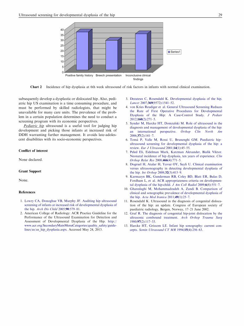
Figure 4 6W postnatal-scan Axial (right) and coronal (left) US scans of same infant with dislocated hip showing measured a- andb-angles.
Table 2 Hip dysplasia results of 6th week ultrasound of risk
factors in infants with normal clinical examination M (male) F
(female).
Risk factor Hip dysplasia
Positive family history 36 (12M-24F)
Breech presentation 28 (8M-20F)
Inconclusive clinical findings 34 (11M-23F)
Total 98 (31M-67F)
28 K.A. Matrawy, M.R. Nouh
3. Results
3540 Hip ultrasound scans were performed (2180 females, 1360
males), of those scanned 12 (0.33%) were found to have dislo-cated hips (nine females, three males) and 98 (2.8%) to havedifferent grades of immaturity or dysplastic hips (67 females,31 males) (Fig. 1).
Among these twelve patients (having dislocated hips); six ofthem had a first degree relative with congenital dislocation ofhips (four females, two males), three had breech presentation
at birth (two females, one male) and three had inconclusiveclinical findings (with persistent positive click on Ortolaniand Barlow maneuvers, 6 weeks post-natally) and they were
all females (Table 1) (Chart 1) (Figs. 2–4).Among these (98) patients (having dysplastic hips); 36 of
them had a first degree relative with congenital dislocation of
hips (24 females, 12 males), 28 had breech presentation at birth(20 females, eight males) and 34 had inconclusive clinicalfindings (23 females, 11 males) (with persistent positive clickon Ortolani and Barlow maneuvers, 6 weeks post-natally)
(Table 2) (Chart 2).Those with dysplastic immature hips were followed up by
serial ultrasound examinations 6 weeks later (at the age of
3 months), none of this group showed any abnormality andthey were considered as normal.
4. Discussion
Consideration for screening with ultrasound is balanced be-
tween the benefits of early detection of DDH and the increasedtreatment as well as cost factors. In addition, randomized trialsevaluating primary ultrasound screening did not find a signif-
icant decrease in late diagnosis of DDH. In the United States,hip ultrasound is selectively performed in infants with risk fac-
tors, such as family history of DDH, breech presentation, andinconclusive findings on physical examination.1
Dr. von Kries and colleagues wrote ‘‘Our data provide evi-dence for effectiveness of a general ultrasound screening pro-gram to prevent operative procedures in developmentaldysplasia of the hip in real life, although the size of the poten-
tially achievable effect of ultrasound screening might beunderestimated’’.4
Early diagnosis of developmental dysplasia of the hip is
very important for proper treatment. Use of ultrasound has re-duced the number of late-presenting cases, shortened treat-ment time, and decreased the number of surgical procedures
of the hip joint.5
Toma et al., stated that ‘‘We support the routine general-ized US screening of neonatal hips. The excess of doubtfulcases and, consequently, of the extra referrals may be limited,
and the overtreatment decreased to the lowest rates reportedby optimization of everyone’s approach’’.6
Dogruel et al., stated that ‘‘our findings suggest that clinical
examination does not reliably detect ultrasonographically de-fined developmental dysplasia of the hip in infants beingscreened for this disease’’.8
Our study is limited by the fact that some cases of DDHmay have normal neonatal hip screening examination and
0
5
10
15
20
25
30
35
40
Positive family history Breech presentation Inconclusive clinicalfindings
Series1
Chart 2 Incidence of hip dysplasia at 6th week ultrasound of risk factors in infants with normal clinical examination.
Ultrasound screening for developmental dysplasia of the hip 29
subsequently develop a dysplastic or dislocated hip. Also, pedi-
atric hip US examination is a time consuming procedure, andmust be performed by skilled radiologists, that might beunavailable for many care units. The prevalence of the prob-
lem in a certain population determines the need to conduct ascreening program with its economic perspectives.
Pediatric hip ultrasound is a useful tool for judging hip
development and picking those infants at increased risk ofDDH warranting further management. It avoids late-adoles-cent disabilities with its socio-economic perspectives.
Conflict of interest
None declared.
Grant Support
None.
References
1. Lowry CA, Donoghue VB, Murphy JF. Auditing hip ultrasound
screening of infants at increased risk of developmental dysplasia of
the hip. Arch Dis Child 2005;90:579–81.
2. American College of Radiology: ACR Practice Guideline for the
Performance of the Ultrasound Examination for Detection and
Assessment of Developmental Dysplasia of the Hip. http://
www.acr.org/SecondaryMainMenuCategories/quality_safety/guide-
lines/us/us_hip_dysplasia.aspx. Accessed May 24, 2013.
3. Dezateux C, Rosendahl K. Developmental dysplasia of the hip.
Lancet 2007;369(9572):1541–52.
4. von Kries Reudiger et al. General Ultrasound Screening Reduces
the Rate of First Operative Procedures for Developmental
Dysplasia of the Hip: A Case-Control Study. J Pediatr
2012;160(2):271–5.
5. Synder M, Harcke HT, Domzalski M. Role of ultrasound in the
diagnosis and management of developmental dysplasia of the hip:
an international perspective. Orthop Clin North Am
2006;37(2):141–7.
6. Toma P, Valle M, Rossi U, Brunenghi GM. Paediatric hip–
ultrasound screening for developmental dysplasia of the hip: a
review. Eur J Ultrasound 2001;14(1):45–55.
7. Peled Eli, Eidelman Mark, Katzman Alexander, Bialik Viktor.
Neonatal incidence of hip dysplasia, ten years of experience. Clin
Orthop Relat Res 2008;466(4):771–5.
8. Dogruel H, Atalar H, Yavuz OY, Sayli U. Clinical examination
versus ultrasonography in detecting developmental dysplasia of
the hip. Int Orthop 2008;32(3):415–9.
9. Karmazyn BK, Gunderman RB, Coley BD, Blatt ER, Bulas D,
Fordham L, et al. ACR appropriateness criteria on developmen-
tal dysplasia of the hip-child. J Am Coll Radiol 2009;6(8):551–7.
10. Gharedaghi M, Mohammadzadeh A, Zandi B. Comparison of
clinical and sonographic prevalence of developmental dysplasia of
the hip. Acta Med Iranica 2011;49(1):25–7.
11. Rosendahl K. Ultrasound in the diagnosis of congenital disloca-
tion of the hip: an update. Congress of European society of
paediatric radiology, Bergen, Norway, 17–21 June 2002.
12. Graf R. The diagnosis of congenital hip-joint dislocation by the
ultrasonic combound treatment. Arch Orthop Trauma Surg
1980;97(2):117–33.
13. Harcke HT, Grissom LE. Infant hip sonography: current con-
cepts. Semin Ultrasound CT MR 1994;15(4):256–63.