Embed Size (px)
Citation preview
M
OFFICIAL NEWSLETTER
Volume 23 – Number 3May/June 2014
The purpose of this article is to disseminate valuable information on the following key points:1) provide a diagram to demonstrate the basic anatomy of a vein, 2) identify the common veinsused for peripheral IV (PIV) access, 3) list the most common drug pH values, 4) list the osmolalityvalues for IV fluid, 5) identify vein complications, 6) discuss basic principles of ultrasound technology,and 7) list the steps for PIV access insertion using ultrasound-guided technology. Nurses having thisupdated knowledge will improve their clinical practice, patient health care outcomes, and patients’quality of life.
M.B. is a 35-year old female admitted to the medical floor, on a Friday evening, for anacute exacerbation of sickle cell crisis. She has been hospitalized at least four times a yearduring the past five years for pain management, blood transfusions, and hydration. M.B. isknown to the nursing staff and her primary care providers as a patient with difficult IV accessissues. Additionally, she refuses to have either an implanted vascular access port or aPeripherally Inserted Central Catheter (PICC) line inserted.
After two failed attempts during M.B.’s most recent admission, she had a 22-gauge IVcatheter placed in a small branch of the median cephalic vein located on her left forearm.However, four hours later, after receiving only two doses of morphine sulfate intravenously,her IV site became red,swollen, tender to the touch,and had to be discontinued.M.B.’s primary care nurse hadthe charge nurse make onemore attempt to restart M.B.’sPIV, without success.
M.B. was crying becauseher joint and back painremained at a 10 on the 0-10pain scale. M.B.’s pain was fur-ther compounded from sev-eral unsuccessful IV attemptsand her pain medication had tobe administered by the intra-muscular (IM) route. M.B. alsocomplained of pain and a burn-ing sensation at her IM injec-tion sites and stated, “Pleasejust let me die. I cannot takethis pain anymore!”
Pain medications, such as morphine sulfate, are known to cause localized inflammationand pain, especially when infused into small diameter veins. IV medications can have eitheran acid or alkaline base (see Table 1), causing inflammation to surrounding endothelial cellsof blood vessels. These cells are fragile and are responsible for the development of the innerlayer for healthy blood vessels (Pries & Kuebler, 2006; Sumpio, Riley, & Dardik, 2002).
continued on page 10
INSIDE THIS ISSUE
Nutrition to Improve Outcomes: New Safety Standards to Prevent Patient Tubing Misconnections . . . . . . . . . . . . . . . . . . . . . . .3
Spasmodic Dysphonia . . . . . . . . . . . . . . . . . . . . . . . . . . . . . . . . . . . . . . . . . . . . . . . . . . . . . . . . . . . . . . . . . . . . . . . . . . . . . .4
Legal Nursing: Are You Ready for a Community Disaster? Part Two . . . . . . . . . . . . . . . . . . . . . . . . . . . . . . . . . . . . . . . . . . . . . .8
Grab Your Sunglasses, and Join us in Orlando . . . . . . . . . . . . . . . . . . . . . . . . . . . . . . . . . . . . . . . . . . . . . . . . . . . . . . . . . . . . . .16
CNE
Ultrasound-Guided Access forPeripheral Intravenous Therapy
Figure 1.Anatomy of an Artery and a Vein
Debbie Arbique, Marilynn Bordelon, Robert Dragoo, and Stephanie Huckaby
Volume 23 – Number 3May/June 2014
2
We are Nurses: We’re Here to HelpSometimes I have a sense of uncertainty and fear when I learn of significant
global events. Russia recently invaded Ukraine. Nearly 300 young Nigerian girlswere abducted while at school.A Malaysian airplane with hundreds of passengersaboard went missing. Unrest continues in the Middle East. Rebels destroyed partsof Syria, dislocating thousands of people. Threats of terrorism are not uncom-mon. Ravaging weather events such as record temperatures, droughts, tsunamis,earthquakes, tornadoes, and hurricanes seem to be happening all the time. Theseoccurrences are bigger than what any one person can fix, but they certainly can-not be ignored.
As I was pondering the complexity and seriousness of national and globalevents, I wondered what nurses could and should do to help. There are about 3million registered nurses in the United States. We have all been trained and knowhow to provide care, education, kindness, and compassion, and we have a uniqueskill set to heal. I would encourage everyone in the nursing profession to knowwhat to do in the event of a disaster. Stay up to date on the disaster-prepared-ness policies in the area where you live and practice. Talk with your colleaguesabout what nurses can do collectively to assist in times of crisis and need. Maybeit’s providing hands-on medical care for people injured from violence or naturaldisasters. Perhaps your group of nurses could educate communities on safety.Maybe you could join with nurses in political arenas to promote needed change.Our colleagues stationed overseas in the Armed Forces appreciate care packagesor letters of support from nurses back home. Because there are more individualsin nursing than in any other health care profession, nurses have the most abilityto help when people need the care we know how to provide. We can’t do italone, but we can certainly make a difference if we all work together.
The world can be a scary and rapidly changing place, but this has been thecase since the beginning of history. As nurses, we must join together to maintaina sense of peace and calm authority that we know how to help. New skills – likestarting IVs through the use of ultrasound and identifying tubing connectionerrors – should always be added to our repertoire. Enhanced knowledge (suchas learning about uncommon disorders like spasmodic dysphonia) needs to beadded to our intelligence banks. Networking with other nurses at conferences(such as the annual AMSN Convention) is essential if we are to work as a col-lective unit in solving some of the world’s problems.
Although global problems can be overwhelming and daunting, nurses needto join together and care for each other so we can all do what we do best – provide care for others.
Molly McClelland, PhD, MSN, RN, CMSRN, ACNS-BCMedSurg Matters! Editor
Reader ServicesMedSurg Matters!Academy of Medical-Surgical NursesEast Holly Avenue, Box 56Pitman, NJ 08071-0056(856) 256-2300 • (866) 877-AMSN (2676)Fax (856) [email protected]
MedSurg Matters! is owned and publishedbimonthly by the Academy of Medical-SurgicalNurses (AMSN). The newsletter is distributed tomembers as a direct benefit of membership.Postage paid at Bellmawr, NJ, and additional mailingoffices.
AdvertisingContact Rick Gabler, Advertising Representative, (856) 256-2314.
Back IssuesTo order, call 866-877-AMSN (2676).
Editorial ContentAMSN encourages the submission of news itemsand photos of interest to AMSN members. Byvirtue of your submission, you agree to the usageand editing of your submission for possible publica-tion in the AMSN newsletter, online, and in otherpromotional and educational materials.
To send comments, questions, or article sugges-tions, or if you would like to write for us, contactthe Editor at [email protected].
AMSN Publications and ProductsTo order, call 866-877-AMSN (2676), or visitwww.amsn.org.
ReprintsFor permission to reprint an article, call 866-877-AMSN (2676).
IndexingMedSurg Matters! is indexed in the CumulativeIndex to Nursing and Allied Health Literature(CINAHL).
© Copyright 2014 by AMSN. All rights reserved.Reproduction in whole or part, electronic ormechanical without written permission of the pub-lisher is prohibited. The opinions expressed inMedSurg Matters! are those of the contributors,authors and/or advertisers, and do not necessarilyreflect the views of AMSN, MedSurg Matters!, or itseditorial staff.
Publication Management is provided byAnthony J. Jannetti, Inc., which is accred-ited by the Association Management
Company Institute.
Molly McClelland
Thoughts from
the Editor
New Safety Standards toPrevent Patient Tubing
MisconnectionsWith increasing patient acuity and technological
advances in health care, a typical patient may be connectedto several delivery systems at one time, including: medica-tions, nutrition, lavage/irrigation, and fluid administration. Thepotential for tubing misconnection (wrong line to wronginfusion site) puts the patient at risk for serious and poten-tially fatal injury. Although errors with misconnection havebeen reported for more than 30 years, there is a new call forsolutions to address this issue from governmental, profes-sional, and patient safety groups (Pugliese, 2013). These agen-cies, along with trade organizations and medical productdevice manufacturers, are collaborating to develop betterconnection standards.
Tubing misconnections are well documented and wellknown, according to the Food and Drug Administration(FDA) (Vockley, 2011). Factors associated with tubing mis-connections included the following risk points: 1) unsecuredor loose line connections; 2) use of unintended adapters per-mitting wrong connection; 3) too many lines close together(spaghetti syndrome); and 4) look alike, unlabeled connec-tors.
Reported misconnections include:• neuraxial (epidural) to vascular lines• topical (wound management) to vascular• urinary to vascular• medical device (sequential compression devices, pneu-matic blood pressure cuffs, oxygen tubing, syringe) tovascular
• respiratory (nebulizer or syringe) to vascular• hemodialysis or peritoneal dialysis fluids to vascular• enteral/oral to vascular, enteral/oral to hemodialysis• intravenous to arterial• Other connectors with the potential for connectionerror include abdominal catheters (t-tubes, wounddrainage, etc.), Blakemore tubes, cantor tubes, chesttubes, cranial catheters, suction tubing, nephrostomytubes, amnioinfusion catheter (intrauterine pressure)and drains.Manufacturers and regulators are working to decrease
the potential for harm by correcting connector compatibili-ties and creating a safety net to avoid misconnections. To
reduce the risk of small-bore connector hazards, a group ofinternational organizations (including the FDA, Associationfor the Advancement of Medical Instrumentation [AAMI],International Standards Organization [ISO], and Centers forMedicare and Medicaid [CMS]) have developed standardsthat provide general requirements for liquids and gas con-nectors. They have also created a framework for testing thatensures incompatibility and non-interconnector fit of Luervascular devices with enteral and other small bore connec-tors to eliminate misconnections. This standard was pub-lished in 2011, and the new enteral connectors reached themarket in 2014.
New enteral tube feeding connectors are now available.You may be seeing these changes in your practice settingtoday. If your unit isn’t using the new, safer connectors, askyour manager or clinical specialist why.
The change in manufacturing is not based on color.Nurses will not be able to assume that all purple connectorsare enteral, for example. IV lines and ports may also be pur-ple colored. The change is based upon connector size andcompatibility. IV connectors, for example, should no longerLuer lock into any enteral device. The ability to connect IVand enteral devices should no longer be possible.
There are several things medical-surgical nurses can doto assure that safe care is provided during the transition ofthe connector standards and acquisition of the new andimproved products:
Develop a risk assessment and action process. BradNoé, Small-Bore Connector Committee Chair at AAMI,shares the following recommendations to reduce risk andpromote a smooth transition (Vockley, 2011):• Evaluate workarounds occurring in the clinical setting(workarounds indicate that the clinicians are doingsomething outside of intended use because what theyhave to work with is not functioning).
• Eliminate adaptors (these are a signal that workaroundsor modifying clinical practice to accomplish a task is anacceptable practice).
• Conduct safety audits (look at your practice within thesafety guidelines).
• Create cross-functional clinical practice teams so that allstakeholders can have a role in decision-making processto ensure continuity of action across the supply chainand pipeline (for example, pharmacy should prepare oralmedication in the oral syringe for administration).
• View reporting of misconnections or near misses asinstructive and informational – versus reacting in a puni-tive or threatening way.Although under standard and product design revision,
enteral misconnections remain a hazard to patient safety inhealth care settings. We have an obligation to our patients toassure safe care. We must ensure that hospital leaders areaware of this potential risk and work with purchasing depart-ments to encourage and demand alternate solutions for
3
Volume 23 – Number 3
NutritionTO IMPROVE OUTCOMES
continued on page 15

Spasmodic Dysphonia
Deadline for Submission: June 30, 2016
MSNN1403
To Obtain CNE Contact Hours1. For those wishing to obtain CNE contact hours,
you must read the article and complete theevaluation through the AMSN Online Library.Complete your evaluation online and print yourCNE certificate immediately, or later. Simplygo to www.amsn.org/library
2. Evaluations must be completed online by June30, 2016. Upon completion of the evaluation, acertificate for 1.1 contact hour(s) may beprinted.
FeesMember: FREE Regular: $20
ObjectivesThe purpose of this continuing nursing educationarticle is to increase nurses’ and other health careprofessionals’ awareness of spasmodic dysphonia.After studying the information presented in this arti-cle, you will be able to:1. Explain spasmodic dysphonia and its symptoms.2. Discuss the process of diagnosing spasmodic
dysphonia.3. Describe treatments available to patients with
spasmodic dysphonia.4. Identify resources available to those affected by
spasmodic dysphonia.
Note: The author, editor, editorial board, andeducation director reported no actual or potentialconflict of interest in relation to this continuingnursing education article.
This educational activity has been co-provided byAnthony J. Jannetti, Inc. and AMSN.
Anthony J. Jannetti, Inc. is accredited as a providerof continuing nursing education by the AmericanNurses Credentialing Center’s Commission onAccreditation.
Anthony J. Jannetti, Inc. is a provider approved bythe California Board of Registered Nursing, providernumber CEP 5387. Licensees in the state of CA mustretain this certificate for four years after the CNE activ-ity is completed.
4
CNECONTINUING
NURSINGEDUCATION
Spasmodic DysphoniaElcy T. Mathew
Spasmodic dysphonia (SD) is a rare,neurogenic voice disorder that is oftenundiagnosed or misdiagnosed due toits similarity to other voice disordersand a lack of research data. SDaffects approximately 1 in 10,000adults in the United States (Revelo,Underbrink, & Quinn, 2009). Theobjective of this article is to enablereaders to gain a general understand-ing of SD, the diagnostic process,treatments, and resources available tothose affected by this disorder.
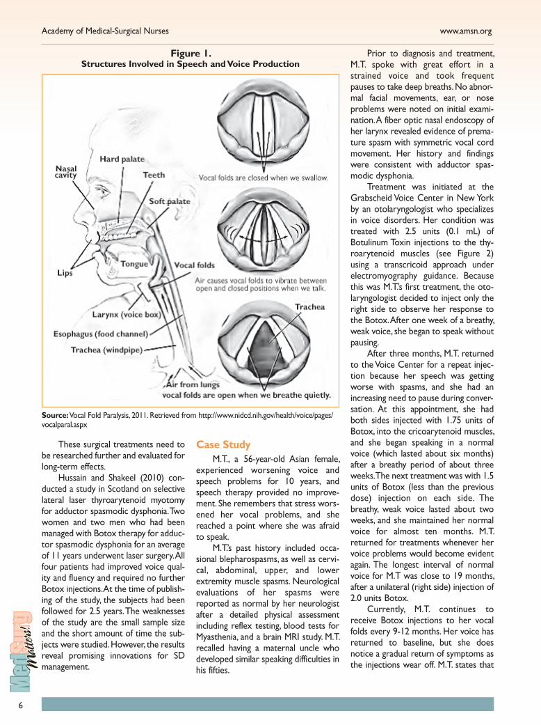
Speech is produced when air fromthe lungs is pushed between the vocalcords, causing them to vibrate (Luc &Zieve, 2012). Spasmodic dysphonia (SD)is a voice disorder characterized byinvoluntary movements or spasms thatprevent the normal vibrations of thevocal cords and the production of anormal voice (see Figure 1). It is alsoreferred to as laryngeal dystonia, whichis a form of focal dystonia (NationalInstitutes of Health [NIH], 2010a).
Dystonia is defined as involuntarymuscle contractions that cause repeatedmovements generating tremors,twitches, or abnormal postures (NationalInstitute of Neurological Disorders andStroke [NINDS], 2011). Dystonias areclassified according to the part of thebody affected with generalized dystoniaaffecting most parts of the body.• Focal dystonia is localized to onepart of the body, such as the intrin-sic laryngeal muscles in SD (Reveloet al., 2009).
• Multifocal dystonia occurs whentwo or more unrelated areas ofthe body are affected.
• Segmental dystonia involves twoor more adjacent parts of thebody.
• Hemidystonia affects the arm andleg on the same side of the body(NINDS, 2011).
Types of SpasmodicDysphonia
There are three types of spas-modic dysphonia. In adductor spas-modic dysphonia, the vocal folds closetogether and stiffen with spasms, mak-ing vibration of the cords difficult.Patients with this condition find it diffi-cult to start talking and words are oftencut off with the spasms. In abductorspasmodic dysphonia, the vocal foldsdo not close properly due to musclespasms that prevent the folds fromvibrating to produce voice. In this situ-ation, the voice will sound weak andbreathy due to air escaping from thelungs during speech. In adductor andabductor spasmodic dysphonia condi-tions, the voice sounds normal whilesinging, shouting, laughing, and crying(NIH, 2010a). Mixed spasmodic dys-phonia, which is rare, is characterizedby spasms of the different muscles thatopen and close the vocal folds. As thename suggests, it has the characteristicsof both adductor and abductor spas-modic dysphonia (NIH, 2010a).
The most common form of spas-modic dysphonia is the adductor type.The symptoms for adductor SD includea choked, strained voice with suddenbreaks in speech in the middle of vow-els. In the less common abductor typeSD, the voice sounds breathy with whis-per quality voice breaks. In mixed typeSD, both types of symptoms are pres-ent (Revelo et al., 2009).
CausesThere are no known causes for
spasmodic dysphonia because no long-term research data is available. SD wasoriginally thought to be psychogenic innature due to lack of readily availableresearch information. However, currentstudies indicate that SD is a neurogenicvoice disorder (Luc & Zieve, 2012).Some clinicians and genetic researchersbelieve SD may be hereditary because
5
866-877-2676 Volume 23 – Number 3Volume 23 – Number 3866-877-2676 Volume 23 – Number 3
ment approach for SD. Speech therapymay be advised initially for mild symp-toms of SD; however, if no improve-ment occurs after 8-10 therapy ses-sions, Botulinum Toxin A (Botox®) ther-apy is second line of treatment (Pitman,Kamat, Bliznikas, & Baredes, 2011).
Injections of very small amounts ofBotox to the affected, hyperfunctionallaryngeal muscles weaken them byblocking the nerve impulses (chemicaldenervation). The weakened musclesreduce spasms and improve voice qual-ity for a few months (Revelo et al.,2009). Laryngeal electromyography isutilized to identify the muscles for theinjection of the Botox. The voice maybecome breathy, and swallowing may bedifficult for a few days after injectiondue to muscle weakening. The injec-tions are repeated every 3-8 monthsper individual requirements for themaintenance of a normal voice (Reveloet al., 2009).
Clinicians and researchers are cur-rently studying several surgical treat-ments; however, the results are conflict-ing. The types of possible surgeriesinclude:• Type 2 thyroplasty – This procedurechanges the shape of the thyroidcartilage to relax and lateralize thevocal folds slightly to enable easierphonation (Isshiki, Yamamoto, &Fukagai, 2004). As of 2009, therewere no long-term studies for thisprocedure according to Revelo andco-authors (2009).
• Recurrent laryngeal denervationand reinnervation – Firstattempted in 1999 by Dr. GeraldBerke at the University ofCalifornia in Los Angeles, this inter-vention involves the resection andreanastomosis of bilateral adduc-tor branches of the laryngeal nerveto the ansa cervicalis (Pitman et al.,2011).
• Bilateral transarytenoid and lateralcricoarytenoid myectomy – Thissurgery is usually performed oneside at a time, at least six monthsapart. The myectomy weakens thevocal folds and prevents spasms(Pitman et al., 2011).
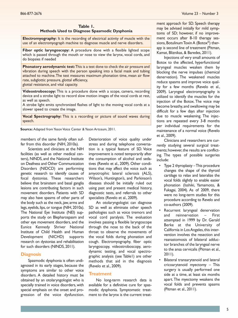
Electromyography: It is the recording of electrical activity of muscle with theuse of an electromyograph machine to diagnose muscle and nerve disorders.
Fiber optic laryngoscopy: A procedure done with a flexible lighted scopewhich is passed through the mouth or nose to view the larynx, vocal cords, anddo biopsies if needed
Phonatory aerodynamic test:This is a test done to check the air pressure andvibration during speech with the person speaking into a facial mask and tubingattached to machine. The test measures maximum phonation time, mean air flowrate, subglottic pressure, glottal efficiency, glottal resistance, and vital capacity.
Videostroboscopy: This is a procedure done with a scope, camera, recordingdevice and a strobe light to record slow motion images of the vocal cords at rest,as well as speech.A strobe light emits synchronized flashes of light to the moving vocal cords at aslower speed to create the image.
Vocal Spectrography: This is a recording or picture of sound waves duringspeech.
Table 1.Methods Used to Diagnose Spasmodic Dysphonia
Source: Adapted from Yeson Voice Center & Yeson Artceum, 2011.
members of the same family often suf-fer from this disorder (NIH, 2010a).
Scientists and clinicians at the NIHfacilities (as well as other medical cen-ters), NINDS, and the National Instituteon Deafness and Other CommunicationDisorders (NIDCD) are performinggenetic research to identify causes offocal dystonias. These researchersbelieve that brainstem and basal ganglialesions are contributing factors in suchmovement disorders. Patients with SDmay also have spasms of other parts ofthe body such as the neck, jaw, arms andlegs, eyelids, lips, or tongue (NIH, 2010a).The National Eye Institute (NEI) sup-ports the study on Blepharospasm andother eye movement disorders, and theEunice Kennedy Shriver NationalInstitute of Child Health and HumanDevelopment (NICHD) supportsresearch on dystonias and rehabilitationfor such disorders (NINDS, 2011).
DiagnosisSpasmodic dysphonia is often undi-
agnosed in its early stages, because thesymptoms are similar to other voicedisorders. A detailed history must beobtained by an otolaryngologist who isspecially trained in voice disorders, withspecial emphasis on the onset and pro-gression of the voice dysfunction.
Deterioration of voice quality understress and during telephone conversa-tion is a typical feature of SD. Voiceimprovement is noted temporarily afterthe consumption of alcohol and seda-tives (Revelo et al., 2009). Other condi-tions that may affect the voice such asamyotrophic lateral sclerosis (ALS),Wilson’s, Huntington’s, and Parkinson’sdiseases should be initially ruled outusing past and present medical history,diagnostic tests, and referrals to otherspecialists (Revelo et al., 2009).
An otolaryngologist can diagnoseSD as well as eliminate other speechpathologies such as voice tremors andvocal cord paralysis. The evaluationinvolves passing a flexible laryngoscopethrough the nose to the back of thethroat to observe the movements ofthe vocal folds during phonation andcough. Electromyography, fiber opticlaryngoscopy, videostroboscopy, aero-dynamic testing, and vocal spectro-graphic analysis (see Table1) are othermethods that aid in the diagnosis(Revelo et al., 2009).
TreatmentNo long-term research data is
available for a definitive cure for spas-modic dysphonia. Symptomatic treat-ment to the larynx is the current treat-
Academy of Medical-Surgical Nurses www.amsn.org
6
These surgical treatments need tobe researched further and evaluated forlong-term effects.
Hussain and Shakeel (2010) con-ducted a study in Scotland on selectivelateral laser thyroarytenoid myotomyfor adductor spasmodic dysphonia. Twowomen and two men who had beenmanaged with Botox therapy for adduc-tor spasmodic dysphonia for an averageof 11 years underwent laser surgery. Allfour patients had improved voice qual-ity and fluency and required no furtherBotox injections. At the time of publish-ing of the study, the subjects had beenfollowed for 2.5 years. The weaknessesof the study are the small sample sizeand the short amount of time the sub-jects were studied. However, the resultsreveal promising innovations for SDmanagement.
Case StudyM.T., a 56-year-old Asian female,
experienced worsening voice andspeech problems for 10 years, andspeech therapy provided no improve-ment. She remembers that stress wors-ened her vocal problems, and shereached a point where she was afraidto speak.
M.T.’s past history included occa-sional blepharospasms, as well as cervi-cal, abdominal, upper, and lowerextremity muscle spasms. Neurologicalevaluations of her spasms werereported as normal by her neurologistafter a detailed physical assessmentincluding reflex testing, blood tests forMyasthenia, and a brain MRI study. M.T.recalled having a maternal uncle whodeveloped similar speaking difficulties inhis fifties.
Prior to diagnosis and treatment,M.T. spoke with great effort in astrained voice and took frequentpauses to take deep breaths. No abnor-mal facial movements, ear, or noseproblems were noted on initial exami-nation. A fiber optic nasal endoscopy ofher larynx revealed evidence of prema-ture spasm with symmetric vocal cordmovement. Her history and findingswere consistent with adductor spas-modic dysphonia.
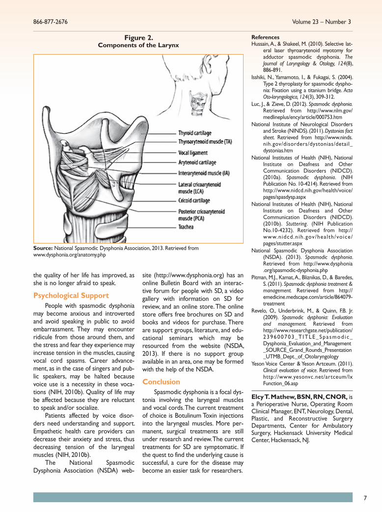
Treatment was initiated at theGrabscheid Voice Center in New Yorkby an otolaryngologist who specializesin voice disorders. Her condition wastreated with 2.5 units (0.1 mL) ofBotulinum Toxin injections to the thy-roarytenoid muscles (see Figure 2)using a transcricoid approach underelectromyography guidance. Becausethis was M.T.’s first treatment, the oto-laryngologist decided to inject only theright side to observe her response tothe Botox. After one week of a breathy,weak voice, she began to speak withoutpausing.
After three months, M.T. returnedto the Voice Center for a repeat injec-tion because her speech was gettingworse with spasms, and she had anincreasing need to pause during conver-sation. At this appointment, she hadboth sides injected with 1.75 units ofBotox, into the cricoarytenoid muscles,and she began speaking in a normalvoice (which lasted about six months)after a breathy period of about threeweeks. The next treatment was with 1.5units of Botox (less than the previousdose) injection on each side. Thebreathy, weak voice lasted about twoweeks, and she maintained her normalvoice for almost ten months. M.T.returned for treatments whenever hervoice problems would become evidentagain. The longest interval of normalvoice for M.T was close to 19 months,after a unilateral (right side) injection of2.0 units Botox.
Currently, M.T. continues toreceive Botox injections to her vocalfolds every 9-12 months. Her voice hasreturned to baseline, but she doesnotice a gradual return of symptoms asthe injections wear off. M.T. states that
Figure 1. Structures Involved in Speech and Voice Production
Source:Vocal Fold Paralysis, 2011. Retrieved from http://www.nidcd.nih.gov/health/voice/pages/vocalparal.aspx
7
866-877-2676 Volume 23 – Number 3
the quality of her life has improved, asshe is no longer afraid to speak.
Psychological SupportPeople with spasmodic dysphonia
may become anxious and introvertedand avoid speaking in public to avoidembarrassment. They may encounterridicule from those around them, andthe stress and fear they experience mayincrease tension in the muscles, causingvocal cord spasms. Career advance-ment, as in the case of singers and pub-lic speakers, may be halted becausevoice use is a necessity in these voca-tions (NIH, 2010b). Quality of life maybe affected because they are reluctantto speak and/or socialize.
Patients affected by voice disor-ders need understanding and support.Empathetic health care providers candecrease their anxiety and stress, thusdecreasing tension of the laryngealmuscles (NIH, 2010b).
The National SpasmodicDysphonia Association (NSDA) web-
site (http://www.dysphonia.org) has anonline Bulletin Board with an interac-tive forum for people with SD, a videogallery with information on SD forreview, and an online store. The onlinestore offers free brochures on SD andbooks and videos for purchase. Thereare support groups, literature, and edu-cational seminars which may beresourced from the website (NSDA,2013). If there is no support groupavailable in an area, one may be formedwith the help of the NSDA.
ConclusionSpasmodic dysphonia is a focal dys-
tonia involving the laryngeal musclesand vocal cords. The current treatmentof choice is Botulinum Toxin injectionsinto the laryngeal muscles. More per-manent, surgical treatments are stillunder research and review. The currenttreatments for SD are symptomatic. Ifthe quest to find the underlying cause issuccessful, a cure for the disease maybecome an easier task for researchers.
ReferencesHussain, A., & Shakeel, M. (2010). Selective lat-
eral laser thyroarytenoid myotomy foradductor spasmodic dysphonia. TheJournal of Laryngology & Otology, 124(8),886-891.
Isshiki, N., Yamamoto, I., & Fukagai, S. (2004).Type 2 thyroplasty for spasmodic dyspho-nia: Fixation using a titanium bridge. ActaOto-laryngologica, 124(3), 309-312.
Luc, J., & Zieve, D. (2012). Spasmodic dysphonia.Retrieved from http://www.nlm.gov/medlineplus/ency/article/000753.htm
National Institute of Neurological Disordersand Stroke (NINDS). (2011). Dystonias factsheet. Retrieved from http://www.ninds.nih.gov/disorders/dystonias/detail_dystonias.htm
National Institutes of Health (NIH), NationalInstitute on Deafness and OtherCommunication Disorders (NIDCD).(2010a). Spasmodic dysphonia. (NIHPublication No. 10-4214). Retrieved fromhttp://www.nidcd.nih.gov/health/voice/pages/spasdysp.aspx
National Institutes of Health (NIH), NationalInstitute on Deafness and OtherCommunication Disorders (NIDCD).(2010b). Stuttering. (NIH PublicationNo.10-4232). Retrieved from http://www.nidcd.n ih .gov/hea l th/voice/pages/stutter.aspx
National Spasmodic Dysphonia Association(NSDA). (2013). Spasmodic dysphonia.Retrieved from http://www.dysphonia.org/spasmodic-dysphonia.php
Pitman, M.J., Kamat, A., Bliznikas, D., & Baredes,S. (2011). Spasmodic dysphonia treatment &management. Retrieved from http://emedicine.medscape.com/article/864079-treatment
Revelo, O., Underbrink, M., & Quinn, F.B. Jr.(2009). Spasmodic dysphonia: Evaluationand management. Retrieved fromhttp://www.researchgate.net/publication/239600703_T ITLE_Spa smod i c_Dysphonia_Evaluation_and_Management_SOURCE_Grand_Rounds_Presentation_UTMB_Dept._of_Otolaryngology
Yeson Voice Center & Yeson Artceum. (2011).Clinical evaluation of voice. Retrieved fromhttp://www.yesonvc.net/artceum/lxFunction_06.asp
Elcy T. Mathew, BSN, RN, CNOR, isa Perioperative Nurse, Operating RoomClinical Manager, ENT, Neurology, Dental,Plastic, and Reconstructive SurgeryDepartments, Center for AmbulatorySurgery, Hackensack University MedicalCenter, Hackensack, NJ.
Figure 2. Components of the Larynx
Source: National Spasmodic Dysphonia Association, 2013. Retrieved fromwww.dysphonia.org/anatomy.php
www.amsn.org
8
Legal Issues and DisasterPreparedness: Are You ReadyFor a Community Disaster?
Part TwoThis article provides information about some of the legal issueshealth care providers may encounter during a disaster. Part one ofthis article (published in the January/February issue) explains rel-evant resources on federal, state, and local levels (Neil, 2014). Parttwo explains the importance of preparation, training, and exercisesto maintain legal standards.
In part one of this series, we discussed the federal, state, andlocal authorities that regulate the planning efforts associatedwith disasters. The Centers for Disease Control and Prevention(CDC, 2009) developed six categories of specific hazards:1. National Disasters & Severe Weather – Wildfires, floods,earthquakes, extreme heat, hurricanes, landslides and mud-slides, tornadoes, tsunamis, volcanoes, and winter weather.
2. Bioterrorism – The deliberate release of viruses, bacteria, orother agents to cause illness or death in people, animals, orplants.
3. Chemical Emergencies – The release of a hazardous chemi-cals that has the potential for harming people’s health.
6. Recent Outbreaks and Incidents – Public health emergencies(for example the outbreak of cyclosporiasis in the U.S. inJune 2013).
5. Mass Casualty Events – Bombing, riots, and civil unrest.6. Radiation Emergencies – Nuclear event, dirty bomb, etc.
This article will address the Emergency Management Lawassociated with health care facilities and their employees.
Health care facilities face many decisions in connectionwith emergency management activities and associated legalissues. Unfortunately, the nature of disasters is that some-thing has gone wrong or is about to go wrong. Whether theevent is the result of an occurrence or imminent occurrence,the threats of severe damage, injury, or loss of life/propertyare real and force health care facilities and their employeesto make very difficult decisions.
Prior to Hurricane Katrina, many hospitals in the NewOrleans area would allow patients’ family members, as wellas employees and pets, to “wait out the storm” in their facil-ities. The aftermath of Katrina proved this to be a poor deci-sion that produced very negative results. Not only werethese hospitals responsible for housing, feeding, and protect-ing their patients and staff, but now they had an unknownnumber of hungry, hot, tired, and frightened guests to care foras well. The extra number of survivors extremely taxed theinsufficient numbers of supplies, equipment, and the facilities.Post Katrina, guests of any type are no longer allowed toremain in hospitals when an emergency is declared.Employees are placed into “Storm Categories” every yearprior to hurricane season. The basic three classifications ofemployees (according to availability) are:
1. The Exempt Employee – These employees have legitimatereasons for not being available during a storm (e.g., preg-nancy, health condition, spouse has to report to duty [mili-tary or health care], care of an elderly parent, ventilatordependent child, etc.).
2. The Activation Team – These employees are willing and ableto report to the facility upon Disaster Activation and remainfor at least 72 hours post event. This requires a great deal ofpreparation and sacrifice. These employees must be self- sufficient and resourceful, and be able to handle stressful sit-uations and the possibility of extreme temperature condi-tions.
3. The Recovery Team – These employees are sent to a desig-nated safe area to “wait out the storm” and return 72 hoursafter the event to relieve the Activation Team. These individ-uals must also be extremely prepared and able to handlestressful situations. Recovery Team members will requirecredentials from community law officials for admission intothe parish, city, and facility. Some facilities house and trans-port their Recovery Team after the first 72 hours. Upondropping off the Recovery Team, they transport theActivation Team to the designated safe area.
Legal Issues and DisastersThe legal challenge for leaders of health care facilities for
emergency management as a whole lies in taking proactivesteps to avoid bad choices like the ones experienced in NewOrleans before, during, and after Katrina. Of course, notevery bad choice can be prevented. However, with carefulplanning in all phases of emergency management, the out-comes of a bad situation may be vastly improved. Duringdevelopment of the Emergency/Disaster Management Plan,legal counsel should be sought to maintain what is known as“litigation mitigation” (Nicholson, 2003).
Litigation mitigation has three goals:1. Reduced exposure to legal claims.2. Improved life safety.3. Enhanced property protection.Many other laws affect emergency management’s daily
activities. Some of these laws spring from duties specific tothe discipline, such as obligations to plan, train, and exercise.Nursing unit managers have obligations that arise from theirpositions, like complying with facility policy and procedures,governmental regulations, and legal and ethical issues.Negligence is a common law doctrine that states: every per-
Legal NursingElement Explanation
Duty A relationship must be established (i.e.,nurse and patient).
Breach of Duty Failure to do what a reasonable andprudent person would do in the same orsimilar situations.
Damages or Injuries Need to define the actual harm thatoccurred as a result of the defendant’saction or inaction.
Legal Causation The harm happened as a reasonably closeresult of the act or failure to act.
Table 1.Four Legal Elements of Negligence
9
866-877-2676 Volume 23 – Number 3
to avoid loss of life are critical to all emergency managementplans. Med-surg nurses and certified legal nurse consultantshave proven to be effective, useful members of the healthcare team during disasters, saving lives and avoiding unneces-sary litigation.
ReferencesCenters for Disease Control and Prevention (CDC). (2009). Emergency
preparedness and response. Retrieved from http://emergency.cdc.gov/
Neil, H.P. (2014). Legal issues and disaster preparedness: Are you readyfor a community disaster? MedSurg Matters!, 23(1), 14-15.
Nicholson, W.C. (2003). Emergency response and emergency managementlaw: Cases and materials. Springfield, IL: Charles C. ThomasPublisher, Ltd.
Helen P. Neil, MSN, RN, CLNC, is President and Owner,Neil Nurse Consulting, LLC, New Orleans, LA. She is the “LegalNursing” Column Editor.
son has a general obligation to act in a reasonable manner atall times, considering the circumstances. When one actsunreasonably (or fails to act), and that act (or failure to act)is the legal cause of an injury to a person or property, liabilityensues (Nicholson, 2003).
In emergency management, negligence (see Table 1) usu-ally arises from:1. Failure to prepare.2. Failure to protect.3. Failure to train or educate staff.4. Failure to provide safe and available operational resources.5. Patient abandonment (i.e., Breach of Duty).6. Failure to comply with legal duty (i.e., OSHA violations).
All health care facilities face the potential of dealing withemployees who act in ways that are contrary to the require-ments of the best interests of the organization. Personnelissues often consume a large percentage of the unit man-ager’s time. While employment law is a subject with manypotential intricacies, understanding the basics will be of greatbenefit to the unit manager. Knowing the fundamental out-lines of this area of law will assist in planning ahead to avoidpersonnel disputes, an important aspect of litigation mitiga-tion. This knowledge will also provide insight into the bestway to react when problems, such as disasters, do occur.Ideally, both the employer and employee should understandthat it is to their mutual benefit to cooperate in helping thefacility successfully survive during a disaster.
Health care providers must comply with a wide varietyof legal responsibilities that spring from federal and statestatutes, local ordinances, case law, and policies. Some ofthese apply specifically to emergency management, havingbeen drafted specifically for nursing. Avoidance of liabilityrequires creating a proactive partnership with legal advisorsthat runs through all phases of emergency management. Inmitigation for example, updating fire and building codes toaddress local hazards can mean a less severe effect from adisaster. Before a disaster strikes, an individual with legalexpertise and a human resources representative can helpdraft plans, evaluate training standards, and monitor exercisesfor potential legal issues, as well as assure that emergencyoperating procedures revisions are legally sufficient. Duringemergency response, the legal expert may advise the leaderof the unit and the emergency management team regardingthe potential legal aspects of various response options.During disaster recovery, the legal expert/human resourcerepresentative can help to make sure that expenses areproperly documented and that the transition into mitigationis properly performed. Only when health care providers andthe legal experts who advise them understand each other’sresponsibilities and contributions can they work together todiminish the potential for litigation.
ConclusionAs a Certified Legal Nurse Consultant, I believe it is very
important to take all steps to avoid the potential for litiga-tion. As a nurse manager of a medical-surgical unit for 22years and a nurse for 28 years, I know that all actions taken
AMSN Corporate Members
Philips Healthcare3000 Minuteman RoadAndover, MA 01810
1-800-934-7372www.philips.com/healthcare
Hospital Corporation of America (HCA)One Park Plaza, Building 2-4W
Nashville, TN 37203615-344-9551
www.hcahealthcare.com
Grand Strand Regional Medical Center809 82nd Parkway
Myrtle Beach, SC 29572 843-692-1000
www.grandstrandmed.com
Dale Medical Products7 Cross Street
Plainville, MA 027621-800-343-3980
www.dalemed.com

Academy of Medical-Surgical Nurses www.amsn.org
10
ing for patients who are known or identified as difficult PIVsticks.
Justification for Using Ultrasound GuidanceFor PIV Access
When nurses are unable to obtain an appropriate PIVsite, serious problems can occur such as a delay in making amedical diagnosis; administration of IV fluids, medications, andblood products; and vascular complications. These delays andvascular complications can place health care professionalsand health care organizations at risk for litigation issues(Arbique & Arbique, 2007; Chinnock et al., 2007; InfusionNurses Society [INS], 2011; Trim, 2005). Other concernsinclude the extra time required for multiple nurses toattempt access of a peripheral vein leading to increased non-productive nursing time, added costs from wasted medicalsupplies, and the unnecessary pain and suffering experiencedby patients (INS, 2011; Trim, 2005).
While most nurses may have some basic education onhow to establish PIV access, some may still lack the technicalskill required to achieve successful PIV access for any patient.Therefore, the authors believe that PIV insertion success willbe best achieved when nurses: a) have an awareness aboutthe anatomy of veins and fragile endothelial cells (see Figure
A phone call was made to M.B.’s physician to report themultiple unsuccessful IV access attempts, consequent poorpain management, and an inability to infuse IV therapy to cor-rect dehydration. M.B.’s nurse requested an order for periph-eral intravenous (PIV) access insertion per ultrasound-guidedtechnique by a skilled infusion nurse. M.B.’s nurse and othernurses working in the hospital that evening were not edu-cated or skilled to use the ultrasound-guided technique.When M.B.’s nurse called the infusion team’s phone number,a recorded message stated, “Services are available Mondaythrough Friday during the hours of 7 a.m. through 5 p.m.”M.B.’s nurse had to place another call to M.B.’s physician toreport that a vascular access nurse was not available untilMonday morning and requested M.B.’s physician to considerordering an implanted vascular access port or a central line,because M.B. had finally consented for either procedure tobe performed.
Does this patient’s experience sound familiar?
OverviewPatients seen in an emergency department (ED) setting
or admitted to the hospital will more than likely require PIVaccess to obtain lab work, receive IV fluids and medications,or to aid the transfusion of blood products (Blaivas, 2005;Chinnock, Thornton, & Hendey, 2007). The ability to establisha PIV access is a technical skill for nurses and other healthcare providers to master. While PIV insertion is considered abasic technical skill for nurses, it still requires nurses to haveformal didactic education along with clinical experience.
PIV catheter insertion is considered the second mostinvasive procedure for patients seeking medical care in a hos-pital or outpatient care setting (Trim, 2005). PIV insertion canpose challenges to nurses caring for patients without visibleor palpable veins and contribute to delays in patient care(Aponte,Acosta, Rigamonti, Silvia, & Austin, 2007; Trim, 2005).
A serious problem arises when nurses lack the appro-priate vein assessment and technical skills required to estab-lish PIV access for patients with no visible or palpable veins(Aponte et al., 2007). The most common contributing factorsleading to failed PIV access include no visible or palpableperipheral veins, obesity, chronic illnesses that require fre-quent PIV insertions resulting in limiting future IV sites, IVdrug abuse, dehydration, steroid use, peripheral edema,and/or health care professionals lacking the necessary tech-nical skills (Aponte et al., 2007; Blaivas, 2005; Costantino,Kirtz, & Satz, 2010; Trim, 2005).
A recent national hospital ambulatory medical care sur-vey identified that the number of ED visits increased from 90million to 114 million annually (McCaig & Burt, 2003). It isapparent that gaining PIV access promptly is of great impor-tance for improving patient health care outcomes and timemanagement. Therefore, a serious need exists for nurses tobe educated on how to use ultrasound technology when car-
Ultrasound-Guided Accesscontinued from page 1
Figure 2.Locations of Peripheral Veins
1), b) know the names and locations of the best peripheralveins recommended for PIV access (see Figure 2), c) knowhow to perform an accurate vein assessment prior toattempting PIV access, d) are proactive to question why a PIVis needed, and e) know the complications of vein injurycaused by administration of IV medications and IV fluids.
There are several advantages for nurses to understandthese key points. When nurses master either the basic oradvanced skills required to insert a PIV successfully, this knowl-edge will help reduce unnecessary pain inflicted from multiplefailed attempts, delays in medical care, waste of expensive med-ical supplies, non-productive nursing time, and improve nursingpractice (Hadaway & Millam, 2005; INS, 2011).
The authors also recommend that nurses review theimportance of how the pH levels of IV medications (see Table1) and the osmolality characteristics for IV fluids (see Table2) can contribute to vascular complications. Nurses mustknow to avoid using small diameter veins when administeringIV medications and/or IV fluids as this causes damage to theinner layer of a vein’s endothelial cells, leading to the inflam-mation process and eliciting painful IV sites and infiltration(Chernecky, Macklin, & Murphy-Ende, 2005; Macklin &Chernecky, 2004).
Nurses must also understand that using large diameterperipheral veins to administer IV medication or IV fluids isalways the best procedure because it reduces the risk forendothelial cell damage due to rapid blood flow and will alsohelp prevent the need for higher risk procedures like a PICCline or an implanted vascular access port. Rapid blood flowhas diluting effects for low pH (acidic base) or high pH (alka-line base) fluids and medications. Nurses must also be awarethat vein damage to surrounding endothelial cells can occurwhen an IV catheter is not properly secured or is placed inan active joint that is not immobilized (INS, 2011).
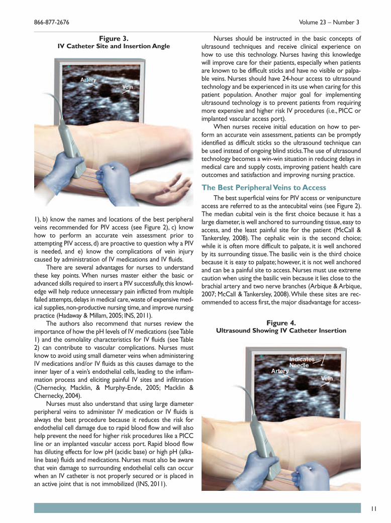
Nurses should be instructed in the basic concepts ofultrasound techniques and receive clinical experience onhow to use this technology. Nurses having this knowledgewill improve care for their patients, especially when patientsare known to be difficult sticks and have no visible or palpa-ble veins. Nurses should have 24-hour access to ultrasoundtechnology and be experienced in its use when caring for thispatient population. Another major goal for implementingultrasound technology is to prevent patients from requiringmore expensive and higher risk IV procedures (i.e., PICC orimplanted vascular access port).
When nurses receive initial education on how to per-form an accurate vein assessment, patients can be promptlyidentified as difficult sticks so the ultrasound technique canbe used instead of ongoing blind sticks. The use of ultrasoundtechnology becomes a win-win situation in reducing delays inmedical care and supply costs, improving patient health careoutcomes and satisfaction and improving nursing practice.
The Best Peripheral Veins to AccessThe best superficial veins for PIV access or venipuncture
access are referred to as the antecubital veins (see Figure 2).The median cubital vein is the first choice because it has alarge diameter, is well anchored to surrounding tissue, easy toaccess, and the least painful site for the patient (McCall &Tankersley, 2008). The cephalic vein is the second choice;while it is often more difficult to palpate, it is well anchoredby its surrounding tissue. The basilic vein is the third choicebecause it is easy to palpate; however, it is not well anchoredand can be a painful site to access. Nurses must use extremecaution when using the basilic vein because it lies close to thebrachial artery and two nerve branches (Arbique & Arbique,2007; McCall & Tankersley, 2008). While these sites are rec-ommended to access first, the major disadvantage for access-
11
866-877-2676 Volume 23 – Number 3
Figure 3.IV Catheter Site and Insertion Angle
Figure 4.Ultrasound Showing IV Catheter Insertion
Invite colleaguesto join AMSN at
amsn.org/MGM
Share how you’ve grownpersonally andprofessionally
as an AMSN member
The more new members you recruit, the more rewards
you earn
amsn.org/MGM
ing these sites is that they are located in an active joint andrequire the joint to be immobilized.
The cephalic and basilic veins are also located in theforearm and upper arms; however, they may be more difficultto palpate due to their location deep within surroundingmuscles and proximity to bone structures (Alexander &
Corrigan, 2004). This specific knowledge of vein placement isonly a portion of the advanced education and technical expe-rience necessary for nurses to be adequately equipped.Nurses must be aware that they are spending valuable timeattempting PIV access and using outdated practices whencaring for patients with poor peripheral vein access.
Ultrasound BasicsUltrasound technique for PIV access is not a new con-
cept and is being utilized more frequently across the nationby nurses in cases where peripheral IV access is difficult orimpossible (Aponte et al., 2007; Gregg, Murthi, Sisley, Stein, &Scalea, 2010). The first use of ultrasound imaging can betraced back to the 1930s (Hadaway & Millam, 2005; Macklin& Chernecky, 2004). The use of ultrasound for PIV access issafe, rapid, effective, improves patient outcomes, andincreases patient satisfaction (Costantino et al., 2010).
Ultrasonography is a non-invasive procedure that useshigh frequency ultrasound waves (7.5-15 MHz) produced bya transducer to create real-time images of organs, tissues, andblood flow (Rumack, 2005). The imaging device contains twobasic components: a transducer and a display screen. Thereare several types of ultrasound transducers available. The lin-ear transducer is recommended for PIV access whenattempting to isolate structures located 1-3 cm beneath theskin (Rumack, 2005).
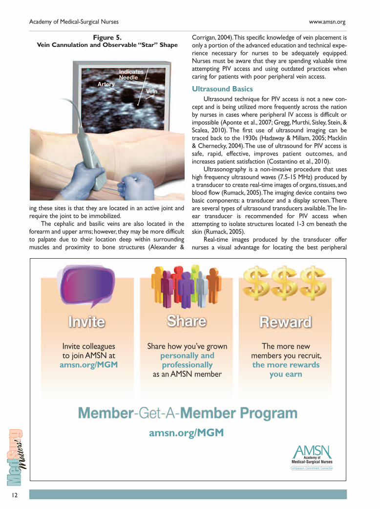
Real-time images produced by the transducer offernurses a visual advantage for locating the best peripheral
Academy of Medical-Surgical Nurses www.amsn.org
12
Figure 5.Vein Cannulation and Observable “Star” Shape
13
866-877-2676 Volume 23 – Number 3
veins to access. This approach provides direct measurementof blood vessel diameter and condition. Ultrasound technol-ogy also allows the visualization of the IV catheter tip insidethe vein, increasing the chance for prompt and successfulvenous access. This helps nurses determine the most appro-priate PIV site and appropriate catheter size, and to verifyplacement of the IV catheter.
Medication pH Acid Neutral Alkaline
Meperidine (Demerol) 3.5-6 √
Morphine 2.5-7 √
Hydromorphone (Dilaudid) 4-5.5 √
Fentanyl 4-7.5 √ √ √
Promethazine (Phenergan) 4-5.5 √
Ondansetron (Zofran) 3-4 √
Prochlorperazine (Compazine) 4.2-6.2 √
Lorazepam (Ativan) 0 √
Diazepam (Valium) 6.2-6.9 √
Haloperidol (Haldol) 3-3.6 √
Midazolam (Versed) 3.0 √
Diphenhydramine (Benadryl) 5-6 √
Hydralazine (Apresoline) 3.4-4 √
Propanolol (Inderal) 2.8-3.5 √
Verapamil (Isoptin) 4.1-6 √
Atenolol (Tenormin) 5.5-6.5 √
Hydrocortisone (Solu-Cortef) 7-8 √ √
Vancomycin 2.4-4.5 √
Tobramycin 3-6.5 √
Gentamycin 3-5.5 √
Cefazolin (Ancef) 4.5-7 √
Regular Insulin 7-7.8 √ √
Dextrose 3.5-6.5 √
Heparin 5-8 √ √ √
Magnesium Sulfate 5.5-7 √
Furosemide (Lasix) 8-9.3 √
Naloxone (Narcan) 3-4 √
Dexamethasone (Decadron) 7-8.5 √ √
Potassium Chloride 4-8 √ √ √
Famotidine (Pepcid) 5-5.6 √
Normal Saline Flush 4.5-7 √
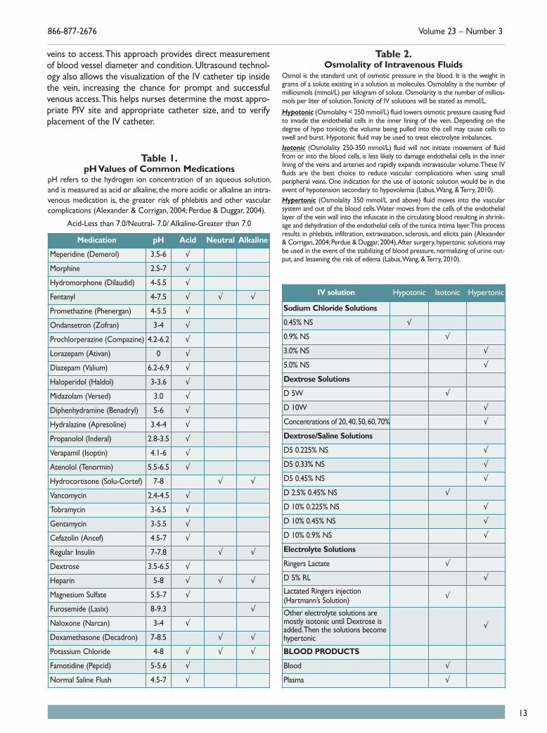
Table 1.pH Values of Common Medications
pH refers to the hydrogen ion concentration of an aqueous solution,and is measured as acid or alkaline; the more acidic or alkaline an intra-venous medication is, the greater risk of phlebitis and other vascularcomplications (Alexander & Corrigan, 2004; Perdue & Duggar, 2004).
Acid-Less than 7.0/Neutral- 7.0/ Alkaline-Greater than 7.0
Table 2.Osmolality of Intravenous Fluids
Osmol is the standard unit of osmotic pressure in the blood. It is the weight ingrams of a solute existing in a solution as molecules. Osmolality is the number ofmilliosmols (mmol/L) per kilogram of solute. Osmolarity is the number of millios-mols per liter of solution. Tonicity of IV solutions will be stated as mmol/L.
Hypotonic (Osmolality < 250 mmol/L) fluid lowers osmotic pressure causing fluidto invade the endothelial cells in the inner lining of the vein. Depending on thedegree of hypo tonicity, the volume being pulled into the cell may cause cells toswell and burst. Hypotonic fluid may be used to treat electrolyte imbalances.Isotonic (Osmolality 250-350 mmol/L) fluid will not initiate movement of fluidfrom or into the blood cells, is less likely to damage endothelial cells in the innerlining of the veins and arteries and rapidly expands intravascular volume. These IVfluids are the best choice to reduce vascular complications when using smallperipheral veins. One indication for the use of isotonic solution would be in theevent of hypotension secondary to hypovolemia (Labus, Wang, & Terry, 2010).Hypertonic (Osmolality 350 mmol/L and above) fluid moves into the vascularsystem and out of the blood cells. Water moves from the cells of the endotheliallayer of the vein wall into the infuscate in the circulating blood resulting in shrink-age and dehydration of the endothelial cells of the tunica intima layer. This processresults in phlebitis, infiltration, extravasation, sclerosis, and elicits pain (Alexander& Corrigan, 2004; Perdue & Duggar, 2004). After surgery, hypertonic solutions maybe used in the event of the stabilizing of blood pressure, normalizing of urine out-put, and lessening the risk of edema (Labus, Wang, & Terry, 2010).
IV solution Hypotonic Isotonic Hypertonic
Sodium Chloride Solutions
0.45% NS √
0.9% NS √
3.0% NS √
5.0% NS √
Dextrose Solutions
D 5W √
D 10W √
Concentrations of 20, 40, 50, 60, 70% √
Dextrose/Saline Solutions
D5 0.225% NS √
D5 0.33% NS √
D5 0.45% NS √
D 2.5% 0.45% NS √
D 10% 0.225% NS √
D 10% 0.45% NS √
D 10% 0.9% NS √
Electrolyte Solutions
Ringers Lactate √
D 5% RL √
Lactated Ringers injection(Hartmann’s Solution) √
Other electrolyte solutions aremostly isotonic until Dextrose isadded. Then the solutions becomehypertonic
√
BLOOD PRODUCTS
Blood √
Plasma √
Academy of Medical-Surgical Nurses www.amsn.org
14
Ultrasound technique locates the best blood vessels toaccess on extremities and will show the structural differencesbetween arteries, veins, and nerve bundles. This will aid in iden-tifying the condition of the blood vessel, preventing or reduc-ing failure rates and vascular complications (Rumack, 2005).
ReferencesAponte, H., Acosta, S., Rigamonti, D., Silvia, B., & Austin, P. (2007). The use
of ultrasound for placement of intravenous catheters. AmericanAssociation of Nurse Anesthetists Journal, 75(3), 212-216.
Alexander, M., & Corrigan, A.M. (2004). Core curriculum for infusion nursing(3rd ed.). Philadelphia: Lippincott, Williams & Wilkins.
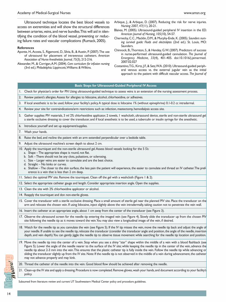
Basic Steps for Ultrasound-Guided Peripheral IV Access
1. Check for physician’s order for PIV. Using ultrasound-guided technique to assess veins is an extension of the nursing assessment process.
2. Review patient’s allergies. Assess for allergies to lidocaine, alcohol, chlorhexidine, or adhesives.
3. If local anesthetic is to be used, follow your facility’s policy. A typical dose is: lidocaine 1% (without epinephrine) 0.1-0.2 cc intradermal.
4. Review your site for contraindications/arm restrictions such as: infection, mastectomy, hemodialysis access site.
5. Gather supplies: PIV materials, 3 ml 2% chlorhexidine applicator, 2 towels, 1 washcloth, ultrasound device, sterile and non-sterile ultrasound gel,a sterile occlusive dressing to cover the transducer, and if local anesthetic is to be used, a tuberculin or insulin syringe for the anesthetic.
6. Introduce yourself and set up equipment/supplies.
7. Wash your hands.
8. Raise the bed, and recline the patient with an arm extended perpendicular over a bedside table.
9. Adjust the ultrasound machine’s screen depth to about 2 cm.
10. Apply the tourniquet and the non-sterile ultrasound gel. Assess blood vessels looking for the 5 S’s:a. Shape – The appropriate shape is round, not flat.b. Soft – There should not be any clots, pulsations, or sclerosing.c. Size – Larger veins are easier to cannulate and are the best choice.d. Straight – No kinks or curves.e. Shallow – The closer to the skin surface, the less pain the patient will experience, the easier to cannulate and thread an IV catheter. The pref-erence is a vein that is less than 2 cm deep.
11. Select the optimal PIV site. Remove the tourniquet. Clean off the gel with a washcloth (Figure 1 & 2).
12. Select the appropriate catheter gauge and length. Consider appropriate insertion angle. Open the supplies.
13. Clean the site with 2% chlorhexidine applicator or alcohol.
14. Reapply the tourniquet and don non-sterile gloves.
15. Cover the transducer with a sterile occlusive dressing. Place a small amount of sterile gel near the planned PIV site. Place the transducer on thearm and relocate the chosen vein. If using lidocaine, inject slightly above the vein intradermally, taking caution not to penetrate the vein wall.
16. Insert the catheter at an appropriate angle, about 1 cm away from the center of the transducer (see Figure 3).
17. Observe the ultrasound screen for the needle tip entering the imaged vein (see Figure 4). Slowly slide the transducer up from the chosen PIVsite following the needle tip as it moves toward the vein. You may also view a longitudinal image of the vein, if desired.
18. Watch for the needle tip as you cannulate the vein (see Figure 5). If the IV tip misses the vein, move the needle tip back and adjust the angle ofyour needle. If unable to see the needle tip, relocate the transducer (consider the transducer angle and position, the angle of the needle, insertiondepth, and vein depth). You can gently jiggle the needle tip to observe tissue movement while searching for the needle tip location and position.
19. Move the needle tip into the center of a vein. Stop when you see a shiny “star” shape within the middle of a vein with a blood flashback (seeFigure 5). Lower the angle of the needle nearer to the surface of the IV site; while keeping the needle tip in the center of the vein, advance theneedle tip about 2-3 mm into the vein. This ensures that the plastic catheter tip is fully inside the vein. Follow the needle tip while advancing orsliding the transducer slightly up from the IV site. Note: If the needle tip is not observed in the middle of a vein during advancement, the cathetermay not advance properly and may kink.
20. Thread the catheter of the needle into the vein. Good blood flow should be achieved after removing the needle.
21. Clean-up the IV site and apply a dressing. Procedure is now completed. Remove gloves, wash your hands, and document according to your facility’spolicy.
Subsumed from literature review and current UT Southwestern Medical Center policy and procedure guidelines.
Arbique, J., & Arbique, D. (2007). Reducing the risk for nerve injuries.Nursing 2007, 47(11), 20-21.
Blaivas, M. (2005). Ultrasound-guided peripheral IV insertion in the ED.American Journal of Nursing, 105(10), 54-57.
Chernecky, C.C., Macklin, D.M., & Murphy-Ende, K. (2005). Saunders nurs-ing survival guide: Fluids and electrolytes (2nd ed.). St. Louis, MO:Saunders.
Chinnock, B., Thornton, S., & Hendey, G.W. (2007). Predictors of successin nurse-performed ultrasound-guided cannulation. The Journal ofEmergency Medicine, 33(4), 401-405. doi:10.1016/j.jemermed.2007.02.027
Costantino, T.G., Kirtz, J.F., & Satz, W.A. (2010). Ultrasound-guided periph-eral venous access vs. the external jugular vein as the initialapproach to the patient with difficult vascular access. The Journal of
15
product connectors. Until this manufacturing change is com-pletely implemented, nurses MUST put safety steps in place.GEDSA, the Global Enteral Device Supplier Association,offers an informational program called “Stay Connected” tofacilitate the transition to safer medical connectors. Formore information, go to www.gedsa.org.
ReferencesPugliese, G. (2013). New standards to prevent tubing misconnections will
have unprecedented impact on supply chair and patient safety.
Nutrition to Improve Outcomescontinued from page 3
Emergency Medicine, 39(4), 462-467. doi:10.1016/j.jemermed.2009.02.004
Gregg, S.C., Murthi, S.B., Sisley, A.C., Stein, D.M., & Scalea, T.M. (2010).Ultrasound-guided peripheral intravenous access in the intensivecare unit. Journal of Critical Care, 25(3), 514-519.
Hadaway, L.C., & Millam, D.A. (2005). On the road to successful I.V. starts.Nursing, 35(Suppl.), 1-16.
Infusion Nurses Society (INS). (2011). Infusion nursing standards of practice(2nd ed.). Norwood, MA: Author.
Labus, D., Wang, R., & Terry, D.P. (Eds.). (2010). Introduction to IV therapy.In IV therapy made incredibly easy (4th ed., pp. 11-23). Philadelphia:Lippincott, Williams & Wilkins.
Macklin, D., & Chernecky, C. (2004). Peripheral complications. In Real worldnursing survival guide: IV therapy (pp. 63-82). St. Louis: Saunders.
McCaig, L.F., & Burt, C.W. (2003). National hospital ambulatory medical caresurvey: 003 emergency department summary. Retrieved fromhttp://www.cdc.gov/nchs/data/ad/ad358.pdf
McCall, R., & Tankersley, T. (2008). Phlebotomy essentials (4th ed.).Philadelphia: Lippincott, Williams & Wilkins.
Perdue, M.A., & Duggar, B. (2004). Technology and clinical application. In M.Alexander & A. Corrigan (Eds.), Core curriculum for infusion nursing(3rd ed., pp. 1-125). Philadelphia: Lippincott, Williams & Wilkins.
Pries, A.R., & Kuebler, W.M. (2006). Normal endothelium. Handbook ofExperimental Pharmacology, 176(1), 1-40.
Rumack, S.R. (2005). Diagnostic ultrasound: Volume I (3rd ed.). St. Louis:Elsevier Mosby.
Sumpio, B.E., Riley, J.T., & Dardik, A. (2002). Cells in focus: Endothelial cell.International Journal of Biochemistry and Cell Biology, 34(12), 1508-1512.
Trim, J.C. (2005). Peripheral intravenous catheters: Considerations in the-ory and practice. British Journal of Nursing, 14(12), 654.
Debbie Arbique, MS, FNP-C, RN, CEN, CFN, DNP, isa Doctor of Nursing Practice, Blood Pressure Specialist,University of Texas Southwestern Medical Center, Dallas, TX.Marilynn Bordelon, MS, RN, CMSRN, is a ClinicalEducator, MedSurg Services, University of Texas SouthwesternMedical Center, Dallas, TX.Robert Dragoo, RN, is a Staff Nurse, Vascular Access,Parallon Vascular Access, Dallas, TX.Stephanie Huckaby, MSN, RN-BC, is Director ofNursing, University of Texas Southwestern Medical Center,Dallas, TX. She is a member of the MedSurg Matters EditorialCommittee.Acknowledgement: The authors would like to thank PamCurry, MA, Biomedical Communications, Senior GraphicsDesigner and Medical Illustrator, Department of ImagingServices, University of Texas Southwestern Medical Center,Dallas, TX.
Retrieved from https://www.premierinc.com/safety/safety-share/bulletin-tubing-misconnect-11132013.jsp
Vockley, M. (2011). Dangerous connections: Healthcare communitytackles tubing risks. Biomedical Instrumentation and Technology,45(6), 426-428, 430-432, 434. doi:10.2345/0899-8205-45.6.426
Suggested ReadingsAdvancing Safety in Medical Technology (AAMI). (2013). Stay connected:
FAQs about small-bore connectors and tubing misconnections.Retrieved from www.aami.org/hottopics/connectors/stay_connected_10152013.pdf
Baxter Healthcare Corporation. (2011). Connections portfolio: Tubing mis-connections self assessment for healthcare facilities. Retrieved fromhttp://www.baxter.com/downloads/healthcare_professionals/clinical_center_of_excellence/baxterselfassessment.pdf
California Legislative Information. (2012). AB-1867 health facilities:Equipment standards. Retrieved from http://leginfo.legislature.ca.gov/faces/billNavClient.xhtml?bill_id=201120120AB1867
Guenter, P., Hicks, R.W., Simmons, D., Crowley, J., Joseph, S., Croteau, R.,… Vanderveen, T.W. (2008). Enteral feeding misconnections: A consor-tium position statement. Retrieved from https://www.premierinc.com/safety/topics/tubing-misconnections/downloads/S5-JQPS-05-08-guenter.pdf
International Organization for Standardization (ISO). (2010).ANSA/AAMI/ISO 80369-1-2010. Small-bore connectors for liquids andgasses in healthcare applications – Part 1: General requirements.Retrieved from http://www.iso.org/iso/catalogue_detail.htm?csnumber=45976
Joint Commission, The. (2006). Tubing misconnections: A persistent andpotentially deadly occurrence. Retrieved from https://www.premierinc.com/safety/topics/tubing-misconnections/downloads/jcaho-sentinel-event-issue-36.pdf
Andie Melendez, MSN, RN, CHTP, HTCP, is a ClinicalNurse Specialist, University of Maryland, Baltimore WashingtonMedical Center, Glen Burnie, MD.
AMSN BOARD OF DIRECTORS
PresidentKathleen Lattavo, MSN, RN, CNS-MS, CMSRN,
RN-BC, ACNS-BCPresident-Elect
Jill Arzouman, MS, RN, CMSRN, ACNS, BCTreasurer
Jane E. Lacovara, MSN, RN, CMSRN, CNS-BCSecretary
Robin Hertel, MSN, RN, CMSRNDirector
Dee A. Eldardiri, MS, RN-BC, CMSRNDirector
Gloria J. Hurst, BSN, RN, CMSRNDirector
Michele George, MBA, BSN, RNDirector
Cynthia C. Barrere, PhD, RN, AHN-BC, RCNSExecutive Director
Cynthia Hnatiuk, EdD, RN, CAE, FAANDirector, Association Services
Suzanne Stott, BS
MedSurg Matters!
EditorMolly McClelland, PhD, MSN, RN, CMSRN, ACNS?
BC
Editorial CommitteeMillicent G. De Jesus, MSN, RN?BC
Deidra B. Dudley, MN, MS, RN?BC, NEA?BC
Michael M. Evans, MSN, MSEd, RN, ACNS, CMSRN, CNE
Dianne J. Gibbs, MSN, RN
Barbara Chamberlain, PhD, APRN, MBA, CCRN,WCC
Perry C. Goldstein, MSN, RN, CMSRN, PCCN
Stephanie Huckaby, MSN, RN?BC
Elizabeth Miller, DNP, RN, CMSRN, CCM
Sally S. Russell, MN, RN, CMSRN
Catherine A. Santori, RN, CMSRN
Elizabeth Thomas, MSN, RN, ACNS?BC
Managing EditorKatie R. Brownlow, ELS
Editorial AssistantJamie Curran
Layout and Design SpecialistRobert Taylor
Education DirectorRosemarie Marmion, MSN, RN?BC, NE?BC
Please think GREEN and recycle!
www.twitter.com/MedSurgNurses
www.facebook.com/MedSurgNurses
AJJ-0614The mission of AMSN is to promote excellence in medical-surgical nursing.
East Holly Avenue, Box 56, Pitman, NJ 08071-0056 • 866-877-AMSN (2676)[email protected] • www.amsn.org
Volume 23 – Number 3 • May/June 2014
Have you ever wished for better nutritionfor your patients? Do you wonder how nurs-ing’s future will affect you? Are you longing tohave a voice in health care change?
You will have the opportunity to getanswers to these questions – and many more– this fall at the 23rdAnnual AMSN Convention,September 11-14, 2014, in Orlando, FL. Therewill be a wide variety of sessions on medical-surgical nursing’s hottest topics,from hands-on practiceupdates to pharmacol-ogy to leadership. Youwill take home infor-mation that is bothinnovative and practi-cal that you can put touse immediately at yourfacility.
Attending on-sitehas many benefits, likenetworking, specialevents, and a dynamicexhibit hall. However,for those of you whocan’t make it in person,AMSN will again be offering live video broad-casts of selected sessions. As with all thecourses, those will be taught by the country’sleading nursing experts.
Several highlights are described for youbelow. After you read, visit www.amsn.org todig deeper into your course options, continu-ing education information, things to do inOrlando, and more. Be sure to register early tosave your spot and take advantage of early birddiscounts.
The convention officially launches onThursday, September 11, with opening cere-monies and the first address, “Do It Well. MakeIt Fun. The Key to Success in Medical-SurgicalNursing,” by author and humorist RonCulberson, MSW, CSP. Culberson will teachyou how to infuse a little lightness into your
day to increase productivity and enhance yourwork environment.
Michael Bleich, PhD, RN, FAAN, FNAP, anexpert in the future of nursing practice andpolicy, will deliver the convention keynoteaddress, “The Lion and the Lamb: A NursingApproach to the Affordable Care Act (ACA).”Bleich will explore the roles nurses have playedin leading change and advancing health care, aswell as shed understanding on the ACA.
One of the convention’s most popularofferings is the interactive Town Hall, which willbe held on Sunday, September 14. The topic,
“Integrating Nutritional Care to OptimizePatient Outcomes: Med-Surg Nurses atthe Forefront,” focuses on one of themost urgent issues today for nurses and
health care providers: better patient nutri-tion.
Beth Quatrara, DNP, RN, ACNS-BC,CMSRN, and Andrea Melendez, MSN, RN,
CHTP, HTCP, will leadthe Town Hall anddescribe the work ofthe Alliance to AdvancePatient Nutrition
(www.malnutrition.com), a group of nationalhealth care organizations, of which AMSN is afounding member. Attendees will be encour-aged to share nutrition practices at their facili-ties. They will also discuss best practices andhow to improve patient outcomes throughbetter nutrition.
The convention will close with anempowering presentation by Cindy Lefton,PhD, RN, “Extraordinary is Ordinary: WhyNurses Make a Difference.” Dr. Lefton willdescribe nurses’ influence on organizationalculture and present research that has sup-ported the tremendous value of nursing.
We hope you will join us in person inOrlando, or online for the live sessions. Pleaseview the convention program and registertoday at http://convention.amsn.org.
Grab Your Sunglasses and Join us in Orlando
Follow Flo as she migrates to Floridafor the AMSN Convention! Visit us onFacebook for the latest on her travels.