Embed Size (px)
Citation preview
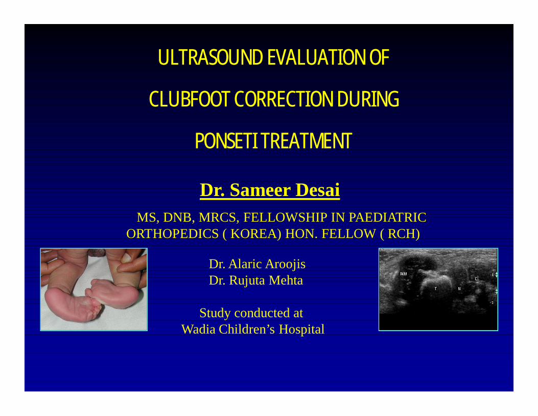
ULTRASOUND EVALUATION OF
CLUBFOOT CORRECTION DURING
PONSETI TREATMENT
Dr. Sameer DesaiMS, DNB, MRCS, FELLOWSHIP IN PAEDIATRIC
ORTHOPEDICS ( KOREA) HON. FELLOW ( RCH)
Dr. Alaric AroojisDr. Rujuta Mehta
Study conducted at Wadia Children’s Hospital
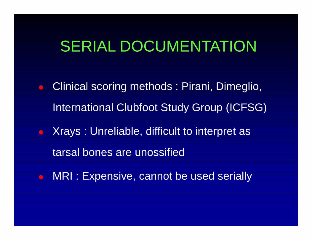
Clinical scoring methods : Pirani, Dimeglio,
International Clubfoot Study Group (ICFSG)
Xrays : Unreliable, difficult to interpret as
tarsal bones are unossified
MRI : Expensive, cannot be used serially
SERIAL DOCUMENTATION
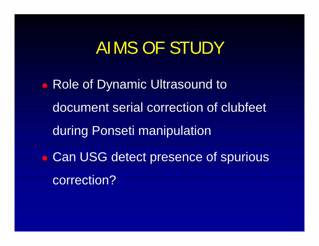
AIMS OF STUDY
Role of Dynamic Ultrasound to
document serial correction of clubfeet
during Ponseti manipulation
Can USG detect presence of spurious
correction?
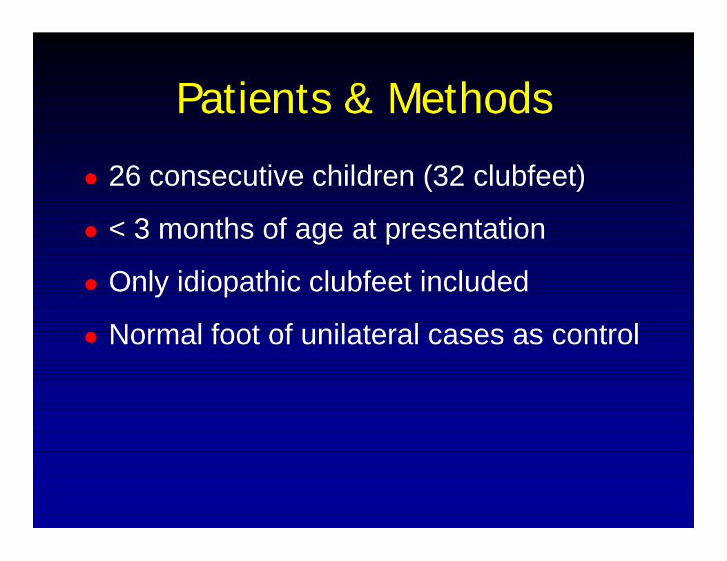
Patients & Methods
26 consecutive children (32 clubfeet)
< 3 months of age at presentation
Only idiopathic clubfeet included
Normal foot of unilateral cases as control
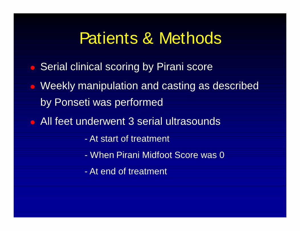
Patients & Methods
Serial clinical scoring by Pirani score
Weekly manipulation and casting as described by Ponseti was performed
All feet underwent 3 serial ultrasounds- At start of treatment
- When Pirani Midfoot Score was 0
- At end of treatment
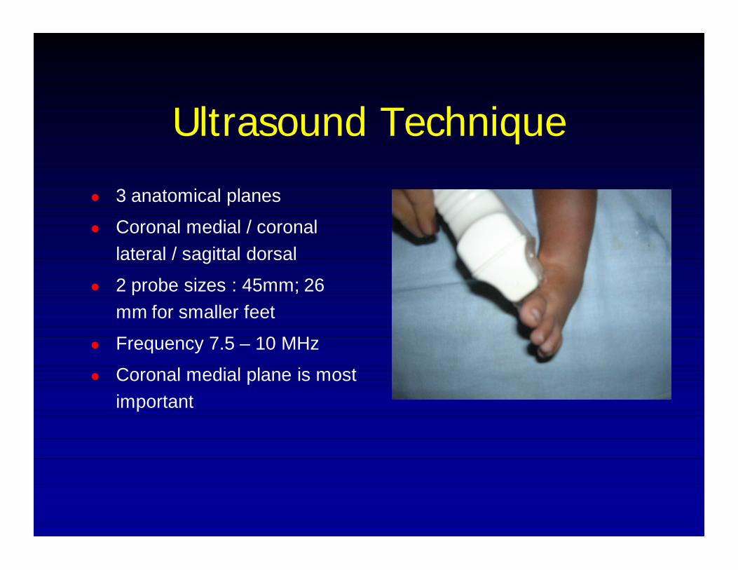
Ultrasound Technique
3 anatomical planes
Coronal medial / coronal lateral / sagittal dorsal
2 probe sizes : 45mm; 26 mm for smaller feet
Frequency 7.5 – 10 MHz
Coronal medial plane is most important
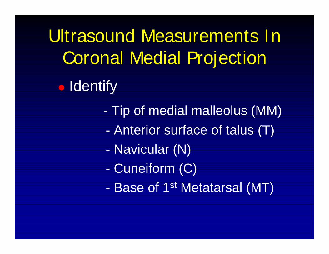
Ultrasound Measurements In Coronal Medial Projection Identify
- Tip of medial malleolus (MM)- Anterior surface of talus (T)- Navicular (N)- Cuneiform (C)- Base of 1st Metatarsal (MT)

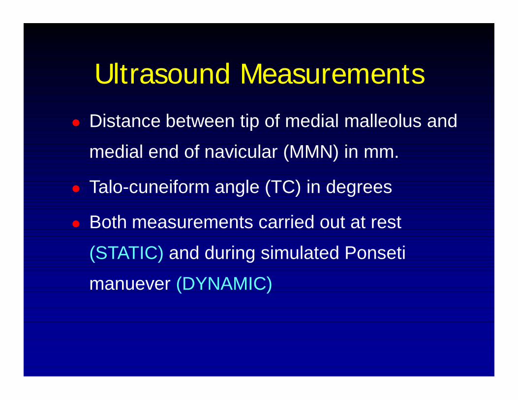
Ultrasound Measurements Distance between tip of medial malleolus and
medial end of navicular (MMN) in mm.
Talo-cuneiform angle (TC) in degrees
Both measurements carried out at rest
(STATIC) and during simulated Ponseti
manuever (DYNAMIC)
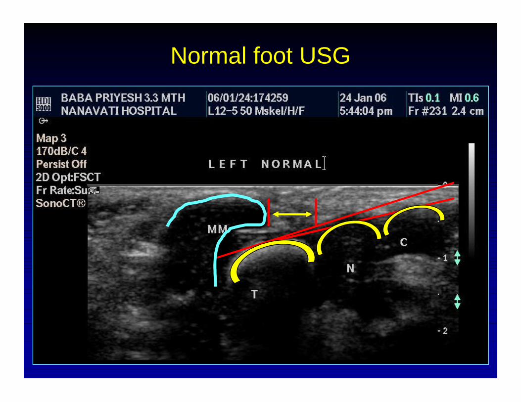
Normal foot USG
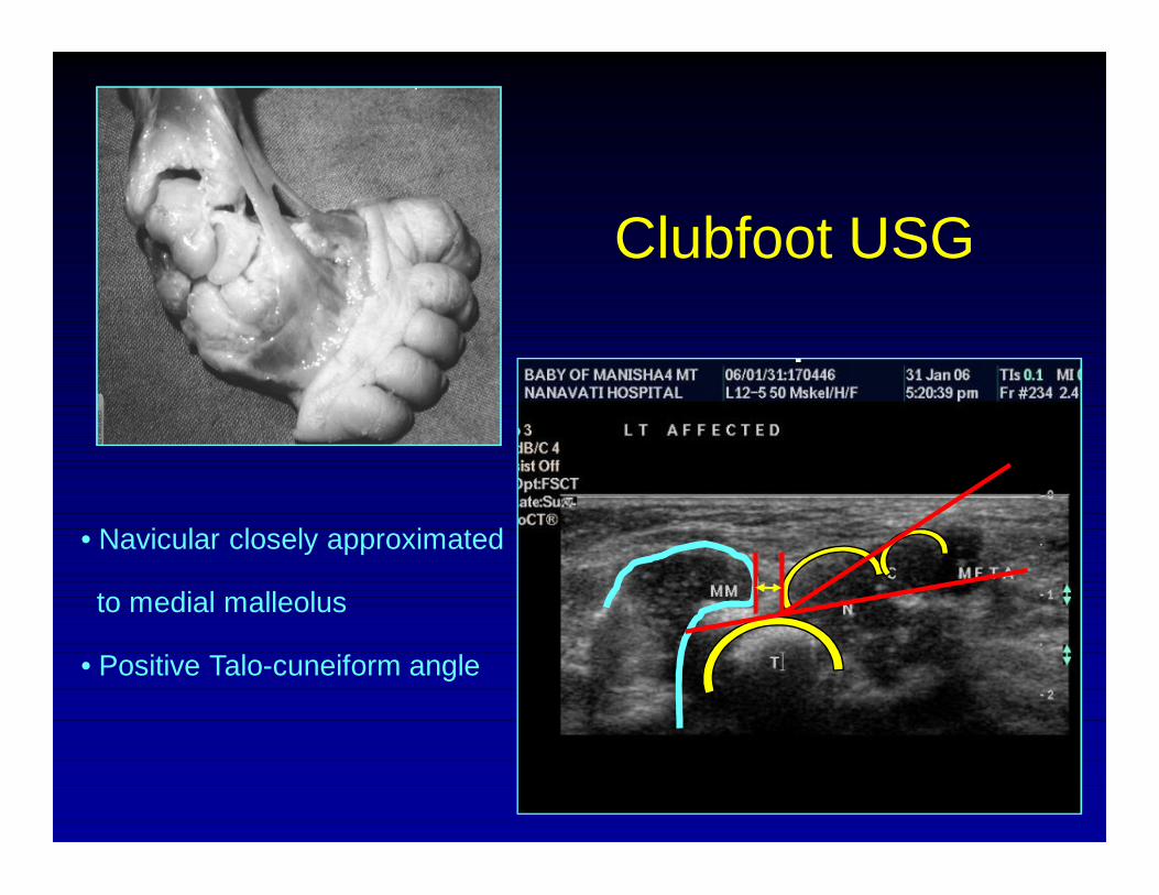
Clubfoot USG
• Navicular closely approximated
to medial malleolus
• Positive Talo-cuneiform angle
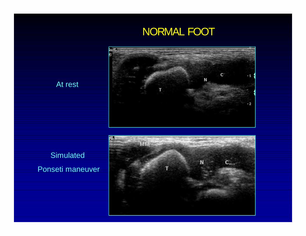
NORMAL FOOT
At rest
Simulated
Ponseti maneuver

CLUBFOOT
At rest
Simulated
Ponseti maneuver
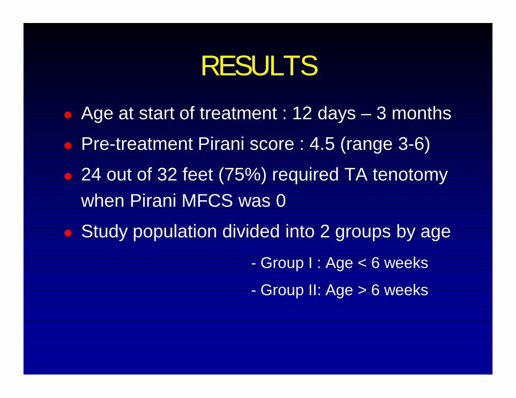
RESULTS Age at start of treatment : 12 days – 3 months
Pre-treatment Pirani score : 4.5 (range 3-6)
24 out of 32 feet (75%) required TA tenotomy when Pirani MFCS was 0
Study population divided into 2 groups by age
- Group I : Age < 6 weeks
- Group II: Age > 6 weeks
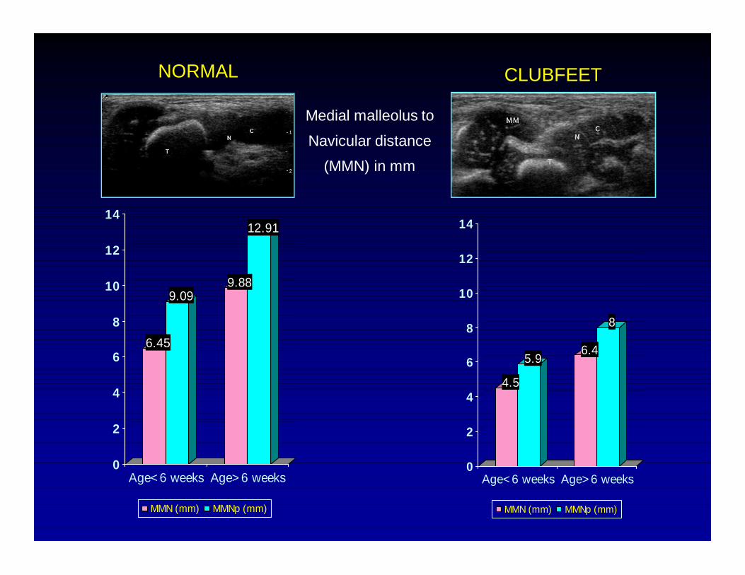
6.45
9.099.88
12.91
0
2
4
6
8
10
12
14
Age<6 weeks Age>6 weeks
MMN (mm) MMNp (mm)
4.5
5.96.4
8
0
2
4
6
8
10
12
14
Age<6 weeks Age>6 weeks
MMN (mm) MMNp (mm)
NORMAL CLUBFEET
Medial malleolus to
Navicular distance
(MMN) in mm
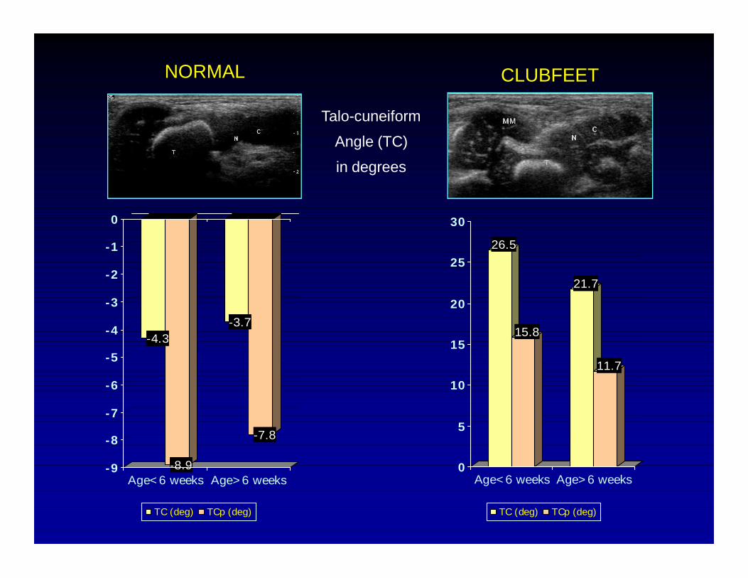
-4.3
-8.9
-3.7
-7.8
-9
-8
-7
-6
-5
-4
-3
-2
-1
0
Age<6 weeks Age>6 weeks
TC (deg) TCp (deg)
26.5
15.8
21.7
11.7
0
5
10
15
20
25
30
Age<6 weeks Age>6 weeks
TC (deg) TCp (deg)
NORMAL CLUBFEET
Talo-cuneiform
Angle (TC)
in degrees
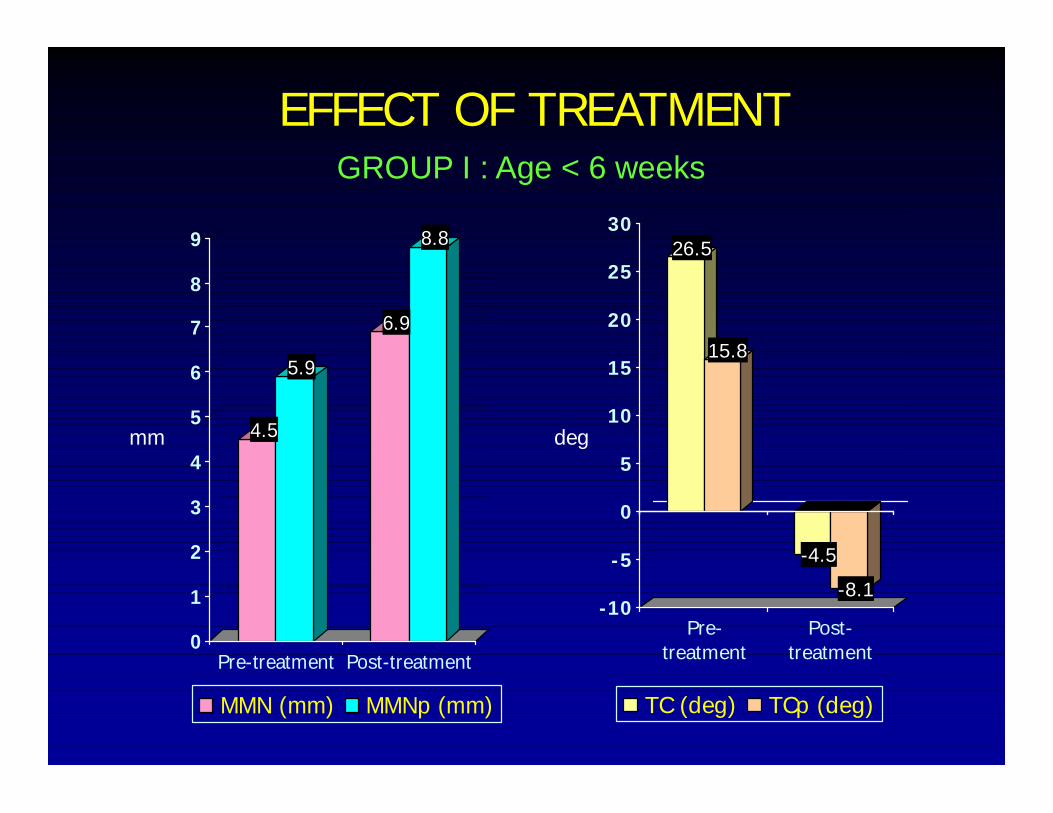
4.5
5.9
6.9
8.8
0
1
2
3
4
5
6
7
8
9
Pre-treatment Post-treatment
MMN (mm) MMNp (mm)
26.5
15.8
-4.5
-8.1-10
-5
0
5
10
15
20
25
30
Pre-treatment
Post-treatment
TC (deg) TCp (deg)
mm deg
EFFECT OF TREATMENTGROUP I : Age < 6 weeks
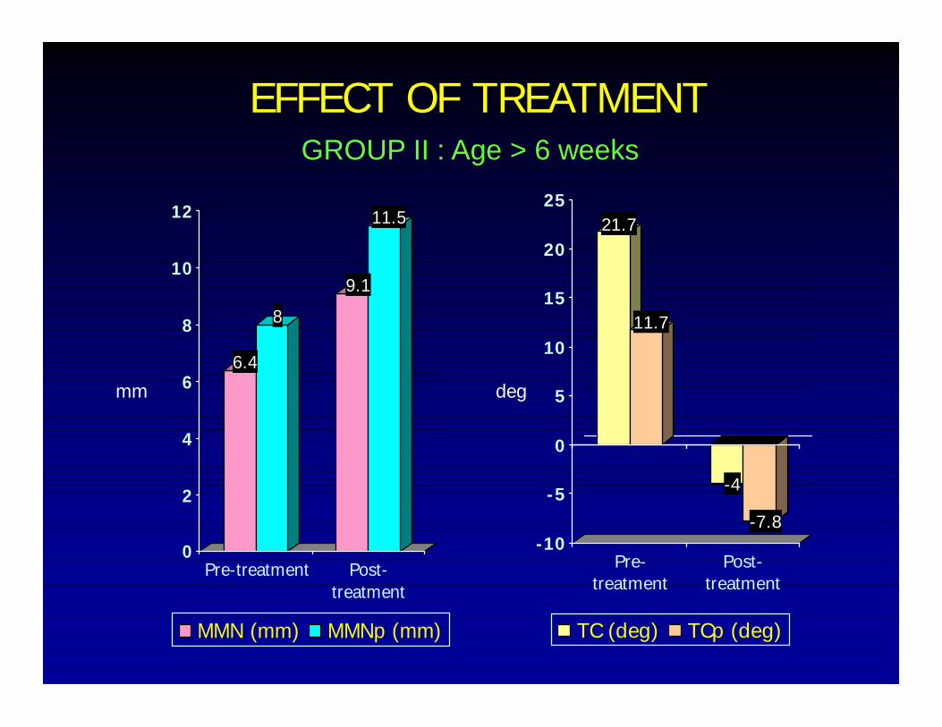
6.4
8
9.1
11.5
0
2
4
6
8
10
12
Pre-treatment Post-treatment
MMN (mm) MMNp (mm)
21.7
11.7
-4
-7.8-10
-5
0
5
10
15
20
25
Pre-treatment
Post-treatment
TC (deg) TCp (deg)
mm deg
EFFECT OF TREATMENTGROUP II : Age > 6 weeks
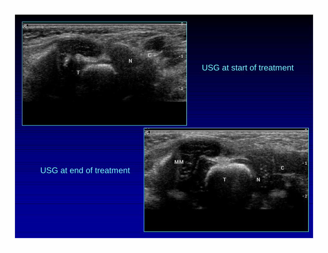
USG at start of treatment
USG at end of treatment
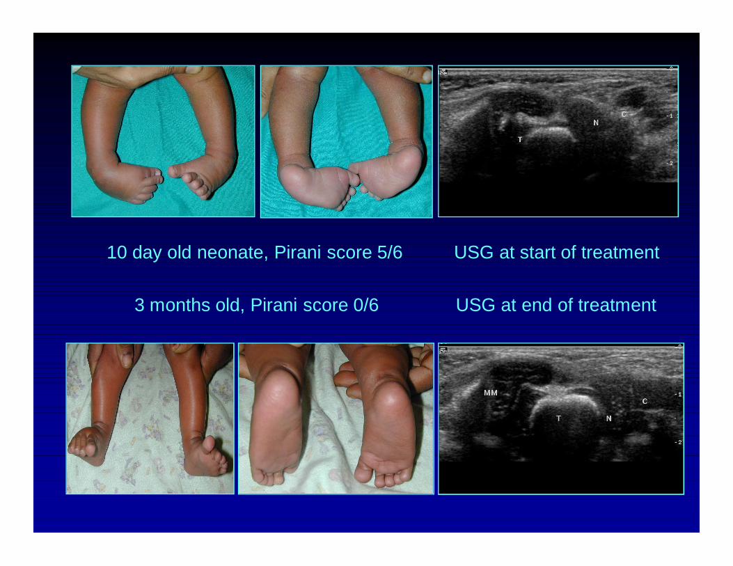
10 day old neonate, Pirani score 5/6 USG at start of treatment
3 months old, Pirani score 0/6 USG at end of treatment
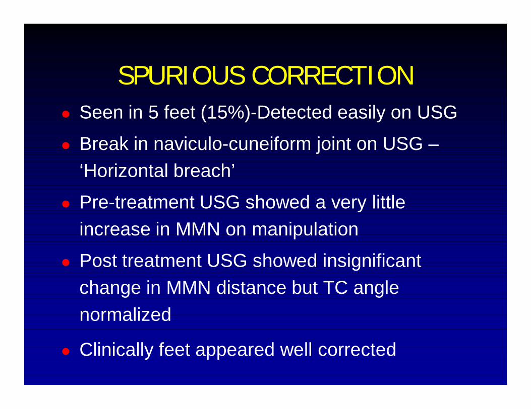
SPURIOUS CORRECTION Seen in 5 feet (15%)-Detected easily on USG
Break in naviculo-cuneiform joint on USG –‘Horizontal breach’
Pre-treatment USG showed a very little increase in MMN on manipulation
Post treatment USG showed insignificant change in MMN distance but TC angle normalized
Clinically feet appeared well corrected
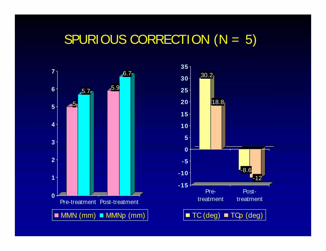
5
5.7 5.9
6.7
0
1
2
3
4
5
6
7
Pre-treatment Post-treatment
MMN (mm) MMNp (mm)
30.2
18.8
-8.6-12
-15
-10
-5
0
5
10
15
20
25
30
35
Pre-treatment
Post-treatment
TC (deg) TCp (deg)
SPURIOUS CORRECTION (N = 5)
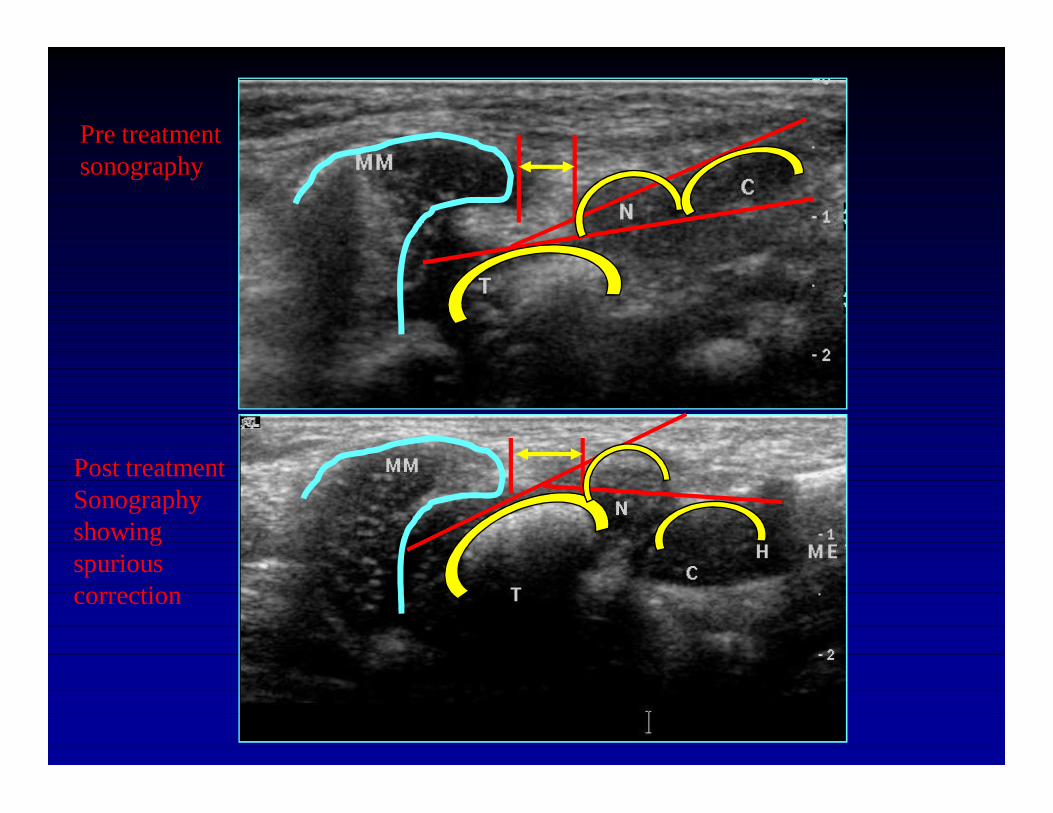
Pre treatmentsonography
Post treatmentSonographyshowing spuriouscorrection
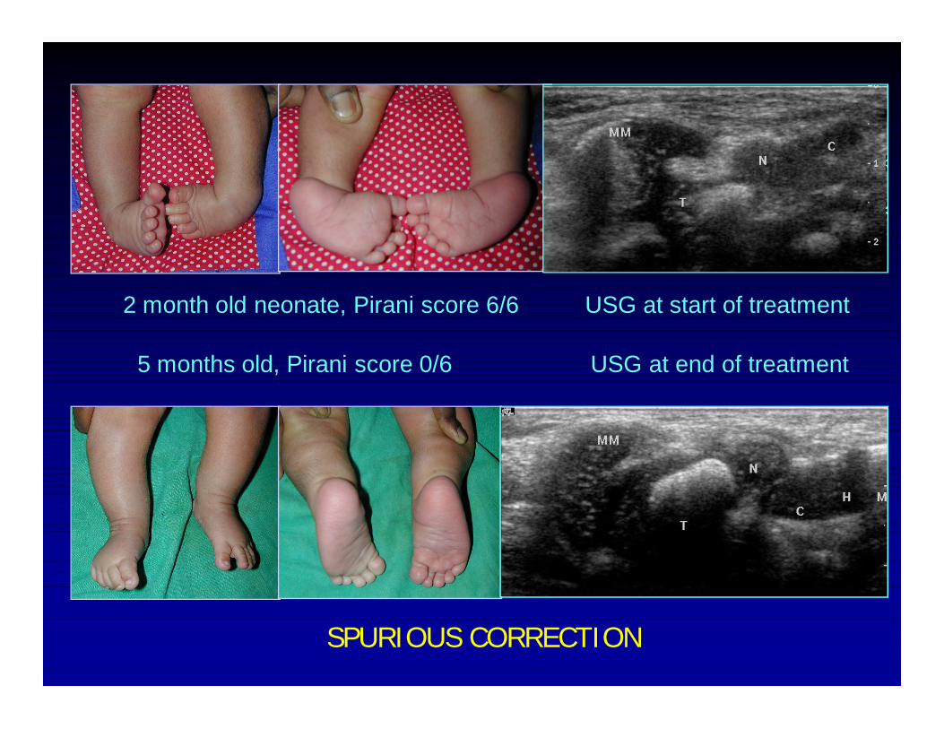
2 month old neonate, Pirani score 6/6 USG at start of treatment
5 months old, Pirani score 0/6 USG at end of treatment
SPURIOUS CORRECTION
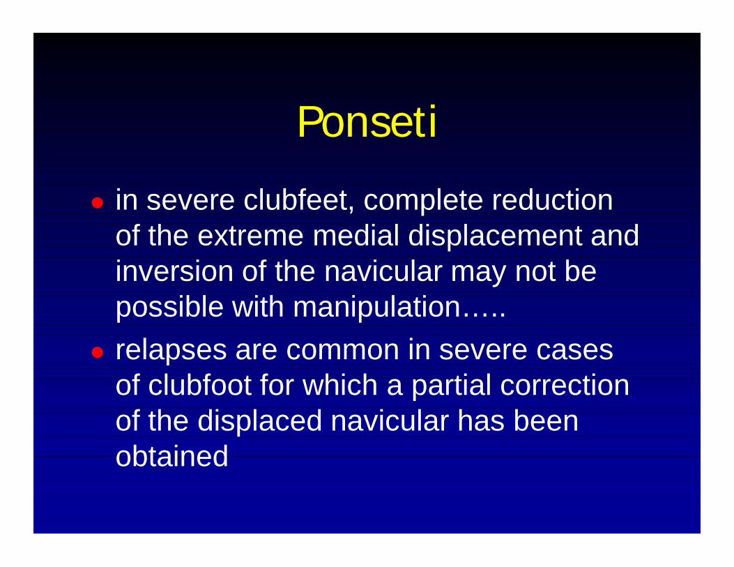
Ponseti
in severe clubfeet, complete reduction of the extreme medial displacement and inversion of the navicular may not be possible with manipulation…..
relapses are common in severe cases of clubfoot for which a partial correction of the displaced navicular has been obtained
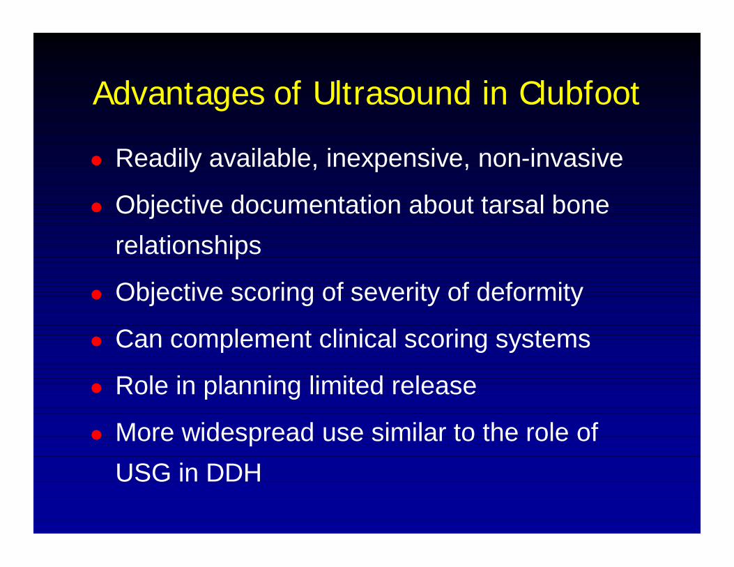
Advantages of Ultrasound in Clubfoot
Readily available, inexpensive, non-invasive
Objective documentation about tarsal bone relationships
Objective scoring of severity of deformity
Can complement clinical scoring systems
Role in planning limited release
More widespread use similar to the role of USG in DDH
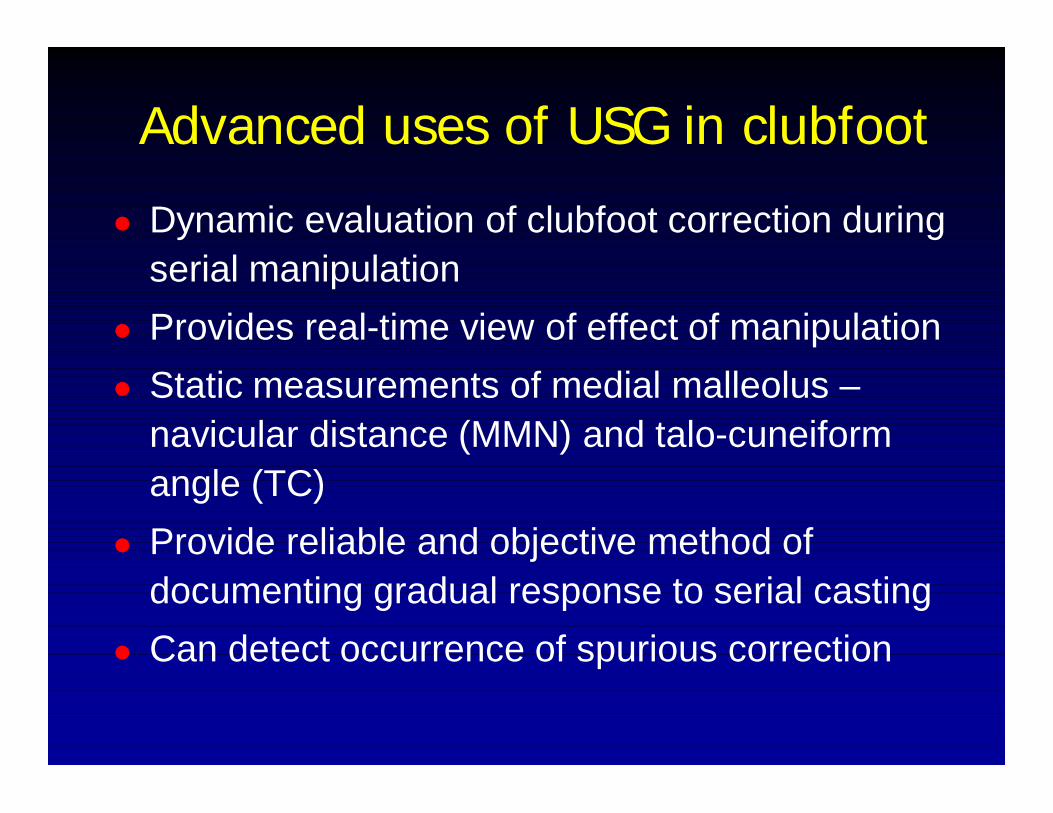
Advanced uses of USG in clubfoot
Dynamic evaluation of clubfoot correction during serial manipulation
Provides real-time view of effect of manipulation Static measurements of medial malleolus –
navicular distance (MMN) and talo-cuneiform angle (TC)
Provide reliable and objective method of documenting gradual response to serial casting
Can detect occurrence of spurious correction
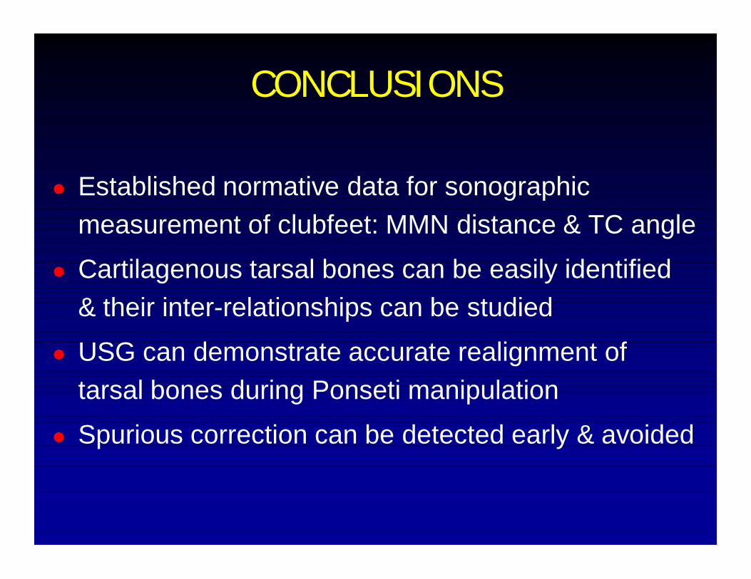
CONCLUSIONS
Established normative data for sonographic measurement of clubfeet: MMN distance & TC angle
Cartilagenous tarsal bones can be easily identified & their inter-relationships can be studied
USG can demonstrate accurate realignment of tarsal bones during Ponseti manipulation
Spurious correction can be detected early & avoided
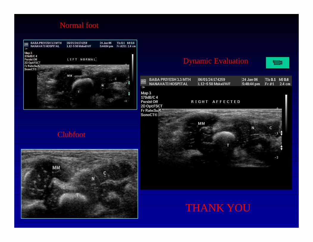
Normal foot
Clubfoot
Dynamic Evaluation
THANK YOU


![Clubfoot: Ponseti Management [Italian]](https://img.pdfslide.us/doc/110x75/613d460c736caf36b75b61e2/clubfoot-ponseti-management-italian.jpg)


![Clubfoot Guide For Parents [2nd Edition]](https://img.pdfslide.us/doc/110x75/618e4bb2659b750eb32ce636/clubfoot-guide-for-parents-2nd-edition.jpg)