Embed Size (px)
Citation preview
Chapter 7: Ultrasound Evaluation of Twin Gestations 134
INTRODUCTION
Since the early eighties and until 2009, there has been a steady and significant increase in the frequency of twins (1, 2). In the United States in 2011, twin birth rate was 33.2 per 1,000 total births and was essentially unchanged from 2009 and 2010 (3). The rate of twin births rose 76% from 1980 to 2009–2011, primarily due to increasing maternal age and widespread use of assisted reproductive technologies, but the pace of increase has slowed in recent years (3).
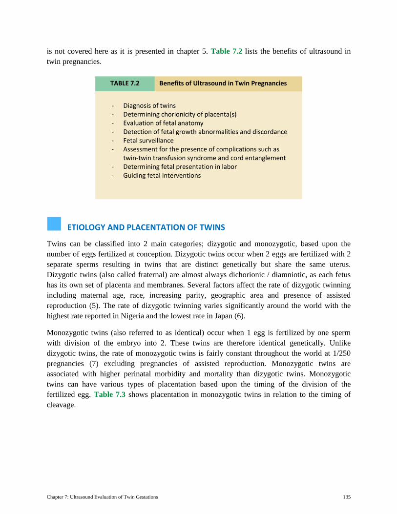
Infants born to twin pregnancies are generally born earlier and smaller than those in singleton pregnancies, and accordingly, are less likely to survive to their first birthday (4). In 2011, 11% of twins were delivered very preterm (less than 32 weeks of gestation), compared with less than 2% of singletons (3). Pregnancies with twins and higher order multiples are at increased risk for many maternal and fetal/child complications. Table 7.1 lists the maternal and fetal/child complications of twin pregnancy.
Ultrasound is an integral part of the diagnosis and management of twin pregnancies. Ultrasound has indeed revolutionized the care of pregnancies with twins from the initial diagnosis to guiding the delivery of the neonates. In this chapter we review the utility of ultrasound in twin pregnancies. The role of ultrasound in the management of high-order multiple pregnancies is beyond the scope of this book. Furthermore, discussion of fetal congenital abnormalities in twins
ULTRASOUND EVALUATION OF TWIN GESTATIONS
7
TABLE 7.1 Maternal and Fetal/Child Complications of Twin Pregnancies
Maternal
- Preterm labor - Preterm premature rupture of membranes - Preeclampsia - Placental abnormalities - Pyelonephritis - Postpartum hemorrhage
Fetal/Child
- Growth abnormalities - Congenital malformations - Admission to neonatal intensive care unit - Cerebral palsy - Perinatal death
Chapter 7: Ultrasound Evaluation of Twin Gestations 135
is not covered here as it is presented in chapter 5. Table 7.2 lists the benefits of ultrasound in twin pregnancies.
ETIOLOGY AND PLACENTATION OF TWINS
Twins can be classified into 2 main categories; dizygotic and monozygotic, based upon the number of eggs fertilized at conception. Dizygotic twins occur when 2 eggs are fertilized with 2 separate sperms resulting in twins that are distinct genetically but share the same uterus. Dizygotic twins (also called fraternal) are almost always dichorionic / diamniotic, as each fetus has its own set of placenta and membranes. Several factors affect the rate of dizygotic twinning including maternal age, race, increasing parity, geographic area and presence of assisted reproduction (5). The rate of dizygotic twinning varies significantly around the world with the highest rate reported in Nigeria and the lowest rate in Japan (6).
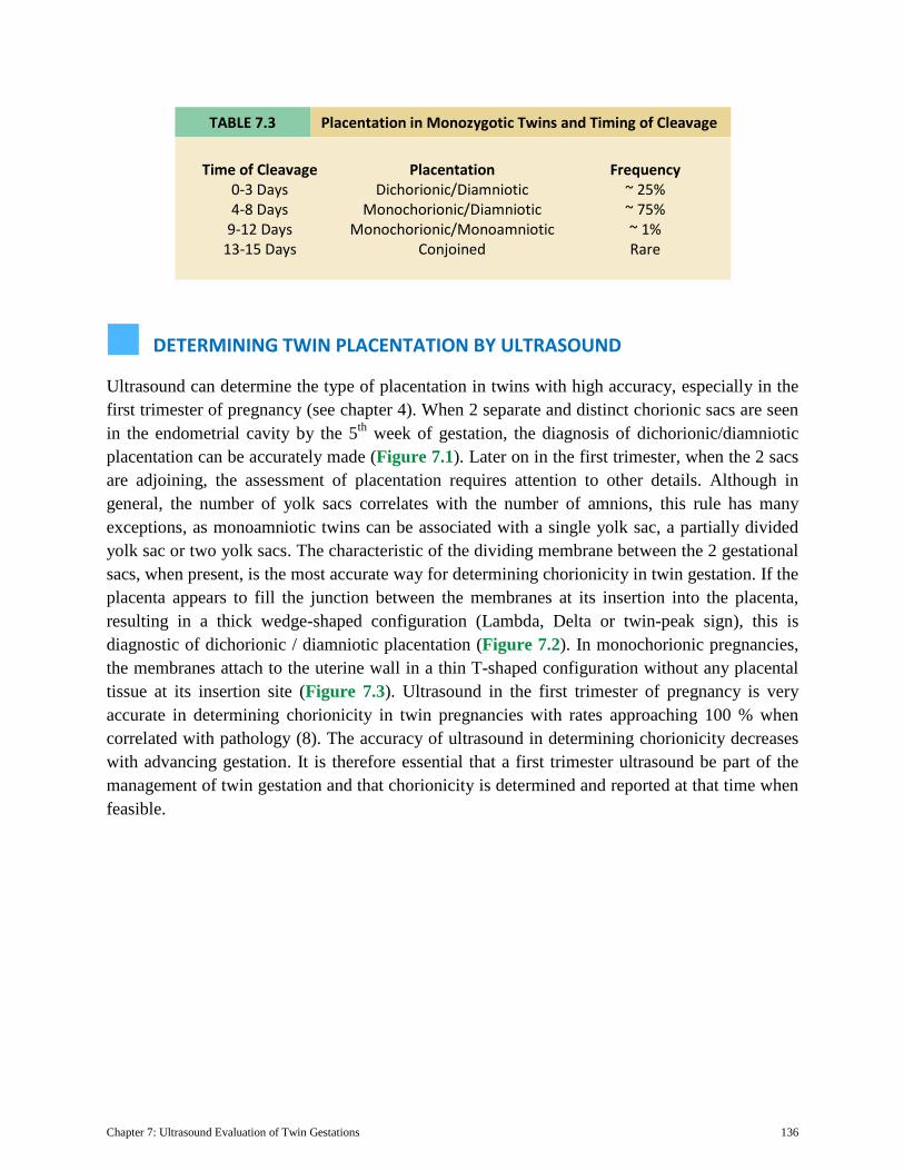
Monozygotic twins (also referred to as identical) occur when 1 egg is fertilized by one sperm with division of the embryo into 2. These twins are therefore identical genetically. Unlike dizygotic twins, the rate of monozygotic twins is fairly constant throughout the world at 1/250 pregnancies (7) excluding pregnancies of assisted reproduction. Monozygotic twins are associated with higher perinatal morbidity and mortality than dizygotic twins. Monozygotic twins can have various types of placentation based upon the timing of the division of the fertilized egg. Table 7.3 shows placentation in monozygotic twins in relation to the timing of cleavage.
TABLE 7.2 Benefits of Ultrasound in Twin Pregnancies
- Diagnosis of twins - Determining chorionicity of placenta(s) - Evaluation of fetal anatomy - Detection of fetal growth abnormalities and discordance - Fetal surveillance - Assessment for the presence of complications such as
twin-twin transfusion syndrome and cord entanglement - Determining fetal presentation in labor - Guiding fetal interventions
Chapter 7: Ultrasound Evaluation of Twin Gestations 136
DETERMINING TWIN PLACENTATION BY ULTRASOUND
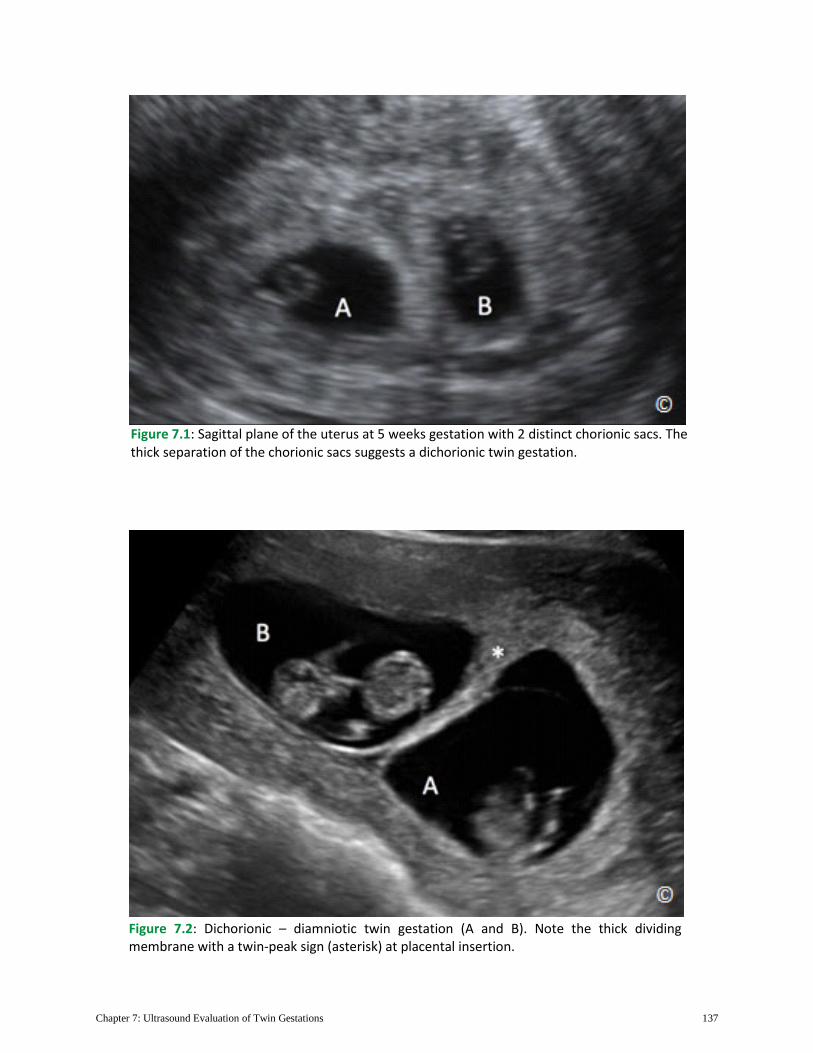
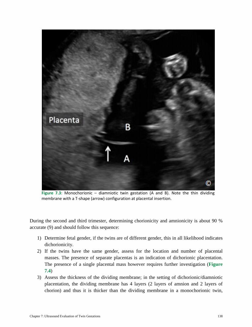
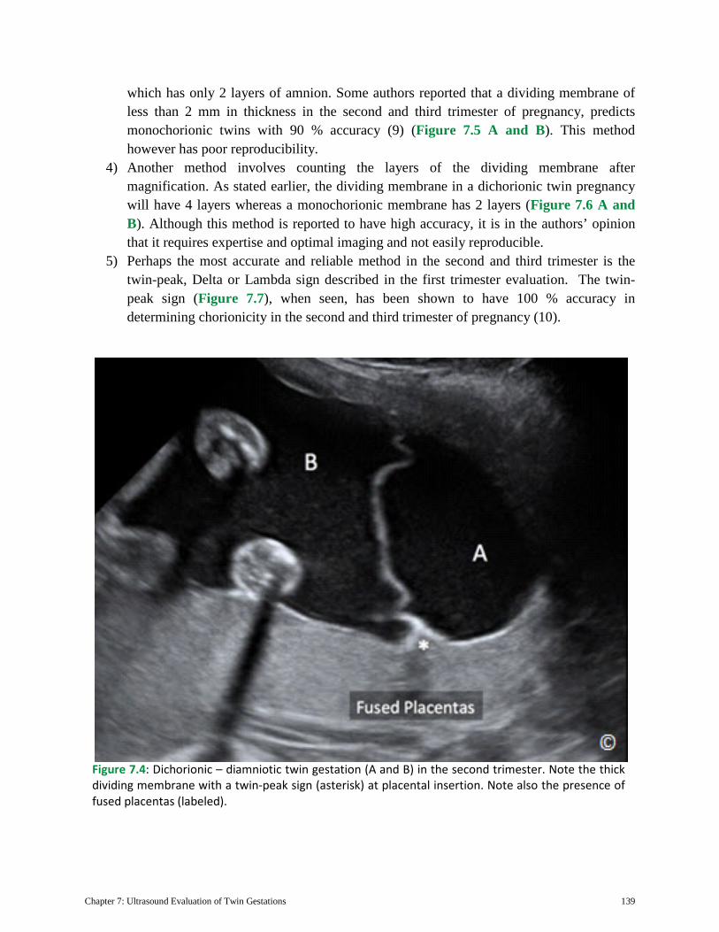
Ultrasound can determine the type of placentation in twins with high accuracy, especially in the first trimester of pregnancy (see chapter 4). When 2 separate and distinct chorionic sacs are seen in the endometrial cavity by the 5th week of gestation, the diagnosis of dichorionic/diamniotic placentation can be accurately made (Figure 7.1). Later on in the first trimester, when the 2 sacs are adjoining, the assessment of placentation requires attention to other details. Although in general, the number of yolk sacs correlates with the number of amnions, this rule has many exceptions, as monoamniotic twins can be associated with a single yolk sac, a partially divided yolk sac or two yolk sacs. The characteristic of the dividing membrane between the 2 gestational sacs, when present, is the most accurate way for determining chorionicity in twin gestation. If the placenta appears to fill the junction between the membranes at its insertion into the placenta, resulting in a thick wedge-shaped configuration (Lambda, Delta or twin-peak sign), this is diagnostic of dichorionic / diamniotic placentation (Figure 7.2). In monochorionic pregnancies, the membranes attach to the uterine wall in a thin T-shaped configuration without any placental tissue at its insertion site (Figure 7.3). Ultrasound in the first trimester of pregnancy is very accurate in determining chorionicity in twin pregnancies with rates approaching 100 % when correlated with pathology (8). The accuracy of ultrasound in determining chorionicity decreases with advancing gestation. It is therefore essential that a first trimester ultrasound be part of the management of twin gestation and that chorionicity is determined and reported at that time when feasible.
TABLE 7.3 Placentation in Monozygotic Twins and Timing of Cleavage
Time of Cleavage Placentation Frequency 0-3 Days Dichorionic/Diamniotic ~ 25% 4-8 Days Monochorionic/Diamniotic ~ 75%
9-12 Days Monochorionic/Monoamniotic ~ 1% 13-15 Days Conjoined Rare
Chapter 7: Ultrasound Evaluation of Twin Gestations 137
Figure 7.1: Sagittal plane of the uterus at 5 weeks gestation with 2 distinct chorionic sacs. The thick separation of the chorionic sacs suggests a dichorionic twin gestation.
Figure 7.2: Dichorionic – diamniotic twin gestation (A and B). Note the thick dividing membrane with a twin-peak sign (asterisk) at placental insertion.
Chapter 7: Ultrasound Evaluation of Twin Gestations 138
During the second and third trimester, determining chorionicity and amnionicity is about 90 % accurate (9) and should follow this sequence:
1) Determine fetal gender, if the twins are of different gender, this in all likelihood indicates dichorionicity.
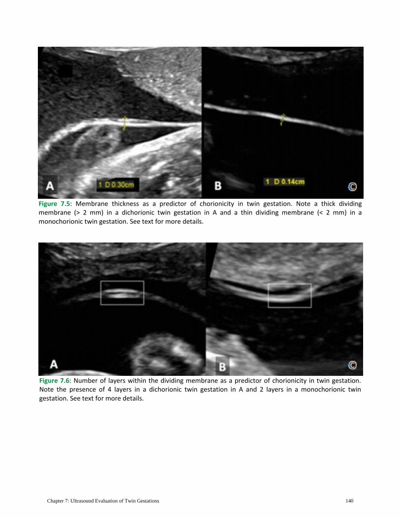
2) If the twins have the same gender, assess for the location and number of placental masses. The presence of separate placentas is an indication of dichorionic placentation. The presence of a single placental mass however requires further investigation (Figure 7.4)
3) Assess the thickness of the dividing membrane; in the setting of dichorionic/diamniotic placentation, the dividing membrane has 4 layers (2 layers of amnion and 2 layers of chorion) and thus it is thicker than the dividing membrane in a monochorionic twin,
Figure 7.3: Monochorionic – diamniotic twin gestation (A and B). Note the thin dividing membrane with a T-shape (arrow) configuration at placental insertion.
Chapter 7: Ultrasound Evaluation of Twin Gestations 139
which has only 2 layers of amnion. Some authors reported that a dividing membrane of less than 2 mm in thickness in the second and third trimester of pregnancy, predicts monochorionic twins with 90 % accuracy (9) (Figure 7.5 A and B). This method however has poor reproducibility.
4) Another method involves counting the layers of the dividing membrane after magnification. As stated earlier, the dividing membrane in a dichorionic twin pregnancy will have 4 layers whereas a monochorionic membrane has 2 layers (Figure 7.6 A and B). Although this method is reported to have high accuracy, it is in the authors’ opinion that it requires expertise and optimal imaging and not easily reproducible.
5) Perhaps the most accurate and reliable method in the second and third trimester is the twin-peak, Delta or Lambda sign described in the first trimester evaluation. The twin-peak sign (Figure 7.7), when seen, has been shown to have 100 % accuracy in determining chorionicity in the second and third trimester of pregnancy (10).
Figure 7.4: Dichorionic – diamniotic twin gestation (A and B) in the second trimester. Note the thick dividing membrane with a twin-peak sign (asterisk) at placental insertion. Note also the presence of fused placentas (labeled).
Chapter 7: Ultrasound Evaluation of Twin Gestations 140
Figure 7.5: Membrane thickness as a predictor of chorionicity in twin gestation. Note a thick dividing membrane (> 2 mm) in a dichorionic twin gestation in A and a thin dividing membrane (< 2 mm) in a monochorionic twin gestation. See text for more details.
Figure 7.6: Number of layers within the dividing membrane as a predictor of chorionicity in twin gestation. Note the presence of 4 layers in a dichorionic twin gestation in A and 2 layers in a monochorionic twin gestation. See text for more details.
Chapter 7: Ultrasound Evaluation of Twin Gestations 141
ULTRASOUND IN FOLLOW-UP OF TWIN GESTATIONS
Twins require heightened evaluation in the antepartum period in order to detect complications such as discordant growth, twin-twin transfusion syndrome, selective intrauterine fetal growth restriction, twin-reversed arterial perfusion and single fetal demise. Surveillance for monochorionic twinning should be performed more frequently given the associated risk involved with such pregnancies. Ultrasound frequency every 4 weeks is adequate to detect growth abnormalities in dichorionic twinning. In monochorionic twins, ultrasound examinations every 2 weeks, starting as early as 16 weeks gestation and until delivery should be considered (11, 12). Doppler ultrasound in twins is reserved for cases where fetal growth restriction is noted or there is twin growth discordance or twin-twin transfusion syndrome. Doppler ultrasound can also be used to evaluate for conditions associated with fetal anemia in twin pregnancies. Table 7.4 and 7.5 provide the indication, timing and type of ultrasound exams recommended in dichorionic and monochorionic twin gestations respectively (11).
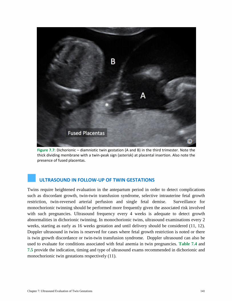
Figure 7.7: Dichorionic – diamniotic twin gestation (A and B) in the third trimester. Note the thick dividing membrane with a twin-peak sign (asterisk) at placental insertion. Also note the presence of fused placentas.
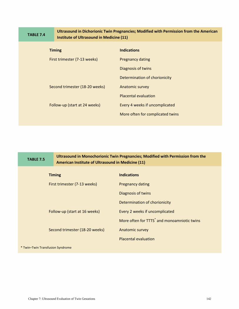
Chapter 7: Ultrasound Evaluation of Twin Gestations 142
Ultrasound in Monochorionic Twin Pregnancies; Modified with Permission from the American Institute of Ultrasound in Medicine (11)
Timing Indications
First trimester (7-13 weeks) Pregnancy dating
Diagnosis of twins
Determination of chorionicity
Follow-up (start at 16 weeks) Every 2 weeks if uncomplicated
More often for TTTS* and monoamniotic twins
Second trimester (18-20 weeks) Anatomic survey
Placental evaluation
* Twin–Twin Transfusion Syndrome
TABLE 7.5
TABLE 7.4 Ultrasound in Dichorionic Twin Pregnancies; Modified with Permission from the American Institute of Ultrasound in Medicine (11)
Timing Indications
First trimester (7-13 weeks) Pregnancy dating
Diagnosis of twins
Determination of chorionicity
Second trimester (18-20 weeks) Anatomic survey
Placental evaluation
Follow-up (start at 24 weeks) Every 4 weeks if uncomplicated
More often for complicated twins
Chapter 7: Ultrasound Evaluation of Twin Gestations 143
DISCORDANT TWINS
Discordance is the difference in weights between twin fetuses and is defined with the larger twin as the standard of growth. It is calculated by the following equation: (larger twin estimated weight-smaller twin estimated weight)/ larger twin estimated weight X 100. A 15-20% or more weight difference among twins is considered discordant (13). Twin-discordance is not a rare event as the likelihood of 20% twin discordance is about 16% in twin gestations (14). Discordant growth is associated with a multitude of problems including increased likelihood of anomalies, intrauterine growth restriction, preterm birth, infection in 1 fetus, admission to the NICU, stillbirth or death within 1 week of birth (13). Serial ultrasound evaluation is essential in twin pregnancies in order to enhance the diagnosis of twin-discordance and for stratification of risk. Once discordance is diagnosed, fetal surveillance should be performed given the associated increase in morbidity and mortality.
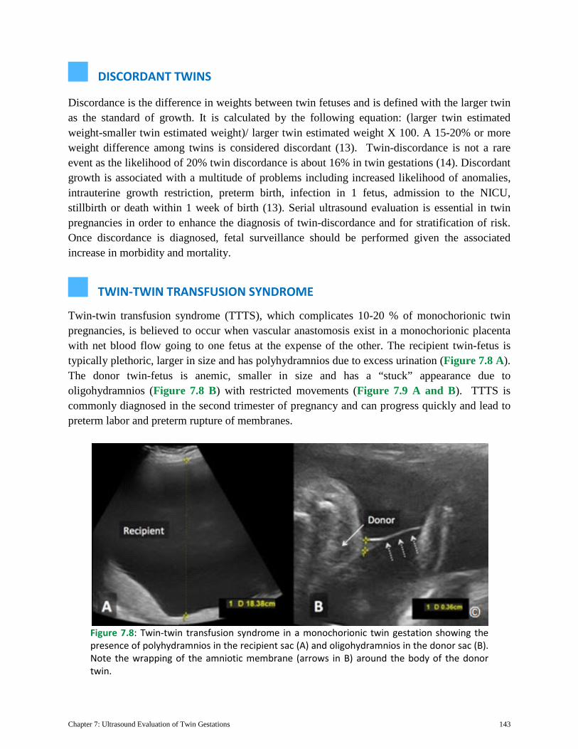
TWIN-TWIN TRANSFUSION SYNDROME
Twin-twin transfusion syndrome (TTTS), which complicates 10-20 % of monochorionic twin pregnancies, is believed to occur when vascular anastomosis exist in a monochorionic placenta with net blood flow going to one fetus at the expense of the other. The recipient twin-fetus is typically plethoric, larger in size and has polyhydramnios due to excess urination (Figure 7.8 A). The donor twin-fetus is anemic, smaller in size and has a “stuck” appearance due to oligohydramnios (Figure 7.8 B) with restricted movements (Figure 7.9 A and B). TTTS is commonly diagnosed in the second trimester of pregnancy and can progress quickly and lead to preterm labor and preterm rupture of membranes.
Figure 7.8: Twin-twin transfusion syndrome in a monochorionic twin gestation showing the presence of polyhydramnios in the recipient sac (A) and oligohydramnios in the donor sac (B). Note the wrapping of the amniotic membrane (arrows in B) around the body of the donor twin.
Chapter 7: Ultrasound Evaluation of Twin Gestations 144
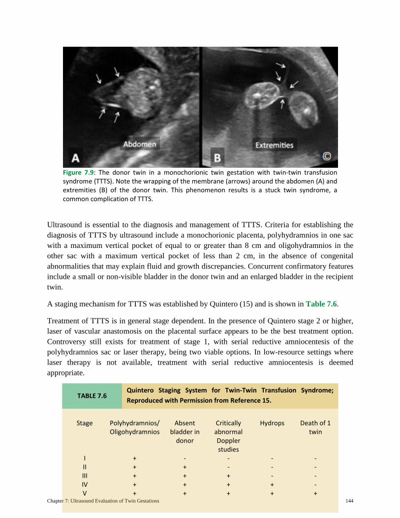
Ultrasound is essential to the diagnosis and management of TTTS. Criteria for establishing the diagnosis of TTTS by ultrasound include a monochorionic placenta, polyhydramnios in one sac with a maximum vertical pocket of equal to or greater than 8 cm and oligohydramnios in the other sac with a maximum vertical pocket of less than 2 cm, in the absence of congenital abnormalities that may explain fluid and growth discrepancies. Concurrent confirmatory features include a small or non-visible bladder in the donor twin and an enlarged bladder in the recipient twin.
A staging mechanism for TTTS was established by Quintero (15) and is shown in Table 7.6.
Treatment of TTTS is in general stage dependent. In the presence of Quintero stage 2 or higher, laser of vascular anastomosis on the placental surface appears to be the best treatment option. Controversy still exists for treatment of stage 1, with serial reductive amniocentesis of the polyhydramnios sac or laser therapy, being two viable options. In low-resource settings where laser therapy is not available, treatment with serial reductive amniocentesis is deemed appropriate.
Figure 7.9: The donor twin in a monochorionic twin gestation with twin-twin transfusion syndrome (TTTS). Note the wrapping of the membrane (arrows) around the abdomen (A) and extremities (B) of the donor twin. This phenomenon results is a stuck twin syndrome, a common complication of TTTS.
TABLE 7.6 Quintero Staging System for Twin-Twin Transfusion Syndrome; Reproduced with Permission from Reference 15.
Stage Polyhydramnios/ Oligohydramnios
Absent bladder in
donor
Critically abnormal Doppler studies
Hydrops Death of 1 twin
I + - - - - II + + - - - III + + + - - IV + + + + - V + + + + +
Chapter 7: Ultrasound Evaluation of Twin Gestations 145
MONOCHORIONIC-MONOAMNIOTIC TWINS
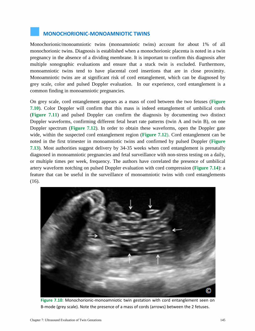
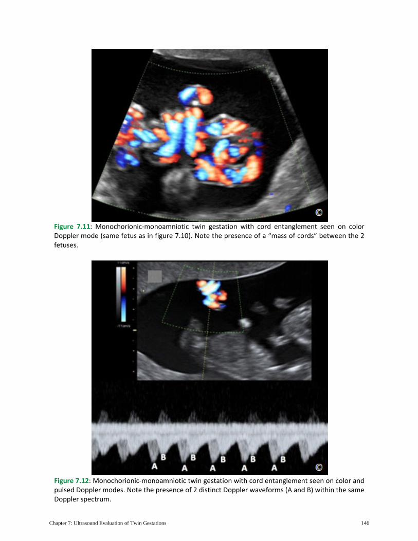
Monochorionic/monoamniotic twins (monoamniotic twins) account for about 1% of all monochorionic twins. Diagnosis is established when a monochorionic placenta is noted in a twin pregnancy in the absence of a dividing membrane. It is important to confirm this diagnosis after multiple sonographic evaluations and ensure that a stuck twin is excluded. Furthermore, monoamniotic twins tend to have placental cord insertions that are in close proximity. Monoamniotic twins are at significant risk of cord entanglement, which can be diagnosed by grey scale, color and pulsed Doppler evaluation. In our experience, cord entanglement is a common finding in monoamniotic pregnancies.
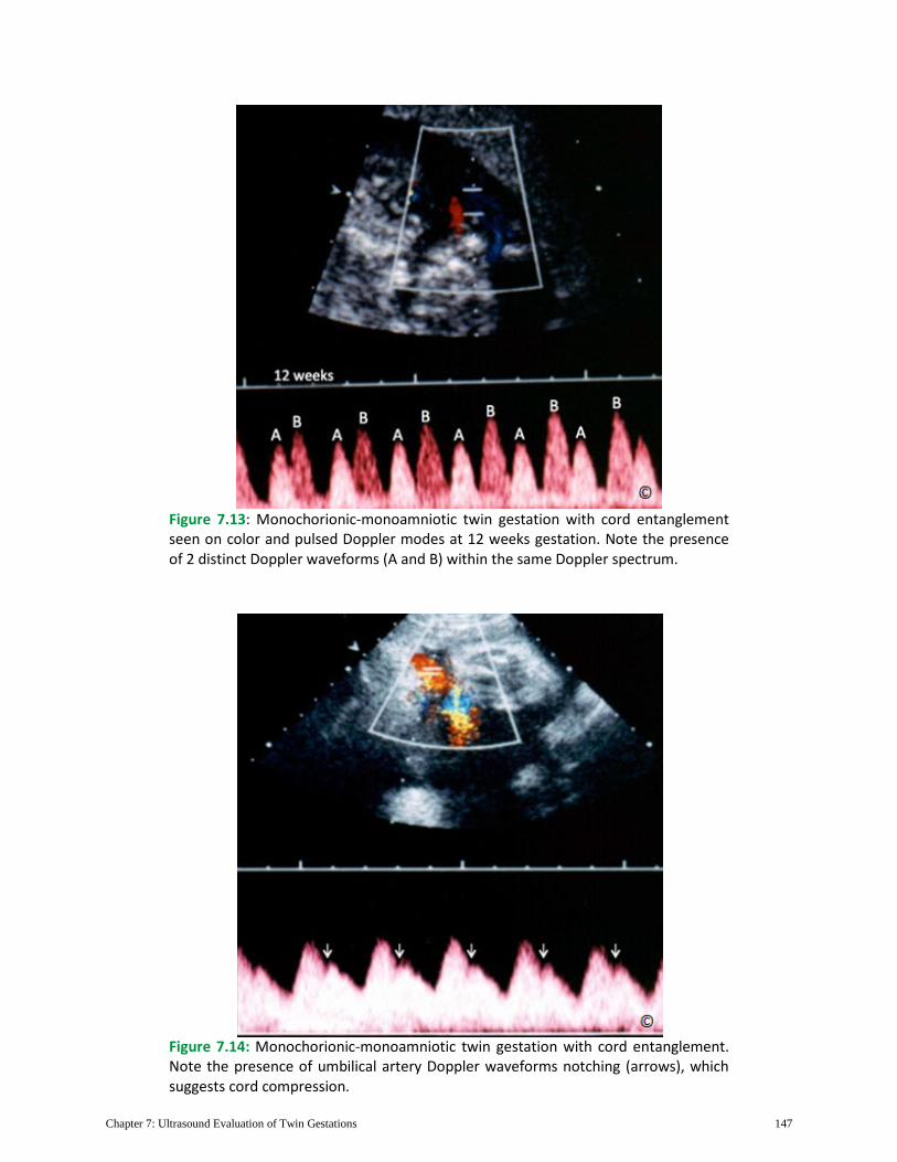
On grey scale, cord entanglement appears as a mass of cord between the two fetuses (Figure 7.10). Color Doppler will confirm that this mass is indeed entanglement of umbilical cords (Figure 7.11) and pulsed Doppler can confirm the diagnosis by documenting two distinct Doppler waveforms, confirming different fetal heart rate patterns (twin A and twin B), on one Doppler spectrum (Figure 7.12). In order to obtain these waveforms, open the Doppler gate wide, within the suspected cord entanglement region (Figure 7.12). Cord entanglement can be noted in the first trimester in monoamniotic twins and confirmed by pulsed Doppler (Figure 7.13). Most authorities suggest delivery by 34-35 weeks when cord entanglement is prenatally diagnosed in monoamniotic pregnancies and fetal surveillance with non-stress testing on a daily, or multiple times per week, frequency. The authors have correlated the presence of umbilical artery waveform notching on pulsed Doppler evaluation with cord compression (Figure 7.14): a feature that can be useful in the surveillance of monoamniotic twins with cord entanglements (16).
Figure 7.10: Monochorionic-monoamniotic twin gestation with cord entanglement seen on B-mode (grey scale). Note the presence of a mass of cords (arrows) between the 2 fetuses.
Chapter 7: Ultrasound Evaluation of Twin Gestations 146
Figure 7.11: Monochorionic-monoamniotic twin gestation with cord entanglement seen on color Doppler mode (same fetus as in figure 7.10). Note the presence of a “mass of cords” between the 2 fetuses.
Figure 7.12: Monochorionic-monoamniotic twin gestation with cord entanglement seen on color and pulsed Doppler modes. Note the presence of 2 distinct Doppler waveforms (A and B) within the same Doppler spectrum.
Chapter 7: Ultrasound Evaluation of Twin Gestations 147
Figure 7.13: Monochorionic-monoamniotic twin gestation with cord entanglement seen on color and pulsed Doppler modes at 12 weeks gestation. Note the presence of 2 distinct Doppler waveforms (A and B) within the same Doppler spectrum.
Figure 7.14: Monochorionic-monoamniotic twin gestation with cord entanglement. Note the presence of umbilical artery Doppler waveforms notching (arrows), which suggests cord compression.
Chapter 7: Ultrasound Evaluation of Twin Gestations 148
CONJOINED TWINS
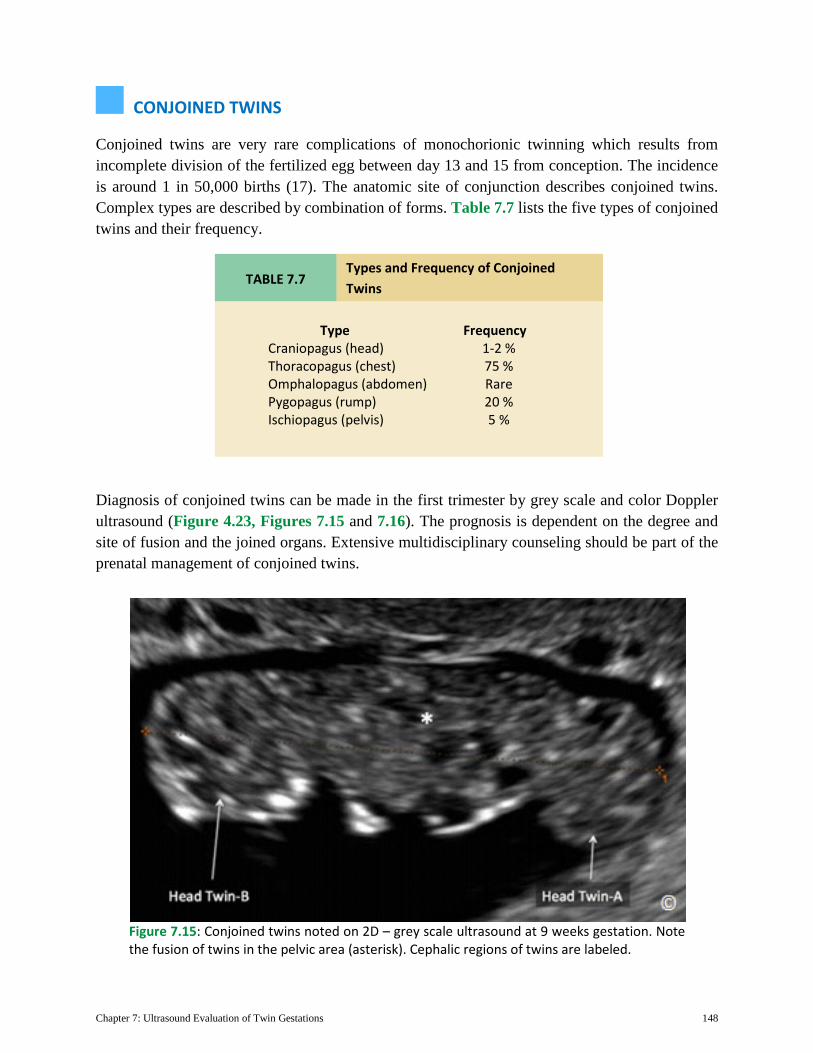
Conjoined twins are very rare complications of monochorionic twinning which results from incomplete division of the fertilized egg between day 13 and 15 from conception. The incidence is around 1 in 50,000 births (17). The anatomic site of conjunction describes conjoined twins. Complex types are described by combination of forms. Table 7.7 lists the five types of conjoined twins and their frequency.
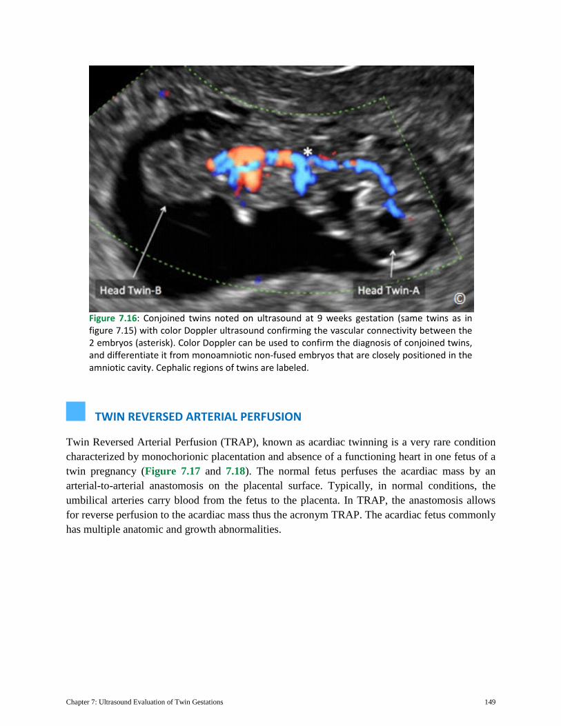
Diagnosis of conjoined twins can be made in the first trimester by grey scale and color Doppler ultrasound (Figure 4.23, Figures 7.15 and 7.16). The prognosis is dependent on the degree and site of fusion and the joined organs. Extensive multidisciplinary counseling should be part of the prenatal management of conjoined twins.
Figure 7.15: Conjoined twins noted on 2D – grey scale ultrasound at 9 weeks gestation. Note the fusion of twins in the pelvic area (asterisk). Cephalic regions of twins are labeled.
TABLE 7.7 Types and Frequency of Conjoined Twins
Type Frequency Craniopagus (head) 1-2 % Thoracopagus (chest) 75 % Omphalopagus (abdomen) Rare Pygopagus (rump) 20 % Ischiopagus (pelvis) 5 %
Chapter 7: Ultrasound Evaluation of Twin Gestations 149
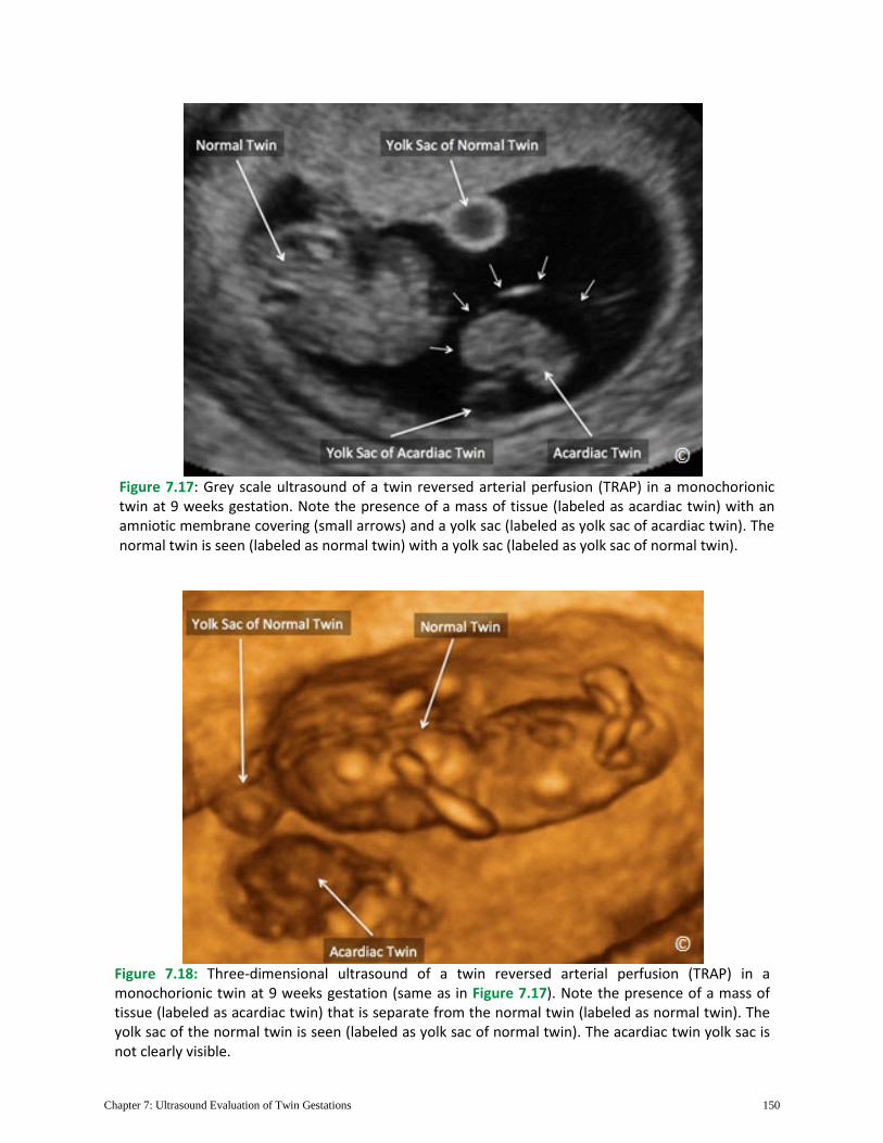
TWIN REVERSED ARTERIAL PERFUSION
Twin Reversed Arterial Perfusion (TRAP), known as acardiac twinning is a very rare condition characterized by monochorionic placentation and absence of a functioning heart in one fetus of a twin pregnancy (Figure 7.17 and 7.18). The normal fetus perfuses the acardiac mass by an arterial-to-arterial anastomosis on the placental surface. Typically, in normal conditions, the umbilical arteries carry blood from the fetus to the placenta. In TRAP, the anastomosis allows for reverse perfusion to the acardiac mass thus the acronym TRAP. The acardiac fetus commonly has multiple anatomic and growth abnormalities.
Figure 7.16: Conjoined twins noted on ultrasound at 9 weeks gestation (same twins as in figure 7.15) with color Doppler ultrasound confirming the vascular connectivity between the 2 embryos (asterisk). Color Doppler can be used to confirm the diagnosis of conjoined twins, and differentiate it from monoamniotic non-fused embryos that are closely positioned in the amniotic cavity. Cephalic regions of twins are labeled.
Chapter 7: Ultrasound Evaluation of Twin Gestations 150
Figure 7.17: Grey scale ultrasound of a twin reversed arterial perfusion (TRAP) in a monochorionic twin at 9 weeks gestation. Note the presence of a mass of tissue (labeled as acardiac twin) with an amniotic membrane covering (small arrows) and a yolk sac (labeled as yolk sac of acardiac twin). The normal twin is seen (labeled as normal twin) with a yolk sac (labeled as yolk sac of normal twin).
Figure 7.18: Three-dimensional ultrasound of a twin reversed arterial perfusion (TRAP) in a monochorionic twin at 9 weeks gestation (same as in Figure 7.17). Note the presence of a mass of tissue (labeled as acardiac twin) that is separate from the normal twin (labeled as normal twin). The yolk sac of the normal twin is seen (labeled as yolk sac of normal twin). The acardiac twin yolk sac is not clearly visible.
Chapter 7: Ultrasound Evaluation of Twin Gestations 151
Given that the normal fetus has to perfuse his/her body and that of the acardiac mass, there is significant increase in cardiac workload and a risk for cardiac failure and hydrops. The overall perinatal mortality of the normal fetus in TRAP syndrome is in the range of 30 – 50 % (18, 19). Frequent echocardiographic evaluation of the normal twin in TRAP syndrome may help recognize cardiovascular stress and help guide management. Management options include expectant management, or cord coagulation of the acardiac twin. Bipolar cord coagulation of the acardiac twin appears to be the most feasible option for cord coagulation and is best performed before 24 weeks’ gestation.
References:
1) Martin JA, Hamilton BE, Sutton PD, Ventura SJ, et al. Births: final data for 2002. Natl Vital Stat Rep 2003; 52(10): 1-102.
2) Jewell SE, Yip R. Increasing trends in plural births in the United States. Obstet Gynecol 1995; 85:229-32.
3) Martin JA, Hamilton BE, Ventura SJ, Osteman JK, et al. Births: final data for 2011. Natl Vital Stat Rep 2013; 62(1): 1-70.
4) Mathews TJ, MacDorman MF. Infant mortality statistics from the 2009 period linked birth/infant death data set. National vital statistics reports; vol 61 no 8. Hyattsville, MD: National Center for Health Statistics. 2013. Available from: http://www.cdc.gov/nchs/data/ nvsr/nvsr61/nvsr61_08.pdf.
5) Nylander PP. The factors that influence twinning rates. Acta Genet Med Gemellol (Roma) 1981;30:189
6) MacGillivray I. Epidemiology of twin pregnancy. Seminars Perinatol 1986; 10:4. 7) Bernirschke K. Multiple pregnancy (First of two parts). N Engl J Med 1973;288:1276 8) Monteagudo A, Timor-Tritsch IE, Sharma S. Early and simple determination of chorionic
and amniotic type in multifetal gestations in the first fourteen weeks by high-frequency transvaginal ultrasonography. Am J Obstet Gynecol 1994; 170(3):824–9.
9) Winn HN, Gabrielli S, Reece EA, et al. Ultrasonographic criteria for the prenatal diagnosis of placental chorionicity in twin gestations. Am J Obstet Gynecol 1989; 161(6 Pt 1):1540–2.)
10) Finberg H. The ‘‘twin peak’’ sign: reliable evidence of dichorionic twining. J Ultrasound Med 1992; 11:571– 7.
11) Reddy UM, Abuhamad AZ, Levine D, Saade GR. Fetal Imaging Executive Summary of a Joint Eunice Kennedy Shriver National Institute of Child Health and Human Development, Society for Maternal-Fetal Medicine, American Institute of Ultrasound in Medicine, American College of Obstetricians and Gynecologists, American College of Radiology, Society for Pediatric Radiology, and Society of Radiologists in Ultrasound Fetal Imaging Workshop. J Ultrasound Med 2014; 33:745–757.

Chapter 7: Ultrasound Evaluation of Twin Gestations 152
12) Society for Maternal-Fetal Medicine, Simpson LL. Twin-twin transfusion syndrome. Am J Obstet Gynecol 2013; 208(1):3-18.
13) American College of Obstetricians and Gynecologists. Multiple gestation: complicated twin, triplet and higher order multifetal pregnancy. ACOG practice bulletin no. 56. Washington, DC: The College; 2004 (reaffirmed 2009).
14) Miller J, Chauhan SP, Abuhamad AZ. Discordant twins, diagnosis, evaluation and management. Am J Obstet Gynecol 2012; FIND NUMBERS.
15) Quintero RA, Morales WJ, Allen MH, et al. Staging of twin-twin transfusion syndrome. J Perinatol 1999; 19(8 Pt 1):550 –5.
16) Abuhamad A, Mari G, Copel JC, Cantwell CJ, Sayed A, Evans AT: Umbilical artery flow velocity waveforms in Monoamniotic Twins with cord enlargement: Can it be used in pregnancy management. Obstet Gynecol 1995; 86:674-7.
17) Malone FD, D’Alton ME. Multiple gestations, clinical characteristics and management. In Creasy RK, Resnik R (eds): Maternal Fetal Medicine, ed 4, Philadelphia, WB Saunders, 2000, p595-615.
18) Moore TR, Gale S, Bernishke K. Perinatal outcome of forty nine pregnancies complicated by acardiac twinning. Am J Obstet Gynecol 1990; 163: 907-912.
19) Healy MG. Acardia: predictive risk factors for the co-twin’s survival. Teratology 1994;50:205-213.