Embed Size (px)
Citation preview
i
ULTRASONIC WOUND DEBRIDEMENT DEVICE
HEALTH TECHNOLOGY ASSESSMENT SECTION
MEDICAL DEVELOPMENT DIVISION
MINISTRY OF HEALTH MALAYSIA
015/2014
ii
DISCLAIMER
Technology review is a brief report, prepared on an urgent basis, which draws on
restricted reviews from analysis of pertinent literature, on expert opinion and / or
regulatory status where appropriate. It has been subjected to an external review
process. While effort has been made to do so, this document may not fully reflect
all scientific research available. Additionally, other relevant scientific findings may
have been reported since completion of this review.
Please contact: [email protected], if you would like further information.
Health Technology Assessment Section (MaHTAS),
Medical Development Division
Ministry of Health Malaysia
Level 4, Block E1, Precinct 1
Government Office Complex
62590 Putrajaya
Tel: 603 88831246
Fax: 603 8883 1230
Available at the following website: http://www.moh.gov.my
iii
Author:
Dr. Syaharatul Patimah Binti Kamarudin
Medical Officer
Senior Assistant Director
Health Technology Assessment Section (MaHTAS)
Medical Development Division
Ministry of Health Malaysia
Reviewed by:
Datin Dr Rugayah Bakri
Public Health Physician
Deputy Director
Health Technology Assessment Section (MaHTAS)
Medical Development Division
Ministry of Health Malaysia
Dr. Izzuna Mudla Mohamed Ghazali
Public Health Physician
Senior Principal Assistant Director
Health Technology Assessment Section (MaHTAS)
Medical Development Division
Ministry of Health Malaysia
External Reviewers:
Dr Harikrishna Ragavan Nair
Head of Wound Care Unit and Internal Medicine Consultant
Department of Medicine
Hospital Kuala Lumpur
Dr Yusniza Mohd Yusof
National Advisor and Rehabilitation Medicine Consultant
Hospital Rehabilitasi Cheras
Kuala Lumpur
DISCLOSURE
The authors of this report have no competing interest in this subject and the
preparation of this report is totally funded by the Ministry of Health, Malaysia.
iv
EXECUTIVE SUMMARY
Introduction
Debridement plays an important role in wound management. It helps to reduce
the bacterial burden within the wound, controls on going inflammation and
malodour, and encourages formation of granulation tissue. The word
debridement derives from the French débridement, which means to remove a
constraint. European Wound Management Association (EMWA) has defined
debridement as the act of removing necrotic material, eschar, devitalised tissue,
serocrusts, infected tissue, hyperkeratosis, slough, pus, haematomas, foreign
bodies, debris, bone fragments or any other type of bioburden from a wound with
the objective to promote wound healing.
Low frequency ultrasound is claimed to provide a debridement alternative to, for
example, surgical debridement. However, it is more commonly used for
therapeutic purposes. Ultrasonic waves are also claimed to lead to destruction of
bacteria and disruption of biofilms.
It has been utilised as a wound debridement and cleansing technique for years in
the United Kingdom, Russia and Germany. While in Malaysia, the first ultrasonic
wound debrider was first launched in August 2012 in collaboration with Malaysian
Society of Wound Care Professional (MSWCP) and Malaysian Enterostomy
Therapy Nurses Association (METNA).
This technology review was conducted to assess the use of the new technology
ultrasonic wound debridement device as a treatment option for wound
debridement focusing on using low frequency high intensity contact ultrasound as
requested by Head of Department and Rehabilitation Medicine Specialist from
Hospital Sungai Buloh.
Objective/aim
The objective of this systematic review was to assess the safety, effectiveness
and cost-effectiveness of ultrasonic wound debridement device using low-
frequency high intensity contact ultrasound for wound debridement.
v
Results and conclusions
Based on the above review:
Safety
There was limited evidence retrieved to show that this device was not associated
with major complications. However, mild pain was one of the reported adverse
events.
Effectiveness
Low frequency high intensity contact ultrasound debridement device or ultrasonic
wound debridement device seemed to have potential benefit as an adjunct to
standard treatment for chronic wounds such as diabetic foot ulcers, venous
ulcers and pressure ulcers. However, there was lack of good quality evidence.
Hence, more quality evidence is required.
Cost / cost effectiveness
There was no retrievable evidence on the cost-effectiveness. The cost of the
device was estimated to be around RM 75 000 to RM 160 000.
Methods
Electronic databases were searched through the Ovid interface: Ovid MEDLINE®
In-process and other Non-indexed citations and Ovid MEDLINE® 1946 to
present, EBM Reviews - Cochrane Central Register of Controlled Trials - June
2014, EBM Reviews - Cochrane Database of Systematic Reviews - 2005 to June
2014, EBM Reviews - Health Technology Assessment – 2nd Quarter 2014, EBM
Reviews - Database of Abstracts of Reviews of Effects - 2nd Quarter 2014, EBM
Reviews – NHS Economic Evaluation Database 2nd Quarter 2014, Embase –
1988 to 2014 Week 29. Searches were also run in PubMed. Google was used to
search for additional web-based materials and information. Limits for human
study and English full article were applied. Additional articles were identified from
reviewing the references of retrieved articles and contacting manufacturers via
email to obtain references in their website. Unpublished articles were attempted
to retrieve by contacting corresponding author by email. Last search was
conducted on 5 August 2014.
1
ULTRASONIC WOUND DEBRIDEMENT DEVICE
1. INTRODUCTION
Debridement plays an important role in wound management. It helps to reduce
the bacterial burden within the wound, controls on going inflammation and
malodour, and encourages formation of granulation tissue.1 The word
debridement derives from the French débridement, which means to remove a
constraint. European Wound Management Association (EMWA) has defined
debridement as the act of removing necrotic material, eschar, devitalised tissue,
serocrusts, infected tissue, hyperkeratosis, slough, pus, haematomas, foreign
bodies, debris, bone fragments or any other type of bioburden from a wound with
the objective to promote wound healing.2
Strohal et al. has summarized the primary targets for debridement which include
removal of bioburden i.e slough, necrotic tissues etc., decreasing odour, excess
moisture and risk of infection, stimulate wound edges and epithelialisation as well
as improving quality of life.2
Various methods of debridement existed today require varying level of expertise
and have their advantages and disadvantages in term of time taken, patient
acceptability and ease of use. Sharp debridement is very quick, using scalpel
and is the current standard for wound debridement. Autolytic debridement is
often slow and unpredictable process uses the body’s own enzymes and
moisture to rehydrate, soften and finally liquefy hard eschar and slough.
Enzymatic debridement utilises chemical enzymes, fast acting products that
produce slough of necrotic tissue. Mechanical debridement using wet to dry
technique. Other techniques of debridement includes laser debridement which
appear to be efficient and precise when utilised in tissue ablation, however carry
the risk of thermal damage to healthy tissue. Maggot therapy utilises maggot to
ingest and break down necrotic tissue. Water debridement utilises a high
pressure water jet.3 Meanwhile, debridement using low frequency ultrasound will
be covered in this review.
Low frequency ultrasound is claimed to provide a debridement alternative to, for
example, surgical debridement.2 It incorporates a probe to selectively excise
nonviable or necrotic tissue and can be used in a variety of setting by trained
personnel.4 Ultrasonic waves are also claimed to lead to destruction of bacteria
and disruption of biofilms.2
2
Ultrasound Assisted Wound Therapy has been utilised as a wound debridement
and cleansing technique for years in the United Kingdom, Russia and Germany.
While in Malaysia, the first ultrasonic wound debrider was first launched in
August 2012 in collaboration with Malaysian Society of Wound Care Professional
(MSWCP) and Malaysian Enterostomy Therapy Nurses Association (METNA) in
which Dr. Harikrishna, the Head of Wound Care Unit Hospital Kuala Lumpur
described the device as one of the great modalities to be held by nurses since it
gives a faster result and portable.5
This technology review was conducted to assess the use of the new technology
ultrasonic wound debridement device as a treatment option for wound
debridement focusing on using low frequency high intensity contact ultrasound
following a request by Head of Department and Rehabilitation Medicine
Specialist from Hospital Sungai Buloh.
2. OBJECTIVE/AIM
The objective of this systematic review was to assess the safety, effectiveness
and cost-effectiveness of ultrasonic wound debridement device using low-
frequency high intensity contact ultrasound for wound debridement.
3. TECHNICAL FEATURES
Therapeutic ultrasound delivers energy through mechanical vibrations in the form
of sound waves at frequencies above detection by human ear (>20 kHz).
Ultrasound is commonly associated with diagnostic imaging which utilised high
frequency ultrasound waves and as well used in physical therapy, physical
medicine, rehabilitation and sports medicine for many years.4
There are mainly two classified effects of ultrasound on tissue: thermal and non-
thermal. Both these effects are inseparable but their respective proportions vary
with the frequency and intensity of ultrasound. Thermal effects are predominant
with high frequency (MHz) and intensity (W/cm2) ultrasound, which raises tissue
temperature and possibly enhances blood flow. Low frequency ultrasound (kHz)
has predominantly mechanical (non-thermal) effects, namely cavitation and
acoustic streaming, although there are some thermal effects on tissue.1
Low frequency ultrasound can be high intensity (~50W/cm2) delivered with direct
contact with wound or low intensity (0.25-0.75W/cm2) delivered without direct
contact with wound bed; both are used with saline as coupling media between
3
the ultrasound probe and wound bed. High intensity ultrasound debrides necrotic
tissue possibly because of the cavitational effect produced by rapid expansion
and implosion of gas bubbles within tissue fluid or coupling media. Whereas low
intensity ultrasound may promote wound healing predominantly by acoustic
streaming effects such as increased protein synthesis and production of growth
factors. In addition, low frequency ultrasound has been reported to have anti-
bacterial effects and enhanced fibrinolysis in vitro.1
Hence, this technology review focuses on ultrasonic wound debridement devices
using low frequency high intensity contact ultrasound.
3.1 Mechanism of action
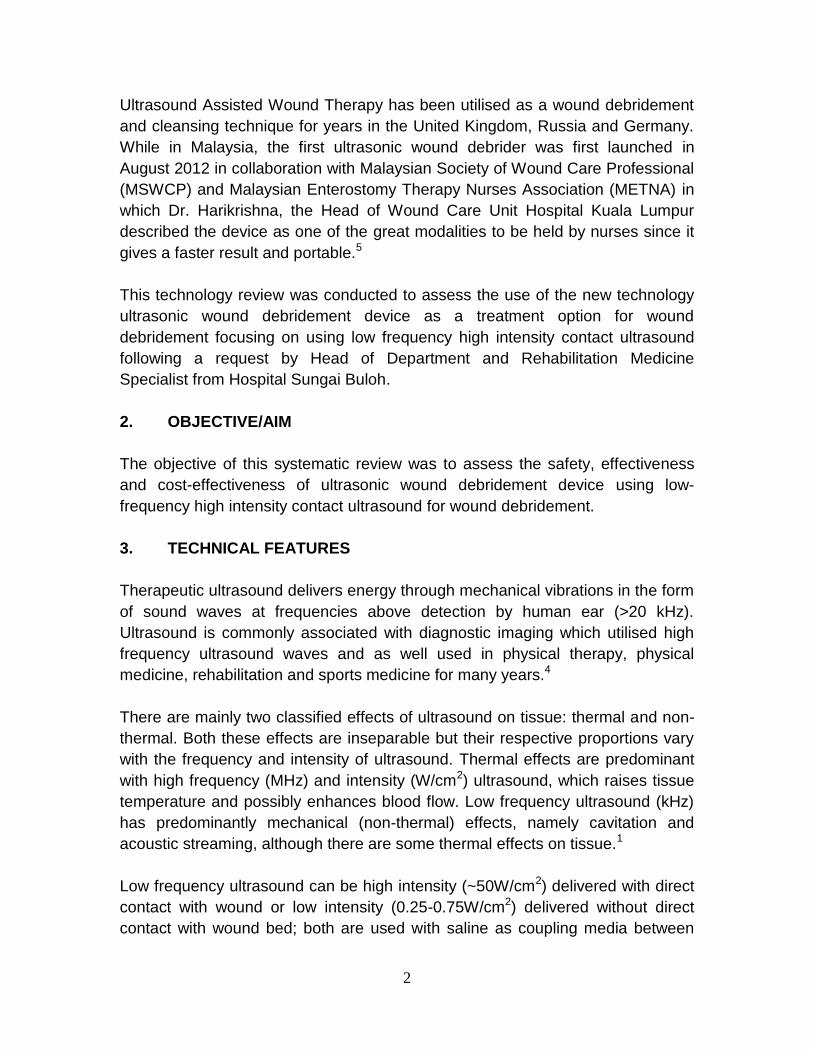
Low frequency ultrasound provides two largely non thermal effects, which are
cavitation and acoustic streaming. The cavitation phenomenon may be described
as the creation of miniscule gas bubbles in tissue fluid and the expansion and
contraction in size of these bubbles in tandem with the variation in the ultrasound
field pressure levels. At certain amplitudes of the sound waves, the bubbles
implode; this implosion results in the formation of tiny shock waves. Because
necrotic tissue has less tensile strength than viable tissue, these locally
generated shock waves in turn liquefy the necrotic tissue, other wound debris,
and associated biofilm, while not injuring viable tissue. The acoustic streaming
initiates a unidirectional movement in fluid in an ultrasound field. This activity
stimulates cell activity and enhances clinical outcome.4
4
Figure 1: The mechanism of action of ultrasonic wound debridement device
A possible alternative mechanism of action, called frequency resonance, is
related to the modification in the structure of proteins and the activation of signal
transduction at nuclear level. This can lead to a range of effects at cellular level
that impact wound healing, such as leucocyte adhesion, increased angiogenesis
and increase of nitric oxide (NO production).2
In SonicOne O.R System for instance is using low frequency (22.5 kHz) high
intensity (~60 W/cm2) ultrasound which has been claimed to be able to disrupt,
inactivate and remove bacterial organisms from surfaces and stimulate the
body’s self-healing ability, resulting in faster healing rates and increased closure
rates. The generator converts standard wall voltage to a 22.5 kHz electrical
signal which is then transferred, via a cable assembly to the hand piece. The
hand piece contains piezoelectric crystals that convert the electrical signal to
mechanical vibrations. The titanium alloy probe, attached to the hand piece distal
end, amplifies the mechanical vibration and then transfers the acoustic energy
into the tissue via direct contact. The resulting cavitation, mechanical and
hydrodynamic effects produce tissue disruption, excision, fragmentation and
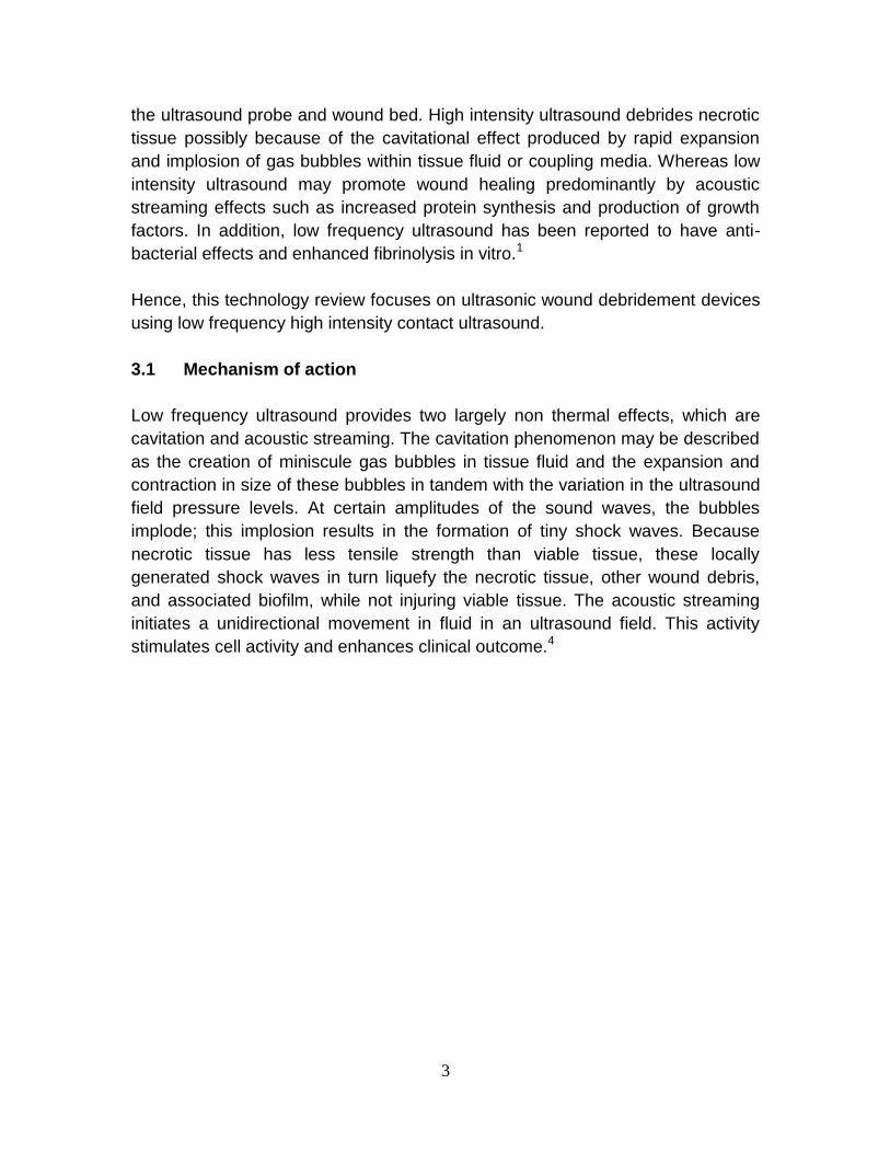
emulsion in the wound bed. The ultrasonic movement of the probe allows the
5
surgeon to remove tissue in a controlled and precise manner, reducing the
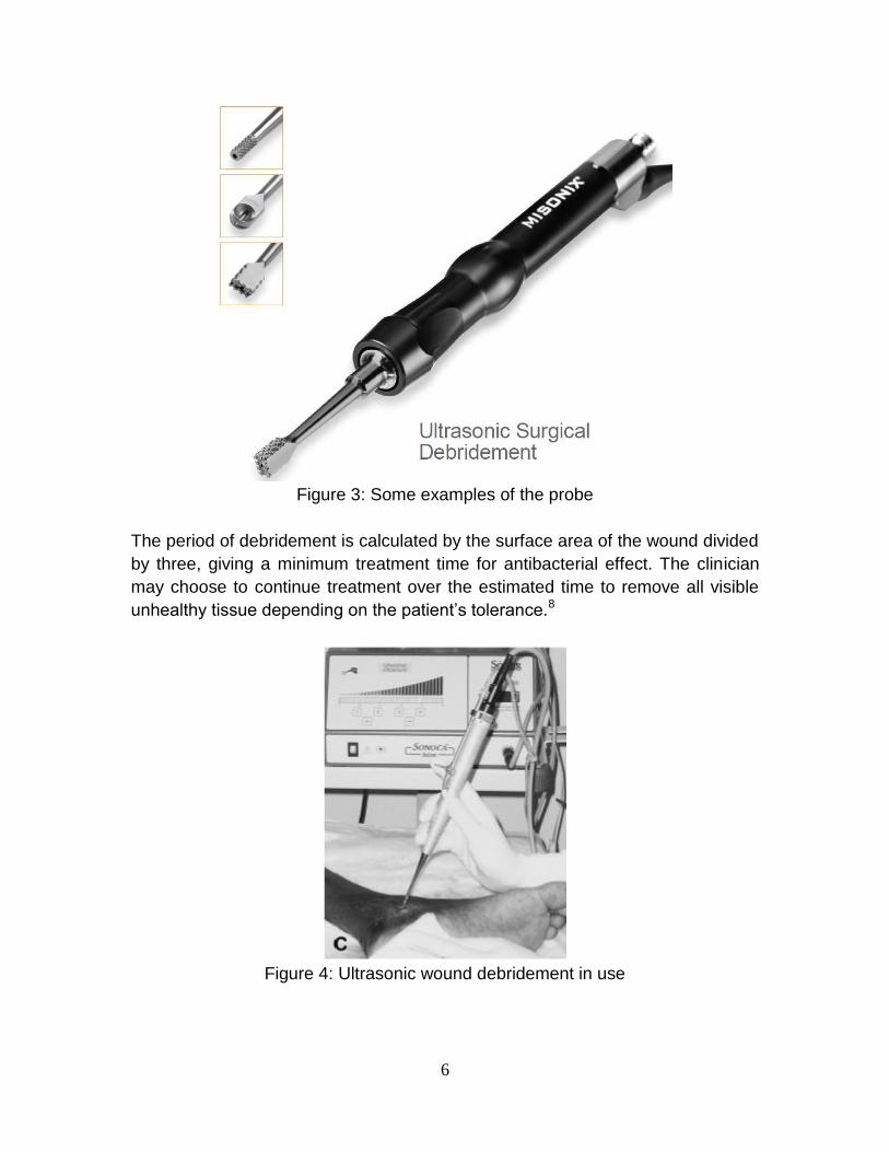
bleeding typically associated with surgical debridement.6 The varieties of probes
that are used are designed to concentrate (sharper tips) or disperse (blunter tips)
the vibratory energy resulting either in differing aggressiveness of the dissection
or fragmentation of the soft tissues.7
Figure 2: An example of ultrasonic wound debridement device
6
Figure 3: Some examples of the probe
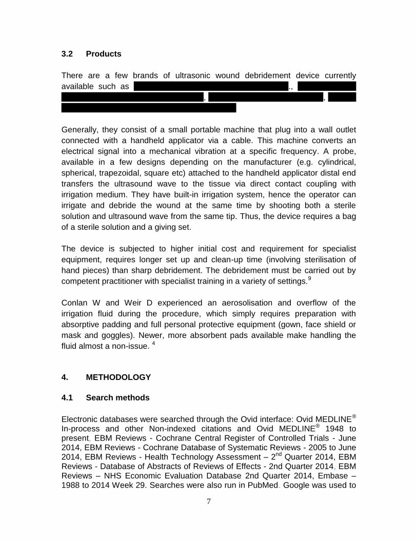
The period of debridement is calculated by the surface area of the wound divided
by three, giving a minimum treatment time for antibacterial effect. The clinician
may choose to continue treatment over the estimated time to remove all visible
unhealthy tissue depending on the patient’s tolerance.8
Figure 4: Ultrasonic wound debridement in use
7
3.2 Products
There are a few brands of ultrasonic wound debridement device currently
available such as SonicOne® O.R. System by Misonix Inc., Qoustic Wound
Therapy SystemTM by Arobella Medical, Sonoca-185® by Soering Gmbh, Debriflo
as well as Ultraclean by InnoSound Technology.
Generally, they consist of a small portable machine that plug into a wall outlet
connected with a handheld applicator via a cable. This machine converts an
electrical signal into a mechanical vibration at a specific frequency. A probe,
available in a few designs depending on the manufacturer (e.g. cylindrical,
spherical, trapezoidal, square etc) attached to the handheld applicator distal end
transfers the ultrasound wave to the tissue via direct contact coupling with
irrigation medium. They have built-in irrigation system, hence the operator can
irrigate and debride the wound at the same time by shooting both a sterile
solution and ultrasound wave from the same tip. Thus, the device requires a bag
of a sterile solution and a giving set.
The device is subjected to higher initial cost and requirement for specialist
equipment, requires longer set up and clean-up time (involving sterilisation of
hand pieces) than sharp debridement. The debridement must be carried out by
competent practitioner with specialist training in a variety of settings.9
Conlan W and Weir D experienced an aerosolisation and overflow of the
irrigation fluid during the procedure, which simply requires preparation with
absorptive padding and full personal protective equipment (gown, face shield or
mask and goggles). Newer, more absorbent pads available make handling the
fluid almost a non-issue. 4
4. METHODOLOGY
4.1 Search methods
Electronic databases were searched through the Ovid interface: Ovid MEDLINE®
In-process and other Non-indexed citations and Ovid MEDLINE® 1948 to present, EBM Reviews - Cochrane Central Register of Controlled Trials - June 2014, EBM Reviews - Cochrane Database of Systematic Reviews - 2005 to June 2014, EBM Reviews - Health Technology Assessment – 2nd Quarter 2014, EBM Reviews - Database of Abstracts of Reviews of Effects - 2nd Quarter 2014, EBM Reviews – NHS Economic Evaluation Database 2nd Quarter 2014, Embase – 1988 to 2014 Week 29. Searches were also run in PubMed. Google was used to

8
search for additional web-based materials and information. Limits for human study and English full article were applied. Additional articles were identified from reviewing the references of retrieved articles and contacting manufacturers via email to obtain references in their website. Unpublished articles were attempted to retrieve by contacting corresponding author by email. Last search was conducted on 5 August 2014. Appendix 1 showed the detailed search strategies.
4.2 Selection
A reviewer screened the titles and abstracts against the inclusion and exclusion
criteria and then evaluated the selected full text articles for final article selection.
The inclusion and exclusion criteria were: Inclusion criteria
Population Patients with wound (chronic wound, diabetic ulcer, pressure ulcer, varicose ulcer)
Interventions Ultrasonic wound debridement device (low-frequency, high intensity, contact ultrasound) for wound debridement
Comparators Conventional wound debridement methods / no comparator
Outcomes i. Safety (adverse events/complications) ii. Effectiveness iii. Economic implication (cost, cost-effectiveness)
Study design Health Technology Assessment (HTA), Systematic Review, Randomised Controlled Trial (RCT), Non Randomised Controlled Trial, Cohort studies, Case Control studies, Cross sectional studies, case series, case reports
English full text articles
Exclusion criteria
Study design
Studies conducted in animals and narrative reviews
Non English full text articles
Relevant articles were critically appraised using Critical Appraisal Skills Programme (CASP) and graded according to US/Canadian preventive services task force (Appendix 2). Data were extracted and summarised in evidence table as in Appendix 3.
9
5. RESULTS
From the search, 137 articles were retrieved. Only nine studies were relevant.
However, only four full text articles were taken as references since they were of
high level of evidence (Appendix 3). These articles include a systematic review
with meta-analysis, a randomised controlled trial and two pre and post
intervention studies. Five case reports were excluded as they have no
comparison and have high risk of bias. The evidence was graded according to
the US/Canadian Preventive Services Task Force (Appendix 2).
5.1 Safety
Pain and bleeding at the wound site were of concerned adverse events
associated with contact ultrasonic debridement device during the procedure. In a
RCT by Herberger K et al., 67 patients with chronic leg ulcers of vascular origin
were randomised into two groups in which 34 patients were treated with
ultrasound assisted wound treatment (UAW) and 33 patients were treated with
surgical wound debridement (WD) in Wound Centre of the University Clinics of
Hamburg. Pain was assessed before, during and 15 minutes after the procedure
measured using a visual analogue scale. Before each treatment, a topical
anaesthetic consisting of lidocaine and prilocaine (EMLA cream) was applied
occlusively to the wound for 60 minutes. The increase in pain during treatment
was not significantly different between the two treatment arms, and lay between
1.1 and 1.7 points for WD and between 1.5 and 1.6 points for UAW.10,Level II-2
Tan J et al. conducted a pre and post intervention study in St Thomas’ Hospital,
London, United Kingdom involved 19 patients with leg ulceration of at least six
months present. Low frequency ultrasound was delivered via a handheld probe.
No analgesia was used in the study. Only three of the 19 patients experienced
mild negligible pain at the start of the treatment. Two of them complained of pain
at the periphery of the ulcer during the treatment and none developed pain that
was severe enough to necessitate interruption or abandonment of the treatment
prematurely. There was a noticeable reduction in pain in subsequent treatments
and by the third treatment pain was no longer reported in any of the patients.
Some patients experienced mild venous oozing from the ulcer surface which
ceased spontaneously within a few minutes. There were no major complications
of treatment. 11,Level I
10
The Food and Drug Administration (USFDA) classified ultrasonic wound
debridement device as a class II device and it complies with USFDA Voluntary
Standards for safety. 12-13 Sonoca-180 by Soering has CE marking.14
5.2 Effectiveness
In a systematic review and meta-analysis done by Voigt et al. in 2011, they
included eight randomised controlled trials (RCTs) which investigated whether
the use of low frequency high intensity contact ultrasound (LFHICU) or low
frequency low intensity noncontact ultrasound (LFLINCU) as an adjunctive
therapy improves the outcome of complete healing of chronic lower limb wounds.
Methodologically, three RCTs included were using LFHICU for intervention
therapy. However, two RCTs used for meta-analysis and two other RCTs were
reviewed individually as they could not be combined for meta-analysis purposes
based on duration of outcome evaluation and lack of specific outcome data.15,Level
I
The primary outcome measure was complete wound healing. However, the
definition of complete wound healing was unclear. The meta-analysis showed a
statistical difference favouring debridement using LFHICU when compared with
sharp debridement (RR=0.64; 95% CI=0.46-0.89; P=0.009; I2=0%). At five
months, this statistical difference persisted, favouring LFHICU (RR=0.52; 95%
CI=0.32-0.85; P=0.008; I2=0%). However, at six months this statistical difference
did not persist (RR=0.66; 95% CI=0.36-1.21; P=0.18; I2=15%). The patients
included were diabetic foot ulcers with osteomyelitis and venous stasis ulcers.
15,Level I
The secondary outcome measure was wound size reduction which included
those patients with diabetic foot ulcers and lower extremity ulcers of various
aetiologies (venous insufficiency, diabetes, pressure, and arterial insufficiency).
Over three months, there was a statistically significant difference in percentage of
wound size reduction between LFHICU and sharps debridement, favouring
LFHICU (mean difference=25.93%; 95% CI=14.2%-37.66%; P<0.0001;
I2=0%).15,Level I
Two of the trials (Li and Singh) could not be combined for meta-analyses
purposes due to differences in the duration of outcome evaluation and lack of
specific outcome data. In the study reported by Li, the percentage of the wound
that had healed over a two week period was significantly higher with LFHICU
versus a saline wash (P=0.006). In a trial by Singh A., it was found that the
11
percentage of the wound that had healed over a two week period was
significantly higher with LFHICU versus a sharp debridement (P=0.001).15,Level I
Voigt et al. concluded that although the quality of the evidence was in general of
lower quality, the evidence does demonstrated a short term clinical beneficial
effect of LFHICU used as an adjunctive therapy on the clinical end points of
complete healing and reduction in wound area size for patients with venous
stasis and diabetic foot ulcers (Wagner 1-3) (see Appendix 4). There may be
longer term completing healing effect (at 6 months) of LFHICU in patients with
venous stasis ulcer.15, Level I
In a RCT by Herberger K et al., 67 patients with chronic leg ulcers of vascular
origin were randomised into two groups in which 34 patients were treated with
ultrasound assisted wound treatment (UAW) and 33 patients were treated with
surgical wound debridement (WD) in Wound Centre of the University Clinics of
Hamburg. The objective was to determine and compare the efficacy, tolerability
and benefit of both wound treatment methods.10,Level II-2
The improvement of the wound status was highly significant (p<0.001) in both
groups in reduction of amount of necrosis and fibrin coatings and an increase in
granulation tissue. However, in epithelisation there was no significant change in
both groups (p=0.267).10,Level II-2
Patients considered both procedures to be equally effective and tolerable. For
UAW, the Patient Benefit Index (wound version) was >1 in 88% of patients, the
mean score being 2.2±1.0 (evaluation without last observation carried forward
(LOCF)). For WD, the score was >1 in 85.1% of patients. The mean score was
2.1±1.1. The differences between the two treatment arms were not significant.
The patients perceived particularly strong benefits in the items ‘to have
confidence in therapy’ with a mean benefit score of 3.2 (both groups), ‘to have a
clean wound’ with 3.4 (both groups) and ‘to receive low-pain treatment’ with 3.0
(UAW) and 3.2 (WD). The patients reported comparatively small benefits for the
item ‘to be able to lead a normal working life’ in both treatment arms, given their
mean age 74.5 years (UAW) and 70.5 years (WD).10,Level II-2
The efficacy was assessed good by patients and practitioners, the mean scores
being 4.2 and 4.3 respectively. There were no differences between the means for
the assessment of the efficacy of UAW and WD. Tolerability was assessed by
the practitioner as slightly better for UAW (2.2 versus 2.1). Overall, both
procedures were assessed as being equally well tolerated.10,Level II-2
12
The global quality of life score increased for all patients over the course of the
treatment (p=0.001). The improvement in quality of life did not differ significantly
between the two groups.10,Level II-2
In a pilot study, Tan J et al. evaluated the use of low frequency ultrasound device
to debride chronic leg ulcers as an adjunct to compression bandages therapy in
19 patients. The leg ulcers were present for more than six months and had failed
to respond to standard compression regimens. Each patient underwent a
minimum of five treatments averaging 9.7 minutes of treatment per session per
ulcer at an interval of two to three weeks. Over half of patients (55%) showed no
visible changes in the ulcer area (ulcers remained static) during the treatment
period and their ulcers remained static. Seven patients (39%) achieved complete
ulcer healing (mean initial ulcer size=4.72±SD 1.872cm2) in the subsequent
mean follow up period of 16 weeks (range 12-24 weeks). These consisted of one
patient with rheumatoid ulcer, two patients with sickle cell ulcers and four patients
with chronic venous ulcers. All healed patients showed a response within the first
five sessions of treatments compared with the ‘non responders’ and remained
successfully healed for more than six months.11,Level II-3
At the end of the 16 week follow up period, one additional patient with an
unhealed ulcer continued to show a steady reduction in ulcer size and eventually
healed 21 weeks into the follow up period. An interesting observation from the
study was that if no improvement of healing had occurred after the fifth treatment,
no additional benefit was gained by continuing treatment.11,Level II-3
Seven of the 18 patients also reported a significant reduction in wound odour.
There was no alteration in skin temperature experienced around the ulcer during
or following treatment. The authors concluded that the beneficial effects
observed may not be related to the ultrasound effects, but as a result of an
increased effort to improve the general condition by simple wound cleaning.11,Level
II-3
Low frequency ultrasonic debridement (LFUD) has had an early favourable
experience in Brigham and Women’s Hospital, Boston. Breuing KH et al did a
study in 17 patients with acute and chronic wounds of varying aetiologies with a
total of 107 procedures done using low frequency ultrasonic debridement
(LFUD). The follow up period was three to eight months with the frequency of
debridement ranged from twice weekly to every third week, depending on the
type and condition of the wound. The average number of treatment per wound
ranged from six (small pressure ulcers) to 15 (venous stasis ulcers). Adjunct
13
wound therapy used were moist saline dressing, alginate and Panafil. Nine of
the wounds (53%) healed primarily or with the aid of a skin graft. Six additional
patients (35%) experienced wound size reduction of at least 50%. The remaining
two patients (12%), one with sickle cells anaemia and one with a venous stasis
ulcer had reductions in wound area of 20% to 30%. Among all 17 wounds
followed, pressure ulcers, arterial insufficiency and non-healing surgical wounds
responded better than venous stasis and diabetic foot ulcers. None of the
patients required initiation of antibiotic treatment after starting LFUD.16,Level II-3
There was no evidence retrieved to claim for its antimicrobial effect and bacterial
biofilm disruption.
5.3 Cost/Cost-Effectiveness
There was no retrievable evidence on cost effectiveness of the device.
Nonetheless, Butcher G and Pinnuck L estimated that overall cost per treatment
including staff time, related consumables and dressings is about £118 (RM 620)
either performed in the ward or in an outpatient setting in the Australian health
service. Ongoing costs are minimal as the hand pieces can be sterilised and
reused and ultrasound assisted wound debridement related consumables
(tubing, saline, drapes, protective equipment and topical anaesthetic products)
only add about £20 (RM105) to the cost of wound treatment.17
Head of Department and Rehabilitation Medicine specialist from Hospital Sg
Buloh and Head of Wound Care Unit from Hospital Kuala Lumpur stated that the
device costs around RM 75 000 to RM 160 000.
5.4 Limitations
This technology review has several limitations. The selection of studies was done
by a reviewer and only English full text articles were included in this report.
14
6. CONCLUSIONS
Based on the above review:
6.1 Safety
There was limited evidence retrieved to show that this device was not associated
with major complications. However, mild pain was one of the reported mild
adverse events.
6.2 Effectiveness
Low frequency high intensity contact ultrasound debridement device or ultrasonic
wound debridement device seemed to have potential benefit as an adjunct to
standard treatment for chronic wounds such as diabetic foot ulcers, venous
ulcers and pressure ulcers. However, there was lack of good quality evidence.
Hence, more quality evidence is required.
6.3 Cost/Cost Effectiveness
There was no retrievable evidence on the cost-effectiveness. The cost of the
device was estimated to be around RM 75 000 to RM 160 000.
15
8. REFERENCES
1. Madhok BM, Vowden K, Vowden P. New Techniques for Wound
Debridement. International Wound Journal. 2013;10:247-251
2. Strohal R, Apelqvist J, Dissemond J et al. EMWA Document: Debridement.
J Wound Care. 2013;22(Supp.1):S1-S52.
3. Lee I, Low Frequency Ultrasound Debridement. Prioritising Summary.
Horizon Scaning Technology. Health Policy Advisory Committee on
Technology. 2007. Available at http://www.horizonscanning.gov.au
4. Conlan W, Weir D. Ultrasound Assisted Wound Therapy: An Exceptional
Adjunct to Wound Bed Preparation.Today’s Wound Clinic. 2008;2(3)
5. The 1st Ultrasound Wound Debrider. Available at
http://www.mswcp.org/events/the-1st-ultrasound-wound-debrider. Accessed
on 18 July 2014.
6. Misonix, Ultrasound Surgical Debridement. Available at www.misonix.com.
Accessed on 18 July 2014.
7. Alvarez OM, DeGroat K, Markowitz L et al. Effective and Long-Lasting
Debridement of Venous Ulcers with Low Frequency, High Intensity Contact
Ultrasound. Wound Supplement–Spring 2013. Available at
http://pubs.royle.com/article/Effective+And+LongLasting+Debridement+Of+
Venous+Ulcers+With+Low+Frequency,+High+Intensity+Contact+Ultrasoun
d/1380183/0/article.html Accessed on 25 July 2014
8. Shannon MK, Williams A, Bloomer M. Low-frequency Ultrasound
Debridement (Sonoca-185) in Acute Wound Management: A Case Study.
Wound Practice and Research. 2012;20(4):200-205
9. Vowden K, Vowden P. Debridement Made Easy. Wounds UK. Nov
2011;7(4):1-4.
10. Herberger K, Franzke N, Blome C et al. Efficacy, Tolerability and Patient
Benefit of Ultrasound-Assisted Wound Treatment versus Surgical
Debridement: A Randomised Clinical Study, Dermatology, April
2011;222:244-249
16
11. Tan J, Abisi S, Smith A et al. A Painless Method of Ultrasonically Assisted
Debridement of Chronic Leg Ulcers: A Pilot Study. Eur J Vasc Endovasc
Surg. 2007;33:234-238
12. Exhibit E 510(k) Summary. Misonix SonicOne Plus Ultrasonic Wound Care
System and Accessories. USFDA. Available at
www.accessdata.fda.gov/cdrh_docs/pdf12/k123980.pdf. Accessed on 5
August 2014
13. 510(k) Summary (K131096). USFDA. Available at
www.accessdata.fda.gov/cdrh_docs/pdf13/k131096.pdf. Accesed on 5
August 2014
14. Available at kapsnovi.com/en/media/djcatalog/Brochure_Sonoca_180.pdf.
Accessed on 18 July 2014.
15. Voigt J, Wendelken M, Driver V et al. Low-Frequency Ultrasound (20-40
kHz) as an Adjunctive Therapy for Chronic Wound Healing: A Systematic
Review of the Literature and Meta-Analysis of Eight Randomized Controlled
Trials, The International Wound Journal of Lower Extremity Wounds,
2011;10:190-199
16. Breuing KH, Bayer L, Neuwalder J et al. Early Experience Using Low-
Frequency Ultrasound in Chronic Wounds. Annals of Plastic Surgery.
August 2005; 55(2):183-187
17. Butcher G & Pinnuck L. Wound Bed Preparation: Ultrasonic-Assisted
Debridement. British Journal of Nursing (Tissue Viability Supplement)
2013;22(6):S36-S43
17
9. APPENDIX
9.1 Appendix 1
LITERATURE SEARCH STRATEGY
Ovid MEDLINE® In-process & other Non-Indexed citations and
OvidMEDLINE® 1946 to present
Search Strategy:
------------------------------------------------------------------------------------------------------------
1. Wound.tw.
2. Chronic wound$.tw.
3. Wound infection/
4. (wound adj1 infection$).tw.
5. Pressure ulcer/
6. (pressure adj1 sore$).tw.
7. (bed adj1 sore$).tw.
8. bedsore$.tw.
9. (pressure adj1 ulcer$).tw.
10. (decubitus adj ulcer$).tw.
11. Leg ulcer/
12. (leg adj1 ulcer$).tw.
13. Pyoderma gangrenosum/
14. Pyoderma gangrenosum.tw.
15. Diabetic foot/
16. ((foot or feet) adj1 diabetic).tw.
17. Foot ulcer diabetic.tw.
18. Diabetic foot ulcer$.tw.
19. Diabetic ulcer$.tw.
20. Foot ulcer/
21. (foot adj1 ulcer$).tw.
22. (plantar adj1 ulcer$).tw.
23. Varicose ulcer/
24. Venous stasis ulcer$.tw.
25. Ulcer$ venous stasis.tw.
26. Stasis ulcer$ venous.tw.
27. (venous adj1 ulcer$).tw.
28. (stasis adj1 ulcer$).tw.
29. (varicose adj1 ulcer$).tw.
18
30. (varicose adj1 ulcer$).tw.
31. Arterial ulcer$.tw.
32. Recalcitrant wound$.tw.
33. 1 or 2 or 3 or 4 or 5 or 6 or 7 or 8 or 9 or 10 or 11 or 12 or 13 or 14 or
15 or 16 or 17 or 18 or 19 or 20 or 21 or 22 or 23 or 24 or 25 or 26 or
27 or 28 or 29 or 30 or 31 or 32
34. Ultrasound wound debridement.tw.
35. Ultrasonic wound debridement.tw.
36. Ultrasonic debridement.tw.
37. Ultrasound debridement.tw.
38. Contact low frequency ultrasonic wound debridement.tw.
39. Contact low frequency ultrasound wound debridement.tw.
40. Contact ultrasonic wound debridement.tw.
41. Contact ultrasound wound debridement.tw.
42. Contact ultrasonic debridement.tw.
43. Contact ultrasound debridement.tw.
44. Low frequency ultrasound.tw.
45. Low frequency contact ultrasound.tw.
46. Ultrasonic wound debridement therapy.tw.
47. Low frequency high intensity contact ultrasound.tw.
48. High intensity low frequency contact ultrasound.tw.
49. Contact low frequency ultrasound debridement.tw.
50. Low frequency ultrasound debridement.tw.
51. Contact low frequency ultrasound.tw.
52. Low frequency high intensity ultrasound wound debridement.tw.
53. High intensity low frequency ultrasound wound debridement.tw.
54. 34 or 35 or 36 or 37 or 38 or 39 or 40 or 41 or 42 or 43 or 44 or 45 or
46 or 47 or 48 or 49 or 50 or 51 or 52 or 53
55. Hydrostatic wound debridement.tw.
56. Hydrostatic debridement.tw.
57. (hydrosurg$ adj system).tw.
58. Hydrosurg$ debridement.tw.
59. Versajet.tw.
60. Waterjet hydrosurgery system.tw.
61. High power parallel waterjet.tw.
62. High pressure waterjet debridement.tw.
63. 55 or 56 or 57 or 58 or 59 or 60 or 61 or 62 or 63
64. 54 or 63
65. 33 or 64
66. Limit 65 to ( English language and humans)
19
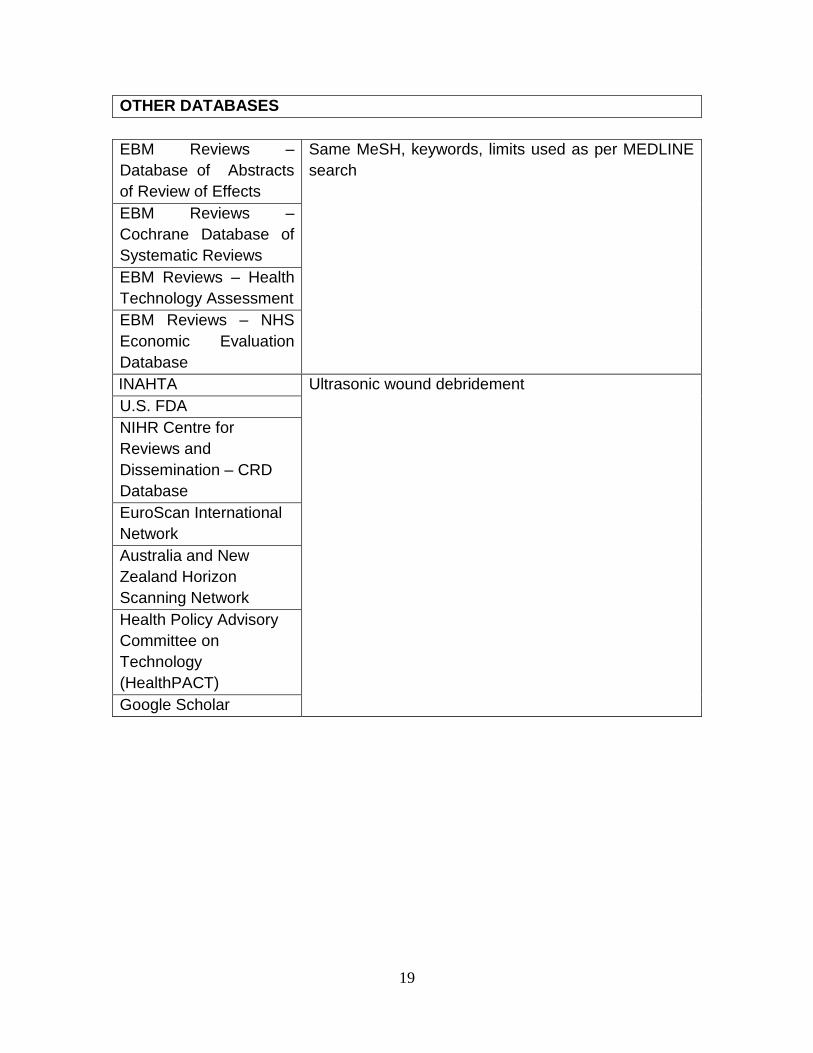
OTHER DATABASES
EBM Reviews –
Database of Abstracts
of Review of Effects
Same MeSH, keywords, limits used as per MEDLINE
search
EBM Reviews –
Cochrane Database of
Systematic Reviews
EBM Reviews – Health
Technology Assessment
EBM Reviews – NHS
Economic Evaluation
Database
INAHTA Ultrasonic wound debridement
U.S. FDA
NIHR Centre for
Reviews and
Dissemination – CRD
Database
EuroScan International
Network
Australia and New
Zealand Horizon
Scanning Network
Health Policy Advisory
Committee on
Technology
(HealthPACT)
Google Scholar
20
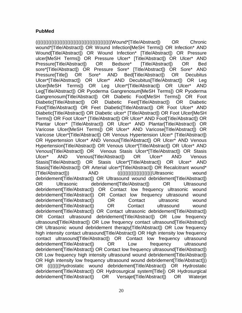
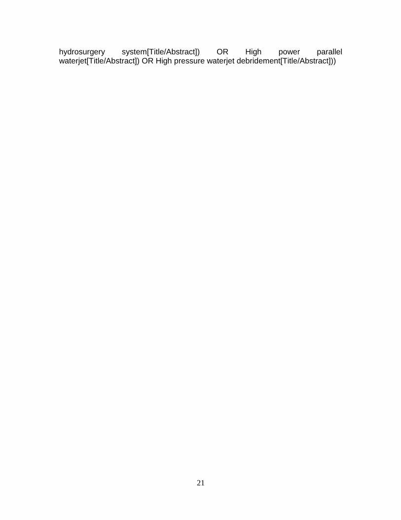
PubMed ((((((((((((((((((((((((((((((((((((((((((((((((Wound*[Title/Abstract]) OR Chronic wound*[Title/Abstract]) OR Wound Infection[MeSH Terms]) OR Infection* AND Wound[Title/Abstract]) OR Wound Infection* [Title/Abstract]) OR Pressure ulcer[MeSH Terms]) OR Pressure Ulcer* [Title/Abstract]) OR Ulcer* AND Pressure[Title/Abstract]) OR Bedsore* [Title/Abstract]) OR Bed sore*[Title/Abstract]) OR Pressure Sore* [Title/Abstract]) OR Sore* AND Pressure[Title]) OR Sore* AND Bed[Title/Abstract]) OR Decubitus Ulcer*[Title/Abstract]) OR Ulcer* AND Decubitus[Title/Abstract]) OR Leg Ulcer[MeSH Terms]) OR Leg Ulcer*[Title/Abstract]) OR Ulcer* AND Leg[Title/Abstract]) OR Pyoderma Gangrenosum[MeSH Terms]) OR Pyoderma Gangrenosum[Title/Abstract]) OR Diabetic Foot[MeSH Terms]) OR Foot Diabetic[Title/Abstract]) OR Diabetic Feet[Title/Abstract]) OR Diabetic Foot[Title/Abstract]) OR Feet Diabetic[Title/Abstract]) OR Foot Ulcer* AND Diabetic[Title/Abstract]) OR Diabetic ulcer* [Title/Abstract]) OR Foot Ulcer[MeSH Terms]) OR Foot Ulcer* [Title/Abstract]) OR Ulcer* AND Foot[Title/Abstract]) OR Plantar Ulcer* [Title/Abstract]) OR Ulcer* AND Plantar[Title/Abstract]) OR Varicose Ulcer[MeSH Terms]) OR Ulcer* AND Varicose[Title/Abstract]) OR Varicose Ulcer*[Title/Abstract]) OR Venous Hypertension Ulcer* [Title/Abstract]) OR Hypertension Ulcer* AND Venous[Title/Abstract]) OR Ulcer* AND Venous Hypertension[Title/Abstract]) OR Venous Ulcer*[Title/Abstract]) OR Ulcer* AND Venous[Title/Abstract]) OR Venous Stasis Ulcer*[Title/Abstract]) OR Stasis Ulcer* AND Venous[Title/Abstract]) OR Ulcer* AND Venous Stasis[Title/Abstract]) OR Stasis Ulcer*[Title/Abstract]) OR Ulcer* AND Stasis[Title/Abstract]) OR Arterial ulcer*[Title/Abstract]) OR Recalcitrant wound* [Title/Abstract])) AND ((((((((((((((((((((((Ultrasonic wound debridement[Title/Abstract]) OR Ultrasound wound debridement[Title/Abstract]) OR Ultrasonic debridement[Title/Abstract]) OR Ultrasound debridement[Title/Abstract]) OR Contact low frequency ultrasonic wound debridement[Title/Abstract]) OR Contact low frequency ultrasound wound debridement[Title/Abstract]) OR Contact ultrasonic wound debridement[Title/Abstract]) OR Contact ultrasound wound debridement[Title/Abstract]) OR Contact ultrasonic debridement[Title/Abstract]) OR Contact ultrasound debridement[Title/Abstract]) OR Low frequency ultrasound[Title/Abstract]) OR Low frequency contact ultrasound[Title/Abstract]) OR Ultrasonic wound debridement therapy[Title/Abstract]) OR Low frequency high intensity contact ultrasound[Title/Abstract]) OR High intensity low frequency contact ultrasound[Title/Abstract]) OR Contact low frequency ultrasound debridement[Title/Abstract]) OR Low frequency ultrasound debridement[Title/Abstract]) OR Contact low frequency ultrasound[Title/Abstract]) OR Low frequency high intensity ultrasound wound debridement[Title/Abstract]) OR High intensity low frequency ultrasound wound debridement[Title/Abstract])) OR ((((((((Hydrostatic wound debridement[Title/Abstract]) OR Hydrostatic debridement[Title/Abstract]) OR Hydrosurgical system[Title]) OR Hydrosurgical debridement[Title/Abstract]) OR Versajet[Title/Abstract]) OR Waterjet
21
hydrosurgery system[Title/Abstract]) OR High power parallel waterjet[Title/Abstract]) OR High pressure waterjet debridement[Title/Abstract]))
22
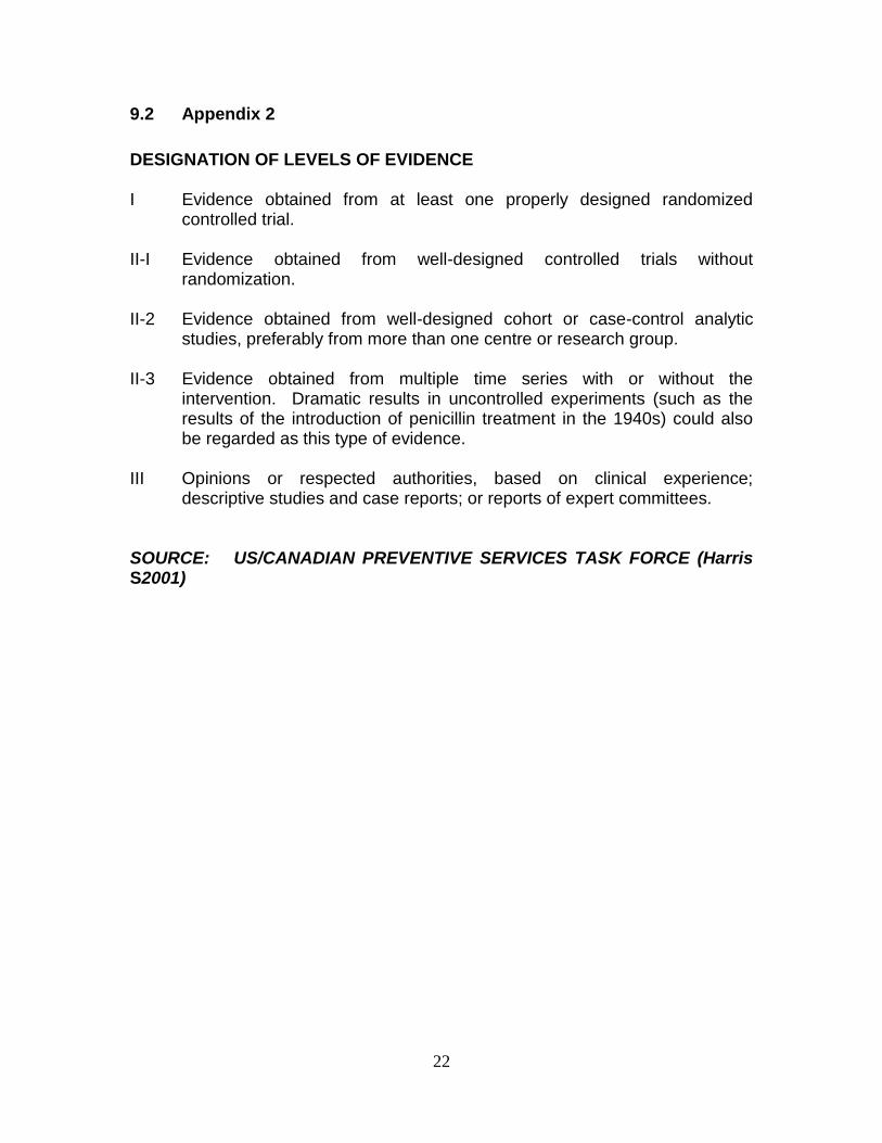
9.2 Appendix 2
DESIGNATION OF LEVELS OF EVIDENCE I Evidence obtained from at least one properly designed randomized
controlled trial.
II-I Evidence obtained from well-designed controlled trials without randomization.
II-2 Evidence obtained from well-designed cohort or case-control analytic
studies, preferably from more than one centre or research group. II-3 Evidence obtained from multiple time series with or without the
intervention. Dramatic results in uncontrolled experiments (such as the results of the introduction of penicillin treatment in the 1940s) could also be regarded as this type of evidence.
III Opinions or respected authorities, based on clinical experience;
descriptive studies and case reports; or reports of expert committees.
SOURCE: US/CANADIAN PREVENTIVE SERVICES TASK FORCE (Harris S2001)
23
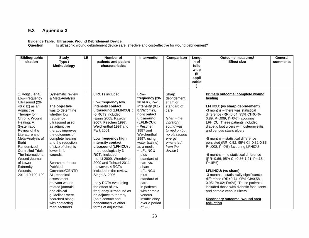
9.3 Appendix 3
Evidence Table: Ultrasonic Wound Debridement Device Question: Is ultrasonic wound debridement device safe, effective and cost-effective for wound debridement?
Bibliographic citation
Study Type /
Methodology
LE Number of patients and patient
characteristics
Intervention Comparison Length of follow up
(if applicable
)
Outcome measures/ Effect size
General comments
1. Voigt J et al. Low-Frequency Ultrasound (20-40 kHz) as an Adjunctive Therapy for Chronic Wound Healing: A Systematic Review of the Literature and Meta-Analysis of Eight Randomized Controlled Trials, The International Wound Journal of Lower Extremity Wounds, 2011;10:190-199
Systematic review & Meta-Analysis The objective
was to determine whether low frequency ultrasound used as adjunctive therapy improves the outcomes of complete healing and the reduction of size of chronic lower limb wounds. Search methods: PubMed, Cochrane/CENTRAL, technical assessment, relevant wound-related journals and clinical guidelines were searched along with contacting manufacturers
I
8 RCTs included Low frequency low intensity contact ultrasound (LFLINCU) :
-5 RCTs included -Ennis 2005, Kavros 2007, Peschen 1997, Weichenthal 1997 and Park 2001 Low frequency high intensity contact ultrasound (LFHICU) :
-methodologically 3 RCTs included -i.e. Li 2009, Wendelken 2009 and Tehrani 2011 -however, 4 RCTs included in the review, Singh A. 2006. -only RCTs evaluating the effect of low-frequency ultrasound as an adjunct to therapy (both contact and noncontact) vs other forms of adjunctive
Low-frequency (20-30 kHz), low intensity (0.1-0.5W/cm2), noncontact ultrasound (LFLINCU):
- Peschen 1997 and Weichenthal 1997, using water (saline) as a medium • LFLINCU
plus standard of care vs. sham LFLINCU plus standard of care in patients with chronic venous insufficiency over a period of 2-3
Sharp debridement, sham or standard of care (sham=the vibratory sound was turned on but no ultrasound energy emanated from the device )
Primary outcome: complete wound healing
LFHICU: (vs sharp debridement)
-3 months – there was statistical difference (RR=0.64; 95% CI=0.46-0.89; P=.009; I
2=0%)-favouring
LFHICU. These patients included diabetic foot ulcers with osteomyelitis and venous stasis ulcers -5 months – statistical difference persisted (RR=0.52; 95% CI=0.32-0.85; P=.008; I
2=0%)-favouring LFHICU
-6 months – no statistical difference (RR=0.66; 95% CI=0.36-1.21; P=.18; I2=15%)
LFLINCU: (vs sham) -3 months – statistically significance
difference (RR=0.74; 95% CI=0.58-0.95; P=.02; I
2=0%). These patients
included those with diabetic foot ulcers and chronic venous ulcers. Secondary outcome: wound area reduction
24
Bibliographic citation
Study Type /
Methodology
LE Number of patients and patient
characteristics
Intervention Comparison Length of follow up
(if applicable
)
Outcome measures/ Effect size
General comments
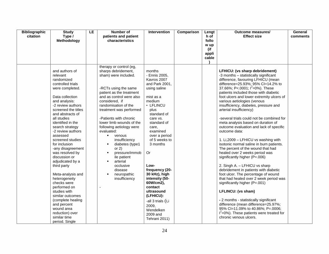
and authors of relevant randomized controlled trials were completed. Data collection and analysis: -2 review authors screened the titles and abstracts of all studies identified in the search strategy -2 review authors assessed screened studies for inclusion -any disagreement was resolved by discussion or adjudicated by a third party Meta-analysis and heterogeneity checks were performed on studies with similar outcomes (complete healing and percent wound area reduction) over similar time period. Single
therapy or control (eg, sharps debridement, sham) were included. -RCTs using the same patient as the treatment and as control were also considered, if randomisation of the treatment was performed -Patients with chronic lower limb wounds of the following aetiology were evaluated:
venous insufficiency
diabetes (type1 or 2)
pressure/immobile patient
arterial occlusive disease
neuropathic insufficiency
-
months - Ennis 2005, Kavros 2007 and Park 2001, using saline mist as a medium • LFLINCU
plus standard of care vs. standard of care, examined over a period of 5 weeks to 3 months
Or Low-frequency (20-30 kHz), high intensity (50-60W/cm2), contact ultrasound (LFHICU):
-all 3 trials (Li
2009, Wendelken 2009 and Tehrani 2011)
LFHICU: (vs sharp debridement)
-3 months – statistically significant difference, favouring LFHICU (mean difference=25.93%; 95% CI=14.2% to 37.66%; P<.0001; I
2=0%). These
patients included those with diabetic foot ulcers and lower extremity ulcers of various aetiologies (venous insufficiency, diabetes, pressure and arterial insufficiency) -several trials could not be combined for meta analysis based on duration of outcome evaluation and lack of specific outcome data: 1. Li,2009 – LFHICU vs washing with isotonic normal saline in burn patients. The percent of the wound that had healed over 2 weeks period was significantly higher (P=.006) 2. Singh A. – LFHICU vs sharp debridement in patients with diabetic foot ulcer. The percentage of wound that had healed over 2 week period was significantly higher (P=.001) LFLINCU: (vs sham) - 2 months - statistically significant
difference (mean difference=25.97%; 95% CI=11.09% to 40.86%; P=.0006; I2=0%). These patients were treated for
chronic venous ulcers.
25
Bibliographic citation
Study Type /
Methodology
LE Number of patients and patient
characteristics
Intervention Comparison Length of follow up
(if applicable
)
Outcome measures/ Effect size
General comments
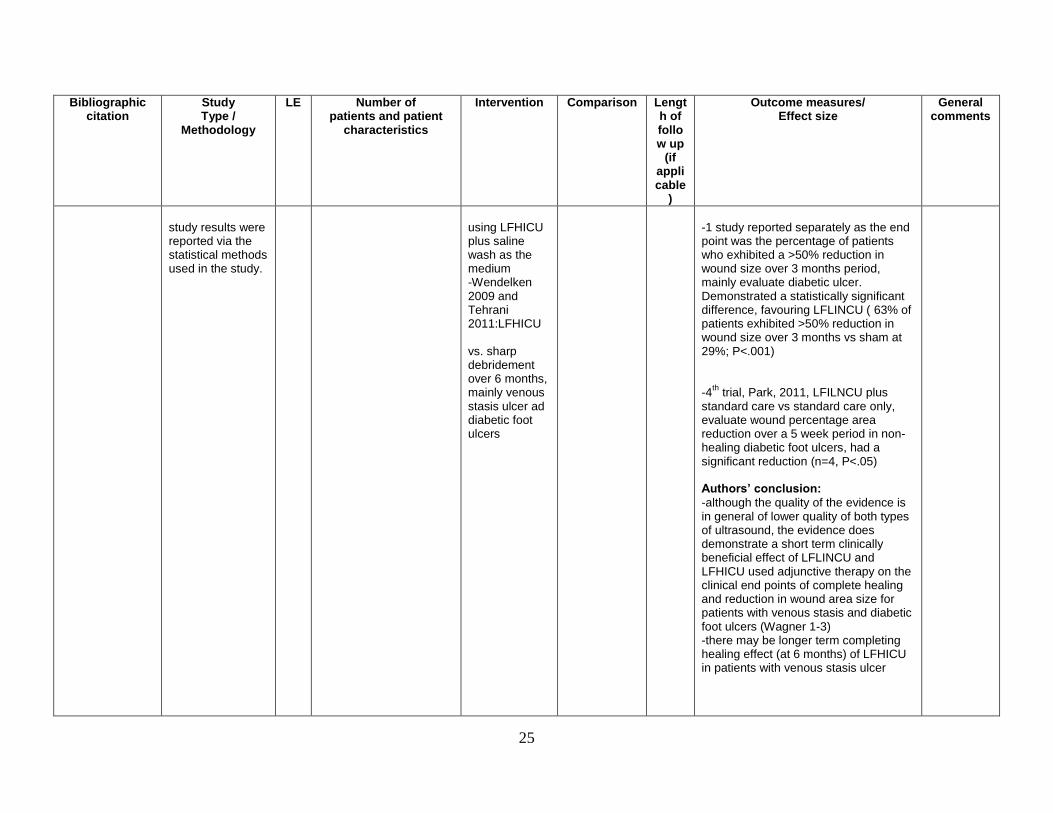
study results were reported via the statistical methods used in the study.
using LFHICU plus saline wash as the medium -Wendelken 2009 and Tehrani 2011:LFHICU vs. sharp debridement over 6 months, mainly venous stasis ulcer ad diabetic foot ulcers
-1 study reported separately as the end point was the percentage of patients who exhibited a >50% reduction in wound size over 3 months period, mainly evaluate diabetic ulcer. Demonstrated a statistically significant difference, favouring LFLINCU ( 63% of patients exhibited >50% reduction in wound size over 3 months vs sham at 29%; P<.001) -4
th trial, Park, 2011, LFILNCU plus
standard care vs standard care only, evaluate wound percentage area reduction over a 5 week period in non- healing diabetic foot ulcers, had a significant reduction (n=4, P<.05) Authors’ conclusion:
-although the quality of the evidence is in general of lower quality of both types of ultrasound, the evidence does demonstrate a short term clinically beneficial effect of LFLINCU and LFHICU used adjunctive therapy on the clinical end points of complete healing and reduction in wound area size for patients with venous stasis and diabetic foot ulcers (Wagner 1-3) -there may be longer term completing healing effect (at 6 months) of LFHICU in patients with venous stasis ulcer
26
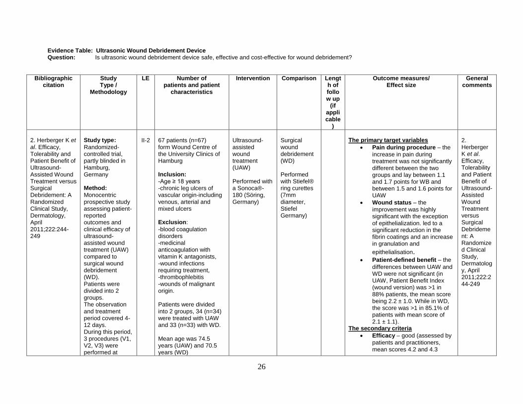
Evidence Table: Ultrasonic Wound Debridement Device Question: Is ultrasonic wound debridement device safe, effective and cost-effective for wound debridement?
Bibliographic citation
Study Type /
Methodology
LE Number of patients and patient
characteristics
Intervention Comparison Length of follow up
(if applicable
)
Outcome measures/ Effect size
General comments
2. Herberger K et al. Efficacy, Tolerability and Patient Benefit of Ultrasound-Assisted Wound Treatment versus Surgical Debridement: A Randomized Clinical Study, Dermatology, April 2011;222:244-249
Study type:
Randomized-controlled trial, partly blinded in Hamburg, Germany Method:
Monocentric prospective study assessing patient-reported outcomes and clinical efficacy of ultrasound-assisted wound treatment (UAW) compared to surgical wound debridement (WD). Patients were divided into 2 groups. The observation and treatment period covered 4-12 days. During this period, 3 procedures (V1, V2, V3) were performed at
II-2
67 patients (n=67) form Wound Centre of the University Clinics of Hamburg Inclusion:
-Age ≥ 18 years -chronic leg ulcers of vascular origin-including venous, arterial and mixed ulcers Exclusion:
-blood coagulation disorders -medicinal anticoagulation with vitamin K antagonists, -wound infections requiring treatment, -thrombophlebitis -wounds of malignant origin. Patients were divided into 2 groups, 34 (n=34) were treated with UAW and 33 (n=33) with WD. Mean age was 74.5 years (UAW) and 70.5 years (WD)
Ultrasound-assisted wound treatment (UAW) Performed with a Sonoca®-180 (Söring, Germany)
Surgical wound debridement (WD) Performed with Stiefel® ring curettes (7mm diameter, Stiefel Germany)
The primary target variables
Pain during procedure – the
increase in pain during treatment was not significantly different between the two groups and lay between 1.1 and 1.7 points for WB and between 1.5 and 1.6 points for UAW
Wound status – the
improvement was highly significant with the exception of epithelialization. led to a significant reduction in the fibrin coatings and an increase in granulation and
epithelialisation. Patient-defined benefit – the
differences between UAW and WD were not significant (in UAW, Patient Benefit Index (wound version) was >1 in 88% patients, the mean score being 2.2 ± 1.0. While in WD, the score was >1 in 85.1% of patients with mean score of 2.1 ± 1.1).
The secondary criteria
Efficacy – good (assessed by
patients and practitioners, mean scores 4.2 and 4.3
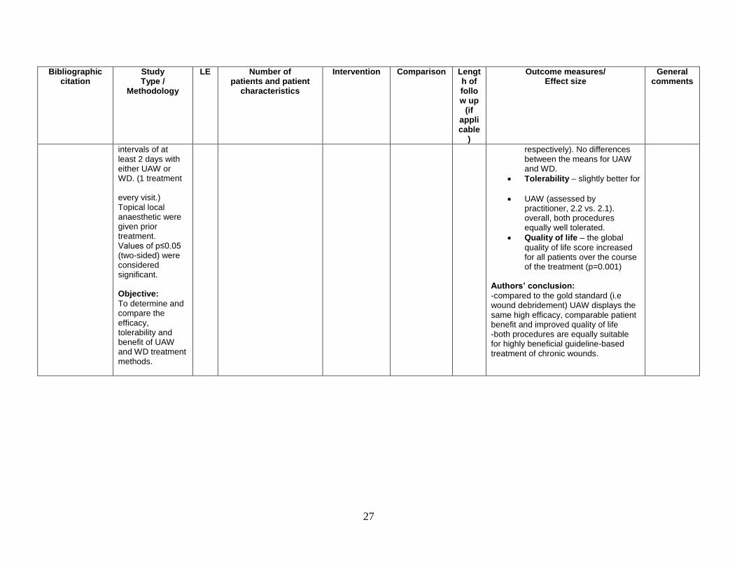
2. Herberger K et al. Efficacy, Tolerability and Patient Benefit of Ultrasound-Assisted Wound Treatment versus Surgical Debridement: A Randomized Clinical Study, Dermatology, April 2011;222:244-249
27
Bibliographic citation
Study Type /
Methodology
LE Number of patients and patient
characteristics
Intervention Comparison Length of follow up
(if applicable
)
Outcome measures/ Effect size
General comments
intervals of at least 2 days with either UAW or WD. (1 treatment every visit.) Topical local anaesthetic were given prior treatment. Values of p≤0.05 (two-sided) were considered significant. Objective:
To determine and compare the efficacy, tolerability and benefit of UAW and WD treatment methods.
respectively). No differences between the means for UAW and WD.
Tolerability – slightly better for
UAW (assessed by practitioner, 2.2 vs. 2.1). overall, both procedures equally well tolerated.
Quality of life – the global
quality of life score increased for all patients over the course of the treatment (p=0.001)
Authors’ conclusion:
-compared to the gold standard (i.e wound debridement) UAW displays the same high efficacy, comparable patient benefit and improved quality of life -both procedures are equally suitable for highly beneficial guideline-based treatment of chronic wounds.
28
Evidence Table: Ultrasonic Wound Debridement Device Question: Is ultrasonic wound debridement device safe, effective and cost-effective for wound debridement?
Bibliographic
citation Study Type /
Methodology
LE Number of patients and patient
characteristics
Intervention Comparison Length of follow up
(if applicable
)
Outcome measures/ Effect size
General comments
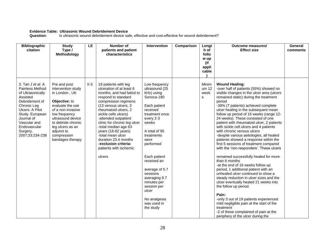
3. Tan J et al. A Painless Method of Ultrasonically Assisted Debridement of Chronic Leg Ulcers: A Pilot Study. European Journal of Vascular and Endovascular Surgery. 2007;33:234-238
Pre and post intervention study in London , UK Objective: to
evaluate the use of a non-invasive low frequency ultrasound device to debride chronic leg ulcers as an adjunct to compression bandages therapy
II-3
19 patients with leg ulceration of at least 6 months, and had failed to respond to standard compression regimens (13 venous ulcers, 3 rheumatoid ulcers, 2 sickle cells ulcers) -attended outpatient clinic for chronic leg ulcer -total median age 63 years (18-82 years) -total mean ulcer duration 23.4 months -exclusion criteria:
patients with ischemic ulcers
Low frequency ultrasound (25 kHz) using Sonoca-180 Each patient received treatment once every 2-3 weeks A total of 95 treatments were performed Each patient received an average of 5.7 sessions averaging 9.7 minutes per session per ulcer No analgesia was used in the study
Minimum 12 weeks
Wound Healing:
-over half of patients (55%) showed no visible changes in the ulcer area (ulcers remained static) during the treatment period -39% (7 patients) achieved complete ulcer healing in the subsequent mean follow up period of 16 weeks (range 12-24 weeks). These consisted of one patient with rheumatoid ulcer, 2 patients with sickle cell ulcers and 4 patients with chronic venous ulcers -despite various aetiologies, all healed patients showed a response within the first 5 sessions of treatment compared with the ‘non-responders’. These ulcers remained successfully healed for more than 6 months -at the end of 16 weeks follow up period, 1 additional patient with an unhealed ulcer continued to show a steady reduction in ulcer sizes and the ulcer eventually healed 21 weeks into the follow up period. Pain:
-only 3 out of 19 patients experienced mild negligible pain at the start of the treatment -2 of these complained of pain at the periphery of the ulcer during the
29
Bibliographic citation
Study Type /
Methodology
LE Number of patients and patient
characteristics
Intervention Comparison Length of follow up
(if applicable
)
Outcome measures/ Effect size
General comments
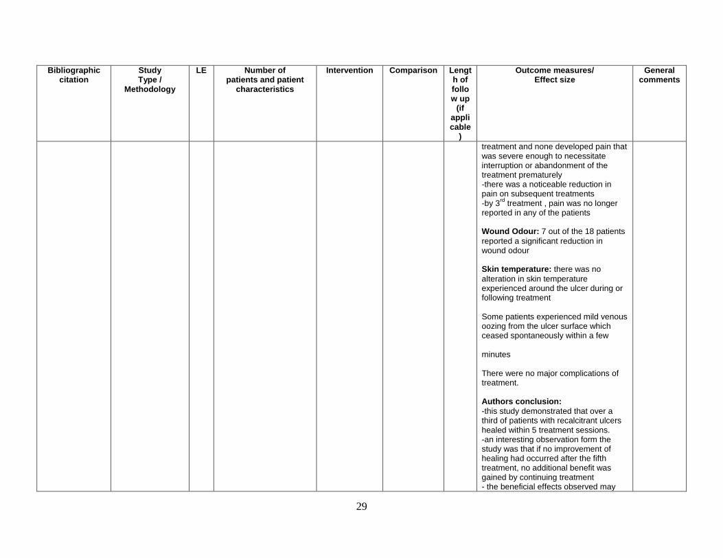
treatment and none developed pain that was severe enough to necessitate interruption or abandonment of the treatment prematurely -there was a noticeable reduction in pain on subsequent treatments -by 3
rd treatment , pain was no longer
reported in any of the patients Wound Odour: 7 out of the 18 patients
reported a significant reduction in wound odour Skin temperature: there was no
alteration in skin temperature experienced around the ulcer during or following treatment Some patients experienced mild venous oozing from the ulcer surface which ceased spontaneously within a few minutes There were no major complications of treatment. Authors conclusion:
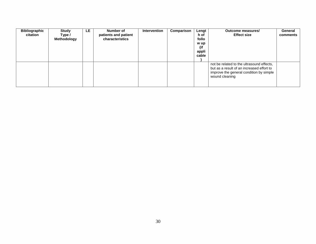
-this study demonstrated that over a third of patients with recalcitrant ulcers healed within 5 treatment sessions. -an interesting observation form the study was that if no improvement of healing had occurred after the fifth treatment, no additional benefit was gained by continuing treatment - the beneficial effects observed may
30
Bibliographic citation
Study Type /
Methodology
LE Number of patients and patient
characteristics
Intervention Comparison Length of follow up
(if applicable
)
Outcome measures/ Effect size
General comments
not be related to the ultrasound effects, but as a result of an increased effort to improve the general condition by simple wound cleaning
31
Evidence Table: Ultrasonic Wound Debridement Device Question: Is ultrasonic wound debridement device safe, effective and cost-effective for wound debridement?
Bibliographic citation
Study Type /
Methodology
LE Number of patients and patient
characteristics
Intervention Comparison Length of follow up
(if applicable
)
Outcome measures/ Effect size
General comments
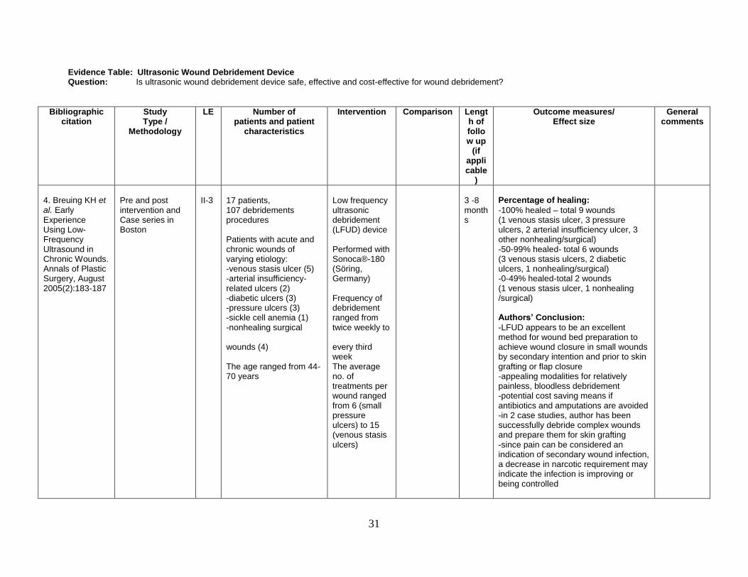
4. Breuing KH et al. Early Experience Using Low-Frequency Ultrasound in Chronic Wounds. Annals of Plastic Surgery, August 2005(2):183-187
Pre and post intervention and Case series in Boston
II-3
17 patients, 107 debridements procedures Patients with acute and chronic wounds of varying etiology: -venous stasis ulcer (5) -arterial insufficiency-related ulcers (2) -diabetic ulcers (3) -pressure ulcers (3) -sickle cell anemia (1) -nonhealing surgical wounds (4) The age ranged from 44-70 years
Low frequency ultrasonic debridement (LFUD) device Performed with Sonoca®-180 (Söring, Germany) Frequency of debridement ranged from twice weekly to every third week The average no. of treatments per wound ranged from 6 (small pressure ulcers) to 15 (venous stasis ulcers)
3 -8 months
Percentage of healing:
-100% healed – total 9 wounds (1 venous stasis ulcer, 3 pressure ulcers, 2 arterial insufficiency ulcer, 3 other nonhealing/surgical) -50-99% healed- total 6 wounds (3 venous stasis ulcers, 2 diabetic ulcers, 1 nonhealing/surgical) -0-49% healed-total 2 wounds (1 venous stasis ulcer, 1 nonhealing /surgical) Authors’ Conclusion:
-LFUD appears to be an excellent method for wound bed preparation to achieve wound closure in small wounds by secondary intention and prior to skin grafting or flap closure -appealing modalities for relatively painless, bloodless debridement -potential cost saving means if antibiotics and amputations are avoided -in 2 case studies, author has been successfully debride complex wounds and prepare them for skin grafting -since pain can be considered an indication of secondary wound infection, a decrease in narcotic requirement may indicate the infection is improving or being controlled
32
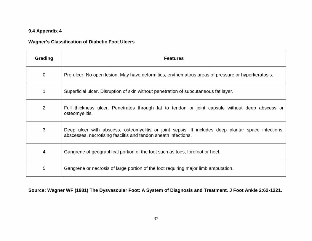
9.4 Appendix 4 Wagner’s Classification of Diabetic Foot Ulcers
Grading
Features
0
Pre-ulcer. No open lesion. May have deformities, erythematous areas of pressure or hyperkeratosis.
1
Superficial ulcer. Disruption of skin without penetration of subcutaneous fat layer.
2
Full thickness ulcer. Penetrates through fat to tendon or joint capsule without deep abscess or osteomyelitis.
3
Deep ulcer with abscess, osteomyelitis or joint sepsis. It includes deep plantar space infections, abscesses, necrotising fasciitis and tendon sheath infections.
4
Gangrene of geographical portion of the foot such as toes, forefoot or heel.
5
Gangrene or necrosis of large portion of the foot requiring major limb amputation.
Source: Wagner WF (1981) The Dysvascular Foot: A System of Diagnosis and Treatment. J Foot Ankle 2:62-1221.