Embed Size (px)
Citation preview
Ulcerative colitis and autoimmunity induced by lossof myeloid !v integrinsAdam Lacy-Hulbert*†‡, Aileen M. Smith*, Hamid Tissire†, Marc Barry†, Denise Crowley†§, Roderick T. Bronson†¶,Jurgen T. Roes!, John S. Savill*, and Richard O. Hynes†§**
*Medical Research Council/University of Edinburgh Centre for Inflammation Research, The Queen’s Medical Research Institute, 47 Little France Crescent,Edinburgh EH16 4TJ, United Kingdom; §Howard Hughes Medical Institute and †Center for Cancer Research, Massachusetts Institute of Technology, 77Massachusetts Avenue, Cambridge, MA 02139; ¶Rodent Histopathology Laboratory, Harvard Medical School, 220 Longwood Avenue, Boston, MA 02115;and !Windeyer Institute for Medical Sciences, University College London, 46 Cleveland Street, London W1T 4JF, United Kingdom
Contributed by Richard O. Hynes, August 7, 2007 (sent for review July 25, 2007)
The gastrointestinal tract is constantly challenged by foreign an-tigens and commensal bacteria but nonetheless is able to maintaina state of immunological quiescence. Recent advances have high-lighted the importance of active suppression by regulatory lym-phocytes and immunosuppressive cytokines in controlling mucosalimmunity. Failures of these mechanisms contribute to the devel-opment of inflammatory bowel disease, but how these regulatorynetworks are established remains unclear. Here, we demonstratekey roles for !v integrins in regulation of mucosal immunity. Wereport that deletion of !v in the immune system causes severecolitis, autoimmunity, and cancer. Mice lacking immune cell !vhave fewer regulatory T (Treg) cells in the colon and correspondingincreases in activated T cells and T cell cytokine production, leadingto colitis. Using conditional gene targeting, we demonstrate thatthis is specifically attributable to loss of !v from myeloid cells.Furthermore, we show that gut-associated macrophages and den-dritic cells fail both to remove apoptotic cells efficiently and toinduce Treg cells. Our results identify a vital role for myeloid !vintegrins in generating mucosal Treg cells and emphasize theimportance of antigen-presenting cells in establishing immunetolerance.
immunoregulation " phagocytosis " apoptotic " dendritic cell "regulatory T cell
Integrins are dimeric cell-surface receptors composed of ! and" subunits (1). !v is the most promiscuous ! subunit, associ-
ating with five different " subunits ("1, "3, "5, "6, and "8) andparticipates in many important cellular processes, including celladhesion, migration, survival, and growth factor signaling. !vintegrins are expressed in many tissues throughout development,and !v knockout mice die at midgestation from placentaldeficiencies or, immediately after birth, with cerebral and intes-tinal hemorrhage and cleft palate (2). The lethality of !v!/! miceis attributable mainly to loss of !v"8, because "8!/! mice showsimilar placental and neural vasculature abnormalities (3), andthese defects are principally attributable to loss of !v"8 fromparenchymal tissues supporting blood vessels (4, 5). Knockoutsof most other !v-associated " integrins ("3, "5, and "6) areviable and reveal roles for !v in repair of tissue injury, inflam-mation, and immune responses (1, 6–8). !v"6 and !v"8 areexpressed in epithelium, where their functions include bindingand activating TGF-" (9), and !v"8 is also expressed by myeloidcells and T cells (10, 11). !v"3 and !v"5 are more widelyexpressed and are up-regulated by many cells after tissue injury.In the immune system, !v"3 and !v"5 are expressed by manydifferent cell types and promote cell adhesion, migration, andsurvival. !v integrins also mediate the uptake of apoptotic cells(12–14), a process central to immune regulation and inflamma-tion resolution (15).
Here, we report that !v integrins are central to the normalregulation of immune responses in the intestine and that deletionof !v in the immune system leads to spontaneous colitis, wasting,
and autoimmunity. Colitis is associated with activated T cells,high expression of T cell cytokines, and corresponding loss oftissue-resident regulatory T (Treg) cells. We dissect the contri-bution of !v on different immune cell populations, identifyingmyeloid cells [macrophages, dendritic cells (DCs), and/or neu-trophils] as the critical expressers of !v integrins. We furthershow that deletion of !v leads to impaired removal of apoptoticcells and failure of mesenteric lymph node (mLN) DCs togenerate Treg cells.
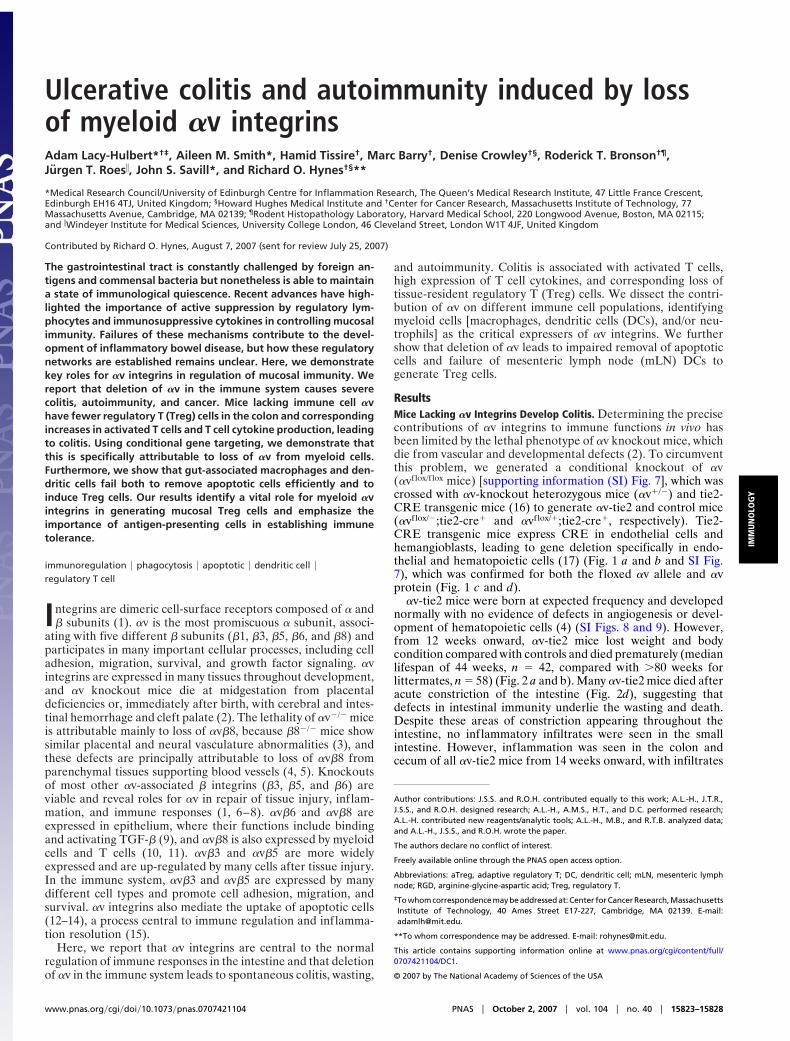
ResultsMice Lacking !v Integrins Develop Colitis. Determining the precisecontributions of !v integrins to immune functions in vivo hasbeen limited by the lethal phenotype of !v knockout mice, whichdie from vascular and developmental defects (2). To circumventthis problem, we generated a conditional knockout of !v(!vflox/f lox mice) [supporting information (SI) Fig. 7], which wascrossed with !v-knockout heterozygous mice (!v"/!) and tie2-CRE transgenic mice (16) to generate !v-tie2 and control mice(!vflox/!;tie2-cre" and !vflox/";tie2-cre", respectively). Tie2-CRE transgenic mice express CRE in endothelial cells andhemangioblasts, leading to gene deletion specifically in endo-thelial and hematopoietic cells (17) (Fig. 1 a and b and SI Fig.7), which was confirmed for both the floxed !v allele and !vprotein (Fig. 1 c and d).
!v-tie2 mice were born at expected frequency and developednormally with no evidence of defects in angiogenesis or devel-opment of hematopoietic cells (4) (SI Figs. 8 and 9). However,from 12 weeks onward, !v-tie2 mice lost weight and bodycondition compared with controls and died prematurely (medianlifespan of 44 weeks, n # 42, compared with $80 weeks forlittermates, n # 58) (Fig. 2 a and b). Many !v-tie2 mice died afteracute constriction of the intestine (Fig. 2d), suggesting thatdefects in intestinal immunity underlie the wasting and death.Despite these areas of constriction appearing throughout theintestine, no inflammatory infiltrates were seen in the smallintestine. However, inflammation was seen in the colon andcecum of all !v-tie2 mice from 14 weeks onward, with infiltrates
Author contributions: J.S.S. and R.O.H. contributed equally to this work; A.L.-H., J.T.R.,J.S.S., and R.O.H. designed research; A.L.-H., A.M.S., H.T., and D.C. performed research;A.L.-H. contributed new reagents/analytic tools; A.L.-H., M.B., and R.T.B. analyzed data;and A.L.-H., J.S.S., and R.O.H. wrote the paper.
The authors declare no conflict of interest.
Freely available online through the PNAS open access option.
Abbreviations: aTreg, adaptive regulatory T; DC, dendritic cell; mLN, mesenteric lymphnode; RGD, arginine-glycine-aspartic acid; Treg, regulatory T.‡To whom correspondence may be addressed at: Center for Cancer Research, MassachusettsInstitute of Technology, 40 Ames Street E17-227, Cambridge, MA 02139. E-mail:[email protected].
**To whom correspondence may be addressed. E-mail: [email protected].
This article contains supporting information online at www.pnas.org/cgi/content/full/0707421104/DC1.
© 2007 by The National Academy of Sciences of the USA
www.pnas.org#cgi#doi#10.1073#pnas.0707421104 PNAS " October 2, 2007 " vol. 104 " no. 40 " 15823–15828
IMM
UNO
LOG
Y
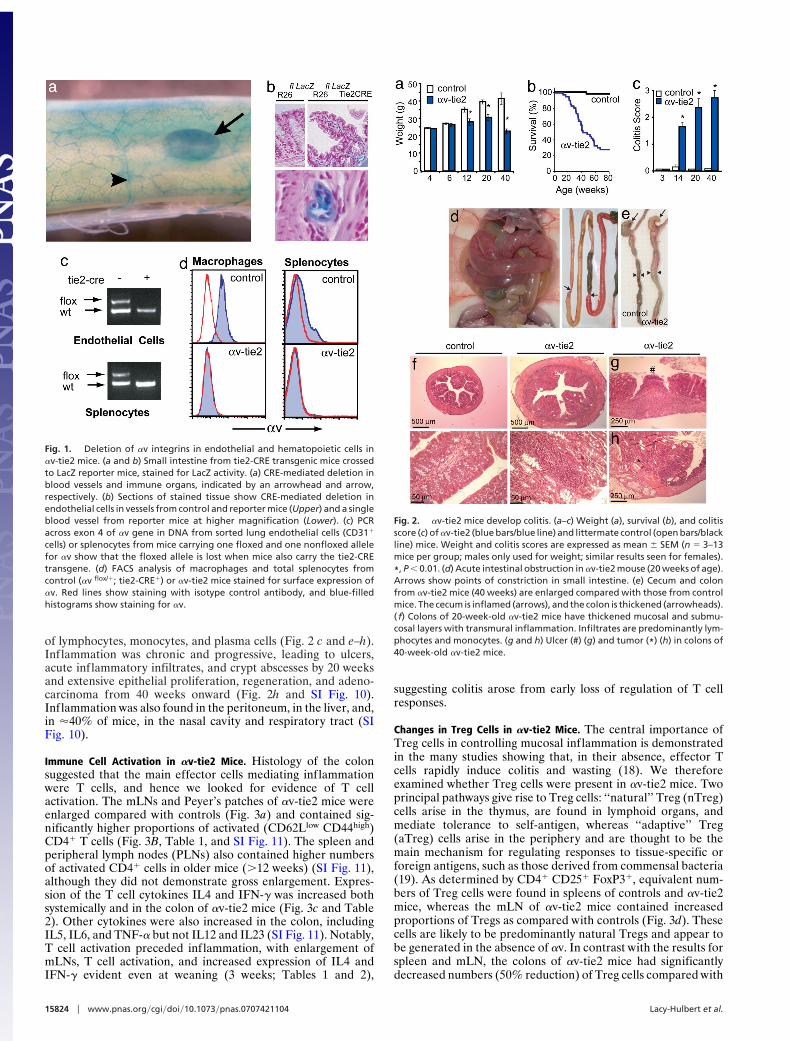
of lymphocytes, monocytes, and plasma cells (Fig. 2 c and e–h).Inflammation was chronic and progressive, leading to ulcers,acute inflammatory infiltrates, and crypt abscesses by 20 weeksand extensive epithelial proliferation, regeneration, and adeno-carcinoma from 40 weeks onward (Fig. 2h and SI Fig. 10).Inflammation was also found in the peritoneum, in the liver, and,in %40% of mice, in the nasal cavity and respiratory tract (SIFig. 10).
Immune Cell Activation in !v-tie2 Mice. Histology of the colonsuggested that the main effector cells mediating inflammationwere T cells, and hence we looked for evidence of T cellactivation. The mLNs and Peyer’s patches of !v-tie2 mice wereenlarged compared with controls (Fig. 3a) and contained sig-nificantly higher proportions of activated (CD62Llow CD44high)CD4" T cells (Fig. 3B, Table 1, and SI Fig. 11). The spleen andperipheral lymph nodes (PLNs) also contained higher numbersof activated CD4" cells in older mice ($12 weeks) (SI Fig. 11),although they did not demonstrate gross enlargement. Expres-sion of the T cell cytokines IL4 and IFN-# was increased bothsystemically and in the colon of !v-tie2 mice (Fig. 3c and Table2). Other cytokines were also increased in the colon, includingIL5, IL6, and TNF-! but not IL12 and IL23 (SI Fig. 11). Notably,T cell activation preceded inflammation, with enlargement ofmLNs, T cell activation, and increased expression of IL4 andIFN-# evident even at weaning (3 weeks; Tables 1 and 2),
suggesting colitis arose from early loss of regulation of T cellresponses.
Changes in Treg Cells in !v-tie2 Mice. The central importance ofTreg cells in controlling mucosal inflammation is demonstratedin the many studies showing that, in their absence, effector Tcells rapidly induce colitis and wasting (18). We thereforeexamined whether Treg cells were present in !v-tie2 mice. Twoprincipal pathways give rise to Treg cells: ‘‘natural’’ Treg (nTreg)cells arise in the thymus, are found in lymphoid organs, andmediate tolerance to self-antigen, whereas ‘‘adaptive’’ Treg(aTreg) cells arise in the periphery and are thought to be themain mechanism for regulating responses to tissue-specific orforeign antigens, such as those derived from commensal bacteria(19). As determined by CD4" CD25" FoxP3", equivalent num-bers of Treg cells were found in spleens of controls and !v-tie2mice, whereas the mLN of !v-tie2 mice contained increasedproportions of Tregs as compared with controls (Fig. 3d). Thesecells are likely to be predominantly natural Tregs and appear tobe generated in the absence of !v. In contrast with the results forspleen and mLN, the colons of !v-tie2 mice had significantlydecreased numbers (50% reduction) of Treg cells compared with
Fig. 1. Deletion of !v integrins in endothelial and hematopoietic cells in!v-tie2 mice. (a and b) Small intestine from tie2-CRE transgenic mice crossedto LacZ reporter mice, stained for LacZ activity. (a) CRE-mediated deletion inblood vessels and immune organs, indicated by an arrowhead and arrow,respectively. (b) Sections of stained tissue show CRE-mediated deletion inendothelial cells in vessels from control and reporter mice (Upper) and a singleblood vessel from reporter mice at higher magnification (Lower). (c) PCRacross exon 4 of !v gene in DNA from sorted lung endothelial cells (CD31"
cells) or splenocytes from mice carrying one floxed and one nonfloxed allelefor !v show that the floxed allele is lost when mice also carry the tie2-CREtransgene. (d) FACS analysis of macrophages and total splenocytes fromcontrol (!v flox/"; tie2-CRE") or !v-tie2 mice stained for surface expression of!v. Red lines show staining with isotype control antibody, and blue-filledhistograms show staining for !v.
Fig. 2. !v-tie2 mice develop colitis. (a–c) Weight (a), survival (b), and colitisscore (c) of !v-tie2 (blue bars/blue line) and littermate control (open bars/blackline) mice. Weight and colitis scores are expressed as mean & SEM (n # 3–13mice per group; males only used for weight; similar results seen for females).*, P ' 0.01. (d) Acute intestinal obstruction in !v-tie2 mouse (20 weeks of age).Arrows show points of constriction in small intestine. (e) Cecum and colonfrom !v-tie2 mice (40 weeks) are enlarged compared with those from controlmice. The cecum is inflamed (arrows), and the colon is thickened (arrowheads).( f) Colons of 20-week-old !v-tie2 mice have thickened mucosal and submu-cosal layers with transmural inflammation. Infiltrates are predominantly lym-phocytes and monocytes. (g and h) Ulcer (#) (g) and tumor (*) (h) in colons of40-week-old !v-tie2 mice.
15824 " www.pnas.org#cgi#doi#10.1073#pnas.0707421104 Lacy-Hulbert et al.
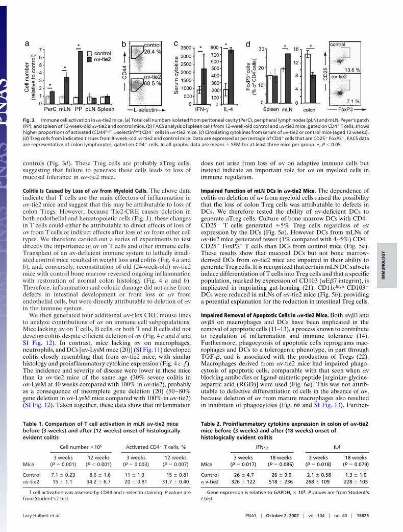
controls (Fig. 3d). These Treg cells are probably aTreg cells,suggesting that failure to generate these cells leads to loss ofmucosal tolerance in !v-tie2 mice.
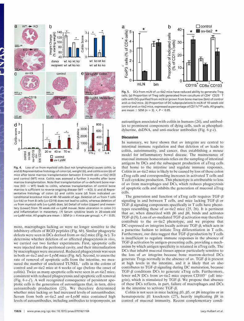
Colitis Is Caused by Loss of !v from Myeloid Cells. The above dataindicate that T cells are the main effectors of inflammation in!v-tie2 mice and suggest that this may be attributable to loss ofcolon Tregs. However, because Tie2-CRE causes deletion inboth endothelial and hematopoietic cells (Fig. 1), these changesin T cells could either be attributable to direct effects of loss of!v from T cells or indirect effects after loss of !v from other celltypes. We therefore carried out a series of experiments to testdirectly the importance of !v on T cells and other immune cells.Transplant of an !v-deficient immune system to lethally irradi-ated control mice resulted in weight loss and colitis (Fig. 4 a andb), and, conversely, reconstitution of old (24-week-old) !v-tie2mice with control bone marrow reversed ongoing inflammationwith restoration of normal colon histology (Fig. 4 a and b).Therefore, inflammation and colonic damage did not arise fromdefects in intestinal development or from loss of !v fromendothelial cells, but were directly attributable to deletion of !vin the immune system.
We then generated four additional !v-f lox CRE mouse linesto analyze contributions of !v on immune cell subpopulations.Mice lacking !v on T cells, B cells, or both T and B cells did notdevelop colitis despite efficient deletion of !v (Fig. 4 c and d andSI Fig. 12). In contrast, mice lacking !v on macrophages,neutrophils, and DCs [!v-LysM mice (20)] (SI Fig. 11) developedcolitis closely resembling that from !v-tie2 mice, with similarhistology and proinflammatory cytokine expression (Fig. 4 c–f ).The incidence and severity of disease were lower in these micethan in !v-tie2 mice of the same age (30% severe colitis in!v-LysM at 40 weeks compared with 100% in !v-tie2), probablyas a consequence of incomplete gene deletion (20) (50–80%gene deletion in !v-LysM mice compared with 100% in !v-tie2)(SI Fig. 12). Taken together, these data show that inflammation
does not arise from loss of !v on adaptive immune cells butinstead indicate an important role for !v on myeloid cells inimmune regulation.
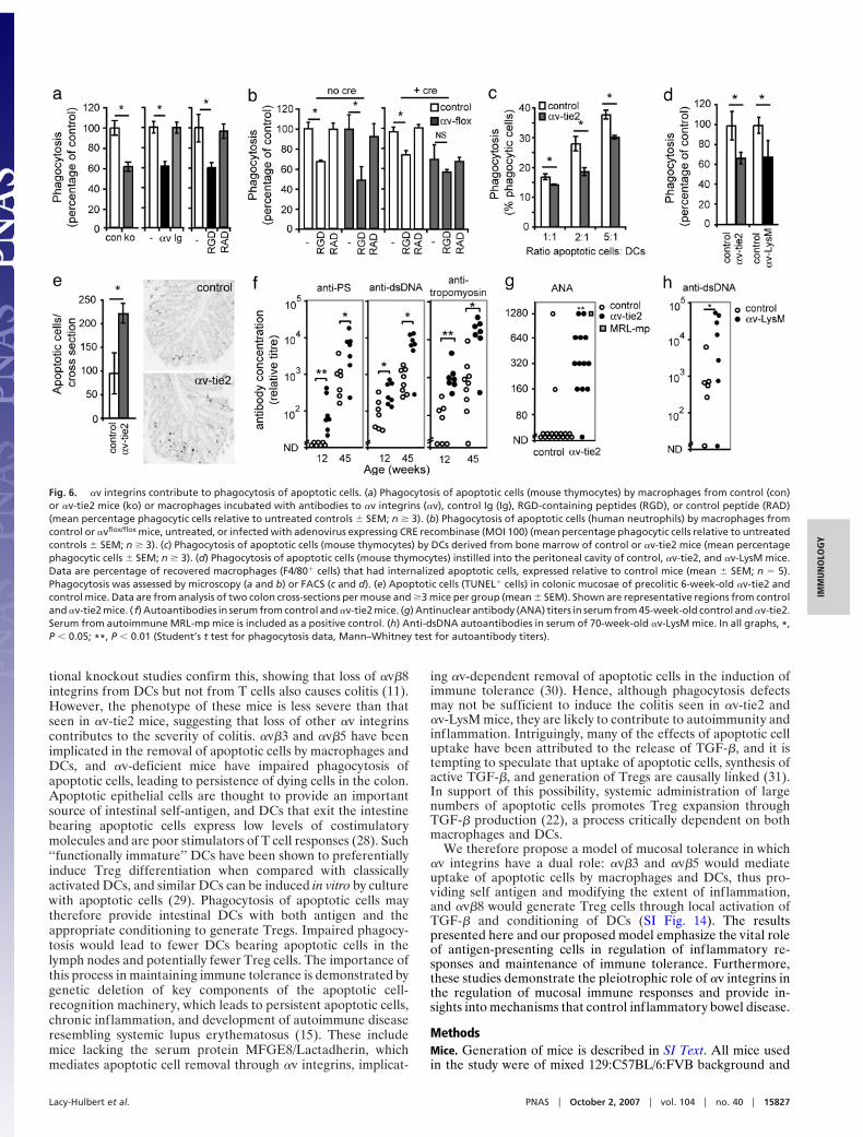
Impaired Function of mLN DCs in !v-tie2 Mice. The dependence ofcolitis on deletion of !v from myeloid cells raised the possibilitythat the loss of colon Treg cells was attributable to defects inDCs. We therefore tested the ability of !v-deficient DCs togenerate aTreg cells. Culture of bone marrow DCs with CD4"
CD25! T cells generated %5% Treg cells regardless of !vexpression by the DCs (Fig. 5a). However DCs from mLNs of!v-tie2 mice generated fewer (1% compared with 4–5%) CD4"
CD25" FoxP3" T cells than DCs from control mice (Fig. 5a).These results show that mucosal DCs but not bone marrow-derived DCs from !v-tie2 mice are impaired in their ability togenerate Treg cells. It is recognized that certain mLN DC subsetsinduce differentiation of T cells into Treg cells and that a specificpopulation, marked by expression of CD103 (!E"7 integrin), isimplicated in imprinting gut-homing (21). CD11chigh CD103"
DCs were reduced in mLNs of !v-tie2 mice (Fig. 5b), providinga potential explanation for the reduction in intestinal Treg cells.
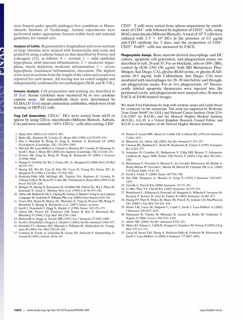
Impaired Removal of Apoptotic Cells in !v-tie2 Mice. Both !v"3 and!v"5 on macrophages and DCs have been implicated in theremoval of apoptotic cells (11–13), a process known to contributeto regulation of inflammation and immune tolerance (14).Furthermore, phagocytosis of apoptotic cells reprograms mac-rophages and DCs to a tolerogenic phenotype, in part throughTGF-", and is associated with the production of Tregs (22).Macrophages derived from !v-tie2 mice had impaired phago-cytosis of apoptotic cells, comparable with that seen when !vblocking antibodies or ligand-mimetic peptide [arginine-glycine-aspartic acid (RGD)] were used (Fig. 6a). This was not attrib-utable to defective differentiation of cells in the absence of !v,because deletion of !v from mature macrophages also resultedin inhibition of phagocytosis (Fig. 6b and SI Fig. 13). Further-
Table 2. Proinflammatory cytokine expression in colon of !v-tie2mice before (3 weeks) and after (18 weeks) onset ofhistologically evident colitis
Mice
IFN-# IL4
3 weeks(P # 0.017)
18 weeks(P # 0.086)
3 weeks(P # 0.018)
18 weeks(P # 0.079)
Control 26 & 4.7 26 & 9.9 2.1 & 0.58 1.3 & 1.0! v-tie2 326 & 122 518 & 236 268 & 109 228 & 105
Gene expression is relative to GAPDH, ( 103. P values are from Student’st test.
Fig. 3. Immune cell activation in !v-tie2 mice. (a) Total cell numbers isolated from peritoneal cavity (PerC), peripheral lymph nodes (pLN) and mLN, Peyer’s patch(PP), and spleen of 12-week-old !v-tie2 and control mice. (b) FACS analysis of spleen cells from 12-week-old control and !v-tie2 mice, gated on CD4" T cells, showshigher proportions of activated (CD44high L-selectinlow) CD4" cells in !v-tie2 mice. (c) Circulating cytokines from serum of !v-tie2 or control mice (aged 12 weeks).(d) Treg cells from indicated tissues from 8-week-old !v-tie2 and control mice. Data are expressed as percentage of CD4" cells that are CD25" FoxP3". FACS dataare representative of colon lymphocytes, gated on CD4" cells. In all graphs, data are means & SEM for at least three mice per group. *, P ' 0.05.
Table 1. Comparison of T cell activation in mLN !v-tie2 micebefore (3 weeks) and after (12 weeks) onset of histologicallyevident colitis
Mice
Cell number (106 Activated CD4" T cells, %
3 weeks(P # 0.001)
12 weeks(P ' 0.001)
3 weeks(P # 0.003)
12 weeks(P # 0.007)
Control 7.1 & 0.23 8.6 & 1.6 11 & 1.3 15 & 0.81!v-tie2 15 & 1.1 34.2 & 6.7 20 & 0.81 31.7 & 0.40
T cell activation was assessed by CD44 and L-selectin staining. P values arefrom Student’s t test.
Lacy-Hulbert et al. PNAS " October 2, 2007 " vol. 104 " no. 40 " 15825
IMM
UNO
LOG
Y
more, macrophages lacking !v were no longer sensitive to theinhibitory effects of RGD peptides (Fig. 6b). Similar phagocyticdefects were seen in DCs derived from !v-tie2 mice (Fig. 6c). Todetermine whether deletion of !v affected phagocytosis in vivo,we carried out two further experiments. First, apoptotic cellswere injected into the peritoneal cavity, and their internalizationby macrophages were measured. Reduced phagocytosis was seenin both !v-tie2 and !v-LysM mice (Fig. 6d). Second, to assess therate of removal of apoptotic cells from the intestine, we mea-sured the number of uncleared TUNEL" cells in the colons ofcontrol and !v-tie2 mice at 6 weeks of age (before the onset ofcolitis). Twice as many apoptotic cells were seen in !v-tie2 mice,consistent with reduced phagocytosis and apoptotic cell removal(Fig. 6 e–f ). A well recognized consequence of persistent apo-ptotic cells is the generation of autoantigens that, in turn, driveautoantibody production (23). We therefore determinedwhether mice lacking !v had increased levels of autoantibodies.Serum from both !v-tie2 and !v-LysM mice contained highlevels of autoantibodies, including antibodies to tropomyosin, an
autoantigen associated with colitis in humans (24), and antibod-ies to prominent components of dying cells, such as phosphati-dylserine, dsDNA, and anti-nuclear antibodies (Fig. 6 g–i).
DiscussionIn summary, we have shown that !v integrins are central tointestinal immune regulation and that deletion of !v leads tocolitis, autoimmunity, and cancer, thus establishing a mousemodel for inflammatory bowel disease. The maintenance ofmucosal immune homeostasis relies on the sampling of intestinalantigens by DCs and the subsequent production of aTreg cellsthat home to the intestine and regulate immune responses.Colitis in !v-tie2 mice is likely to be caused by loss of these colonaTreg cells and corresponding increases in activated T cells andT cell cytokine production. This phenotype is attributable to lossof !v from macrophages and DCs, which reduces phagocytosisof apoptotic cells and inhibits the generation of mucosal aTregcells.
The generation and function of aTreg cells requires TGF-"signaling in and between T cells, and mice lacking TGF-" orTGF-" signaling components specifically in T cells have pheno-types resembling those of !v-tie2 mice (25, 26). It is pertinentthat !v, when dimerized with "6 and "8, binds and activatesTGF-" (9). Loss of !v-mediated TGF-" activation may thereforecontribute to the !v-tie2 phenotype, and we propose thatDC-expressed !v integrins locally activate TGF-", which acts ina paracrine fashion to initiate Treg differentiation in T cells.Furthermore, our data suggest that TGF-" production by T cellsis insufficient to regulate immune responses in the absence ofTGF-" activation by antigen-presenting cells, providing a mech-anism by which antigen specificity is retained in aTreg cells. TheDCs that inhabit mucosal tissues appear particularly sensitive tothe loss of !v integrins because bone marrow-derived DCsgenerate Tregs normally in the absence of !v. TGF-" is presentat high levels in the intestine, and it is likely that !v alsocontributes to TGF-" signaling during DC maturation, becauseTGF-" conditions DCs to generate aTreg cells. Furthermore,fewer mLN DCs from !v-tie2 mice express CD103" (!E inte-grin), which is stimulated by TGF-". We propose that absenceof these DCs reflects, in part, failure of macrophages and DCsin the intestine to activate TGF-".
Colitis is not seen in mice lacking "3, "5, or "6 integrins or inhematopoietic "1 knockouts (27), heavily implicating "8 incontrol of mucosal immunity. Recent complementary condi-
Fig. 4. Loss of !v from myeloid cells (but not lymphocytes) causes colitis. (aand b) Representative histology of colon (a), weight (b), and colitis score (b) ofmice after bone marrow transplantation between 3-month-old !v-tie2 (KO)and control (WT) mice. Colitis was assessed a further 3 months after bonemarrow transplantation. Note that transplantation of !v-deficient bone mar-row (KO 3 WT) leads to colitis, whereas transplantation of control bonemarrow is sufficient to reverse ongoing disease (WT3 KO). (c and d) Repre-sentative histology of colon (c) and colitis score (d) from indicated !v-conditional knockout mice at 40–50 weeks of age. Deletion of !v from T cells(!v-lck) or from B cells (!v-CD19) does not lead to colitis, whereas deletion of!v from myeloid cells (!v-LysM) does. (e) Detail of colon (Upper) and mesen-tery (Lower) from 70 week-old !v-LysM mouse. Note ulceration in colon (*)and inflammation in mesentery. ( f) Serum cytokine levels in 20-week-old!v-LysM mice. All graphs are mean & SEM (n $ 4 mice per group). *, P ' 0.05.
Fig. 5. DCs from mLN of !v-tie2 mice have reduced ability to generate Tregcells. (a) Proportion of Treg cells generated from coculture of CD4" CD25! Tcells with DCs purified from mLN or grown from bone marrow (bm) of controland !v-tie2 mice. (b) Proportion of DC subpopulations in mLN of 10-week-oldcontrol and !v-tie2 mice, expressed as percentage of CD11chigh cells. All graphsare mean & SEM (n $ 3). *, P ' 0.05.
15826 " www.pnas.org#cgi#doi#10.1073#pnas.0707421104 Lacy-Hulbert et al.
tional knockout studies confirm this, showing that loss of !v"8integrins from DCs but not from T cells also causes colitis (11).However, the phenotype of these mice is less severe than thatseen in !v-tie2 mice, suggesting that loss of other !v integrinscontributes to the severity of colitis. !v"3 and !v"5 have beenimplicated in the removal of apoptotic cells by macrophages andDCs, and !v-deficient mice have impaired phagocytosis ofapoptotic cells, leading to persistence of dying cells in the colon.Apoptotic epithelial cells are thought to provide an importantsource of intestinal self-antigen, and DCs that exit the intestinebearing apoptotic cells express low levels of costimulatorymolecules and are poor stimulators of T cell responses (28). Such‘‘functionally immature’’ DCs have been shown to preferentiallyinduce Treg differentiation when compared with classicallyactivated DCs, and similar DCs can be induced in vitro by culturewith apoptotic cells (29). Phagocytosis of apoptotic cells maytherefore provide intestinal DCs with both antigen and theappropriate conditioning to generate Tregs. Impaired phagocy-tosis would lead to fewer DCs bearing apoptotic cells in thelymph nodes and potentially fewer Treg cells. The importance ofthis process in maintaining immune tolerance is demonstrated bygenetic deletion of key components of the apoptotic cell-recognition machinery, which leads to persistent apoptotic cells,chronic inflammation, and development of autoimmune diseaseresembling systemic lupus erythematosus (15). These includemice lacking the serum protein MFGE8/Lactadherin, whichmediates apoptotic cell removal through !v integrins, implicat-
ing !v-dependent removal of apoptotic cells in the induction ofimmune tolerance (30). Hence, although phagocytosis defectsmay not be sufficient to induce the colitis seen in !v-tie2 and!v-LysM mice, they are likely to contribute to autoimmunity andinflammation. Intriguingly, many of the effects of apoptotic celluptake have been attributed to the release of TGF-", and it istempting to speculate that uptake of apoptotic cells, synthesis ofactive TGF-", and generation of Tregs are causally linked (31).In support of this possibility, systemic administration of largenumbers of apoptotic cells promotes Treg expansion throughTGF-" production (22), a process critically dependent on bothmacrophages and DCs.
We therefore propose a model of mucosal tolerance in which!v integrins have a dual role: !v"3 and !v"5 would mediateuptake of apoptotic cells by macrophages and DCs, thus pro-viding self antigen and modifying the extent of inflammation,and !v"8 would generate Treg cells through local activation ofTGF-" and conditioning of DCs (SI Fig. 14). The resultspresented here and our proposed model emphasize the vital roleof antigen-presenting cells in regulation of inflammatory re-sponses and maintenance of immune tolerance. Furthermore,these studies demonstrate the pleiotrophic role of !v integrins inthe regulation of mucosal immune responses and provide in-sights into mechanisms that control inflammatory bowel disease.
MethodsMice. Generation of mice is described in SI Text. All mice usedin the study were of mixed 129:C57BL/6:FVB background and
Fig. 6. !v integrins contribute to phagocytosis of apoptotic cells. (a) Phagocytosis of apoptotic cells (mouse thymocytes) by macrophages from control (con)or !v-tie2 mice (ko) or macrophages incubated with antibodies to !v integrins (!v), control Ig (Ig), RGD-containing peptides (RGD), or control peptide (RAD)(mean percentage phagocytic cells relative to untreated controls & SEM; n $ 3). (b) Phagocytosis of apoptotic cells (human neutrophils) by macrophages fromcontrol or !vflox/flox mice, untreated, or infected with adenovirus expressing CRE recombinase (MOI 100) (mean percentage phagocytic cells relative to untreatedcontrols & SEM; n $ 3). (c) Phagocytosis of apoptotic cells (mouse thymocytes) by DCs derived from bone marrow of control or !v-tie2 mice (mean percentagephagocytic cells & SEM; n $ 3). (d) Phagocytosis of apoptotic cells (mouse thymocytes) instilled into the peritoneal cavity of control, !v-tie2, and !v-LysM mice.Data are percentage of recovered macrophages (F4/80" cells) that had internalized apoptotic cells, expressed relative to control mice (mean & SEM; n # 5).Phagocytosis was assessed by microscopy (a and b) or FACS (c and d). (e) Apoptotic cells (TUNEL" cells) in colonic mucosae of precolitic 6-week-old !v-tie2 andcontrol mice. Data are from analysis of two colon cross-sections per mouse and $3 mice per group (mean & SEM). Shown are representative regions from controland !v-tie2 mice. ( f) Autoantibodies in serum from control and !v-tie2 mice. (g) Antinuclear antibody (ANA) titers in serum from 45-week-old control and !v-tie2.Serum from autoimmune MRL-mp mice is included as a positive control. (h) Anti-dsDNA autoantibodies in serum of 70-week-old !v-LysM mice. In all graphs, *,P ' 0.05; **, P ' 0.01 (Student’s t test for phagocytosis data, Mann–Whitney test for autoantibody titers).
Lacy-Hulbert et al. PNAS " October 2, 2007 " vol. 104 " no. 40 " 15827
IMM
UNO
LOG
Y
were housed under specific pathogen free conditions at Massa-chusetts Institute of Technology. Animal experiments wereperformed under appropriate licenses within local and nationalguidelines for animal care.
Analysis of Colitis. Representative longitudinal and cross-sectionsof large intestine were stained with hematoxylin and eosin andgraded by using a scheme based on that described by Powrie andcolleagues (32), as follows: 0 # normal; 1 # mild epithelialhyperplasia, mild mucosal inflammation; 2 # moderate hyper-plasia, mucin depletion, moderate inflammation; 3 # severehyperplasia, transmural inflammation, ulceration. The highestscore seen in sections from the length of the colon and cecum wasreported for each mouse. All scoring was on coded samples andindependently confirmed by two pathologists (M.B. and R.T.B.).
Immune Analysis. Cell preparation and staining are described inSI Text. Serum cytokines were measured by in vivo cytokinecapture assay. All autoantibody titers were determined byELISA (SI Text) except antinuclear antibodies, which were fromstaining of HEP-G2 cells.
Treg Cell Generation. CD11c" DCs were sorted from mLN orspleen by using CD11c microbeads (Miltenyi Biotech, Auburn,CA) and were routinely $85% CD11c" cells after sorting. CD4"
CD25! T cells were sorted from spleen suspensions by enrich-ment of CD4" cells followed by depletion of CD25" cells, usingMACs microbeads (Miltenyi Biotech). A total of 105 T cells werecultured with 2–5 ( 104 DCs in the presence of 0.5 %g/mlanti-CD3 antibody for 3 days, and the proportion of CD4"
CD25" FoxP3" cells was measured by FACS.
Phagocytosis Assays. Bone marrow-derived macrophage and DCculture, apoptotic cell generation, and phagocytosis assays aredescribed in refs. 29 and 33. For !v blockade, anti-!v (H9–2B8),control Ig (E36–239) (50 %g/ml; both BD Biosciences Phar-Mingen, San Diego, CA), glycine-RGD-serine, or glycine-RAD-serine (0.5 mg/ml; both Calbiochem, San Diego, CA) wereincubated with macrophages for 20–30 min before and through-out phagocytosis assays. For in vivo phagocytosis, 107 fluores-cently labeled apoptotic thymocytes were injected into theperitoneal cavity, and phagocytosis were assayed after 20 min byFACS of F4/80-stained lavages.
We thank Fred Finkelman for help with cytokine assays and Lynda Stuartfor comments on the manuscript. This work was supported by WellcomeTrust Grant 064487 (to J.S.S.) and National Cancer Institute Grant U54-CA112967 (to R.O.H.) and the Howard Hughes Medical Institute(R.O.H.). A.L.-H. is a United Kingdom Research Council Fellow, andR.O.H. is an Investigator of the Howard Hughes Medical Institute.
1. Hynes RO (2002) Cell 110:673–687.2. Bader BL, Rayburn H, Crowley D, Hynes RO (1998) Cell 95:507–519.3. Zhu J, Motejlek K, Wang D, Zang K, Schmidt A, Reichardt LF (2002)
Development (Cambridge, UK) 129:2891–2903.4. McCarty JH, Lacy-Hulbert A, Charest A, Bronson RT, Crowley D, Housman D,
Savill J, Roes J, Hynes RO (2005) Development (Cambridge, UK) 132:165–176.5. Proctor JM, Zang K, Wang D, Wang R, Reichardt LF (2005) J Neurosci
25:9940–9948.6. Huang X, Griffiths M, Wu J, Farese RV, Jr, Sheppard D (2000) Mol Cell Biol
20:755–759.7. Huang XZ, Wu JF, Cass D, Erle DJ, Corry D, Young SG, Farese RV, Jr,
Sheppard D (1996) J Cell Biol 133:921–928.8. Hodivala-Dilke KM, McHugh KP, Tsakiris DA, Rayburn H, Crowley D,
Ullman-Cullere M, Ross FP, Coller BS, Teitelbaum S, Hynes RO (1999) J ClinInvest 103:229–238.
9. Munger JS, Huang X, Kawakatsu H, Griffiths MJ, Dalton SL, Wu J, Pittet JF,Kaminski N, Garat C, Matthay MA, et al. (1999) Cell 96:319–328.
10. Abbas AR, Baldwin D, Ma Y, Ouyang W, Gurney A, Martin F, Fong S, van LookerenCampagne M, Godowski P, Williams PM, et al. (2005) Genes Immun 6:319–331.
11. Travis MA, Reizis B, Meton AC, Masteller E, Tang Q, Proctor JM, Wang Y,Bernstein X, Huang X, Reichardt, et al. (2007) Nature, in press.
12. Savill J, Dransfield I, Hogg N, Haslett C (1990) Nature 343:170–173.13. Albert ML, Pearce SF, Francisco LM, Sauter B, Roy P, Silverstein RL,
Bhardwaj N (1998) J Exp Med 188:1359–1368.14. Rubartelli A, Poggi A, Zocchi MR (1997) Eur J Immunol 27:1893–1900.15. Savill J, Dransfield I, Gregory C, Haslett C (2002) Nat Rev Immunol 2:965–975.16. Kisanuki YY, Hammer RE, Miyazaki J, Williams SC, Richardson JA, Yanagi-
sawa M (2001) Dev Biol 230:230–242.17. Constien R, Forde A, Liliensiek B, Grone HJ, Nawroth P, Hammerling G,
Arnold B (2001) Genesis 30:36–44.
18. Powrie F, Leach MW, Mauze S, Caddle LB, Coffman RL (1993) Int Immunol5:1461–1471.
19. Bluestone JA, Abbas AK (2003) Nat Rev Immunol 3:253–257.20. Clausen BE, Burkhardt C, Reith W, Renkawitz R, Forster I (1999) Transgenic
Res 8:265–277.21. Annacker O, Coombes JL, Malmstrom V, Uhlig HH, Bourne T, Johansson-
Lindbom B, Agace WW, Parker CM, Powrie F (2005) J Exp Med 202:1051–1061.
22. Kleinclauss F, Perruche S, Masson E, de Carvalho Bittencourt M, Biichle S,Remy-Martin JP, Ferrand C, Martin M, Bittard H, Chalopin JM, et al. (2006)Cell Death Differ 13:41–52.
23. Savill J, Fadok V (2000) Nature 407:784–788.24. Das KM, Dasgupta A, Mandal A, Geng X (1993) J Immunol 150:2487–
2493.25. Gorelik L, Flavell RA (2000) Immunity 12:171–181.26. Li MO, Wan YY, Flavell RA (2007) Immunity 26:579–591.27. Brakebusch C, Fillatreau S, Potocnik AJ, Bungartz G, Wilhelm P, Svensson M,
Kearney P, Korner H, Gray D, Fassler R (2002) Immunity 16:465–477.28. Huang FP, Platt N, Wykes M, Major JR, Powell TJ, Jenkins CD, MacPherson
GG (2000) J Exp Med 191:435–444.29. Stuart LM, Lucas M, Simpson C, Lamb J, Savill J, Lacy-Hulbert A (2002)
J Immunol 168:1627–1635.30. Hanayama R, Tanaka M, Miyasaka K, Aozasa K, Koike M, Uchiyama Y,
Nagata S (2004) Science 304:1147–1150.31. Albert ML (2004) Nat Rev Immunol 4:223–231.32. Maloy KJ, Salaun L, Cahill R, Dougan G, Saunders NJ, Powrie F (2003) J Exp
Med 197:111–119.33. Lucas M, Stuart LM, Zhang A, Hodivala-Dilke K, Febbraio M, Silverstein R,
Savill J, Lacy-Hulbert A (2006) J Immunol 177:4047–4054.
15828 " www.pnas.org#cgi#doi#10.1073#pnas.0707421104 Lacy-Hulbert et al.