Embed Size (px)
Citation preview
Published Ahead of Print 11 April 2012. 2012, 86(12):7003. DOI: 10.1128/JVI.00411-12. J. Virol.
Baines and Carol DuffyEkaette F. Mbong, Lucille Woodley, Elizabeth Frost, Joel D. Replication CycleEarly Stages of the Herpes Simplex Virus 1
21 Causes a Delay in theLDeletion of U
http://jvi.asm.org/content/86/12/7003Updated information and services can be found at:
These include:
REFERENCEShttp://jvi.asm.org/content/86/12/7003#ref-list-1at:
This article cites 20 articles, 14 of which can be accessed free
CONTENT ALERTS more»articles cite this article),
Receive: RSS Feeds, eTOCs, free email alerts (when new
http://journals.asm.org/site/misc/reprints.xhtmlInformation about commercial reprint orders: http://journals.asm.org/site/subscriptions/To subscribe to to another ASM Journal go to:
on October 24, 2013 by U
NIV
OF
CA
LIF S
AN
DIE
GO
http://jvi.asm.org/
Dow
nloaded from
on October 24, 2013 by U
NIV
OF
CA
LIF S
AN
DIE
GO
http://jvi.asm.org/
Dow
nloaded from
on October 24, 2013 by U
NIV
OF
CA
LIF S
AN
DIE
GO
http://jvi.asm.org/
Dow
nloaded from
on October 24, 2013 by U
NIV
OF
CA
LIF S
AN
DIE
GO
http://jvi.asm.org/
Dow
nloaded from
on October 24, 2013 by U
NIV
OF
CA
LIF S
AN
DIE
GO
http://jvi.asm.org/
Dow
nloaded from
on October 24, 2013 by U
NIV
OF
CA
LIF S
AN
DIE
GO
http://jvi.asm.org/
Dow
nloaded from
Deletion of UL21 Causes a Delay in the Early Stages of the HerpesSimplex Virus 1 Replication Cycle
Ekaette F. Mbong,a Lucille Woodley,a Elizabeth Frost,a Joel D. Baines,b and Carol Duffya
Department of Biological Sciences, University of Alabama, Tuscaloosa, Alabama, USA,a and Department of Microbiology and Immunology, Cornell University, Ithaca, NewYork, USAb
The herpes simplex virus 1 (HSV-1) UL21 gene encodes a 62-kDa tegument protein with homologs in the alpha-, beta-, and gam-maherpesvirus subfamilies. In the present study, we characterized a novel UL21-null virus and its genetic repair to determinewhether this protein plays a role in early stages of the HSV-1 replication cycle. Single-step growth analyses, protein synthesistime courses, and mRNA quantifications indicated that the absence of UL21 results in a delay early in the HSV-1 replicationcycle.
Herpes simplex virus 1 (HSV-1) virions, like those of all her-pesviruses, contain a proteinaceous layer, termed the tegu-
ment, located between the nucleocapsid and viral envelope. TheHSV-1 tegument is composed of �20 different viral proteins ofvarying stoichiometries. Tegument proteins have been shown toplay a variety of roles in infection, including the regulation of viraland host gene expression and the promotion of virus assembly andegress (2, 5, 12, 13). Tegument proteins are delivered to the hostcell upon infection and, thus, can play roles at early times in in-fection as well as at late times when they are produced.
The UL21 protein is a 535-amino-acid component of theHSV-1 tegument (1, 17). The majority of work on UL21 has beenperformed with HSV-1 and pseudorabies virus (PRV), thoughhomologs of this protein have been identified in the alpha-, beta-,and gammaherpesvirus subfamilies. Although the role of UL21late in infection has been studied in both HSV-1 and PRV, the rolethis protein plays at early times in infection is unknown. In thepresent study, we sought to characterize the role(s) of the UL21protein at early times in the HSV-1 replication cycle.
To identify the function(s) of UL21 at early times in HSV-1infection, we generated both a UL21-null virus (UL21�), whichlacks the entire UL21 ORF, and a repair virus (UL21R), in whichthe UL21 ORF was restored using the HSV-1(F) BAC pYEbac102(18) with the “en passant” recombination system (19) and thePCR primers 5=-CCGTAGGGGGCCTCTGGGCCGTGTT-3=,ACGTCGCCGCCCGCGAAGACCCCAATAAACGTATATAGGGATAACAGGGTAATCGATTT-3=, and 5=-ACACAAGGGTGTAGTAGCGATATACGTTTATTGGGGTCTTCGCGGGCGGCGACGTAACACGCCAGTGTTACAACCAATTAACC-3=. The de-letion in the UL21-null virus spanned HSV-1 bp 42074 to 43678, aregion beginning with the start codon and ending with the stopcodon of the UL21 open reading frame (ORF). This entire se-quence was restored to its original location in the UL21R virus.Following transfection of BAC DNAs into Vero cells and subse-quent virus stock production, restriction fragment length poly-morphism analysis of purified viral DNAs was performed andshowed the genotype was as expected (data not shown). ViralDNAs were also used for PCR amplification of the manipulatedareas. Sequencing of the PCR products showed that the geneticmanipulations were made as planned (data not shown). Southernblot analysis showed that the deletion in the UL21-null genomeand the repair in the UL21R genome were of the correct sizes (data
not shown). Immunoblot analysis showed that the UL21 proteinwas present in lysates of Vero cells infected with the wild-type andUL21R viruses but not the UL21-null virus (data not shown).
To determine whether the newly generated UL21-null viruspossessed a defect in virus replication, single-step growth analyseswere performed. Vero cells infected at a multiplicity of infection(MOI) of 5 PFU/cell with the wild-type (WT), UL21�, and UL21Rviruses were collected at 0, 6, 12, 18, and 24 h postinfection andlysed, and intracellular virus was quantified by plaque assay (Fig.1A). Medium overlying the infected cell monolayers was clarifiedand assayed separately (Fig. 1B). The UL21-null virus showed a99% (two-log-unit) reduction in both intracellular and extracel-lular virus yields as early as 6 h postinfection compared to thewild-type and UL21 repair viruses. This reduction was less pro-nounced late in infection, with intracellular and extracellular virusyields being reduced by approximately 5- and 10-fold, respec-tively, at 18 and 24 h postinfection (hpi). These data indicate thatthe absence of UL21 causes a delay in the production of infectiousvirus.
A delay in virus production could result from a delay in theinitiation of virus infection. To measure the kinetics and efficiencyof UL21-null virus attachment, 25-cm2 flasks of Vero cells wereinfected at room temperature with 100 PFU of WT, UL21�, orUL21R viruses for 5, 10, 20, 30, or 45 min. At each time point,infected cells were washed to remove unattached virus and over-laid with medium containing human gamma globulins to neutral-ize any remaining unattached virus. Infected cells were then incu-bated at 37°C for �2 days, at which time viral plaques werecounted to determine the percentage of the original infecting dosethat had initiated viral infection by each time point. We found nodifference in the number of viral plaques produced by the wild-type, UL21-null, and UL21 repair viruses (data not shown).
The reduced intra- and extracellular virus yields observed earlyin UL21-null virus infections could be caused by a defect or delay
Received 16 February 2012 Accepted 2 April 2012
Published ahead of print 11 April 2012
Address correspondence to Carol Duffy, [email protected].
Copyright © 2012, American Society for Microbiology. All Rights Reserved.
doi:10.1128/JVI.00411-12
June 2012 Volume 86 Number 12 Journal of Virology p. 7003–7007 jvi.asm.org 7003
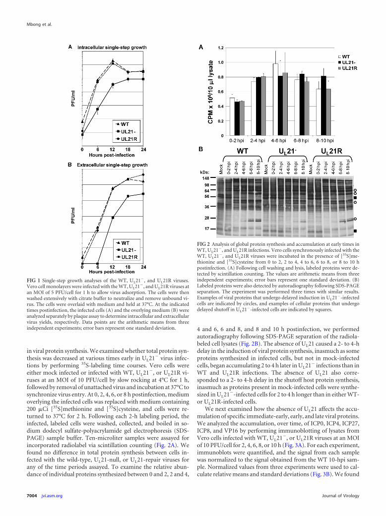
in viral protein synthesis. We examined whether total protein syn-thesis was decreased at various times early in UL21� virus infec-tions by performing 35S-labeling time courses. Vero cells wereeither mock infected or infected with WT, UL21�, or UL21R vi-ruses at an MOI of 10 PFU/cell by slow rocking at 4°C for 1 h,followed by removal of unattached virus and incubation at 37°C tosynchronize virus entry. At 0, 2, 4, 6, or 8 h postinfection, mediumoverlying the infected cells was replaced with medium containing200 �Ci [35S]methionine and [35S]cysteine, and cells were re-turned to 37°C for 2 h. Following each 2-h labeling period, theinfected, labeled cells were washed, collected, and boiled in so-dium dodecyl sulfate-polyacrylamide gel electrophoresis (SDS-PAGE) sample buffer. Ten-microliter samples were assayed forincorporated radiolabel via scintillation counting (Fig. 2A). Wefound no difference in total protein synthesis between cells in-fected with the wild-type, UL21-null, or UL21-repair viruses forany of the time periods assayed. To examine the relative abun-dance of individual proteins synthesized between 0 and 2, 2 and 4,
4 and 6, 6 and 8, and 8 and 10 h postinfection, we performedautoradiography following SDS-PAGE separation of the radiola-beled cell lysates (Fig. 2B). The absence of UL21 caused a 2- to 4-hdelay in the induction of viral protein synthesis, inasmuch as someproteins synthesized in infected cells, but not in mock-infectedcells, began accumulating 2 to 4 h later in UL21� infections than inWT and UL21R infections. The absence of UL21 also corre-sponded to a 2- to 4-h delay in the shutoff host protein synthesis,inasmuch as proteins present in mock-infected cells were synthe-sized in UL21�-infected cells for 2 to 4 h longer than in either WT-or UL21R-infected cells.
We next examined how the absence of UL21 affects the accu-mulation of specific immediate-early, early, and late viral proteins.We analyzed the accumulation, over time, of ICP0, ICP4, ICP27,ICP8, and VP16 by performing immunoblotting of lysates fromVero cells infected with WT, UL21�, or UL21R viruses at an MOIof 10 PFU/cell for 2, 4, 6, 8, or 10 h (Fig. 3A). For each experiment,immunoblots were quantified, and the signal from each samplewas normalized to the signal obtained from the WT 10-hpi sam-ple. Normalized values from three experiments were used to cal-culate relative means and standard deviations (Fig. 3B). We found
FIG 1 Single-step growth analyses of the WT, UL21�, and UL21R viruses.Vero cell monolayers were infected with the WT, UL21�, and UL21R viruses atan MOI of 5 PFU/cell for 1 h to allow virus adsorption. The cells were thenwashed extensively with citrate buffer to neutralize and remove unbound vi-rus. The cells were overlaid with medium and held at 37°C. At the indicatedtimes postinfection, the infected cells (A) and the overlying medium (B) wereanalyzed separately by plaque assay to determine intracellular and extracellularvirus yields, respectively. Data points are the arithmetic means from threeindependent experiments; error bars represent one standard deviation.
FIG 2 Analysis of global protein synthesis and accumulation at early times inWT, UL21�, and UL21R infections. Vero cells synchronously infected with theWT, UL21�, and UL21R viruses were incubated in the presence of [35S]me-thionine and [35S]cysteine from 0 to 2, 2 to 4, 4 to 6, 6 to 8, or 8 to 10 hpostinfection. (A) Following cell washing and lysis, labeled proteins were de-tected by scintillation counting. The values are arithmetic means from threeindependent experiments; error bars represent one standard deviation. (B)Labeled proteins were also detected by autoradiography following SDS-PAGEseparation. The experiment was performed three times with similar results.Examples of viral proteins that undergo delayed induction in UL21�-infectedcells are indicated by circles, and examples of cellular proteins that undergodelayed shutoff in UL21�-infected cells are indicated by squares.
Mbong et al.
7004 jvi.asm.org Journal of Virology
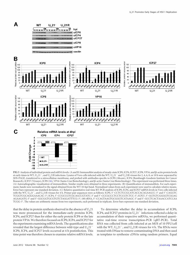
that the delay in protein synthesis observed in the absence of UL21was more pronounced for the immediate-early proteins ICP0,ICP4, and ICP27 than for either the early protein ICP8 or the lateprotein VP16. We therefore focused on ICP0, ICP4, and ICP27 forthe experiments examining mRNA levels. The quantifications alsorevealed that the largest difference between wild-type and UL21�
ICP0, ICP4, and ICP27 levels occurred at 4 h postinfection. Thistime point was therefore chosen to examine relative mRNA levels.
To determine whether the delay in accumulation of ICP0,ICP4, and ICP27 proteins in UL21� infections reflected a delay inaccumulation of their respective mRNAs, we performed quanti-tative real-time reverse transcription-PCR (qRT-PCR). TotalRNA was collected from cells infected at an MOI of 10 PFU/cellwith the WT, UL21�, and UL21R viruses for 4 h. The RNAs weretreated with DNase to remove contaminating DNA and then usedas templates to synthesize cDNAs using random primers. Equal
FIG 3 Analysis of individual protein and mRNA levels. (A and B) Immunoblot analysis of steady-state ICP0, ICP4, ICP27, ICP8, VP16, and �-actin protein levelsat early times in WT, UL21�, and UL21R infections. Lysates of Vero cells infected with the WT, UL21� and UL21R viruses for 2, 4, 6, 8, or 10 h were separated bySDS-PAGE, transferred to a nitrocellulose membrane, and probed with antibodies specific to ICP0 (Abcam), ICP4 (Rumbaugh-Goodwin Institute for CancerResearch), ICP27 (Virusys), ICP8 (16), VP16 (Santa Cruz Biotechnology), and �-actin (Santa Cruz Biotechnology). The experiment was performed three times.(A) Autoradiographic visualization of immunoblots. Similar results were obtained in three experiments. (B) Quantification of immunoblots. For each experi-ment, bands were normalized to the signal obtained from the WT 10-hpi band. Normalized values from each experiment were used to calculate relative means.Error bars represent one standard deviation. (C) Relative quantitative real-time RT-PCR analysis of ICP0, ICP4, and ICP27 mRNA levels in Vero cells infectedwith the WT, UL21�, and UL21R viruses for 4 h. Primer pair sequences were as follows: ICP0, 5=-CCTCTCCGCATCACCACAGAAGCC-3= and 5=-CAGGTCTCGGTCGCAGGGAAACAC-3=; ICP4, 5=-CCGCCGTCGCAGCCGTATC-3= and 5=-CCCGCCCTCCTCCGTCTCC-3=; ICP27, 5=-GGTGTCGGGGTCGGAGAGAAGATG-3= and 5=-GGCGGTGCGTGTCTAGGATTTCG-3=; 18S rRNA, 5=-CCAGTAAGTGCGGGTCATAAGC-3= and 5=-GCCTCACTAAACCATCCAATCGG-3=. The values are arithmetic means from two experiments, each performed in triplicate. Error bars represent one standard deviation.
UL21 Promotes Early Stages of HSV-1 Replication
June 2012 Volume 86 Number 12 jvi.asm.org 7005
amounts of cDNA were used in qRT-PCR mixtures containingprimers specific to the gene of interest and SYBR green for productquantification. Parallel qRT-PCR mixtures contained primersspecific to 18S rRNA as a control to normalize template input. The18S rRNA cycle threshold (CT) value for a given template wassubtracted from the CT obtained for each mRNA of interest (ICP0,ICP4, and ICP27) from the same template to obtain the normal-ized CT value (�CT) for each template. The change (fold, relativeto the wild type) was then calculated using the ��CT method. At 4h postinfection, steady-state ICP4 and ICP27 mRNA levels weredecreased �10-fold and �13-fold, respectively, in UL21�-in-fected cells compared to WT- and UL21R-infected cells (Fig. 3C).ICP0 mRNA levels were also affected by the absence of UL21,although to a lesser extent, showing a 2-fold decrease in UL21�-infected cells compared to WT- and UL21R-infected cells.
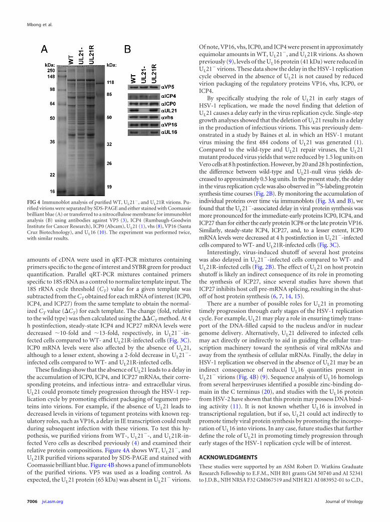
These findings show that the absence of UL21 leads to a delay inthe accumulation of ICP0, ICP4, and ICP27 mRNAs, their corre-sponding proteins, and infectious intra- and extracellular virus.UL21 could promote timely progression through the HSV-1 rep-lication cycle by promoting efficient packaging of tegument pro-teins into virions. For example, if the absence of UL21 leads todecreased levels in virions of tegument proteins with known reg-ulatory roles, such as VP16, a delay in IE transcription could resultduring subsequent infection with these virions. To test this hy-pothesis, we purified virions from WT-, UL21�-, and UL21R-in-fected Vero cells as described previously (4) and examined theirrelative protein compositions. Figure 4A shows WT, UL21�, andUL21R purified virions separated by SDS-PAGE and stained withCoomassie brilliant blue. Figure 4B shows a panel of immunoblotsof the purified virions. VP5 was used as a loading control. Asexpected, the UL21 protein (65 kDa) was absent in UL21� virions.
Of note, VP16, vhs, ICP0, and ICP4 were present in approximatelyequimolar amounts in WT, UL21�, and UL21R virions. As shownpreviously (9), levels of the UL16 protein (41 kDa) were reduced inUL21� virions. These data show the delay in the HSV-1 replicationcycle observed in the absence of UL21 is not caused by reducedvirion packaging of the regulatory proteins VP16, vhs, ICP0, orICP4.
By specifically studying the role of UL21 in early stages ofHSV-1 replication, we made the novel finding that deletion ofUL21 causes a delay early in the virus replication cycle. Single-stepgrowth analyses showed that the deletion of UL21 results in a delayin the production of infectious virions. This was previously dem-onstrated in a study by Baines et al. in which an HSV-1 mutantvirus missing the first 484 codons of UL21 was generated (1).Compared to the wild-type and UL21 repair viruses, the UL21mutant produced virus yields that were reduced by 1.5 log units onVero cells at 8 h postinfection. However, by 20 and 28 h postinfection,the difference between wild-type and UL21-null virus yields de-creased to approximately 0.5 log units. In the present study, the delayin the virus replication cycle was also observed in 35S-labeling proteinsynthesis time courses (Fig. 2B). By monitoring the accumulation ofindividual proteins over time via immunoblots (Fig. 3A and B), wefound that the UL21�-associated delay in viral protein synthesis wasmore pronounced for the immediate-early proteins ICP0, ICP4, andICP27 than for either the early protein ICP8 or the late protein VP16.Similarly, steady-state ICP4, ICP27, and, to a lesser extent, ICP0mRNA levels were decreased at 4 h postinfection in UL21�-infectedcells compared to WT- and UL21R-infected cells (Fig. 3C).
Interestingly, virus-induced shutoff of several host proteinswas also delayed in UL21�-infected cells compared to WT- andUL21R-infected cells (Fig. 2B). The effect of UL21 on host proteinshutoff is likely an indirect consequence of its role in promotingthe synthesis of ICP27, since several studies have shown thatICP27 inhibits host cell pre-mRNA splicing, resulting in the shut-off of host protein synthesis (6, 7, 14, 15).
There are a number of possible roles for UL21 in promotingtimely progression through early stages of the HSV-1 replicationcycle. For example, UL21 may play a role in ensuring timely trans-port of the DNA-filled capsid to the nucleus and/or in nucleargenome delivery. Alternatively, UL21 delivered to infected cellsmay act directly or indirectly to aid in guiding the cellular tran-scription machinery toward the synthesis of viral mRNAs andaway from the synthesis of cellular mRNAs. Finally, the delay inHSV-1 replication we observed in the absence of UL21 may be anindirect consequence of reduced UL16 quantities present inUL21� virions (Fig. 4B) (9). Sequence analysis of UL16 homologsfrom several herpesviruses identified a possible zinc-binding do-main in the C terminus (20), and studies with the UL16 proteinfrom HSV-2 have shown that this protein may possess DNA bind-ing activity (11). It is not known whether UL16 is involved intranscriptional regulation, but if so, UL21 could act indirectly topromote timely viral protein synthesis by promoting the incorpo-ration of UL16 into virions. In any case, future studies that furtherdefine the role of UL21 in promoting timely progression throughearly stages of the HSV-1 replication cycle will be of interest.
ACKNOWLEDGMENTS
These studies were supported by an ASM Robert D. Watkins GraduateResearch Fellowship to E.F.M., NIH R01 grants GM 50740 and AI 52341to J.D.B., NIH NRSA F32 GM067519 and NIH R21 AI 083952-01 to C.D.,
FIG 4 Immunoblot analysis of purified WT, UL21�, and UL21R virions. Pu-rified virions were separated by SDS-PAGE and either stained with Coomassiebrilliant blue (A) or transferred to a nitrocellulose membrane for immunoblotanalysis (B) using antibodies against VP5 (3), ICP4 (Rumbaugh-GoodwinInstitute for Cancer Research), ICP0 (Abcam), UL21 (1), vhs (8), VP16 (SantaCruz Biotechnology), and UL16 (10). The experiment was performed twice,with similar results.
Mbong et al.
7006 jvi.asm.org Journal of Virology
a Howard Hughes Medical Institute Undergraduate Science Educationgrant to The University of Alabama, and the Louis Stokes Alliance forMinority Participation Bridge to the Doctorate Program supported byNSF HRD-0602359.
We thank Duncan Wilson for the gift of the anti-vhs antibody and BillRuyechan for the gift of the anti-ICP8 antibody.
REFERENCES1. Baines JD, Koyama AH, Huang T, Roizman B. 1994. The UL 21 gene of
herpes simplex virus 1 is dispensable for replication in cell culture. J. Virol.68:2929 –2936.
2. Campbell ME, Palfreyman JW, Preston CM. 1984. Identification ofherpes simplex virus DNA sequences which encode a trans-acting poly-peptide responsible for stimulation of immediate early transcription. J.Mol. Biol. 180:1–19.
3. Cohen GH, et al. 1980. Structural analysis of the capsid polypeptides ofherpes simplex virus types 1 and 2. J. Virol. 34:521–531.
4. Duffy C, et al. 2006. Characterization of a UL49-null mutant: VP22 ofherpes simplex virus type 1 facilitates viral spread in cultured cells and themouse cornea. J. Virol. 80:8664 – 8675.
5. Fuchs W, Granzow H, Klupp BG, Kopp M, Mettenleiter TC. 2002. TheUL48 tegument protein of pseudorabies virus is critical for intracytoplas-mic assembly of infectious virions. J. Virol. 76:6729 – 6742.
6. Hardwicke MA, Sandri-Goldin RM. 1994. The herpes simplex virusregulatory protein ICP27 contributes to the decrease in cellular mRNAlevels during infection. J. Virol. 68:4797– 4810.
7. Hardy WR, Sandri-Goldin RM. 1994. Herpes simplex virus inhibits hostcell splicing, and regulatory protein ICP27 is required for this effect. J.Virol. 68:7790 –7799.
8. Lee GE, Church GA, Wilson DW. 2003. A subpopulation of tegumentprotein vhs localizes to detergent-insoluble lipid rafts in herpes simplexvirus-infected cells. J. Virol. 77:2038 –2045.
9. Meckes DG, Jr, Marsh JA, Wills JW. 2010. Complex mechanisms for thepackaging of the UL16 tegument protein into herpes simplex virus. Virol-ogy 398:208 –213.
10. Nalwanga D, Rempel S, Roizman B, Baines JD. 1996. The UL 16 geneproduct of herpes simplex virus 1 is a virion protein that colocalizes withintranuclear capsid proteins. Virology 226:236 –242.
11. Oshima S, et al. 1998. Characterization of the UL16 gene product ofherpes simplex virus type 2. Arch. Virol. 143:863– 880.
12. Pellett PE, McKnight JL, Jenkins FJ, Roizman B. 1985. Nucleotidesequence and predicted amino acid sequence of a protein encoded in asmall herpes simplex virus DNA fragment capable of trans-inducing alphagenes. Proc. Natl. Acad. Sci. U. S. A. 82:5870 –5874.
13. Read GS, Karr BM, Knight K. 1993. Isolation of a herpes simplex virustype 1 mutant with a deletion in the virion host shutoff gene and identifi-cation of multiple forms of the vhs (UL41) polypeptide. J. Virol. 67:7149 –7160.
14. Sacks WR, Greene CC, Aschman DP, Schaffer PA. 1985. Herpes simplexvirus type 1 ICP27 is an essential regulatory protein. J. Virol. 55:796 – 805.
15. Sandri-Goldin RM, Mendoza GE. 1992. A herpesvirus regulatory proteinappears to act post-transcriptionally by affecting mRNA processing.Genes Dev. 6:848 – 863.
16. Shelton LS, Albright AG, Ruyechan WT, Jenkins FJ. 1994. Retention ofthe herpes simplex virus type 1 (HSV-1) UL37 protein on single-strandedDNA columns requires the HSV-1 ICP8 protein. J. Virol. 68:521–525.
17. Takakuwa H, et al. 2001. Herpes simplex virus encodes a virion-associated protein which promotes long cellular processes in over-expressing cells. Genes Cells 6:955–966.
18. Tanaka M, Kagawa H, Yamanashi Y, Sata T, Kawaguchi Y. 2003.Construction of an excisable bacterial artificial chromosome containing afull-length infectious clone of herpes simplex virus type 1: viruses recon-stituted from the clone exhibit wild-type properties in vitro and in vivo. J.Virol. 77:1382–1391.
19. Tischer BK, Smith GA, Osterrieder N. 2010. En passant mutagenesis: atwo step markerless red recombination system. Methods Mol. Biol. 634:421– 430.
20. Wing BA, Lee GCY, Huang ES. 1996. The human cytomegalovirus UL94open reading frame encodes a conserved herpesvirus capsid/tegument-associated virion protein that is expressed with true late kinetics. J. Virol.70:3339 –3345.
UL21 Promotes Early Stages of HSV-1 Replication
June 2012 Volume 86 Number 12 jvi.asm.org 7007