Embed Size (px)
Citation preview
TOBYregister
coolingUK
UK TOBY Cooling Register Clinician’s Handbook
Version 4, May 2010
2
3
Contents
1. IntRoDUCtIon 5
2. WHen to ConsIDeR tReAtMent WItH CooLInG 6
2.1 treatment criteria 6
2.2 When to start cooling 7
2.3 other assessment 7
2.4 When is cooling not appropriate? 8
3. WHeRe sHoULD InFAnts Be tReAteD WItH CooLInG? 9
4. CooLInG FoR InFAnts BoRn oUtsIDe tReAtInG CentRes 9
5. Consent 10
6. MAnAGeMent 10
6.1 Maintaining cooling 10
6.2 seizures 10
6.3 Ventilation 11
6.4 Cardiovascular support 11
6.5 Anti-oedema therapy 12
6.6 Analgesic and sedative therapy 12
6.7 Fluid Management 12
6.8 sepsis 12
6.9 Rewarming procedures 12
7. DAtA CoLLeCtIon 13
8. aeeG MonItoRInG 13
8.1 scalp needle electrodes 13
8.2 Adhesive electrodes 14
8.3 Artefacts 14
9. GUIDe to stARtInG CooLInG In CooLInG CentRes 15
10. MAGnetIC ResonAnCe IMAGInG 16
4
11. LonG teRM oUtCoMe 17
12. CHeCKLIst 18
13. DeFInItIons oF teRMs In DAtA CoLLeCtIon FoRM 19
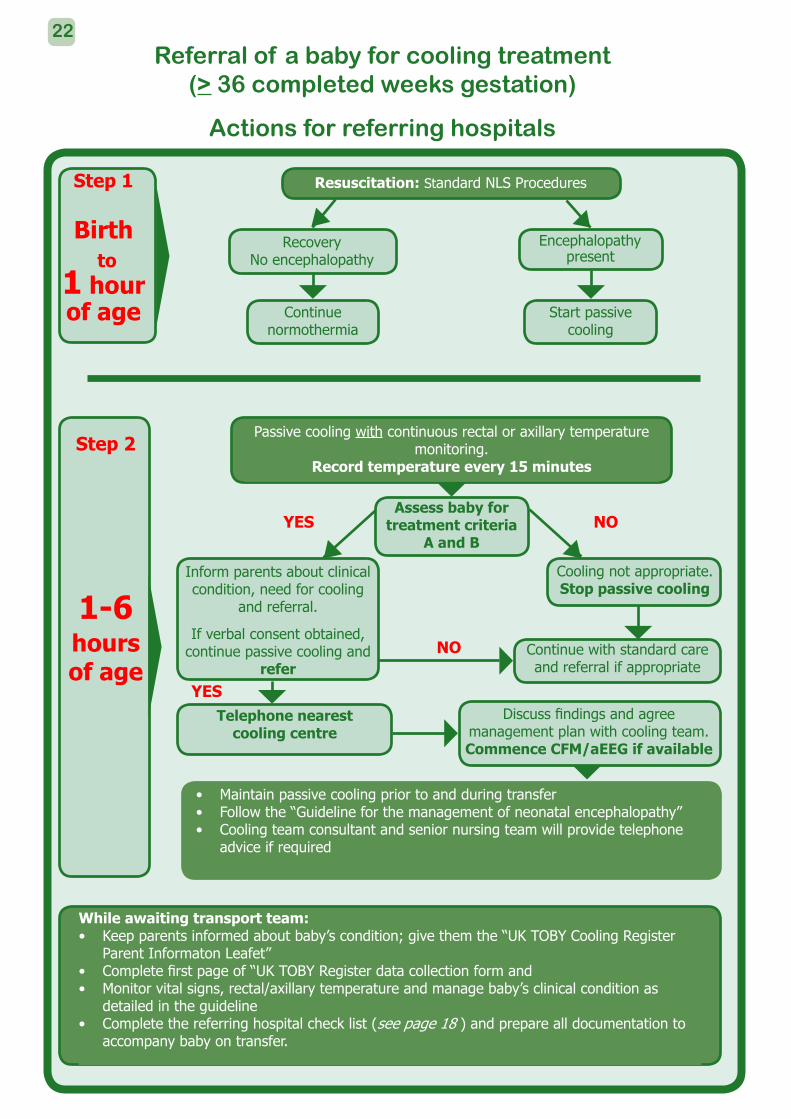
14. ReFeRRAL oF A BABY FoR CooLInG tReAtMent 22
15. PAssIVe CooLInG 23
5
1. INTRODUCTION
Current evidence indicates that moderate induced hypothermia (cooling) to a rectal temperature of 33-34oC improves survival and neurological outcomes to 18 months of age in infants with moderate or severe perinatal asphyxial encephalopathy (BMJ. 2010 Feb 9;340:c363).
The UK TOBY Cooling Register administered from the National Perinatal Epidemiology Unit (www.npeu.ox.ac.uk/tobyregister) has been set up to guide clinicians and audit treatment with cooling in the UK.
All infants receiving therapeutic hypothermia should be registered with the UK toBY Cooling Register.
6
2. WHEN TO CONSIDER TREATMENT WITH COOLING
Consider treatment with cooling in infants that meet the following criteria:
2.1 treatment criteria
A. Infants≥36completedweeksgestationadmittedtotheneonatalunitwith at least one of the following:
Apgar score of ≤5 at 10 minutes after birth•
Continued need for resuscitation, including endotracheal or mask ventilation, at • 10 minutes after birth
Acidosis within 60 minutes of birth (defined as any occurrence of umbilical cord, • arterial or capillary pH <7.00)
Base Deficit ≥ 16 mmol/L in umbilical cord or any blood sample (arterial, venous • or capillary) within 60 minutes of birth
Infants that meet criteria A should be assessed for whether they meet the neurological abnormality entry criteria (B):
B. seizures or moderate to severe encephalopathy, consisting of:
Altered state of consciousness (reduced response to stimulation or absent • response to stimulation) and
Abnormal tone (focal or general hypotonia, or flaccid) and•
Abnormal primitive reflexes (weak or absent suck or Moro response) •
Infants who meet criteria A and B may be considered for treatment with cooling.
If an infant meets these criteria, but cooling is NOT offered, the reasons for this should be clearly documented in the medical notes. It is possible that this decision may need to be justified in the future, should litigation ensue for example.
7
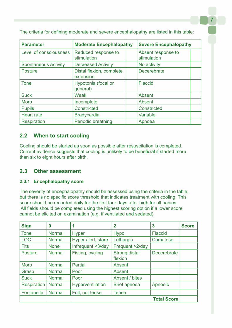
The criteria for defining moderate and severe encephalopathy are listed in this table:
Parameter Moderate encephalopathy severe encephalopathyLevel of consciousness Reduced response to
stimulationAbsent response to stimulation
Spontaneous Activity Decreased Activity No activityPosture Distal flexion, complete
extensionDecerebrate
Tone Hypotonia (focal or general)
Flaccid
Suck Weak AbsentMoro Incomplete AbsentPupils Constricted ConstrictedHeart rate Bradycardia VariableRespiration Periodic breathing Apnoea
2.2 When to start cooling
Cooling should be started as soon as possible after resuscitation is completed. Current evidence suggests that cooling is unlikely to be beneficial if started more than six to eight hours after birth.
2.3 other assessment
2.3.1 encephalopathy score
The severity of encephalopathy should be assessed using the criteria in the table, but there is no specific score threshold that indicates treatment with cooling. This score should be recorded daily for the first four days after birth for all babies. All fields should be completed using the highest scoring option if a lower score cannot be elicited on examination (e.g. if ventilated and sedated).
sign 0 1 2 3 scoreTone Normal Hyper Hypo FlaccidLOC Normal Hyper alert, stare Lethargic ComatoseFits None Infrequent <3/day Frequent >2/dayPosture Normal Fisting, cycling Strong distal
flexionDecerebrate
Moro Normal Partial AbsentGrasp Normal Poor AbsentSuck Normal Poor Absent / bitesRespiration Normal Hyperventilation Brief apnoea Apnoeic
Fontanelle Normal Full, not tense Tensetotal score
8
2.3.2 aeeG assessment
The amplitude integrated EEG (aEEG) must be recorded in all infants treated with cooling but cooling need not be delayed until the aEEG is initiated.
A normal aEEG record (confirmed by assessing the underlying EEG and excluding artefact distortion of aEEG) indicates a high probability of normal outcome, and clinicians may consider that treatment with cooling is not required.
Continued aEEG recording during the treatment period is helpful clinically, to assess occurrence of seizures and monitor the severity of encephalopathy. IV anticonvulsant therapy may cause transient suppression of EEG activity. Ideally the aEEG should be performed before administering anticonvulsant therapy.
Information on the use of the aEEG is available from the suppliers of the cerebral function monitors, from text books and on the following website:www.neoweb.org.uk/cfm. Refer to section 8 for further information on aEEG.
If the aEEG becomes normal and the infant no longer has encephalopathy by 6 hours of age the need for continuing cooling can be reconsidered. Apparent improvement of the aEEG after 6 hours of age is not an indication for discontinuing cooling.
2.4 When is cooling not appropriate?
Cooling is not appropriate if:
• The infant is likely to require surgery during the first 3 days after birth
• There are other abnormalities indicative of poor long term outcome
Cooling may not be appropriate if the infant appears moribund or has persisting extremely severe encephalopathy such that further treatment is likely to be futile, for example if the aEEG/EEG is isoelectric beyond 12-24 hours of age.
Cooling may produce adverse respiratory or cardiovascular effects and should be used with caution in infants with an unstable respiratory or cardiovascular condition.
There is limited evidence to support treatment with cooling in infants less than 36 weeks gestational age or with other conditions such as postnatal collapse or cerebral infarction. Compassionate use of treatment with cooling outside the published protocols requires special arrangements for consent and explanation to the parents about the lack of evidence for safety and efficacy in these situations.
9
3. WHERE SHOULD INFANTS BE TREATED WITH COOLING?
Neonatal networks will decide which centres should provide cooling to meet local need. These centres should become familiar with the TOBY Cooling Register protocol and the monitoring and cooling equipment before providing this treatment, and should have facilities for providing full intensive care, recording the aEEG and carrying out appropriate investigations, neuroimaging, including MRI, and follow-up.The TOBY clinical and administrative co-ordinating centres will continue to assist centres treating infants with cooling.
4. COOLING FOR INFANTS BORN OUTSIDE TREATING CENTRES
(See flow-charts on ‘Referral of a baby for cooling treatment’ page 22 and ‘Passive cooling’ page 23) All infants that require resuscitative measures at birth should have a neurological assessment once physiological stability is achieved.
The possibility of treatment with cooling should be considered for babies that meet the clinical criteria A and B above.
The local network cooling treatment centre should be contacted to agree a management plan.
Clinicians should discuss the option of treatment with the parents and seek parental assent for the baby to be transferred for treatment with cooling.
Following parental assent, cooling should be initiated prior to and during the transfer to the cooling centre.
Cooling outside the treatment centre is started by turning off heating equipment, and removing coverings from the baby. If necessary a fan can help induce cooling (see passive cooling protocol). The baby’s age at the time heating equipment is turned off should be entered as the time cooling started on the data collection form.
The baby should be monitored and observation data collected during this period according to the TOBY Cooling Register protocol. This includes continuous monitoring of rectal temperature, blood pressure and heart rate.
If immediate transfer to a cooling treatment centre is not possible, for example because of a lack of intensive care cots, passive cooling should be continued with guidance from the cooling centre.
10
5. CONSENT
Clinicians should always discuss the option of cooling treatment with parents and seek parental assent as soon as practically possible.
Details of all discussion with parents about their baby’s treatment with cooling should be documented in the baby’s notes.
Local Trust clinical governance procedures and policy for consent for treatment should be followed.
6. MANAGEMENT Please note that drug dosages stated here are a guide only, local protocols should be followed where appropriate.
6.1 Maintaining cooling
Cooling should be maintained using appropriate selective head cooling or whole body cooling equipment. Only certified equipment should be used to provide treatment with cooling. The manufacturer’s instructions should be followed when using the cooling equipment.
The target rectal temperature when using selective head cooling is 34.5 °C, maintained for 72 hours, followed by slow rewarming (over 12 hours) to 37.2 °C (normothermia).
During whole body cooling the target rectal temperature is 33-34° C, for 72 hours, followed by slow rewarming (over 12 hours) to 37.2 °C (normothermia).
The investigations and monitoring listed in the TOBY Register data form should be carried out on each infant treated with cooling. It is essential that continuous rectal temperature monitoring should be performed, with hourly recordings documented.
6.2 seizures
The management of seizures will be guided by local protocols.
In general, symptomatic seizures or frequent (>3/hr) subclinical (EEG) seizures will be treated with anticonvulsants.
Cooling may affect the metabolism of several drugs, including anticonvulsants and sedatives, and toxic drug levels may occur even with normal doses.
1st line anticonvulsants may be phenobarbital or phenytoin. Phenytoin should be administered at a rate no faster than 1mg/minute.
10
11
Benzodiazapines such as midazolam, or clonazapam are commonly used if seizures persist. The dose should be adjusted according to response.
Lidocaine is sometimes used as a further anticonvulsant, but should be avoided if phenytoin has previously been administered. There is a risk of toxicity if the lidocaine infusion is continued more than 6 hours at 6mg/kg/hr, or more than 12 hours at lower doses.
Severe bradycardia or ventricular tachycardia has been noted following phenytoin or lidocaine administration. Treatment consists of immediately discontinuing the agent and initiating standard resuscitative measures. It is not known if the risk of arrhythmia is increased with cooling.
6.3 Ventilation
Almost all infants treated with cooling will initially require mechanical ventilation.
Ventilatory care will be managed according to the Treatment Centre’s standard policy and this may include treatment with high frequency oscillation and inhaled nitric oxide if necessary.
Blood gases will guide ventilatory requirements; as a guide PaO2 should be maintained between 6-10 KPa and the PaCO2 between 5-7 KPa.
Ventilator gases should be warmed and humidified in the normal way, according to local policy.
6.4 Cardiovascular support
Alterations in heart rate and blood pressure are common during cooling. In general the heart rate is reduced and blood pressure increases with a reduction in body temperature.
Most infants with a rectal temperature of 33-34°C (the target rectal temperature for whole body cooling) will have a heart rate around 100 bpm and a mean blood pressure greater than 40 mmHg.
A rapid rise in body temperature may cause hypotension by inducing peripheral vasodilatation.
Causes of hypotension should be sought and appropriate treatment provided. An echocardiogram may help guide treatment.
Treatment with volume replacement and/or inotropes should be considered if the mean arterial blood pressure is less than 40 mmHg. A bolus of 10-20 ml/kg of normal saline should be given initially, and repeated if necessary. If the blood pressure remains low following treatment with normal saline the infant should be treated with dopamine 5-10 micrograms/kg/min, and/or dobutamine 5-10 micrograms/kg/min. Persistent failure of response may be treated by increasing the dose of dopamine and dobutamine up to 20 micrograms/kg/min.
12
6.5 Anti-oedema therapy
Infants being treated with cooling should not be treated with steroids (other than for treatment of hypotension), or mannitol.
6.6 Analgesic and sedative therapy
Stress may have adverse effects in asphyxiated infants and may influence the therapeutic effect of hypothermia. In addition, neonatal intensive care procedures may cause considerable stress to infants and cooling may also be associated with stress.
Signs of distress include tachycardia, facial grimacing and irritability. A heart rate consistently above 110 bpm in cooled infants suggests that the infant is distressed.
Ventilated infants may be sedated with intravenous morphine, maximum loading dose 50 micrograms/kg over 30 minutes followed by 10-20 micrograms/kg/hour. Morphine should be discontinued after 24-48 hours to lessen the risk of accumulation and toxicity.
Non-ventilated infants who appear distressed will also require sedative therapy, for example with chloral hydrate, 50 mg/kg. Respiratory function must be monitored in these babies.
6.7 Fluid Management
Renal function is commonly impaired following severe perinatal asphyxia. The infant’s weight, blood creatinine and electrolytes and urine output will guide fluid management.
As a guide infants will require about 40-60 ml/kg/day. Infants in renal failure should receive a total of 30 ml/kg/24 hours plus any measured losses. Boluses of 0.9% saline may be required to avoid hypovolaemia if diuresis occurs in the infant or if vasodilatation occurs during rewarming.
Enteral feeding can be cautiously introduced once the initial biochemical and metabolic disturbance are corrected, usually after about 24 hours.
6.8 sepsis
Antibiotic therapy may be given if clinically indicated.
6.9 Rewarming procedures
Cooling is concluded after 72 hours (or earlier if clinical circumstances dictate).
13
The rectal temperature should be allowed to rise by no more than 0.2-0.3°C per hour, to 37+/- 0.2°C.
The manufacturer’s instructions should be carefully followed to avoid excessive rewarming.
Re-warming can be carried out by adjusting the thermostat set point of the cooling equipment or the incubator in increments of 0.2-0.3°C hourly.
The infant’s temperature must be carefully monitored for 24 hours after normothermia has been achieved to prevent rebound hyperthermia, as this might be detrimental.
7. DATA COLLECTION
The data collection form (based on the TOBY Study data collection form) should be used for all babies being treated with cooling for perinatal asphyxial encephalopathy.
Data collection covers the first 4 days following induction of hypothermia, recording clinical information about the baby’s condition at birth, pregnancy and delivery information, hourly temperature recordings and basic daily information summarising the care and treatments required by the baby.
Outcome data are also collected, including MRI information. If MRI findings are not available at the time the form is submitted then a subsequent request will be made to collect this information.
All data submitted to the Register must be anonymised, and identified only by the baby’s Register PIN. Please ensure that ALL items sent to the Register are clearly marked with the PIN.
Keep photocopies of all completed forms, clearly marked with the Register PIN and hospital patient identifiers for your own records.
8. aEEG MONITORING
Adhesive or needle electrodes may be used. These should be placed in the parietal area according to the instructions specific to the cerebral function monitor (CFM) used.
8.1 scalp needle electrodes
• Choose site of insertion and part the hair using damp gauze, then dry the area, maintaining the parting
14
• Clean exposed skin according to local policy
• Insert needle under the skin, in line with the parting
• If baby is to wear a hat for securing ET tube, direct the leads to the front of the head so that they sit easily leading away from the hat towards the forehead
• Using collodion (supplied in a tube, not the liquid variety from a bottle) place a spot on the hub of the needle and exposed skin, and hold in place until the collodion is dry. Once the collodion is dry this is very secure, it can be easily inspected and there is no need for tape. Acetone (or substitute) will aid removal
8.2 Adhesive electrodes
The skin needs meticulous preparation to result in a good trace with low impedance.
• Part any hair using a wet swab and clean the application site according to local policy
• Gently rub the skin using abrasive paste such as Nu-Prep or use the edge of an “orange stick”
• Apply the electrode and hold in place for about 30 seconds
• Check impedance. If impedance is greater than 5Kohms, gently dribble some sterile water through a syringe to edge of electrode to improve conduction
• Place a bonnet over head to help keep electrodes in place
Needle electrodes will probably be quicker to insert, and more likely to result in a good trace. If the electrodes cannot be attached on the parietal area because of hair, they can be attached at the hair line in the fronto-temporal area but the trace may be artefacted by muscle movement.
8.3 Artefacts
Artefacts are common during long term monitoring of aEEG. Always inspect the underlying EEG for artefact at the start of a recording or if the aEEG trace alters.
A common artefact is elevation of the baseline due to ECG signal. The underlying EEG will display a regular artefact with the same frequency as the heart rate. Common movement artefacts are due to head movement due to gasping or caused
15
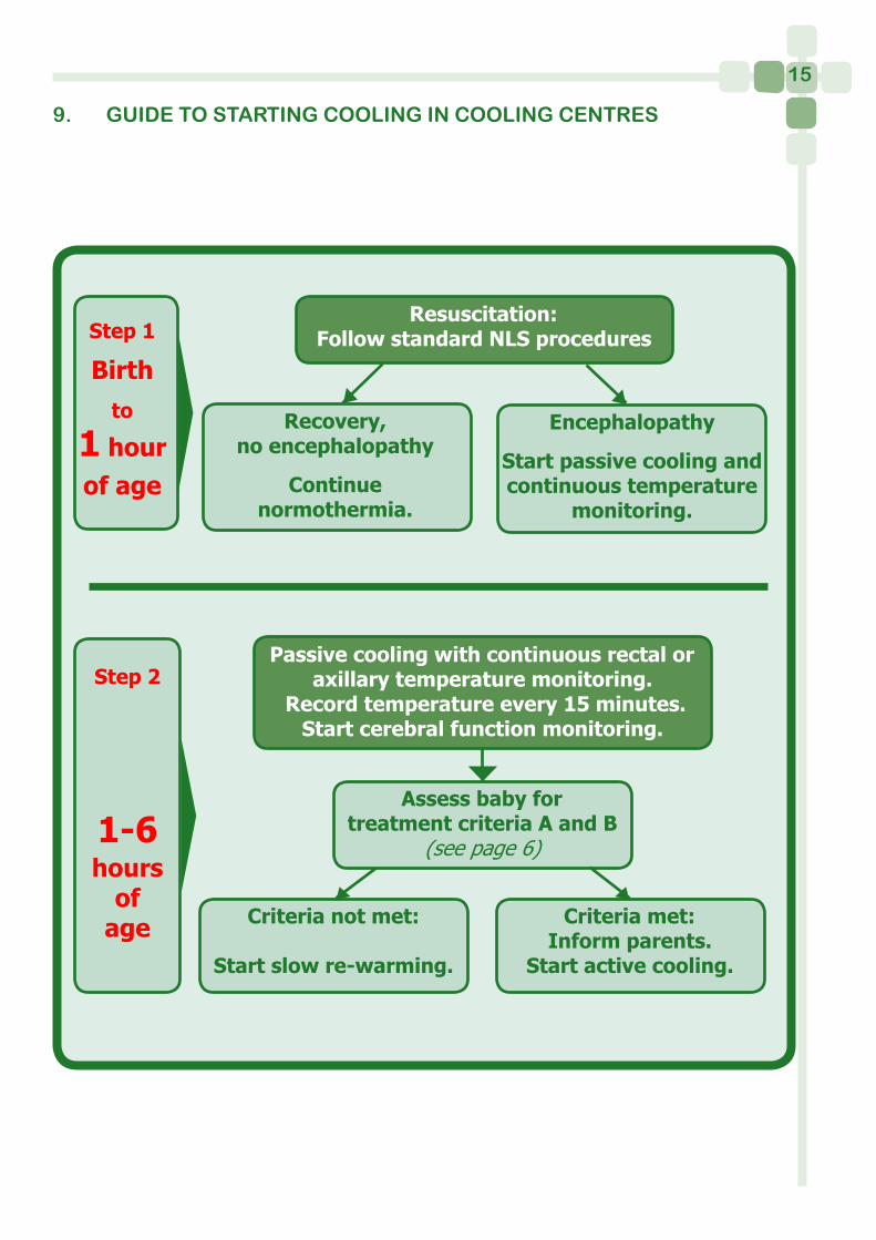
9. GUIDE TO STARTING COOLING IN COOLING CENTRES
Criteria met:Inform parents.
Start active cooling.
Recovery, no encephalopathy
Continue normothermia.
Encephalopathy
Start passive cooling and continuous temperature
monitoring.
Passive cooling with continuous rectal or axillary temperature monitoring.
Record temperature every 15 minutes. Start cerebral function monitoring.
Assess baby for treatment criteria A and B
(see page 6)
Criteria not met:
Start slow re-warming.
Resuscitation:Follow standard NLS proceduresStep 1
Birthto
1 hour of age
Step 2
1-6 hours
of age
16
by mechanical ventilation. This type of artefact often causes widening of the aEEG trace.
Examples of artefacts affecting the EEG are provided in the aEEG handbook. (www.npeu.ox.ac.uk/toby)
10. MAGNETIC RESONANCE IMAGING
An MRI should be done in all babies within about 2 weeks of birth. MRI is the imaging modality of choice for assessing the distribution of injury and likely prognosis and to support a diagnosis of hypoxic ischaemic encephalopathy.
Images should be obtained in the transverse plane with T1 weighted spin echo, T2 weighted spin echo and age related inversion recovery sequences†, and assessed for abnormal signal intensities by an experienced observer. Additionally, where possible, a set of sagittal T1 weighted volume images should also be acquired.
† suggested sequences:
T1 weighted spin echo (SE 860/20)•
T2 weighted spin echo (SE 3000/120)•
Inversion recovery (IR 3800/30/950)•
Normal anatomical features of the brain with no evidence of antenatal injury or malformation e.g. dilated ventricles, widened extra cerebral space or abnormal cortical folding, should be confirmed.
The pattern of abnormal signal intensities observed should be documented as follows:
The posterior limb of the internal capsule (PLIC) assessed as normal, equivocal • or abnormal
The basal ganglia and thalami assessed as normal, or with minimal, moderate or • severe abnormality
Minimal: focal abnormalities but normal signal within the PLIC•
Moderate: focal abnormalities involving the posterior lentiform nuclei and • ventrolateral nuclei of the thalami with equivocal or abnormal signal intensity within the PLIC
Severe: widespread abnormalities in all regions of the basal ganglia and thalami • and abnormal signal intensity within the PLIC
White matter abnormalities should be assessed according to which lobes of the • brain are involved, whether there is a haemorrhagic element to the lesion and whether it is subcortical, periventricular or widespread
Abnormalities in the white matter should be described as moderate or severe•

17
Moderate: small focal lesions with a short T1 and short T2, consistent with • haemorrhage and/or areas with an exaggerated long T1 and long T2 but no loss of grey/white matter differentiation
Severe: more marked areas of abnormality with larger haemorrhages or • exaggerated long T1 and T2 with loss of grey/white matter differentiation, consistent with infarction
The presence of extra cerebral haemorrhage or collections should be noted, in particular, evidence of subdural or large subarachnoid haemorrhage. Where possible the presence of normal flow in the sinuses should be confirmed and any thrombosis documented. The latter will require a set of both transverse and sagittal images.
A copy of the MRI report should be obtained from the radiologist.
11. LONG TERM OUTCOME
Infants should be followed up regularly after discharge and a formal neurological examination and psychomotor assessment should be carried out at approximately 2 years of age.
Anonymised data from the 2 year follow-up assessment should be submitted to the Cooling Register, on the form provided, and using the baby’s Register PIN as identification.
A reminder will be sent to the hospital that provided the cooling treatment at birth when 2 year follow-up data are due to be collected.
18
12. CHECKLIST
• Baby meets Group A and Group B criteria
• No contraindications to using cooling present
• Discuss baby’s condition and treatment options, including cooling, with parents
• Initiate cooling as soon as possible, or document reasons for not doing so
• Start CFM (aEEG) recording if equipment available as soon as possible, but not necessarily before cooling is commenced
• Obtain Register number for baby and document relevant information on a Register data form
• Ensure that PIN and baby details are added to local central record of cooled babies so that all PINs can be linked to patients in the future (to enable follow up data collection for example).
• Monitor rectal temperature continuously, record hourly until rewarming completed
• Maintain hypothermia for 72 hours then rewarm.
• Complete all sections of the data form
• Arrange MRI between 5 and 14 days of age if possible
• Submit MRI findings to the Register, either on the data form or when available subsequently. Remember to remove identifiers apart from Register PIN
• Explain to parents need for continued follow up
• Submit follow-up data to the Register following 2 year assessment, using the form provided
• NB: all data submitted to the Register must be anonymised, and identified only by the baby’s register PIN. Please ensure that ALL items sent to the Register are clearly marked with the PIN
• Keep photocopies of all completed forms, clearly marked with the Register PIN and hospital patient identifiers for your own records
19
13. DEFINITIONS OF TERMS IN DATA COLLECTION FORM
DEFINITIONS OF TERMS IN DATA COLLECTION FORM
Age when cooling commenced Age of baby in hours and minutes when passive cooling is allowed to start, if this takes place prior to active cooling using cooling equipment
Arrhythmia Sinus bradycardia below 80 bpm and other arrhythmias identified on ECG
CoagulopathyAny disorder requiring treatment in order to maintain or recover normal haemostasis
Delivery complications This can include prolapsed cord, abruption, shoulder dystocia, ruptured uterus, head entrapment etc
DiabetesExisting diagnosis of diabetes, or gestational diabetes requiring treatment
EDDUse the best estimate (dates or ultrasound) based on a 40 week gestation
Head entrapmentDelayed second stage during breech delivery, vaginally or at caesarean section
Hypoglycaemia (infant)Blood glucose below 2.6mmol/litre
Hypotension (infant)Persistent mean blood pressure of < 40mmHg
Illicit drug useRecorded drug or alcohol use that may lead to social, occupational, psychological, or physical problems
Late onset sepsis (>72 hours after birth) confirmed by blood or CSF culture. Any evidence of infection requiring antibiotic therapy which is confirmed on culture
Major cerebral anomaly Including evidence of parenchymal haemorrhage as determined by ultrasound, ventricular dilatation (defined as >97th centile for gestational age) or the presence of porencephalic cysts or cystic leukomalacia
Maternal seizure Convulsions due to eclampsia or other causes, e.g. epilepsy
20
Meconium aspiration syndrome The presence of meconium stained liquor at birth and severe respiratory distress within 1 hour of birth and compatible X-ray changes
Necrotising enterocolitisInfants with abdominal distension, gastric aspirate and/or blood in stools together with abdominal X-ray showing bowel oedema, pneumatosis or pneumoperitoneum, i.e. Bell’s staging 2 or 3
Placental abruptionSeparation of a normally situated placenta after 28th week of pregnancy
Placenta PraeviaPlacenta partially or wholly covering the internal cervical os
Pre-eclampsiaHypertension greater than 140/90 mmHg, during pregnancy
Pregnancy complicationsThis can include: pre-eclampsia, maternal seizure, thyroid disorder, diabetes, placenta praevia, known illicit drug use etc
Prolapsed cordCord presentation following rupture of membranes
Pulmonary airleakAny radiologically confirmed airleak serious enough to affect management (including pneumothorax, pulmonary interstitial emphysema, pneumopericardium, pneumoperitoneum and pneumomediastinum)
Pulmonary haemorrhageCopious bloody secretions with clinical deterioration requiring change(s) in ventilatory management.
Pulmonary hypertensionSevere hypoxaemia disproportionate to the severity of lung disease and evidence of a right to left shunt
Respiratory supportUse of mechanical ventilation, CPAP or supplementary oxygen
Ruptured uterusSpotaneous full-thickness tear in the uterine wall due to existing scar, obstructed labour, etc
Seizures.Clinical or subclinical, identified on CFM /EEG
21SepsisAny evidence of infection requiring antibiotic therapy which is confirmed on culture
Shoulder dystociaFailure of the shoulders to rotate into the anteroposterior diameter of the pelvis following delivery of the head, resulting in a substantial delay in delivery
Maternal Thyroid disorderThyroid dysfunction requiring treatment during pregnancy
22
Passive cooling with continuous rectal or axillary temperature monitoring.
Record temperature every 15 minutes
Referral of a baby for cooling treatment (> 36 completed weeks gestation)
Actions for referring hospitals
Inform parents about clinical condition, need for cooling
and referral.
If verbal consent obtained, continue passive cooling and
refer
Assess baby for treatment criteria
A and B
Cooling not appropriate.Stop passive cooling
Continue with standard care and referral if appropriate
YES
YES
NO
Telephone nearest cooling centre
Discuss findings and agree management plan with cooling team.Commence CFM/aEEG if available
Maintain passive cooling prior to and during transfer• Follow the “Guideline for the management of neonatal encephalopathy” • Cooling team consultant and senior nursing team will provide telephone • advice if required
While awaiting transport team:Keep parents informed about baby’s condition; give them the “UK TOBY Cooling Register • Parent Informaton Leafet” Complete first page of “UK TOBY Register data collection form and • Monitor vital signs, rectal/axillary temperature and manage baby’s clinical condition as • detailed in the guidelineComplete the referring hospital check list (• see page 18 ) and prepare all documentation to accompany baby on transfer.
NO
Resuscitation: Standard NLS ProceduresStep 1
Birth to
1 hour of age
RecoveryNo encephalopathy
Continue normothermia
Encephalopathy present
Start passive cooling
Step 2
1-6
hours of age
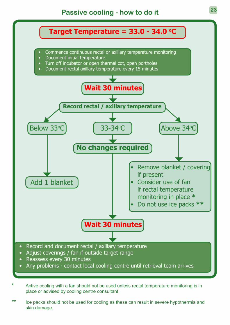
Passive cooling - how to do it
Remove blanket / covering • if presentConsider use of fan • if rectal temperature monitoring in place *Do not use ice packs • **
Commence continuous rectal or axillary temperature monitoring• Document initial temperature• Turn off incubator or open thermal cot, open portholes• Document rectal axillary temperature every 15 minutes•
Target Temperature = 33.0 - 34.0 oC
Add 1 blanket
Record and document rectal / axillary temperature• Adjust coverings / fan if outside target range• Reassess every 30 minutes• Any problems - contact local cooling centre until retrieval team arrives•
Below 33oC 33-34oC Above 34oC
No changes required
Wait 30 minutes
Record rectal / axillary temperature
* Active cooling with a fan should not be used unless rectal temperature monitoring is in place or advised by cooling centre consultant.
** Ice packs should not be used for cooling as these can result in severe hypothermia and skin damage.
Wait 30 minutes
23
TOBYregister
coolingUK
Clinical Co-ordinating Centre:Department of PaediatricsFaculty of MedicineImperial College LondonHammersmith CampusDu Cane RoadLondon Tel: 0208 383 3326W12 0NN Fax: 0208 740 8281
Data Co-ordinating Centre:UK TOBY Cooling Register NPEU Clinical Trials UnitNational Perinatal Epidemiology UnitUniversity of OxfordOld Road CampusHeadingtonOxford Tel: 01865 289735 / 617919OX3 7LF Fax: 01865 289740
Web: www.npeu.ox.ac.uk/tobyregister email: [email protected]
7 June 2010