Embed Size (px)
Citation preview
Issued by the Standards Unit, Microbiology Services, PHE Bacteriology | B 2 | Issue no: 6.1 | Issue date: 04.05.17 | Page: 1 of 24
© Crown copyright 2017
UK Standards for Microbiology Investigations
Investigation of Bacterial Eye Infections

Investigation of Bacterial Eye Infections
Bacteriology | B 2 | Issue no: 6.1 | Issue date: 04.05.17 | Page: 2 of 24 UK Standards for Microbiology Investigations | Issued by the Standards Unit, Public Health England
Acknowledgments UK Standards for Microbiology Investigations (SMIs) are developed under the auspices of Public Health England (PHE) working in partnership with the National Health Service (NHS), Public Health Wales and with the professional organisations whose logos are displayed below and listed on the website https://www.gov.uk/uk-standards-for-microbiology-investigations-smi-quality-and-consistency-in-clinical-laboratories. SMIs are developed, reviewed and revised by various working groups which are overseen by a steering committee (see https://www.gov.uk/government/groups/standards-for-microbiology-investigations-steering-committee). The contributions of many individuals in clinical, specialist and reference laboratories who have provided information and comments during the development of this document are acknowledged. We are grateful to the Medical Editors for editing the medical content. For further information please contact us at: Standards Unit Microbiology Services Public Health England 61 Colindale Avenue London NW9 5EQ E-mail: [email protected] Website: https://www.gov.uk/uk-standards-for-microbiology-investigations-smi-quality-and-consistency-in-clinical-laboratories UK Standards for Microbiology Investigations are produced in association with:
Logos correct at time of publishing.
Investigation of Bacterial Eye Infections
Bacteriology | B 2 | Issue no: 6.1 | Issue date: 04.05.17 | Page: 3 of 24 UK Standards for Microbiology Investigations | Issued by the Standards Unit, Public Health England
Contents ACKNOWLEDGMENTS .......................................................................................................... 2
AMENDMENT TABLE ............................................................................................................. 4
UK SMI: SCOPE AND PURPOSE ........................................................................................... 5
SCOPE OF DOCUMENT ......................................................................................................... 7
SCOPE .................................................................................................................................... 7
INTRODUCTION ..................................................................................................................... 7
TECHNICAL INFORMATION/LIMITATIONS ......................................................................... 11
1 SAFETY CONSIDERATIONS .................................................................................... 13
2 SPECIMEN COLLECTION ......................................................................................... 13
3 SPECIMEN TRANSPORT AND STORAGE ............................................................... 14
4 SPECIMEN PROCESSING/PROCEDURE ................................................................. 14
5 REPORTING PROCEDURE ....................................................................................... 19
6 NOTIFICATION TO PHE OR EQUIVALENT IN THE DEVOLVED ADMINISTRATIONS .................................................................................................. 20
APPENDIX: INVESTIGATION OF BACTERIAL EYE INFECTIONS ..................................... 21
REFERENCES ...................................................................................................................... 22
Investigation of Bacterial Eye Infections
Bacteriology | B 2 | Issue no: 6.1 | Issue date: 04.05.17 | Page: 4 of 24 UK Standards for Microbiology Investigations | Issued by the Standards Unit, Public Health England
Amendment Table Each SMI method has an individual record of amendments. The current amendments are listed on this page. The amendment history is available from [email protected]. New or revised documents should be controlled within the laboratory in accordance with the local quality management system.
Amendment No/Date. 12/04.05.17
Issue no. discarded. 6
Insert Issue no. 6.1
Section(s) involved Amendment
4.5.2 Supplementary. Provider of non-pathogenic control strain of Acanthamoeba species changed.
Amendment No/Date. 11/29.12.14
Issue no. discarded. 5.4
Insert Issue no. 6
Section(s) involved Amendment
Whole document. Hyperlinks updated to gov.uk.
Page 2. Updated logos added.
Whole document.
B 2 – Investigation of eye swabs and canalicular pus has been merged with B 52 – Investigation of intraocular fluids and corneal scrapings to form a new B 2 – Investigation of Bacterial Eye Infections.
Types of specimen. This section has been expanded to cover the new title.
Introduction. Expanded to include keratitis and endophthalmitis.
4.5 Culture and investigation. A note added to make it clear that E. coli seeded plates should not be sent to the patient’s bedside.
References. References reviewed and updated.
Investigation of Bacterial Eye Infections
Bacteriology | B 2 | Issue no: 6.1 | Issue date: 04.05.17 | Page: 5 of 24 UK Standards for Microbiology Investigations | Issued by the Standards Unit, Public Health England
UK SMI#: Scope and Purpose Users of SMIs Primarily, SMIs are intended as a general resource for practising professionals operating in the field of laboratory medicine and infection specialties in the UK. SMIs also provide clinicians with information about the available test repertoire and the standard of laboratory services they should expect for the investigation of infection in their patients, as well as providing information that aids the electronic ordering of appropriate tests. The documents also provide commissioners of healthcare services with the appropriateness and standard of microbiology investigations they should be seeking as part of the clinical and public health care package for their population.
Background to SMIs SMIs comprise a collection of recommended algorithms and procedures covering all stages of the investigative process in microbiology from the pre-analytical (clinical syndrome) stage to the analytical (laboratory testing) and post analytical (result interpretation and reporting) stages. Syndromic algorithms are supported by more detailed documents containing advice on the investigation of specific diseases and infections. Guidance notes cover the clinical background, differential diagnosis, and appropriate investigation of particular clinical conditions. Quality guidance notes describe laboratory processes which underpin quality, for example assay validation. Standardisation of the diagnostic process through the application of SMIs helps to assure the equivalence of investigation strategies in different laboratories across the UK and is essential for public health surveillance, research and development activities.
Equal Partnership Working SMIs are developed in equal partnership with PHE, NHS, Royal College of Pathologists and professional societies. The list of participating societies may be found at https://www.gov.uk/uk-standards-for-microbiology-investigations-smi-quality-and-consistency-in-clinical-laboratories. Inclusion of a logo in an SMI indicates participation of the society in equal partnership and support for the objectives and process of preparing SMIs. Nominees of professional societies are members of the Steering Committee and Working Groups which develop SMIs. The views of nominees cannot be rigorously representative of the members of their nominating organisations nor the corporate views of their organisations. Nominees act as a conduit for two way reporting and dialogue. Representative views are sought through the consultation process. SMIs are developed, reviewed and updated through a wide consultation process.
Quality Assurance NICE has accredited the process used by the SMI Working Groups to produce SMIs. The accreditation is applicable to all guidance produced since October 2009. The process for the development of SMIs is certified to ISO 9001:2008. SMIs represent a good standard of practice to which all clinical and public health microbiology
# Microbiology is used as a generic term to include the two GMC-recognised specialties of Medical Microbiology (which includes Bacteriology, Mycology and Parasitology) and Medical Virology.
Investigation of Bacterial Eye Infections
Bacteriology | B 2 | Issue no: 6.1 | Issue date: 04.05.17 | Page: 6 of 24 UK Standards for Microbiology Investigations | Issued by the Standards Unit, Public Health England
laboratories in the UK are expected to work. SMIs are NICE accredited and represent neither minimum standards of practice nor the highest level of complex laboratory investigation possible. In using SMIs, laboratories should take account of local requirements and undertake additional investigations where appropriate. SMIs help laboratories to meet accreditation requirements by promoting high quality practices which are auditable. SMIs also provide a reference point for method development. The performance of SMIs depends on competent staff and appropriate quality reagents and equipment. Laboratories should ensure that all commercial and in-house tests have been validated and shown to be fit for purpose. Laboratories should participate in external quality assessment schemes and undertake relevant internal quality control procedures.
Patient and Public Involvement The SMI Working Groups are committed to patient and public involvement in the development of SMIs. By involving the public, health professionals, scientists and voluntary organisations the resulting SMI will be robust and meet the needs of the user. An opportunity is given to members of the public to contribute to consultations through our open access website.
Information Governance and Equality PHE is a Caldicott compliant organisation. It seeks to take every possible precaution to prevent unauthorised disclosure of patient details and to ensure that patient-related records are kept under secure conditions. The development of SMIs are subject to PHE Equality objectives https://www.gov.uk/government/organisations/public-health-england/about/equality-and-diversity. The SMI Working Groups are committed to achieving the equality objectives by effective consultation with members of the public, partners, stakeholders and specialist interest groups.
Legal Statement Whilst every care has been taken in the preparation of SMIs, PHE and any supporting organisation, shall, to the greatest extent possible under any applicable law, exclude liability for all losses, costs, claims, damages or expenses arising out of or connected with the use of an SMI or any information contained therein. If alterations are made to an SMI, it must be made clear where and by whom such changes have been made. The evidence base and microbial taxonomy for the SMI is as complete as possible at the time of issue. Any omissions and new material will be considered at the next review. These standards can only be superseded by revisions of the standard, legislative action, or by NICE accredited guidance. SMIs are Crown copyright which should be acknowledged where appropriate.
Suggested Citation for this Document Public Health England. (2017). Investigation of Bacterial Eye Infections. UK Standards for Microbiology Investigations. B 2 Issue 6.1. https://www.gov.uk/uk-standards-for-microbiology-investigations-smi-quality-and-consistency-in-clinical-laboratories
Investigation of Bacterial Eye Infections
Bacteriology | B 2 | Issue no: 6.1 | Issue date: 04.05.17 | Page: 7 of 24 UK Standards for Microbiology Investigations | Issued by the Standards Unit, Public Health England
Scope of Document Type of Specimen Eye swabs, canalicular pus, aqueous and vitreous humour, corneal scrapings, contact lens cases and cleaning fluid
Scope This SMI describes the processing and bacteriological investigation of specimens from the eyes. Separate swabs in appropriate transport media are needed for the diagnosis of viral and chlamydial infections (see V 37 – Chlamydia trachomatis Infection – Testing by Nucleic Acid Amplification Tests (NAATs)). Invasive specimens may be required for optimal investigation of severe eye infections. Molecular techniques are now available to diagnose chlamydia infections from eye swabs. These are not covered in this SMI. This SMI should be used in conjunction with other SMIs.
Introduction Infections of the eye can be caused by a variety of microorganisms. Swabs from eyes may be contaminated with skin microflora, but any organism may be considered for further investigation if clinically indicated. Exogenous organisms may be introduced to the eye via hands, fomites (eg contact lenses), traumatic injury, or following surgery. Haematogenous spread from a focus elsewhere in the body can also occur1. The most common causes of viral conjunctivitis are adenoviruses, HSV and varicella-zoster viruses. More information on this area can be found in V 21 – Isolation of Viruses Associated with Infections of the Eye: Keratoconjunctivitis.
Infections Common mild eye infections include conjunctivitis (inflammation of the conjunctiva). Conjunctivitis may occur in association with infection of the eyelid (blepharoconjunctivitis), inflammation of the eye lid (blepharitis) or of the cornea (keratoconjunctivitis). Less common and more severe infections include keratitis (inflammation of the cornea) and endophthalmitis (infection inside the eye itself)2. Other periocular infections include dacryoadenitis (inflammation of the lacrimal gland), dacryocystitis (inflammation of the lacrimal sac), canaliculitis (infection of the lacrimal puncta and canaliculi), and preseptal and orbital cellulitis. Hypopyon is a condition associated with a leukocytic exudate, seen in the anterior chamber of the eye, usually accompanied by redness of the conjunctiva and the underlying episclera. Eye swabs may be received from patients with any of these conditions, but may need handling differently according to the type and severity of infection.

Investigation of Bacterial Eye Infections
Bacteriology | B 2 | Issue no: 6.1 | Issue date: 04.05.17 | Page: 8 of 24 UK Standards for Microbiology Investigations | Issued by the Standards Unit, Public Health England
Conjunctivitis Conjunctivitis may be acute or chronic. The conjunctiva is the most frequently infected ocular tissue, and infectious conjunctivitis is one of the causes of red or sticky eyes. Common bacterial causes in adults include1:
• S. aureus
• Streptococcus pneumoniae
• Haemophilus influenzae Conjunctivitis in neonates is caused by the pathogens commonly found in adult cases3. Additional organisms include1:
• N. gonorrhoeae
• Haemophilus parainfluenzae
• Lancefield group B streptococci and enterococci
• Enterobacteriaceae eg Klebsiella pneumoniae and Proteus mirabilis
• Pseudomonas aeruginosa
Keratitis2,4,5 Keratitis is infection of the cornea. This is a serious condition requiring prompt and meticulous investigation, and may progress to perforation and blindness if treatment is unsuccessful. Initial infection with keratitis may progress to endophthalmitis if inappropriately treated6. Predisposing factors include contact lens wear followed by pre-existing ocular disease including herpes simplex keratitis, ocular trauma, ocular surgery, laser refractive surgery and use of topical steroids. The condition may be caused by a wide range of bacteria, fungi and parasites including:
• Staphylococci
• Streptococci
• Pseudomonads
• Enterobacteriaceae
• Corynebacterium species
• Moraxella species
• Serratia species
• Haemophilus
• N. gonorrhoea
• Acanthamoebae
• Aspergillus species
• Candida species
• Fusarium species
• Propionbacterium species
Investigation of Bacterial Eye Infections
Bacteriology | B 2 | Issue no: 6.1 | Issue date: 04.05.17 | Page: 9 of 24 UK Standards for Microbiology Investigations | Issued by the Standards Unit, Public Health England
Endophthalmitis7,8 Infectious endophthalmitis is a relatively uncommon but potentially sight-threatening infection of intraocular fluids and tissue. It may develop as a result of surgery, trauma, or by haematogenous spread of organisms. The most appropriate specimens for investigation of endophthalmitis are intraocular fluids (aqueous humour from the anterior chamber and vitreous humour from the vitreous cavity/body). Eye swabs may also be taken. The organisms most often involved include8:
• Coagulase negative staphylococci
• Staphylococcus aureus
• Streptococci
• Propionibacterium acnes
• Yeast and moulds Acute post-operative endophthalmitis occurs within 1 to 7 days of intraocular surgery9. The source of infection is usually the patient's own ocular or eyelid surface flora making diagnosis by culture difficult. To address these issues quantitative PCR can now be used to diagnose infection and can help to clarify the causative organisms10. Although virtually any organism may be introduced and cause infection, the commoner causes include:
• Coagulase negative staphylococci
• S. aureus
• Streptococci
• Enterobacteriaceae
• P. aeruginosa
• P. acnes Glaucoma filtering-bleb-associated endophthalmitis can be either localised to the bleb itself (‘blebitis’) or present as a fulminant intraocular infection9. It can occur within weeks or years after the original surgery including pars plana vitrectomy, penetrating keratoplasty and glaucoma drainage implant. The range of organisms responsible is similar to that listed above. Chronic endophthalmitis occurs months to years after intraocular surgery. Patients typically present with a persistent low-grade uveitis or they may present the same ways as acute-endophthalmitis. Causative organisms include8,9,11:
• P. acnes
• Coagulase negative staphylococci
• Corynebacterium species
• Yeasts and moulds
• P. aeruginosa
• S. aureus
• Mycobacterium species
Investigation of Bacterial Eye Infections
Bacteriology | B 2 | Issue no: 6.1 | Issue date: 04.05.17 | Page: 10 of 24 UK Standards for Microbiology Investigations | Issued by the Standards Unit, Public Health England
Post-traumatic endophthalmitis occurs after penetrating or perforating ocular injuries. This condition is much worse if organic material is associated with the penetrating injury. Pathogens may include any organism, well recognised examples being8,9:
• Bacillus cereus
• Fungi
• Streptococci
• Clostridium species
• Microsporidium species Endogenous endophthalmitis is rare and occurs in patients with bacteraemia or fungaemia, often associated with treatment with immunosuppressive therapy, intravenous drug abuse or invasive surgical procedures. Blood cultures are indicated in these conditions. Causative organisms include9:
• Yeasts
• Moulds
• S. aureus
• Streptococci
• Enterobacteriaceae
• Bacillus species
Orbital cellulitis Orbital cellulitis is the infection of orbital tissue. It can result from trauma, surgery, or an extension of paranasal sinus infections. It is a serious infection and may cause blindness, septic thrombosis of the cavernous sinus or intracranial infections. The most common pathogens in adults are S. aureus, streptococci and anaerobes. In children H. influenzae still remains prevalent, but the capsulated (type b) strain is rarely seen following the introduction of the vaccine programme12. Streptococci, staphylococci, peptostreptococci and P. aeruginosa may cause necrosis. Eye swabs are of limited value in the investigation of orbital and preseptal cellulitis. Ideally aspirates from the affected tissues should be obtained and treated according to the procedures outlined in B 26 - Investigation of Fluids from Normally Sterile Sites. Blood cultures are also useful in diagnosis (refer to B 37 - Investigation of Blood Cultures (for Organisms other than Mycobacterium species)).
Canaliculitis Canaliculitis is a rare condition. Infections are usually chronic and caused by anaerobic actinomycetes such as Actinomyces israelii or by Propionibacterium propionicum13,14. Swabs of samples of the canalicular pus are preferable to eye swabs for diagnosis.
Blepharitis Blepharitis is an acute or chronic inflammatory process involving the eyelids that can be associated with conjunctivitis and other eye infections15. Patients are not routinely

Investigation of Bacterial Eye Infections
Bacteriology | B 2 | Issue no: 6.1 | Issue date: 04.05.17 | Page: 11 of 24 UK Standards for Microbiology Investigations | Issued by the Standards Unit, Public Health England
swabbed for this condition unless it is associated with other ocular infections. Bacteria associated with this infection include16:
• Staphylococcus aureus
• Staphylococcus epidermidis
• Streptococcus species
• Moraxella species
• Corynebacterium species
• P. acnes However, these organisms may be isolated from the eyelids of normal healthy individuals, necessitating careful interpretation of such cultures.
Bacteria
Chlamydia1 Inclusion conjunctivitis and trachoma are caused by various serotypes of Chlamydia trachomatis. Trachoma is associated with serotypes A-C. These serotypes are associated with sexual transmission and can also cause infection in neonates.
Protozoa
Acanthamoeba species17 Acanthamoeba species can cause severe keratitis, usually in contact lens wearers or after ocular trauma. If not treated promptly, it may lead to corneal ulceration, and eventually to blindness. These protozoa may be isolated from corneal scrapings, as well as from contact lenses and storage cases. However, Acanthamoeba species may be isolated from contact lens fluid from individuals with no signs or symptoms of disease18. PCR is becoming the tool of choice for diagnosis.
Technical Information/Limitations Superficial swabs, although not ideal, may be all that is available. Deep-seated samples if available should be sought.
Limitations of UK SMIs The recommendations made in UK SMIs are based on evidence (eg sensitivity and specificity) where available, expert opinion and pragmatism, with consideration also being given to available resources. Laboratories should take account of local requirements and undertake additional investigations where appropriate. Prior to use, laboratories should ensure that all commercial and in-house tests have been validated and are fit for purpose.
Selective Media in Screening Procedures Selective media which does not support the growth of all circulating strains of organisms may be recommended based on the evidence available. A balance therefore must be sought between available evidence, and available resources required if more than one media plate is used.
Investigation of Bacterial Eye Infections
Bacteriology | B 2 | Issue no: 6.1 | Issue date: 04.05.17 | Page: 12 of 24 UK Standards for Microbiology Investigations | Issued by the Standards Unit, Public Health England
Specimen Containers19,20 SMIs use the term “CE marked leak proof container” to describe containers bearing the CE marking used for the collection and transport of clinical specimens. The requirements for specimen containers are given in the EU in vitro Diagnostic Medical Devices Directive (98/79/EC Annex 1 B 2.1) which states: “The design must allow easy handling and, where necessary, reduce as far as possible contamination of, and leakage from, the device during use and, in the case of specimen receptacles, the risk of contamination of the specimen. The manufacturing processes must be appropriate for these purposes”.
Investigation of Bacterial Eye Infections
Bacteriology | B 2 | Issue no: 6.1 | Issue date: 04.05.17 | Page: 13 of 24 UK Standards for Microbiology Investigations | Issued by the Standards Unit, Public Health England
1 Safety Considerations19-35 1.1 Specimen Collection, Transport and Storage19-24 Use aseptic technique. Collect specimens in appropriate CE marked leak proof containers and transport in sealed plastic bags. Collect swabs into appropriate transport medium and transport in sealed plastic bags. Compliance with postal, transport and storage regulations is essential.
1.2 Specimen Processing19-35 Containment Level 2. Laboratory procedures that give rise to infectious aerosols must be conducted in a microbiological safety cabinet27. If infection with a Hazard Group 3 organism, for example Mycobacterium tuberculosis, Brucella, or an agent of exotic imported mycosis, is suspected, all work must be undertaken in a microbiological safety cabinet under full Containment Level 3 conditions27. Although N. meningitidis, Cryptococcus neoformans and Acanthamoeba species are in Hazard group 2, suspected and known isolates of N. meningitidis, Cryptococcus neoformans and Acanthamoeba species should always be handled in a microbiological safety cabinet25. Sometimes the nature of the work may dictate that full containment level 3 conditions should be used eg for the propagation of N. meningitidis in order to comply with COSHH 2004 Schedule 3 (4e). Refer to current guidance on the safe handling of all organisms documented in this SMI. The above guidance should be supplemented with local COSHH and risk assessments.
2 Specimen Collection 2.1 Type of Specimens Eye swabs, canalicular pus, aqueous and vitreous humour, corneal scrapings, contact lens cases and cleaning fluid
2.2 Optimal Time and Method of Collection For safety considerations refer to Section 1.1. Unless otherwise stated, swabs for bacterial and fungal culture should be placed in appropriate transport medium36-40. Collect specimens other than swabs into appropriate CE marked leakproof containers and place in sealed plastic bags. Collect specimens before antimicrobial therapy where possible41.
Investigation of Bacterial Eye Infections
Bacteriology | B 2 | Issue no: 6.1 | Issue date: 04.05.17 | Page: 14 of 24 UK Standards for Microbiology Investigations | Issued by the Standards Unit, Public Health England
Corneal scrapings and intraocular fluids will be collected by an ophthalmic surgeon. Because of the small amounts of material involved, inoculation of plates and preparation of slides may need to be done at the patients’ side. Laboratories should agree a protocol for the collection of specimens, inoculation of media, and transport to the laboratory with their local ophthalmologists, and supply kits for this purpose when required. Under appropriate agreed protocols consider issuing corneal scrape kits to the ophthalmologists. They would scrape the cornea and send the blade in 1mL BHI broth in a bijou (inside an appropriate CE marked leakproof containers and placed in sealed plastic bags) and this is cultured. Any available pus should be sampled as well as the lesion of interest. It may also be useful to sample the contact lens itself and the contact lens case if still available and cleaning solutions. Separate samples must be collected into appropriate transport media for detection of viruses or chlamydiae (see V 37 – Chlamydia trachomatis Infection – Testing by Nucleic Acid Amplification Tests (NAATs)). Alcohol or acetone fixed smears for immunofluorescence is also used for chlamydial investigations.
2.3 Adequate Quantity and Appropriate Number of Specimens41 Numbers and frequency of specimen collection are dependent on clinical condition of patient. Culture should be the priority with corneal scrapings if there is insufficient sample for both an impression smear and inoculation of plates.
3 Specimen Transport and Storage19,20 3.1 Optimal Transport and Storage Conditions For safety considerations refer to Section 1.1. Specimens should be transported and processed as soon as possible41. If processing is delayed, refrigeration is preferable to storage at ambient temperature41. If specimens for investigation for amoebae cannot be processed within 8hr, it is preferable to store them at ambient temperature. Do not freeze specimens.
4 Specimen Processing/Procedure19,20 4.1 Test Selection Separate samples should be collected for detection of viruses. Corneal scrapings should be inoculated to media for culture of Acanthamoeba species if indicated by clinical details (eg the use of contact lenses or corneal ulceration). Smears and media for detection of Mycobacterium species should be processed according to B 40 - Investigation of Specimens for Mycobacterium species.
Investigation of Bacterial Eye Infections
Bacteriology | B 2 | Issue no: 6.1 | Issue date: 04.05.17 | Page: 15 of 24 UK Standards for Microbiology Investigations | Issued by the Standards Unit, Public Health England
4.2 Appearance N/A
4.3 Sample Preparation For safety considerations refer to Section 1.2.
4.4 Microscopy
4.4.1 Standard Refer to TP 39 – Staining Procedures. Gram stain Eye swabs (from neonates with sticky eyes and others as appropriate) and canalicular pus. Prepare a thin smear from the swab or pus on a clean microscope slide for Gram staining.
4.5 Culture and Investigation
4.5.1 Standard Inoculate each agar plate with swab or pus (Q 5 - Inoculation of Culture Media for Bacteriology). For inoculation methods performed at the patient’s side, refer to local protocols. Aqueous and vitreous humour If fluids are received, one or two drops of fluid should be inoculated to each of the agar plates and streaked out with a sterile loop for the isolation of individual colonies. Enrichment media should also be inoculated if sufficient specimen is available (see Q 5 - Inoculation of Culture Media for Bacteriology). Agar plates maybe inoculated directly at the patient’s side and should be streaked out with a sterile loop for the isolation of individual colonies, and immediately incubated on receipt in the laboratory (see Q 5 - Inoculation of Culture Media for Bacteriology).
Corneal scrapings Agar plates for bacterial culture, inoculated directly at the patient’s side, should be streaked out with a sterile loop for the isolation of individual colonies, and incubated immediately on receipt in the laboratory (see Q 5 - Inoculation of Culture Media for Bacteriology). Note: E. coli seeded plates should not be sent to the patient’s bedside.
4.5.2 Supplementary Culture for free-living amoebae42 Cultures for Acanthamoeba species must be processed in a safety cabinet. A non-pathogenic control strain of Acanthamoeba species, A. castellani (Neff strain), can be obtained from the Culture Collection of Algae and Protozoa (CCAP) on request (CCAP Ref. 1501/1A).
Investigation of Bacterial Eye Infections
Bacteriology | B 2 | Issue no: 6.1 | Issue date: 04.05.17 | Page: 16 of 24 UK Standards for Microbiology Investigations | Issued by the Standards Unit, Public Health England
Medium for inoculation of specimens 1. Autoclave a 1.5% concentration (15g/L) of purified agar in quarter-strength
Ringer's solution (or preferably Page's saline if available). Allow to cool and pour into small Petri dishes. Dry plates before use
2. Flood the blood agar plate with Escherichia coli* (NCTC 10418) and incubate at 37°C overnight
3. Recover all the growth with a sterile cotton-tipped swab and suspend in 2mL of the preferred saline (as used in 1) or sterile distilled water
4. Add two drops of the suspension to the surface of the purified agar plates and spread evenly over the surface with a swab. The plates are now ready for inoculation with the specimen
5. On grounds of patient safety it is preferred to spread the coliform suspension after receipt of plates by the laboratory after sampling has been performed in theatre. In which case the plates should be flooded on their return to the laboratory and excess fluid removed.
Note: An even distribution of the organisms is required on the plate. *Klebsiella species and Enterobacter aerogenes are acceptable alternatives to E. coli.
Inoculation of specimen42
1. Inoculate the specimen to a clean microscope slide (examine with a low-power objective) and to the surface of a bacteria-coated purified agar plate. Ring the inoculated area on the base of the plate with a permanent felt-tipped pen for easy reference. Include a plate with a non-pathogenic Acanthamoeba control
2. Place the plate in a sealable bag or moist box 3. Incubate plates at 30°C. Incubation at 37°C is not recommended as some
strains grow poorly at this temperature 4. Examine the surface of the plate after 24hr and then daily for up to 7 days with
an inverted microscope with a low-power objective. The plate need not be opened
5. Trophozoite stage amoebae may be seen to have made tracks in the bacterial layer. Characteristic polygonal cysts may also be seen
Contact lens solutions42 1. Transfer fluid from contact lens storage case to a sterile universal container.
Rub the inside of the storage case with a sterile cotton-tipped swab moistened with sterile distilled water. Express the liquid from the swab into the fluid in the sterile universal container
2. Centrifuge at 800 x g for 5 mins 3. Using a sterile pipette discard the supernatant into disinfectant, leaving
approximately 0.5mL of centrifuged deposit 4. Resuspend the centrifuged deposit in the remaining fluid and place 2 drops in
the centre of a bacteria-coated purified agar plate After the fluid has been absorbed incubate and examine the plate as described for corneal scrapings (above).

Investigation of Bacterial Eye Infections
Bacteriology | B 2 | Issue no: 6.1 | Issue date: 04.05.17 | Page: 17 of 24 UK Standards for Microbiology Investigations | Issued by the Standards Unit, Public Health England
For Mycobacterium species – see B 40 - Investigation of Specimens for Mycobacterium species.
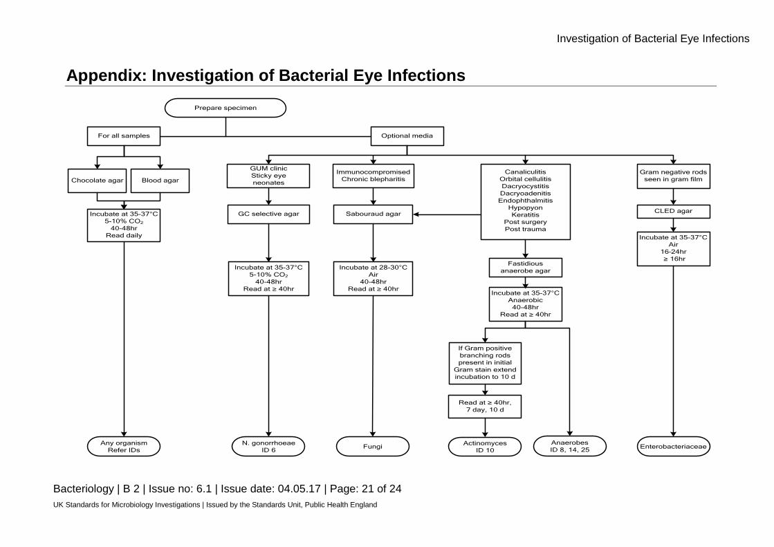
4.5.3 Culture media, conditions and organisms Clinical details/
conditions
Specimen Standard media
Incubation Cultures read
Target organism(s)
Temp °C
Atmos Time
Conjunctivitis
Sticky eye
If no clinical details available, treat as a ‘sticky eye’
Blepharitis (associated with other infections)
All samples
Chocolate agar
Blood agar
35-37
35-37
5-10% CO2
5-10% CO2
40-48hr
40-48hr
daily
daily
H. influenzae
Lancefield group A,B,C and G streptococci
Moraxella species
N. gonorrhoeae
N. meningitidis
P. aeruginosa
S. aureus
S. pneumoniae
Other organisms (see section 4.6.1)
For these situations, add the following:
Clinical details/
conditions
Specimen Supplementary media
Incubation Cultures read
Target organism(s)
Temp °C
Atmos Time
GUM clinic sticky eye
Neonates
Swabs GC selective agar
35-37 5-10% CO2
40-48hr ≥40hr N. gonorrhoeae
Immunocompromised
Chronic blepharitis
Swabs Sabouraud agar
28-30 air 40-48hr* ≥40hr Fungi
Canaliculitis †
Orbital cellulitis
Dacryocystitis †
Dacryoadenitis †
Keratitis‡
Endophthalmitis
Hypopyon
Post surgery
Post trauma
canalicular pus, aqueous and vitreous humour, corneal scrapings
Fastidious anaerobe agar
35-37 anaerobic
40-48hr*
10d
≥40hr
≥40hr, at 7d and 10d
Anaerobes
Actinomycetes
Sabouraud agar
28-30 air 40-48hr* ≥40hr Fungi
If Gram negative rods seen in Gram film
All samples
CLED agar 35-37 air 16-24hr ≥16hr Enterobacteriaceae
Other organisms for consideration - Chlamydia species and viruses and Mycobacterium species.
Investigation of Bacterial Eye Infections
Bacteriology | B 2 | Issue no: 6.1 | Issue date: 04.05.17 | Page: 18 of 24 UK Standards for Microbiology Investigations | Issued by the Standards Unit, Public Health England
*incubation may be extended to 5 days; in such cases plates should be read at ≥40hr and then left in the incubator/cabinet until day 5.
†extend incubation time to 10 days if clinically suspected or Gram positive branching rods present in Gram stain.
4.6 Identification Refer to individual SMIs for organism identification.
4.6.1 Minimum level of identification in the laboratory Actinomycetes "actinomycetes" level
Anaerobes "anaerobes" level
Bacillus species genus level
Coagulase negative staphylococci "coagulase negative" level
Diphtheroids "diphtheroid" level
Enterobacteriaceae "coliforms" level
Enterococci species level
Yeast and Moulds genus level
Haemophilus influenzae species level
Lancefield groups A, B, C and G streptococci
Lancefield group level
Moraxella species species level
Neisseria meningitidis species level
P. aeruginosa species level
Pseudomonads "pseudomonads" level
S. aureus species level
S. pneumoniae species level
α-haemolytic streptococci "α-haemolytic" level
Yeasts "yeasts" level
Mycobacterium species B 40 - Investigation of Specimens for Mycobacterium species
Parasites B 31 - Investigation of Specimens other than Blood for Parasites
Note 1: Organisms may be further identified if clinically or epidemiologically indicated. Any organism isolated from a normally sterile site should be identified to species level. Note 2: Any organism considered to be a contaminant (or part of normal flora) may not require identification to species level. Note 3: All work on suspected N. meningitidis and C. neoformans isolates which is likely to generate aerosols must be performed in a microbiological safety cabinet43.
Investigation of Bacterial Eye Infections
Bacteriology | B 2 | Issue no: 6.1 | Issue date: 04.05.17 | Page: 19 of 24 UK Standards for Microbiology Investigations | Issued by the Standards Unit, Public Health England
4.7 Antimicrobial Susceptibility Testing Refer to British Society for Antimicrobial Chemotherapy (BSAC) and/or EUCAST guidelines.
4.8 Referral for Outbreak Investigations N/A
4.9 Referral to Reference Laboratories For information on the tests offered, turn around times, transport procedure and the other requirements of the reference laboratory click here for user manuals and request forms. Organisms with unusual or unexpected resistance, and whenever there is a laboratory or clinical problem, or anomaly that requires elucidation should be sent to the appropriate reference laboratory. Contact appropriate devolved national reference laboratory for information on the tests available, turn around times, transport procedure and any other requirements for sample submission: England and Wales https://www.gov.uk/specialist-and-reference-microbiology-laboratory-tests-and-services Scotland http://www.hps.scot.nhs.uk/reflab/index.aspx Northern Ireland http://www.publichealth.hscni.net/directorate-public-health/health-protection
5 Reporting Procedure 5.1 Microscopy Report on WBCs and organisms detected. Microscopy for Mycobacterium species (B 40 - Investigation of Specimens for Mycobacterium species) and parasites (B 31 - Investigation of Specimens other than Blood for Parasites).
5.1.1 Microscopy reporting time Urgent microscopy results to be telephoned or sent electronically. Written report, 16–72hr.
5.2 Culture Report: Clinically significant organisms isolated or other growth, eg “No significant growth” or absence of growth Report presence or absence of Acanthamoeba species if applicable.
Investigation of Bacterial Eye Infections
Bacteriology | B 2 | Issue no: 6.1 | Issue date: 04.05.17 | Page: 20 of 24 UK Standards for Microbiology Investigations | Issued by the Standards Unit, Public Health England
5.2.1 Culture reporting time Clinically urgent requests: telephone when available. Written report: 16–72hr stating, if appropriate, that a further report will be issued. For Acanthamoeba species written report on day 4 stating if appropriate, that a further report will be issued. Supplementary investigations: Mycobacterium species (B 40 - Investigation of Specimens for Mycobacterium species).
5.3 Antimicrobial Susceptibility Testing Report susceptibilities as clinically indicated. Prudent use of antimicrobials according to local and national protocols is recommended.
6 Notification to PHE44,45 or Equivalent in the Devolved Administrations46-49 The Health Protection (Notification) regulations 2010 require diagnostic laboratories to notify Public Health England (PHE) when they identify the causative agents that are listed in Schedule 2 of the Regulations. Notifications must be provided in writing, on paper or electronically, within seven days. Urgent cases should be notified orally and as soon as possible, recommended within 24 hours. These should be followed up by written notification within seven days. For the purposes of the Notification Regulations, the recipient of laboratory notifications is the local PHE Health Protection Team. If a case has already been notified by a registered medical practitioner, the diagnostic laboratory is still required to notify the case if they identify any evidence of an infection caused by a notifiable causative agent. Notification under the Health Protection (Notification) Regulations 2010 does not replace voluntary reporting to PHE. The vast majority of NHS laboratories voluntarily report a wide range of laboratory diagnoses of causative agents to PHE and many PHE Health protection Teams have agreements with local laboratories for urgent reporting of some infections. This should continue. Note: The Health Protection Legislation Guidance (2010) includes reporting of Human Immunodeficiency Virus (HIV) & Sexually Transmitted Infections (STIs), Healthcare Associated Infections (HCAIs) and Creutzfeldt–Jakob disease (CJD) under ‘Notification Duties of Registered Medical Practitioners’: it is not noted under ‘Notification Duties of Diagnostic Laboratories’. Note: Isolation of N. meningitidis should be reported to the CCDC. https://www.gov.uk/government/organisations/public-health-england/about/our-governance#health-protection-regulations-2010 Other arrangements exist in Scotland46,47, Wales48 and Northern Ireland49.
Investigation of Bacterial Eye Infections
Bacteriology | B 2 | Issue no: 6.1 | Issue date: 04.05.17 | Page: 21 of 24 UK Standards for Microbiology Investigations | Issued by the Standards Unit, Public Health England
Appendix: Investigation of Bacterial Eye Infections
Prepare specimen
For all samples Optional media
CanaliculitisOrbital cellulitisDacryocystitisDacryoadenitisEndophthalmitis
HypopyonKeratitis
Post surgeryPost trauma
Chocolate agar
Fastidious anaerobe agar
Incubate at 35-37°C5-10% CO2
40-48hrRead daily
Incubate at 35-37°CAnaerobic40-48hr
Read at ≥ 40hr
Any organismRefer IDs
If Gram positive branching rods present in initial
Gram stain extend incubation to 10 d
Read at ≥ 40hr, 7 day, 10 d
ActinomycesID 10
AnaerobesID 8, 14, 25
Gram negative rods seen in gram film
Incubate at 28-30°CAir
40-48hrRead at ≥ 40hr
CLED agar
Incubate at 35-37°CAir
16–24hr ≥ 16hr
Enterobacteriaceae
Blood agar
Sabouraud agar
Fungi
ImmunocompromisedChronic blepharitis
GUM clinicSticky eyeneonates
GC selective agar
N. gonorrhoeaeID 6
Incubate at 35-37°C5-10% CO2
40-48hrRead at ≥ 40hr

Investigation of Bacterial Eye Infections
Bacteriology | B 2 | Issue no: 6.1 | Issue date: 04.05.17 | Page: 22 of 24 UK Standards for Microbiology Investigations | Issued by the Standards Unit, Public Health England
References 1. Syed NA, Hyndiuk RA. Infectious conjunctivitis. Infect Dis Clin North Am 1992;6:789-805.
2. Karsten E, Watson SL, Foster LJ. Diversity of microbial species implicated in keratitis: a review. Open Ophthalmol J 2012;6:110-24.
3. Sandstrom KI, Bell TA, Chandler JW, Kuo CC, Wang SP, Grayston JT, et al. Microbial causes of neonatal conjunctivitis. J Pediatr 1984;105:706-11.
4. Liesegang TJ. Bacterial keratitis. Infect Dis Clin North Am 1992;6:815-29.
5. Foster CS. Fungal keratitis. Infect Dis Clin North Am 1992;6:851-7.
6. Henry CR, Flynn HW, Jr., Miller D, Forster RK, Alfonso EC. Infectious keratitis progressing to endophthalmitis: a 15-year study of microbiology, associated factors, and clinical outcomes. Ophthalmology 2012;119:2443-9.
7. Keynan Y, Finkelman Y, Lagace-Wiens P. The microbiology of endophthalmitis: global trends and a local perspective. Eur J Clin Microbiol Infect Dis 2012;31:2879-86.
8. Pflugfelder SC, Flynn HW, Jr. Infectious endophthalmitis. Infect Dis Clin North Am 1992;6:859-73.
9. Brod RD, Flynn HW, Jr. Endophthalmitis: current approaches to diagnosis and therapy. Current Opinion in Infectious Diseases 1993;6:628-37.
10. Joseph CR., Lalitha P, Sivaraman KR, Ramsamy K, Behera UC. Real-time polymerase chain reaction in the diagnosis of acute postoperative endophthalmitis. Am J Ophthalmol 2012;153:1031-7.
11. Block SL. Etiologic and therapeutic pitfalls of newborn conjunctivitis. Pediatr Ann 2012;41:310-3.
12. Hauser A, Fogarasi S. Periorbital and orbital cellulitis. Pediatr Rev 2010;31:242-9.
13. McKellar MJ, Aburn NS. Cast-forming Actinomyces israelii canaliculitis. Australian & New Zealand Journal of Ophthalmology 1997;25:301-3.
14. Brazier JS, Hall V. Propionibacterium propionicum and infections of the lacrimal apparatus. Clinical Infectious Diseases 1993;17:892-3.
15. Bernardes TF, Bonfioli AA. Blepharitis. Semin Ophthalmol 2010;25:79-83.
16. Raskin EM, Speaker MG, Laibson PR. Blepharitis. Infect Dis Clin North Am 1992;6:777-87.
17. Clarke B, Sinha A, Parmar DN, Sykakis E. Advances in the diagnosis and treatment of acanthamoeba keratitis. J Ophthalmol 2012;2012:484892.
18. Visvesvara GS, Moura H, Schuster FL. Pathogenic and opportunistic free-living amoebae: Acanthamoeba spp., Balamuthia mandrillaris, Naegleria fowleri, and Sappinia diploidea. FEMS Immunol Med Microbiol 2007;50:1-26.
19. European Parliament. UK Standards for Microbiology Investigations (SMIs) use the term "CE marked leak proof container" to describe containers bearing the CE marking used for the collection and transport of clinical specimens. The requirements for specimen containers are given in the EU in vitro Diagnostic Medical Devices Directive (98/79/EC Annex 1 B 2.1) which states: "The design must allow easy handling and, where necessary, reduce as far as possible contamination of, and leakage from, the device during use and, in the case of specimen
Investigation of Bacterial Eye Infections
Bacteriology | B 2 | Issue no: 6.1 | Issue date: 04.05.17 | Page: 23 of 24 UK Standards for Microbiology Investigations | Issued by the Standards Unit, Public Health England
receptacles, the risk of contamination of the specimen. The manufacturing processes must be appropriate for these purposes".
20. Official Journal of the European Communities. Directive 98/79/EC of the European Parliament and of the Council of 27 October 1998 on in vitro diagnostic medical devices. 7-12-1998. p. 1-37.
21. Health and Safety Executive. Safe use of pneumatic air tube transport systems for pathology specimens. 9/99.
22. Department for transport. Transport of Infectious Substances, 2011 Revision 5. 2011.
23. World Health Organization. Guidance on regulations for the Transport of Infectious Substances 2013-2014. 2012.
24. Home Office. Anti-terrorism, Crime and Security Act. 2001 (as amended).
25. Advisory Committee on Dangerous Pathogens. The Approved List of Biological Agents. Health and Safety Executive. 2013. p. 1-32
26. Advisory Committee on Dangerous Pathogens. Infections at work: Controlling the risks. Her Majesty's Stationery Office. 2003.
27. Advisory Committee on Dangerous Pathogens. Biological agents: Managing the risks in laboratories and healthcare premises. Health and Safety Executive. 2005.
28. Advisory Committee on Dangerous Pathogens. Biological Agents: Managing the Risks in Laboratories and Healthcare Premises. Appendix 1.2 Transport of Infectious Substances - Revision. Health and Safety Executive. 2008.
29. Centers for Disease Control and Prevention. Guidelines for Safe Work Practices in Human and Animal Medical Diagnostic Laboratories. MMWR Surveill Summ 2012;61:1-102.
30. Health and Safety Executive. Control of Substances Hazardous to Health Regulations. The Control of Substances Hazardous to Health Regulations 2002. 5th ed. HSE Books; 2002.
31. Health and Safety Executive. Five Steps to Risk Assessment: A Step by Step Guide to a Safer and Healthier Workplace. HSE Books. 2002.
32. Health and Safety Executive. A Guide to Risk Assessment Requirements: Common Provisions in Health and Safety Law. HSE Books. 2002.
33. Health Services Advisory Committee. Safe Working and the Prevention of Infection in Clinical Laboratories and Similar Facilities. HSE Books. 2003.
34. British Standards Institution (BSI). BS EN12469 - Biotechnology - performance criteria for microbiological safety cabinets. 2000.
35. British Standards Institution (BSI). BS 5726:2005 - Microbiological safety cabinets. Information to be supplied by the purchaser and to the vendor and to the installer, and siting and use of cabinets. Recommendations and guidance. 24-3-2005. p. 1-14
36. Rishmawi N, Ghneim R, Kattan R, Ghneim R, Zoughbi M, Abu-Diab A, et al. Survival of fastidious and nonfastidious aerobic bacteria in three bacterial transport swab systems. J Clin Microbiol 2007;45:1278-83.
37. Barber S, Lawson PJ, Grove DI. Evaluation of bacteriological transport swabs. Pathology 1998;30:179-82.

Investigation of Bacterial Eye Infections
Bacteriology | B 2 | Issue no: 6.1 | Issue date: 04.05.17 | Page: 24 of 24 UK Standards for Microbiology Investigations | Issued by the Standards Unit, Public Health England
38. Van Horn KG, Audette CD, Sebeck D, Tucker KA. Comparison of the Copan ESwab system with two Amies agar swab transport systems for maintenance of microorganism viability. J Clin Microbiol 2008;46:1655-8.
39. Nys S, Vijgen S, Magerman K, Cartuyvels R. Comparison of Copan eSwab with the Copan Venturi Transystem for the quantitative survival of Escherichia coli, Streptococcus agalactiae and Candida albicans. Eur J Clin Microbiol Infect Dis 2010;29:453-6.
40. Tano E, Melhus A. Evaluation of three swab transport systems for the maintenance of clinically important bacteria in simulated mono- and polymicrobial samples. APMIS 2011;119:198-203.
41. Baron EJ, Miller JM, Weinstein MP, Richter SS, Gilligan PH, Thomson RB, Jr., et al. A Guide to Utilization of the Microbiology Laboratory for Diagnosis of Infectious Diseases: 2013 Recommendations by the Infectious Diseases Society of America (IDSA) and the American Society for Microbiology (ASM). Clin Infect Dis 2013;57:e22-e121.
42. Kilvington S, White DG. Acanthamoeba: biology, ecology and human disease. Rev Med Microbiol 1994;5:12-20.
43. Advisory Committee on Dangerous Pathogens. Categorisation of biological agents according to hazard and categories of containment. 4th ed. Suffolk: HSE Books; 1995. p. Supplements 1, 1998 and 2, 2000.
44. Public Health England. Laboratory Reporting to Public Health England: A Guide for Diagnostic Laboratories. 2013. p. 1-37.
45. Department of Health. Health Protection Legislation (England) Guidance. 2010. p. 1-112.
46. Scottish Government. Public Health (Scotland) Act. 2008 (as amended).
47. Scottish Government. Public Health etc. (Scotland) Act 2008. Implementation of Part 2: Notifiable Diseases, Organisms and Health Risk States. 2009.
48. The Welsh Assembly Government. Health Protection Legislation (Wales) Guidance. 2010.
49. Home Office. Public Health Act (Northern Ireland) 1967 Chapter 36. 1967 (as amended).