Embed Size (px)
Citation preview

UJPAH Vol. I No. 18 June 2015
1
Dr. S. Farooq - Chief Editor, Universities’ Journal of Phytochemistry and Ayurvedic Heights,Dehradun, UK., India
Dr. I.P. Saxena - Editor, Universities Journal of Phytochemistry and Ayurvedic Heights,Dehradun, UK., India Former Principal DAV (PG) College, Dehradun, Ex.V. C. H.N.B. Garhwal University, Srinagar, UK., India
Dr. A.N. Purohit - Ex. V. C. H.N.B. Garhwal University, Srinagar, UK., IndiaDr. Maya Ram Uniyal - Ex-Director Ayurved (Govt. of India) and Advisor, Aromatic and MedicinalPlant (Govt. of Uttarakhand), IndiaDr. G.K. Pathak - Former H.O.D., Chemistry Deptt., D.A.V. (PG) College, Dehradun, UK., IndiaDr. Himmat Singh - Former Advisor, R N D, BPCL, Mumbai, IndiaB.B. Raizada - Former Principal, D.B.S. College, Dehradun, U.K., IndiaDr. Iqbal Ahmad - Professor, Deptt. of Agricultural Microbiology, A.M.U., Aligarh, UP., IndiaMs. Alka Shiva - President and Managing Director, Centre of Minor Forest Products
(COMFORPTS), Dehradun, UK., IndiaDr. I.P. Pandey - Eminent Scientist, Dehradun, UK., IndiaDr. Versha Parcha - Head, Chemistry Deptt. SBSPGI of Biomedical Sciences and Research,
Dehradun, UK., IndiaDr. Umar Farooq - Associate Professor, Shoolini University, Solan, H.P., IndiaDr. Sanjay Naithani - Head, Chemistry Forest Products, FRI Bangaloru, Karnataka, India
Dr. G. K. Pathak Dr. I. P. Saxena Dr. I. P. Pandey Dr. S. Farooq Patron Editor Treasurer Chief Editor
Dr. I.P. Pandey, Co-ordinatiorUniversities’ Journal of Phyto-Chemistry and Ayurvedic Heights,
1-Inder Road, Dehradun - 248001 (Uttarakhand) IndiaE-mail : [email protected]; [email protected]
Website: www.ujpah.in
Inland annual subscription Rs. 300/-Inland annual scholar’s subscription Rs. 125/-Inland annual institutional subscription Rs. 400/-Overseas annual institutional subscription $ 40.00/-
Contact regarding any enquiry related to publication of research papers, articles etc.
Subscription may be sent by Bank Draft in favour of Universities’ Journal of Phyto-Chemistry and AyurvedicHeights, 1-Inder Road, Dehradun - 248001 (Uttarakhand), India. The Journal will be despatched under certificate of
posting. Send an additional amount of Rs. 100.00, if delivery is required through registered post.
Subscription
Full Page Rs. 10000.00 2 IssuesHalf Page Rs. 5000.00 2 IssuesQuarter Page Rs. 2500.00 2 Issues
Advertisement Tariff
Abstracted & Indexed by CAS, a division of American Chemical Society
Advisory Board

UJPAH Vol. I No. 18 June 2015
UJPAH Vol. I No. 18 June 2015
2
Editorial 3TlC and Spectroscopic Based Determination of Free 4-10Radical Scavenging Activity of Seven Ayurvedic Medicinal PlantsAfsheen, Mohd. Shavez Khan, Meenu Maheswari, Ait Sidi Brahim Malika and Iqbal AhmadChemical Constituents From Bark of Euonymus tingen (Celastraceae) 11-14S.C. Sati, Maneesha D. Sati and I P PandeyAlterneriol: Secondary Metabolites Derived From Endophytic 15-17Fungi Alternaria spp Isolated From Catharanthus roseusMonika Tickoo, Umar Farooq, Nidhi Bhatt, Mohini Dhiman,Afroz Alam, M. Azhar Khan and Sandeep JaglanGrowth Hormones Stimulate the Enzyme Activities and 18-23Total Nitrogen Content in Salinity Stressed Seeds of Triticum aestivum cvs.Shweta Tyagi and Sanjeev kumarPhytochemical Screening and Antimicrobial Activity of Zanthoxylum 24-27armatum DC.Ajay Singh , Vinod Kumar and Deepak SinghComparision of Physicochemical Properties of Some Edible Oils 28-30Afshan Tarranum, Renu Chauhan, Raj Kumar and Mona ChauhanQualitative Phytochemical Analysis of Allium humile and 31-34Ocimum gratissimum (Linn.) Leaves ExtractYogita Dobhal, Versha Parcha and D.C. DhasmanaAntioxidant Effect and Antimicrobial Activity of Cassia auriculata 35-36Piyush Mani Dubey and Neerja GuptaAntimicrobial Activity of Cleome viscose (seed) 37-38Arti Tomar and Suman RawatPhysico-chemical Characteristics of Diospyros peregrina 39-42Fruits at Different Ripening StagesDeepika Chauhan and P. K. GuptaForced Degradation Studies for Drug Substances and 43-47Drug Products- Scientific and Regulatory Considerations- ReviewTrivikram Rawat and I.P. PandeyAbout Mango (Mangifera indica) shown on the cover page 48-49Forth Coming Events 50-51Instructions to Contributors 52-53
Contents

UJPAH Vol. I No. 18 June 2015
3
EDITORIAL
India urgently needs specific measures for the development of Pharmaceuticals from all quarters.The recently passed Budget for 2015-16 has no incentives on R&D and investments in Pharma orany significant tax incentives for export and innovation, therefore, it does not enable the cultureof “India innovates” to encourage young scientists and give them a line to focus on innovation.Even the current Budget does not reverse the service tax imposed on Clinical research organizationsintroduced last year. Anyhow whatever may be the political thinking, the young scientists and nonpolitical organizations follow their respective duties towards the development of Indian PharmaIndustry through innovation and documentation of the therapeutic effects of Indian herbs. UJPAHis also committed and have not received/took any financial assistance/monetary help for the servicesUJPAH is rendering since its inception in the year 2005.
I am happy about the relationship I have in person with the Members of the Board of UJPAH whereeach one of them voluntarily is supportive for the cause of encouraging the young Scientistsworking on Indian herbs.
Similarly, I congratulate the young scientists of the Universities, their departmental Heads andother officials who deserve gratitude for the support rendered in our endeavour by sending studentsand staff members for making this mission successful.
In the end, my special thanks to Dr. Rajendra Dobhal, DG-UCOST who always stood for the causelike my younger brother.
(Dr. S. Farooq)Chief Editor

UJPAH Vol. I No. 18 June 2015
UJPAH Vol. I No. 18 June 2015
4
TlC and Spectroscopic Based Determination of Free Radical Scavenging Activity ofSeven Ayurvedic Medicinal Plants
Afsheena, Mohd. Shavez Khana, Meenu Maheswaria, Ait Sidi Brahim Malikaa,b and Iqbal Ahmada *
aDepartment of Agricultural Microbiology, Aligarh Muslim University, Aligarh, IndiabPhytochemistry and Pharmacology of Medicinal plants unit, Cadi Ayyad University, Marrakesh, Morocco
Abstract- Antioxidant activity of medicinal plants iswidely recognized for their therapeutic value. Variousin-vitro and in-vivo methods are used to assessantioxidants in plant extracts. However, in thescreening of large number of plants, rapid method ofdetection of active compounds is required to developfurther activity guided fractionation of the extracts.In this study, methanolic crude extracts of sevenplants namely Balsamodendron mukul, Boerhaviadiffusa, Catharanthus roseus, Curcuma longa, Lawsoniainermis, Laurus nobilis and Piper nigrum weresubjected to phytochemical analysis by color test andTLC for detection of phytocompounds, particularlyflavonoids. DPPH bio-autographic method was usedto detect the antioxidant activity of particular spoton the pre developed TLC plate. Further each extractwas also assessed by standard spectroscopic analysisfor DPPH radical scavenging potential. The dataobtained is comparable and highlights the importanceof TLC based method for fast detection of antioxidantactive phytocompounds in the crude extract of theplant/Ayurvedic drugs.Key words: Antioxidant activity, Medicinal plant,Phytocompound, TLC.
IntroductionFree radical facilitated cell damage plays an importantrole in the development of numerous human diseases.It has been widely assumed that progression ofdiseases such as cardiovascular disease, neuronaldisease, cataracts, and several types of cancer aremediated with free radicals generation thatultimately leads to the onset of oxidative stress(Thomas & Kalyanaraman, 1997). Recent clinicalstudies have indicated positive role of naturalantioxidant in significant reduction in the risk ofcoronary heart disease and ageing related disease
(Valko et al., 2007). On the other hand, certain toxicitypotential and carcinogenicity of syntheticantioxidants such as BHA and BHT have been reported(Sasaki et al., 2002). Therefore, there is an increasinginterest in search of natural antioxidant from plantssuch as vegetables, fruits and medicinal herbs.Antioxidant activity in plants is very commonlyreported but it is challenging to rapidly screen themost active antioxidants in the plant extracts due toits complex nature and presence of large number ofinterfering constituents. Numerous methods havebeen developed for screening of plant extracts fortheir antioxidant potential, among these TLC-bio-autography method (Cimpoiu, 2006) can quicklydetect and separate the antioxidant activeconstituents from complex crude extracts (Gu et al.,2009).Various plants used in the traditional medicinal systemproduce a lot of antioxidants and represent richsource of new and comparably nontoxicphytocompounds with antioxidant activity. It has beenwidely accepted that antioxidant active secondarymetabolites including simple phenolic acids to highlypolymerized tannins impart therapeutic potential tovarious plants (Pandey and Rizvi, 2009). Moreover,majority of plants derived antioxidant activecompounds falls in the category of polyphenolics (e.g.phenolic acids, flavonoids, and tannins) and amongthese flavonoids represent most versatile group interms of their antioxidant and other therapeuticvalues (Cimpoiu, 2006). Depending upon the chemicalmodif ication in the basic benzo--pyrane ringstructure, flavonoids chemically subdivided intoflavonols, flavones, isoflavones, flavanones andanthocyanidins (Harborne and Baxter, 1993).Characterization of these biologically activephytochemicals present in the plant along with the

UJPAH Vol. I No. 18 June 2015
5
knowledge of their antioxidant potential can give fairestimate of their therapeutic potential (Chanda etal., 2011). Furthermore, simultaneous separation anddetection of phtycompounds with antioxidant activityfrom crude mixture of plant extracts can ultimatelylead to identif ication of most potent antioxidantmolecules.In our previous studies, a large number of Indianmedicinal plants were screened for their broadspectrum antioxidant activity and also identified theactive fractions/compounds (Ahmad et al., 2008; Zahinet al., 2009, 2010, 2013). In the present study, DPPH-TLC bio-autography of crude extract of seventraditionally used medicinal plants namely B. mukul,B. diffusa, C. roseus, C. longa, L. inermis, L. nobilis and P.nigrum was performed along with spectroscopicanalysis for DPPH free radical scavenging activity. Inaddition, TLC based detection of flavonoids andphytochemicals profiling was also carried out usingstandard protocol.
Material and MethodsCollection of essential oil samples: The authentic plantmaterials were collected in the vicinity of Universitycampus (AMU) or obtained from local authorizedshops. The identification of the samples was furtherconfirmed by the Department of Botany, AligarhMuslim University, Aligarh, India. The voucherspecimens have been deposited in the Departmentof Agricultural Microbiology, Faculty of AgriculturalSciences, AMU, Aligarh. The detail of the plantsamples along with their parts used has beensummarized in table 1.Preparation of plant extract: Plants extract wasprepared as described earlier (Ahmad and Beg, 2001)with a little modif ication. The collected plants-material was dried in the shade. 100 gm of groundplants material was soaked in 300ml of methanol for3-4 days and stirred with glass rod after every 18 hrs.Plants-material was filtered using Whattman filterpaper No.1. The filtrate was concentrated in a rotatoryevaporator at 400 C temperature under vacuum. Allextracts were stored at 40 C for further use.Phytochemical analysis of plant extracts: Plantsextract were screened for the presence of major
phytocompounds by colour test using standardmethod as describe by Jamil et al.(2012).TLC based detection of Flavonoids in plant extracts:TLC was performed on silica gel 60 F254 plates (Merck,Germany) using a solvent system of n-hexane–toluene–ethyl acetate–formic acid (2:5:2.5:0.5) asdescribed by Wagner and Bladt (1996). Aliquot ofplant extracts-methanolic solution (1 mg/mL) wasdirectly spotted onto the TLC plates for development,followed by spraying with Natural product-polyethylene glycol reagent. Typical intensefluorescence indicates different classes of flavonoidsas follows:Flavonols: Orange-Yellow Fluorescence, Yellow-greenFluorescenceFlavones: Orange Fluorescence, Yellow-greenFluorescencePhenol carboxylic acid: Blue FluorescenceTLC-DPPH bio-autography analysis: An aliquot of plant-extract methanolic solution of concentration 1mg/mL, was directly applied (as spot) using Pasteurpipette onto the TLC plates (Silica gel 60 F254). TLCplates were developed in a presaturated solventchamber with n-hexane–toluene–ethyl acetate–formic acid (2:5:2.5:0.5) as developing reagents. Thedeveloped TLC plates were then removed from thechamber sprayed with a 1 mM DPPH radical methanolsolution for derivatization. Bands with the DPPHradical scavenging activity were observed as whiteyellow bands on a purple background (Gu et al., 2009).Total phenolic compound analysis: The amount of totalphenolic in plant extract was determined with theFolin-Ciocalteu reagent using the method of Spanosand Wrolstad (1990), as modified by Lister and Wilson(2001). To 5 ml of each sample (three replicates), 2.5ml 1/10 dilution of Folin-Ciocalteau’s reagent and 2 mlof Na2CO3 (7.5% w/v) were added and incubated at450C 15 min. The absorbance of all samples wasmeasured at 765 nm using a Spectronic 20D+ UV–Visspectrophotometer. Result was expressed asmilligrams of gallic acid equivalent per dry weight ofplant extract (mg GAE/g dw).Estimation of flavonoids content: The flavonoidscontent in extracts was determined

UJPAH Vol. I No. 18 June 2015
UJPAH Vol. I No. 18 June 2015
6
spectrophotometrically according to Lamaison andCarnat as describe by Quettier-Deleu et al. (2000),using a method based on the formation of a complexflavonoid–aluminium, having the absorbtivitymaximum at 430 nm. Rutin was used as control tomake the calibration curve. 1 ml of diluted samplewas separately mixed with 1 ml of 2% aluminumchloride methanolic solution. After incubation atroom temperature for 15 min, the absorbance of thereaction mixture was measured at 430 nm with aSpectronic 20D+ UV–Vis spectrophotometer and theflavonoids content was expressed in mg per g of rutinequivalent (RE).
Free radical scavenging assay: The scavenging activity
of DPPH free radicals by different plants-extracts wasassayed according to the method described by Gyamfiet al., (1999). Fifty micro liters of the plants-extract inmethanol, yielding 12.5, 25, 50 and 100μg extractrespectively in each reaction was mixed with 1ml of 0.1mM DPPH in methanol solution and 450μl of 50mMTris-HCL buffer (pH 7.4). Methanol (50μl) only was usedas control of experiment. After 30min of incubation atroom temperature the reduction of the DPPH free radialwas measured at 517nm. Vitamin C was used as controls.The percent inhibition was calculated from the followingequation:
% Inhibition = [Absorbance of control – Absorbanceof test sample / Absorbance of control] X 100
ResultsPhytochemical Analysis: Preliminary phytochemicalanalysis of methanolic crude extract of seven selectedplant extracts revealed the presence of Alkaloids,flavonoids, tannins, glycosides, and saponins in one ormore plant extracts as summarized in Table-1.TLC analysis of plant extract for detection ofFlavonoids: Different classes of flavonoids weredetected in the methanolic crude extract. All theplant extract showed varying degree fluorescencequenching band at UV-254 nm that showed thepossible presence of flavonoids and phenolic acids (fig.1A). Typical intense fluorescence in UV-365 nm isproduced immediately after spraying the naturalproduct reagent (f ig. 1 B) which conf irmed thepresence of flavonoids in the plant-extract. Based on
the fluorescence type, the presence of flavanols,flavanones and phenol carboxylic acid werecharacterized in each extract as major compounds.Number and antioxidant activity of flavonoid bandsobserved in each plant extract varied are shown infig. 3.Calorimetric estimation of Total Phenolics andflavonoids : The amount of total phenol wasdetermined with the Folin-Ciocalteu reagent. Gallicacid was used as a standard compound and the totalphenols were expressed as mg/g gallic acid equivalent.The results for total phenolic and flavonoid contentin crude-methanolic extracts are presented in table-1. Total phenolic contents were significantly high inthe crude extract of B. diffusa (589.92 mg GAE 10g-1)and least was in L. nobilis (210.34 mg GAE 10g-1). Theamount of phenolic compound ranged from 702.34to 210.34 mg GAE g-1dry weight of all the medicinalplants examined. The contents of total Falvonoid thatwere measured by AlCl3 reagent in terms of Rutinequivalent are shown in table-1. Flavonoid content ofextracts varied from 32.51 (L. nobilis) to 95.38 (L.inermis) mg Ru 10g-1. The results of table show thatthe total phenolic content is higher in B. diffusa,whereas the flavonoids presence is most significancein L. inermis.TLC based detection of antioxidant activity and Freeradical scavenging assay: In the present study,methanolic plant-extracts are monitored by a TLC bio-autography method because this method gives quickaccess for detection and localization of the activecompounds in a complicated plant extract (Tasdemiret al., 2004). In the method, the DPPH scavengingactivity was observed visually as white yellow spotson a purple background. Fig.1C shows a profile of theantioxidant components in methanolic extract of B.mukul, B. diffusa, C. roseus, C. longa, L. inermis, L. nobilisand P. nigrum under visible light. The chromatogramsof all the plant-extracts were observed to have DPPHscavenging activities. The entire band that werepreviously detected as flavonoids (fig. 1B), observedto have radical scavenging activity (fig. 3). Our findingsof TLC bioautography are in agreement with thereports of Cimpoiu (2006) who has detected suchactivity of flavonoids by TLC. However, DPPH-bio-
UJPAH Vol. I No. 18 June 2015
7
autography of different plant-extracts also revealedthat bands other than of flavonoid, also exhibit potentantioxidant activity.DPPH is usually used as a reagent to evaluate freeradical scavenging activity of antioxidants (Oyaizu,1986). DPPH is a stable free radical and accepts anelectron or hydrogen radical to become a stablediamagnetic molecule (Soares et al., 1997). Thereduction capability of DPPH radical is determined bythe decrease in absorbance at 517 nm induced byantioxidants. Ascorbic acid was used as standard. Theextracts are able to reduce the stable radical DPPH tothe yellow-coloured diphenylpicrylhydrazine. Thescavenging activity of the seven extracts tested wascompared to those of ascorbic acid, used as positivecontrol, and was found to be concentration-dependent. This activity can be evaluated by thedetermination of the IC50values, corresponding to theamount of extract required to scavenge 50% of DPPHradicals present in the reaction mixture. High IC50values indicate low antioxidant activity. As shown inTable-1, the scavenging effect in terms of IC50 value ofmethanolic extracts and standards radical is in thefollowing order: B. mukul (47.61 µg/ml) > B. diffusa(49.53 µg/ml) > L. inermis (52.02 µg/ml) > L.nobilis (53.98 µg/ml)> C. longa (55.28 µg/ml)> C. roseus(60.26 µg/ml) > P. nigrum (62.61 µg/ml), controlcompound (ascorbic acid) shows the IC50 value of 8.26µg/ml. From fig. 2, a dose dependent response wasfound in the DPPH radical scavenging activity; theactivity increased as the concentration increased foreach individual crude methanolic extracts.Our f indings are in accordance with the report ofother workers (Hsouna et al, 2011; Shisode andKareppa 2011; Tiong et al, 2013; Shabana and Afif i,
2014; Khan et al., 2014). Free radical scavenging andantioxidant activity of the phenolics/flavonoids mainlyattributed to the numbers and position of hydrogendonating hydroxyl groups on the aromatic ring of thephenolic molecules, and also affected by othersfactors such as glycosylation of aglycones, other H-donating groups (-NH, -SH), and so on. Substitutionpatterns in the B-ring and A-ring as well as the 2,3double bond (unsaturation) and the 4-oxo group inthe C-ring also affect antioxidant activity of phenolics(Cai et al., 2006). As a result, of their abilities toscavenge free radicals, phenolics exhibited excellentantioxidant, antimutagenic, antiinflammatory, andanticancer properties (Ahmad and Mukhtar 1999).Therefore, therapeutic properties of the plants testedmay possibly be attributed to the presence ofphenolic compounds in particular, the flavonoidspresent in the plant extracts.
ConclusionOn the basis of this preliminary study, it is concludedthat TLC based detection and identif ication ofantioxidant compounds is a simple and fast methodfor screening of large number of extracts.Antioxidant potency of the extracts by spectroscopicmethod revealed similar trend. Further, the TLC basedapproach may be developed into more eff icienttechnique using HPTLC for fast detection and qualityassurance of herbal extracts and isolation of mostactive antioxidant.
AcknowledgementWe are grateful to Dr. S. Farooq (Director, TheHimalaya Drug Company, Dehradun) for his guidanceand support in above work.

UJPAH Vol. I No. 18 June 2015
UJPAH Vol. I No. 18 June 2015
8
Fig.1 TLC analysis of plant extracts A) shows chromatogram of plant extracts at 254 nm. B) tlc chromatogramshowing the detection of flavonoids corresponding to different classes at 366nm and C) DPPH-TLCbioautography showing different antioxidant active bands. Plant extracts spot lane: A) Curcuma longa, B)Piper nigrum, C) Lawsonia inermis, D) Boerhavia diffusa, E) Catharanthus roseus, F) Laurus nobilis and G)Balsamodendron mukul
Fig. 2 (A) Free radicle inhibition activity of plant extracts and (B) standard curve of Ascorbic Acid.

UJPAH Vol. I No. 18 June 2015
9
Fig. 3 Band profile of different plant extracts as observed on their respective chromatograms.
References1. Ahmad, I. and Beg, A. Z. Antimicrobial and
phytochemical studies on 45 Indian medicinalplants against multidrug resistant humanpathogens, J Ethnopharmacol., 2001, 74: 113-123.
2. Ahmad, I.; Zahin, M.; Aqil, F.; Hasan, S.; Khan, M.S. A. and Owais, M. Bioactive compounds fromPunica granatum, Curcuma longa and Zingiberofficinale and their therapeutic potential. Drugsof the Future, 2008, 33(4): 329-346.
3. Chanda, S.; Dave R. and Kaneria, M. In vitroantioxidant property of some Indian medicinal plants. Res J Med Plant, 2011, 5(2): 169-179.
4. Gu, L.; Tao W. and Zhengtao W. TLC bio-autography-guided isolation of antioxidants fromfruit of Perilla frutescens var. acuta. LWT-Food SciTech., 2009, 42 (1): 131-136.
5. Gyamfi, M.A.; Yonamine, M. and Aniya, Y. Free-radical scavenging action of medicinal herbs fromGhana: Thonningia sanguinea on experimentally-

UJPAH Vol. I No. 18 June 2015
UJPAH Vol. I No. 18 June 2015
10
induced liver injuries. General Pharmacology: TheVascular System, 1999, 32(6): 661-667.
6. Harborne, J. B. and Baxter, H. Phytochemicaldictionary. A handbook of bioactive compoundsfrom plants, 1993, Taylor & Francis Limited. ISBN:0850667364.
7. Hsouna, A. B.; Trigui, M.; Culioli, G.; Blache, Y. andJaoua, S. Antioxidant constituents from Lawsoniainermis leaves: Isolation, structure elucidation andantioxidative capacity. Food Chem., 2011, 125(1):193-200.
8. Jamil, M.; Ul Haq, I., Mirza, B. and Qayyum, M.Isolation of antibacterial compounds fromQuercus dilatata L. through bioassay guidedfractionation. Ann Clin Microbiol Antimicrob., 2012,11: 11.
9. Khan, A.; Nazar, H.; Sabir, S. M.; Irshad, M.; Awan,S. I.; Abbas, R. and Malik, F. Antioxidant activityand inhibitory effect of some commonly usedmedicinal plants against lipid per-oxidation in micebrain. Afr J Trad Complemt & Alter Med (AJTCAM),2014, 11(5): 83-90.
10. Oyaizu, M. Studies on product of browningreaction prepared from glucose amine. Jap J Nutr.,1986, 44: 307–315.
11. Quettier-Deleu, C.; Gressier, B.; Vasseur, J.; Dine,T.; Brunet, C.; Luyckx, M. and Trotin, F. Phenoliccompounds and antioxidant activities ofbuckwheat (Fagopyrum esculentum Moench)hulls and flour. J Ethnopharmacol, 2000, 72(1): 35-42.
12. Sasaki, Y.F.; Kawaguchi, S.; Kamaya, A.; Ohshita,M.; Kabasawa, K.; Iwama, K.; Taniguchi, K. andTsuda, S. The comet assay with 8 mouse organs:results with 39 currently used food additives.Mutat Res., 2002, 519: 103–109.
13. Shabana, M. H. and Afif i, M. S. A new acylatedluteolin glycoside from Curcuma Longa L. and freeradical scavenging potential of its extracts. J MedPlants Res., 2014, 8(1): 1-5.
14. Shisode, K. S. and Kareppa, B. M. In-vitroantioxidant activity and phytochemical studiesof Boerhaavia diffusa Linn. roots. Int Res J Phar.,2011, 2(12): 3171-3176.
15. Soares, J.R.; Dinis, T.C.; Cunha, A.P. and Almeida,L. Antioxidant activities of some extracts ofThymus zygis. Free Rad Res., 1997, 26(5), 469-478.
16. Spanos, G.A. and Wrolstad, R.E. Influence ofprocessing and storage on the phenoliccomposition of Thompson Seedless grape juice.J Agric Food Chem., 1990, 38: 1565-1571.
17. Tasdemir, D.; Donmez, A. A.; Calis, I. and Ruedi, P.Evaluation of biological activity of Turkish plants.Rapid screening for the antimicrobial,antioxidant, and acetylcholinesterase inhibitingpotential by TLC bioautographic method.Pharmac Biol., 2004, 42(4–5), 374–383.
18. Thomas, Craig. Oxygen radicals and the diseaseprocess 1998. CRC Press, ISBN: 9789057022272.
19. Tiong, S. H.; Looi, C. Y.; Hazni, H.; Arya, A.; Paydar,M.; Wong, W. F. and Awang, K. Antidiabetic andantioxidant properties of alkaloids fromCatharanthus roseus (L.) G. Don. Molecules, 2013,18(8), 9770-9784.
20. Valko, M.; Leibfritz, D.; Moncol, J.; Cronin, M. T.;Mazur, M. and Telser, J. Free radicals andantioxidants in normal physiological functions andhuman disease. Int J Biochem & Cell Biol., 2007,39(1), 44-84.
21. Wagner, H. 1996 Plant drug analysis: a thin layerchromatography atlas. Springer Science &Business Media, ISBN: 97835405867603540586768.
22. Zahin, M.; Ahmad, I. and Aqil, F. Antioxidant andantimutagenic activity of Carum copticum fruitextracts. Toxicol in Vitro, 2010, 24(4), 1243-1249.
23. Zahin, M.; Aqil, F. and Ahmad, I. The in vitroantioxidant activity and total phenolic contentof four Indian medicinal plants. Int J Phar & PharmSci., 2009, 1(1), 88-95.
24. Zahin, M., Aqil, F., Husain, F. M., & Ahmad, I.Antioxidant capacity and antimutagenic potentialof Murraya koenigii . Biomed ResearchInternational, 2013, ID 263509 (2013).

UJPAH Vol. I No. 18 June 2015
11
Chemical Constituents From Bark of Euonymus tingen (Celastraceae)1S.C. Sati, 1Maneesha D. Sati and 2 I P Pandey
1Department of Chemistry,H.N.B.Garhwal University
(A Central University)Srinagar, Uttarakhand, India.
2Department of Chemistry,DAV(PG) College Dehradun,IndiaCorresponding author: [email protected]
Abstract- 1,5 di-hydroxy 3,6,7 tri methoxy 8-allyloxyxanthone and 5-O--L xylopyranosyl (1 6) -D-glucopyranosyl 1,6 dihydroxy 3,5dimethoxy xanthone were isolated from bark ofEuonymus tingen. The structures of these compoundswere characterized by means of chemical and spectralmethods including advanced 2D NMR studies. Thesecompounds were first time isolated from this species.Key words: Euonymus tingen Celastraceae, 1,5-di-hydroxy 3,6,7 tri methoxy 8-allyloxyxanthone, 5-O--L Xylopyranosyl (1 6)-D-glucopyranosyl 1,6 dihydroxy 3,5
dimethoxy xanthone.
IntroductionEuonymus tingens (Roxb.), vern. Kumkum, familyCelastraceae, is a evergreen tree or large shrubs,often attaining to 8m high; bark dark ash-coloured,bright yellow inside. Leaves opposite, elliptic or ovate-lanceolate. Flowers pale-white, calyx lobes fimbriate.Petals orbicular, with yellowish tingen; stemns shorterthan petals; seeds dark brown, half enclosed in orange-red aril. Flowering season is March to April and fruitingfrom May to September. The plant is distributed fromW. Himalaya, Shimla to Bhutan; Myanmar and Tibet1.The bark paste of the plant is useful in eye diseases.Number of T ingenone, hydroxytingenone,triterpenoid quinine methides has been isolated fromEuonymus tingen2,3,4.
Material and MethodsPlant material- Stem bark (6 kg) of Euonymus tingenwas collected from Moling Ghat, District ChamoliGarhwal during October 2014 and identif ied bytaxonomist in the Department of Botany, H.N.B.Garhwal University Srinagar. A voucher specimen(GUH-8327) of the plant is deposited at the Herbariumof Department.Extraction and isolation- Shade dried and coarselypowdered stem bark of Euonymus tingen (4 kg) was
extracted thrice with 95% ethanol (5L) at 50°C (15 hrs)on a heating mantle. The reaction mixture was filteredoff and the filtrate was concentrated under reducedpressure to yield brownish residue (440 g). Thisresidue was fractionated with EtOAc (repeatedly 3-4times) yield EtOAc soluble and insoluble fraction.EtOAc soluble portion (310 g) with n-hexane:Chloroform (93:7) as eluting solvent with increasingpolarity of CHCl3, afforded two compounds in pureform tentatively designed as compound 1 and 2respectively. These compounds were purif ied byrecrystallization.General experimental procedure- UV spectra weretaken in MeOH using AlCl3 as shift reagent. IRrecorded in KBr on a Perkin Elmer FT IRSpectrophotometer. 1H-NMR were run at 300MHzusing TMS as internal standard and C5D5N and DMSOas solvent. 13C-NMR recorded in 125MHz, using C5D5Nand DMSO as solvent, FAB-MS on JOEL. JMS700Mstaion Spectrophotometer. MPs were incorrected.
Result and Discussion
Compound 1 It was crystallized from methanol as yellow stickymass.Melting Point : 322-325 °CMolecular Formula : C21H22O8
U.V.( maxMeOH) nm : 269,260
I.R.(maxKBr ) cm-1 : 1640
FAB-MS (m/z) : 402[M]+, 371[M-OCH3], 340[M-2xOCH3],292[M-3xOCH3+OH],
283[M2xOCH3+2xOH],199[M-3xOCH3+2xOH+C5H8O]
1H-NMR(CHCl3,ppm) : 1.65(s), 1.78(s),
2.57(s) 3.2(s), 3.44(s), 3.9(d,J=7.4Hz), 6.93
(d,J=8.6Hz), 7.6(d, J=7.4Hz), 9.8(s), 12.4(s,-OH).

UJPAH Vol. I No. 18 June 2015
UJPAH Vol. I No. 18 June 2015
12
It was crystallized from MeOH, yellow sticky mass. Itgave characteristic greenish brown colour withmethanolic Fecl3 and also positive to Gibbs test. Itspositive FAB-MS spectra showed a molecular ion peakat m/z 402[M]+, which correspond to the molecularformula C21H22O8. Its UV spectrum in methanol showeda characteristic band at 269 nm and IR spectrumshowed a bands at 3260 and 1640 cm 1 werecharacteristic for xanthone5. 1H-NM of compoundshowed presence of two AB type doublets at 6.89(d,J=8.6Hz) and 7.5(d,J=7.4Hz) suggesting thepresence of tetra substituted aromatic ring in thecompound. The downfield signal at12.4(s) and 9.8(s)exchangeable with D2O indicate the presence ofchelated hydroxyl group at C-1 position and one freephenolic OH group. The UV spectrum of MH-6 showedabsence of AlCl3 induced shift there by suggesting abulky substituent located ortho to the hydrogenbonded group. Besides the [M]+ ion peak, the FAB-MSof compound showed other significant peaks at m/z340, 309, 292, 283 and 199 which correspond to thesubsequent loss of three OCH3, two OH and one C5H8Oion from molecular ion peak. The presence of dimethylallyl group was further confirmed by 1H-NMR spectrumof compound, which showed two methyl signals at 1.65(s) and 1.78(s), one 2H proton doublet at 3.9(d,J=7.4Hz) and a doublet at 7.5(d, J=7.4Hz). The
peak at 6.89(d,j=8.6Hz) and7.45(d, J=7.4Hz) showedthe presence of three aromatic protons in ring A. Thepresence of three singlets at 2.57, 3.44 and 3.20confirms the presence of three methoxyl group incompound.13C-NMR spectrum of compound showed the presenceof twenty carbon signals. The downfield signal at176.7was attributed to -unsaturated carbonyl carbonatom of xanthone6. The other signal at 56.7, 61.3 and60.4 were assigned for methoxyl groups. The positionof methoxyl groups were found at C-3, C-6 and C-7atoms (by comparison of 13C-NMR data with relatedcompound7. The 13C-NMR peaks at 11.82 and 15.14showed the presence of two tertiary methyl groups,whereas the downfield peaks at 122.6 and 122.8 werecompatible with a double bonded carbon. Two methylsignals at1.65 and 1.78 9 (1H-NMR) and tertiary carbonsignal at 29.0 denoted the presence of gem-di methylgroup. The carbon resonances at 161.6, 164.7, 148.2,148.5, 145.9 and 160.0 showed the presence of sixoxygenated carbon atoms and were compatible with1, 3, 5, 6, 7, and 8 oxygenated carbon atoms of xanthone.Thus all these values were in agreement with reporteddata of 3, 3-di methyl allyl 3-methyl 2-hydroxy substitutedxanthone. Hence on the basis of above spectral studiescompound 1 was identified as 1,5-di-hydroxy 3,6,7 tri -methoxy 8-allyloxy xanthone (Figure- 1).
13C-NMR (CHCl3, ppm)

UJPAH Vol. I No. 18 June 2015
13
Compound 2It was crystallized as yellow solid from methanol.Melting Point : 185-187 °CMolecular Formula : C26H30O15Molecular Weight : 582 amu
U.V.(maxMeOH )nm : 360,306,239,205.
I.R (maxKBr ) cm-1 : 3541(OH), 2926(C-H
stretching), 1664(C=O), 1589(C=C stretching)FAB-MS (m/z) : 6 2 1 [ M + K ] + ,605[M+Na]+, 583[M+H]+, 503, 460.1H-NMR (C5D5N,ppm)3.82(3H,s,CH3), 4.08(3H,s,CH3), 13.10(s,OH), 4.33-4.40(-CH), 5.42(d,J=7.6Hz), 7.23(d,J=8.7Hz) 7.53(d,J=8.7Hz),6.63(d, J=2.4Hz), 7.21(d,J=2.4Hz), 4.36,4.84(CH2),4.95(1H,d,J=7.3Hz), 4.04 (1H,dd, J=7.6,8.8Hz),4.13(1H,t), 3.67(1H, d, J=10.2,11.2Hz).13C-NMR (C5D5N,ppm)Aglycone160.7(C-1), 102.0(C-2), 165.0(C-3), 95.9(C-4), 158.6(C-4a), 149.7(C-4b), 146.5(C-5), 148.3(C-6), 123.4(C-7)113.2(C-8), 118.3(C-8a), 176.2(C-9), 108.8(C-9a), 56.0(OCH3), 61.5(OCH3).GlyconeGlucose : 105.8 (C-1’), 75.1(C-2’), 77.6(C-3’), 71.4,(C-4’), 77.8(C-5’), 70.4(C-6’)Xylose : 106.6(C-1’’), 75.0(C-2’’), 78.2(C-3’’),71.1(C-4’’), 67.2(C-5’’)It was crystallized from methanol as yellow solid. TheFAB-MS of compound showed the molecular ion peakat m/z 621[M+K]+, 605[M+Na]+ and 583[M+H]+ whichcorrespond the molecular formula C26H30O15 andmolecular weight 582. Its UV spectrum showedcharacteristic bands at 360, 306, 239 and 206,suggesting a xanthone skeleton of compound. The IRspectrum displayed a bands at 3541 cm-1 (OH), 1664cm -1 (C=O) and 1620 cm-1, 1589 cm -1 for (C=C)stretching. The 1H-NMR spectrum showed a pair of
ortho coupled protons which appeared at 7.53(1H.d,J=8.7Hz) and 7.23 (1H,d,J=8.7Hz) wereassigned to H-7 and H-8, while two doublets of metacoupled protons at 7.21(1H,d,J=2.4Hz) and6.63(1H,d,J=2.4Hz) were assigned to H-2 and H-4respectively. Its 1H-NMR spectrum also showed twoseparate singlet each integrating for 3H appeared at3.82 and 4.06 were attributed for two methoxylgroups, attached to C-3 and C-5 carbon atoms.The 1H-NMR spectrum also displayed two sharpdoublets at 5.42 (d,J=7.6Hz) and 4.95(d,J=7.3Hz)suggesting the presence of two sugar moieties in thecompound, and were assigned for anomeric protonsof glucose (H-1’) and xylose (H-1’’), while other protonsof glucose moieties resonated between 4.22-4.82and that of xylose at3.67-4.35. The 13C-NMR spectrumalso supported that C-1’ and C-2’’ carbon atomsappeared at 105.8 and 106.6. Its 13C-NMR spectrumfurther support the presence of twenty six carbonatoms. A signal for one proton appeared at 13.20was assigned for chelated hydroxyl group. Adownfield signal at 176.2 in 13C-NMR spectrumattributed forunsaturated carbonyl group, whichalso confirms the xanthone skeleton of the compound2 .Thus on the basis of above studies compound 2 wasidentif ied as 5-O--L Xylopyranosyl (1 6) -D-glucopyranosyl 1,6 -dihydroxy 3,5 -dimethoxy xan-thone (Figure- 2). It is the first reported compoundin the family Celastraceae.
References
1. Gaur, R.D. Flora of District Garhwal ,1999.2. Ayhan, U. and Battop, T. Phyochemistry, 1973, 12,
1824.3. Brown, P. M.; Moir, M.; Thomson, R.H.; King, T.
J.; Krishnamoorthy, V. and Seshadri, T. R. J.Chem. Soc., Perkin Trans., 1973, 1, 2721-2725.
ë

UJPAH Vol. I No. 18 June 2015
UJPAH Vol. I No. 18 June 2015
14
4. Baek, N. I.; Lee,Y. H.; Park, J.D.; Kim, S. I. and Ahn,B. Z. Planta Med., 1994, 60, 26-29.
5. Despandey, V.H.; Rama Rao, A.V.; Varadan, M. andVenkataraman, K. Ind. J. Chem., 1973, 1, 518.
6. Hostettman, K. and Wagner, H. Phytochemistry,1977, 16,821.
7. Mandal, S. and Chetterjee, A. Tetrahedron Lett.,1987, 28, 1309.

UJPAH Vol. I No. 18 June 2015
15
Alterneriol: Secondary Metabolites Derived From Endophytic Fungi Alternaria sppIsolated From Catharanthus roseus
Monika Tickoo1, Umar Farooq1, Nidhi Bhatt1, Mohini Dhiman1, Afroz Alam2, M. Azhar Khan1 and SandeepJaglan*
1Molecular and immune-parasitological research laboratory, Faculty of Applied Sciences and Biotechnology,Shoolini University,Solan, 173212, Himachal Pradesh, India.
2Faculty of Pharmacy, Shoolini University, Solan, HP*Microbiology Lab, Indian Institute of Integrative Medicine, Jammu (J&K), India
Corresponding AuthorDr. Umar Farooq,
Associate Professor, Microbiology,Faculty of applied Sciences and Biotechnology,
Shoolini University,Solan, 173212, Himachal Pradesh, India.Email: [email protected], [email protected]
Abstract- Endophytic fungi produce bioactivemetabolites which play an essential role to provideprotection to their host against attack by anotherpathogens and environmental factors. Isolation,identification and cultivation of endophytic fungi frommedicinal plants are important for the production andcharacterization of bioactive fungal metabolites.Present study was aimed to isolate and characterizeendophytic fungi from the medicinal plant,Catharanthus roseus collected from garden area ofMuthi and Ramnagar forest in Jammu. Five samplesof leaves, stem and bark were collected from C. roseus.The sample of leave, stem and bark were inoculatedin three media PDA (Potato dextrose agar), YMA(Yeast maltose agar), WA (water Agar). A total ofthree strains of endophytic fungi (VRE–1, VRE-3, andVRE-4) were isolated using PDA media and YMA media.Out of these three strains, only one strain VRE-1 hasbeen identified as Alternaria spp. by microscopicallyand biochemically. Alternaria spp further used forfermentation by using shake flask method and thenthe secondary metabolites were extracted by usingColumn Chromatography. One pure compound,Alternariol was obtained from methanolic extract,further study is undertaken.Key words: Endophytic fungi, Catharanthus roseus,Secondary Metabolites, Alternaria spp.
IntroductionFungi have been widely used as a source of bioactivecompounds. The first milestone in this field was thediscovery of penicillinnotatum by Alexander Flemingin 1928(Ligon B., 2004). Fungi are heterotrophicorganisms and have different lifestyle; they live inmutualistic, antagonistic, or neutral symbiosis with awide variety of autotrophic organism. Variouseffective antifungal agents like Griseofulvin,
Cyclosporine, Mevastatin, Taxolare isolated fromdifferent fungus and used for the treatment of fungalinfection (Stierle et al., 1993). The gold bioactivecompound taxol discover from the endophytic fungusTaxomycesandreanae (Stierle et al., 1993). Endophyticfungi found in leaves, stem and bark of Catharanthusroseus having medicinal properties (Ansari and Naved.,2006). Fungus produce metabolites (primary andsecondary) which play an important role in plantdefense against herbivores and other interspeciesdefense. The primary and secondary metabolitesproduced by the fungi while some secondarymetabolites are often derived from the primarymetabolites and some environmental factors areresponsible for the production of secondarymetabolites which protect the fungi as well as plantsfrom external factors (Vanisree lee et al., 2004). It issuggested that secondary metabolites also play animportant role in chemical defense andcommunication of fungi. The present study was aimedto isolate the endophytic fungi from plants and alsoextracts the secondary metabolites produced by theentophytic fungi.
Material and MethodsCollection of samples: A total of f ive samples werecollected during January 2013. Leavesstem and barkof Catharanthus roseus were collected from Muthiarea of Jammu (J&K) and Kangra Distt. (H.P.).Isolation of endophytic fungi: Media used for theisolation of endophytic fungus were WA, YMA andPDA. Fungal growth was observed on the plates aswhite cottony outgrowth. The fungal growth wasinitiated within 7 to 10 days of inoculation. Isolationfrom the master plate was done by transfer of hyphaltips to PDA plates from water agar plates withoutaddition of antibiotics to obtain pure culture.

UJPAH Vol. I No. 18 June 2015
UJPAH Vol. I No. 18 June 2015
16
Characterization of endophytes: The fungi wereidentif ied on the basis of their morphological andculture characteristics. Production of extracellularenzymes – amylase, protease, chitinase, lipase,oxidase and catalase was measured by biochemicalanalysis. Seed preparation on Potato Dextrose Agar(PDA) was done to obtain secondary metabolites. Bulkgrowth of endophytes on PDA was examined everyday. The cell mass was separated out from thesupernatant by the process of filtrations with the helpof muslin cloth and kept the supernatant as well asbiomass separately (Carroll, 1978).Extraction of biomass: Extraction of endophytes fromfermented broth was homogenized with tissuehomogenizer and 10% methanol. The extractionprocedure was done with organic solvent,chloroform. The compound which was left, bind tothe methanol due to vigorous shaking. After 24 hours,separate the supernatant and biomass by filtration.Drying of biomass by evaporation: The sample volumeof the extracted solution to dryness containingmetabolites of the fungal mycelium was done usingrota- vapour. The main function of the rota- vapour isto separate the solvent from biomass.Detection of secondary metabolites from Alternariaspp.: The secondary metabolites from endophyticfungi Alternaria spp was extracted by using thin layerchromatography. Then the separated compoundswere purified by using Column chromatography. Thephysical and chemical properties of extractedcompound was determined by nuclear magneticresonance (NMR).
ResultsIsolation: In this study three numbers of fungalendophytes (VRE-1, VRE-3, and VRE-4) were isolatedfrom Catharanthus roseus as pure culture and thesewere isolated from stem and leaves of the plant. The
DiscussionPlant endophytic fungi are important and novelresource of natural bioactive compounds as they havewide potential applications in agriculture, medicineand food industries. In the past two decades, many
The present study revealed that the endophyticfungus isolated from C. roseus was characterized asAlternaria spp. These endophytes present in a plantproduced several secondary metabolites which helpin survival of endophytes as a commensal as well asthese secondary metabolites have great medicinalvalue.In the present study, secondary metabolite wasextracted by column chromatography frommethanolic extract of Alternaria spp. The meldingpoint of the compound was 205.88 °C, physical statewas solid and molecular weight was 258.23 g·mol”1.
The extracted bioactive compound was identified asAlternariol (C14H10O5) (Figure) by NMR.
Figure- Chemical structure of Alternariol derivedfrom Endophytic fungi Alternaria spp.
VRE-1 isolate was identif ied as Alternaria spp. ofendophytic fungi by Morphological (Table-1) andBiochemical analysis (Table-2). We have used WA, PDA,YMA media for the extraction of endophytic fungi.Out of these three media, PDA and YMA were foundhighly suitable for the isolation of endophytic fungi.

UJPAH Vol. I No. 18 June 2015
17
valuable bioactive compounds have been extractedfrom the endophytic fungi as beneficial productshaving various antimicrobial, insecticidal, catatonicand anti cancer activities.In the present study, a total f ive samples whichinclude stem, leave and bark of Catharanthus roseuswas collected from different areas. Out of f ivesamples endophytic fungi were isolated from threesamples of stem and leaves of Catharanthus roseus.The cultural characterization of fungal endophyte wasperformed using PDA, YMA and WA media. It wasobserved that the endophytes grow best on PDAmedium in comparison to YMA and WA media. Onthe basis of these characteristics, three isolates VRE-1, VRE-2 and VRE-3 were obtained. The isolates werefurther screened for the presence of various enzymesi.e. amylase, chitinase, lipase, protease, oxydase andcatalase using biochemical activity assay. Afterdetermining the morphological and biochemicalobservations, VRE-1 was identified as Alternaria spp.,while VRE-3 and VRE-4 remains unidentified due tono similarity found with other fungal species. Earlier,a study conducted by Jagetia and Venkatesh (2005)reported the presence of 10 endophytic fungi fromthe family Aegelmarmelose while in the present studyonly one endophytic fungi of family Alternatia spp.were found. The fermentation of VRE-1 was performedby using shake flask method on PDB (Potato DextroseBroth) after inoculating the pure culture andincubated at appropriate temperature. The extractionof the fermented broth was done withdichloromethane (DCM) to get both high polar andlow polar compounds. The extract of Alternaria spp.was then subjected to column chromatography forisolation of pure compounds and one pure fractionwas isolated. Then NMR of pure fraction wasperformed to know the structure of compound andthe compound identified as alternariol. Alternariolwas first isolated and structurally characterized as 3,7, 9-trihydroxy-1-methyl-6H-dibenzo [b, d] pyran-6-one(Raistrick et al., 1953). Further research is required toexplore the role and importance of Alternariol.
References
1. Ansari, S.H. and Naved, T. A review of chemicaland biological activity of Catharanthus roseus. J.Pharm Res., 2006, 5: 46-59.
2. Carroll, G.C. and Carroll, F.E. Studies on theincidence of coniferous needle endophytes in thePacific Northwest. Can. J. Bot., 1978, 56: 3034-3043.
3. Ligon, B. Penicillin: its discovery and earlydevelopment. SeminPediatr Infect Dis., 2004,15 (1): 52–7.
4. Raistrick, H.; Stickings, C.E. and Thomas, R. Studiesin the biochemistry of micro-organisms.90Alternariol and alternariolmonomethylether,metabolic products of Alternariatenuis. BiochemJ., 1953, 55:421–433.
5. Stierle, A.; Strobel, G. and Stierle, D. Taxol andtaxane production by Taxomycesandreanae, anendophytic fungus of Pacific yew., 1993,260:214-216.
6. Vanisree, L.M.; Lo, C.Y.; Nalawade, S.F. and Tsay,S.M. Studies on the production of some importantsecondary metabolites from medicinal plants byplant tissue culture. Bot. Bull. Acad. Sin., 2004, 45:1-22.

UJPAH Vol. I No. 18 June 2015
UJPAH Vol. I No. 18 June 2015
18
Growth Hormones Stimulate the Enzyme Activities and Total Nitrogen Content inSalinity Stressed Seeds of Triticum aestivum cvs.
Shweta Tyagi and Sanjeev kumarDepartment of Botany, D.A.V. (PG) College, Muzaffarnagar (U.P.), India
Email: [email protected] salinity (1x10⁻¹M) stressed seeds ofTriticum aestivum cv. PBW-502 and WH-711 treatedwith growth hormones (IAA, GA and Kn), an increasedlevel of nitrogen and total activities of - amylase, -amylase, protease, acid phophatase and alkalinephosphatase were found. The content of protein andsugar was not altered.The response exhibited inorder: IAA < Kn < GA < mixed (IAA + Kn + GA). However,these effects were cultivar and parameter specific.Key words:- amylase,- amylase, protease, acid andalkaline phosphatase, IAA, Kn, GA.
IntroductionSoil, water salinity is known to cause considerabledecifit in plant growth and yield. (Ashraf 2009; Cha-um et al., 2011 ; Allakhverdiev et al., 2000 ; Zhu, 2007).The reduction in seed germination (Das and Panda,2001), seedling growth (Ashraf et al., 2002), enzymeactivities (Seckin et al., 2009) is also reported. Saltsinhibit the physiological process by affecting thebiochemical process such as N and CO2 assimilationand protein biosynthesis (Cusido et al., 1987). Somereport indicate that exogenous supply of IAA (Khanet al., 2004), GA (Afjal et al., 2005) and Kn (Gul et al.,2000) to plant can alleviate the adverse effect of saltand improve the germination, growth and yield(Egamberdieva , 2009). So, further to explore theseexperiments, the present studies has been carriedout at biochemical level and enzyme activities inimbibed seeds of Triticum aestivum cv. WH-711 andPBW-502.
Material and MethodsThe seeds of cereals – Triticum aestivum cvs. ( PBW-502 & WH-711) with uniform size, shape, colour andweight as far as possible, were selected, surfacesterilized with 0.1% HgCl2 solution, thoroughly washedwith distilled water and treated with inhibitory doseof NaCl and promotory dose of growth hormone i.e.IAA, GA and Kn and the combination of promotorydose of all these growth hormones for 24 hr. Afterimbibitions, the estimation of level of biochemicalcomponents (total sugar , reducing sugar, totalprotein and total nitrogen) and total activities ofcertain enzymes (-amylase,- amylase, protease, acidphosphatase and alkaline phosphatase) in theseimbibed seeds was measured,
The estimation of total carbohydrate was done byAnthrone Method (Hedge et al., 1962). The amountof carbohydrate was calculated in term of mg/gm dryweight with the help of calibration curve.Estimation of reducing sugar: The estimation ofreducing sugar was done by DNS method (Bernfeldet al., 1955). The total reducing sugar was estimatedin terms of glucose released in mg/gm fresh weightusing glucose as a standard.Estimation of Total Nitrogen: Estimation of nitrogenwas done according to Snell & Snell (1945). Theamount of nitrogen was calculated in terms of mg/gm dry weight.Estimation of total protein: Protein estimation wasdone with the help of Lowry method (Lowry et al.,1951). Protein was estimated in terms of mg/gm freshweight. Determination of enzyme activity: A common Tris –NaOH buffer at 6.8 pH was prepared. (Vimla,1983).This was used as extraction cum assay medium foramalyses and proteases. Crude enzyme was extractedby homogenising 1 gm material in 10 ml buffer andcentrifuging the extract to get a clear supernatant,which was made to 20 ml with the buffer. Theprepration constituted the crude enzyme extract.Further , each enzyme was assayed as per the methodgiven here under:- amylase activity: Alpha amylase was assayed using0.2 M tris-malate buffer of pH-6.8 was used as commonextraction cum assay medium for amylase. Crudeenzyme was extracted by homogenizing 50mgsample in 8 ml of buffer and centrifuging the extractat 5000rpm for 15 minutes to get a clear supernatant,which was made to 8ml with the buffer. Take 1ml ofenzyme extract and 1ml substrate i.e. starch (0.15%)added to it and then incubate it at room temperaturefor 10 minutes. Now add 3ml of quinching reagentand read O.D. at 620nm with the help ofspectrophotometer. Alpha- amylase activity wasdetermined in term of mg starch degraded per minuteper gram fresh weight (Filner and Varner, 1967).- amylase activity: Pipette 0.5 ml of respectiveenzyme dilutions into a series of numbered test tubes.Incubated a blank with 0.5 ml distilled water.Incubated the tubes at 25°C for 3 to 4 minutes toachieved temperature equilibrium. After that added0.5ml starch solution (1%). Incubated exactly 3 minutesand add 1ml DNS color reagent to each tube. Incubate

UJPAH Vol. I No. 18 June 2015
19
all tubes in a boiling water bath at 100ºC for 5 minutes.Cool at room temperature and mix well then readabsorbance at 540 nm using calibration curve ofmaltose (Bernfeld, 1955). Activity was determined interm of mg maltose degraded per minute per gramfresh weight.Protease activity: 1 ml of enzyme extract wasincubated for 1 hr at 400C with 1 ml substrate (4mg/ml casein in buffer). The reaction was quenched byaddition of 2 ml of TCA and chilling for 3 hr. Thesupernatant was collected by centrifugation, madeslightly alkaline by addition of 1 ml 1.5 N NaOH andfinal volume made to 5ml with buffer. 1 ml of this wasmixed with 5 ml of copper sulphate reagent after 10minutes , 1 ml Folin’s reagent ( Lowry et al.) was addedto the reaction mixture, kept for 30 minutes andthen take O.D. at 620nm against blank. (Yomo andVarner, 1973). The amount of amino acids released , interms of tyrosine was calculated. Activity wasexpressed as mg or µg tyrosine released / h / gm freshweight.Acid and alkaline phophatase: Crude enzyme wasextracted by homogenizing 50mg plant material inextraction buffer and centrifuging the extract at6000rpm for 15 minutes to get a supernatant. Nowtake 50 µl of sample and 25 µl of pNpp were added init and then make up the volume by 2.925ml of acetatebuffer (pH-5) for acid phophatase and tris buffer (pH-7.5) for alkaline phosphatase. Then incubate at 37ºC
for 30 minutes. After incubation, 2ml of 0.1N NaOHwas added in it. After that O.D. was taken at 430nmwith the help of spectrophotometer. The activity ofacid phosphatase was determined by (Lea, 1990;Prince et al., 1982; Wilson et al., 1996 and Sawhney2007) in terms of pNpp as a substrate at 430nm.Activity was determined in term of mg pNppdegraded/min / gram fresh weight.
Result and DiscussionTable 1 exhibit the interactive effect of 1x10⁻¹M doseof NaCl and doses of growth hormones (i.e. 1x10⁻⁵M ofIAA, 1x10⁻⁹M of Kn and 1x10⁻¹M of GA) on level ofbiochemical component in imbibed seeds of Triticumaestivum cv. PBW-502. At the treatment of NaCl+IAA,the total protein content is 104%, total sugar is 102%,reducing sugar is 102% of control, total nitrogen is146% of control, at the treatment of NaCl+GA, thetotal protein content is 105%, total sugar is 103%,reducing sugar is 103% of control, total nitrogen is236% of control, at the treatment of NaCl+Kn, thetotal protein content is 105%, total sugar is 103%,reducing sugar is 103% of control, total nitrogen is208% of control and at the treatment ofNaCl+IAA+GA+Kn, the total protein content is 109%,total sugar is 106%, reducing sugar is 107% of control,total nitrogen is 364% of control. Increase is maximumin total nitrogen content at NaCl + all growthhormone, a slight stimulation in other parameters.
UJPAH Vol. I No. 18 June 2015
UJPAH Vol. I No. 18 June 2015
20
.
Table- 2 indicate the interactive effect of 1x10⁻¹M doseof NaCl and doses of growth hormones (i.e. 1x10⁻⁶M ofIAA, 1x10⁻⁸M of Kn and 1x10⁻⁷M of GA) on level ofbiochemical component in imbibed seeds of Triticumaestivum cv. WH-711. On the treatment of NaCl+IAA,the total protein content is found 102%, total sugar101%, reducing sugar 102% of control, total nitrogen105% of control, at the treatment of NaCl+GA, thetotal protein content 104%, total sugar 102%, reducingsugar 103% of control, total nitrogen 112% of control,at the treatment of NaCl+Kn, the total proteincontent 104%, total sugar 102%, reducing sugar 104%of control, total nitrogen 109% of control and the totalprotein content 107%, total sugar 106%, reducing sugar
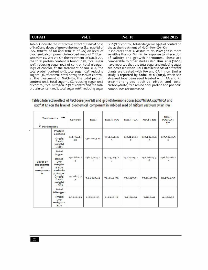
is 109% of control, total nitrogen is 144% of control onthe at the treatment of NaCl+IAA+GA+Kn.It indicates that T. aestivum cv. PBW-502 is moresensitive than cv. WH-711 in response to interactionof salinity and growth hormones. These arecomparable to other studies also. Kim et al (2006)have reported that the total sugar and reducing sugarare increased when NaCl stressed seeds of differentplants are treated with IAA and GA in rice. Similarstudy is reported by Sadak et al (2013), when saltstressed faba bean seed treated with IAA and Kntreatment gives positive effect and totalcarbohydrate, free amino acid, proline and phenoliccompounds are increased .

UJPAH Vol. I No. 18 June 2015
21
Table- 3 shows the interactive effect of 1x10⁻¹M doseof NaCl and doses of growth hormones (i.e. 1x10⁻⁵Mof IAA, 1x10⁻⁹M of Kn and 1x10⁻¹M of GA) on activitiesof certain enzyme in imbibed seeds of Triticumaestivum cv. PBW-502. On the treatment of NaCl+IAA,the total activity of -amylase is 125%, -amylase ac-tivity 120%, protease activity 133%, acid phosphataseactivity 130% and alkaline phosphatase activity 122%of control, on the treatment NaCl+GA, , the totalactivity of-amylase is 141%, -amylase activity 130%,
protease activity 147%, acid phosphatase activity 155%and alkaline phosphatase activity 113% of control, onthe treatment of NaCl+Kn, , the total activity of -amylase 144%, -amylase activity 134%, protease activ-ity 156%, acid phosphatase activity 182% and alkalinephosphatase activity 107% of control and while on thetreatment of NaCl+IAA+GA+Kn, the total activity of-amylase 176%, -amylase activity 157%, protease ac-tivity 208%, acid phosphatase activity 234% and alka-line phosphatase activity 178% of control respectively
Table- 4 indicate the interactive effect of 1x10⁻¹M doseof NaCl and doses of growth hormones (i.e. 1x10⁻⁶M ofIAA, 1x10⁻⁸M of Kn and 1x10⁻⁷M of GA) on activities ofcertain enzyme in imbibed seeds of Triticum aestivumcv. WH-711. On the treatment of NaCl+IAA, the totalactivity of-amylase is 104%, -amylase activity 105%,protease activity 138%, acid phosphatase activity 142%and alkaline phosphatase activity 123% of control. Onthe treatment NaCl+GA, , the total activity of -amylase 102%, -amylase activity 101%, proteaseactivity 272%, acid phosphatase activity 180% andalkaline phosphatase activity 149% of control. On thetreatment of NaCl+Kn, , the total activity of-amylase104%, -amylase activity 103%, protease activity 354%,acid phosphatase activity 192% and alkalinephosphatase activity 137% of control and on the
treatment of NaCl+IAA+GA+Kn, , the total activity of-amylase 111%, -amylase activity 112%, proteaseactivity 474%, acid phosphatase activity is 217% andalkaline phosphatase activity is 183% of controlrespectively.Similarly, Kim et al. (2006) have also reported thatthe activity of -amylase increased when NaClstressed seeds of different plants is treated with IAAand GA in rice. The effects of plant hormones on seedgermination, researchers have found that both understress and non-stress conditions, N compounds,including nitrous oxide can enhance seed germinationthrough enhancing amylase activities (Zhang et al.,2005; Hu et al., 2007; Zheng et al., 2009). Throughdecreasing the production of O2 and H2O2 suchproducts can also alleviate the stress by controlling

UJPAH Vol. I No. 18 June 2015
UJPAH Vol. I No. 18 June 2015
22
Refrences1. Afzal, I.; Basra, S. and Iqbal, A. The effect of seed
soaking with plant growth regulators on seedlingvigor of wheat under salinity stress. J StressPhysiol Biochem., 2005, 1: 6-14.
2. Allakhverdiev, S.I.; Sakamoto, A.; Nishijama, Y.;Inaba, M. and Murata, N. Ionic and osmotic effectsof NaCl-induced inactivation of photo system Iabd II in Synechoccus sp. Plant Physiology, 2000,123:1047-1056.
3. Ashraf, M. Biotechnological approach ofimproving plant salt tolerance using antioxidantsas markers, Biotech adv., 2009, 27: 84-93.
4. Bernfeld, P. Amylase á and â methods,Enzymology, Vol.1, 1995, 149-158.
5. Cha-um, S.; Pokasombat, Y.; and Kirdmanee, C.Remediation of salt-affected soil by gypsum andfarmyard manure – Importance for theproduction of Jasmine rice. Aust J Crop. Sci.,2011, 5: 458-465.
6. Cusido, R.M.; Palazon, J. And Altabella. Effect ofsalinity on soluble protein. Free amino acids andnicotine contents in Nicotiana rustica L, soil, 1987,102: 55-60.
7. Das, M. and Panda, S.K. Salt stress inducedchanges in growth and enzyme activities ingerminating Phaseolus muingo seeds. BiolPlantarum, 2001, 44:587-589.
8. Egamberdieva, D. Alleviation of salt stress byplant growth regulators and IAA producingbacteria in wheat, Acta Physiol Plant, 2009, 31:861-864.
9. Filner, P. and Varner, J.E. A test for de novosynthesis of enzyme. Density labelling with H2O
18
of Barley, alpha amylase indused by gibberellicacid, Prac. Notl Acad. Sci., 1967, Vol.58:1520-1526.
10. Gul, B.; Khan, M.A. and Weber, D.J. Alleviationsalinity and dark enforced dormancy in Allenrolfeaoccidentalis seeds under various thermoperiods.Aust J Bot., 2000, 48:745–752.
the likely oxidative damage, similar to the effects ofantioxidant enzymes including superoxidedismutase (SOD), catalse (CAT) and peroxidase(POD) on plant growth undervarious stresses (Song
et al., 2006; Tian and Lei, 2006; Tseng et al., 2007; Li etal., 2008; Tuna et al., 2008; Zheng et al., 2009; Sajediet al., 2011). Increase of enzyme activities still remainsa question, it needs further kinetic studies.

UJPAH Vol. I No. 18 June 2015
23
11. Hedge, J.E. and Hofreiter, B.T. Carbohydratechemistry 17(eds Whistler RL and Be Miller JN)Academic press,New York, 1962.
12. Hu, K.D.; Hu, L.Y.; Li, Y.H.; Zhang, F.Q. and Zhang,H. Protective roles of nitric oxide on germinationand antioxidant metabolism in wheat seedsunder copper stress, Plant Growth Regul., 2007,53: 173–183.
13. Khan, M.A.; Gul, B. and Weber, D.J. Action of plantgrowth regulators and salinity on seedgermination of Ceratoides lanata. Can J Bot., 2004,82:37-42.
14. Kim, S.K.; Son, T.K.; Park, S.Y.; Lee, I.J.; Lee, B.H.;Kim, H.Y. and Lee, S.C. Influences of gibberellinsand auxin on endogenous plant hormone andstarch mobilization during rice seed germinationunder salt stress. Journal of environmental biology,2006, 27(2):181-186.
15. Lea, P.J. Methods in plant biochemistry, 1990 Vol.3 Enzymes of primary metabolism , Acadmic press,New York.
16. Lowry, O.H.; Rosenbrough, F.J.; Farr, A.L. andRandle, R.J. Protein measurement with Folinphenol reagent, J. Bio. Chem., 1951, Vol.193:265-275.
17. Prince, N.C. and Steven, L. Fundamentals ofEnzymology, Oxford University Press, Oxford,1982.
18. Sadak, M.S.; Dawood, M.G.; Bakry, B.A. andKaramany, M.F.E. Synergistic effect of indoleacetic acid and kinetin on performance, somebiochemical constituents and yield of faba beanplant crown under newly reclaimed sandy soil.World journal of agricultural science , 2013,9(4):335-344.
19. Sajedi, N.; Ardakani, M.; Madani, H.; Naderi, A. andMiransari, M. The effects of selenium and othermicronutrients on the antioxidant activities andyield of corn (Zea mays L.) under drought stress,Physiol. Mol. Biol. Plants , 2011, 17:215–222.
20. Sawhney, S.K. and Singh, Randhir. IntroductoryPratical Biochemistry,Narosa Publishing House,2007, 135-142. New Delhi.
21. Seckin, B.; Sekmen, A.H. and Turkan, I. Anenhancing effect of exogenous mannitol on theantioxidant enzyme activities in roots of wheat
under salt stress. J Plant Growth Regul., 2009,28:12-20.
22. Snell, F.D. and Snell, C.T. Colorimetric methodsof analysis, ed. (1954) 3rd Vol.4 D-Van Nostrandcompany Inc. New York.
23. Song, L.; Ding, W.; Zhao, M.; Sun, B. and Zhang, L.Nitric oxide protects against oxidative stressunder heat stress in the calluses from twoecotypes of reed, Plant Sci., 2006, 171: 449–458.
24. T ian, X. and Lei, Y. Nitric oxide treatmentalleviates drought stress in wheat seedlings, BiolPlant, 2006, 50: 775–778.
25. Tseng, M.J.; Liu, C.W. and Yiu, J.C. Enhancedtolerance to sulphur dioxide and salt stress oftransgenic Chinese cabbage plants expressingboth super-oxide dismutase and catalase inchloroplasts, Plant Physiol Biochem., 2007, 45:822–833.
26. Tuna, A.; Kaya,; Cengiz, K.; Dikilitas, M. and Higgs,D. The combined effects of gibberellic acid andsalinity on some antioxidant enzyme activities,plant growth parameters and nutritional statusin maize plants, Envorin. Exp. Bot., 2008, 62: 1–9.
27. V imla, Y. Change in the certain enzymesaccompanying natural and induced loss of seedviability, 6th Bot. Conf., J. Indian Botanical Science(Soppl.), 1983, Vol. 62:71-X-12.
28. Wilson, K. and Walker, J. Practical Biochemistryprinciples and technique, 4 edu, CambridgeUniversity Press, London, 1996.
29. Yomo, H. and Varner, J.E. Control of the formationof amylase and protease in the cotyledons ofgerminating peas, Plant Physiol., 1973, Vol.51:708-713.
30. Zhang, H.; Shen, W.B.; Zhang, W. and Xu, L.L. Arapid response of-amylase to nitric oxide but notgibberellin in wheat seeds during the early stageof germination, Planta, 2005, 220: 708–716.
31. Zheng, C.; Jiang, D.; Liub, F.; Dai, T.; Liu, W.; Jing,Q. and Cao, W. Exogenous nitric oxide improvesseed germination in wheat against mitochondrialoxidative damage induced by high salinity, Env.Exp. Bot., 2009, 67: 222–227.
32. Zhu, J.K. Plant salt tolerance, Trends plant Sci.,2001, 6: 66-71.
33. Zhu, J.K. Plant salt stress, Encyclopedia of lifesciences, 2007.

UJPAH Vol. I No. 18 June 2015
UJPAH Vol. I No. 18 June 2015
24
Phytochemical Screening and Antimicrobial Activity of Zanthoxylumarmatum DC.
Ajay Singh , Vinod Kumar and Deepak SinghDepartment of Chemistry, Uttaranchal University, Dehradun, Uttarakhand, India, 248006
Email Id- [email protected]
Abstract- Zanthoxylum armatum is used in traditionalmedicinal systems for number of disorders inUttarakhand and other parts of India. Theantimicrobial activity of different extracts of plant-stem bark in different solvents (water, ethanol,methanol, hexane) were tested against 8 pathogenicbacteria and 3 isolated fungal strain by disc diffusionmethod. Two extracts of bark show mild antibacterialactivity and limited antifungal activity but methanolicand aqueous extract gives wide range antibacterialactivity against some pathogenic bacteria. MICs ofmethanol extract of Z. armatum root produced bestinhibition zones against P. Mirabilis (18mm), S.Mutons(20mm), Gardoni (18mm) and aqueous extractproduce inhibition zone against Listeria(18mm) andGardoni (20mm). Chemical analysis revealed thepresence of terpenoid, saponin and reducing sugarsas major compounds in the stem bark of the plant.Key words: Zanthoxylum armatum, antibacterial,antifungal activities, inhibition zone, stem bark.
IntroductionIndia has richest plant based medicinal traditionalsystem because of its rich biodiversity. These herbalmedicines are mainly used for health care due to theircost value, effectiveness and lesser side effects onhuman body. Among different medicinal plants of TheIndian Himalayan region (IHR), Zanthoxylum (family:Rutaceae) is one of such genus which possess highmedicinal importance and have about 250 speciesspreading all over the world. In India, 11 species ofthis genus is reported and 4 species; Z. armatum DC(Syn. Z. alatum Roxb.), Z. acanthopodium DC. Z.oxyphyllum Edgew, and Z. budrunga are present inUttarakhand. Zanthoxylum armatum is widelydistributed in India from Kashmir to Bhutan upto2500m altitudes. The bark is utilized as traditional dyeyielding resource. Chemical studies carried out onZanthoxylum species have revealed the occurrencemainly of alkaloids, flavonoids, terpenoids, aliphatic
and aromatic amides, lignans, coumarins, sterols,carbohydrate residues etc. Some of these metaboliteshave reported, antibacterial, antifungal, antioxidant,cytotoxic, molluscicidal, anti-sickling, anesthetic anti-hypertensive and anti-inflammatory properties.
Material and MethodsPreparation of extract: The extracts of medicinalplants were prepared by dissolving sample in 1:10 withdifferent solvents separately (Distilled water, ethanol,methanol and hexane respectively) and a constantheating was provided by heating mental for 6 hoursin soxhlet appratus. Then extracts in round bottomflask were transferred to pre-weighted apenldrof.Apenldrof containing extracts were weighted andnoted down and finally, the percentage yield wascalculated. Then the extracts were stored at 40C.
Yield of extract= Weight of empty apendrof – weight
of apendrof with sample
%%yield = Yield of extract
Weight of raw material taken%XX 100
Bacterial Culture: The human pathogenic bacteriasuch as Staphylococcus sp., Escherichia coli, Listeria, P.Vulgaris, S.Gardoni, P. Mirabilis, S.Mutons, Clostridiumwere obtained from Bioinformatics Centre, IMTECH,Chandigarh and were maintained in Nutrient agar slantat 40C for experimental studies. The fungus strain ofPenicillin was isolated from Potato Dextrose Agar.Preparation of standard culture inoculum of testorganism: The colonies of different bacteria and strainof Fungus were inoculated in the 20 ml nutrient brothand incubated for 24 hours and 72 hours respectively.Assay of antimicrobial activity: Assay of Anti bacterialactivity done by Disc Diffusion method (Kirby-Bauermethod, 1966). All plates were incubated at 37°C for24 h. Zone of inhibition was noted down. Eachexperiment was performed 3 times. Discs

UJPAH Vol. I No. 18 June 2015
25
impregnated with only solvents were used as negativecontrols.Antifungal assays: Assay of Anti Fungal activity wasdone by using disc diffusion method (Kirby-Bauermethod, 1966). Each experiment was performed 3times.Minimum Inhibitory Concentration (MIC) Assay: TheMIC method was applied on extracts that proved theirhigh efficacy against microorganisms by serial dilutionmethod. All plates were incubated at 37°C for 24 h.Zone of inhibition was noted down. Each experimentwas performed 3 times.
Phytochemical studies: Chemical Tests were carriedout on the ethanol, methanol, water and –hexaneextracts of Zanthoxylum Armatum plant usingstandard procedures (Abdul Wadoodet al.,2013 andPravin S. Jogi et al.,2012).Results and DiscussionThe percentage yields of extracts and thephytochemical constituents of the plants are shownin Table 1. Among all the extracts the Aqueous extractof bark of Zanthoxylum Armatum contain the higheramount of soluble solids followed by ethanol,methanol and hexane.
Table- 1 The yields of plants extracted in different solvents.
Antibacterial activity: The methanolic extract of plantonly shows some antibacterial properties againstMirabilis (18mm), Mutons (20mm) and Gardoni(18mm)while aqueous extract shows zone of inhibition forListeria(18mm) and Gardoni(20mm) shown in table 2.Barkatullah et al. in 2012 reported that the ZLE extractshowed highest inhibition against M. leutus (18.00 ±0.71 mm), P. multocida (18.00 ± 0.71 mm), E. coli (17.00± 0.71) and B. subtilis (15.33 ± 0.81 mm). The ZFEshowed topmost action against M. leutus (21.33 ± 0.41mm) and P. multocida (18.33 ± 0.41 mm) while theZFH showed inhibitory action against M. leutus (19.67
± 0.41 mm) as compared to other tests species. TheZBH extract was found active against M. leutus (20.33± 0.41 mm). The minimum inhibitory concentration(MIC) values for most of the bacterial species werefound to be 0.65 g/ml. The crude ethanolic and n-hexane of all parts were proved a rich source offungicidal effect. Highest flavonoids was found inethanolic extract of Z. armatum fruit (ZFE) (22.8 ±1.33 mg/g) followed by ethanolic extract of Z. armatumbark (ZBE) (18.33 ± 1.22 mg/g) while highest phenoliccontents were found in ZFE (21.68 ± 0.44 mg/g)followed by ZBE (16.48 ± 1.33 mg/g).
Table- 2 Zone of Inhibition (in mm) of bark extracts of Zanthoxylum Armatum against Gram-positive and Gram-negative bacteria at different concentration.
UJPAH Vol. I No. 18 June 2015
UJPAH Vol. I No. 18 June 2015
26
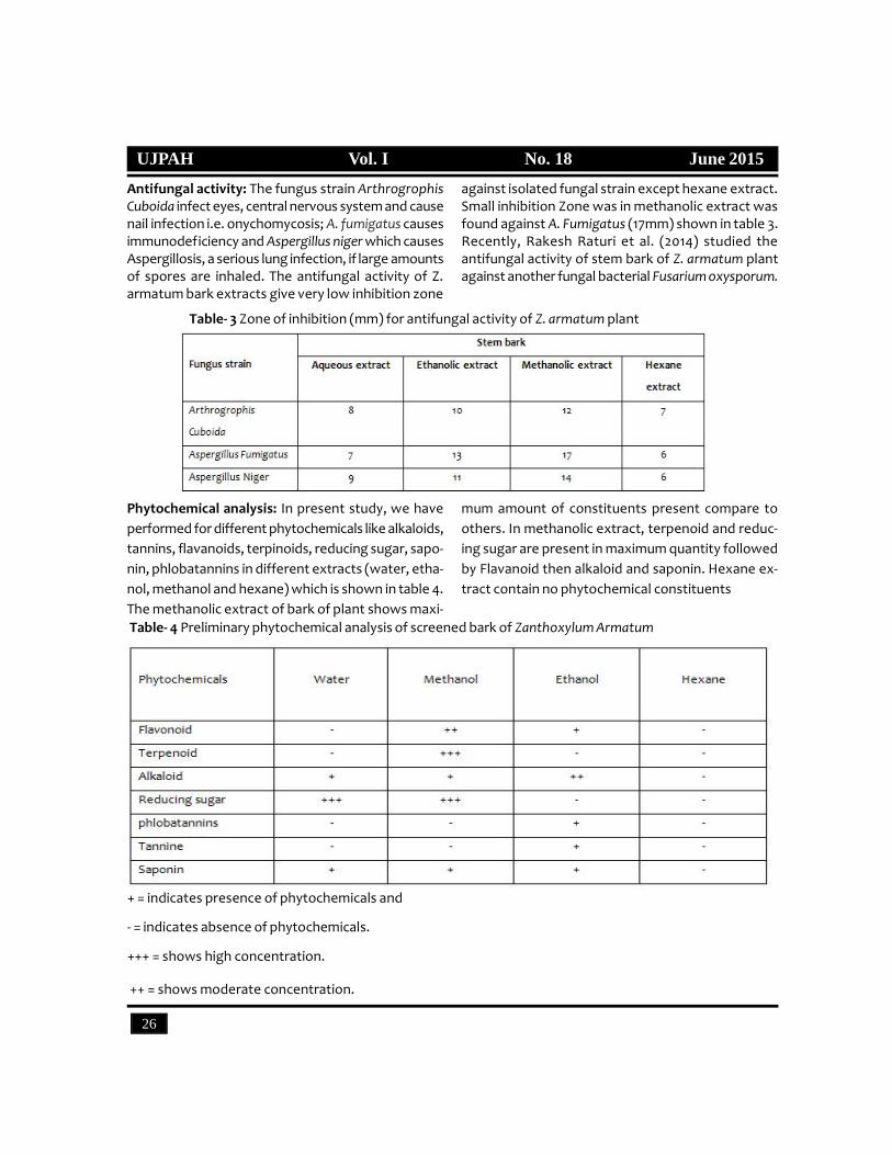
Antifungal activity: The fungus strain ArthrogrophisCuboida infect eyes, central nervous system and causenail infection i.e. onychomycosis; A. fumigatus causesimmunodeficiency and Aspergillus niger which causesAspergillosis, a serious lung infection, if large amountsof spores are inhaled. The antifungal activity of Z.armatum bark extracts give very low inhibition zone
against isolated fungal strain except hexane extract.Small inhibition Zone was in methanolic extract wasfound against A. Fumigatus (17mm) shown in table 3.Recently, Rakesh Raturi et al. (2014) studied theantifungal activity of stem bark of Z. armatum plantagainst another fungal bacterial Fusarium oxysporum.
Table- 3 Zone of inhibition (mm) for antifungal activity of Z. armatum plant
Phytochemical analysis: In present study, we haveperformed for different phytochemicals like alkaloids,tannins, flavanoids, terpinoids, reducing sugar, sapo-nin, phlobatannins in different extracts (water, etha-nol, methanol and hexane) which is shown in table 4.The methanolic extract of bark of plant shows maxi-
mum amount of constituents present compare toothers. In methanolic extract, terpenoid and reduc-ing sugar are present in maximum quantity followedby Flavanoid then alkaloid and saponin. Hexane ex-tract contain no phytochemical constituents
Table- 4 Preliminary phytochemical analysis of screened bark of Zanthoxylum Armatum
+ = indicates presence of phytochemicals and
- = indicates absence of phytochemicals.
+++ = shows high concentration.
++ = shows moderate concentration.
UJPAH Vol. I No. 18 June 2015
27
ConclusionThe present study deals with the antibacterial,antifungal activity and phytochemical screening of Z.armatum. This plant has many medicinal uses especiallyas antibacterial agent due to the presence ofmedicinal compounds. On the basis of results ofpresent study we have concluded that the methanolicextract of stem bark of plant possess antibacterialactivity against S. Gardoni (18mm), S. Mutons (20mm)and P.Mirabilies (18 mm). The stem bark of plant showsunsatisfactory result against Arthrogrophis Cuboida,A. Niger and A. Fumigantus. So, Z. armatum plant havelimited antibacterial and antifungal activity againstpathogenic bacteria. From phytochemical screeningit concluded that the plant bark contain thosecompounds which are responsible for medicial valueslike reducing sugar, terpenoid, saponin as a majorcompound and other constituents like alkaloid andflavonoid.
References1. Barkatullah; Muhammad Ibrar; Naveed
Muhammad and Lubna Tahir. Antimicrobialevaluation, determination of total phenolic andflavoniod contents in Zanthoxylum armatum DC.Journal of Medicinal Plants Research, 2012,6(11),2105-2110.
2. Bauer, A.W.; Kirby, W.M.; Sherris, J.C. and Turck,M. Antibiotic susceptibility testing by astandardized single disk method. Am J Clin Pathol.,1966 Apr, 45(4):493–496.
3. Pravin S. Jogi and Akkewar, D. M. Phytochemicalscreening and antimicrobial activity of medicinalplant Pergularia daemia From Chandrapur ForestRegion. International Journal of Natural ProductsResearch, 2012, 1(3): 61-63
4. Rakesh Raturi; Badoni, P.P. and Radha Ballabha.Insecticidal and fungicidal activities of stem barkof zanthoxylum armatum (rutaceae). WorldJournal of Pharmacy and Pharmaceutical Sciences,2014. Vol. 3, 1838-1843.
5. Wadood, A.; Ghufran, M.; Jamal, S. B.; Naeem, M.and Khan, A. Phytochemical Analysis of MedicinalPlants Occurring in Local Area of Mardan. BiochemAnal Biochem., 2013, 2:144.

UJPAH Vol. I No. 18 June 2015
UJPAH Vol. I No. 18 June 2015
28
Comparison of Physicochemical Properties of Some Edible OilsAfshan Tarranum, Renu Chauhan, Raj Kumar and Mona Chauhan
Department of Life Science, Beehive College of Advanced Studies, Dehradun (UK.), IndiaEmail: [email protected]
Abstract- Oils and fats are important parts of humandiet. These are the rich sources of dietary energy andcontain more than twice the caloric value ofequivalent amount of sugar. Their functional andtextural characteristics contribute to the flavour andpalatability of the natural and the prepared foods.They contain certain fatty acids which play animportant role in nutrition and are also carrier of fatsoluble vitamins. In the present paper, we haveexperimentally determined the saponification value,iodine value, acid value, and peroxide value for threedifferent edible oils. The iodine value (IV) indicatesthe degree of unsaturation of the oil. The acid value(AV) give an idea of the amount of free acid presentin the oil. The peroxides value (PV) gives the initialevidence of rancidity in unsaturated fats and oils. Itgives a measure of the extent to which oil hasundergone primary oxidation. The Saponif icationvalue (SV) is expressed by the amount of potassiumhydroxide in mg required to saponify one gram of fat.It was observed on the basis of said values thatsoyabean oil and mustard oil showed higher values ofunsaturation and auto-oxidation than those given byolive, coconut and pea-nut oils.Key words: Edible oils, Iodine value, Acid value,Saponification value, Peroxide value.
IntroductionEdible oils are triglyceride extracted from a plant.Such oils have been part of human culture formillennia. The term edible oil can be narrowly definedas referring only to plant oils that are liquid at roomtemperature, or broadly defined without regard to asubstance’s state of matter at given temperature(D.Robin,1999). The estimation of the physico-chemical properties of edible oils is essential in thedesign of unit processes such as distillation, heatexchangers, reactors and piping. On the other side,physico- chemical properties are an important factorthat determines the overall quality and stability of afood system. ). Density, saponification value, iodinevalue, acid value, peroxide values are some of theimportant characteristics of a vegetable oil. Thecoconut (Cocosnucifera L.) is an important fruit treein the tropical regions and the fruit can be made intoa variety of foods and beverages. Solid at roomtemperature, coconut oil is white and buttery intexture; it has a distinctive coconut flavor and is widely
used in South India. Soya bean oil (Glycine max) is avegetable oil extracted from the seeds of the soyabean. It is one of the most widely consumed cookingoils and is an important source of dietary proteinwidely used in a variety of dishes by oriental peoplefor many centuries. The sunflower, (Helianthus annuusL.) seed itself is edible and its oil is used throughoutthe world for frying and cooking. It is also used aspoultry feed. Peanuts (Arachishypogaea L.)are a verygood source of monounsaturated fats, the type of fatthat is emphasized in the heart-healthy Mediterraneandiet. The olive tree ( Oleaeuropea L.) is one of themost cultivated fruit trees since ancient times (Fabbri,2006).Olive oil is a natural green vegetable oil whichis abundant in vitamin, carotene and many traceelements and has many functions innutrition and health care (Mar´ýa-Isabel Covas, 2006). Mustard Oil (Brassicanigra) is extracted at a low pressure and lowtemperature (40-60ºC). It contains 0.30-0.35 %essential oil (AllylIso- Thiocynate) which acts aspreservative. Ghee, the most famous traditional dairyproduct in India, many countries and the Middle East,is made from milk, cream, or butter of several animalspecies (G. S. Rajorhia, 2003).Therefore, the aim of the present investigation wasto ascertain the saponif ication, iodine, acid andperoxide value of these oils as a means to comparethe physicochemical properties of the oil mentioned.
Material and MethodsCollection of oils: Edible oil viz., olive oil, peanut oil,mustard oil, soya bean oil, sunflower oil, coconut oiland desi ghee were collected from local areas ofDehradun.Saponification Value: The saponification value of oil isdefined as the number of milligram of KOH requiredto neutralize the fatty acid resulting from thecomplete hydrolysis of 1gm of oil or fat .Themagnitude of its value gives an idea about the averagemolecular weight of the oil or fat. The higher themolecular weight of a fat, the smaller is itssaponification value .The higher the saponificationvalue, the greater is the percentage of the short chainacids present in the glycerides of the oil and fat.Iodine value: The iodine value of an oil or fat is definedas the no. of grams of iodine taken up by 100 gm ofthe oil. Iodine value is an indication of the degree ofunsaturated monochloride in acetic acid with the oil

UJPAH Vol. I No. 18 June 2015
29
dissolved in CHCl3 or carbon tetra chloride and thenback titrating the residual iodine with a standardsodium thiosulphate solution taking starch as theindicator.Acid value: The acid value of oil is defined as the no. ofmg of potassium hydroxide required to neutralize 1gof oil or the no of mg of KOH neutralized by the freeacid presents in 1gm of oil. Such a value gives an ideaof the amount of the free acids presents in the oilunder study. Normally fresh and recently extractedoil or fats have very little or no free fatty acid present.Peroxide value: Peroxide value (PV) measures themilli equivalents of oxygen (hydro peroxides) per 1000
gram of oil. The peroxide value is a measure of theconcentration of substances that oxidize potassiumiodide to iodine. The amount of free iodine isdetermined by titration with sodium thiosulphateusing starch as indicator. A corresponding blankreagent is simultaneously prepared.(P.C.kamboj,2007)Results and DiscussionThe aim of this work was to characterize differentedible oils on the basis of saponification, iodine, acidand peroxide values. The values obtained are given inthe following table.
Table
Graph Physico-chemical properties of respective oils tested

UJPAH Vol. I No. 18 June 2015
UJPAH Vol. I No. 18 June 2015
30
Saponification value is a measure of oxidation duringstorage. It also indicates deterioration of the oils.From the above table, it can be seen that peanut oilhas the highest value of SV while olive oil has thelowest among the oils tested. These results are inagreement with the studies conducted by Naegu, etal., 2013.The higher the iodine value, the less stable is the oiland is more vulnerable to oxidation and free radicalproduction. High iodine value oils are prone tooxidation and polymerization. During heating, suchas when used in cooking, oils with high iodine valuereadily oxidize and polymerize. Polymerization is anirreversible process which causes the fatty acids tobecome hard, insoluble, plastic-like solids. We haveobserved that highest iodine value is of soya bean oilfollowed by mustard and peanut oil.Acid Value is a measure of the content of free fattyacids in the vegetable oil and describes the quantityof caustic potash solution which is necessary for theneutralization of the free fatty acids. The acid numberdepends strongly on the refining degree and theaging degree of oil. Since the acid value of soyabeanoil is high, it depicts less comparative refining ofsoyabean oil as compared to other oils tested.Detection of peroxides gives the initial evidence ofrancidity in unsaturated fats and oils. It gives ameasure of the extent to which oil has undergoneprimary oxidation. The double bonds found in fatsand oils play a role in auto-oxidation. Oils with a highdegree of unsaturation are most susceptible to auto-oxidation. Peroxides are intermediates in the auto-oxidation reaction. Here, we found that peroxidevalue of soya bean is highest followed by mustard oiland sunflower oil indicative of it being undergoneauto-oxidation.From the above values it can be concluded thatsoyabean oil followed by mustard oil and sunfloweroil exhibit a higher level of unsaturation and auto-oxidation as compared to that of olive, cococnut andpeanut oil. These results are in agreement with thestudies conducted by Tasic et al., 1999 who observedlowest value of saponification for olive oil.References1. Robin, D. The International Cocoa Trade.
Woodhead Publishing. 1999, p.169. ISBN 1-85573-434-6.
2. R u d a n, D. and T a s i è a n dKlofutar, C. Character-istics of vegetableoils of some solvene manufacturers. Acta Chim.Slov., 1999, 146(4), pp. 511-521.
3. Fabbri, A. Olive propagation new challenges andscientific research. Olive bioteq., 2006, 1: 411-421.
4. Rajorhia,G. S. -GHEE, Encyclopedia of FoodSciences and Nutrition, 2003, pp. 2883-2889.
5. Mar´ýa-Isabel Covas, DPharm. Minorcomponents of olive oil: evidence to date of healthbeneûts in humans. Nutrition Reviews, 2006, 64:S20-S30.
6. Naegu Anisoara Arlezania, Irina Nita, ElisabetaBotez and Sibel Geacai. A physicochemical studyfor some edible oil properties. Ovidus UniversityAnnals of Chemistry, 2013, 24(2) pp 121-126.
7. Kamboj, P.C. University practical chemistry( Anadvanced practical text book) printed at NewSimran Printing Press, Jalandhar, 2007, pp.1-543.

UJPAH Vol. I No. 18 June 2015
31
Qualitative Phytochemical Analysis of Allium humile and Ocimum gratissimum(Linn.) Leaves Extract
Yogita Dobhal1*, Versha Parcha2 and D.C. Dhasmana3
1 Department of Pharmaceutical Sciences, Sardar Bhagwan Singh PG Institute of Biomedical Sciences &Research, Balawala, Dehradun, Uttarakhand, India.
2Department of Chemistry, Sardar Bhagwan Singh PG Institute of Biomedical Sciences & Research,Balawala, Dehradun, Uttarakhand, India.
3 Department of Pharmacology, H.I.M.S.Doiwala, Dehradun, Uttarakhand, India.*Corresponding author: [email protected]
[email protected]@gmail.com
Abstract- The present study has been designed toanalyse the phyto-constituents of various extracts ofAllium humile and Ocimum gratissimum (Linn.) leaves.Various extracts of A. humile and O. gratissimum (Linn.)leaves were prepared in viz. Pet.ether, chloroform,acetone and methanol (12 hrs each) using Soxhletextraction technique to get four extracts. Theseextracts were then subjected to qualitative chemicaltests for the identif ication of various phyto-constituents viz, alkaloids, carbohydrates, glycosides,proteins, tannins, sterols, saponins, amino acids etc.This study demonstrates that methanolic extract ofAllium humile leaves was rich in alkaloids, saponins,carbohydrate, glycosides, proteins and amino acids,whereas the chloroform extract of O. gratissimum(Linn.) gave test for alkaloids, triterpenoids,carbohydrates and glycosides. These results suggestthat these phyto-constituents may be responsible forpharmacological activities of these plants.Key words: Allium humile, Ocimum gratissimum(Linn.), Phyto-constituents, Extraction.
IntroductionOver the past few decades, the traditional knowledgeon the use of medicinal plants has been widelyacknowledged across the world. According to theWorld Health Organization, 80%of the world’spopulation in developing countries uses traditionalmedicine1. In India, the knowledge of traditionalherbal medicine is synonymous with its rich culturalheritage and has found its mention in Vedic literature,particularly Rigveda, Charak Samhita and SusrutaSamhita. In the North of India, the Himalayas covering18% of the Indian subcontinent accounts for more than50% of India’s forest and contains 30% of India’seconomic species. The area harbours about 8,000species of higher plants of which 1,748 are used formedicinal purposes2.Roughly 40% of plants provideactive ingredients for modern drugs, and because oftheir use in traditional medicines, they draw theinterest of researchers. Furthermore, the traditional
knowledge with its holistic and systematic approachsupported by experimental base can serve as aninnovative and powerful discovery engine for newer,safer and affordable medicines.Small Alpine Onion Allium humile (A.nivale;A.gowanianum), belongs to family Alliaceae, found athigh altitudes in the Himalayas at altitudes of 3000-4000 m. It is found on open alpine slopes from Pakistanto W. Nepal and China3. Allium humile is used as acondiment with pulses, blood purification, swelling,asthma, stomach disease, cold, cough, jaundice. Theleaf and bulb parts are used locally in alleviation ofinflammation and pain full conditions4. Leaves andinflorescences are used as seasoning agents. Fewresearch articles reported the antibacterial activity5.Ocimum gratissimum (Linn.) is well-known plant usedin the Indian system of medicine. A lot of researchhas been carried out on O. gratissimum (Linn.). Whichis herbaceous plant (Family- Labiatae), indigenous totropical areas especially India and in West Africa. InNigeria, it is found in the Savannah and coastal areas.It is cultivated in Ceylon, South Sea Islands, and alsowithin Nepal, Bengal, Chittagong and Deccan6. Theplant is commonly used in folk medicine to treatdifferent diseases such as upper respiratory tractinfections, diarrhoea, headache, diseases of the eye,skin diseases, pneumonia, cough, fever andconjunctivitis7. In the present study, the varioussolvent extracts of A.humile and O.gratissimum (Linn.)were subjected to qualitative chemical tests for theidentification of various phyto-constituents which aresolely responsible for biological activity of these plants.
Material and MethodsPlant material: Leaves of Allium humile were collectedfrom Chamoli. The plant material was identified fromBotanical Survey of India, Northern Regional Centre,Dehradun, India with the reference number BSI/NRC9 (Tech.)/2010-03/839/12796. Leaves of Ocimumgratissimum (Linn.) was collected from IndianInstitute of Integrative Medicine, Jammu (Formerly

UJPAH Vol. I No. 18 June 2015
UJPAH Vol. I No. 18 June 2015
32
Regional Research Laboratory), CSIR, India. Thereference number was [RRL(J)OG-14]. The leaves ofAllium humile and Ocimum gratissimum (Linn.) weredried in shade at room temperature and groundseparately at controlled temperature.Chemicals and reagents were of analytical grade- Thesolvents and chemicals were of analytical grade.Petroleum ether, Chloroform, Acetone, Methanol,Ethyl acetate were purchased from Merck India Ltd.,Mumbai. Benedicts reagent, Fehling reagent, Mayer’sreagent, Dragendroff reagent, Hager reagent,Wagner reagent, Molish reagent, Millon reagent,Ninhydrin reagent, NaOH pellets, FeCl 3 werepurchased from Ranbaxy Fine Chemicals Ltd, NewDelhi. Picric acid and Hydrochloric acid was purchasedfrom S.D. Fine Chemicals Ltd. Mumbai. -naphthol,H2SO4 was purchased from Merck India Ltd. Mumbai.
Preparation of extracts: The fresh leaves of A. humileand O.gratissimum were dried in shade and at roomtemperature for 2 days followed by tray drying [40-50oC] for 3-4hrs and powdered to obtain coarsepowder. 980g of powder of A. humile leaves and 2 kgO. gratissimum leaves were extracted with pet ether,chloroform, acetone and methanol (12 hrs each) usingSoxhlet extraction technique to get four extracts.
The solvent was removed by evaporation underreduced pressure to obtain a semisolid mass. Theresultant concentrated extracts were then scoopedinto a pre-weighed small sterile container andweighed. The difference between the two weightswas recorded as the percentage yield. The containerswere then covered, labelled accordingly and theresultant extract was kept in desiccators8.Qualitative phytochemical analysis: The varioussolvent extracts of A.humile and O.gratissimum (Linn.)were subjected to qualitative chemical tests for theidentif ication of various phyto-constituents viz,alkaloids, carbohydrates, glycosides, proteins, tannins,sterols, saponins, amino acids etc. These phyto-constituents are solely responsible for biologicalactivity of these plants9-11.
Results and DiscussionQualitative phytochemical constituents of Alliumhumile leaves: All the extracts were screened forphytochemical constituents. Results showed thepresence of triterpenoids in pet. ether extract;alkaloids, carbohydrates and glycosides were foundin acetone extract, while methanol extract was richin alkaloids, saponins, carbohydrate, glycosides,proteins and amino acids (Table-1).

UJPAH Vol. I No. 18 June 2015
33
Qualitative phytochemical constituents of Ocimumgratissimum (Linn.) leaves: All the extracts werescreened for phytochemical constituents. Resultsshowed the presence of alkaloids, triterpenoids,carbohydrates and glycosides in chloroform extract;
acetone extract showed the presence of alkaloids,proteins and amino acids, while methanol extract wasrich in alkaloids, carbohydrate, glycosides andphenolic compounds (Table- 2).

UJPAH Vol. I No. 18 June 2015
UJPAH Vol. I No. 18 June 2015
34
ConclusionFrom extensive literature survey, it was found that A.humile and O. gratissimum (Linn.) possess differentpharmacological activities that have been reported.The Allium Genus includes approximately 500 speciesthat most widely used and have been employed forcenturies for the pungency and flavouring value, andfor their medicinal properties. The scientificcommunity is increasingly getting interested in thestudy of pharmalogical properties of Allium plantsand their chemical constituents particularly withregard to their ability to prevent various diseases12, 13
whereas various leaf extracts of O. gratissimum (Linn.)showed antidiabetic, anti-diarrheal, anti-fertility,hepatoprotective, analgesic activity aphrodisiac,immuno stimulatory, Antitumor, anti-mutagenic andanti-cancer effects have been reported in in-vitro andin-vivo experiments14-15. The widespread plant is knownfor its chemopreventive, anticarcinogenic, free radicalscavenging effect and used as traditional herb inEuropean and Asian countries since ancient times16.Allium humile and Ocimum gratissimum (Linn.) leavesare widely used in folklore medicine for treatment ofvarious diseases in the different regions of Himalaya.
In present study, the plant materials were subjectedto soxhlet extraction using various solvents in an orderof increasing polarity viz., petroleum ether,chloroform, acetone and methanol. The extracts thusobtained were concentrated and used for furtherevaluation. Qualitative phytochemical analysis ofdifferent extract was carried out. Methanol extract ofA. humile was rich in alkaloids, saponins,carbohydrate, glycosides, proteins and amino acidsand chloroform extract of O. gratissimum (Linn.) gavetest for alkaloids, triterpenoids, carbohydrates andglycosides.
AcknowledgementThe authors are grateful to Director and managementof SBSPGI, Balawala, Dehradun, India for providingthe necessary facilities during this experimentalstudy.
References1. Cotton, C.M. Ethnobotany, Principles and
applications, (Wiley & Sons, UK), 1997.2. Gaur, R.D.; Semwal, J.K. and Tiwari, J.K. A survey
of high altitude medicinal plants of GarhwalHimalayas. Bull Med Ethnobot Res., 1983, 4: 102-116.
3. Prabhu, K.S.; Lobo, R.; Shirwaikar, A.A. andShirwaikar, A. Ocimum gratissimum: A Review ofits Chemical, Pharmacological and
Ethnomedicinal Properties. The OpenComplementary Medicine Journal 2009, 1: 1-15.
4. Woodward, P.W.; Garlic and Friends: The History,Growth and Use of Edible Alliums Hyland House,Melbourne, Australia 1996, 2-22.
5. Elnima, E.I.; Ahmed, S.A.; Mekkawi, A.G. andMossa, J.S. The Antimicrobial Activity of Garlicand Onion Extracts. Pharmazie 1983, 38 (11):747–8.
6. Nadkarni, K.M. Indian Materia Medica, 3rd ed.Popular Prakashan Pvt Ltd: India 1999.
7. Adebolu, T.T. and Salau, A.O. Antimicrobial activityof leaf extracts of Ocimum gratissimum onselected diarrhoea causing bacteria insouthwestern Nigeria. Afr J Biotech. 2005, 4: 682-4.
8. Mukherjee, P.K. Quality Control of Herbal Drugs.Business Horizone, New Delhi, 2002, 405-406.
9. Trease, G.E. and Evans, W.C. Pharmacognosy. 1st
edition, ELBS, London, 1989, 3, 223-249.10. Kokate, C.K.; Purohit, A.P. and Gokhale, S.B.
Pharmacognosy. 30th edition, Nirali Prakashan,Pune, India, 1996, 447 -460.
11. Khandelwal, K.R. Practical Pharmacognosy. 13th
edition, Nirali Prakashan, Pune, India, 2005, 149-156.
12. Sengupta, A.; Ghosh, S. and Bhattacharjee, S.Allium Vegetables in Cancer Prevention: AnOverview. Asian Pacific J Cancer Prev., 2004, 5:237-245.
13. Banerjee, S.K. and Maulik, S.K. Effect of garlic oncardiovascular disorders: a review. NutritionJournal 2002, 1:4
14. Ahmed M.M.; Khan, M.Y. and Khan, A.A. Effectsof Ocimum gratissimum (Tulsi) on thereproductive system: an updated review. BiomedRes., 2002, 13: 63-67.
15. Surana, V. and Jain, D. Protective effect of Ocimumgratissimum against carbon tetrachloride inducedhepatic damage in rats. Pharmacology online2010, 2:1111-1119.
16. Gupta, S.K.; Prakash, J. and Srivastava, S. Validationof traditional claim of Tulsi, Ocimum sanctum Linn.as a medicinal plant. Indian J Exp Biol., 2002,40(7):765–773.

UJPAH Vol. I No. 18 June 2015
35
Antioxidant Effect and Antimicrobial Activity of Cassia auriculataPiyush Mani Dubey and Neerja Gupta
Department of Chemistry, A.N.D.N.N.M.College, Kanpur (U.P.), IndiaEmail: [email protected]
Abstract- Cassia auriculata (Fabaceae) commonlyknown as “Tanner’s cassia”is a shrub with large brightyellow flowers found growing wild in central andwestern India and cultivated in other areas of thecountry. It is widely used in Indian folk medicines forthe treatment of various diseases like diabetes,rheumatism and conjunctivitis. Free radicals aregenerated inside the body due to variousphysiological and environmental factors which causevarious ill effects on the body including ageing andcancer. The objective of, the present study is todiscover a new entity which can inhibit the generationof these free radicals and also its anti-microbial activity.Consuming antioxidant supplements would help onmaintaining immune system in the best conditionspossible. It has been found that when large numbersof antioxidants are mixed together in small doses,they can help in protecting against severe diseaseslike cancer and heart diseases.
IntroductionAntioxidants are substances that reduce oxidativedamage in cells caused by free radicals.1 Medicinalproperties of plants are due to presence of definitechemical constituents in them. In many cases thesephysiologically active constituents of medicinal plantsgive a clue for the preparation of synthetic drugs ofgreater potency. With the development in technologyand recent research, it has been proved that certainnon-nutritive chemicals in plants such as Terpenoidsand Flavanoids possess antioxidant properties 2,3.Antioxidants are a type of complex compounds foundin our diet and act as protective shield for our bodyagainst certain disastrous diseases and strengthen theimmune system of our body4. Medicinal plants containvarious compounds like carotenoids, vitamins,terpenes, flavanoids and phytosterols etc. that possessantioxidative components which prevent from freeradical damage and premature ageing.5-7Some of theimportant medicinal plants having characteristicantioxidant properties are tabulated here inalphabetical order with their common name, family,part of the plant used and their active chemicalconstituents8-11. Cassia auriculata (Fabaceae)commonly known as “Tanner’s cassia” is a shrub withlarge bright yellow flowers found growing wild incentral and western India and cultivated in otherareas of the country. It is widely used in Indian folkmedicines for the treatment of various diseases likediabetes, rheumatism and conjunctivitis.
Material and MethodsThis study is comprised of mainly two activities listedbelow as separate headings for which different testsare carried out.
In-vitro Antioxidant Activity1
DPPH radical scavenging method: The differentconcentrations of extracts, the compound andstandards were added to DPPH in methanolic solution(100 µM) in a 96-well microtitre plate. After incubationat 370C for 20 min, the absorbance of each solutionwas determined at 490 nm using ELISA reader.
Scavenging of super oxide radical by alkaline DMSOmethod: The reaction mixture is prepared containingNBT (1 mg/ml solution in DMSO) and alkaline DMSO(1ml DMSO containing, 5mM NaOH in 0.1ml water)and to its different concentrations of the extractsand standards were added and the absorbance wasmeasured at 560 nm.
Nitric oxide radical inhibition assay: The reactionmixture containing sodium nitroprusside (10 mM, 4ml), phosphate buffer saline (1 ml) and the extracts,the compound and standard solutions (1 ml) wereincubated at 25 0C for 150 min. After incubation, 0.5mlof the reaction mixture was removed and 1ml ofsulphanilic acid reagent (0.33% in 20% glacial aceticacid) was mixed and allowed to stand for 5 min forcompletion of diazotization reaction and then 1ml ofNEDD was added, mixed and allowed to stand foranother 30 min in diffused light. The absorbance wasmeasured at 540 nm against the corresponding blanksolutions in a 96-well microtitre plate using ELISAreader.
Scavenging of ABTS radical caution: To variousconcentrations of the extracts, the compound orstandard, distilled DMSO and ABTS solutions wereadded and incubated for 20 min. Absorbance wasmeasured at 734 nm.
Anti-microbial Activity3
Agar Diffusion Method- Media prepared according tothe manufacturers instruction (for each organism).Wells were prepared and 40 µl of the different

UJPAH Vol. I No. 18 June 2015
UJPAH Vol. I No. 18 June 2015
36
concentrations of the extracts were introduced intothe well and was incubated at 37ÚC for bacteria for48 hrs. Anti-microbial activity was determined bymeasuring the zone of inhibition.
The crude extract of Cassia auriculata showedsignificant antioxidant activity in DPPH and NBT. Italso showed significant anti-microbial activity.
ConclusionFrom these studies it can be concluded that thecomponents in the crude extract of Cassia auriculatacontains active components responsible forantimicrobial and antioxidant activity. Furtherisolation of these active components from thefraction would lead to the identif ication of themolecule.
Refrences1. Srinivasan, R. et al. Journal of Ethnopharmacology
2007, 113:284–291.2. Elizabeth, K. et al. International Journal Of
Pharmaceutics, 58:237-40.3. Parekh, J. et al. Indian Journal of Pharmacology,
2005, 37: 408-409.4. Bhattacharjee, S.K. Handbook of Medicinal Plants,
Pointer Publishers, Jaipur, India, 1998, 302003; 1-6.
5. Mayoclinic Health Letter, 1995, 11, 1-2.6. Fendall, R. Ayurvedic Medicine and Primary Health
Care, World Health Forum, 1982, 3(1);90-94.7. Annual Report, Institute of Food Research, 1997.
8. Halliwell, B. Free Radical Res., 1999, 31, 261-272.9. Bray, T. M. Society for Experimental Biology and
Medicine, 1994, (3a-5s).10. Mayoclinic Health Letter, 1997, 10, 1-2.11. The Amer. J. of Med., 1994, 3a-5a.12. Institute of Food Research, Annual Report, 1999.13. Freiz, Balz. Amer. J. of Med.,1994, 97, 58.14. Halliwell, Barry, The Lancet, 1994, 721-724.

UJPAH Vol. I No. 18 June 2015
37
Antimicrobial Activity of Cleome viscose (seed)Arti Tomar1* and Suman Rawat 2
1Department of Pharmaceutical Chemistry,Dolphin (PG)Institute Of Biomedical and Natural Sciences,Dehradun , Uttarakhand (India)
2Department of Chemistry,Dolphin (PG)Institute Of Biomedical and Natural Sciences, Dehradun ,Uttarakhand (India)
Contact no:9412558706*E. mail: [email protected]
Abstract- The present study was aimed at detectingand evaluating antimicrobial activities of Cleomeviscosa known for their medicinal properties in folkmedicine. Methanol and acetone extracts of seedsshow good activity against some bacterial strains suchas proteus vulgaris, bacillus cereus, pseudomonasaeruginosa, klebsella pneumonia. Methanol extractsshowed maximum antibacterial activity in comparisonto other extracts. Methanol extract also shows goodantifungal activity against Aspergillus niger. It showedmaximum antifungal activity in comparison to otherextracts.Key words: Cleome viscosa, Antimicrobial activity.
IntroductionCleome viscosa is a plant belonging to familyCapparaceae. It is a weed distributed throughout thetropics of the world and the plains of india. The plantis an annual; sticky herb with a strong penetratingodour. It is known as Hurhur (Hindi), hurhuria(Bengali), Nayikkadugu (Tamil) in indian traditionalmedicine1-2. Leaves are digitately compound with 3-5leaflets. Fruit 30-75 mm long, 3-5 mm broad, linear-oblong, erect, obliquely striated, tapering at bothends, glandular-pubescent, slender; style 2-5 mm long;seeds many, 1-1.4 mm in diam., glabrous withlongitudinal striations and transverse ridges, darkbrown. Cleome viscosa is highly effective in widespectrum of disease and reported to possesantidiarrhoeal, analgesic, pharmacological,antimicrobial properties including in vitroHelicobacter pylori and wound healing activity3.
Material and MethodsCollection of seed: Leaves of Cleome viscosa werecollected from area around Tilak nagar, Delhi duringthe month of Oct to Dec. The collected plant materialwas washed with water to remove mud and otherundesirable material and then dried under shade.
Extraction of seed: The collected plant material waswashed with water to remove other undesirablematerials and dried under shade. The air-dried seeds(300 gm) of Cleome viscosa were crushed. The crushedseeds extracted with methanol at room temperature.The extract was evaporated till dryness to obtainresidue. These extracts were concentrated underreduced pressure. The extract was used forantimicrobial activity.Anti-microbial activity: The anti-microbial activity ofseed of Cleome viscose was carried out. The seedextracts were screened for anti bacterial and antifungal activities.Anti bacterial activity of seed extract: In this study,the anti bacterial activity was studied against themicro organism and the bacterial cultures used in thestudy were:1) Pseudomonas2) Klebsiella species3) B.cereus4) ProtiusThese bacterial cultures were maintained on nutrientagar slants at first being incubated at 370c for about18-24 hours and then stored at 40c as stock for antibacterial activity. Fresh cultures were obtained bytransferring a loop full of cultures into nutrient brothand then incubated at 370c overnight. To test antibacterial activity, the well diffusion method was used.Culture media preparation: The microbiological mediaprepared as per standard instructions provided by theHI-Media Laboratories, Mumbai. The media used foranti-bacterial activity were Muller- Hinton Agar(MHA) and Nutrient broth (NB). They were preparedand sterilized at 1210C at 15 psi for 15-30 minutes inautoclave.Plate preparations: 25 ml of pre autoclaved Muller-Hinton agar (MHA) was poured into 90 mm diameterpre sterilized petri-plates. These petri-plates wereallowed to solidify at room temperature.

UJPAH Vol. I No. 18 June 2015
UJPAH Vol. I No. 18 June 2015
38
Table-1 Antibacterial Activity of the Extract of Cleome viscose Seed
Table- 2 Antifungal Activity of the Extract of Cleome viscose Seed
Well Diffusion method: After the plated solidif ied,the freshly prepared microbial growth culturesuspension (about 20µl) was spread over the Muller –Hinton agar (MHA) media using L shaped sterilizedglass spreader separately under the aseptic conditionusing laminar air flow. Then, well were made in eachplate with the help of borer of 8 mm diameter. Inthese well, about 100µl of each seed extractsindividually was loaded. This method depends uponthe diffusion of seed extracts from hole through thesolidified agar layer of petri-dish to such an extentthat the growth of added micro organism is preventedentirely in a circular area or zone around the holecontaining seed extract. Petri plates were incubatedfor overnight at 370C ± 0.50C in the incubator.
Inhibition Measurement of zone of inhibition: Afterincubation, the diameter of clear zone of incubationproduced around the well or holes were measured inmm by ESR Tube and compared with standard drug.Results and DiscussionAntimicrobial studies reveal that methanol and ac-
etone extracts of seeds show good activity againstsome bacterial strains such as proteus vulgaris, bacil-lus cereus, pseudomonas aeruginosa, klebsella pneu-monia. Methanol extracts showed maximum antibac-terial activity in comparison to other extracts.All ex-tracts showed antifungal activity against all bacterialculture at a concentration of 200 mg/ml.Methanolextract showed maximum antifungal activity in com-parison to other extracts.
References1. Sulekha, M.; Yadav, S.; Yadav, S. and Kumar, N.R.
J. Chem. Pharma. Res.,2009, 1(1):102-104.2. Joseph, S. and Peter, K.V. Econ Botany, 1985, 39
(1): 68-73.3. Bajpai, A.; Ojha, J.K. and Sant, H.R. Medico botany
of the Varanasi district. Int J Pharm., 1983, 33(2):172-176.
4. Kokate, C.K. Plant constituents: PracticalPharmacognosy, Delhi, Vallabh Prakashan:2005,107-111.

UJPAH Vol. I No. 18 June 2015
39
Physico-chemical Characteristics of Diospyros peregrina Fruits at DifferentRipening Stages
Deepika Chauhan* and P. K. GuptaChemistry Division, Forest Research Institute, Dehradun (UK.), India
Email: [email protected]
Abstract- Diospyros is a large genus of shrubs and treescomprising of 500 species distributed in the warmerregions. It belongs to the family Ebenaceae. About 41species occur in India mostly on evergreen forests ofDeccan, Assam, and Bengal; only few are found inNorth India. Ripening stage of this fruit affects physicalparameters of fruit which ultimately affect the qualityof processed products prepared from them. Hence,an experiment was undertaken to study the effect ofdifferent stages of ripening on physical parametersand chemical composition of Diospyros fruits. In thisstudy, it was observed that the ripening stages of thefruit cause variations in volume and weight of fruit,its pulp specific gravity as well as in its amount ofreducing sugar, tannin, ascorbic acid. These phsico-chemical variations in fruit may be taken as maturityindices to judge its ripening stages.
IntroductionThe fruits of Diospyros peregrina fall to ground fromJune to July onwards and, under favorable conditions,the seeds germinate during rainy season. Its fruit isspherical berry with a leathery rind, containing 4-8seeds embedded in a viscid glutinous pulp. It is yellowwhen ripe and is covered with a rusty easily detachablescurfiness. A good tree produces about 4000 fruits ina season. Its fully ripe fruits have a mawkish sweettaste and are edible. Unripe fruits are rich in tanninand are employed for tanning hides and dyeing cloth.The fruit and the stem bark possess astringentproperties. The unripe fruit is acrid, bitter andoleaginous. An infusion of the fruit is used as gargle inaphthae and sore throat. The juice forms a usefulapplication for wounds and ulcers.Its fruits are used for making jams, jellies,osmodehydrated slices and squash (Reddy, 1959).Products like sweet chutney, dried pieces, milk shake,nectar, blended drinks, pickle, preserve and candy canalso be prepared with good sensory quality (Sawant,1989). Even wine can be prepared from Diospyrosfruit (Gautam and Chundawat, 1998).The present investigation was conducted at theDepartment of Chemistry, Forest Research Institute,Dehradun (India) during the years 2009 to 2012.Experiments at Dehradun have shown that theviability of seeds, which is high when fresh, suffersduring storage. Collection of fruit at different stagesduring its ripening was done and the fruits of different
stages during fruit ripening were stored and kept intodeep freeze in the laboratory at –20°C temperature.These fruits of different stages of ripening were usedfor physico-analysis.
Material and MethodsThe fruits of Diospyros peregrina used in the studywere collected at four different stages viz., immaturegreen, mature green, colour initiation and fully ripefrom the Forest Research Institute garden, Dehradun,India. The experiment was conducted by followingcomplete randomized design with 4 levels of ripeningstages with 5 replications. To study the physicalcharacteristics, 10 Diospyros fruits from each ripeningstage were randomly selected and examinedindividually for various physical parameters. Theaverage of 10 fruits has been reported for eachphysical parameter.The weights of fruit, pulp, skin and seed weremeasured by Electronic balance at different stages offruit ripening. Volume of fruit was measured bymeasuring cylinder with reference to water, lengthand diameter, pH of fruit by vernier calipers and pHindicator at different stages of fruit ripening.
Results and DiscussionIt is seen from Table- 1 that all the ripening stages ofDiospyros fruit had significant influence on physicalparameters of Diospyros fruit. Weight of fruit andseed, volume of fruit, length and diameter of fruitshowed increasing trend from immature stage to fullyripe stage during ripening of Diospyros fruit andsignificantly decreasing trend of pulp: seed ratio.Signif icantly, increasing trend of weight of pulp,weight of skin and specific gravity at different stagesof fruit ripening in Diospyros fruit has been shown.This increasing trend obtained during ripening ofDiospyros fruit may be attributed to gain of moisturedue to respiration and transpiration during ripeningprocess. The present findings of decreasing trends inweight, volume, length and diameter of fruit aresupported by Pawar (1988) in karonda fruit and Honde(1995) in sapota for juice recovery. Minimum weightof skin observed at mature stage (1.19g) was theimpact of loss of maximum moisture from skin.However, increase in specific gravity of fruits from1.07 (immature stage) to 1.30 (fully ripe stage) during

UJPAH Vol. I No. 18 June 2015
UJPAH Vol. I No. 18 June 2015
40
ripening indicated that the increase in weight of fruitwas more than the corresponding increase in itsvolume. But, specif ic gravity of Diospyros fruit atdifferent stages of ripening did not show significantdifference. Similar observations have been reportedby Joshi et al. (1986) for karonda fruits. Weight ofpulp and pulp: seed ratio of Diospyros fruit at differentstages of ripening did not show significant difference.Decrease in pulp: seed ratio observed during ripening
of Diospyros fruit may be due to more increase inweight of pulp as compared to seed. Observationanalogous to this finding was reported by Raut (1999)in sapota fruits. The colour of Diospyros fruit changedfrom dark green (immature stage) to yellow (fullyripe stage) during ripening. Identical observationsduring ripening were also reported by Suryanarayanaand Goud (1984) and Raut (1999) in sapota.
Table 1. Effect of different stages of ripening on physical parameters of Diospyros peregrina fruits

UJPAH Vol. I No. 18 June 2015
41
The pH in immature stage (6.27), mature stage (5.91),colour initiation (6.51) and fully ripe (6.14) found duringripening of Diospyros fruit may be attributed to thedecrease in acidity during ripening. Results of thepresent study are supported by Paralkar (1985) insapota and Pawar (1988) in karonda. The pH valueswere signif icant at different stages during fruitripening in Diospyros fruit.It is observed from Table- 2 that all the chemicalparameters studied showed significant differencewith respect to ripening stages of fruit exceptmoisture content of fruit. Total and reducing sugar
Ascorbic acid content of Diospyros fruit declinedthroughout the ripening process from 20.50 mg/100g(immature green stage) to 7.28 mg/100g (fully ripestage) due to oxidative destruction of ascorbic acidby enzymes, mainly ascorbic acid oxidase, duringripening (Hulme, 1970). Identical observations duringripening were also reported by Suryanarayana andGoud (1984) and Raut (1999) in sapota. Sharp decreasein tannins was observed during ripening of Diospyrosfruit. This may be due to the fact that tannins arehydrolyzed into components like sugars, acids andother compounds during ripening and also due to its
ConclusionFrom the present study, it is concluded that physicalparameters viz. weight of fruit, pulp, skin and seed,volume of fruit, specific gravity, length and diameterof fruit, fruit colour, and pulp: seed ratio and chemicalparameters viz. total and reducing sugars, titratableacidity, ascorbic acid, tannins and moisture ofDiospyros fruits could be considered as maturityindices to judge the ripening stages of Diospyros fruitfor various value added products preparation.
References1. Gautam, S.K. and Chundawat, B.S.
Standardization of technology of sapota winemaking. Indian Food Packer, 1998, 52(1): 17-21.
2. Hulme, A. C. The Biochemistry of Fruits and TheirProducts. Academic Press, London and New York,1970, Vol. 1, p. 107 and 376.
3. Honde, V. M. Studies on preparation of wine fromsapota (Manilkara achras (Mill) Forsberg) M.Sc.(Agri.) Thesis, Mahatma Phule Krishi Vidyapeeth,Rahuri, Ahmednagar, Maharashtra (India), 1995.
content of Diospyros fruit at different stages ofripening increased significantly from immature green(13.20% and 7.80%) to colour initiation stage (18.23%and 10.09%) with a slight decline at fully ripe stage(19.40% and 9.98%). An increase in sugars duringripening process in Diospyros fruit may probably bedue to accumulation of more sugars in the fruit dueto hydrolysis of starch and slight decline at over ripestage was due to utilization of sugars duringrespiration process. The results of this investigationare in agreement with the results obtained by Raut(1999) in sapota fruit.
oxidation by polyphenol oxidase to form colourpigment. Similar results were reported by Sawant(1989) in sapota. The moisture content of Diospyrosfruit decreased continuously from immature green(75.70%) to fully ripe stage (69.70%). However theresults were nonsignificant. Decline in moisture ofDiospyros fruit during ripening could be attributedto the loss of moisture through respiration andtranspiration. Similar findings were also reported byRaut (1999) in sapota fruits cv. Kalipatti.

UJPAH Vol. I No. 18 June 2015
UJPAH Vol. I No. 18 June 2015
42
4. Joshi, G. D., Prabhudesai, V. G. and Salvi, M. J.Physico-chemical characteristics of karonda(Carissa carandas L.) fruits. Maharashtra J. Hort.,1986, 3 (1): 39-44.
5. Paralkar, P. S. Studies on physico-chemicalchanges in sapota (Manilkara achras (mill)Forsberg) Cv. Kalipatti fruits during growth,development and storage, M.Sc. (Agri.) Thesis,Dr. Balasaheb Sawant Konkan Krishi Vidyapeeth,Ratnagiri, (India), 1985.
6. Pawar, C. D. Studies of post-harvest handling andpreparation of different products of karonda(Carissa carandas L.) fruits, M.Sc. (Agri.) Thesis,Dr. Balasaheb Sawant Konkan Krishi Vidyapeeth,Ratnagiri (India), 1988.
7. Raut, V. U. Studies on maturity indices, harvesting,integrated post - harvest handling and processingof sapota (Manilkara achras (mill) Forsberg) Cv.Kalipatti. Ph.D. Thesis, Dr. Balasaheb SawantKonkan Krishi Vidyapeeth, Ratnagiri (India), 1999.
8. Reddy, M. G. Physico chemical investigations onsapota and its products. M.Sc. (Food Tech) Thesis,Central Fd. Tech Res. Inst., Mysore, (India), 1959.
9. Sawant, V. S. Studies on post-harvest handlingand preservation of sapota (Manilkara achras(Mill) Forsberg) fruit Cv. Kalipatti, M.Sc. (Agri.)Thesis, Dr. Balasaheb Sawant Konkan KrishiVidyapeeth, Ratnagiri (India), 1989.
10. Suryanarayana, V. and Goud, V. Effect of post ethreltreatment on ripening of sapota fruits. TheAndhra Agric. J., 1984, 31 (4): 308-311.

UJPAH Vol. I No. 18 June 2015
43
Forced Degradation Studies for Drug Substances and Drug Products- Scientificand Regulatory Considerations-Review
Trivikram Rawat1* and I.P. Pandey2
1Member, American Association of Pharmaceutical Scientists, United States of America2Department of Chemistry, DAV(PG) College, Dehradun
Uttaranchal, INDIA 248001*Corresponding author- 4321 Foxfire Trl, Portage, MI- 49024 USA
E-mail: [email protected], Tel: 001-732-829-6189
Abstract- Forced degradation is the process ofsubjecting drug compounds to extreme chemical andenvironmental conditions to determine productbreakdown levels and preliminary degradationkinetics, and to identify potential degradationproducts. They are used to facilitate the developmentof analytical methodology, to gain a betterunderstanding of active pharmaceutical ingredient(API) and drug product (DP) stability, and to provideinformation about degradation pathways anddegradation products. It is particularly useful whenlittle information is available about potentialdegradation products. In addition to develop stability-indicating analytical methods, these studies alsoprovide information about the degradation pathwaysand degradation products that could form duringstorage, transportation. Forced degradation studiesmay help facilitate pharmaceutical development inareas such as formulation development,manufacturing, and packaging, in which knowledgeof chemical behavior can be used to improve a drugproduct. This publication provides information aboutregulatory needs and scientific guidance to performforced degradation.
IntroductionForced degradation studies are also known as stresstesting, stress studies, stress decomposition studies,forced decomposition studies, etc. Forceddegradation is a process that involves degradation ofdrug products and drug substances at conditions moresevere than accelerated conditions and thusgenerates degradation products that can be studiedto determine the stability of the molecule. The ICHguideline states that stress testing is intended toidentify the likely degradation products which further
helps in determination of the intrinsic stability of themolecule and establishing degradation pathways, andto validate the stability indicating procedures used1.But these guidelines are very general in conduct offorced degradation and do not provide details aboutthe practical approach towards stress testing.Knowledge of the stability of molecule helps inselecting proper formulation and package as well asproviding proper storage conditions and shelf life,which is essential for regulatory documentation.Forced degradation is a process that involvesdegradation of drug products and drug substances atconditions more severe than accelerated conditionsand thus generates degradation products that can bestudied to determine the stability of the molecule2.Although forced degradation studies are a regulatoryrequirement and scientif ic necessity during drugdevelopment, it is not considered as a requirementfor formal stability program.Overview of regulatory guidance: According to theavailable guidance, forced degradation studies arecarried out for the following reasons:1. Development and validation of stability-indicating
methodology.2. Determination of degradation pathways of drug
substances and drug products.3. Discernment of degradation products in
formulations that are related to drug substancesversus those that are related to non–drugsubstances (e.g., excipients).
4. Structure elucidation of degradation products.5. Determination of the intrinsic stability of a drug
substance molecule. Degradation studies haveseveral defining characteristics.
6. They are carried out in solution and/or the solidstate.
UJPAH Vol. I No. 18 June 2015
UJPAH Vol. I No. 18 June 2015
44
7. Involve conditions more severe than acceleratedtesting (e.g., 40 C; 75% relative humidity; in excessof ICH light conditions; high and low pH, oxidation,etc.) (1, 2).
8. Are typically carried out on one batch of material(1, 2).
9. Include conditions that analyze thermolytic,hydrolytic, oxidative, and photolytic degradationmechanisms in the drug substance and drugproduct (as appropriate) (1, 2).
10. Is not part of the formal stability program.
FDA perspectives and scientific considerations:Ragine Maheswaran provided a clear perspective onFDA regarding the scientif ic considerations withrespect to forced degradation studies. If thesubstance does not show any degradation under anyof the stress conditions then the Stress studies shallbe repeated to obtain adequate degradation. Ifdegradation is not achievable, rationale shall beprovided. The conditions employed for stress studyare too harsh and that most of the drug substancehas degraded. The stress studies using milderconditions or shorter exposure time to generaterelevant degradation products. Stressed samples shallbe performed as per the assay method conditions.For the related substances method to be stabilityindicating, the stressed samples should be analyzedusing related substances method conditions. Theattempts shall be made to ensure that all theimpurities including the degradation products of theunstressed and the stressed samples are captured bythe final analytical method. Summary of the amountof degradation products (known and unknown) in thestressed samples shall be provided. The puritydeterminations shall be performed as per theestablished software. Mass imbalance of the stressedsamples shall be justified. The degradation productsshall be identif ied that are formed due to drug-excipients interactions. Photo stability studies shallbe determined whether the drug product is verysensitive to light or not. This shall be documented inthe analytical method, manufacturing process,product handling and etc3.
Forced degradation in QbD paradigm: A systematicprocess of manufacturing quality drug products thatmeet the predefined targets for the critical qualityattributes (CQA) necessitates the use of knowledgeobtained in forced degradation studies.A well-designed, forced degradation study isindispensable for analytical method development ina QbD paradigm. It helps to establish the specificityof a stability indicating method and to predictpotential degradation products that could form duringformal stability studies. Incorporating all potentialimpurities in the analytical method and establishingthe peak purity of the peaks of interest helps to avoidunnecessary method re-development andrevalidation.Knowledge of chemical behavior of drug substancesunder various stress conditions can also provide usefulinformation regarding the selection of the excipientsfor formulation development. Excipientscompatibility is an integral part of understandingpotential formulation interactions during productdevelopment and is a key part of productunderstanding. Degradation products due to drug-excipient interaction or drug-drug interaction incombination products can be examined by stressingsamples of drug substance, drug product, and placeboseparately and comparing the impurity prof iles.Information obtained regarding drug-related peaksand non-drug-related peaks can be used in theselection and development of more stableformulations. For instance, if a drug substance is labileto oxidation, addition of an antioxidant may beconsidered for the formulation. For drug substancesthat are labile to acid or undergo stereo-chemicalconversion in acidic medium, delayed-releaseformulations may be necessary. Acid/base hydrolysistesting can also provide useful insight in theformulation of drug products that are liquids orsuspensions.Knowledge gained in forced degradation studies canfacilitate improvements in the manufacturing process.If a photostability study shows a drug substance to bephotolabile, caution should be taken during themanufacturing process of the drug product. Usefulinformation regarding process development (e.g.,wet versus dry processing, temperature selection)

UJPAH Vol. I No. 18 June 2015
45
can be obtained from thermal stress testing of drugsubstance and drug product3.In addition to develop stability indicating methods,forced degradation studies provide information fordegradation pathways and degradation products thatcould form during storage and transportation. Forceddegradation studies may help facilitatepharmaceutical development as well in areas such asformulation development, manufacturing, andpackaging, in which knowledge of chemical behaviorcan be used to improve a drug product.
Table Conditions generally used for forced degradation studies
whenever a new stability-indicating method isrequired during development. Therefore, for later-phase molecules, the forced degradation conditionsare defined by the earlier work. By reprocess thesame forced degradation conditions throughoutdevelopment a consistent approach is maintained4.Some conditions mostly used for forced degradationstudies are presented in Table-1.
testing has been proposed to achieve purposefuldegradation that is predictive of long-term andaccelerated storage conditions5. The generallyrecommended degradation varies between 5-20%degradation5-8. This range covers the generallypermissible 10% degradation for small moleculepharmaceutical drug products, for which the stabilitylimit is 90%-110% of the label claim. Although thereare references in the literature that mention a widerrecommended range (e.g., 10-30%), the more extremestress conditions often provide data that areconfounded with secondary degradation products.
1. Hydrolytic condition: Hydrolysis is one of the mostcommon degradation chemical reactions over widerange of pH. Hydrolysis is a solvolytic process in whichdrug reacts with water to yield breakdown productsof different chemical compositions. Water either as asolvent or as moisture in the air comes in contactwith pharmaceutical dosage forms is responsible fordegradation most of the drugs. For example, aspirin
Selection of experimental conditions: There are manyexamples in the literature of experimental conditionsit not possible to identify a generic set of conditionsfor a forced degradation study. For an early phasemolecule, using a set of normal conditions by firstintention makes sense since very little may be knownabout the intrinsic stability. If early stability data areavailable which suggest the molecule is labile at aparticular condition (e.g., high pH), the conditions canbe modified to take into account the instability (e.g.,reduced temperature or time of study). Once a set ofconditions have been found, they may be repeated
Degradation Conditions: Typical stress tests includefour main degradation mechanisms: heat, hydrolytic,oxidative, and photolytic degradation. Selectingsuitable reagents such as the concentration of acid,base, or oxidizing agent and varying the conditions(e.g., temperature) and length of exposure canachieve the preferred level of degradation. Over-stressing a sample may lead to the formation ofsecondary degradents that would not be seen informal shelf-life stability studies and under-stressingmay not serve the purpose of stress testing.Therefore, it is necessary to control the degradationto a desired level. A generic approach for stress

UJPAH Vol. I No. 18 June 2015
UJPAH Vol. I No. 18 June 2015
46
combines with water and hydrolyzed to salicylic acidand acetic acid9-10. Hydrolytic study under acidic andbasic condition involves catalyzation of ionisablefunctional groups present in the molecule. HCl andNaOH are employed for generating acidic and basicstress samples, respectively 11. The hydrolyticdegradation of a new drug in acidic and alkalinecondition can be studied by refluxing the drug in 0.1N HCl / 0.1 N NaOH. If reasonable degradation is seen,testing can be stopped at this point. However in caseno degradation is seen under these conditions thedrug should be refluxed in acid/alkali of higherstrength and for longer duration of time.Alternatively if total degradation is seen aftersubjecting the drugs to initial condition, acid/alkalistrength can be decreased with decrease in reactiontemperature. In general temperature and pH are themajor determinant in stability of the drug prone tohydrolytic decomposition. Hydrolysis of most of thedrugs is dependent upon the relative concentrationof hydronium and hydroxyl ions. Hence pH at whicheach drug is optimaly stable can be determined.
2. Oxidation conditions: Hydrogen peroxide is widelyused for oxidation of drug substances in forceddegradation studies but other oxidizing agents suchas metal ions, oxygen, and radical initiators (e.g.,azobisisobutyronitrile, AIBN) can also be used.Selection of an oxidizing agent, its concentration, andconditions depends on the drug substance. It isreported that subjecting the solutions to 0.1–3%hydrogen per oxide at neutral pH and roomtemperature for seven days or upto a maximum 20%degradation could potentially generate relevantdegradation products12. The mechanism of oxidativedegradation of drug substance involves an electrontransfer mechanism to form reactive anions andcations. Amines, sulphides and phenols are susceptibleto electron transfer oxidation to give N-oxides,hydroxylamine, sulphones and sulphoxide 13. Thefunctional group with labile hydrogen like benzyliccarbon, allylic carbon, and tertiary carbon or –positions with respect to hetro atom is susceptible tooxidation to form hydroperoxides, hydroxide orketone14, 15.
3. Photo degradation: According to ICH Q1B guidelinefor photo degradation, samples should be exposed tolight providing an overall illumination of not less than1.2 million lux hours and an integrated near ultravioletenergy of not less than 200 watt hours/square meterwith spectral distribution of 320-400nm to allow directcomparisons to be made between the drug substanceand drug product. Samples may be exposed side-by-side with a validated chemical actinometric systemto ensure the specified light exposure is obtained, orfor the appropriate duration of time when conditionshave been monitored using calibrated radiometers/lux meters16. The photolytic degradation can occurthrough non-oxidative or oxidative photolyticreaction. The nonoxidative photolytic reaction includeisomerization, dimerization, cyclization,rearrangements, decarboxylation and hemolyticcleavage of X-C hetero bonds, N-alkyl bond(dealkylation and deamination), SO2- C bonds etc andwhile oxidative photolytic reaction occur througheither singlet oxygen (1O2) or triplet oxygen (3O2)mechanism. The singlet oxygen reacts with theunsaturated bonds, such as alkenes, dienes,polynuclear aromatic hydrocarbon to formphotoxidative degradation products whereas tripletoxygen react with free radical of the drug molecule,which than react with a triplet oxygen molecule toform peroxide. Hence, light can also act as a catalystto oxidation reactions17, 18.4. Thermal condition: In general, rate of a reactionincrease with increase in temperature. Hence, thedrugs are susceptible to degradation at highertemperature. Many APIs are sensitive to heat ortropical temperatures. For example, vitamins,peptides, etc. Thermal degradation involves differentreactions like pyrolysis, hydrolysis, decarboxylation,isomerization, rearrangement and polymerization.Effect of temperature on thermal degradation of asubstance is studied through Arrhenius equation:
K = Ae-Ea/RT
Where k is specific reaction rate, A is frequency factor,Ea is energy of activation, R is gas constant (1.987 cal/deg mole) and T is absolute temperature.
UJPAH Vol. I No. 18 June 2015
47
Thermal degradation study is carried out at 400C to800C. The most widely accepted temperature is 700Cat low and high humidity for 1-2 months. Hightemperature (>800C) may not produce predictivedegradation pathway. The use of high-temperaturesin predictive degradation studies assumes that thedrug molecule will follow the same pathway ofdecomposition at all temperatures. This assumptionmay not hold true for all drug molecules, andtherefore great care must be taken in using theextreme temperatures easily accessible in a sealed-vessel microwave experiment for predictivedegradation studies19.
ConclusionForced degradation is important part of the drugdevelopment process as it provides knowledge aboutthe degradation chemistry of drug substances anddrug products. This knowledge is used primarily todevelop stability- indicating analytical methods butalso useful for other purposes such as formulationdevelopment, packaging development and the designof the official stability studies. As there is no formalregulatory guidance for forced degradation, it isrecommended to use appropriate conditions toachieve 5-20% degradation.
References1. ICH guidelines, Q1A (R2): Stability Testing of New
Drug Substances and Products (revision2),International Conference on Harmonization.
2. Blessy, M.; Patel, R.D.; Prajapati, P.N. and Agrawal,Y.K. Development of forced degradation andstability indicating studies of drugs: A review,Journal of Pharmaceutical Analysis, 2013, 09, 003.
3. Ragine Maheswaran. FDA Perspectives: ScientificConsiderations of Forced Degradation Studies inANDA Submissions. Pharmaceutical Technology,2012, 36:73-80.
4. Singh, R. and Rehman, Z. Current trends in forceddegradation study for pharmaceutical productdevelopment. Journal of pharmaceutical andeducational research, 2012, 2:54-63.
5. Klick, S.; et al., Pharm.Technol. 2005, 29(2) 48-66.6. Alsante, K. M.; Martin, L. and Baertschi, S.W.,
Pharm.Technol., 2003, 27(2) 60-72.7. Reynolds, D.W.; Facchine, K.L.; Mullaney, J.F.;
Alsante, K.M.; Hatajik, T. D.; and Motto, M.G.,Pharm.Technol., 2002, 26(2), 48-56.
8. Alsante, K.M.; Ando, A.; Brown, R.; Ensing, J.;Hatajik, T.D.; Kong, W. and Tsuda, Y. AdvancedDrug Delivery Reviews, 2007, 59, 29-37.
9. Qiu, F. and Norwood, D.L. Identif ication ofpharmaceutical impurities. J Liq Chromatogr R T,2007, 30: 877-935.
10. Kovarikova, P.; Jiri, K.; Jiri, D. and Lucie, T. HPLCstudy of glimepiride under hydrolytic stressconditions. J. Pharmaceut Biomed., 2004, 36: 205-209.
11. Singh, S. and Bakshi, M. Guidance on conduct ofstress tests to determine inherent stability ofdrugs. Pharma Tech., 2000, 24: 1-14.
12. Alsante, K.M.; Ando, A.; Brown, R.,etal.,Theroleofdegradant prof iling inactivepharmaceutical ingredients and drug products,Adv. Drug Deliv.Rev., 2007, 59(1) 29–37.
13. Gupta, A.; Yadav, J.S.; Rawat, S., etal., Methoddevelopment and hydrolytic degradation studyof Doxofylline by RPHPLC and LC–MS/MS, AsianJ.Pharm.Anal., 2011, 1, 14–18.
14. Boccardi, G. Oxidative susceptibility testing, in:S.W. Baertschi (Ed.), Pharmaceutical StressTesting-Predicting Drug Degradation, Taylor andFrancis, New York, 2005, p. 220.
15. Alsante, K.M.; Hatajik, T.D.; Lohr, L.L., et al., Solvingimpurity/ degradation problems: case studies, in:S. Ahuja, K.M. Alsante (Eds.), Handbook ofIsolation and Characterization of Impurities inPharmaceutical, Academics Press, New York,2003, p. 380.
16. ICH Harmonised Tripartite Guideline stabilitytesting: Photostability testing of new drugsubstances and products Q1B.
17. Baertschi, S.W. and Alsante, K.M. Stress testing:The chemistry of the drug degradation, pp: 99, inPharmaceutical Stress Testing, Baertschi SW,editors, Taylor & Francis, New York, 2005.
18. Singh, R. and Rehman, Z. Current trends in forceddegradation study for pharmaceutical productdevelopment, Journal of Pharmaceutical Educationand Research, 3, June 2012,issue no.1.
19. Hotha, K.K.; Reddy, Phani Kumar; Raju, V. Kishore;Ravindanath, L.K. Int. Res. J. Pharma., 2013, 4(5)

UJPAH Vol. I No. 18 June 2015
UJPAH Vol. I No. 18 June 2015
48
Mango ( Mangifera indica) shown on the cover page- its Tree, Fruits, Flowers, Leavesand Bark.
Aam, Amra (Mangifera indica) is a species ofmango in the Anacardiaceae family.
IntroductionMango is a tropical and subtropical fruit cropgrown in India over an area of 2.31 millionhectares with production of 15.03 million tonnes(2009-10). Apart from as a delicious fruit its allparts (Unripe fruits, leaves, barks and flowers)are medicinally important.
Fruit: Mango is the “king of fruits”, it has strongaroma, delicious taste, and high nutritive value.Mango fruit are rich in pre-biotic dietary fiber,vitamins (Vitamin-A, vitamin-B6 (pyridoxine),vitamin-C and vitamin-E, Vitamin B-6), mineralslike copper, and poly-phenolic flavonoidantioxidant compounds. These protect against
colon, breast, leukemia and prostate cancers.Unripe green mangos are acidic, acrid, digestiveand carminative and widely used in pickles,Panah, jam (marmalade), and chutney.Bark: The bark is astringent, acrid, refrigerant,styptic, anti syphilitic, vulnerary, anti emetic,anti inflammatory and constipating.Leaves: The leaves are useful in vitiatedconditions of cough and pitta, hiccup,hyperdipsia, burning sensation, hemorrhages,wounds, ulcers, diarrhoea, dysentery. Thesmoke from burning leaves is inhaled for reliefin hiccup and throat diseases.Flowers: The flowers are astringent, refrigerant,styptic, vulnerary, constipating and haematinic.Stone: The seed kernel in rich source of protein(8.5%) and gallic acid. It also used in manydigestive Choorans
Anti-diabetic constituents of Mango. (b) mangiferin, (c) lupeol

UJPAH Vol. I No. 18 June 2015
49

UJPAH Vol. I No. 18 June 2015
UJPAH Vol. I No. 18 June 2015
50
Forth Coming Events1- 19th Annual Green Chemistry & Engineering
ConferenceDates: July 14-16, 2015Location: North Bethesda, Maryland, USAWeblink: http://www.acs.org/content/acs/en/meetings/greenchemistryconferences.html
2- International Conference on Renewable Energyand Sustainable Environment (RESE – 2015) 3rdto 5th August 2015 Pollachi, Tamilnadu, IndiaWebsite: http://www.drmcet.ac.in/conference
3- 2nd International Conference and Exhibition onBiotechnology Conference 3rd to 4th August 2015Hyderabad, Telangana, India; Website: http://www.brightice.org/index.php?option=com_content&view=article&id=6&Itemid=109International Conference and Expo onPharmaceutical Sciences conference 3rd to 4thAugust 2015 Hyderabad, Andhra Pradesh, IndiaWebsite: http://www.brightice.org/
4- Carbohydrate Processing Enzymes in Medicineand BiotechnologyDates: 19 Aug 2015 - 21 Aug 2015Location: University of St Andrews, UnitedKingdomW e bl in k : h t t p : / /w w w . bio ch e m is t ry . org /Con fere nces /Al lConferen ces/tabid/379/MeetingNo/SA169/view/Conference/Default.aspx
5- Asia Labex 201525-27 August 2015, New Delhi,India.Venue : Pragati Maidan, New Delhi, India.A prodigious international exhibition andconference on analytical, biotechnology,diagnostic, laboratory, research, testing &scientific instruments. The event will be the onlyshow for entire lab customization where 2000brands from around the world will be displayed.It will also provide best opportunity to “interact’with lab professionals and dealers from the
neighbourhood and also “discuss” technicality ofproducts during the seminar.Organizers : Mr Pallav, Manager-InternationalSales, M/s Fenza Exhibitions, 21, 3rd Floor, BehindSamrat Restaurant, Outram Lane, Near G.T.B.Nagar Metro Station Gate No. 1, New Delhi- 110009, India.Phone: +91-11-4508 8781 / 8782. Mobile: +91-9711182040.Email:[email protected], [email protected]: www.asialabex.com
6- 4th International Conference on Applied LifeSciences (ICALS2015) , 15th to 17th September2015 Mumbai, India ; Website: http://www.icals2015.com/
7- Indo-Global Summit and Expo On Healthcare 5thto 7th October 2015, New Delhi, India Website:http://healthcare.indoglobalsummit.com/
8- 14th International Conference on the Chemistryof Antibiotics and other Bioactive CompoundsDates: 13 Oct 2015 ’! 16 Oct 2015Location: Galveston, United StatesWeblink: http://www.icca-conference.com/
9- Pharmaceutical Summit and Expo, 8th to 10thOctober 2015, New Delhi, India Website: http://global.pharmaceuticalconferences.com/index.php
10- International Conference on Natural Products andExhibition - INCONPE-2015, 8th to 10th October2015, Namakkal, Tamil Nadu, India; Website: http://www.mypadacademia.com/inconpe2015
11- Global Conference on Renewable Energy (GCRE), 19th to 21st October 2015 , Patna, Bihar, IndiaWebsite: http://www.weentech.co.uk/gcre2015/
12- Current Scenario and Future Prospects ofBiotechnology in Diverse Sectors (CSFPB-2015),22nd to 24th October 2015, Sathyamangalam-

UJPAH Vol. I No. 18 June 2015
51
Erode, Tamil Nadu, India; Website: http://www.bitsathy.ac.in/csfpb2015/
13- 3rd International Conference and Exhibition onPharmacognosy, Phytochemistry & NaturalProducts during October 26- 28, 2015, India,Hyderabad, India, Website:http://pharmacognosyphytochemis try -nat uralproducts.pharmaceuticalconferences.com/ 15.
14- 1st International Conference on AppliedChemistry – (ICAC 2015)
Date: 18–19 November 2015This conference will cover the following areas:
• Water and Environmental Applications
• Oil, Gas and Energy
• Petrochemicals and Catalysis
• Nanotechnology and Advanced Materials
• Organic Synthesis, Natural Products and MedicinalChemistry Conference VenueThe conference will be held at King FAISALCONVENTION CENTER- King Abdulaziz University– Jeddah, Saudi Arabia.For Further information Please contact Prof. Abdullah M. AsiriE-Mail: [email protected]
15- 6th International Conference and Exhibition on
Pharmaceutics & Novel Drug Delivery SystemsDates: March 07-09, 2016Location: Madrid, SpainW e b l i n k : h t t p : / / n o v e l - d r u g d e l i v e r y -systems.pharmaceuticalconferences.com/
16- WBC2016 — 10th World Biomaterials CongressDates: 17 May 2016 ’! 22 May 2016Location: Montreal, CanadaWeblink: http://www.wbc2016.org
UJPAH Vol. I No. 18 June 2015
UJPAH Vol. I No. 18 June 2015
52
Instructions to Contributors
The Universities’ Journal of Phyto-Chemistry andAyurvedic Heights is a bi-annual Journal publishing re-search on herbs, natural products, phytochemicals andindigenous traditional system on Indian medicines forHuman Health Protection, Nutrition and Plant Defence.It is open to research workers in India and abroad.Original articles, Research articles, short communica-tions and Letters to the Editor on relevant topics arepublished in English. The copyright lies with the pub-lisher. Publication of paper will be in clear understand-ing that they have not been submitted to anotherJournal for publication.The Journal has a Helping Board of Editors which as-sists young researchers free of charge for modifica-tion, correction and preparation of proper format ofResearch paper for publication. In that event suchpaper/s may be addressed to ‘The Director HelpingBoard of Editors’, Universities’ Journal of Phytochem-istry and Ayurvedic Heights, 1- Inder Road, Dehradun(U.K.). The papers are accepted for publication on theadvice of the refrees.1. The typed manuscript should not exceed 10 pages
and tables and graphs should not exceed 10% ofthe typed manuscript.
2. Manuscript should be typed on one side of thepaper with double spaces. The usual format of amanuscript comprises of Abstract (not exceed-ing 150 words), Introduction, Keywords, Materialand Methods, Observations, Results, Discussion,Acknowledgements, References and Tables & Fig-ures.
3. Tables & Figures should be double spaced on sepa-rate pages, numbers consecutively in Roman nu-merals and placed at the end of manuscript.
4. Those students who want their papers to be
evaluated, corrected and formatted may addressto the Director Helping Board only.
5. The reprint of their articles will be supplied tothe authors (First author) free of charge. Ordersfor additional reprints should be made along withthe manuscript. They will be charged for postageand packing.
6. It is emphasized that the manuscripts should beneatly typed, double spaced throughout includ-ing tables on pages of uniform size with at least2.5 cms margin on all sides. Author(s) should notbreak or hyphenate the words. Authors are re-quired to include:i. The chemical structure, formula and propri
etary name of novel or ill defined compounds.ii. The w/w yield of prepared extracts in terms
of starting crude material.iii. Complete form ulation details of all crude
drug mixtures.iv. The herbarium specimen number of plant(s)
studied in case of less well known plants becited using the collector and collection number (eg. Doe 123) as well as indicating thenames of the herbarium institution whereit has been deposited.
7. Keywords: Authors are requested to assign 3-6keywords to the manuscript preferably takenfrom index Medicus of Excerpta Medica Indexfor abstracting and indexing purposes. Theyshould be mentioned after abstract and beforeintroduction.
8. References to literature are to be arrangedalphabetically and to be placed at the end of thearticle and referred to by name and year chrono-logically. Standard abbreviations listed in the

UJPAH Vol. I No. 18 June 2015
53
World list of Science Periodicals should be used.A sample citation is given here: Singh,G.; Dhar, U.and Singhal, A.K. Origin of Kashmir Saffron – apossible clue from weeds . Sci.Cult., 1976, 42:485-487.
9. Contributors are requested to send their manu-scripts via email. The software for text should pref-erably be MS Word for Windows version 6.0 on-wards. Corel Draw or any other compatible soft-ware or as picture in MS Word may be used forgiving illustrations. Images and PDF files are notacceptable.
10. Research papers and correspondence pertainingto Journal be sent to the following email [email protected]@ujpah.in
11. The publisher or editors have no responsibilityfor the statements, opinions and views expressedby contributors in the Journal and in case of anydispute. New Delhi shall be the place of jurisdic-tion for all legal purposes.