Embed Size (px)
DESCRIPTION
U pper air way obstruction & Tracheotomy. Dr. Lamia AlMaghrabi Consultant ENT King Saud Medical City. Malignant tumours 1 Advanced malignant disease of the tongue, larynx, pharynx or upper trachea. 2 As part of a surgical procedure for the treatment of laryngeal cancer. - PowerPoint PPT Presentation
Citation preview

Upper air way obstruction
&Tracheotomy
Dr. Lamia AlMaghrabiConsultant ENT
King Saud Medical City
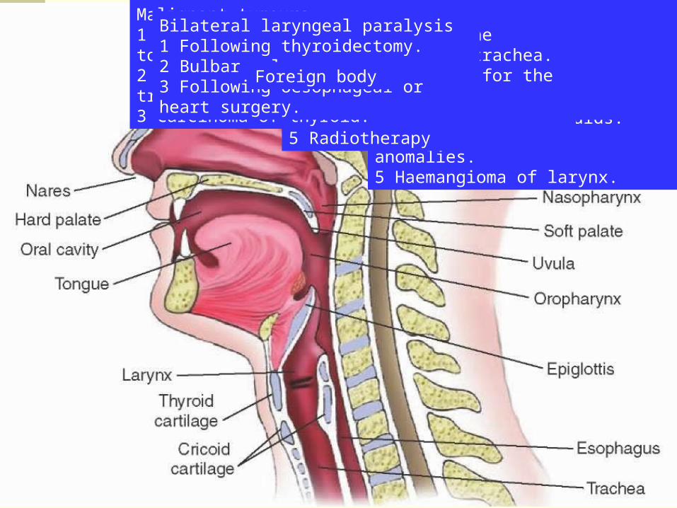
Congenital1 Subglottic or upper tracheal stenosis.2 Laryngeal web.3 Laryngeal and vallecular cysts.4 Tracheo-oesophageal anomalies.5 Haemangioma of larynx.
Trauma1 Prolonged endotracheal intubation.2 Gunshot wounds and cut throat, laryngeal fracture.3 Inhalation of steam or hot vapour.4 Swallowing of corrosive fluids.5 Radiotherapy
Infections1 Acute epiglottitis2 Laryngotracheobronchitis.3 Diphtheria.4 Ludwig’s angina.
Malignant tumours1 Advanced malignant disease of the tongue, larynx, pharynx or upper trachea.2 As part of a surgical procedure for the treatment of laryngeal cancer.3 Carcinoma of thyroid.
Bilateral laryngeal paralysis1 Following thyroidectomy.2 Bulbar palsy.3 Following oesophageal or heart surgery.
Foreign body
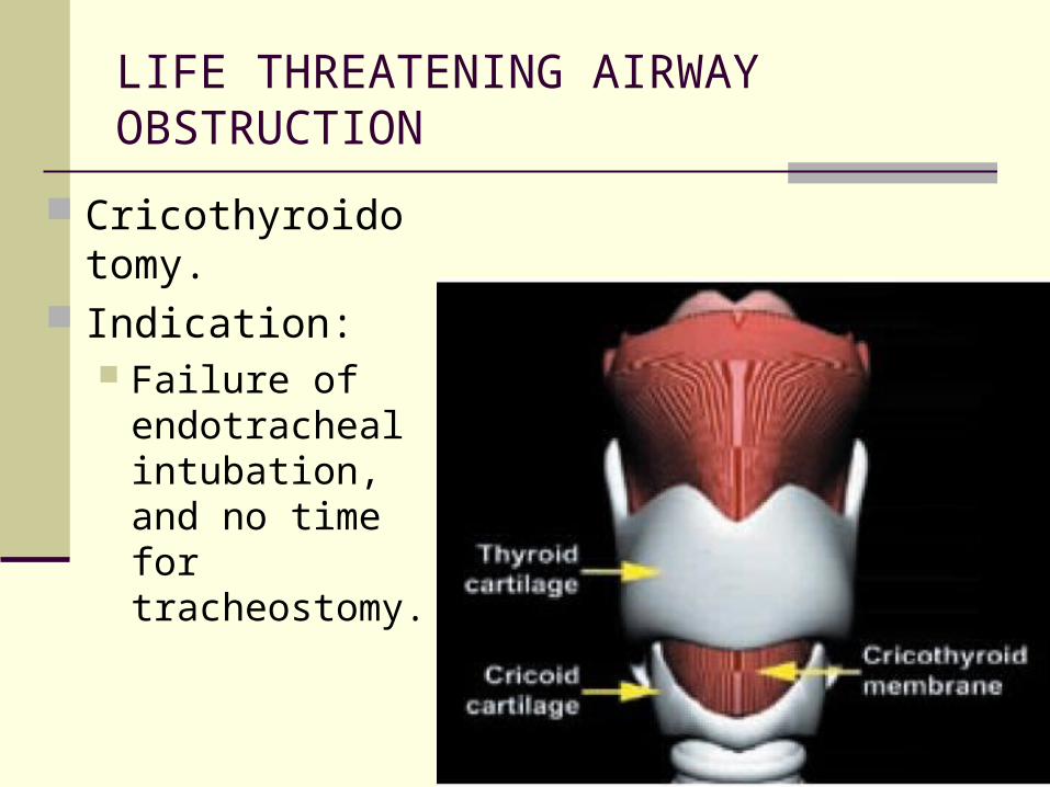
LIFE THREATENING AIRWAY OBSTRUCTION
Cricothyroidotomy. Indication:
Failure of endotracheal intubation, and no time for tracheostomy.

Tracheotomy
Indications Technique
Open and percutaneous Complications Physiology of a tracheotomy Decannulation

Tracheotomy
Creation of communication between the trachea and the cervical skin with insertion of a tube.
Indications
Upper Airway obstruction. Pulmonary Secretions. Ventilation. Prolonged mechanical ventilation.
May assist in weaning from mechanical ventilation.
Prevention of glottic stenosis/complication of prolonged endotracheal tube.
Pulmonary Secretion Clearance
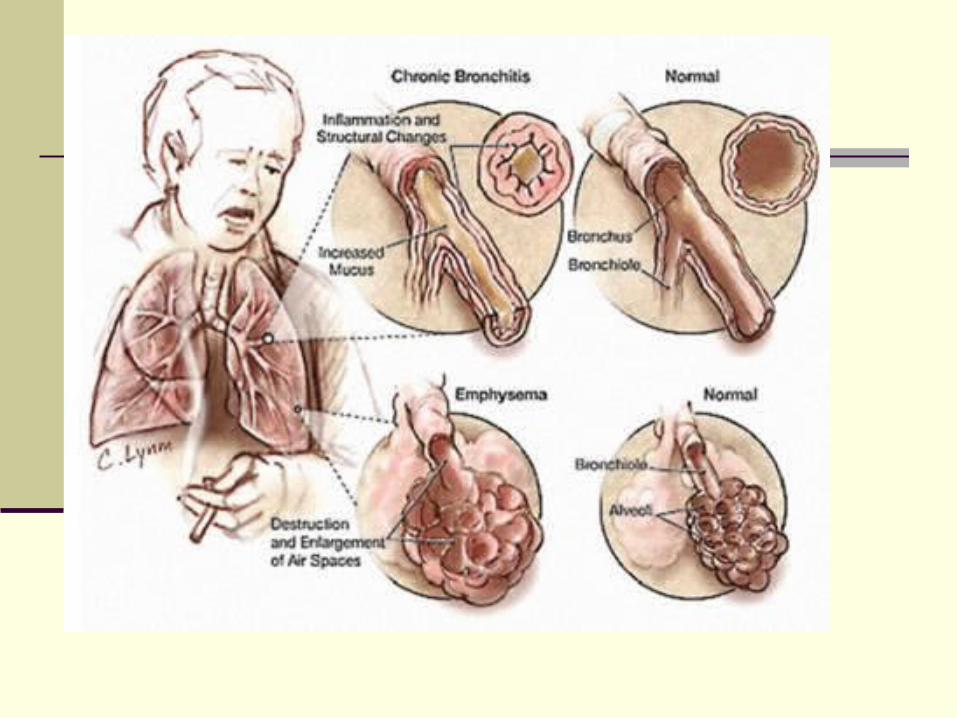
Aspiration / dysphagia COPD Bronchiectesis Stasis of secretions
Poor cough Poor respiratory reserve
Ventilation Neuromuscular disorder affecting respiratory
muscles Reduced respiratory effort
Limited pulmonary reserve COPD, Scoliosis, bronchiectesis
Central respiratory depression Reduced level of consciousness
Severe obstructive sleep apnea Cor pulmonale, failure CPAP
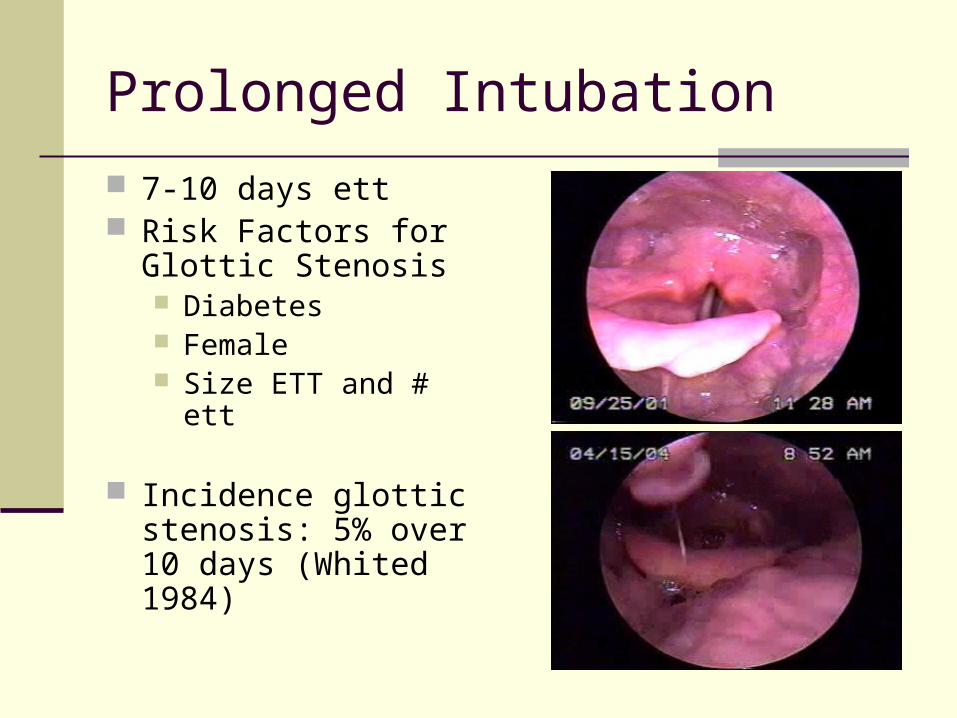
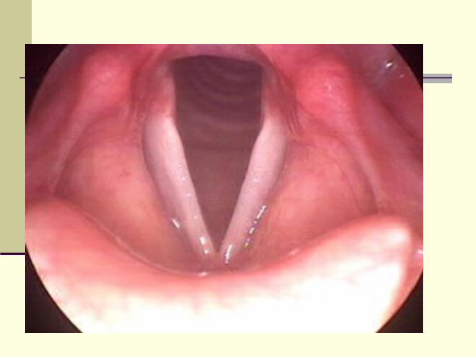
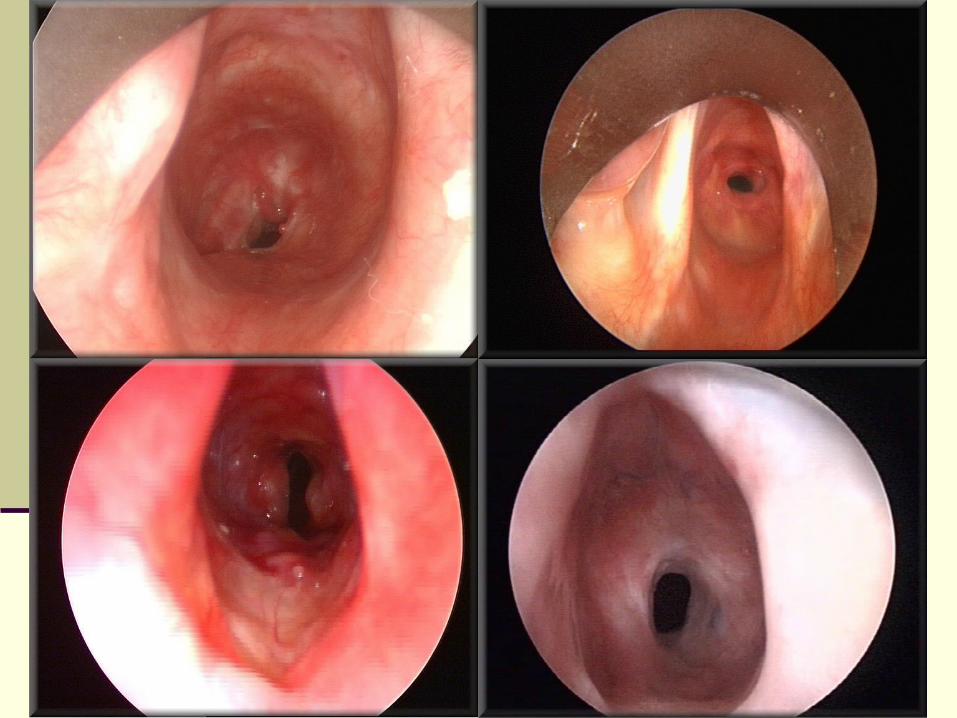
Prolonged Intubation
7-10 days ett Risk Factors for Glottic
Stenosis Diabetes Female Size ETT and # ett
Incidence glottic stenosis: 5% over 10 days (Whited 1984)
Tracheotomy
Decision made patient requires tracheotomy.
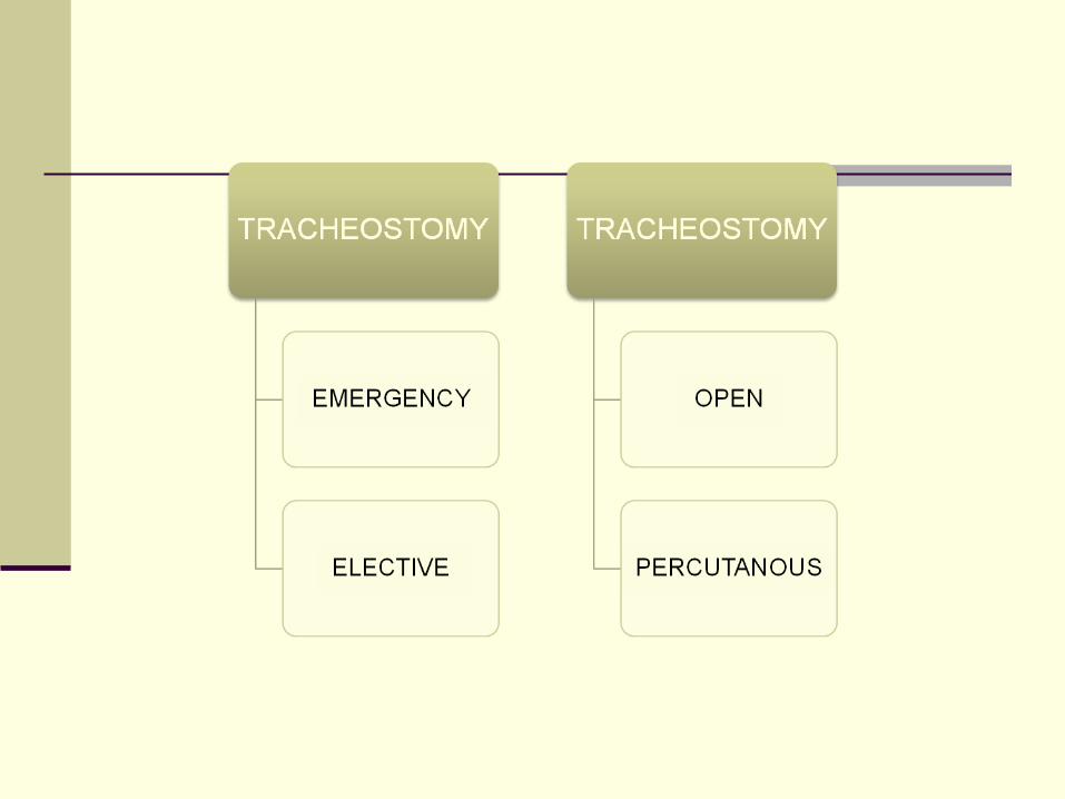
Open or percutaneous technique.
75% of tracheotomies done are done percutaneously in ICU at bedside.
General principles: External approach through neck soft tissue. Creation of opening in trachea. Placement of tube to maintain airway.
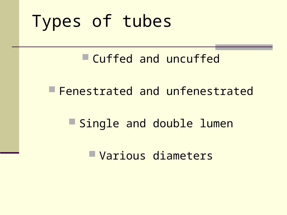
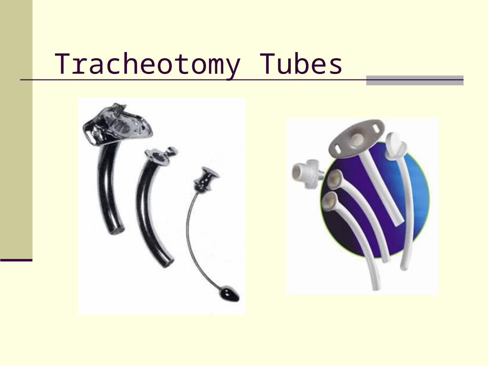
Types of tubes
Cuffed and uncuffed
Fenestrated and unfenestrated
Single and double lumen
Various diameters
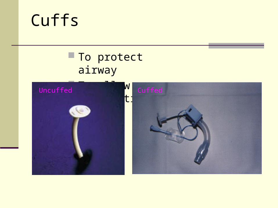
Cuffs
To protect airway To allow ventilation
Uncuffed Cuffed
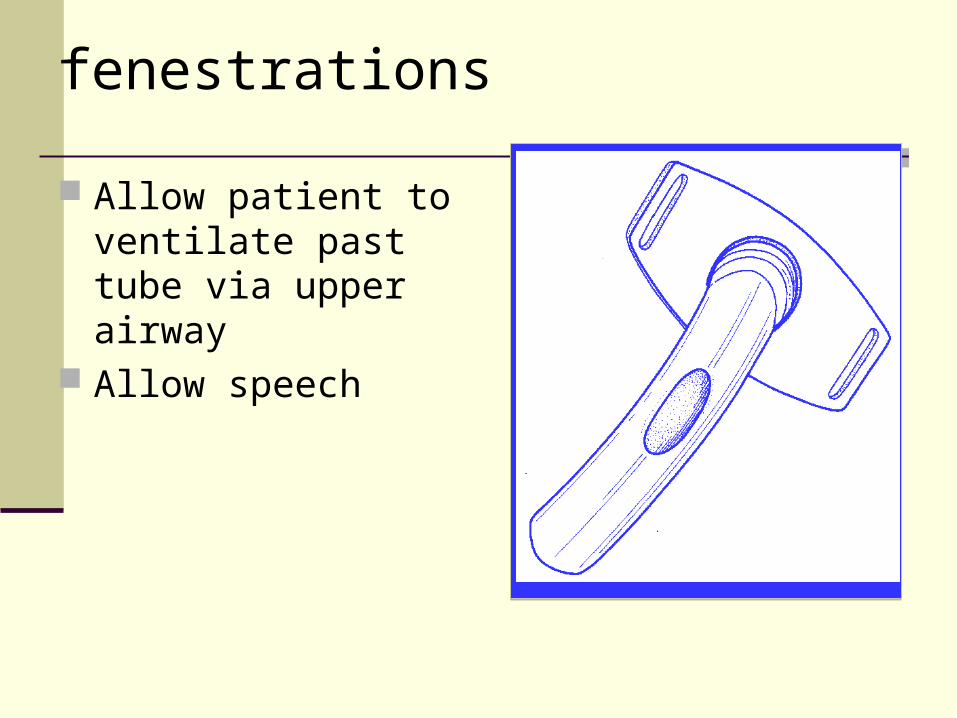
fenestrations
Allow patient to ventilate past tube via upper airway
Allow speech
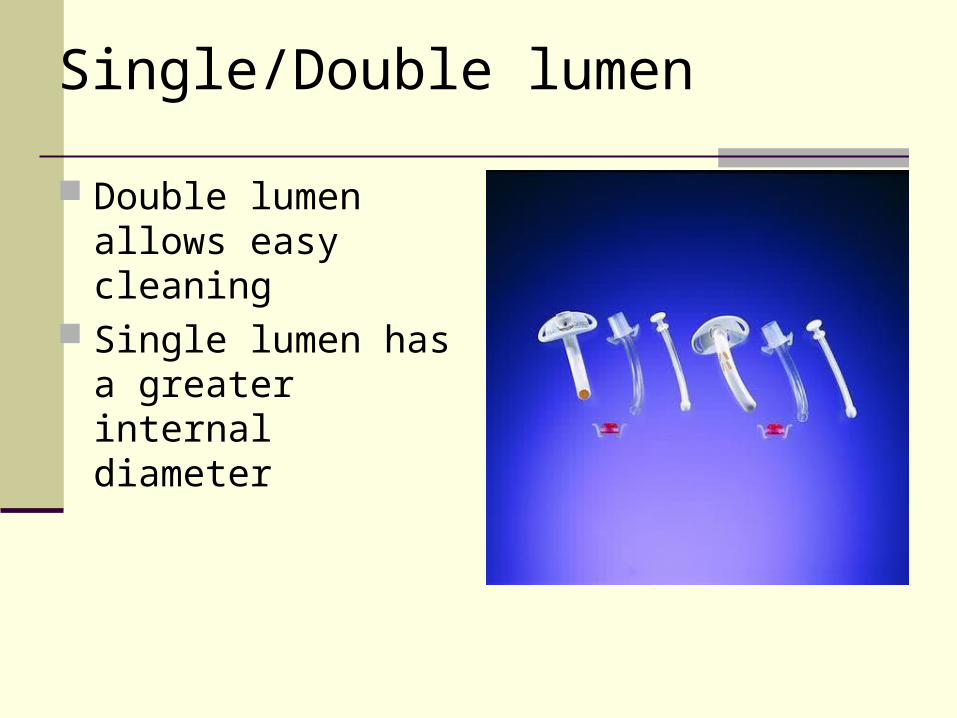
Single/Double lumen
Double lumen allows easy cleaning
Single lumen has a greater internal diameter
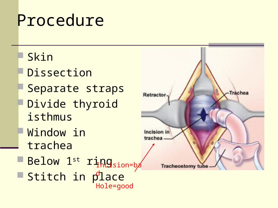
Procedure
Skin Dissection Separate straps Divide thyroid isthmus Window in trachea Below 1st ring Stitch in place
Incision=bad
Hole=good
Contraindications
Medically well enough for GA Uncontrolled coagulopathy Airway pathology below tracheotomy site
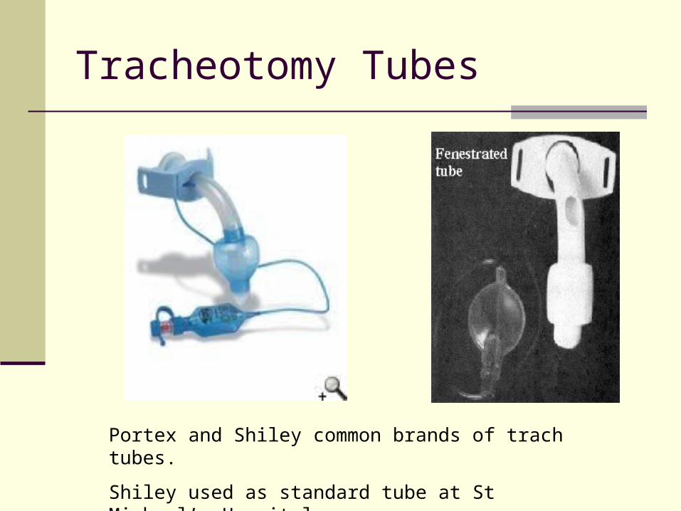
Tracheotomy Tubes
Portex and Shiley common brands of trach tubes.
Shiley used as standard tube at St Michael’s Hospital.
Tracheotomy Tubes
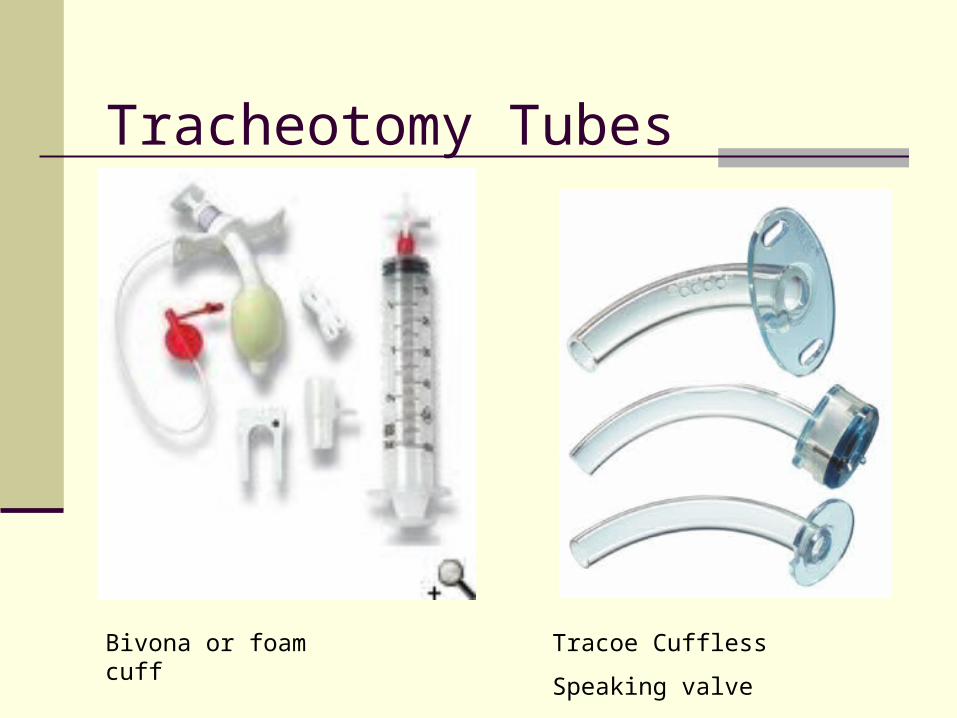
Tracheotomy Tubes
Bivona or foam cuff Tracoe Cuffless
Speaking valve
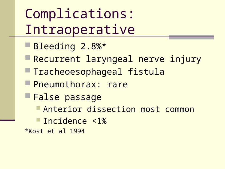
Complications: Intraoperative
Bleeding 2.8%* Recurrent laryngeal nerve injury Tracheoesophageal fistula Pneumothorax: rare False passage
Anterior dissection most common Incidence <1%
*Kost et al 1994
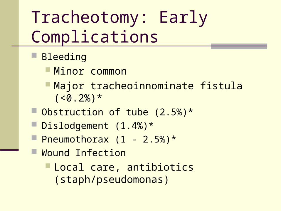
Tracheotomy: Early Complications
Bleeding Minor common Major tracheoinnominate fistula (<0.2%)*
Obstruction of tube (2.5%)* Dislodgement (1.4%)* Pneumothorax (1 - 2.5%)* Wound Infection
Local care, antibiotics (staph/pseudomonas)

Late Complications
Tracheal stenosis Tracheal chondritis Subglottis stenosis- high tracheotomy Tracheomalacia Tracheoesophageal fistula Failure of stoma closure when decannulated
Overall complication rate 15-30% in ICU patients largely minor with no long term morbidity
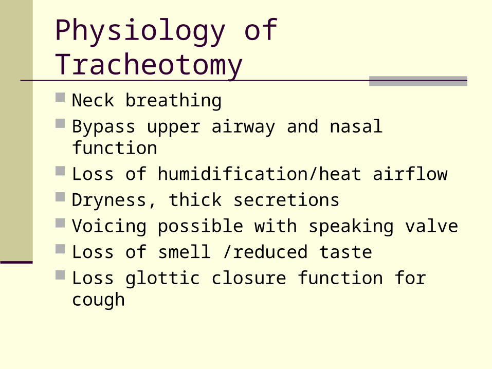
Physiology of Tracheotomy
Neck breathing Bypass upper airway and nasal function Loss of humidification/heat airflow Dryness, thick secretions Voicing possible with speaking valve Loss of smell /reduced taste Loss glottic closure function for cough

Physiology of Tracheotomy Respiration
AdvantagesAdvantages Lower work of breathing (30%) c/w normal airway Facilitates secretion clearance
Aspiration or thick secretions Less dead space (100 mL) Reduced airway resistance Assists in patient independence from mechanical
ventilation Patient comfort (better than ett)
Epstein 2005 Respiratory Care
Physiology of Tracheotomy Respiration
Disadvantages Tube diameter and shape
increases turbulent airflow, secretions adhere inside tube Loss of humidification/heat function of upper airway
Ciliary function affected Biofilm colonization
Diminish cough/loss glottic closure Reduce laryngeal elevation during swallow Patient comfort (better no tube at all)

Postoperative Tracheotomy Care
Humidification via trach mask/Instill saline Clear secretions, prevent crust Inner cannula cleaning tid at least If non-ventilated, change cuffed tube to non-
cuffed at 5-7 days Ties changed 2 people if possible Most hospital have nursing/RT protocol Teach everyone trach care including patient,
family
Decannulation
Decannulation
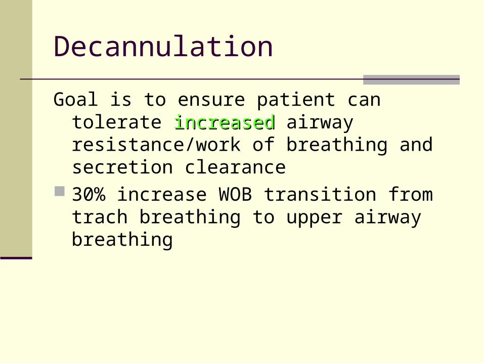
Goal is to ensure patient can tolerate increasedincreased airway resistance/work of breathing and secretion clearance
30% increase WOB transition from trach breathing to upper airway breathing
Decannulation
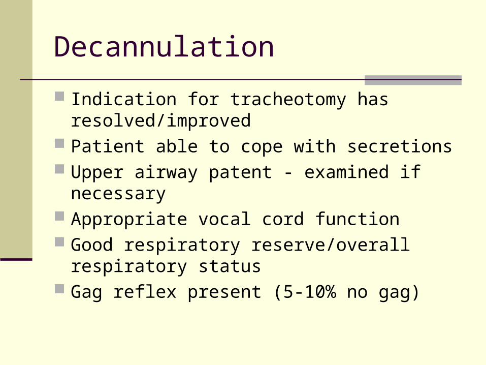
Indication for tracheotomy has resolved/improved
Patient able to cope with secretions Upper airway patent - examined if necessary Appropriate vocal cord function Good respiratory reserve/overall respiratory
status Gag reflex present (5-10% no gag)
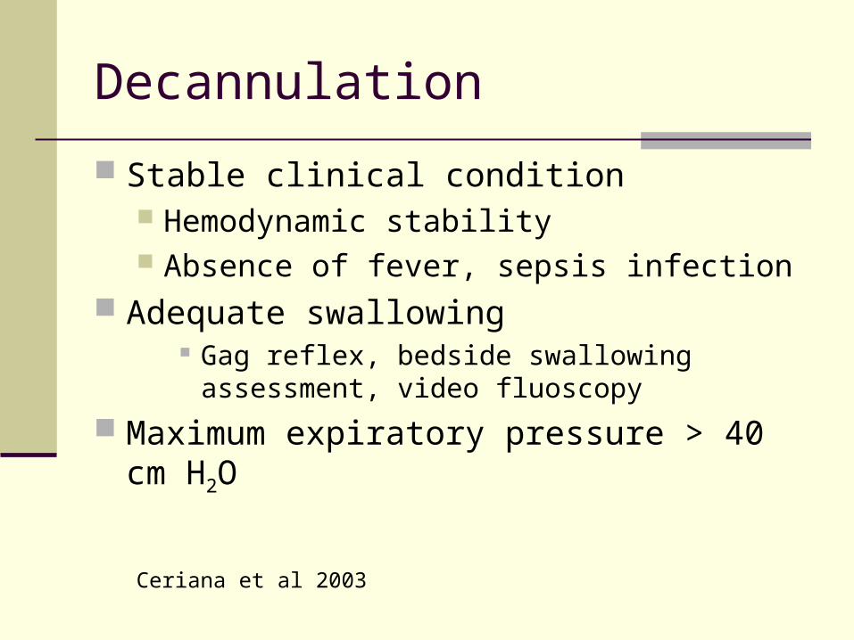
Decannulation
Stable clinical condition Hemodynamic stability Absence of fever, sepsis infection
Adequate swallowing Gag reflex, bedside swallowing assessment,
video fluoscopy
Maximum expiratory pressure > 40 cm H2O
Ceriana et al 2003