Embed Size (px)
Citation preview

Open Access
Alassali et al., Adv Tech Biol Med 2016, 4:1 DOI: 10.4172/2379-1764.1000163
Open Access
Volume 4 • Issue 1 • 1000163Adv Tech Biol MedISSN: 2379-1764 ATBM, an open access journal
Keywords: Algae; Algal phytochemicals; Analysis methods;Biological activity; Extraction methods
Introduction Nature is the first source of basic compounds and molecules, from
which bigger molecules are being formed. Since early 50s, natural substrates have been a source of various products that can be applied in food, drug, cosmetic, textile, and energy. Great emphasis has been given to the secondary metabolites of different natural species, especially plants [1-3].
Aquatic bodies cover a big portion of the earth (≈ 70% of the total area of the planet) providing a wide habitat for a large number of organisms [4,5]. Marine organisms are considered to be important feedstock for future bio refineries (co-producing biofuel and bioenergy). They have been found to be producing great amounts of secondary metabolites, which are divers in nature and efficacy [6-8].
Algae are diverse aquatic, photosynthetic organisms [9-11], representing 10% of the flora kingdom [12]. They are categorized into two groups, based on their biological structure; macroalgae (i.e., red, green and brown seaweeds) and microalgae (blue-green algae; normally a unicellular organism).
Because of the fluctuating environmental conditions and nutrient content from one place to another, algae are known for their diverse high-content of fatty acids, fibers, antioxidants, carotenoids, sterols, proteins, phycocolloids, lectins, oils, amino acids, unsaturated fatty acids, and vitamins, which could be commercially utilized [6,9,12-22]. Different algae cultivation methods produce different chemical compounds or different content thereof, which helps in cultures specification (i.e., due the secondary metabolites produced). This makes algae very attractive for drug research [16,18,23-25]. Algae research started in 1970s, emphasizing on four main areas including bioactive metabolites [15], toxins [26-28], chemical ecology [4,29] and biodiesel/bioethanol [9,14,30-36].
Microalgae are found to be rich in carbohydrates, mostly in form of starch and other polysaccharides [37]. The average lipid content of algal cells is high and can reach up to 70 wt.% [38,39]. Especially
macro algae are reported to be mini-factories of sugars, protein, and a wide range of bio-compounds with pharmaceutical, bio medicinal, and nutritional importance [40]. Furthermore, macro algae generally have a greater hydrolysable carbohydrate content than microalgae, hence higher ethanol production potential than some of current bioethanol feedstock [41].
This paper presents a review of molecular characterization and their potential applications. Furthermore, the extraction and analysis methods that were conducted on algae biomass to analyze for their metabolites were summarized and evaluated in this review.
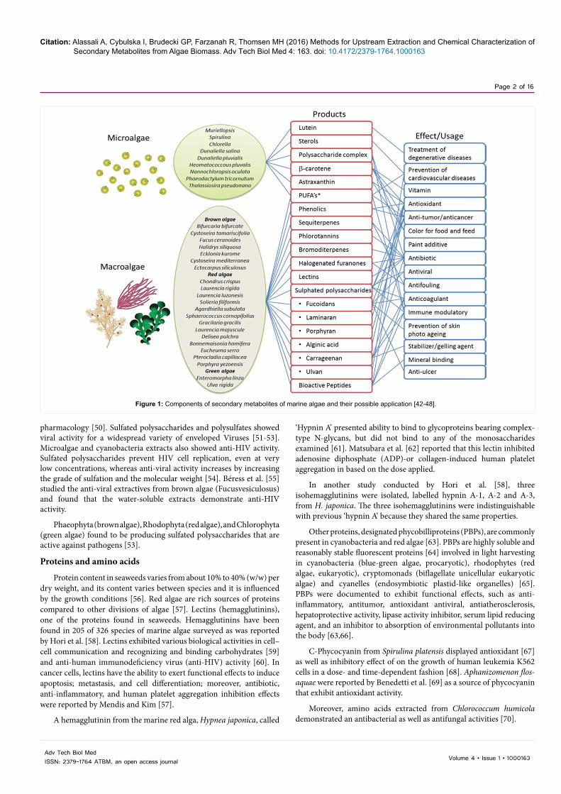
Figure 1 illustrates some of the interesting components of secondary metabolites and their possible usage in e.g. the pharmaceutical industry (Figure 1).
Molecular Characterization of Algae-extracted Chemi-cals and their Possible ApplicationsSulfated polysaccharides
Marine algae are the most significant source of non-animal Sulfated polysaccharides (SPs), where the chemical structure varies from one species to another [42-49]. The major SPs found in marine algae include fucoidan and laminarans of brown algae, carrageenan of red algae, and ulvan of green algae [50]. Recently, numerous SPs isolated from marine algae have attracted much attention in the fields of food, cosmetic, and
*Corresponding author: Ayah Alassali, The Institute Centre for Energy, MasdarInstitute of Science and Technology, P.O. Box 54224, Abu Dhabi, UAE, E-mail:[email protected]
Received January 25, 2016; Accepted January 28, 2016; Published February 04, 2016
Citation: Alassali A, Cybulska I, Brudecki GP, Farzanah R, Thomsen MH (2016) Methods for Upstream Extraction and Chemical Characterization of Secondary Metabolites from Algae Biomass. Adv Tech Biol Med 4: 163. doi: 10.4172/2379-1764.1000163
Copyright: © 2016 Alassali A, et al. This is an open-access article distributed under the terms of the Creative Commons Attribution License, which permits unrestricted use, distribution, and reproduction in any medium, provided the original author and source are credited.
AbstractMarine life is very rich in producing various and distinctive chemical components, both basic and complex.
Due to the harsh conditions such as high salinity, deficiency of nutrients, light and space, which make the marine environment competitive, organisms adapt to the environment by producing various chemicals and metabolites to help them survive under such conditions. In many studies great emphasis has been given to the secondary metabolites produced by algae (macro and microalgae). Certain species of algae are known for their high content of fatty acids, fibers, antioxidants, carotenoids, sterols, proteins, phytocolloids, lectins, oils, amino acids, unsaturated fatty acids, and vitamins, which could be commercially utilized.
Current algae studies emphasize on four main research areas: fuels, bioactive metabolites, toxins, and chemical ecology. This paper focuses on reviewing interesting biochemicals from algae biomass and their therapeutic applications. To achieve optimum extraction of high-value products, extraction methods and conditions were thoroughly presented in this review. Finally, different analytical approaches and techniques to identify the extracted chemicals were discussed.
Methods for Upstream Extraction and Chemical Characterization of Secondary Metabolites from Algae BiomassAyah Alassali*, Iwona Cybulska, Grzegorz Przemyslaw Brudecki, Rashed Farzanah and Mette Hedegaard ThomsenThe Institute Centre for Energy, Masdar Institute of Science and Technology, P.O. Box 54224, Abu Dhabi, UAE
Advanced Techniques in Biology & MedicineAdv
ance
d Te
chniques in Biology & M
edicine
ISSN: 2379-1764
Review Article
Citation: Alassali A, Cybulska I, Brudecki GP, Farzanah R, Thomsen MH (2016) Methods for Upstream Extraction and Chemical Characterization of Secondary Metabolites from Algae Biomass. Adv Tech Biol Med 4: 163. doi: 10.4172/2379-1764.1000163
Page 2 of 16
Volume 4 • Issue 1 • 1000163Adv Tech Biol MedISSN: 2379-1764 ATBM, an open access journal
pharmacology [50]. Sulfated polysaccharides and polysulfates showed viral activity for a widespread variety of enveloped Viruses [51-53]. Microalgae and cyanobacteria extracts also showed anti-HIV activity. Sulfated polysaccharides prevent HIV cell replication, even at very low concentrations, whereas anti-viral activity increases by increasing the grade of sulfation and the molecular weight [54]. Béress et al. [55] studied the anti-viral extractives from brown algae (Fucusvesiculosus) and found that the water-soluble extracts demonstrate anti-HIV activity.
Phaeophyta (brown algae), Rhodophyta (red algae), and Chlorophyta (green algae) found to be producing sulfated polysaccharides that are active against pathogens [53].
Proteins and amino acids
Protein content in seaweeds varies from about 10% to 40% (w/w) per dry weight, and its content varies between species and it is influenced by the growth conditions [56]. Red algae are rich sources of proteins compared to other divisions of algae [57]. Lectins (hemagglutinins), one of the proteins found in seaweeds. Hemagglutinins have been found in 205 of 326 species of marine algae surveyed as was reported by Hori et al. [58]. Lectins exhibited various biological activities in cell–cell communication and recognizing and binding carbohydrates [59] and anti-human immunodeficiency virus (anti-HIV) activity [60]. In cancer cells, lectins have the ability to exert functional effects to induce apoptosis; metastasis, and cell differentiation; moreover, antibiotic, anti-inflammatory, and human platelet aggregation inhibition effects were reported by Mendis and Kim [57].
A hemagglutinin from the marine red alga, Hypnea japonica, called
‘Hypnin A’ presented ability to bind to glycoproteins bearing complex-type N-glycans, but did not bind to any of the monosaccharides examined [61]. Matsubara et al. [62] reported that this lectin inhibited adenosine diphosphate (ADP)-or collagen-induced human platelet aggregation in based on the dose applied.
In another study conducted by Hori et al. [58], three isohemagglutinins were isolated, labelled hypnin A-1, A-2 and A-3, from H. japonica. The three isohemagglutinins were indistinguishable with previous ‘hypnin A’ because they shared the same properties.
Other proteins, designated phycobilliproteins (PBPs), are commonly present in cyanobacteria and red algae [63]. PBPs are highly soluble and reasonably stable fluorescent proteins [64] involved in light harvesting in cyanobacteria (blue-green algae, procaryotic), rhodophytes (red algae, eukaryotic), cryptomonads (biflagellate unicellular eukaryotic algae) and cyanelles (endosymbiotic plastid-like organelles) [65]. PBPs were documented to exhibit functional effects, such as anti-inflammatory, antitumor, antioxidant antiviral, antiatherosclerosis, hepatoprotective activity, lipase activity inhibitor, serum lipid reducing agent, and an inhibitor to absorption of environmental pollutants into the body [63,66].
C-Phycocyanin from Spirulina platensis displayed antioxidant [67] as well as inhibitory effect of on the growth of human leukemia K562 cells in a dose- and time-dependent fashion [68]. Aphanizomenon flos-aquae were reported by Benedetti et al. [69] as a source of phycocyanin that exhibit antioxidant activity.
Moreover, amino acids extracted from Chlorococcum humicola demonstrated an antibacterial as well as antifungal activities [70].
Figure 1: Components of secondary metabolites of marine algae and their possible application [42-48].
Citation: Alassali A, Cybulska I, Brudecki GP, Farzanah R, Thomsen MH (2016) Methods for Upstream Extraction and Chemical Characterization of Secondary Metabolites from Algae Biomass. Adv Tech Biol Med 4: 163. doi: 10.4172/2379-1764.1000163
Page 3 of 16
Volume 4 • Issue 1 • 1000163Adv Tech Biol MedISSN: 2379-1764 ATBM, an open access journal
Lipids
Some microalgae species have been recognized as commercially promising biomass for third-generation biofuel production, along with their nutritional and pharmaceutical applications. This is due to the high and unique lipid composition generated by the influence of microalgae rapid growth under a range of environmental conditions [71]. Adapting to harsh conditions resulted in production of diverse types of algae with extensive lipid patterns [72]. According to Metting Jr [73], the average lipid content of algal cells varies between 1% and 70% and can reach up to 90% of dry weight under certain growing conditions [38].
Long-chain (C35-C40) alkenones and their derivatives have been identified in haptophyte Chrysotila lamellose using gas chromatography–electron impact mass spectrometry [74]. These compounds were fairly stable during aging of C. lamellose when compared to sterols [74]. In addition, an uncommon polar lipid has been isolated from the red alga Gracilaria verrucosa, where it showed an inositol phosphoceramide structure [75].
Microalgae contain polar lipids, which were identified to be anti-inflammatory and anti-thrombotic [76]. Monogalactosyl diacylglycerols, digalactosyl diacylglycerols and phosphatidylglycerol are examples of polar lipids. The most studied lipids in algae are represented by polyunsaturated fatty acids (PUFAs) which showed different beneficial effects, such as enhanced heart health [77]. PUFAs are useful in the prevention of cardiovascular diseases [78]. Furthermore, Venugopal [79] indicated that the consumption of PUFAs can possibly prevent atherosclerosis, arrhythmias, and chronic obstructive pulmonary diseases, reduce blood pressure, reduce symptoms in asthma patients, fight against manic-depressive illness, protect against chronic obstructive pulmonary diseases, alleviate symptoms of cystic fibrosis, prevent relapses in patients with Crohn’s disease, prevent various cancers, provide bone health, and improve brain functions in children.
Sterols: Sterols are considered one of the most important chemical produced by algae, specifically by microalgae [80] with larger diversity when compared to higher plants [81,82]. They are essential components of the membranes of all eukaryotes, since they control membrane fluidity and consequently permeability. Sterols can be significant in cell proliferation, signal transduction and membrane-bound enzymes activity regulation [83].
Sterol structures range from the predominance of a single sterol, such as cholesterol in marine eustigmatophytes and 24-methylcholesta-5,22E-dien-3β-ol in some diatoms and haptophytes (prymnesiophytes), to mixtures of ten or more 4-desmethyl and 4-methylsterols in some species of dinoflagellates [84].
Generally, sterol composition in microalgae differs from one strain to another and is influenced by growth conditions, such as temperature, light intensity, and growth stage [84-86].
Terpenes and terpenoids: Diterpenes were reported as an antiviral compounds [87]. Manzo et al. [88] analyzed the secondary metabolites of the brown algae Dictyota ciliolate, which contained diterpenes as Dictyodial, Dictyol C, Dicytol H, those showed antiviral activities. For instance, anti-HIV-1 effects were exhibited by diterpenes extracted from D. menstrualiswere [89]. Also, diterpenes extracted from Dictyota pfaffii and Dictyota menstrualis possessed in-vitro inhibitory effects on herpes simplex virus [90].
Epitaondiol is a terpenoid isolated from the brown algae Stypopodium
flabelliforme, which is a tropical, polycyclic meroditerpendoids-rich species [91]. This component showed many biological activities as mentioned by Areche et al. [92]. Epitaondiol diacetate showed pharmacological effects in the rat cardiovascular system; where negative inotropic and chronotropic effects were observed. The compound also possesses noticeable anti-inflammatory effects [93].
Furthermore, the pharmaceutical applications of several meroditerpenoids were discussed by Sabry et al. [94]. For instance, stypolactone and atomaric acid are potent against human lung and colon carcinoma cells.
Pacifenol is a terpenoid isolated from seaweeds of the marine alga Laurencia claviformis and Laurencia tasmanica. An antimicrobial activity of pacifenol derivatives has previously been reported, after testing against some microrganisms, especially against Pseudomonas aeruginosa and Streptococcus enteriditis [93]. Furthermore, pacifenol exerts an inhibitory activity on both, inflammation [95] and allergy [93].
The genus Stypopodium is a rich source of polycyclic meroditerpenoids, such as stypodiol, epistypodiol, stypotriol, taondiol, epitaondiol, 2β,3α-epitaondiol, flabellinol, flabellinone, stypotriolaldehyde, stypohydroperoxide, isoepitaondiol, and 14-ket-ostypodiol. Among all, epitaondiol has displayed potent topical anti-inflammatory activity [96]. Also, antimicrobial effects against gram-positive and gram-negative bacteria, especially against E. faecalis. Antiviral activity against herpes simplex, and antiproliferative properties were exhibited by epitaondiol [93].
The derivative of 14-keto-stypodiol diacetate (SDA) was extracted from the algae Stypopodium flabelliforme, which showed some anti-inflammatory effects [97,98]. 14-ketostypodiol diacetate was proved by Depix et al. [99] to have a potential to microtubules’ assembly and cell proliferation in human prostatic cancer cells. Moreover, Sabry et al. [94] discussed the microtubule-assembly inhibitory effects of stypoldione and 14-keto-stypodiol diacetate.
Carotenoids
Carotenoids from marine sources found to be structurally different from those found on land, algae are rich in carotenoids [100].
From the health benefits of carotenoids that they offer provitamin A activity. Those carotenoids can be converted enzymatically to produce retinal and ultimately retinol (vitamin A), which is essential for vision, maintenance of differentiated epithelia, and reproduction [101]. Numerous studies have shown that by consuming a relatively large quantity of carotenoid-rich food, risk of cancer at several tumor sites is decreased [102]. Also, carotenoids provide dermal photoprotection against UV light photooxidation [101]. Carotenoids showed anti-inflammatory effects [103], hence was suggested to be used for cardiovascular diseases [104].
Carotenoids have been extensively studied and implicated as cancer preventive agents, seaweed-originated carotenoids, including fucoxanthin, neoxanthin, canthaxanthin, and peridinin tempt apoptosis in cancer cells [100,105,106]. Fucoxanthin has been observed to be a very effective inhibitor of cellular growth and promotes apoptosis in human cancer cell lines [107].
Mendes et al. [108] explained the biological activity of β-Carotene as cancer preventive component, where canthaxanthin showed immunoenhancement activity against cancer [109].
Gouveia and Empis [110] studied the carotene composition of

Citation: Alassali A, Cybulska I, Brudecki GP, Farzanah R, Thomsen MH (2016) Methods for Upstream Extraction and Chemical Characterization of Secondary Metabolites from Algae Biomass. Adv Tech Biol Med 4: 163. doi: 10.4172/2379-1764.1000163
Page 4 of 16
Volume 4 • Issue 1 • 1000163Adv Tech Biol MedISSN: 2379-1764 ATBM, an open access journal
Dunaliella salina. D. salina is one of the richest sources of natural β-carotene (90% of the total carotene composition), where the other 10% is composed of other carotenoids such as α-carotene and xanthophylls like lutein, zeaxanthin, and cryptoxanthins [110]. These xanthophylls have extensive applications, specifically for pharmaceuticals, cosmetics, and animal feed production [111].
The green algae Chlorellu vulgaris has produced canthaxanthin and astaxanthin in fairly high yields [112]. Aliquots of the green alga Caldophora glomerata were analyzed and quantified, results revealed that lutein and zeaxanthin were the carotenoids present at the highest concentrations, with lower concentrations of astaxanthin and its esters [113].
Schubert, García-Mendoza and Pacheco-Ruiz [114] studied the carotenoid content of different red algae species illustrative for the numerous rhodophyte families. The carotenoid content was representative to the evolution of the Rhodophyta and of the other algal groups. The study indicated that it is difficult to understand the function of these pigments due to the high diversity as well as the inconsistency in the presence or absence of certain carotenoids [114].
The carotenoids of Euglenophyceae have been identified to embrace β-carotene, diatoxanthin, heteroxanthin, diadinoxanthin, and neoxanthin as was reviewed by Deli et al. [115]. A quantitative carotenoid analysis of a natural bloom of Euglena sanguinea, Ehrenberg showed that the highest carotenoid content was exemplified by diesters of (3S,3'S)-astaxanthin (75%) followed by diesters of (3S,3'R)-adonixanthin (13%) and (3R, 3'R)-diatoxanthin (6%). Yet the rest was identified to be β-carotene monoesters of (3S)-adonirubin, 19-monoester of (3R,3'R,6R)-loroxanthin, diadinoxanthin and traces of neoxanthin (trace) [116].
Bromophenols
A review presented by Lin and Liu [117] discussed the algae-chemicals potential for anti-diabetic drugs; where marine algae are rich supplier of bromophenols; hundreds were isolated and studied. Bromophenols extracted from marine algae exhibit hyperglycemic effects by showing inhibition activity against protein tyrosine phosphatase 1B (PTP1B) when tested on rats [117]. The enzyme PTP1B dephosphorylates the insulin receptor and consequently down regulates insulin [118,119].
Bromophenols were isolated from the marine alga, Rhodomela confervoides and studied for their antibacterial activity by Xu et al.
[120], extracted bromophenols could inhibit the growth of some bacteria. Various antibacterial potencies were observed by the different bromophenols studied on several gram-positive and gram-negative strains [52].
Phlorotannins
As highly hydrophilic components, phlorotannins exist in abundance in marine brown algae and lower amounts accumulate in red algae [49]. Ecklonia cava; edible brown algae, was seen to have effective antioxidant activity [121], which was explained by the high phenolic content [121].
Brown seaweeds, including Sargassum tennerimum and Sargassum cervicorne, Sargassum graminifolium turn, Spireae thunbergii, and Laminaria japonica, are capable of inhibiting hyaluronidase, which is an enzyme used to speed dispersion and delivery and in case of histamines it increases their permeability [122]. The anti-allergic activity as other activities, such as anti-cancer, anti-diabetic and anti-HIV, are exhibited by phlorotannins, hydrophilic compounds with wide range of molecular weights [122].
Zhang et al. [49] indicated various health beneficial activities of phlorotannins, including, anti-HIV [123], anti-diabetic [124], anti-inflammatory [125], antihypertensive [126], radioprotective [127], anti-proliferative, anti-Alzheimer’s disease [128], antimicrobial [129], and antimatrix metalloproteinase activities (anti-MMP) [130]. MMPs play an important role in physiologic degradation of extracellular matrix (ECM) [131], which could extent to pathologic conditions characterized by excessive degradation of ECM such as chronic inflammation, wrinkle formation, arthritis, osteoporosis, tumor invasion and metastasis [130]. Table 1 summarizes different phlorotannins extracted from algae species and their potential applications [49] (Table 1).
Value-Added Chemicals ExtractionThe literature discussed numerous and extensive extraction
methods used for both micro and macro algae samples. Those different methods were applied for various chemicals extraction. Some of the methods were applied for method evaluation, where others were studied for process optimization.
Biomass extraction for natural products generation could be achieved with a single solvent (typically methanol) or a combination of solvents, especially when a large number of compounds with various polarities are pursued. Sequential extraction is also approached by
Phlorotannin Species Health effect Reference
6,6'-Bieckol Ecklonia cavaAnti-HIV-1 [123]Matrix metalloproteinase (MMP-2 and MMP-9) inhibitors [130-132]
6,6'-Bieckol Ishige okamurae Potential Acetylcholinesterase(AChE) inhibitors that could be used for preventing Alzheimer’s disease [128]
Triphlorethol-A and ecol Ecklonia cava Antioxidant activity [133]
8,8´-bieckol and 8,4´´´-dieckol Ecklonia cava Inhibitory effect onHIV-1 RT and protease [134]
Fucodiphloroethol G and phlorofucofuroeckol A Eckolonia cava Anti-histamine activity (anti-allergic) [135]Dieckol Eckolonia cava Anti-diabetic [124]Dieckol and phlorofucofuroeckol (PFF) Eckolonia cava memory-enhancing abilities [136]Phlorofucofuroeckol-B Eisenia arborea Anti-allergic [125,137]Eckol and 2-phloroeckol Ecklonia stolonifera Hepatoprotective constituents [138]Eckol Ecklonia kurome Antiplasmin inhibitory effects, makes it potentially useful for thrombolytic activity [139]Eckol, phlorofucofuroeckol A, dieckol and 8,8′-bieckol Ecklonia kurome Bactericidal effects [129]Phlorofucofuroeckol A, dieckol, and eckol Ecklonia stolonifera Antihypertensive [126]
Table 1: Phlorotannins extracted from algae and their potential applications.

Citation: Alassali A, Cybulska I, Brudecki GP, Farzanah R, Thomsen MH (2016) Methods for Upstream Extraction and Chemical Characterization of Secondary Metabolites from Algae Biomass. Adv Tech Biol Med 4: 163. doi: 10.4172/2379-1764.1000163
Page 5 of 16
Volume 4 • Issue 1 • 1000163Adv Tech Biol MedISSN: 2379-1764 ATBM, an open access journal
nonpolar and polar solvents to generate a series of samples [140]. Later in this review, different high-value chemicals extraction methods are presented, which are varied between simple and complex. Moreover, an overview of the conditions applied and the analysis approaches was stated.
Traditional extraction procedures
Simple extraction procedures such as decoction, maceration [141], liquid-liquid extraction, infusion, percolation, digestion and hot continuous extraction (Soxhlet) were extensively studied for biomass processing [142].
Polysaccharide extraction: Soxhlet method was suggested by Alves et al. [143] to extract the water soluble ulvan polysaccharide. Dichloromethane and acetone were used in a Soxhlet extraction in order to remove interfering substances such as coloring material and lipids. The output of this extraction was processed with hot water extraction under continuous stirring, applying the following conditions: 0.5 L, 8 h at 75-85°C. Further treatment processes were applied: centrifugation, concentration (using a rotary evaporator), deproteinization, and decolourization. This method is effective for polysaccharide extraction, with a yield of ca. 10-20%. A flow diagram of the extraction procedure is shown in Figure 2 [143]. The polysaccharide product obtained was qualitatively analyzed using 1H NMR spectroscopy (Figure 2).
A study to extract polysaccharides from three types of algae: green, brown and red algae (i.e. Ulva pertusa, Laminaria japonica, Enteromorpha linza, Bryopsis plumose and Porphyra haitanensis) was conducted and described by Zhang et al. [144]. The antioxidant activity was also studied by examining superoxide and hydroxyl radical scavenging effects, and reducing power of the compound. The main target was to obtain high yields of the polysaccharides. The method was generally summarized as follows: 100 g of dry algae was crushed then autoclaved, with the following autoclaving conditions: 115-125°C for duration of 3 to 4 h. Dialysis was done twice for the resulted solution: one against tap water for 48 h and another against distilled water for another 48h. The solution was concentrated with vacuum. Precipitation of the polysaccharides was achieved by adding 75% (v/v) ethanol. To extract the polysaccharides, the precipitate should be washed by ethanol and freeze-dried [144]. The extraction conditions (time and temperature) varied as shown in Table 2.
Lipid extraction: Soxhlet method for lipids extraction from Calluna vulgaris (Chlorellaceae family) was studied by Araujo et al.
[146]. Five grams of the biomass was used for the trial. Lipids extraction was done selectively by 110 mL acetone. Temperature applied was in the range of 120-180°C and the extraction was run for 8 consecutive hours. The extracted material was collected by solvent evaporation applying rotary evaporation. The resultant liquid fraction consisted of lipids and the solvent, which was filtered to keep the solution separated from the solid. 75 mL of KCl (0.88 w/v) solution was added to the sample and kept for 24 h in a separatory funnel, so that the oil phase separates from the aqueous phase. The oil phase (top phase) was transferred to a rotary evaporator and dried at 45°C under 200 mm Hg vacuum.
Fatty acid methyl esters were studied in P. cruentum samples, which were prepared by using 0.3 g of lyophilized material and 5 mL of a mixture containing 1 acetylchloride: 19 methanol (v/v%). The material was esterified at 80°C for 1 h. After chilling, water and n-heptane were added to the mixture (1 ml and 2 ml respectively), stirred and centrifuged. The organic phase was taken, filtered and dried using anhydrous sodium sulphate. The effluent was analyzed using gas chromatography [23].
Phlorotannins extraction: Generally, there is no specific extraction procedure for extraction of all plant phenolics, since phenolics may also be attached to other plant components, including proteins and carbohydrates. Nonetheless, the solvent and solvent system used is very important to decide for the phenolics to be extracted. Furthermore, additional steps may be required to separate the targeted components [147].
In a study done by Kim et al. [130], Ecklonia cava extract was collected and washed to remove salt and sand residues attached to the surface before being stored at -20°C. The frozen samples were lyophilized and homogenized using a mill before undergoing extraction. 1.0 kg of freeze-dried E. cava was extracted with 95% ethanol in a ratio of 1:10
Dried algae•Soxhlet extraction
Algae residue (off-white)•Static batch extraction (water as solvent)
Resultant solution (extract)•Filtration (using cloth)•Concentration to 10-20% its initial volume (using rotary evaporator)•Deproteinization, deodoration and decoloration.
Resultant solution•Precipitated solition is liophilized
Figure 2: Ulvan extraction from green algae [143].
Chemicals extracted/application Autoclave temperature (°C)
Autoclaving duration (h)
Sulfate 125 4Total sugars 120 3Uronic acid 115 3Neutral sugars 115 3Molecular weight identification 120 4
Table 2: Autoclaving conditions applied by Sathish and Sims [145] for each chemical extraction.

Citation: Alassali A, Cybulska I, Brudecki GP, Farzanah R, Thomsen MH (2016) Methods for Upstream Extraction and Chemical Characterization of Secondary Metabolites from Algae Biomass. Adv Tech Biol Med 4: 163. doi: 10.4172/2379-1764.1000163
Page 6 of 16
Volume 4 • Issue 1 • 1000163Adv Tech Biol MedISSN: 2379-1764 ATBM, an open access journal
(w:v) and evaporated under reduced pressure. The concentrated E. cava extract was freshly dissolved in Dimethyl sulfoxide before use.
In another study done by Xiao-jun, Xiao-jun et al. [148] 3.0 kg Sargassum kjellmanianum was extracted twice with 85% ethanol, the extract was first filtered with cheesecloth and then Buchner funnel filtration was applied, a deep brown liquid was obtained. After ethanol was removed by distillation under vacuum, the aqueous phase was cooled. The aqueous phase was then filtered through 0.45 µm Millipore membrane. The filtrate was extracted two times by ethyl acetate. After the organic phase was removed, vacuum was applied to concentrate the aqueous phase. The crude extract (weighed 30 g) was re-dissolved in distilled water and dialyzed against tap water for 2 days, then against distilled water for l day. The dialysate was dried by applying vaccum. The brown phlorotannin crude crystals obtained weighed 2.5 g.
Lee et al. [138] conducted extraction of phlorotannins based on the procedure applied by from Kim, Shin, Lee, Park, Park, Yoon, Kim, Choi, Jang and Byun [149]. 3.0 kg of the dried powder E. stolonifera was refluxed with 96% ethanol (EtOH, 3 x 9 L) for 3 consecutive hours. The concentrated extract was suspended in water, after which it was partitioned with n-hexane, ethyl acetate, n-butanol solvents in sequence.
Pigments extraction: Liquid-liquid extraction is one of the extensively used methods, especially for organic material extraction. This method was demonstrated by Cha et al. [150] for carotenoids extraction from Chlorella ellipsoidea and Chlorella vulgaris by the following procedure: 100 mL ethanol solution containing 0.1% (w/v) butylated hydroxytoluene (BHT) was used as the extraction solvent for 1 g of freeze-dried sample. The extraction took place in dark, room temperature conditions. Shaking was done continuously for three consecutive hours, followed by filtration of the mixture. Potassium hydroxide was added for saponification (120 µL for 5 ml of the content). For carotenoids extraction, 3 mL of hexane was added, shaken and then diluted with 3 mL water. An amber separatory funnel was used for phase separation. The layer containing the carotenoid was set to dry by evaporation, with continuous nitrogen purging [150].
Vitamin isolation: The impact of the low temperature growth condition on the composition of tocopherol in Prophyridium cruentum was studied by Durmaz et al. [23]. The samples were harvested by flocculation and then were centrifuged. In order to analyze tocopherols, sonication and liquid-liquid extraction were consecutively applied according to Chen et al. [151] as mentioned by Durmaz et al. [23], where 2 mL ethanol and 10 mg ascorbic acid were added to 0.5 g of freeze-dried material. 3 mL of n-hexane was added and swirling was applied. The sample was sonicated using an ultrasound bath (for 20 minutes). This step is necessary to disrupt the cell walls. 2 mL of distilled water was added and the mixture underwent stirring followed by centrifugation for 10 min. After the organic solvent filtration, drying was performed using anhydrous sodium sulphate. A second extraction was achieved by adding 1 mL and 0.5 mL of n-hexane individually.
Advanced extraction procedures
Studies were generally focused on overcoming the drawbacks of traditional methods as they are time-consuming and use large amounts of organic solvents, which is considered environmentally hazardous. Traditional methods also showed low extraction yields [22]. Recently, new extraction methods were developed, which could be able to overcome those negative aspects of the traditional extraction methods [152,153].
Pressurized liquid extraction (PLE): Pressurized liquid extraction (PLE) under the trade name ASE (for accelerated solvent extraction) is one of the non-traditional methods, which is described as an environmentally-clean technology [153]. PLE showed promising extraction outcomes as demonstrated by Herrero et al. [152] to extract active chemicals from microalga Spirulina platens. Different conditions were applied as follows:
Different solvents: hexane (dielectric constant of 1.9), light petroleum (dielectric constant of 4.3), ethanol (dielectric constant of 24.3) and water (dielectric constant of 78.5).
Different temperatures: 115 and 170°C
Extraction time varied between 9 and 15 minutes.
This work was conducted in order to study the effectiveness of Pressurized Liquid Extraction (PLE) by applying different extraction conditions, mentioned above. Herrero et al. [152] discussed a new Capillary Electrophoresis- Diode Array Detection (CE–DAD) method for extracts identification where the in vitro assay is used to describe the biological activity of those extracts. The author stated that this work shows a first-time confirmation for a possible application of PLE in-vitro-assay Micellar Electrokinetic Chromatography - Diode Array Detection (MEKC–DAD) for analyzing antioxidants from natural sources.
High Pressure Liquid Extraction (HPLE) application was demonstrated by Plaza et al. [15] to screen bioactive materials from macro algae and microalgae. Advanced analytical methods (HPLC-DAD or GC–MS) were used in this study for chemical characterization of extracts from Himanthalia elongata and Synechocystis sp.
This study used different extraction conditions, as follows:
Different extraction solvents (i.e., hexane, ethanol, and water).
Different extraction temperatures (50, 100, 150, and 200°C).
The extraction time was fixed for all trials to be 20 minutes.
Using the mentioned extraction techniques, added to it the proper analysis practices, biochemical characterization could be conducted such as analyses of antimicrobial and antioxidant activities. From the results of the different trials (applying different solvents or temperatures), water was found the most effective when used to extract non-polar to slightly polar compounds, and it showed good results in antioxidant extraction. Solvent polarity and extraction yield were directly proportional. Temperature showed a positive effect on the extraction yield as well. The results illustrated in Plaza et al. [15] for both Himanthalia elongata and Synechocystis sp showed the highest extractives yield for water extraction, then for ethanol and the lowest for hexane, and the results increased with temperature.
The choice of the solvent is based on the need of extracting various chemicals with wide range of polarities [152]. Herrero et al. [152] and Plaza et al. [15] demonstrated similar results: water and ethanol have been found more effective in extractives removal than the nonpolar solvents, while applying the same extraction conditions. Water showed the most complex MEKC electropherogram among all other solvents used, while the antioxidant activity of the extract was the lowest. Light petroleum MEKC electropherogram though comparable to hexane’s MEKC electropherogram was slightly different in the half maximal effective concentration (EC50) values obtained from the 2, 2-diphenyl-1-picrylhydrazyl (DPPH) in vitro experiments. Hexane showed lower EC50 indicating that the antioxidant activity of hexane extracts is

Citation: Alassali A, Cybulska I, Brudecki GP, Farzanah R, Thomsen MH (2016) Methods for Upstream Extraction and Chemical Characterization of Secondary Metabolites from Algae Biomass. Adv Tech Biol Med 4: 163. doi: 10.4172/2379-1764.1000163
Page 7 of 16
Volume 4 • Issue 1 • 1000163Adv Tech Biol MedISSN: 2379-1764 ATBM, an open access journal
superior to the ones obtained by using light petroleum. Ethanol showed quantitative results lower than that of water but higher than what hexane and light petroleum showed. Ethanol extracts exhibited antioxidant activity higher than water extracts, but comparable to the light petroleum extracts. Longer extraction times and higher temperature showed better extraction yields [152].
Supercritical fluid extraction (SFE): Supercritical CO2 extraction has the ability to extract compounds with pharmaceutical significance [154,155]. This extraction method was studied by Mendes et al. [154] for value-added chemicals extraction from four species of microalgae (Botryococcus braunni, Chlorella vulgaris, Dunaliella salina and the cyanobacteria Arthrospira (Spirulina) maxima). Using supercritical CO2 as an extraction media resulted in uncontaminated and undamaged extract components [156]. Supercritical extraction can be used to extract lipids, which could be determined gravimetrically. Hydrocarbons extracted using hexane and supercritical CO2, were evaluated by GC [8,157,158].
A study was conducted by Nobre et al. [159] to assess the extraction of astaxanthin and its esters in addition to other carotenoids using supercritical CO2. The extraction was conducted on Haematococcus pluvialis and different extraction conditions were applied to optimize the process. The extraction conditions were as follows: pressure between 200 and 300 bar, temperature between 40 and 60°C. The study evaluated grade of crushing and the effect of using a co-solvent (ethanol).
UV-Visible spectra (between 380 and 700 nm) were run to quantify the total carotenoid extracts. Carotenoid extracts attained by SFE were filtered and analyzed using HPLC [159]. Carotenoid extraction showed a yield of 100% by applying acetone L-L extraction, and using SFE extraction by supercritical CO2, without excessive grinding and without using a co-solvent (ethanol) gave a yield of 46.0%. Adding 10 Ethanol: 90 CO2 (v/v%) increased the yield of carotenoid extraction by 25.0% to achieve 58.7%. A much higher yield could be attained by further crushing of the biomass, which disrupts the cells and makes the carotenoids exposed and easier to extract. Having SFE extraction adjusted by adding a co-solvent and having a more crushed biomass can give a yield of 91.8% [159]. This paper also demonstrates a positive influence of both temperature and pressure on carotenoids extraction using SFE, where the pressure showed higher influence on the extraction yield than temperature; by keeping the temperature constant and increasing the pressure, an apparent increase in the extraction yield was obtained, on the other hand, by fixing the pressure and increasing the temperature, a slight extraction-yield-increase was obtained [159].
A study was conducted by Mendes et al. [154] discussing the extraction efficiency of antioxidants and antimicrobial components from Spirulina platensis, applying supercritical fluid extraction (SFE) and using CO2 and the mixture of CO2 and ethanol, similar to Nobre et al. [159]. Different conditions were applied to optimize the extraction and fractionation methods used.
The extraction using CO2 with 10% co-solvent (ethanol) gave a higher yield (1.9%) where the extract obtained using CO2 without using 10% ethanol was only (0.23%). Adding the co-solvent to the extraction agent also resulted in higher antioxidant activity. Supercritical CO2 has a low polarity which caused an inadequate interaction between CO2 and the matrix. Therefore adding a co-solvent can overcome this drawback and accordingly the extraction efficiency could be increased [154].
When pure CO2 was used as the extraction agent (no co-solvent addition), antioxidant activity of the extracts increased with pressure. The influence of temperature at low pressure had a negative impact
on the antioxidant activity, whereas high pressure affected the temperature’s activity positively. The optimum extraction conditions were found to be 360 bar and 74°C for the mentioned extraction agent. The optimum extraction conditions were confirmed to be at 275 bars and 57°C. Moreover, pressure of 220 bar seemed to be optimal to extract substances with antimicrobial activity [154] (Table 3).
Analysis TechniqueOverview
Throughout the conducted studies, different analytical techniques were applied as illustrated in Table 4. Each analytical technique had one or multiple components to analyze, since extracts are mixed with impurities and it is very important to separate them.
High-performance liquid chromatography (HPLC) and gas chromatography (GC) are very popular methods used for the analysis of algal extractives [152]. HPLC is the most sensitive and extensively used method, which can separate a wide range of compounds. (LC-MS) and (GC-MS) have been applied to perform a pharmaceutical grade analysis [162]. Reversed-Phase High-Performance Liquid Chromatography (RP-HPLC) was mentioned by Herrero et al. [152] as one of widely applied analysis methods, however this technique fails to separate highly polar compounds from the less polar ones [154].
Capillary electrophoresis using diode array detection (CE-DAD) is considered a good substitute of the RP-HPLC for fast SFE extracts characterization, which shows shorter application time, higher efficiency and selectivity when compared to the HPLC [152].
Complete structure evaluation was performed by many researchers to study bioactive natural products, and methods used were: 1D, 2D NMR, MS/MS, HPLC and chiral GC-MS analysis [163-166].
According to what Bernal et al. [167] explained regarding analysis procedures through a literature review of studies between 2005 and 2010, algae fatty acids, as methyl or ethyl esters, were analyzed by LC-MS and GC-FID [8,167]. For lipid detection, gas chromatography (GC) with various detection techniques such as ECD, FID and MS and also HPLC joined to one of the following detection systems: PDA, UV, MS and MS/MS were recommended. Nuclear magnetic resonance (NMR) and mass spectrometry (MS) were also among the suggested analysis techniques.
For terpenes, GC-MS, HPLC-UV-MS or NMR were found to be applied, nevertheless NMR is preferred for structure analysis.
Bernal et al. [167] have mentioned different analytical techniques for identification and/or quantification of carotenoids, such as HPLC that equipped with DAD or UV detectors. Liquid chromatography coupled with PDA and MS detectors showed high sensitivity for carotenoids and carotenoid esters detection. For antioxidant and anti-cancer carotenoids analysis, such as β-carotene, HPLC-UV/Vis or DAD was suggested to be used.
In most cases, especially for the characterizing composition of nutraceuticals, it is necessary to evaluate the suitability of the analytical techniques. The following sections review the popular analysis techniques, especially for marine-species extract (i.e., macroalgae and microalgae) (Table 4).
Analysis of polysaccharides
Zhang et al. [144] identified neutral sugars content of different algae

Citation: Alassali A, Cybulska I, Brudecki GP, Farzanah R, Thomsen MH (2016) Methods for Upstream Extraction and Chemical Characterization of Secondary Metabolites from Algae Biomass. Adv Tech Biol Med 4: 163. doi: 10.4172/2379-1764.1000163
Page 8 of 16
Volume 4 • Issue 1 • 1000163Adv Tech Biol MedISSN: 2379-1764 ATBM, an open access journal
Extraction method Bio-mass Chemicals used Temp. (°C)/ Pressure Time min.) Chemicals extracted Analytical Method References
Soxhlet extraction followed by hot
water extraction;Hot water
extractions
Green Algae
Dichloromethane and acetone for
Soxhlet extraction and water for hot water extraction
75-85°C during hot water extraction
480 (hot water
extraction)
Ulvan polysaccharide
Elemental analysis (% C, H, N and S content) by combustion
[143]
Proteins were measured as nitrogen content through Kjedhal
analysis.chemical structure of the
extracted polysaccharide by IR and 1H NMR
Soxhlet extraction Chlorella vulgaris Acetone 120-180°C 480 Lipids Lipids were separated applying L-L separation and then drying [146]
Liquid-Liquid Extraction
Chlorella ellipsoidea and Chlorella vulgaris
Ethanol solution containing BHT.
KOH was added in the procedure, refer
to the text.
Room temperature and dark conditions ----- Carotenoids HPLC and carotenoids were
identified by HPLC-ESI-MS [150]
Sonication in an ultrasound.
The output was re-extracted applying
L-L extraction.
Porphyridium cruentum
Ethanol, ascorbic acid , hexane, and
distilled water__________ ____ Tocopherols HPLC [23]
Esterification followed by Water and n-heptane L-L
extraction.
Porphyridium cruentum
Acetylchloride, methanol,
n-heptane and water
Esterification at 80°C 60 min for esterification
Fatty acid methyl esters Gas chromatography [23]
Pressurized liquid extraction (PLE) Himanthalia
elongate(macroalgae)and Synechocystis sp.
(microalgae)
HexaneEthanolWater
Three different temp.:50, 100, 150, and
200°C 20
Various for the aim of characteri-
zation
GC–MS and HPLC-DAD [15]
Pressurized Liquid Extraction (PLE)
Microalgae samples (Spirulina platensis)
Hexane, light petroleum, ethanol,
and water
Two different extraction
temperatures (115 and 170 °C
9 and 15 Antioxidant activity
Reversed phase high performance liquid chromatography
[152]Capillary electrophoresis
with diode array detection (CE–DAD)
Supercritical fluid extraction
(SFE)
Supercritical fluid extraction
(SFE)
Supercritical fluid extraction
(SFE)
Dunaliella salina Acetone
39.95 and 59.95 and pressures up to
35.0 MPa
----
Β-carotene LC-UV/vis
[154] as in [21,160]
Chlorella vulgaris Supercritical CO2 Carotenoids Spectrophotometry
Chlorella vulgaris Supercritical CO2Astaxanthin and Canthaxanthin HPLC
Lipids Supercritical CO2 Lipids Gravimetric
Botryococcus braunii Hexane and supercritical CO2
Hydrocarbons GC
Haematococcus pluvialis (microalgae) Ethanol
(40 and 60),pressure (200 and
300 bar), ----- Β-carotene LC-UV/vis
[159] as mentioned in
[160]
Spirulina platensis(microalgae)
Ethanol and CO2
275 bar and 57°C for antioxidant activity and 220 bar and 27 for optimum
antibacterial activity
75
Antioxidant activities
The β-carotene bleaching method and DPPH free radical-scavenging assay were used to determine the optimal extraction
conditions for antioxidant compounds
[161]Antimicrobial
activity
Broth microdilution method was used
for determination of the minimuminhibitory concentration
Pure CO2
360 bar and 74°C for antioxidant activity. At 361 bar, 55°C for highest antimicrobial
activity
Lipid composition
analysis
Gas chromatography, coupled to a flame ionization detector
(GC-FID)
Citation: Alassali A, Cybulska I, Brudecki GP, Farzanah R, Thomsen MH (2016) Methods for Upstream Extraction and Chemical Characterization of Secondary Metabolites from Algae Biomass. Adv Tech Biol Med 4: 163. doi: 10.4172/2379-1764.1000163
Page 9 of 16
Volume 4 • Issue 1 • 1000163Adv Tech Biol MedISSN: 2379-1764 ATBM, an open access journal
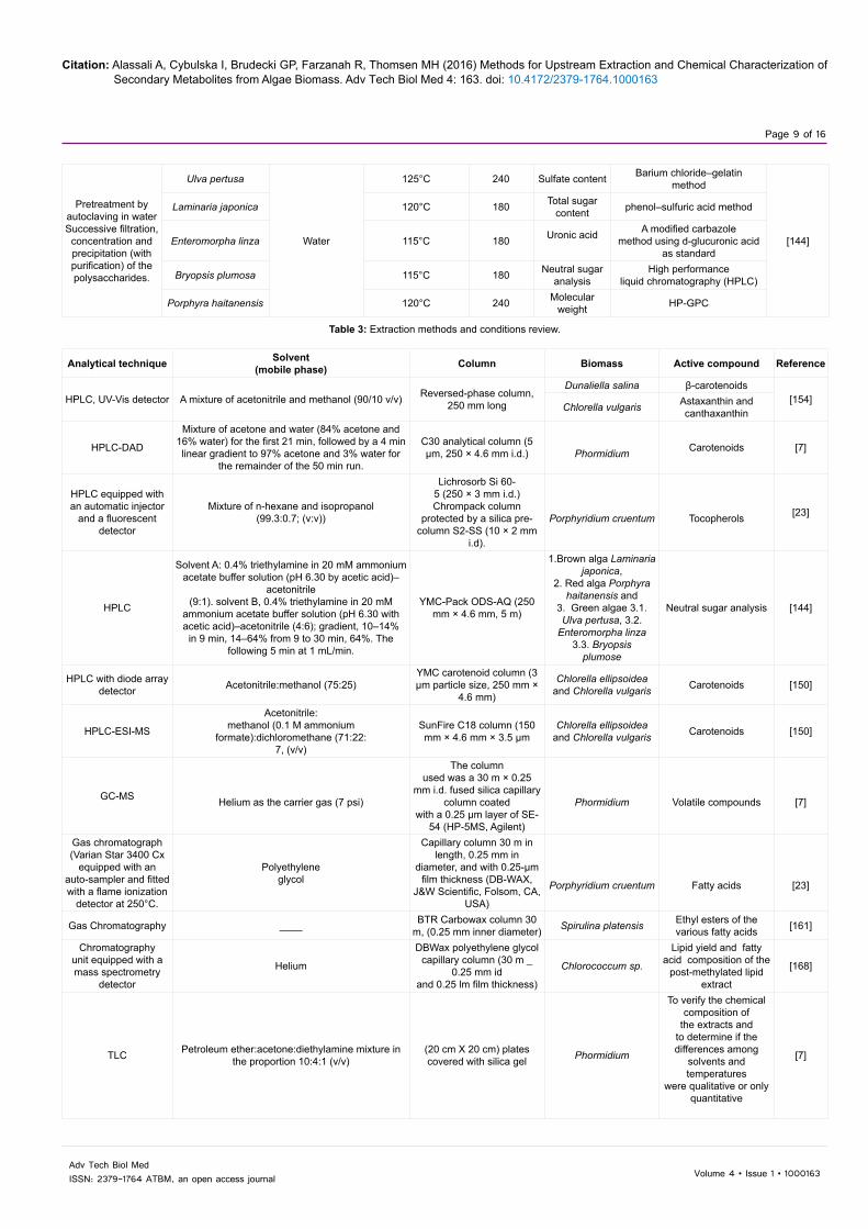
Pretreatment byautoclaving in water Successive filtration,
concentration and precipitation (with purification) of the polysaccharides.
Ulva pertusa
Water
125°C 240 Sulfate content Barium chloride–gelatinmethod
[144]
Laminaria japonica 120°C 180 Total sugar content phenol–sulfuric acid method
Enteromorpha linza 115°C 180 Uronic acid A modified carbazolemethod using d-glucuronic acid
as standard
Bryopsis plumosa 115°C 180 Neutral sugar analysis
High performanceliquid chromatography (HPLC)
Porphyra haitanensis 120°C 240 Molecular weight HP-GPC
Table 3: Extraction methods and conditions review.
Analytical technique Solvent(mobile phase) Column Biomass Active compound Reference
HPLC, UV-Vis detector A mixture of acetonitrile and methanol (90/10 v/v) Reversed-phase column, 250 mm long
Dunaliella salina β-carotenoids[154]
Chlorella vulgaris Astaxanthin and canthaxanthin
HPLC-DAD
Mixture of acetone and water (84% acetone and 16% water) for the first 21 min, followed by a 4 min linear gradient to 97% acetone and 3% water for
the remainder of the 50 min run.
C30 analytical column (5 μm, 250 × 4.6 mm i.d.) Phormidium Carotenoids [7]
HPLC equipped with an automatic injector
and a fluorescent detector
Mixture of n-hexane and isopropanol(99.3:0.7; (v:v))
Lichrosorb Si 60-5 (250 × 3 mm i.d.) Chrompack column
protected by a silica pre-column S2-SS (10 × 2 mm
i.d).
Porphyridium cruentum Tocopherols [23]
HPLC
Solvent A: 0.4% triethylamine in 20 mM ammonium acetate buffer solution (pH 6.30 by acetic acid)–
acetonitrile(9:1). solvent B, 0.4% triethylamine in 20 mM
ammonium acetate buffer solution (pH 6.30 with acetic acid)–acetonitrile (4:6); gradient, 10–14%
in 9 min, 14–64% from 9 to 30 min, 64%. The following 5 min at 1 mL/min.
YMC-Pack ODS-AQ (250 mm × 4.6 mm, 5 m)
1.Brown alga Laminaria japonica,
2. Red alga Porphyra haitanensis and
3. Green algae 3.1. Ulva pertusa, 3.2.
Enteromorpha linza3.3. Bryopsis
plumose
Neutral sugar analysis [144]
HPLC with diode array detector Acetonitrile:methanol (75:25)
YMC carotenoid column (3 µm particle size, 250 mm ×
4.6 mm)
Chlorella ellipsoidea and Chlorella vulgaris Carotenoids [150]
HPLC-ESI-MS
Acetonitrile:methanol (0.1 M ammonium
formate):dichloromethane (71:22:7, (v/v)
SunFire C18 column (150 mm × 4.6 mm × 3.5 μm
Chlorella ellipsoidea and Chlorella vulgaris Carotenoids [150]
GC-MS Helium as the carrier gas (7 psi)
The columnused was a 30 m × 0.25
mm i.d. fused silica capillary column coated
with a 0.25 μm layer of SE-54 (HP-5MS, Agilent)
Phormidium Volatile compounds [7]
Gas chromatograph (Varian Star 3400 Cx
equipped with an auto-sampler and fitted with a flame ionization
detector at 250°C.
Polyethyleneglycol
Capillary column 30 m in length, 0.25 mm in
diameter, and with 0.25-μm film thickness (DB-WAX,
J&W Scientific, Folsom, CA, USA)
Porphyridium cruentum Fatty acids [23]
Gas Chromatography ____ BTR Carbowax column 30 m, (0.25 mm inner diameter) Spirulina platensis Ethyl esters of the
various fatty acids [161]
Chromatographyunit equipped with a mass spectrometry
detector
Helium
DBWax polyethylene glycol capillary column (30 m _
0.25 mm idand 0.25 lm film thickness)
Chlorococcum sp.
Lipid yield and fatty acid composition of the
post-methylated lipid extract
[168]
TLC Petroleum ether:acetone:diethylamine mixture in the proportion 10:4:1 (v/v)
(20 cm X 20 cm) plates covered with silica gel Phormidium
To verify the chemical composition of
the extracts and to determine if the differences among
solvents and temperatures
were qualitative or only quantitative
[7]

Citation: Alassali A, Cybulska I, Brudecki GP, Farzanah R, Thomsen MH (2016) Methods for Upstream Extraction and Chemical Characterization of Secondary Metabolites from Algae Biomass. Adv Tech Biol Med 4: 163. doi: 10.4172/2379-1764.1000163
Page 10 of 16
Volume 4 • Issue 1 • 1000163Adv Tech Biol MedISSN: 2379-1764 ATBM, an open access journal
species by the HPLC method, which analyzed sugar derivatives. The process was run at 25°C. Two different solvents with different gradients were used: 0.4% triethylamine in 20 mM ammonium acetate buffer solution (pH 6.30 by acetic acid)–acetonitrile (9:1) and solvent B, 0.4% triethylamine in 20 mM ammonium acetate buffer solution (pH 6.30 with acetic acid)–acetonitrile (4:6) at a flow rate of 1.0 mL/min [144]. In another study, HPLC system with a ZORBAX Eclipse XDB-C18 analytical column was used for rebaudioside A and stevioside content analysis. The column was eluted with acetonitrile-water 70:30, at a flow rate of 1.0 mL/min, and an ambient temperature of 25°C. UV absorption was used for detection at 210 nm, column pressure of 80 atm was applied [169].
IR identification technique was applied by Alves et al. [143] to identify ulvan chemical components. An IR Prestige-21 apparatus was used for disc formed sample (mixing the powder sample with KBr and then pressing it into a disc).
1H NMR spectroscopy has been suggested as a good analytical tool for studying algal polysaccharides. However, this technique was only suggested for chemical identification and not quantification, as there are possible structural irregularities, which could give misleading and complex signals.
The advantage of this technique over others, like chromatography, is its simple calibration and application besides the faster optimization of the experiment practice [143].
Ulvan thermal properties were also analyzed by Alves et al. [143] using thermogravimetric analysis (TGA), differential scanning calorimetry (DSC) and dynamic mechanical analysis (DMA). Combustion was applied throughout this study for elemental analysis of the major components and Carlo Erba CHNS-O EA 1108 apparatus was used for this purpose.
Analysis of proteins
Chiral HPLC method, as well as 2D NMR spectral analysis were reviewed by Tan [163] based on the study of Han et al. [170] for complete lipopeptides structural determination on extracted Wewak peptins.
Total protein content is usually measured by a standard Kjeldahl method [143,171].
Analysis of lipids and fatty acids
GC–MS analysis with a split/splitless injector coupled to a quadrupole mass spectrometer was used to identify the volatile material and fatty acids extracted by PLE method [15].
Hydrocarbons and fatty acids from supercritical extract were analyzed by Mendes et al. [154] using gas chromatography. The fatty acid analyses were achieved in a gas chromatograph joined with flame ionization (FID) detector.
Analyses of the fatty acid methyl ester content in the post-methylated lipids were also performed using gas chromatography connected with a mass spectrometry detector [8,154,168].
For lipid composition analysis, Mendiola et al. [161] and Sathish and Sims [145] used gas chromatography, joined with a flame ionization detector (GC-FID). Sample solution was injected into a gas chromatograph with a 30 m column with inner diameter of 0.25 mm. Temperature applied started at 100°C then increased to 180°C and afterwards to 220°C with heating rates of 20°C/min and 15°C/min, respectively.
Long-chain (C35-C40) alkenones and their derivatives have been identified in Chrysotila lamellose using gas chromatography–electron impact mass spectrometry (GC–EIMS) [72].
TLC Petroleum ether:acetone (75:25) (10 cm × 20 cm) plates covered with silica gel Spirulina platensis
To investigate inquire into the types of
compoundsresponsible for the antioxidant activity
[161]
Combustion ------ ----- Green algae Elemental analysis
[143]
KjedhalAnalysis
----- ----- Green algae Protein measurement
IR and 1H NMR----- ----- Green algae
Chemical structure of the extracted polysaccharide
Spectrophotometry:(Neutralization
of free radicals of DPPH by the extract
antioxidants)
Methanol ----- Microalgae samples (Spirulina platensis) Antioxidants activity [152]
Micellar electrokinetic chromatography with diode array detection
(MEKC–DAD)
50 mM sodium tetraborate, 100 mM SDS at pH 8.8
Fused silica capillary with 75 _m i.d., 37 cm total length and 30 cm length to the
detector
Microalgae samples (Spirulina platensis)
To provide a preliminary analysis on the
composition of the extracts
[152]
Barium chloride–gelatinMethod
-------- -------1. Brown alga Laminaria
japonica,2. Red alga Porphyra
haitanensis and3. Green algae 3.1. Ulva pertusa, 3.2.
Enteromorpha linza3.3. Bryopsis
plumose
Sulfate content
[144]Phenol–sulfuric acid
method ----------- ---------- Total sugar content
Table 4: Review of analytical techniques used for algae and microalgae extracts.

Citation: Alassali A, Cybulska I, Brudecki GP, Farzanah R, Thomsen MH (2016) Methods for Upstream Extraction and Chemical Characterization of Secondary Metabolites from Algae Biomass. Adv Tech Biol Med 4: 163. doi: 10.4172/2379-1764.1000163
Page 11 of 16
Volume 4 • Issue 1 • 1000163Adv Tech Biol MedISSN: 2379-1764 ATBM, an open access journal
HPLC can also be applied to analyze lipids, e.g., Jones et al. [8] used normal-phase HPLC coupled to an evaporative light scattering detector (ELSD) and MS for crude lipid-extracts analysis. Furthermore, Guella et al. [172] reported that the structural interpretation of some galactolipids produced by Glenodinium sanguineum and Chaetoceros has been obtained using high performance liquid chromatography–electrospray ionization ion trap mass spectrometry HPLC/ESI-ITMS.
To determine the position of double bonds in minor fatty acids (below 1% of total fatty acids) in a golden alga, Schizochytrium spp., acetonitrile chemical ionization tandem MS has been efficiently applied [173].
For a qualitative identification of the lipids in brown algae, Obluchinskaya [171] applied thin layer chromatography method, with 50:50 benzene–chloroform as the solvent. The spots were urbanized by 5% phosphomolybdic acid solution in methanol.
Lipids from supercritical extracts could be examined gravimetrically as demonstrated by Mendes et al. [154], Araujo et al. [146] and Cha et al. [150].
Analysis of pigments (carotenoids)
HPLC was used by Mendes et al. [154] to analyze astaxanthin and canthaxanthin. The same study used HPLC for β-carotene. A liquid chromatograph Perkin-Elmer, Series 10, with an UV-Vis detector, coupled to a Perkin-Elmer LCI-100 integrating unit, and a reversed phase column, 250 mm long, Vydac 201 TP54 was used. The eluent was a mixture of acetonitrile and methanol (90:10, v/v) at a flow rate of 1 ml/min [154].
For Liquid-Liquid pigment-containing extracts, HPLC coupled with diode array detector was utilized as an analytical method. Cha et al. [150] demonstrated the sample pre-treatment before being analysed; methanol was used for dissolving the residue, while fat-soluble impurities were extracted with hexane. Sample of 20 µL was injected into YMC carotenoid column (3 µm particle size, 250 mm × 4.6 mm) with an acetonitrile-methanol (75:25) mobile phase under flow rate of 1.0 mL/min. Cha et al. [150] also used HPLC-ESI-MS for carotenoids identification. Separation was done using a C18 column (150 mm × 4.6 mm × 3.5 μm) and using isocratic system, with acetonitrile:methanol (or 0.1 M ammonium formate):dichloromethane (71:22:7, v/v)) as the eluent.
The use of Diode Array Detection (DAD) in the HPLC system has proved its powerful application in compounds identification. A challenge is presented in the simultaneous extraction and analysis of dissimilar compounds [152].
HPLC-DAD analysis was done by dissolving the dry extract in the compatible solvents prior to the HPLC analysis. The extract was analyzed by the HPLC (Agilent 1100 Liquid Chromatograph equipped with a DAD). The column used was a C18 column (150 mm × 3.9 mm). A mixture of two solvents; A: (methanol/ammonium acetate 0.1N; 7:3) and B: (methanol) was used as the mobile phase. The identification was performed by two means: either using a standard or using UV–vis spectral characteristics and assessment based on the literature [15].
HPLC-DAD analysis was applied by Rodríguez-Meizoso et al. [7] for carotenoid compounds identification. The mobile phase was a mixture of acetone and water in ratios presented in Figure 3 and at a flow rate of 1 mL/min. Detection using a diode array system was achieved at a wavelength of 480 nm (Figure 3).
Analysis of polyphenols (phlorotannins)
Kim et al. [130] determined the total content of phlorotannins using spectrophotometric methods as was described by Waterman and Mole [174], by applying a modified version of Folin-Ciocalteu method. Phloroglucinol was used as the standard, where samples were diluted while considering the range of the spectrophotometer. A 0.1-ml aliquot of the diluted sample was mixed with 1.0 ml of 1 N Folin-Ciocalteu reagent. 2.0 ml of 20% Na2CO3 was added, after which, the mixture was allowed to stand for 3 minutes. Samples were kept in the dark for 45min at room temperature before being centrifuged at 1600 × g for 8 min. Optical density (OD) of the supernatant was measured at 730 nm using a GENios® microplate reader (Tecan Austria GmbH, Austria). Total phlorotannin content is calculated using the standard graph plotted and expressed as a percentage.
In another study, the IR spectrum and fluorescence spectrum of phlorotannins were verified by PHP Fourier Infrared Spectroscopy, and Hitachi 850 Flourescent Spectroscopy respectively [148].
Future PerspectivesFuture utilization of algae will be decisively influenced by the effort
put into and the results algae and seaweed research [175]. Mainly, studies are looking into the scalability and commercial production of algae-derived products.
One of the major commercial markets for algal products is the food industry, including nutraceuticals products [176]. The commercial applications are dominated by four strains: Aphanizomenon flos-aquae, Arthrospira, Chlorella and D. salina [38]. Alagae-based nutraceutical products include but not restricted to pure powder, tablets, chips, pasta and liquid extract [38]. Moreover, algae are used in commercial aquaculture operations for the production of fish feed. The algae supply important nutrients such as polyunsaturated fatty acids (PUFAs), carotenoids, and proteins, those are needed for fish [79,177]. Species with large quantities of PUFAs are of high interest, such as Cryptomonas sp. and Nannochloropsis sp. [176,178].
Algae represent a single photosynthetic production system that is capable of extremely versatile biofuels applications. It was indicated by Jones and Mayfield [179] that algae can be used as a biomass to replace oil seeds plants (in production of biodiesel), corn and sugar cane (in production of bioethanol), lignocellulosic biomass (in production of bioethanol and biogas) and organic waste (in production of biogas and biohydrogen).
The concept of using microalgae as renewable source of lipid-rich feedstock for biofuels has been explored over years due to the substantial amounts of triacylglycerols (TAG); yet, a scalable, commercially viable system has yet to emerge [180]. In a study done by Harun et al. [181] showed that the green algae Chlorococum sp. produces 60%
The remainder of the 50 minutes
97% acetone 3% water
First 21 minutes
84% acetone 16% water
Figure 3: The mobile phase composition used HPLC-DAD carotenoid analysis [15].
Citation: Alassali A, Cybulska I, Brudecki GP, Farzanah R, Thomsen MH (2016) Methods for Upstream Extraction and Chemical Characterization of Secondary Metabolites from Algae Biomass. Adv Tech Biol Med 4: 163. doi: 10.4172/2379-1764.1000163
Page 12 of 16
Volume 4 • Issue 1 • 1000163Adv Tech Biol MedISSN: 2379-1764 ATBM, an open access journal
higher ethanol concentrations for samples that are pre-extracted for lipids versus those that remain as dried intact cells. This indicates that microalgae can be used as a biomass for the production of different fuels to increase their overall economic value [179].
Producing biofuels from algae is challenging, mainly due to the high cost associated with the infrastructure and the energy needed for growing and harvesting algae [182]. Metabolic and genetic engineering of biofuel producing organisms will likely play a critical role in strain development to optimize the biofuel producing strains, together with creating designer triacylglycerides through lipid chain length manipulation [183]. Despite the high cost associated with viable production of biofuels from algae, algae still represent one of the best possibilities available as a source of bioenergy. Optimum utilizations of algae, by considering a biorefinery concept that is economically feasible and environmentally sustainable are subjects of process optimization and system efficiency. Hence, minimizing the generated waste and using every component of the algal biomass will help in producing viable biofuels from algae. Accordingly, we focused in this review on the added-value chemicals, which will generate a great profit to algae-based bio refinery along with the need of finding a good source of such valuable components.
ConclusionAlgae are photosynthetic diverse organisms, which can survive
under harsh conditions and accordingly produce various high-value metabolites. They are estimated to represent a huge number of species ranging between 30,000 and more than 1 million [184]. Based on the great potential of chemicals production from algae, researchers conducted studies to characterize different algae strains and to study chemical extraction feasibility. Different extraction and analysis studies were carried out. The extraction methods applied varied between simple and sophisticated ones. It was observed that simple (traditional) extraction methods are time consuming, and environmentally hazardous, besides the inefficient downstream treatment. Therefore advanced methods such as PLE and SFE were studied and effective extraction was observed in these trials. SFE extraction is valuable, as the diffusivity and the viscosity values are between those of the gases and liquids. Faster and more efficient extraction was obtained by supercritical fluid due to its deeper penetration into the structure of biomass. Since the great discovery of supercritical extraction, SFE is now applied as a very efficient extraction method in several areas [21,154]. Nevertheless, high operation cost, especially on large scales, influenced this method negatively [168].
Development of advanced analytical techniques is essential in algae research, in order to characterize, identify and quantify natural components in the studied species. Among different analysis technologies discussed, such as HPLC, GC, TLC, MS, NMR, some methods used two or more combined techniques for identification, such as HPLC-MS, GC-MS and HPLC DAD for more detail and accurate identification and quantification of the targeted compounds.
References1. Chin YW, Balunas MJ, Chai HB, Kinghorn AD (2006) Drug discovery from
natural sources. AAPS J 8: E239-253.
2. Cragg GM, Newman DJ (2001) Natural product drug discovery in the next millennium. Pharm Biol 39 Suppl 1: 8-17.
3. Schmutterer H (1995) The neem tree Azadirachta indica A. Juss. and other meliaceous plants: sources of unique natural products for integrated pest management, medicine, industry and other purposes, VCH Verlagsgesellschaft mbH, USA.
4. Ioannou E, Vagias C, Roussis V (2010) Bioactive metabolites from marine algae. Bio Environment 26: 68-72.
5. Ebada S, Proksch P (2011) Marine Organisms and Their Prospective Use in Therapy of Human Diseases. In: H. Mehlhorn (Edr) Nature Helps. Springer Berlin Heidelberg, New York.
6. Cardozo KHM, Colepicolo P, Pinto E, Guaratini T, Barros MP, et al. (2007) Metabolites from algae with economical impact. Comparative Biochemistry and Physiology Part C 146: 60-78.
7. Rodríguez-Meizoso I, Jaime L, Santoyo S, Cifuentes A, García-Blairsy Reina G, et al. (2008) Pressurized fluid extraction of bioactive compounds from Phormidium species. J Agric Food Chem 56: 3517-3523.
8. Jones J, Manning S, Montoya M, Keller K, Poenie M (2012) Extraction of Algal Lipids and Their Analysis by HPLC and Mass Spectrometry. J. Am. Oil Chem Soc 89: 1371-1381.
9. Rosenberg JN, Oyler GA, Wilkinson L, Betenbaugh MJ (2008) A green light for engineered algae: redirecting metabolism to fuel a biotechnology revolution. Curr Opin Biotechnol 19: 430-436.
10. Lane AL, Mular L, Drenkard EJ, Shearer TL, Engel S, et al. (2010) Ecological leads for natural product discovery: novel sesquiterpene hydroquinones from the red macroalga Peyssonnelia sp Tetrahedron 66: 455-461.
11. Fryxell GA (1983) Survival strategies of the algae, CUP Archive.
12. Ioannou E, Roussis V (2009) Natural Products from Seaweeds. In: AE Osbourn, V Lanzotti (Eds) Plant-derived Natural Products. Springer, USA.
13. Shimizu Y (1993) Microalgal metabolites, Chemical Reviews 93: 1685-1698.
14. Menetrez MY (2012) An overview of algae biofuel production and potential environmental impact. Environ Sci Technol 46: 7073-7085.
15. Plaza M, Santoyo S, Jaime L, García-Blairsy Reina G, Herrero M, et al. (2010) Screening for bioactive compounds from algae. J Pharm Biomed Anal 51: 450-455.
16. Kim SK, Ravichandran YD, Khan S, Kim Y (2008) Prospective of the cosmeceuticals derived from marine organisms. Biotechnol Bioproc E 13: 511-523.
17. Gupta S, Abu-Ghannam N (2011) Recent developments in the application of seaweeds or seaweed extracts as a means for enhancing the safety and quality attributes of foods. Innovative Food Science & Emerging Technologies 12: 600-609.
18. Rastogi RP, Richa, Sinha RP, Singh SP, Häder DP (2010) Photoprotective compounds from marine organisms. J Ind Microbiol Biotechnol 37: 537-558.
19. Yasuhara-Bell J, Lu Y (2010) Marine compounds and their antiviral activities. Antiviral Res 86: 231-240.
20. Gallardo-Rodríguez J, Sánchez-Mirón A, García-Camacho F, López-Rosales L, Chisti Y, et al. (2012) Bioactives from microalgal dinoflagellates. Biotechnol Adv 30: 1673-1684.
21. Kadam SU, Tiwari BK, O'Donnell CP (2013) Application of novel extraction technologies for bioactives from marine algae. J Agric Food Chem 61: 4667-4675.
22. Ibañez E, Herrero M, Mendiola JA, Castro-Puyana M (2012) Extraction and characterization of bioactive compounds with health benefits from marine resources: macro and micro algae, cyanobacteria, and invertebrates. Marine Bioactive Compounds, Springer.
23. Durmaz Y, Monteiro M, Bandarra N, Gökpinar S, Isik O (2007) The effect of low temperature on fatty acid composition and tocopherols of the red microalga, Porphyridium cruentum. J Appl Phycol 19: 23-227.
24. Caicedo N, Heyduck-Söller B, Fischer U, Thöming J (2011) Bioproduction of antimicrobial compounds by using marine filamentous cyanobacterium cultivation. J Appl Phycol 23: 811-818.
25. McKee TC, Bokesch HR, McCormick JL, Rashid MA, Spielvogel D, et al. (1997) Isolation and characterization of new anti-HIV and cytotoxic leads from plants, marine, and microbial organisms. J Nat Prod 60: 431-438.
26. Mynderse JS, Moore RE (1978) Toxins from blue-green algae: structures of oscillatoxin A and three related bromine-containing toxins. The Journal of organic chemistry 43: 2301-2303.
27. van Apeldoorn ME, van Egmond HP, Speijers GJ, Bakker GJ (2007) Toxins of cyanobacteria. Mol Nutr Food Res 51: 7-60.

Citation: Alassali A, Cybulska I, Brudecki GP, Farzanah R, Thomsen MH (2016) Methods for Upstream Extraction and Chemical Characterization of Secondary Metabolites from Algae Biomass. Adv Tech Biol Med 4: 163. doi: 10.4172/2379-1764.1000163
Page 13 of 16
Volume 4 • Issue 1 • 1000163Adv Tech Biol MedISSN: 2379-1764 ATBM, an open access journal
28. Gerssen A, Mulder PPJ, de Boer J (2011) Screening of lipophilic marine toxins in shellfish and algae: Development of a library using liquid chromatography coupled to orbitrap mass spectrometry. Anal Chim Acta 685: 176-185.
29. Efstathia Ioannou CVAVR, Bioactive metabolites from marine algae. Bioenvironment.
30. Scott SA, Davey MP, Dennis JS, Horst I, Howe CJ, et al. (2010) Biodiesel from algae: challenges and prospects. Curr Opin Biotechnol 21: 277-286.
31. Demirbas A, Fatih Demirbas M (2011) Importance of algae oil as a source of biodiesel. Energy Conversion and Management 52: 163-170.
32. Borowitzka M, Moheimani N (2013) Sustainable biofuels from algae. Mitig Adapt Strateg Glob Change 18: 13-25.
33. Yeang K (2008) Biofuel from Algae. Architectural Design 78: 118-119.
34. Demirbas A, Demirbas MF (2010) Biofuels, Algae Energy. Springer, London.
35. Najafi G, Ghobadian B, Yusaf TF (2011) Algae as a sustainable energy source for biofuel production in Iran: A case study, Renewable and Sustainable Energy Reviews 15: 3870-3876.
36. Powell EE, Hill GA (2010) Carbon dioxide neutral, integrated biofuel facility. Energy 35: 4582-4586.
37. Brown MR, Jeffrey SW, Volkman JK, Dunstan GA (1997) Nutritional properties of microalgae for mariculture. Aquaculture 151: 315-331.
38. Spolaore P, Joannis-Cassan C, Duran E, Isambert A (2006) Commercial applications of microalgae. J Biosci Bioeng 101: 87-96.
39. Chisti Y (2007) Biodiesel from microalgae. Biotechnol Adv 25: 294-306.
40. Soletto D, Binaghi L, Lodi A, Carvalho JCM, Converti A (2005) Batch and fed-batch cultivations of Spirulina platensis using ammonium sulphate and urea as nitrogen sources. Aquaculture 243: 217-224.
41. Borines MG, de Leon RL, McHenry MP (2011) Bioethanol production from farming non-food macroalgae in Pacific island nations: Chemical constituents, bioethanol yields, and prospective species in the Philippines. Renewable and Sustainable Energy Reviews 15: 4432-4435.
42. Wijesekara I, Pangestuti R, Kim SK (2011) Biological activities and potential health benefits of sulfated polysaccharides derived from marine algae. Carbohydrate Polymers 84: 14-21.
43. Schaeffer DJ, Krylov VS (2000) Anti-HIV activity of extracts and compounds from algae and cyanobacteria. Ecotoxicol Environ Saf 45: 208-227.
44. Jiménez-Escrig A, Gómez-Ordóñez E, Rupérez P (2011) Seaweed as a source of novel nutraceuticals: sulfated polysaccharides and peptides. Adv Food Nutr Res 64: 325-337.
45. Swamy M (2011) Marine algal sources for treating bacterial diseases. Advances in Food and Nutrition Research 64: 71-84.
46. M Zubia, MS Fabre, V Kerjean, KL Lann, V Stiger-Pouvreau, et al. (2009) Antioxidant and antitumoural activities of some Phaeophyta from Brittany coasts. Food Chem 116: 693-701.
47. EP Stout, J Prudhomme, KL Roch, CR Fairchild, SG Franzblau, et al. (2012) Unusual antimalarial meroditerpenes from tropical red macroalgae. Bioorganic & Medicinal Chemistry Letters 20: 5662-5665.
48. Kannan RRR, Arumugam R, Anantharaman P (2010) Antibacterial potential of three seagrasses against human pathogens. Asian Pacific Journal of Tropical Medicine 3: 890-893.
49. Zhang C, Li X, Kim SK (2012) Application of marine biomaterials for nutraceuticals and functional foods. Food Sci Biotechnol 21: 625-631.
50. Li YX, Kim SK (2011) Utilization of seaweed derived ingredients as potential antioxidants and functional ingredients in the food industry: An overview. Food Sci Biotechnol 20: 1461-1466.
51. Baba M, Snoeck R, Pauwels R, Clercq E De (198) Sulfated polysaccharides are potent and selective inhibitors of various enveloped viruses, including herpes simplex virus, cytomegalovirus, vesicular stomatitis virus, and human immunodeficiency virus. Antimicrob Agents Chemother 32: 1742-1745.
52. Schols D, Clercq E De, Balzarini J, Baba M, Witvrouw M, et al. (1990) Sulphated polymers are potent and selective inhibitors of various enveloped viruses, including herpes simplex virus, cytomegalovirus, vesicular stomatitis virus, respiratory syncytial virus, and toga-, arena-and retroviruses. Antiviral Chem Chemother 1: 233-240.
53. Zhu W, Ooi VE, Chan PK, Ang PO Jr (2003) Isolation and characterization of a sulfated polysaccharide from the brown alga Sargassum patens and determination of its anti-herpes activity. Biochem Cell Biol 81: 25-33.
54. Witvrouw M, Clercq E De (1997) Sulfated Polysaccharides Extracted from Sea Algae as Potential Antiviral Drugs. General Pharmacology: The Vascular System 29: 497-511.
55. Béress A, Wassermann O, Tahhan S, Bruhn T, Béress L, et al. (1993) A new procedure for the isolation of anti-HIV compounds (polysaccharides and polyphenols) from the marine alga Fucus vesiculosus. J Nat Prod 56: 478-488.
56. Murata M, Nakazoe JI (2001) Production and use of marine algae in Japan. Japan Agricultural Research Quarterly 35: 281-290.
57. Mendis E, Kim SK (2011) Present and future prospects of seaweeds in developing functional foods. Adv Food Nutr Res 64: 1-15.
58. Hori K, Matsubara K, Miyazawa K (2000) Primary structures of two hemagglutinins from the marine red alga, Hypnea japonica. Biochim Biophys Acta 1474: 226-236.
59. Sharon N, Lis H (2004) History of lectins: from hemagglutinins to biological recognition molecules. Glycobiology 14: 53R-62R.
60. Mori T, O'Keefe BR, Sowder RC 2nd, Bringans S, Gardella R, et al. (2005) Isolation and characterization of griffithsin, a novel HIV-inactivating protein, from the red alga Griffithsia sp. J Biol Chem 280: 9345-9353.
61. Hori K, Miyazawa K, Fusetani N, Hashimoto K, Ito K (1986) Hypnins, low-molecular weight peptidic agglutinins isolated from a marine red alga Hypnea japonica. Biochimica et Biophysica Acta (BBA) - Protein Structure and Molecular Enzymology 873: 228-236.
62. Matsubara K, Sumi H, Hori K (1996) Platelet aggregation is inhibited by phycolectins. Experientia 52: 540-543.
63. Sekar S, Chandramohan M (2008) Phycobiliproteins as a commodity: trends in applied research, patents and commercialization. J Appl Phycol 20: 113-136.
64. Su HN, Xie BB, Chen XL, Wang JX, Zhang XY, et al. (2010) Efficient separation and purification of allophycocyanin from Spirulina (Arthrospira) platensis. J Appl Phycol 22: 65-70.
65. Glazer AN (1994) Phycobiliproteins—a family of valuable, widely used fluorophores. J Appl Phycol 6: 105-112.
66. Romay Ch, González R, Ledón N, Remirez D, Rimbau V (2003) C-phycocyanin: a biliprotein with antioxidant, anti-inflammatory and neuroprotective effects. Curr Protein Pept Sci 4: 207-216.
67. Bhat VB, Madyastha KM (2000) C-phycocyanin: a potent peroxyl radical scavenger in vivo and in vitro. Biochem Biophys Res Commun 275: 20-25.
68. Liu Y, Xu L, Cheng N, Lin L, Zhang C (2000) Inhibitory effect of phycocyanin from Spirulina platensis on the growth of human leukemia K562 cells. J Appl Phycol 12: 125-130.
69. Benedetti S, Benvenuti F, Pagliarani S, Francogli S, Scoglio S, et al. (2004) Antioxidant properties of a novel phycocyanin extract from the blue-green alga Aphanizomenon flos-aquae. Life Sci 75: 2353-2362.
70. Bhagavathy S, Sumathi P, Bell IJS (2011) Green algae Chlorococcum humicola-a new source of bioactive compounds with antimicrobial activity. Asian Pacific Journal of Tropical Biomedicine 1: S1-S7.
71. Yao L, Gerde JA, Lee SL, Wang T, Harrata KA (2015) Microalgae lipid characterization. J Agric Food Chem 63: 1773-1787.
72. Guschina IA, Harwood JL (2006) Lipids and lipid metabolism in eukaryotic algae. Prog Lipid Res 45: 160-186.
73. Metting FB Jr (1996) Biodiversity and application of microalgae. J Ind Microbiol 17: 477-489.
74. Rontani JF, Beker B, Volkman JK (2004) Long-chain alkenones and related compounds in the benthic haptophyte Chrysotila lamellosa Anand HAP 17. Phytochemistry 65: 117-126.
75. Khotimchenko SV, Vas'kovsky VE (2004) An Inositol-Containing Sphingolipid from the Red Alga Gracilaria verrucosa. Russ J Bioorg Chem 30: 168-171.
76. Bruno A, Rossi C, Marcolongo G, Di Lena A, Venzo A, et al. (2005) Selective in vivo anti-inflammatory action of the galactolipid monogalactosyldiacylglycerol. Eur J Pharmacol 524: 159-168.

Citation: Alassali A, Cybulska I, Brudecki GP, Farzanah R, Thomsen MH (2016) Methods for Upstream Extraction and Chemical Characterization of Secondary Metabolites from Algae Biomass. Adv Tech Biol Med 4: 163. doi: 10.4172/2379-1764.1000163
Page 14 of 16
Volume 4 • Issue 1 • 1000163Adv Tech Biol MedISSN: 2379-1764 ATBM, an open access journal
77. Ibañez E, Cifuentes A (2013) Benefits of using algae as natural sources of functional ingredients. J Sci Food Agric 93: 703-709.
78. Calzolari I, Fumagalli S, Marchionni N, Di Bari M (2009) Polyunsaturated fatty acids and cardiovascular disease. Curr Pharm Des 15: 4094-4102.
79. Venugopal V (2009) Marine Products for Healthcare CRC Pres Taylor & Francis Group. Printed in the United States of America on acid-free paper, USA.
80. Cardozo KHM, Guaratini T, Barros MP, Falcão VR, Tonon VP, et al. (2007) Metabolites from algae with economical impact. Comparative Biochemistry and Physiology Part C: Toxicology & Pharmacology 146: 60-78.
81. Boutry JL, Saliot A, Barbier M (1979) The diversity of marine sterols and the role of algal bio-masses; from facts to hypothesis. Experientia 35: 1541-1543.
82. Ponomarenko L, Stonik I, Aizdaicher N, Orlova TY, Popovskaya G, et al. (2004) Sterols of marine microalgae Pyramimonas cf. cordata (Prasinophyta), Attheya ussurensis sp. nov.(Bacillariophyta) and a spring diatom bloom from Lake Baikal. Comp Biochem Physiol B Biochemd Mol Biol 138: 65-70.
83. Hartmann MA (1998) Plant sterols and the membrane environment. Trends Plant Sci 3: 170-175.
84. Volkman JK (2003) Sterols in microorganisms. Appl Microbiol Biotechnol 60: 495-506.
85. Ballantine JA, Lavis A, Morris RJ (1979) Sterols of the phytoplankton—effects of illumination and growth stage. Phytochemistry 18: 1459-1466.
86. Shifrin NS, Chisholm S (1980) Phytoplankton lipids: environmental influences on production and possible commercial applications. Food and Agriculture Organization of the United Nations, USA.
87. Michalak I, Chojnacka K (2015) Algae as production systems of bioactive compounds. Eng Life Sci 15: 160-176.
88. Manzo E, Ciavatta ML, Bakkas S, Villani G, Varcamonti M, et al. (2009) Diterpene content of the alga Dictyota ciliolata from a Moroccan lagoon. Phytochemistry Letters 2: 211-215.
89. Pereira H, Leão-Ferreira L, Moussatché N, Teixeira V, Cavalcanti D, et al. (2004) Antiviral activity of diterpenes isolated from the Brazilian marine alga Dictyota menstrualis against human immunodeficiency virus type 1 (HIV-1). Antiviral Res 64: 69-76.
90. Barbosa JP, Pereira RC, Abrantes JL, Cirne dos Santos CC, Rebello MA, et al. (2004) In vitro antiviral diterpenes from the Brazilian brown alga Dictyota pfaffii. Planta Med 70: 856-860.
91. Areche C, San-Marti´n C, Rovirosa J, Mun~oz MA, Herna´ndez-Barraga´n A, et al. (2009) Stereostructure Reassignment and Absolute Configuration of Isoepitaondiol, a Meroditerpenoid from Stypopodium flabelliforme. J Nat Prod 73: 79-82.
92. Areche C, Vaca I, Labbe P, Soto-Delgado J, Astudillo J, et al. (2011) Biotransformation of Stypotriol triacetate by Aspergillus niger. J Mol Struct 998: 167-170.
93. D’Orazio N, Gammone MA, Gemello E, Girolamo M De, Cusenza S, et al. (2012) Marine Bioactives: Pharmacological Properties and Potential Applications against Inflammatory Diseases. Mar Drugs 10: 812-833.
94. Sabry OM, Andrews S, McPhail KL, Goeger DE, Yokochi A, et al. (2005) Neurotoxic meroditerpenoids from the tropical marine brown alga Stypopodium flabelliforme. J Nat Prod 68: 1022-1030.
95. San-Martín A, Rovirosa J, Astudillo L, Sepúlveda B, Ruiz D, et al. (2008) Biotransformation of the marine sesquiterpene pacifenol by a facultative marine fungus. Nat Prod Res 22: 1627-1632.
96. Areche C, San-Martin A, Rovirosa J, Munoz MA, Hernández-Barragán A, et al. (2009) Stereostructure reassignment and absolute configuration of isoepitaondiol, a meroditerpenoid from Stypopodium flabelliforme. J Nat Prod 73: 79-82.
97. Pereira DM, Cheel J, Areche C, San-Martin A, Rovirosa J, et al. Anti-proliferative activity of meroditerpenoids isolated from the brown alga stypopodium flabelliforme against several cancer cell lines. Mar Drugs 9: 852-862.
98. Farooqi AA, Butt G, Razzaq Z (2012) Algae extracts and methyl jasmonate anti-cancer activities in prostate cancer: choreographers of 'the dance macabre'. Cancer Cell Int 12: 50.
99. Depix M, Martínez J, Santibañez F, Rovirosa J, San Martín A, et al. (1998)
The compound 14-keto-stypodiol diacetate from the algae Stypopodium flabelliforme inhibits microtubules and cell proliferation in DU-145 human prostatic cells. Mol Cell Biochem 187: 191-199.
100. Ganesan P, Noda K, Manabe Y, Ohkubo T, Tanaka Y, et al. (2011) Siphonaxanthin, a marine carotenoid from green algae, effectively induces apoptosis in human leukemia (HL-60) cells. Biochimica et Biophysica Acta (BBA) - General Subjects 1810: 497-503.
101. Mayne ST (1996) Beta-carotene, carotenoids, and disease prevention in humans. FASEB J 10: 690-701.
102. Block G, Patterson B, Subar A (1992) Fruit, vegetables, and cancer prevention: a review of the epidemiological evidence. Nutr Cancer 18: 1-29.
103. Shiratori K, Ohgami K, Ilieva I, Jin XH, Koyama Y, et al. (2005) Effects of fucoxanthin on lipopolysaccharide-induced inflammation in vitro and in vivo. Exp Eye Res 81: 422-428.
104. Riccioni G, Mancini B, Di Ilio E, Bucciarelli T, D'Orazio N (2008) Protective effect of lycopene in cardiovascular disease. Eur Rev Med Pharmacol Sci 12: 183-190.
105. Ishikawa C, Tafuku S, Kadekaru T, Sawada S, Tomita M, et al. (2008) Anti-adult T-cell leukemia effects of brown algae fucoxanthin and its deacetylated product, fucoxanthinol. Int J Cancer 123: 2702-2712.
106. Spirt SD, Lutter K, Stahl W (2010) Carotenoids in photooxidative stress. Current Nutrition & Food Science 6: 36-43.
107. Kotake-Nara E, Kushiro M, Zhang H, Sugawara T, Miyashita K, et al. (2001) Carotenoids affect proliferation of human prostate cancer cells. J Nutr 131: 3303-3306.
108. Mendes RL, Fernandes HL, Coelho J, Reis EC, Cabral JMS, et al. (1995) Supercritical CO2 extraction of carotenoids and other lipids from Chlorella vulgaris. Food Chem 53: 99-103.
109. Bendich A (1991) Non-provitamin A activity of carotenoids: immunoenhancement. Trends Food Sci. Technol 2: 127-130.
110. Gouveia L, Empis J (2003) Relative stabilities of microalgal carotenoids in microalgal extracts, biomass and fish feed: effect of storage conditions. Innovative Food Science & Emerging Technologies 4: 227-233.
111. Venkatesan S, Senthil M, Senthil C, Bashkar S, Rengasamy R (2013) Culturing marine green microalgae Dunaliella salina Teod. and Dunaliella tertiolecta Masjuk in Dewalne’s Medium for valuable feeds stock. Journal of modern biotechnology 2: 40-45.
112. Novais J, Veloso V, Gouveia L, Reis A, Fernandes H, et al. (1993) Carotenoids production (from Chlorella vulgaris), Microalgae and Health. 1st European (Symposium, Biocim).
113. Welker C, Negro De P, Sarti M (2001) Green algal carotenoids and yellow pigmentation of rainbow trout fish. Aquacult Int 9: 87-93.
114. Schubert N, García-Mendoza E, Pacheco-Ruiz I (2006) Carotenoid Composition of Marine Red Algae. J Phycol 42: 1208-1216.
115. Deli J, Gonda S, Nagy LZ, Szabó I, Gulyás-Fekete G, et al. (2014) Carotenoid composition of three bloom-forming algae species. Food Res Int 65: 215-223.
116. Grung M, Liaaen-Jensen S (1993) Algal carotenoids 52*; secondary carotenoids of algae 3; carotenoids in a natural bloom of Euglena sanguinea. Biochem Syst Ecol 21: 757-763.
117. Lin X, Liu M (2012) Bromophenols from marine algae with potential anti-diabetic activities. J Ocean Univ China 11: 533-538.
118. Barr AJ, Knapp S (2006) MAPK-specific tyrosine phosphatases: new targets for drug discovery? Trends Pharmacol Sci 27: 525-530.
119. Zhong-Yin Z (2002) Protein tyrosine phosphatases: Structure and function, substrate specificity, and inhibitor development. Annu Rev Pharmacol Toxicol 42: 209-209.
120. Xu N, Fan X, Yan X, Li X, Niu R, et al. (2003) Antibacterial bromophenols from the marine red alga Rhodomela confervoides. Phytochemistry 62: 1221-1224.
121. Heo SJ, Lee KW, Song CB, Jeon YJ (2003) Antioxidant activity of enzymatic extracts from brown seaweeds. ALGAE-INCHON 18: 71-81.
122. Vo TS, Ngo DH, Kim SK (2012) Marine algae as a potential pharmaceutical source for anti-allergic therapeutics. Process Biochem 47: 386-394.

Citation: Alassali A, Cybulska I, Brudecki GP, Farzanah R, Thomsen MH (2016) Methods for Upstream Extraction and Chemical Characterization of Secondary Metabolites from Algae Biomass. Adv Tech Biol Med 4: 163. doi: 10.4172/2379-1764.1000163
Page 15 of 16
Volume 4 • Issue 1 • 1000163Adv Tech Biol MedISSN: 2379-1764 ATBM, an open access journal
123. Artan M, Li Y, Karadeniz F, Lee SH, Kim MM, et al. (2008) Anti-HIV-1 activity of phloroglucinol derivative, 6,6'-bieckol, from Ecklonia cava. Bioorg Med Chem 16: 7921-7926.
124. Lee SH, Yong L, Karadeniz F, Kim MM, Kim SK (2009) α-Glucosidase and a-amylase inhibitory activities of phloroglucinal derivatives from edible marine brown alga, Ecklonia cava. J Sci Food Agric 89: 1552-1558.
125. Shibata T, Ishimaru K, Kawaguchi S, Yoshikawa H, Hama Y (2008) Antioxidant activities of phlorotannins isolated from Japanese Laminariaceae. J Appl Phycol 20: 705-711.
126. Jung HA, Hyun SK, Kim HR, Choi JS (2006) Angiotensin-converting enzyme I inhibitory activity of phlorotannins from Ecklonia stolonifera. Fish Sci 72: 1292-1299.
127. Zhang R, Kang KA, Piao MJ, Ko DO, Wang ZH, et al. (2008) Eckol protects V79-4 lung fibroblast cells against?-ray radiation-induced apoptosis via the scavenging of reactive oxygen species and inhibiting of the c-Jun NH 2-terminal kinase pathway. Eur J Pharmacol 591: 114-123.
128. Yoon NY, Lee SH, Kim SK (2009) Phlorotannins from Ishige okamurae and their acetyl-and butyrylcholinesterase inhibitory effects. Journal of Functional Foods 1: 331-335.
129. Nagayama K, Iwamura Y, Shibata T, Hirayama I, Nakamura T (2002) Bactericidal activity of phlorotannins from the brown alga Ecklonia kurome. J Antimicrob Chemother 50: 889-893.
130. Kim MM, Ta QV, Mendis E, Rajapakse N, Jung WK, et al. (2006) Phlorotannins in Ecklonia cava extract inhibit matrix metalloproteinase activity. Life Sci 79: 1436-1443.
131. Chakrabarti S, Patel KD (2005) Matrix metalloproteinase-2 (MMP-2) and MMP-9 in pulmonary pathology. Exp Lung Res 31: 599-621.
132. Zhang C, Li Y, Shi X, Kim SK (2010) Inhibition of the expression on MMP-2, 9 and morphological changes via human fibrosarcoma cell line by 6,6'-bieckol from marine alga Ecklonia cava. BMB Rep 43: 62-68.
133. Kang KA, Lee KH, Chae S, Koh YS, Yoo BS, et al. (2005) Triphlorethol-A from Ecklonia cava protects V79-4 lung fibroblast against hydrogen peroxide induced cell damage. Free Radical Res 39: 883-892.
134. Ahn MJ, Yoon KD, Min SY, Lee JS, Kim JH, et al. (2004) Inhibition of HIV-1 reverse transcriptase and protease by phlorotannins from the brown alga Ecklonia cava. Biol Pharm Bull 27: 544-547.
135. Li Y, Lee SH, Le QT, Kim MM, Kim SK (2008) Anti-allergic effects of phlorotannins on histamine release via binding inhibition between IgE and Fc epsilonRI. J Agric Food Chem 56: 12073-12080.
136. Myung CS, Shin HC, Bao HY, Yeo SJ, Lee BH, et al. (2005) Improvement of memory by dieckol and phlorofucofuroeckol in ethanol-treated mice: possible involvement of the inhibition of acetylcholinesterase. Arch Pharm Res 28: 691-698.
137. Sugiura Y, Matsuda K, Yamada Y, Nishikawa M, Shioya K, et al. (2006) Isolation of a new anti-allergic phlorotannin, phlorofucofuroeckol-B, from an edible brown alga, Eisenia arborea. Biosci Biotechnol Biochem 70: 2807-2811.
138. Lee MS, Shin T, Utsuki T, Choi JS, Byun DS, et al. (2012) Isolation and identification of phlorotannins from Ecklonia stolonifera with antioxidant and hepatoprotective properties in tacrine-treated HepG2 cells. J Agric Food Chem 60: 5340-5349.
139. Fukuyama Y, Kodama M, Miura I, Kinzyo Z, Kido M, et al. (1989) Structure of an anti-plasmin inhibitor, eckol, isolated from the brown alga Ecklonia kurome Okamura and inhibitory activities of its derivatives on plasma plasmin inhibitors. Chem Pharm Bull (Tokyo) 37: 349-353.
140. Buss AD, Butler MS (2004) A new model for utilising chemical diversity from natural sources. Drug Dev Res 62: 362-370.
141. Han L, Huang X, Dahse HM, Moellmann U, Fu H, et al. (2007) Unusual naphthoquinone derivatives from the twigs of Avicennia marina. J Nat Prod 70: 923-927.
142. Organization UNID, Handa SS, Khanuja SPS, Longo G, Rakesh DD (2008) Extraction technologies for medicinal and aromatic plants. Earth, Environmental and Marine Sciences and Technologies, Italy.
143. Alves A, Caridade SG, Mano JF, Sousa RA, Reis RL (2010) Extraction and physico-chemical characterization of a versatile biodegradable polysaccharide obtained from green algae. Carbohydrate Research 345: 2194-2200.
144. Zhang Z, Wang F, Wang X, Liu X, Hou Y, et al. (2010) Extraction of the polysaccharides from five algae and their potential antioxidant activity in vitro. Carbohydrate Polymers 82: 118-121.
145. Sathish A, Sims RC (2012) Biodiesel from mixed culture algae via a wet lipid extraction procedure. Bioresour Technol 118: 643-647.
146. Araujo GS, Matos LJBL, Fernandes JO, Cartaxo SJM, Gonçalves LRB, et al. (2013) Extraction of lipids from microalgae by ultrasound application: Prospection of the optimal extraction method. Ultrasonics Sonochemistry 20: 95-98.
147. Dai J, Mumper RJ (2010) Plant phenolics: extraction, analysis and their antioxidant and anticancer properties. Molecules 15: 7313-7352.
148. Xiao-jun Y, Xian-cui L, Xiao F, Cheng-xu Z (1997) Studies on extraction procedure and antioxidative activity of phlorotannins from Sargassum kjellmanianum. Chin J Oceanol Limnol 15: 42-45.
149. Kim AR, Shin TS, Lee MS, Park JY, Park KE, et al. (2009) Isolation and identification of phlorotannins from Ecklonia stolonifera with antioxidant and anti-inflammatory properties. J Agric Food Chem 57: 3483-3489.
150. Cha KH, Koo SY, Lee DU (2008) Antiproliferative effects of carotenoids extracted from Chlorella ellipsoidea and Chlorella vulgaris on human colon cancer cells. J Agric Food Chem 56: 10521-10526.
151. Chen JY, Latshaw JD, Lee HO, Min DB (1998) alpha-Tocopherol content and oxidative stability of egg yolk as related to dietary alpha-tocopherol. J Food Sci 63: 919-922.
152. Herrero M, Ibáñez E, Señoráns J, Cifuentes A (2004) Pressurized liquid extracts from Spirulina platensis microalga: Determination of their antioxidant activity and preliminary analysis by micellar electrokinetic chromatography. J Chromatogr 1047: 195-203.
153. Ong ES (2004) Extraction methods and chemical standardization of botanicals and herbal preparations. J Chromatogr B Analyt Technol Biomed Life Sci 812: 23-33.
154. Mendes RL, Nobre BP, Cardoso MT, Pereira AP, Palavra AF (2003) Supercritical carbon dioxide extraction of compounds with pharmaceutical importance from microalgae. Inorganica Chimica Acta 356: 328-334.
155. Henry MC, Yonker CR (2006) Supercritical fluid chromatography, pressurized liquid extraction, and supercritical fluid extraction. Analytical chemistry 78: 3909-3916.
156. Demirbas AD, Fatih M (2010) Energy from Algae, Algae Energy, Springer, London.
157. Ryckebosch E, Muylaert K, Foubert I (2012) Optimization of an Analytical Procedure for Extraction of Lipids from Microalgae. J Am Oil Chem Soc 89: 189-198.
158. Kim YH, Choi YK, Park J, Lee S, Yang YH, et al. (2012) Ionic liquid-mediated extraction of lipids from algal biomass. Bioresour Technol 109: 312-315.
159. Nobre B, Marcelo F, Passos R, Beirão L, Palavra A, et al. (2006) Supercritical carbon dioxide extraction of astaxanthin and other carotenoids from the microalga Haematococcus pluvialis. Eur Food Res Technol 223: 787-790.
160. Plaza M, Herrero M, Cifuentes A, Ibáñez E (2009) Innovative natural functional ingredients from microalgae. J Agric Food Chem 57: 7159-7170.
161. Mendiola JA, Jaime L, Santoyo S, Reglero G, Cifuentes A, et al. (2007) Screening of functional compounds in supercritical fluid extracts from Spirulina platensis. Food Chem 102: 1357-1367.
162. Babić S, Asperger D, Mutavdzić D, Horvat AJ, Kastelan-Macan M (2006) Solid phase extraction and HPLC determination of veterinary pharmaceuticals in wastewater. Talanta 70: 732-738.
163. Tan LT (2007) Bioactive natural products from marine cyanobacteria for drug discovery. Phytochemistry 68: 954-979.
164. Arman M, Qader SAU (2012) Structural analysis of kappa-carrageenan isolated from Hypnea musciformis (red algae) and evaluation as an elicitor of plant defense mechanism. Carbohydrate Polymers 88: 1264-1271.
165. Hou Y, Liang W, Zhang L, Cheng S, He F, et al. (2011) Freshwater algae chemotaxonomy by high-performance liquid chromatographic (HPLC) analysis. Front Environ Sci Eng China 5: 84-91.
166. Xu N, Fan X, Yan X, Tseng CK (2004) Screening marine algae from China for their antitumor activities. J Appl Phycol 16: 451-456.

Citation: Alassali A, Cybulska I, Brudecki GP, Farzanah R, Thomsen MH (2016) Methods for Upstream Extraction and Chemical Characterization of Secondary Metabolites from Algae Biomass. Adv Tech Biol Med 4: 163. doi: 10.4172/2379-1764.1000163
Page 16 of 16
Volume 4 • Issue 1 • 1000163Adv Tech Biol MedISSN: 2379-1764 ATBM, an open access journal
167. Bernal J, Mendiola JA, Ibáñez E, Cifuentes A (2011) Advanced analysis of nutraceuticals. J Pharm Biomed Anal 55: 758-774.
168. Halim R, Gladman B, Danquah MK, Webley PA (2011) Oil extraction from microalgae for biodiesel production. Bioresour Technol 102: 178-185.
169. Liu J, Li JW, Tang J (2010) Ultrasonically assisted extraction of total carbohydrates from Stevia rebaudiana Bertoni and identification of extracts. Food Bioprod Process 88: 215-221.
170. Han B, Goeger D, Maier CS, Gerwick WH (2005) The wewakpeptins, cyclic depsipeptides from a Papua New Guinea collection of the marine cyanobacterium Lyngbya semiplena. J Org Chem 70: 3133-3139.
171. Obluchinskaya ED (2008) Comparative chemical composition of the Barents Sea brown algae. Appl Biochem Microbiol 44: 305-309.
172. Guella G, Frassanito R, Mancini I (2003) A new solution for an old problem: the regiochemical distribution of the acyl chains in galactolipids can be established by electrospray ionization tandem mass spectrometry. Rapid Commun Mass Spectrom 17: 1982-1994.
173. Michaud AL, Diau GY, Abril R, Brenna JT (2002) Double bond localization in minor homoallylic fatty acid methyl esters using acetonitrile chemical ionization tandem mass spectrometry. Anal Biochem 307: 348-360.
174. Waterman PG, Mole S (1994) Analysis of phenolic plant metabolites. Blackwell Scientific Oxford, Boston.
175. Jensen A (1993) Present and future needs for algae and algal products. Hydrobiologia 260: 15-23.
176. Hudek K, Davis LC, Ibbini J, Erickson L (2014) Commercial Products from Algae. In: R Bajpai, A Prokop, M Zappi (Edr) Algal Biorefineries. Springer, Netherlands.
177. Muller-Feuga A (2000) The role of microalgae in aquaculture: situation and trends. J Appl Phycol 12: 527-534.
178. Doan TTY, Obbard JP (2012) Enhanced intracellular lipid in Nannochloropsis sp. via random mutagenesis and flow cytometric cell sorting. Algal Research 1: 17-21.
179. Jones CS, Mayfield SP (2012) Algae biofuels: versatility for the future of bioenergy. Curr Opin Biotechnol 23: 346-351.
180. Hu Q, Sommerfeld M, Jarvis E, Ghirardi M, Posewitz M, et al. (2008) Microalgal triacylglycerols as feedstocks for biofuel production: perspectives and advances. Plant J 54: 621-639.
181. Harun R, Danquah MK, Forde GM (2010) Microalgal biomass as a fermentation feedstock for bioethanol production. J Chem Technol Biotechnol 85: 199-203.
182. Davis R, Aden A, Pienkos PT (2011) Techno-economic analysis of autotrophic microalgae for fuel production. Applied Energy 88: 3524-3531.
183. Radakovits R, Eduafo PM, Posewitz MC (2011) Genetic engineering of fatty acid chain length in Phaeodactylum tricornutum. Metab Eng 13: 89-95.
184. Guiry MD (2012) How many species of Algae are there? J Phycol 48: 1057-1063.
OMICS International: Publication Benefits & Features Unique features:
• Increasedglobalvisibilityofarticlesthroughworldwidedistributionandindexing• Showcasingrecentresearchoutputinatimelyandupdatedmanner• Specialissuesonthecurrenttrendsofscientificresearch
Special features:
• 700OpenAccessJournals• 50,000editorialteam• Rapidreviewprocess• Qualityandquickeditorial,reviewandpublicationprocessing• IndexingatPubMed(partial),Scopus,EBSCO,IndexCopernicusandGoogleScholaretc• SharingOption:SocialNetworkingEnabled• Authors,ReviewersandEditorsrewardedwithonlineScientificCredits• Betterdiscountforyoursubsequentarticles
Submityourmanuscriptat:http://www.omicsonline.org/submission/
Citation: Alassali A, Cybulska I, Brudecki GP, Farzanah R, Thomsen MH (2016) Methods for Upstream Extraction and Chemical Characterization of Secondary Metabolites from Algae Biomass. Adv Tech Biol Med 4: 163. doi: 10.4172/2379-1764.1000163