Embed Size (px)
Citation preview
JOURNAL OF CLINICAL MICROBIOLOGY, Dec. 1994, p. 2904-29120095-1137/94/$04.00+0Copyright ©) 1994, American Society for Microbiology
Typing of Pneumocystis carinii Strains That Infect HumansBased on Nucleotide Sequence Variations of Internal
Transcribed Spacers of rRNA GenesJANG-JIH LU,' MARILYN S. BARTLETT,' MARGARET M. SHAW,' SHERRY F. QUEENER,2
JAMES W. SMITH,' MARIA ORTIZ-RIVERA,3 MICHAEL J. LEIBOWITZ,3AND CHAO-HUNG LEE`*
Department of Pathology and Laboratory Medicine' and Department of Pharmacology and Toxicology, 2Indiana University School of Medicine, Indianapolis, Indiana 46202, and Department of Molecular Geneticsand Microbiology, University of Medicine and Dentistry ofNew Jersey-Robert Wood Johnson Medical School,
Piscataway, New Jersey 08854-56353
Received 16 May 1994/Returned for modification 16 August 1994/Accepted 1 September 1994
Small portions of the 18S and the 26S rRNA genes, the entire 5.8S rRNA gene, and internal transcribedspacers ITS1 and ITS2 (located between the 18S and 5.8S rRNA genes and between the 5.8S and 26S rRNAgenes, respectively) of Pneumocystis carinii that infect humans were cloned and sequenced. The nucleotidesequences of the 18S, 5.8S, and 26S rRNA genes determined in the study were approximately 90% homologousto those of P. carinii that infect rats, while the sequences of ITS1 and ITS2 of P. carinii from the two differenthosts were only 60% homologous. The 18S, 5.8S, and 26S rRNA gene sequences of P. carinii from 15 patientspecimens were determined and were found to be identical to each other, whereas the ITS sequences were foundto be variable. With the observed sequence variation, it was possible to classify the ITS1 sequences into twotypes and the ITS2 sequences into three types. P. carinii strains that had the same type of ITS1 sequence couldhave a different type of ITS2 sequence. On the basis of the sequence types of the two ITS regions, P. carinii fromthe 15 patients were classified into four groups. P. carinii from three patient specimens were found to containtwo different ITS sequence patterns. More surprisingly, one additional specimen was found to have one ITSsequence typical of P. carinii isolates that infect humans and another typical of P. carinii isolates that infectrats. The studies indicate that it is possible to type P. carinii strains on the basis of their ITS sequences andthat more than one ITS sequence pattern may be demonstrated in P. carinii from one patient, suggesting thatcoinfection with more than one strain of P. carinii may occur in the same patient.
Pneumocystis cannii is a major cause of pneumonia in immu-nocompromised patients, especially those with AIDS. Althoughthere is evidence of variation (21, 23, 29, 30, 32), diversity amongstrains of P. carinii that infect humans has not been clearlydefined. The existence of multiple strains of P. cannii that infecthumans (hereafter referred to as human P. cannii for simplicity)was first demonstrated by examining the restriction fragmentlength polymorphisms of P. carinii genomes from three patients(32). However, restriction fragment length polymorphism analy-ses are not readily applicable to human clinical specimens be-cause they require enormous numbers of organisms, and there isno culture system in which human P. carinii can be grown. Incontrast, P. cannii isolates that infect rats (hereafter referred to as
rat P. cafinii) can be grown in tissue culture or passaged in rats (3,5, 7, 9, 20, 27, 28); therefore, it has been possible to analyze the ratP. cannii genome more extensively. Up to five types of rat P.
cannii strains have been described by molecular karyotyping onthe basis of the numbers and the sizes of chromosomes separatedby pulsed-field gel electrophoresis (6, 17, 35). Like restrictionfragment length polymorphism analysis, pulsed-field gel electro-phoresis also requires an enormous number of organisms, andclinical specimens, such as bronchoalveolar lavage (BAL) fluids,are inadequate for analysis of the genomes of human P. carinii.
* Corresponding author. Mailing address: Department of Pathologyand Laboratory Medicine, Indiana University School of Medicine,1120 South Dr., FH 419, Indianapolis, IN 46202. Phone: (317)274-2596. Fax: (317) 278-0643.
The characterization of P. carinii rRNA genes has allowed thedevelopment of another approach to the typing of strains of thisorganism. The entire rRNA gene cluster of rat P. carinii has beencloned and sequenced (10, 11, 25, 31), and its transcripts havebeen characterized (22, 24). As with other organisms, P. carindihas three species of nuclear rRNA transcripts derived from a
single precursor: 16S, 5.8S, and 26S. The 16S rRNA of P. cariniihas also been referred to as 18S rRNA (11); in this report it isreferred to as 18S rRNA since it has been shown to be similar insize to the 18S rRNA of Sacchromyces cerevisiae (22). Both the18S and the 26S rRNA genes of rat P. carinii have been shown tocontain a group I self-splicing intron (10, 22, 24, 25), although ratP. carinii strains lacking the 18S rRNA gene group I self-splicingintron exist (22, 24). Portions of the rRNA genes of human P.
carnii have also been examined, and a group I self-splicing intronwas found to be present in the 26S but not in the 18S rRNA gene(24). On the basis of the presence and absence of the group Iself-splicing intron, P. carinii strains have been classified into threetypes: two rat (Pcl and Pc2) and one human (Pc3) (24).
Recently, PCR was used to amplify a portion of the largesubunit mitochondrial rRNA gene of human P. carinii strainsdirectly from biopsy specimens or BAL fluids (34). Theamplified products were sequenced, and nucleotide sequencevariations were detected (21). This approach has been ex-
tended to examine the internal transcribed spacer (ITS) re-gions of the rRNA genes of human P. carinii in the presentstudy. Analysis of the nucleotide sequences obtained revealedthat sequence variations also exist in the ITS regions and that
2904
Vol. 32, No. 12
on June 9, 2018 by guesthttp://jcm
.asm.org/
Dow
nloaded from
P. CARINII STRAIN TYPING 2905
TABLE 1. Characteristics of specimens
Date of Under-Patient no. or Group ITS type specimen lying Sex- AgeDNA specimen collection disease (yr)
Patient no.6 I Ac 5/22/92 AIDS M 317 II Bb 3/30/93 AIDS M 3812 III and rat Ba and Rat 11/07/83 ALLb F 413 II Bb 2/09/93 AIDS F 2521 III Ba 3/24/92 AIDS M 2622 II Bb 5/18/90 AIDS M 4429 I Ac 4/22/93 AIDS M 3734 I and II Ac and Bb 6/29/92 AIDS M 4035 II and IV Bb and Bc 10/01/93 AIDS M 3337 I and III Ac and Ba 5/01/92 BMT` F 4838 I Ac 5/18/92 AIDS M 30
DNA specimenA II Bb 1/06/86 AIDS M 48W III Ba ? ? F ?Z II Bb 9/?/85 AIDS M 32114 III Ba 1/30/91 AIDS M 29
a M, male; F, female.b ALL, acute lymphocytic leukemia.' BMT, bone marrow transplant recipient.
this sequence variation can be used to type human strains of P.cainii.
MATERIALS AND METHODSSpecimens. The majority of specimens used in the present
study were obtained from the Clinical Microbiology Labora-tory, Indiana University Medical Center (IUMC), and were
remnants of BAL fluid or biopsy specimens after all diagnostictests had been completed. One specimen was collected in 1983;the others were obtained during the period 1990 through 1993(Table 1). All specimens were stored frozen at -70°C until use.
Patients from IUMC included one individual diagnosed withacute lymphocytic leukemia, one bone marrow transplantrecipient, and nine individuals with AIDS. Specimens were
numbered according to the order in which they were processedfor the study. Four DNA specimens (designated A, W, Z, and114) isolated from infected humans were provided by JosephA. Kovacs (National Institutes of Health) and have beendescribed previously (24).
Processing of specimens for PCR and PCR conditions.Specimens collected at IUMC were processed for PCR by theprocedures described by Lee et al. (21), and DNA specimensA, W, Z, and 114 were prepared as described by Ortiz-Riveraet al. (26). The PCR mixture contained template DNA, PCRbuffer (21), 0.2 ,uM (each) PCR primer, 0.2 ,uM (each)deoxynucleoside triphosphates, and 2.5 U of Taq DNA poly-merase (obtained from Perkin-Elmer, Norwalk, Conn.) in atotal volume of 100 ,ul and was overlaid with 10 ,ul of mineraloil prior to amplification. For PCR with the primer set1724F-3454R, the initial stage was a 10-min denaturation at94°C; the second stage was 35 cycles of 94°C for 1 min, 47°C for1 min, and 72°C for 3 min; and the final stage was a 10-minextension at 72°C. When the ITS1F-ITS2R1 primer set was
used, the initial stage was a 10-min denaturation at 94°C; thesecond-stage PCR was 35 cycles of 94°C for 1 min, 55°C for 1min, 72°C for 2 min; and the final stage was a 10-min extensionat 72°C. For DNA specimens A, W, Z, and 114, amplificationwas performed by a two-step PCR method with the PCRmixture described above, except that primers were used at 0.4,uM. For the primary PCR, primers 7056 (5'-GGTAATCTT
GTGAAACTCTG-3'; the positive strand of the 18S rRNAgene; 5' coordinate 1514) and 7057 (5'-GTAATCCTACCTGA1T1TG-3'; the negative strand of the 26S rRNA gene; 5'coordinate 27) were used. For the secondary PCR, which wasperformed with the gel-purified product of the primary PCR asthe template, primers 4443 (25) and 7057 were used; coordi-nate numbers are based on the sequence of type Pcl genomicDNA (25). Primary PCR was performed by two cycles of 94°Cfor 2 min, 58°C for 1 min, and 72°C for 1 min; this was followedby 30 cycles of 94°C for 1 min, 58°C for 1 min, and 72°C for 1.5min, with a final extension at 72°C for 5 min. Secondary PCRwas done by two cycles of 94°C for 2 min, 51°C for 1 min, and72°C for 1 min; this was followed by 30 cycles of 94°C for 1 min,52°C for 1 min, and 72°C for 1 min, with a final extension of72°C for 5 min.
Purification of PCR products. The PCR products wereelectrophoresed in a low-melting-point agarose gel, and theband representing the desired fragment was removed. TheDNA fragment was isolated by melting the gel slice at 70°C ina 1.5-ml tube and was then purified by using the Magic PCRPreps kit (Promega, Madison, Wis.) according to the manu-facturer's instructions. PCR products derived from DNA spec-imens A, W, Z, and 114 were purified as described previously(25).
Cloning and sequencing of PCR products. The purified PCRproducts were ligated into the TA-cloning vector pCRII (In-vitrogen, San Diego, Calif.). The ligated products were intro-duced into Escherichia coli INVotF' [F' endAl recAl hsdR17(rk, IMk) X- supE44 thi-I gyrA96 reLA1 480 AlacAM15A(lacZYA-argF)U169 deoR] by transformation. The clonedPCR products were sequenced with the Sequenase 2.0 DNAsequencing kit (U.S. Biochemical Corp., Cleveland, Ohio) andappropriate primers. At least three clones of each PCRproduct were sequenced to confirm the insert sequence. Puri-fied PCR products derived from DNA specimens A, W, Z, and114 were blunt-ended and ligated into the SmaI site of plasmidpUC18 as described previously (25). Cloned PCR productswere sequenced at least twice on each strand by using appro-priate primers on a Genesis 2,000 Automated DNA Sequencer(DuPont, Wilmington, Del.).
RESULTS
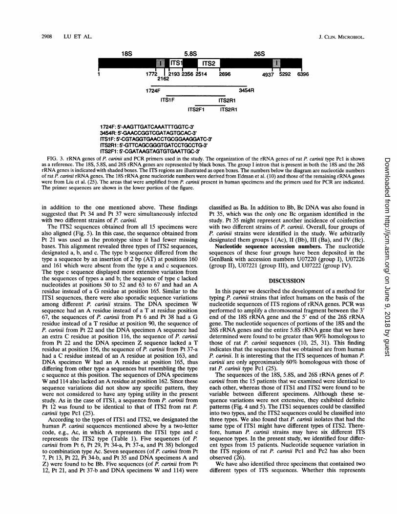
Since the ITS nucleotide sequences of rRNA genes in otherorganisms were shown to be variable (2, 19, 33, 36) and thisregion showed the greatest variation between rat-derived P.carinii type Pcl and Pc2 (26), primers were designed to amplifythe area containing both the ITS1 and ITS2 regions of P.carinii. The area between nucleotides 1724 and 3454 (nucle-otide numbers of rat P. carinii type Pcl rRNA genes) was firstamplified with primers 1724F and 3454R. These two primersequences were chosen because they were found to be moreconserved among all rat P. carinii strains that have beensequenced (10, 25, 31). P. carinii in only 2 of the 11 BALspecimens from patient (Pt) 21 and Pt 29 that we examinedgenerated PCR products of the expected size, suggesting thatthe rRNA gene sequences of rat and human P. carinii are notidentical. The PCR products of P. carinii from Pt 21 and Pt 29were cloned into pCRII and then sequenced.The nucleotide sequence of P. carinii obtained from Pt 21
was compared with that of rat P. carinii (Fig. 1). The 18S rRNAgene of P. carinii from Pt 21 lacked the intron sequence thatwas shown to be present in some rat P. carinii isolates (10, 24,31), denoted type Pcl (24, 25). This finding was consistent withthat described previously (24). The ITS1 sequence of P. carinjifrom Pt 21 was found to be 156 bp in length, which was 7 bp
VOL. 32, 1994
on June 9, 2018 by guesthttp://jcm
.asm.org/
Dow
nloaded from
2906 LU ET AL.
Rat PCHu PC (Pt 21)
Rat PCHu PC (Pt 21)
AAGTTGATCA AATTTGGTCA TTTAGAGGAA********** ********** **********
<intron>TT CCGTAGGTGA ACCTGCGGAA<.--_*_ >
GTAAAAGTCG TAACAAGGT<********** *****
18S+-I 1-4ITS1GGATCATTA< <ATGAAA-TG*********< <--****A*-
Rat PCHu PC (Pt 21)
Rat PCHu PC (Pt 21)
TTGTCAAG-- -AA------- -CTAGTTTAT CTGGTTCTT- GACATTTTCA---***-*CT T**ACACTTC C*****__ ___***-**A *-***C**--
TCATAACA-C TTGTGAACAT TAAAGAT--- TTGCTTTGAC -AGGA--TGG***-****T* *---*****--**_TT*TTT ***-****G* G****GC***
Rat PCHu PC (Pt 21)
Rat PCHu PC (Pt 21)
GAG-----TA AGCTTTCGTC CT-GTC--AG___CTTTT*T ***** **C**CA*
CTTTTT---T GGTG--TTTC -GGTTAAAAA--****AAA* --**AA**** A*-**-----
AGGT-TTTCA ATT---AAAA 250****G***-- ***TTT****
ITS14-l i-+5.8STAT--AATTT TT-AA>>>AA 300*-*AG***** **T**>>>**
Rat PCHu PC (Pt 21)
Rat PCHu PC (Pt 21)
ACTTTCAGCA ATGGATCTCT TGGTTCCCGC GTCGATGAAG AACGTGGCAA*******A** ********** ***C**T*** ********** **********
AATGCGATAA GTAGTGTGAA TTGCAGAATT CAGTGACTCA TCGAATTTTT********** ********** ********** T*****A*** **********
Rat PCHu PC (Pt 21)
Rat PCHu PC (Pt 21)
GAACGCATAT TGCGCTCCTC********C* **GC*****T5.8S+-I I-4ITS2TCATTT<<<< TTATACTTGA*T****<<<< ***-*G**--
AGTATTCTGT GGAGCATGCC TGTTTGAGCG********AG *
ACCTTTTT-- ---------- AAGGTTTGTG-*******TC AAGCAGAAAA ****___*GA
Rat PCHu PC (Pt 21)
Rat PCHu PC (Pt 21)
Rat PCHu PC (Pt 21)
Rat PCHu PC (Pt 21)
TTGGGCTATG CA--T-T--T TAGTA-T--- __TTTTACAA G-ATGCTAGT*******T** **AA*A*AA* ***A*-*AAA ATA****T*T *C********
CTAAAATGGA ATCCAGAATA ---TTATTTC GT-GCAGCGT AATAGGGT--**G****--- -**A*A*G** GCT**T**** T*T**C . __***T**CG
TAAATTCCAA TTCGCTGTTT TTAGAAATGA TAGACTGGTT TGTCTATTGT****----** *******--- --G****-** -**GAAAAAA GC*______*
TCCTAG---- AGAGCAATTT*T--*TAGAT *C*AG*****
ITS2+-I 1-26sTTGAAC>CTT TGACCTCAAA TCAGGTAGGA------>--- *A*T****** *****C****
550
600
650
700
Rat PCHu PC (Pt 21)
Rat PCHu PC (Pt 21)
TTACCCGCTG AACTTAAGCA TATCAATAAG CGGAGGAAAA GAAACTAACA*C******** ********** ********** ********** **********
AGGATTCCCT CAGTAACGGC GA********** T********* **
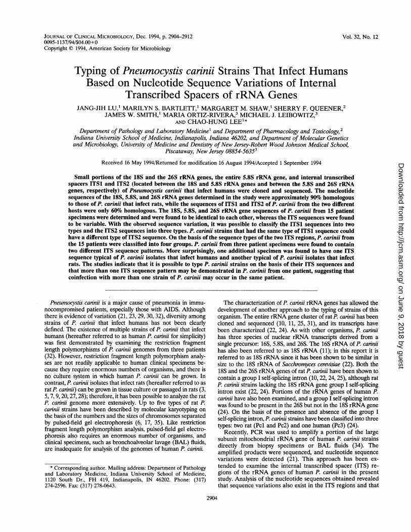
FIG. 1. Comparison of nucleotide sequences of portions of the rRNA genes and the ITS regions of P. carinii from rat and human hosts. Thesequence of P. carinii from Pt 21 [Hu PC (Pt 21)] is compared with that of rat P. carinii type Pcl (Rat PC) published by Liu et al. (25). Identicalsequences are represented with asterisks, missing bases are shown with dashes, and different bases are indicated. These same symbols are also usedfor Fig. 2, 4, and 5. The numbers shown here and in Figures 2, 4, and 5 indicate position, not nucleotide numbers. Different regions of the rRNAgenes are bracketed and are indicated by the arrows above the sequence.
shorter than that of rat P. carinii type Pcl (25). The 5.8S rRNAgene was found to be composed of 158 nucleotides, the samelength as that of rat P. carinii. The ITS2 sequence of P. cariniifrom Pt 21 was 173 bp long, which was 9 bp shorter than thatof rat P. carinii type Pcl (25). A close analysis of the sequencesrevealed that the 18S, 5.8S, and 26S rRNA genes were quiteconserved between rat (type Pcl) and human P. carinii (100,93.7, and 91.6% identities, respectively), but both ITS1 andITS2 sequences differed significantly (56.3 and 69.4% identi-ties, respectively). The GC content of the 18S, 5.8S, and 26SrRNA genes of both human and rat P. carinii was approxi-mately 40%, whereas it was 30% in the ITS regions.The same homology patterns were found between the
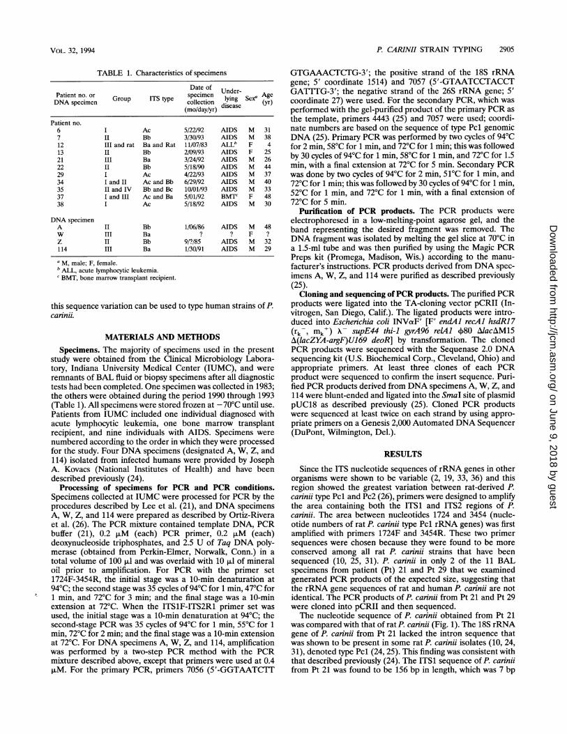
sequences of P. carinii from Pt 21 and Pt 29 (Fig. 2). The
nucleotide sequences of the 18S, 5.8S, and 26S rRNA genesthat we determined from P. carinii from Pt 21 and Pt 29 wereidentical. However, six nucleotides in ITS1 and 11 nucleotidesin ITS2 were different. In order to determine whether thesequence variations found in the ITS regions could be used todifferentiate between different types of human P. carinii,additional patient specimens were examined. Since the primerset 1724F-3454R was designed on the basis of the rat P. carindisequence and did not amplify all human P. carinii sequences, anew set of primers was developed on the basis of the human P.carinii sequences (from Pt 21 and Pt 29) that we had deter-mined. This primer set (ITS1F-ITS2R1) amplified the regionbetween nucleotide positions 53 and 602 (Fig. 2), including thelast 28 bp of the 18S rRNA gene, the entire ITS1, 5.8S, and
50
100
150
200
350
400
450
500
750
772
J. CLIN. MICROBIOL.
on June 9, 2018 by guesthttp://jcm
.asm.org/
Dow
nloaded from
P. CARINII STRAIN TYPING 2907
1898FAAGTTGGTCA
ITS1Frr%rr-rnr ar-r-pr
AATTTGGTCA TTTAGAGGAA GTAAAAGTCG TAACAAGGTT********** ********** ********** **********
18SE- -+ITS1AACCTGCGGA AGGATCATTA GAAAATTCAG CTT-AAACAC
*****C**** ***T******
Pt 21 TTCCCTAGTG TTTTAGCATC TTTCAAACAT CTGTGAATTT TTTTTTTGTTPt 29 ********** *********T * ********** ******-***
50
100
150
Pt 21 TGGCGAGGAG CTGGCTTTTT TGCTTGCCTCPt 29 *****__*** ********** **********
Pt 21 AAATTTTAAA TTGAATTTCA GTTTTAGAATPt 29 ********** ********** **********
Pt 21 TGGATCTCTT GGCTCTCGCGPt 29 ********** **********
Pt 21Pt 29
TAGTGTGAAT TGCAGAATTT********** **********
GCCAAAGGTG TTTATTTTTA********** **********
ITS14--+5.8STTTTTAAAAA CTTTCAACAA********** **********
ITS2F1TCGATGAAGA ACGTGGCAAA ATGCGaATAAG********** ********** **********
AGTGAATCAT** *******
CGAATTTTTG AACGCATCTT********** **********
5.8S-I--*ITS2
200
250
300
350
Pt 21 GGCCTCCTTA GTATTCTAGG GAGCATGCCT GTTTGAGCGT TATTTTTAAG 400Pt 29 ********** ********** ********** ********** *******
Pt 21 TTCCTTTTTT CAAGCAGAAA AAAGGGGATT GGGCTTTGCA AATATAATTA 450Pt 29 ********** ********** ********** ********** ****___***
Pt 21 GAA-TAAAAT ATTTATATGC ATGCTAGTCT GAAATTCAAA AGTAGCTTTT 500Pt 29 ***A***--- ******** ********** ********** **********
Pt 21 TTTCTTTGCC TAGTGTCGTA AAAATTCGCT GGGAAAGAAG GAAAAAAGCT 550Pt 29 ********** ********** ********** ********** **********
ITS2*-I-+26S ITS2R1Pt 21 TTTATAGATA CAAGAATTTT AATCTCAAAT CAGGCAGGAT CACCCGCTGA 600Pt 29 ******A*** ********** ********** ********** **********
Pt 21 ACTTAAGCAT ATCAATAAGC GGAGGAAAAG AAACTAACAA GGATTCCCTT 650Pt 29 ********** ********** ********** ********** ****
Pt 21 AGTAACGGCG APt 29 ********** *
FIG. 2. Comparison of sequences of P. carinii from Pt 21 and Pt 29. The sequences shown include the last 80 bp of the 18S rRNA gene, thefirst 93 bp of the 26S rRNA gene, the entire 5.8S rRNA gene, ITS1, and ITS2. The underlined sequences were used to design the PCR primersfor the present study. Representations of symbols and numbers are as described in the legend to Fig. 1. Regions of the rRNA genes are indicatedabove the sequence.
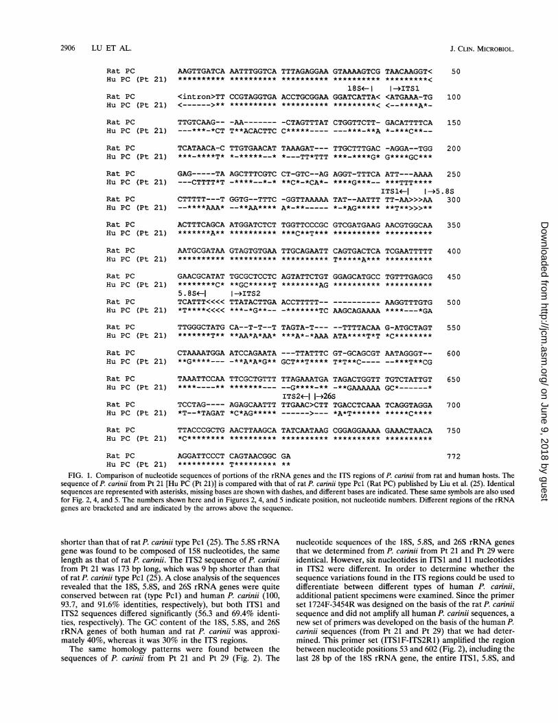
ITS2 regions, and the first 34 bp of the 26S rRNA gene.Another primer, ITS2F1, was also developed; in combinationwith primer ITS2R1, ITS2F1 amplified mainly the ITS2 region(Fig. 2 and 3). As indicated above, different primers were usedto amplify the ITS regions from DNA specimens A, W, Z, and114.
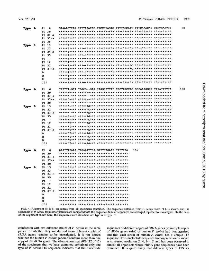
P. carinii isolates in BAL specimens collected from nineadditional patients at IUMC and the four DNA specimensobtained from the National Institutes of Health were exam-ined. The region spanning ITS1 and ITS2 was amplified withthe primer set ITS1F-ITS2R1 (Fig. 3). The resulting 550-bpPCR products were cloned into the TA-cloning vector pCRIIand were then sequenced. The ITS1 sequences of P. canniifrom all 15 specimens (17 sequences) were aligned as shown inFig. 4. The sequence obtained from Pt 6 was chosen as theprototype in Fig. 4 because 6 was the lowest patient number inthe present study, and all other sequences were compared withthis sequence. This comparison revealed that human P. carinjiITS1 sequences could be classified into two major types. Thesetwo types, designated A and B, differed in sequence at
positions 6, 14, 76, and 77. Type A had a C residue at position6 and a T residue at position 14, and there was a 2-bp deletionat positions 76 and 77. Type B had a T residue at position 6, anA residue at position 76, and G residue at position 77 and was
missing a base at position 14. According to this typing system,5 sequences were classified as type A and 12 were classified astype B. There were some sporadic variations within type B.Sequences of P. carinii from Pt 12 and DNA specimen 114 hadan A residue at position 21, whereas P. carinii sequences fromall other patients had a T residue at this position. Thesequences of P. carinii from Pt 21 and Pt 37-b had a C residueinstead of a T residue at position 40. The sequences of P.carinii from Pt 12, 21, and 37-b and DNA specimens A, W, Z,and 114 had an extra T residue at position 67, which mightrepresent another type of ITS1 sequence. Of particular impor-tance was that specimens from Pt 34 and Pt 37 contained bothtype A and type B ITS1 sequences. These sequences were
designated Pt 34-a, Pt 34-b, Pt 37-a, and Pt 37-b, respectively(Fig. 4). Surprisingly, the specimen from Pt 12 contained anITS1 sequence identical to that of rat P. carinii type Pcl (24),
Pt 21Pt 29
Pt 21Pt 29
VOL. 32, 1994
on June 9, 2018 by guesthttp://jcm
.asm.org/
Dow
nloaded from
2908 LU ET AL.
18S 5.8S 26S
1772 1 2193 2356 2514 26962162
1724F
IIl4937 5292 6396
3454R
ITS1 F ITS2RI
ITS2F1 ITS2R1
1724F: 5'-AAGTTGATCAAATTGGTC-3'3454R: 5!-GAACCGGTCGATAGTGCAC-3'ITSI F: '-CGTAGGTGAACCTGCGGAAGGATC-3'ITS2R1: -GT1TCAGCGGGTGATCCTGCCTG-3'ITS2F1: 5'-CGATMGTAGTGTGAATTGC-3'
FIG. 3. rRNA genes of P. carinii and PCR primers used in the study. The organization of the rRNA genes of rat P. carinii type Pcl is shownas a reference. The 18S, 5.8S, and 26S rRNA genes are represented by black boxes. The group I intron that is present in both the 18S and the 26SrRNA genes is indicated with shaded boxes. The ITS regions are illustrated as open boxes. The numbers below the diagram are nucleotide numbersof rat P. carinii rRNA genes. The 18S rRNA gene nucleotide numbers were derived from Edman et al. (10) and those of the remaining rRNA genes
were from Liu et al. (25). The areas that were amplified from P. carinii present in human specimens and the primers used for PCR are indicated.The primer sequences are shown in the lower portion of the figure.
in addition to the one mentioned above. These findingssuggested that Pt 34 and Pt 37 were simultaneously infectedwith two different strains of P. carinii.The ITS2 sequences obtained from all 15 specimens were
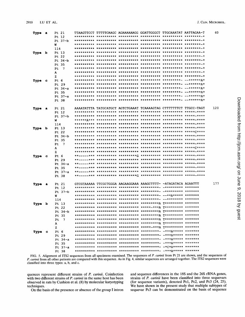
also aligned (Fig. 5). In this case, the. sequence obtained fromPt 21 was used as the prototype since it had fewer missingbases. This alignment revealed three types of ITS2 sequences,designated a, b, and c. The type b sequence differed from thetype a sequence by an insertion of 2 bp (AT) at positions 160and 161 which were absent from the type a and c sequences.The type c sequence displayed more extensive variation fromthe sequences of types a and b; the sequence of type c lackednucleotides at positions 50 to 52 and 63 to 67 and had an Aresidue instead of a G residue at position 165. Similar to theITS1 sequences, there were also sporadic sequence variationsamong different P. carinii strains. The DNA specimen Wsequence had an A residue instead of a T at residue position67, the sequences of P. carinii from Pt 6 and Pt 38 had a Gresidue instead of a T residue at position 90, the sequence ofP. carinii from Pt 22 and the DNA specimen A sequence hadan extra C residue at position 116, the sequence of P. carinjifrom Pt 22 and the DNA specimen Z sequence lacked a Tresidue at position 156, the sequence of P. caninii from Pt 37-ahad a C residue instead of an A residue at position 163, andDNA specimen W had an A residue at position 165, thusdiffering from other type a sequences but resembling the typec sequence at this position. The sequences of DNA specimensW and 114 also lacked anA residue at position 162. Since thesesequence variations did not show any specific pattern, theywere not considered to have any typing utility in the presentstudy. As in the case of ITS1, a sequence from P. carinii fromPt 12 was found to be identical to that of ITS2 from rat P.carinii type Pcl (25).
According to the types of ITS1 and ITS2, we designated thehuman P. carinii sequences mentioned above by a two-lettercode, e.g., Ac, in which A represents the ITS1 type and crepresents the ITS2 type (Table 1). Five sequences (of P.carinii from Pt 6, Pt 29, Pt 34-a, Pt 37-a, and Pt 38) belongedto combination type Ac. Seven sequences (of P. cannii from Pt7, Pt 13, Pt 22, Pt 34-b, and Pt 35 and DNA specimens A andZ) were found to be Bb. Five sequences (of P. cannii from Pt12, Pt 21, and Pt 37-b and DNA specimens W and 114) were
classified as Ba. In addition to Bb, Bc DNA was also found inPt 35, which was the only one Bc organism identified in thestudy. Pt 35 might represent another incidence of coinfectionwith two different strains of P. caninii. Overall, four groups ofP. carinii strains were identified in the study. We arbitrarilydesignated them groups I (Ac), II (Bb), III (Ba), and IV (Bc).
Nucleotide sequence accession numbers. The nucleotidesequences of these four groups have been deposited in theGenBank with accession numbers U07220 (group I), U07226(group II), U07221 (group III), and U07222 (group IV).
DISCUSSION
In this paper we described the development of a method fortyping P. carinii strains that infect humans on the basis of thenucleotide sequences of ITS regions of rRNA genes. PCR was
performed to amplify a chromosomal fragment between the 3'end of the 18S rRNA gene and the 5' end of the 26S rRNAgene. The nucleotide sequences of portions of the 18S and the26S rRNA genes and the entire 5.8S rRNA gene that we havedetermined were found to be greater than 90% homologous tothose of rat P. carini sequences (10, 25, 31). This findingindicates that the sequences that we obtained are from humanP. caninii. It is interesting that the ITS sequences of human P.caninii are only approximately 60% homologous with those ofrat P. cannii type Pcl (25).The sequences of the 18S, 5.8S, and 26S rRNA genes of P.
carinii from the 15 patients that we examined were identical toeach other, whereas those of ITS1 and ITS2 were found to bevariable between different specimens. Although these se-
quence variations were not extensive, they exhibited definitepatterns (Fig. 4 and 5). The ITS1 sequences could be classifiedinto two types, and the ITS2 sequences could be classified intothree types. We also found that P. carinii isolates that had thesame type of ITS1 might have different types of ITS2. There-fore, human P. carinii strains may have six different ITSsequence types. In the present study, we identified four differ-ent types from 15 patients. Nucleotide sequence variation inthe ITS regions of rat P. carinii Pcl and Pc2 has also beenobserved (26).We have also identified three specimens that contained two
different types of ITS sequences. Whether this represents
1
J. CLIN. MICROBIOL.
on June 9, 2018 by guesthttp://jcm
.asm.org/
Dow
nloaded from
P. CARINII STRAIN TYPING 2909
Type A Pt 6 GAAAACTCAG CTTTAAACAC TTCCCTAGTG TTTTAGCATTPt 29Pt 34-aPt 37-aPt 38
Type B Pt 13Pt 22Pt 34-bPt 35Pt 7Pt 12Pt 21Pt 37-bAwz114
Type A Pt 6Pt 29Pt 34-aPt 37-aPt 38
Type B Pt 13Pt 22Pt 34-bPt 35Pt 7Pt 12Pt 21Pt 37-bAwz114
Type A Pt 6Pt 29Pt 34-aPt 37-aPt 38
Type B Pt 13Pt 22Pt 34-bPt 35Pt 7Pt 12Pt 21Pt 37-bAwz114
**********
**********
**********
**********
*****T*********T*********T*********T*********T*********T*********T*********T*********T*********T*********T*********T****
TTTTTT-GTT
* ** ** *T_** ** ** ** *T_** ** ** ** *T_** ** ** ** *T_** *
******T***
AAATTTTAAA
*********
*********
**********
**********
**********
**********
TGGCG--GAG
* * * * *AG* * *
* * * * *AG* * *
* * * * *AG* * *
* * * * *AG* * *
*****AG***
*****AG***
*****AG********AG********AG********AG********AG********AG***
TTGAATTTCA**********
**********
**********
**********
**********
**********
**********
* * * * * * * * * *
**********
**********
* * * * * * * * * *
**********
**********
**********
**********
**********
CTGGCTTTTT**********
**********
**********
**********
**********
**********
**********
**********
**********
**********
**********
**********
**********
**********
* * * * * * ** * *
**********
GTTTTAGAAT**********
**********
**********
**********
**********
**********
**********
**********
**********
**********
**********
**********
**********
**********
**********
**********
TTTCAAACAT**********
**********
**********
**********
**********
* * * * * * * * * *
**********
**********
**********
* * * * * * * * * *
**********
**********
**********
**********
**********
* * * ** * * * * *
CTGTGAATTT* * * * * * * * * *
**********
**********
**********
**********
* * * * * * * * * *
**********
**********
**********
**********
**********
**********
**********
**********
**********
**********
60**********
**********
**********
**********
**********
**********
**********
**********
**********
**********
*********C*********C**********
**********
**********
**********
TGCTTGCCTC* * * * * * * * * *
* * * * * * * * * *
**********
**********
**********
**********
**********
**********
**********
**********
**********
* * * * * * * * * *
* * * * * * * * * *
**********
**********
**********
TTTTTAA*******
*******
*******
*******
*******
*******
* * * * * * *
*******
*******
* * * * * * *
* * * * * * *
*******
* * * * * * *
* * * * * * *
* * * * * * *
*******
GCCAAAGGTG**********
**********
* * * * * * * * * *
**********
**********
* * * * * * * * * *
* * * * * * * * * *
**********
**********
**********
**********
**********
**********
**********
**********
**********
TTTATTTTTA**********
* * * * * * * * * *
**********
* * * * * * * * * *
**********
**********
**********
**********
* * * * * * * * * *
* * * * * * * * * *
**********
* * * * * * * * * *
* * * * * * * * * *
**********
**********
* * * * * * * * * *
120
157
FIG. 4. Alignment of ITS1 sequences from all specimens examined. The sequence obtained from P. carinii from Pt 6 is shown, and thesequences of P. carinii from other patients are compared with this sequence. Similar sequences are arranged together to reveal types. On the basisof the alignment shown here, the sequences were classified into type A or type B.
coinfection with two different strains of P. carinii in the samepatient or whether they are derived from different copies ofrRNA genes remains to be investigated. It is not knownwhether the human P. carinii genome contains more than onecopy of the rRNA genes. The observation that 80% (12 of 15)of the specimens that we have examined contained only onetype of P. carinii ITS sequence indicates that the nucleotide
sequences of different copies ofrRNA genes (if multiple copiesof rRNA genes exist) of human P. carinii had homogenizedand that each strain of human P. carinii has a unique ITSsequence. This nucleotide sequence homogenization is knownas concerted evolution (1, 4, 14-16) and has been observed inalmost all organisms whose rRNA gene sequences have beenexamined. It is quite likely that different types of ITS se-
VOL. 32, 1994
on June 9, 2018 by guesthttp://jcm
.asm.org/
Dow
nloaded from
2910 LU ETAL.J.LN.MCOI.
Type a Pt 21 TTAAGTTCCT TTTTTCAAGC AGAAAAAAGG GGATTGGGCT TTGCAAATAT AATTAGAA-TPt 12Pt 37-bw114
Type b Ptl13Pt 22Pt 34-bPt 35Pt 7Az
Type c Pt 6Pt 29Pt 34-aPt 35Pt 37-aPt 38
Type a Pt 21Pt 12Pt 37-bw114
Type b Ptl13Pt 22Pt 34-bPt 35Pt 7Az
Type c Pt 6Pt 29Pt 34-aPt 35Pt 37-aPt 38
Type a Pt 21Pt 12Pt 37-bw114
Type b Ptl13Pt 22Pt 34-bPt 35Pt 7Az
Type c PtE6Pt 29Pt 34-aPt 35Pt 37-aPt 38
** * * * *
AAAATATTTA
**.
**.
**.
**.
**.
**.
GTCGTAAAAA
TATGCATGCT
TTCGCTGGGA
AGTCTGAAAT
AAGAAGGAAA
TCAAAAGTAG
AAAGCTTTT-
** *** ** **
* ********A
CTTTTTTTCT
-ATAGATACA
T*******
T** * * * *
* * ** *
..*****
* * ** *
* * ** *
TTGCC-TAGT
* * * * *c* * * *
*****.*** *
AGAATTT
60
120
177
FIG. 5. Alignment of ITS2 sequences from all specimens examined. The sequences of P. carinii from Pt 21 are shown, and the sequences ofP. carinii from all other patients are compared with this sequence. As in Fig. 4, similar sequences are arranged together. The ITS2 sequences wereclassified into three types: a, b, and c.
quences represent different strains of P. carinii. Coinfection and sequence differences in the 18S and the 26S rRNA genes,with two different strains of P. carinii in the same host has been strains of P. carinii have been classified into three sequevarsobserved in rats by Cushion et al. (8) by molecular karyotyping (for sequence variants), denoted Pcl, Pc2, and Pc3 (24, 25).techniques. We have shown in the present study that multiple subtypes ofOn the basis of the presence or absence of the group I intron sequevar Pc3 can be demonstrated on the basis of sequence
J. CLIN. MICROBIOL.
on June 9, 2018 by guesthttp://jcm
.asm.org/
Dow
nloaded from
P. CARINII STRAIN TYPING 2911
variation within ITS1 and ITS2. Additional DNA specimenswill need to be sequenced to determine if more sequevars andsubtypes exist, including presently unknown subtypes withinPcl and Pc2.We have previously examined sequences within the mito-
chondrial rRNA gene from human P. carinii (21) and foundone patient infected with a strain that appeared to be a hybridof rat and human P. carinii. In the present study we obtaineddata which further substantiate this possibility. The specimenfrom this same patient (Pt 12) was found to contain P. cariniiwith two different rRNA gene sequences. One of the se-quences was a typical human P. carinii sequence, while theother was identical to that of rat P. carinii. It is unlikely that thespecimen from Pt 12 was contaminated with rat P. carinii sincethis specimen was analyzed in a laboratory where no P.carinii-infected rats were ever present. In addition, the threemajor PCR steps, specimen processing (preparation of tem-plate DNA), PCR setup, and analysis of PCR products, weredone in three separate rooms to prevent contamination oftemplate DNA by PCR products. The coexistence of humanand rat P. carinii DNAs in the specimen from Pt 12 suggeststhat this patient was coinfected with human and rat strains ofP. carindi and that a portion of the genomes recombined, givingrise to a hybrid sequence. If this possibility is proven, the term"rat P. carinii" and the proposal that P. carinii be classified onthe basis of its host (12, 18) would be inappropriate. It appearsthat the strain which commonly infects rats, termed type Pcl(24, 25), can also infect humans, although Gigliotti et al. (13)have shown that P. carinii from one mammalian host does notinfect other animal species.Although sequence variation was also found in the mito-
chondrial rRNA gene of P. caninii, this variation was irregular(21); therefore, it was not useful for typing human P. cariniistrains. The use of ITS sequence variation to type human P.carindi will make it possible to explore the question of whetherdifferent strains of P. carinii differ in prevalence and virulenceby conducting more extensive epidemiologic studies. Addi-tional patient specimens should be examined to expand thedata summarized in Table 1 to determine whether a specificstrain of P. carinii is associated with a unique underlyingdisease. The methods developed in the present study will alsoenable us to determine whether a second episode of P. canniiinfection in the same patient represents a relapse because ofthe failure of chemotherapy or a reinfection by the acquisitionof a different strain of P. cannii. If it can be shown that the P.carinii strains isolated from patients with a second infectiondiffer significantly from the strains isolated initially, it wouldsuggest that immunosuppressed patients require protectionfrom environmental exposure.
ACKNOWLEDGMENTSThe work done at IUMC was supported in part by National
Institutes of Health contracts N01-AI-72647 and U01-AI-25859-01(to J.W.S.) and grant 001996-14-RG from the American Foundationfor AIDS Research (to M.J.L.). M.O.-R. was supported by a fellowshipfrom the minority Advancement Program of Rutgers University.We thank Gerald L. MacLaughlin for assistance in designing two of
the PCR primers used at IUMC, Joseph A. Kovacs (National Institutesof Health) for providing four DNA samples, and Regina Felder forsequencing the four samples examined at UMDNJ.
REFERENCES1. Arnheim, N. 1983. Concerted evolution of multi-gene families, p.
38-61. In M. Nei and R. K. Koehn (ed.), Evolution of genes andproteins. Sinauer, Sunderland, England.
2. Barry, T., G. Colleran, M. Glennon, L. K. Dunican, and F.Gannon. 1991. The 16S/23S ribosomal spacer region as a target for
DNA probes to identify eubacteria. PCR Methods Applic. 1:51-56.
3. Bartlett, M. S., P. A. Verbanac, and J. W. Smith. 1979. Cultivationof Pneumocystis carinii with WI-38 cells. J. Clin. Microbiol. 10:796-799.
4. Cohen, E. S., T. Strachan, and G. A. Dover. 1982. Dynamics ofconcerted evolution of ribosomal DNA and histone gene familiesin melanogaster species subgroup of Drosophila. Cell 33:849-855.
5. Cushion, M. T., and D. Ebbets. 1990. Growth and metabolism ofPneumocystis carinii in axenic culture. J. Clin. Microbiol. 28:1385-1394.
6. Cushion, M. T., M. Kaselis, S. L. Stringer, and J. R. Stringer.1993. Genetic stability and diversity of Pneumocystis carinii infect-ing rat colonies. Infect. Immun. 61:4801-4813.
7. Cushion, M. T., and P. D. Walzer. 1984. Growth and serial passageof Pneumocystis carinii in the A549 cell line. Infect. Immun.44:245-251.
8. Cushion, M. T., J. Zhang, M. Kaselis, S. L Stringer, and J. R.Stringer. 1993. Evidence for two species of Pneumocystis cariniico-infecting laboratory rats. J. Clin. Microbiol. 31:1217-1223.
9. Durkin, M. M., M. S. Bartlett, S. F. Queener, M. M. Shaw, andJ. W. Smith. 1989. A culture method allowing production ofrelatively pure Pneumocystis carinii trophozoites. J. Protozool.36:31S-32S.
10. Edman, J. C., J. A. Kovacs, H. Masur, D. V. Santi, H. J. Elwood,and M. L Sogin. 1988. Ribosomal RNA sequence shows Pneumo-cystis carinii to be a member of the fungi. Nature (London)334:519-522.
11. Edman, J. C., J. A. Kovacs, H. Masur, D. V. Santi, H. J. Elwood,and M. L. Sogin. 1991. Ribosomal RNA genes of Pneumocystiscarinii. J. Protozool. 38:18S-20S.
12. Frenkel, J. K. 1976. Pneumocystis jiroveci n. sp. from man: mor-phology, physiology, and immunology in relation to pathology.Natl. Cancer Inst. Monogr. 43:13-30.
13. Gigliotti, F., A. G. Harmsen, C. G. Haidaris, and P. J. Haidaris.1993. Pneumocystis carinii is not universally transmissible betweenmammalian species. Infect. Immun. 61:2886-2890.
14. Hillis, D. M., and S. K. Davis. 1988. Ribosomal DNA: intraspecificpolymorphism, concerted evolution, and phylogeny reconstruc-tion. Syst. Zool. 37:63-66.
15. Hillis, D. M., and M. T. Dixon. 1991. Ribosomal DNA: molecularevolution and phylogenetic inference. Q. Rev. Biol. 66:411-451.
16. Hillis, D. M., C. Moritz, C. A. Porter, and R. J. Baker. 1991.Evidence of biased gene conversion in concerted evolution ofribosomal DNA. Science 251:308-310.
17. Hong, S. T., P. E. Steele, M. T. Cushion, P. D. Walzer, S. L.Stringer, and J. R Stringer. 1990. Pneumocystis cannii karyotypes.J. Clin. Microbiol. 28:1785-1795.
18. Hughes, W. T., and F. Gigliotti. 1988. Nomenclature of Pneumo-cystis carinii. J. Infect. Dis. 157:432-433.
19. Kostman, J. R, T. D. Edlind, J. J. LiPuma, and T. L. Stull. 1992.Molecular epidemiology of Pseudomonas cepacia determined bypolymerase chain reaction ribotyping. J. Clin. Microbiol. 30:2084-2087.
20. Lee, C. H., N. L. Bauer, M. M. Shaw, M. M. Durkin, M. S. Bartlett,S. F. Queener, and J. W. Smith. 1993. Proliferation of ratPneumocystis carinii on cells sheeted on microcarrier beads inspinner flasks. J. Clin. Microbiol. 31:1659-1662.
21. Lee, C. H., J. J. Lu, M. S. Bartlett, M. M. Durkin, T. H. Liu, J.Wang, B. Juang, and J. W. Smith. 1993. Nucleotide sequencevariation in Pneumocystis carinii strains that infect humans. J. Clin.Microbiol. 31:754-757.
22. Lin, H., M. T. Niu, T. Yoganathan, and G. A. BucLk 1992.Characterization of the rRNA-encoding genes and transcripts, anda group-I self-splicing intron in Pneumocystis carinii. Gene 119:163-173.
23. Linke, M. J., M. T. Cushion, and P. D. Walzer. 1989. Properties ofthe major antigens of rat and human Pneumocystis carinii. Infect.Immun. 57:1547-1555.
24. Liu, Y., and M. J. Leibowitz. 1993. Variation and in vitro splicingof group I introns in rRNA genes of Pneumocystis carinii. NucleicAcids Res. 21:2415-2421.
25. Liu, Y., M. Rocourt, S. Pan, C. Liu, and M. J. Leibowitz. 1992.
VOL. 32, 1994
on June 9, 2018 by guesthttp://jcm
.asm.org/
Dow
nloaded from
2912 LU ET AL. J. CLIN. MICROBIOL.
Sequence and variability of the 5.8S and 26S rRNA genes ofPneumocystis carinii. Nucleic Acids Res. 20:3763-3772.
26. Ortiz-Rivera, M., Y. Liu, R. Felder, and M. J. Leibowitz. Compar-ison of coding and spacer region sequences of chromosomalrRNA-coding genes of two sequevars of Pneumocystis caninii. J.Euk. Microbiol., in press.
27. Sloand, E., B. Laughon, M. Armstrong, M. S. Bartlett, W. Blu-menfeld, M. T. Cushion, A. Kalica, J. A. Kovacs, W. Martin, E.Pitt, E. L. Pesanti, F. Richards, R. Rose, and P. Walzer. 1993. Thechallenge of Pneumocystis caninii culture. J. Euk. Microbiol. 40:188-195.
28. Smith, J. W., and M. S. Bartlett. 1984. In vitro cultivation ofPneumocystis caninii, p. 107-137. In L. S. Young (ed.), Pneumo-cystis caninii pneumonia. Lung biology in health and disease, vol.22. Marcel Dekker, Inc., New York.
29. Smulian, A. G., D. W. Smulian, M. J. Linke, N. A. Halsey, T. C.Quinn, A. P. MacPhail, M. A. Hernandez-Avila, S. T. Hong, andP. D. Walzer. 1993. Geographic variation in humoral response toPneumocystis caninii. J. Infect. Dis. 167:1243-1247.
30. Stringer, J. R., S. L. Stringer, J. Zhang, R. Baughman, A. G.Smulian, and M. T. Cushion. 1993. Molecular genetic distinctionof Pneumocystis carinii from rats and humans. J. Euk. Microbiol.40:733-741.
31. Stringer, S. L., J. R. Stringer, M. A. Blase, P. D. Walzer, and M. T.Cushion. 1989. Pneumocystis carinii: sequence from ribosomalRNA implies a close relationship with fungi. Exp. Parasitol.68:450-461.
32. Tanabe, K., M. Fuchimoto, K. Egawa, and Y. Nakamura. 1988.Use of Pneumocystis caninii genomic DNA clones for DNAhybridization analysis of infected human lungs. J. Infect. Dis.157:593-596.
33. Vodkin, M. H., D. K. Howell, G. S. Visvesvara, and G. L.McLaughlin. 1992. Identification of Acanthamoeba at the geneticand specific levels using the polymerase chain reaction. J. Proto-zool. 39:378-385.
34. Wakefield, A. E., F. J. Pixley, S. Banerji, K. Sinclair, R. F. Miller,E. R. Moxon, and J. M. Hopkin. 1990. Amplification of mitochon-drial ribosomal RNA sequences from Pneumocystis carinii DNA ofrat and human origin. Mol. Biochem. Parasitol. 43:69-76.
35. Weinberg, G. A., and P. J. Durant. 1994. Genetic diversity ofPneumocystis caninii derived from infected rats, mice, ferrets, andcell cultures. J. Euk. Microbiol. 41:219-224.
36. Xue, B., P. H. Goodman, and S. L. Annis. 1992. Pathotypeidentification of Leptosphaeria maculans with PCR and oligonu-cleotide primers from ribosomal internal transcribed spacerssequences. Physiol. Mol. Plant Pathol. 41:179-188.
on June 9, 2018 by guesthttp://jcm
.asm.org/
Dow
nloaded from


![Isolation and characterization of adenoviruses infecting ......[10, 11]. SAdVs that infect great apes are closely related to types that infect humans, which belong to the species Human](https://img.pdfslide.us/doc/110x75/6145de718f9ff812541fe6a5/isolation-and-characterization-of-adenoviruses-infecting-10-11-sadvs.jpg)