Embed Size (px)
Citation preview
TYPES OF HYPOXIA(HYPOXIC,ANEMIC,STAGNANT,HISTOTOXIC,TUMOUR)
AND DISSOCIATION CURVES IN THESE STATES
G.S.ZAKYNTHINOS
2nd faculty CharlesUniversity

TABLE OF CONTENTS
QUICK DEFINITION OF HYPOXIA
SYMPTOMS OF HYPOXIA
SIGNS OF HYPOXIA
TYPES OF HYPOXIA

QUICK DEFINITION OF HYPOXIA
INADEQUATE O2 SUPPLY TO THE BODY TISSUES
(ENTIRE BODY) OR (LOCALIZED REGION)
HYPOXIA MEANS

SYMPTOMS OF HYPOXIA
• DEPEND ON:
RAPIDITY AND SEVERITY
OF THE
DECREASE OF ARTERIAL Po2

1) FULMINANT hypoxia (Arterial Po2<20mmHg)
(eg.aircraft loses cabin pressure above 30,000 feet and no supplemental O2 available)
Occurs in seconds Unconsciousness in 15-20 sec Brain death in 4-5 min
2) ACUTE hypoxia(25mmHg<Arterial Po2<40mmHg)
(eg.altitudesof 18,000-25,000 feet)
Symptoms similar to those of ethyl alcohol(lack of coordination,slowed reflexes,overconfidence)
Unconsciousness
Coma and death(in minutes to hours) if the regulatory mechanisms of the body are inadequate
eventually

3) CHRONIC hypoxia
(40mmHg<Arterial Po2<60mmHg)(eg.at altitudes of 10,000-18,000 feet for extended periods of time)
FOR EXTENDED PERIODS OF TIME!!!
Most clinical causes of hypoxia are in these category
Symptoms similar to those of severe fatigue
DYSPNEA
SHORTNESS OF BREATH
+
RESPIRATORY ARRHYTHMIAS

SIGNS OF HYPOXIA
1. Cyanosis (bluish color of tissue) caused by more than 5g of deoxyhemoglobin/dl in capillary blood(or less than
13ml O2 per 100ml of blood)
NOT RELIABLE SIGN OF HYPOXIA!!!
ANEMIC PATIENTS never develop
cyanosis but are extremely hypoxic
PATIENTS WITH POLYCYTHEMIA may be
cyanotic but they are perfectly oxygenated
2. Tachycardia
(peripheral chemoreceptor reflex response to Po2 )
3. Tachypnea and Hyperpnea (arterial chemoreceptor reflex response to Po2 )

TYPES OF HYPOXIA
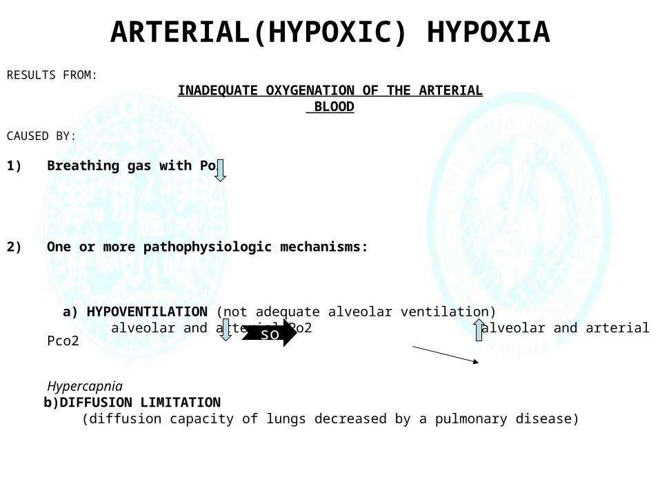
ARTERIAL(HYPOXIC) HYPOXIARESULTS FROM:
INADEQUATE OXYGENATION OF THE ARTERIAL BLOOD
CAUSED BY:
1) Breathing gas with Po2
2) One or more pathophysiologic mechanisms:
a) HYPOVENTILATION (not adequate alveolar ventilation) alveolar and arterial Po2 alveolar and arterial Pco2 Hypercapnia b)DIFFUSION LIMITATION
(diffusion capacity of lungs decreased by a pulmonary disease)
so
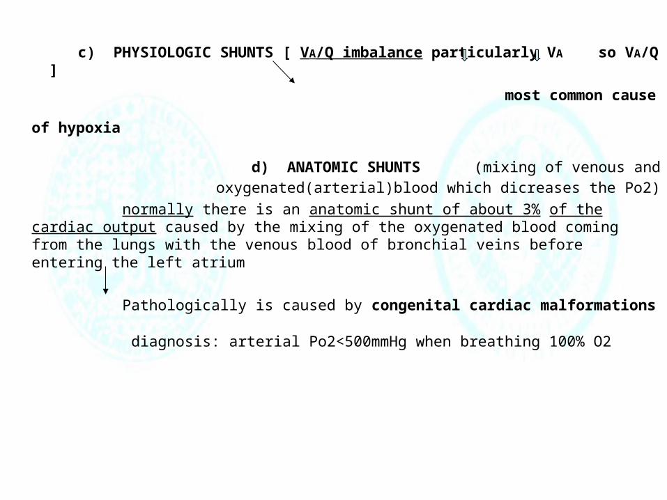
c) PHYSIOLOGIC SHUNTS [ VA/Q imbalance particularly VA so VA/Q ]
most common cause of hypoxia d) ANATOMIC SHUNTS (mixing of venous and oxygenated(arterial)blood which dicreases
the Po2)
normally there is an anatomic shunt of about 3% of the cardiac output caused by the mixing of the oxygenated blood coming from the lungs with the venous blood of bronchial veins before entering the left atrium
Pathologically is caused by congenital cardiac malformations
diagnosis: arterial Po2<500mmHg when breathing 100% O2
Po2(mmHg)
O2
in b
lood
(vol
umes
%)
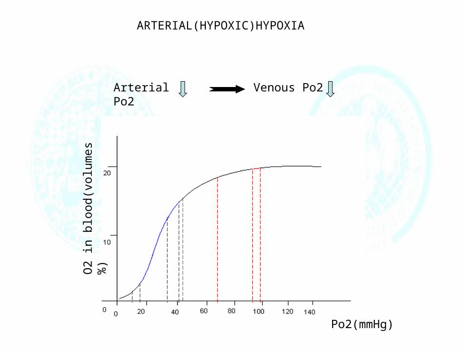
ARTERIAL(HYPOXIC)HYPOXIA
Arterial Po2 Venous Po2

STAGNANT(ISCHEMIC) HYPOXIA
RESULTS FROM:
INADEQUATE BLOOD FLOW
entire body or localized area
caused by
Congestive heart failure Arteriosclerosis
Arterial Po2 may be normal BUT because Q (blood flow),tissues withdraw larger amounts of O2 from the blood ,so, Venous Po2

Po2(mmHg)
O2
in b
lood
(vol
umes
%)
STAGNANT(ISCHEMIC)HYPOXIA
Arterial Po2
Venous Po2
BUT

ANEMIC HYPOXIA
RESULTS FROM:
INSUFFICIENT AMOUNT OF FUNCTIONAL HEMOGLOBIN
CAUSED BY:
1) Deficiency of essential nutrients(iron,B12 vitamin) 2) Blood loss
Patients with Anemic hypoxia have reduced O2 capacity so they have
reduced concentration of O2 in their blood
Arterial Po2is Normal Venous Po2
but
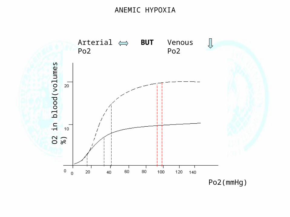
ANEMIC HYPOXIA
Po2(mmHg)
O2
in b
lood
(vol
umes
%)
Arterial Po2 BUT Venous Po2
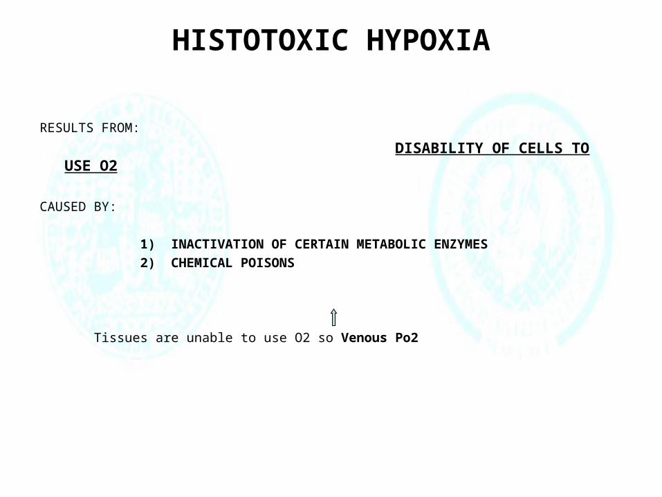
HISTOTOXIC HYPOXIA
RESULTS FROM:
DISABILITY OF CELLS TO USE O2
CAUSED BY:
1) INACTIVATION OF CERTAIN METABOLIC ENZYMES
2) CHEMICAL POISONS
Tissues are unable to use O2 so Venous Po2
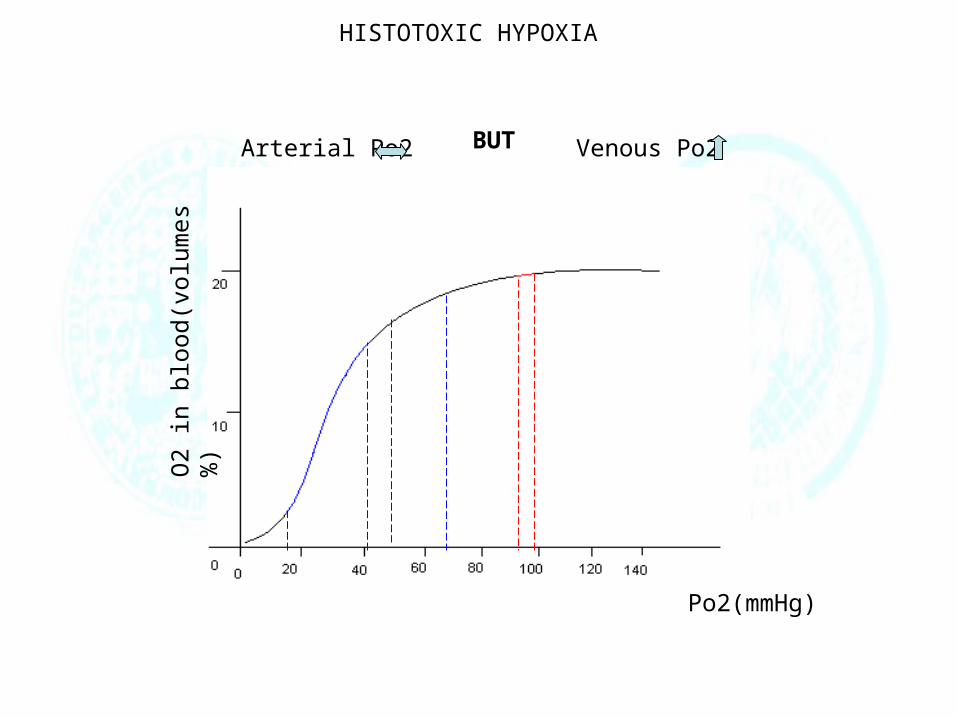
HISTOTOXIC HYPOXIA
Po2(mmHg)
O2
in b
lood
(vol
umes
%)
Arterial Po2 Venous Po2 BUT

SUMMARY
TYPE OF HYPOXIA
Arterial Po2
Venous Po2
Arterial Pco2
Arterial Po2 during exercise
Effect of 100% O2
ARTERIAL HYPOXIA Hypoventilation Arterial Pco2
Diffusion limitation
Arterial Po2>600mmHg
Physiologic shunt
Arterial Po2>600mmHg
Anatomic shunt Arterial Po2<500mmHg
STAGNANT HYPOXIA
dissolved O2
ANEMIC HYPOXIA
dissolved O2
HISTOTOXIC HYPOXIA
dissolved O2

TUMOUR HYPOXIA
1) Hypoxia is widespread in tumors
2) Most human solid tumors have pO2 values lower than their normal tissues of origin.
Tumor blood vessels are highly irregular and disorganized.
Tumours do not get enough O2 and nutrients
THIS IS CAUSED BECAUSE
SO

But tumor cells are usually proliferating
faster than normal cells.
the ability of tumor cells to
sense and adapt to low oxygen (hypoxia)
is essential for tumor growth.
SO
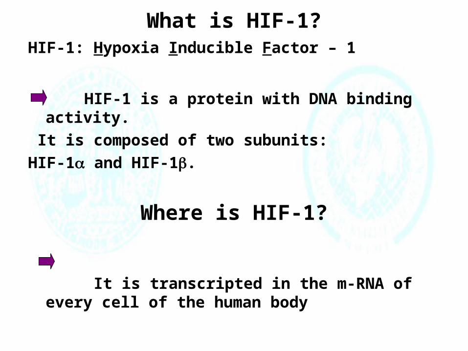
What is HIF-1?HIF-1: Hypoxia Inducible Factor – 1
HIF-1 is a protein with DNA binding activity.
It is composed of two subunits:
HIF-1 and HIF-1.
Where is HIF-1?
It is transcripted in the m-RNA of every cell of the human body

What does HIF-1 do?
• Helps normal tissues as well as tumors to survive under hypoxic conditions
• HIF-1 is a transcription factor that turns on genes needed for survival under hypoxic conditions.
• So far, more than 40 target genes have been found to be regulated by HIF-1.
• These genes can be classified into 3 main groups:

HIF-1 Target Genes
Erythropoeitin (EPO)
Nitric oxide synthase 2
(NOS2)
Transferrin
Transferrin receptor
Vascular endothelial
growth factor (VEGF)
VEGF receptor FLT-1
Group 1: O2 Delivery

Aldolase A
Aldolase C
Enolase 1 (ENO1)
Glucose transporter 1
Glyceraldehyde phosphate
dehydrogenase Hexokinase 1
Hexokinase 2
Lactate dehydrogenase A
Phosphofructokinase L
Phosphoglycerate kinase 1
Pyruvate kinase M
Group 2: Glucose/Energy Metabolism

Insulin-like growth
factor 2 (IGF-2)IGF binding protein 1
IGF binding protein 3
p21
p35srj
Group 3: Cell Proliferation/Viability

How does HIF-1 do the job?
Oxygen Concentration
Rel
ativ
e H
IF-1
Exp
ress
ion
HIF-1 expression increases exponentially when O2

HOW DOES HIF-1 HELPS THE TUMOUR CELL
Among the first responses at the onset of hypoxia
is an increase in the protein levels of
hypoxia-inducible factor-1 (HIF-1)
gradientO2, glucose,growth factors
The oxygen and nutrientsdisplay a gradient awayfrom the necrotic center
An idealized diagram of a tumor cross section

SUMMARY:HIF-1 Correlates with Tumor Vascularity
The expression of HIF-1 is positively
correlated with tumor vascularity, indicating HIF-1 plays a crucial role in tumor angiogenesis progression.
Low oxygen tension is associated with increased
metastasis and decreased survival of patients

REFERENCES
• NMS PHYSIOLOGY(ed.2000)
• GUYTON AND HALL MEDICAL PHYSIOLOGY
• MIN WANG Hypoxia Inducible Factor – 1 (HIF-1): A High Impact Factor
• Review Article:Predictive assays for tumour hypoxia: where are we going?
from the 1993 Gray Laboratory Annual Report