Embed Size (px)
Citation preview
Proc. Natl. Acad. Sci. USAVol. 94, pp. 1852–1856, March 1997Developmental Biology
Type III collagen is crucial for collagen I fibrillogenesis and fornormal cardiovascular development
(gene targetingyEhlers–Danlos syndrome type IVyaortic rupture)
XIN LIU*, HONG WU*, MICHAEL BYRNE†, STEPHEN KRANE†, AND RUDOLF JAENISCH*‡
*The Whitehead Institute for Biomedical Research and ‡Department of Biology, Massachusetts Institute of Technology, Nine Cambridge Center, Cambridge, MA02142; and †Department of Medicine, Harvard Medical School and Medical Services (Arthritis Unit), Massachusetts General Hospital, Boston, MA 02214
Communicated by Jerome Gross, Waban, MA, December 24, 1996 (received for review October 6, 1996)
ABSTRACT Type III collagen is a fibrillar forming col-lagen comprising three a1(III) chains and is expressed inearly embryos and throughout embryogenesis. In the adult,type III collagen is a major component of the extracellularmatrix in a variety of internal organs and skin. Mutations inthe COL3A1 gene have been implicated as a cause of type IVEhlers–Danlos syndrome, a disease leading to aortic rupturein early adult life. To directly study the role of Col3a1 indevelopment and disease, we have inactivated the Col3a1 genein embryonic stem cells by homologous recombination. Themutated allele was transmitted through the mouse germ lineand homozygous mutant animals were derived from heterozy-gous intercrosses. About 10% of the homozygous mutantanimals survived to adulthood but have a much shorter lifespan compared with wild-type mice. The major cause of deathof mutant mice was rupture of the major blood vessels, similarto patients with type IV Ehlers–Danlos syndrome. Ultrastruc-tural analysis of tissues from mutant mice revealed that typeIII collagen is essential for normal collagen I fibrillogenesis inthe cardiovascular system and other organs.
Patients with type IV Ehlers–Danlos syndrome, a geneticdisorder associated with fragile blood vessels and skin, oftencarry mutations in the COL3A1 gene coding for type IIIprocollagen (1–3). This suggests that type III collagen isimportant for the development of skin and the cardiovascularsystem and for maintaining the normal physiological functionsof these organs in adult life (4, 5). Type III collagen is amember of the fibrillar collagen family and is colocalized withthe most abundant member of the family, type I collagen, insuch tissues as blood vessels and skin (6–10). Previous studieshave shown that fibrillogenesis may involve coassembly ofdifferent types of collagens. For example, type III collagen isfound to be colocalized with type I collagen within the samefibril (11–14). It was thought that type III collagen maymodulate the size of type I collagen fibrils because thediameter of these fibrils formed by self-assembly in vitroappeared uniform (15–17), but variable in different tissues orin the same tissue at different developmental stages which havedifferent ratios of type III to type I fibrils (11, 12, 18, 19). Todefine the role of type III collagen in fibrillogenesis, we derivedCol3a12/2 mutant mice by gene targeting. Most homozygousCol3a1 mutant mice died perinatally. The phenotype of sur-viving mutant mice resembled the clinical manifestations ofpatients with type IV Ehlers–Danlos syndrome (20) includingsudden death due to rupture of the large vessels. Electronmicroscopic analysis revealed that, in Col3a12/2mice, collagen
fibrils in the media of aorta were missing and collagen fibrilsin the adventitia of the aorta and skin were irregular in size.
MATERIALS AND METHODS
Targeting Vector. Genomic DNA of the Col3a1 gene wascloned from a J1 library (21), and a 14-kb fragment that coversthe promoter and the first two exons of the gene was subclonedinto a pGEM7 vector. At the 59 portion of this 14-kb genomicDNA fragment, a 0.7-kb XbaI–KpnI fragment containing thepromoter and the first exon of the col3a1 gene was replacedwith a 1.8-kb PGKneo cassette (see Fig. 1).Production of Col3a1 Mutant Mice. About 50 mg of the
targeting vector DNA linearized at the 39 end of the 14-kbfragment was electroporated into 2 3 107 J1 embryonic stem(ES) cells. The cells were subsequently cultured in the presenceof G418, and 384 clones were picked after 9 days of culture.Among the 384 clones, 192 were analyzed by BamHI digestionand hybridization to probe (see Fig. 1), and 6 of them,including clones 5 and 67, were identified as correctly targetedclones. Clones 5 and 67 were injected into C57-BLy6 andBALByc embryos as described (22). Chimeras were identifiedon the basis of agouti pigmentation in the coat and back-crossed. Their agouti offspring were genotyped by Southernblot analysis.Collagen Analysis of Col3a1 Mutant Mice. Collagens were
extracted from mouse tail or skin by digestion with pepsin, 50mgymg, in 0.5 M acetic acid at 0–48C for 2–4 days. Thecollagen extracts were resolved with SDSyPAGE. When thedye front had migrated into the running gel for about 1.5 cm,the sample buffer was changed and 2% of 2-mercaptoethanolwas added. This method permits the separation of disulfide-bonded type III collagen from type I collagen. The collagenswere visualized by staining with Coomassie blue.Histological Analysis. Samples were fixed in 10% buffered
formalin. They were then placed in successive ethanol andxylene baths and finally embedded in Paraplast Plus (OxfordLabware, Oxford, U.K.) using an Autotechnicon embedder(Technicon). The embedded tissues were sectioned to 5 mmusing a Reichert–Jung (Vienna) microtome and stained withMasson’s trichrome (23) where collagens are blue in color.Transmission Electron Microscopy. Samples were fixed
with 2.5% glutaraldehyde in 0.1 M Na cacodylate buffer (pH7.2) for 1 hr on ice. Samples were then rinsed in 10% sucrosein 0.1 M Na cacodylate buffer, postfixed with 1% OsO4 incacodylate buffer, and en bloc stained with 1% aqueous uranylacetate. Samples were dehydrated in graded ethanol andembedded in polyBed 812 (Polysciences). Thin sections werecut on a Ultracut E (Reichert–Jung), stained with uranylacetate and lead citrate, and examined under the Philips 410transmission election microscope.
The publication costs of this article were defrayed in part by page chargepayment. This article must therefore be hereby marked ‘‘advertisement’’ inaccordance with 18 U.S.C. §1734 solely to indicate this fact.
Copyright q 1997 by THE NATIONAL ACADEMY OF SCIENCES OF THE USA0027-8424y97y941852-5$2.00y0PNAS is available online at http:yywww.pnas.org. Abbreviation: ES cell, embryonic stem cell.
1852
RESULTS AND DISCUSSION
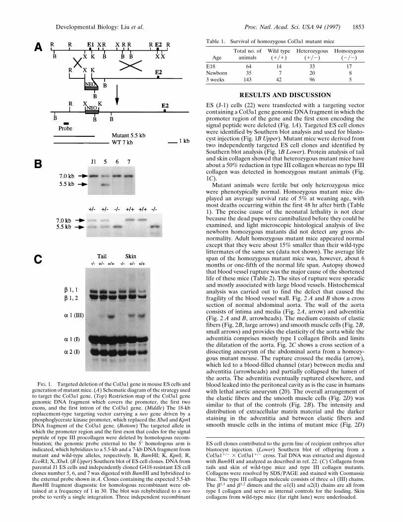
ES (J-1) cells (22) were transfected with a targeting vectorcontaining a Col3a1 gene genomic DNA fragment in which thepromoter region of the gene and the first exon encoding thesignal peptide were deleted (Fig. 1A). Targeted ES cell cloneswere identified by Southern blot analysis and used for blasto-cyst injection (Fig. 1B Upper). Mutant mice were derived fromtwo independently targeted ES cell clones and identified bySouthern blot analysis (Fig. 1B Lower). Protein analysis of tailand skin collagen showed that heterozygous mutant mice haveabout a 50% reduction in type III collagen whereas no type IIIcollagen was detected in homozygous mutant animals (Fig.1C).Mutant animals were fertile but only heterozygous mice
were phenotypically normal. Homozygous mutant mice dis-played an average survival rate of 5% at weaning age, withmost deaths occurring within the first 48 hr after birth (Table1). The precise cause of the neonatal lethality is not clearbecause the dead pups were cannibalized before they could beexamined, and light microscopic histological analysis of livenewborn homozygous mutants did not detect any gross ab-normality. Adult homozygous mutant mice appeared normalexcept that they were about 15% smaller than their wild-typelittermates of the same sex (data not shown). The average lifespan of the homozygous mutant mice was, however, about 6months or one-fifth of the normal life span. Autopsy showedthat blood vessel rupture was the major cause of the shortenedlife of these mice (Table 2). The sites of rupture were sporadicand mostly associated with large blood vessels. Histochemicalanalysis was carried out to find the defect that caused thefragility of the blood vessel wall. Fig. 2 A and B show a crosssection of normal abdominal aorta. The wall of the aortaconsists of intima and media (Fig. 2A, arrow) and adventitia(Fig. 2 A and B, arrowheads). The medium consists of elasticfibers (Fig. 2B, large arrows) and smooth muscle cells (Fig. 2B,small arrows) and provides the elasticity of the aorta while theadventitia comprises mostly type I collagen fibrils and limitsthe dilatation of the aorta. Fig. 2C shows a cross section of adissecting aneurysm of the abdominal aorta from a homozy-gous mutant mouse. The rupture crossed the media (arrow),which led to a blood-filled channel (star) between media andadventitia (arrowheads) and partially collapsed the lumen ofthe aorta. The adventitia eventually ruptured elsewhere, andblood leaked into the peritoneal cavity as is the case in humanswith lethal aortic aneurysm (20). The overall arrangement ofthe elastic fibers and the smooth muscle cells (Fig. 2D) wassimilar to that of the controls (Fig. 2B). The intensity anddistribution of extracellular matrix material and the darkerstaining in the adventitia and between elastic fibers andsmooth muscle cells in the intima of mutant mice (Fig. 2D)
FIG. 1. Targeted deletion of the Col3a1 gene in mouse ES cells andgeneration of mutant mice. (A) Schematic diagram of the strategy usedto target the Col3a1 gene. (Top) Restriction map of the Col3a1 genegenomic DNA fragment which covers the promoter, the first twoexons, and the first intron of the Col3a1 gene. (Middle) The 18-kbreplacement-type targeting vector carrying a neo gene driven by aphosphoglycerate kinase promoter, which replaced the XbaI and KpnIDNA fragment of the Col3a1 gene. (Bottom) The targeted allele inwhich the promoter region and the first exon that codes for the signalpeptide of type III procollagen were deleted by homologous recom-bination; the genomic probe external to the 59 homologous arm isindicated, which hybridizes to a 5.5-kb and a 7-kb DNA fragment frommutant and wild-type alleles, respectively. B, BamHI; K, KpnI; R,EcoRI; X, XbaI. (B Upper) Southern blot of ES cell clones. DNA fromparental J1 ES cells and independently cloned G418-resistant ES cellclones number 5, 6, and 7 was digested with BamHI and hybridized tothe external probe shown in A. Clones containing the expected 5.5-kbBamHI fragment diagnostic for homologous recombinant were ob-tained at a frequency of 1 in 30. The blot was rehybridized to a neoprobe to verify a single integration. Three independent recombinant
Table 1. Survival of homozygous Col3a1 mutant mice
AgeTotal no. ofanimals
Wild type(1y1)
Heterozygous(1y2)
Homozygous(2y2)
E18 64 14 33 17Newborn 35 7 20 83 weeks 143 42 96 5
ES cell clones contributed to the germ line of recipient embryos afterblastocyst injection. (Lower) Southern blot of offspring from aCol3a11/2 3 Col3a11/2 cross. Tail DNA was extracted and digestedwith BamHI and analyzed as described in ref. 22. (C) Collagens fromtails and skin of wild-type mice and type III collagen mutants.Collagens were resolved by SDSyPAGE and stained with Coomassieblue. The type III collagen molecule consists of three a1 (III) chains.The b1,1 and b1,2 dimers and the a1(I) and a2(I) chains are all fromtype I collagen and serve as internal controls for the loading. Skincollagens from wild-type mice (far right lane) were underloaded.
Developmental Biology: Liu et al. Proc. Natl. Acad. Sci. USA 94 (1997) 1853
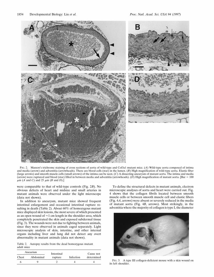
were comparable to that of wild-type controls (Fig. 2B). Noobvious defects of heart and midsize and small arteries inmutant animals were observed under the light microscope(data not shown).In addition to aneurysm, mutant mice showed frequent
intestinal enlargement and occasional intestinal rupture re-sulting in death (Table 2). About 60% of homozygous mutantmice displayed skin lesions, the most severe of which presentedas an open wound of'1 cm length in the shoulder area, whichcompletely penetrated the skin and exposed subdermal tissue(Fig. 3). The wounds were not due to fighting between animals,since they were observed in animals caged separately. Lightmicroscopic analysis of skin, intestine, and other internalorgans including liver and lung did not detect any overtabnormality in mutant animals (data not shown).
To define the structural defects in mutant animals, electronmicroscopic analyses of aorta and heart were carried out. Fig.4 shows that the collagen fibrils located between smoothmuscle cells or between smooth muscle cell and elastic fibers(Fig. 4A, arrows) were absent or severely reduced in the mediaof mutant aorta (Fig. 4B, arrows). Most strikingly, in theadventitia where themajority of collagen is type I, the diameter
FIG. 2. Masson’s trichrome staining of cross sections of aorta of wild-type and Col3a1 mutant mice. (A) Wild-type aorta composed of intimaand media (arrow) and adventitia (arrowheads). There are blood cells (star) in the lumen. (B) High magnification of wild-type aorta. Elastic fiber(large arrows) and smooth muscle cells (small arrows) of the intima can be seen. (C) A dissecting aneurysm of mutant aorta. The intima and media(arrow) were ruptured and blood (star) filled in between media and adventitia (arrowheads). (D) High magnification of mutant aorta. [Bar 5 100mm (A and C) and 25 mm (B and D).]
FIG. 3. A type III collagen-deficient mouse with a skin wound onits left shoulder.
Table 2. Autopsy results from the dead homozygous mutantadult mice
Aneurism Intestinalrupture Infection
Cause notdeterminedChest Abdominal
6 9 2 4 4
1854 Developmental Biology: Liu et al. Proc. Natl. Acad. Sci. USA 94 (1997)
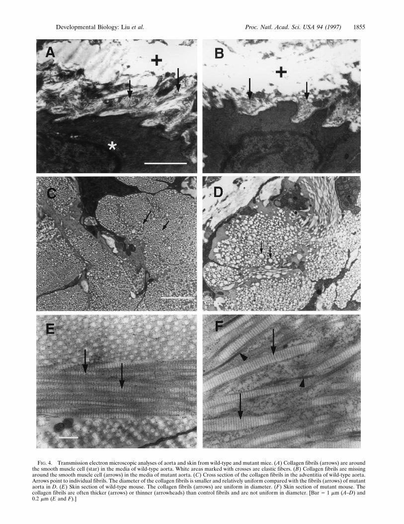
FIG. 4. Transmission electron microscopic analyses of aorta and skin from wild-type and mutant mice. (A) Collagen fibrils (arrows) are aroundthe smooth muscle cell (star) in the media of wild-type aorta. White areas marked with crosses are elastic fibers. (B) Collagen fibrils are missingaround the smooth muscle cell (arrows) in the media of mutant aorta. (C) Cross section of the collagen fibrils in the adventitia of wild-type aorta.Arrows point to individual fibrils. The diameter of the collagen fibrils is smaller and relatively uniform compared with the fibrils (arrows) of mutantaorta in D. (E) Skin section of wild-type mouse. The collagen fibrils (arrows) are uniform in diameter. (F) Skin section of mutant mouse. Thecollagen fibrils are often thicker (arrows) or thinner (arrowheads) than control fibrils and are not uniform in diameter. [Bar 5 1 mm (A–D) and0.2 mm (E and F).]
Developmental Biology: Liu et al. Proc. Natl. Acad. Sci. USA 94 (1997) 1855
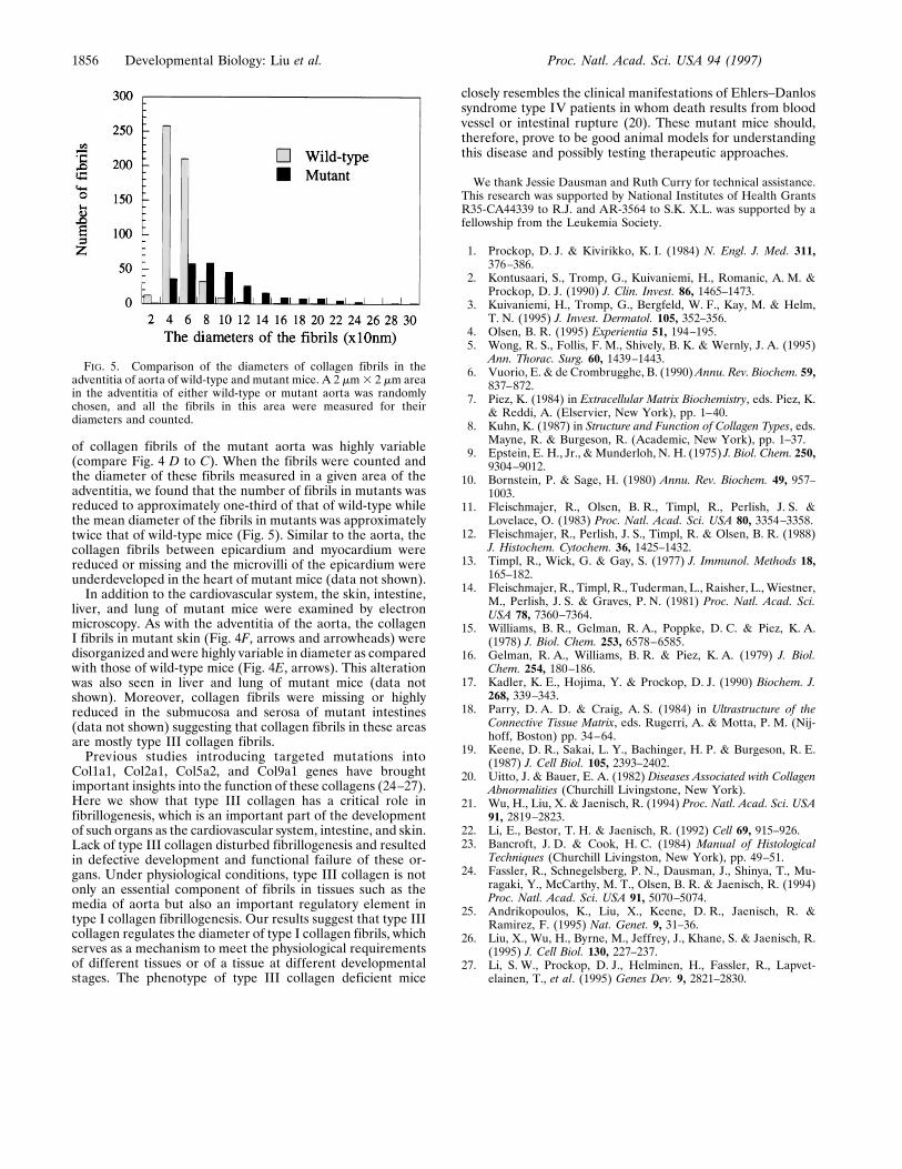
of collagen fibrils of the mutant aorta was highly variable(compare Fig. 4 D to C). When the fibrils were counted andthe diameter of these fibrils measured in a given area of theadventitia, we found that the number of fibrils in mutants wasreduced to approximately one-third of that of wild-type whilethe mean diameter of the fibrils in mutants was approximatelytwice that of wild-type mice (Fig. 5). Similar to the aorta, thecollagen fibrils between epicardium and myocardium werereduced or missing and the microvilli of the epicardium wereunderdeveloped in the heart of mutant mice (data not shown).In addition to the cardiovascular system, the skin, intestine,
liver, and lung of mutant mice were examined by electronmicroscopy. As with the adventitia of the aorta, the collagenI fibrils in mutant skin (Fig. 4F, arrows and arrowheads) weredisorganized and were highly variable in diameter as comparedwith those of wild-type mice (Fig. 4E, arrows). This alterationwas also seen in liver and lung of mutant mice (data notshown). Moreover, collagen fibrils were missing or highlyreduced in the submucosa and serosa of mutant intestines(data not shown) suggesting that collagen fibrils in these areasare mostly type III collagen fibrils.Previous studies introducing targeted mutations into
Col1a1, Col2a1, Col5a2, and Col9a1 genes have broughtimportant insights into the function of these collagens (24–27).Here we show that type III collagen has a critical role infibrillogenesis, which is an important part of the developmentof such organs as the cardiovascular system, intestine, and skin.Lack of type III collagen disturbed fibrillogenesis and resultedin defective development and functional failure of these or-gans. Under physiological conditions, type III collagen is notonly an essential component of fibrils in tissues such as themedia of aorta but also an important regulatory element intype I collagen fibrillogenesis. Our results suggest that type IIIcollagen regulates the diameter of type I collagen fibrils, whichserves as a mechanism to meet the physiological requirementsof different tissues or of a tissue at different developmentalstages. The phenotype of type III collagen deficient mice
closely resembles the clinical manifestations of Ehlers–Danlossyndrome type IV patients in whom death results from bloodvessel or intestinal rupture (20). These mutant mice should,therefore, prove to be good animal models for understandingthis disease and possibly testing therapeutic approaches.
We thank Jessie Dausman and Ruth Curry for technical assistance.This research was supported by National Institutes of Health GrantsR35-CA44339 to R.J. and AR-3564 to S.K. X.L. was supported by afellowship from the Leukemia Society.
1. Prockop, D. J. & Kivirikko, K. I. (1984) N. Engl. J. Med. 311,376–386.
2. Kontusaari, S., Tromp, G., Kuivaniemi, H., Romanic, A. M. &Prockop, D. J. (1990) J. Clin. Invest. 86, 1465–1473.
3. Kuivaniemi, H., Tromp, G., Bergfeld, W. F., Kay, M. & Helm,T. N. (1995) J. Invest. Dermatol. 105, 352–356.
4. Olsen, B. R. (1995) Experientia 51, 194–195.5. Wong, R. S., Follis, F. M., Shively, B. K. & Wernly, J. A. (1995)
Ann. Thorac. Surg. 60, 1439–1443.6. Vuorio, E. & de Crombrugghe, B. (1990)Annu. Rev. Biochem. 59,
837–872.7. Piez, K. (1984) in Extracellular Matrix Biochemistry, eds. Piez, K.
& Reddi, A. (Elservier, New York), pp. 1–40.8. Kuhn, K. (1987) in Structure and Function of Collagen Types, eds.
Mayne, R. & Burgeson, R. (Academic, New York), pp. 1–37.9. Epstein, E. H., Jr., &Munderloh, N. H. (1975) J. Biol. Chem. 250,
9304–9012.10. Bornstein, P. & Sage, H. (1980) Annu. Rev. Biochem. 49, 957–
1003.11. Fleischmajer, R., Olsen, B. R., Timpl, R., Perlish, J. S. &
Lovelace, O. (1983) Proc. Natl. Acad. Sci. USA 80, 3354–3358.12. Fleischmajer, R., Perlish, J. S., Timpl, R. & Olsen, B. R. (1988)
J. Histochem. Cytochem. 36, 1425–1432.13. Timpl, R., Wick, G. & Gay, S. (1977) J. Immunol. Methods 18,
165–182.14. Fleischmajer, R., Timpl, R., Tuderman, L., Raisher, L., Wiestner,
M., Perlish, J. S. & Graves, P. N. (1981) Proc. Natl. Acad. Sci.USA 78, 7360–7364.
15. Williams, B. R., Gelman, R. A., Poppke, D. C. & Piez, K. A.(1978) J. Biol. Chem. 253, 6578–6585.
16. Gelman, R. A., Williams, B. R. & Piez, K. A. (1979) J. Biol.Chem. 254, 180–186.
17. Kadler, K. E., Hojima, Y. & Prockop, D. J. (1990) Biochem. J.268, 339–343.
18. Parry, D. A. D. & Craig, A. S. (1984) in Ultrastructure of theConnective Tissue Matrix, eds. Rugerri, A. & Motta, P. M. (Nij-hoff, Boston) pp. 34–64.
19. Keene, D. R., Sakai, L. Y., Bachinger, H. P. & Burgeson, R. E.(1987) J. Cell Biol. 105, 2393–2402.
20. Uitto, J. & Bauer, E. A. (1982) Diseases Associated with CollagenAbnormalities (Churchill Livingstone, New York).
21. Wu, H., Liu, X. & Jaenisch, R. (1994) Proc. Natl. Acad. Sci. USA91, 2819–2823.
22. Li, E., Bestor, T. H. & Jaenisch, R. (1992) Cell 69, 915–926.23. Bancroft, J. D. & Cook, H. C. (1984) Manual of Histological
Techniques (Churchill Livingston, New York), pp. 49–51.24. Fassler, R., Schnegelsberg, P. N., Dausman, J., Shinya, T., Mu-
ragaki, Y., McCarthy, M. T., Olsen, B. R. & Jaenisch, R. (1994)Proc. Natl. Acad. Sci. USA 91, 5070–5074.
25. Andrikopoulos, K., Liu, X., Keene, D. R., Jaenisch, R. &Ramirez, F. (1995) Nat. Genet. 9, 31–36.
26. Liu, X., Wu, H., Byrne, M., Jeffrey, J., Khane, S. & Jaenisch, R.(1995) J. Cell Biol. 130, 227–237.
27. Li, S. W., Prockop, D. J., Helminen, H., Fassler, R., Lapvet-elainen, T., et al. (1995) Genes Dev. 9, 2821–2830.
FIG. 5. Comparison of the diameters of collagen fibrils in theadventitia of aorta of wild-type and mutant mice. A 2 mm3 2 mm areain the adventitia of either wild-type or mutant aorta was randomlychosen, and all the fibrils in this area were measured for theirdiameters and counted.
1856 Developmental Biology: Liu et al. Proc. Natl. Acad. Sci. USA 94 (1997)