Embed Size (px)
Citation preview

Barrett et al. Retrovirology (2017) 14:25 DOI 10.1186/s12977-017-0349-2
SHORT REPORT
Type I interferon signaling is required for the APOBEC3/Rfv3-dependent neutralizing antibody response but not innate retrovirus restrictionBradley S. Barrett1, Michael S. Harper1,2, Sean T. Jones1,2, Kejun Guo1, Karl J. Heilman1, Ross M. Kedl2, Kim J. Hasenkrug3 and Mario L. Santiago1,2,4*
Abstract
Background: APOBEC3/Rfv3 restricts acute Friend retrovirus (FV) infection and promotes virus-specific neutralizing antibody (NAb) responses. Classical Rfv3 studies utilized FV stocks containing lactate-dehydrogenase elevating virus (LDV), a potent type I interferon inducer. Previously, we showed that APOBEC3 is required for the anti-FV activity of exogenous IFN-alpha treatment. Thus, type I interferon receptor (IFNAR) signaling may be required for the APOBEC3/Rfv3 response.
Results: To test if the APOBEC3/Rfv3 response is dependent on type I IFN signaling, we infected IFNAR knockout versus IFNAR/APOBEC3 double-knockout mice with FV/LDV or LDV-free FV, and evaluated acute FV infection and subsequent NAb titers. We show that LDV co-infection and type I IFN signaling are not required for innate APOBEC3-mediated restriction. By contrast, removal of LDV and/or type I IFN signaling abrogated the APOBEC3-dependent NAb response.
Conclusions: APOBEC3 can restrict retroviruses in a type I IFN-independent manner in vivo. By contrast, the ability of APOBEC3 to promote NAb responses is type I IFN-dependent. These findings reveal novel insights on the interplay between type I IFNs and APOBEC3 in vivo that may have implications for augmenting antiretroviral NAb responses.
Keywords: IFNAR, Friend retrovirus, Neutralizing antibody, Deaminase, LDV
© The Author(s) 2017. This article is distributed under the terms of the Creative Commons Attribution 4.0 International License (http://creativecommons.org/licenses/by/4.0/), which permits unrestricted use, distribution, and reproduction in any medium, provided you give appropriate credit to the original author(s) and the source, provide a link to the Creative Commons license, and indicate if changes were made. The Creative Commons Public Domain Dedication waiver (http://creativecommons.org/publicdomain/zero/1.0/) applies to the data made available in this article, unless otherwise stated.
BackgroundInnate immune mechanisms provide a means for the host to control pathogens before more slowly develop-ing adaptive immune responses come into play. Innate immunity was linked to the production of type I inter-ferons (IFN), which orchestrate an antiviral state through the expression of hundreds of interferon-stimulated genes (ISGs) [1]. Some ISGs encoded proteins known as ‘restriction factors’, which directly inhibit invading pathogens. These restriction factors include the seven
human APOBEC3 enzymes (hA3A to hA3H): deoxy-cytidine deaminases that counteract a broad range of retroviruses including HIV-1 [2, 3], and exhibit a wide range of sensitivities to type I IFN induction [4–7]. In the absence of the HIV-encoded antagonist Vif, APOBEC3 gets incorporated into budding HIV-1 particles, inhibit-ing replication in the next target cell either by physically impeding reverse transcription or hypermutating single-stranded reverse transcripts [8]. However, the majority of APOBEC3 studies involved the ectopic expression of APOBEC3 in cell lines, which may not be physiologically relevant. In particular, transfecting APOBEC3 expression constructs into cells would bypass potential upstream regulatory pathways such as type I IFN signaling that
Open Access
Retrovirology
*Correspondence: [email protected] 4 Division of Infectious Diseases, University of Colorado Denver, Mail Stop B-168, 12700 E 19th Avenue, Aurora, CO 80045, USAFull list of author information is available at the end of the article
Page 2 of 9Barrett et al. Retrovirology (2017) 14:25
may be required for APOBEC3 to restrict retroviral rep-lication in vivo.
In contrast to humans, mice encode only one APOBEC3 gene, mA3. Thus, mA3 knockout (KO) mice provided the field with a powerful means to understand the in vivo impact of APOBEC3 on retroviral pathogen-esis and immunity [9–12]. Our group utilized the murine Friend retrovirus (FV) infection model, which was key to the identification of host genes that control retrovirus infection, including the first retrovirus restriction factor, Fv1 [13, 14]. FV is a complex of a replication-competent Friend murine leukemia virus (F-MuLV) and a replica-tion-defective spleen-focus forming virus (SFFV) that causes splenomegaly and erythroleukemia [15]. We pre-viously showed that C57BL/6 (B6) wild-type (WT) and mA3 KO mice had similar levels of plasma viral load at 7 days post-infection (dpi), but the infectivity of the viri-ons was significantly higher in mA3 KO mice [10, 16–18]. Importantly, administration of recombinant IFNα inhib-ited FV replication in WT mice, but exogenously admin-istered IFNα had no antiviral effect in mA3 KO mice [17]. These findings indicated that mA3 acts downstream of exogenous IFNα to inhibit FV replication. However, it remains unclear if mA3 can inhibit retroviral infection in vivo in the absence of endogenous type I IFN signaling.
The impact of mA3 extends beyond retrovirus restric-tion. Our group and others reported that mA3 encoded Rfv3, a classical resistance gene in B6 mice that promoted recovery from FV viremia by stimulating a stronger NAb response [10, 11, 19, 20]. These early studies on Rfv3 were conducted in F1 hybrid mice harboring the Fv2 susceptibility allele from BALB or A.BY strains, which promotes higher FV replication by driving erythroblast proliferation [21–23]. Nevertheless, the impact of mA3/Rfv3 on NAb responses was also observed in pure B6 mice. Compared to B6 WT mice, B6 mA3 KO mice had significantly lower FV-specific NAb responses by 28 days post-infection (dpi) [10]. The underlying mechanism was multifaceted. Compared to mA3 KO mice, WT mice exhibited: (1) enhanced germinal center (GC) responses due to noninfectious virion particle release [16]; (2) aug-mented GC responses due to contraction of the marginal zone B cell compartment [24]; (3) higher levels of anti-viral IgG2b and IgG2c antibodies [25]; and (4) enhanced somatic hypermutation of virus-specific IgG antibod-ies [26]. Antibody neutralization was dependent on Fcγ receptors, as removal of the common γ chain (FcRγ) and particularly FcγRIV, which bind to IgG2b and IgG2c anti-bodies, rendered 28 dpi antisera incapable of neutralizing FV in vivo [25]. By contrast, removal of complement C3 had no effect on the in vivo neutralization capacity of 28 dpi antisera from mA3-sufficient mice [25].
Interestingly, Rfv3 was initially discovered using FV stocks that contained lactate-dehydrogenase elevat-ing virus (LDV), an endemic RNA virus in wild mouse populations and component of the murine ‘virome’ [27, 28]. Since Rfv3 was discovered using FV/LDV stocks, our studies on the role of mA3 in NAb responses utilized FV/LDV [10, 16, 20, 26]. LDV has potent immunostimulatory properties; thus, data obtained using FV/LDV may not necessarily be reproduced using ‘LDV-free’ FV. LDV can suppress T and B cell responses in FV infection [29–31], and can induce high levels of type I IFNs through Toll-like receptor 7 (TLR7) sensing [32]. LDV-free FV infec-tion of B6 mice resulted in very low or undetectable IFNα expression compared to FV/LDV co-infection [33]. Nota-bly, type I IFNs can also augment and shape humoral immune responses in vivo [34–37]. In certain contexts, LDV may also enhance antibody responses [38, 39]. Thus, we hypothesized that type I IFN signaling might be required for the mA3/Rfv3-dependent NAb response during FV/LDV infection.
In order to investigate the impact of type I IFN signal-ing in mA3 restriction and NAb responses, we prepared mice doubly-deficient in mA3 and the type I IFN receptor (IFNAR). IFNAR is a heterodimer consisting of IFNAR-1 and IFNAR-2 subunits that together form a binding site for the antiviral cytokines IFNβ and IFNα subtypes [40]. IFNAR KO mice lacked the IFNAR-1 receptor chain and were unresponsive to JAK/STAT signaling cascades trig-gered by type I IFNs [41]. Many viral infections, includ-ing FV, replicated to significantly higher levels in IFNAR KO compared to WT mice [41, 42]. However, the down-stream effector mechanisms remain under intense inves-tigation. In this report, we tested whether mA3 can inhibit FV and FV/LDV infection and promote NAb responses in the absence of IFNAR signaling.
ResultsMurine APOBEC3 inhibited infectious virus release in the absence of type I IFN signalingWe previously demonstrated that B6 mA3 KO mice had higher infectious viremia compared to wild-type (WT) mice in experimental infections using LDV-containing FV stocks (FV/LDV) [10, 16] or LDV-free FV stocks (FV) [18, 43]. These data suggested that the induction of type I IFNs by LDV might not be required for mA3 to inhibit FV infection in vivo. To test this hypothesis directly, IFNAR KO and IFNAR/mA3 double KO (dKO) mice (<1 year old) were infected i.v. with 104 spleen focus forming units (SFFU) of FV/LDV or FV (Fig. 1a). At 7 days post-infection (dpi), infectious viremia in the plasma was evaluated. Plasma infectious viremia was 5 to eightfold higher in IFNAR/mA3 dKO mice compared to IFNAR KO mice in both FV/LDV (Fig. 1b, left panel)

Page 3 of 9Barrett et al. Retrovirology (2017) 14:25
and FV (Fig. 1b, right panel) infections. We next quan-tified the levels of viral RNA in the plasma (Fig. 1c), an indirect measure of the total number of particles released. Consistent with our previous findings on WT versus mA3 KO mice [17], we found no significant dif-ference in plasma viral RNA load between IFNAR and IFNAR/mA3 dKO mice at 7 dpi in both FV/LDV and FV infections (Fig. 1c). The ratio of infectious titers (Fig. 1b) and plasma viral RNA load (Fig. 1c) provides a measure of virion infectivity [16, 17]. As shown in Fig. 1d, virions in the 7 dpi plasma of IFNAR/mA3 dKO mice had sig-nificantly higher infectivity than those from IFNAR KO mice. Our findings reveal that type I IFN signaling and LDV co-infection are not required for mA3-mediated inhibition of virion infectivity during acute infection.
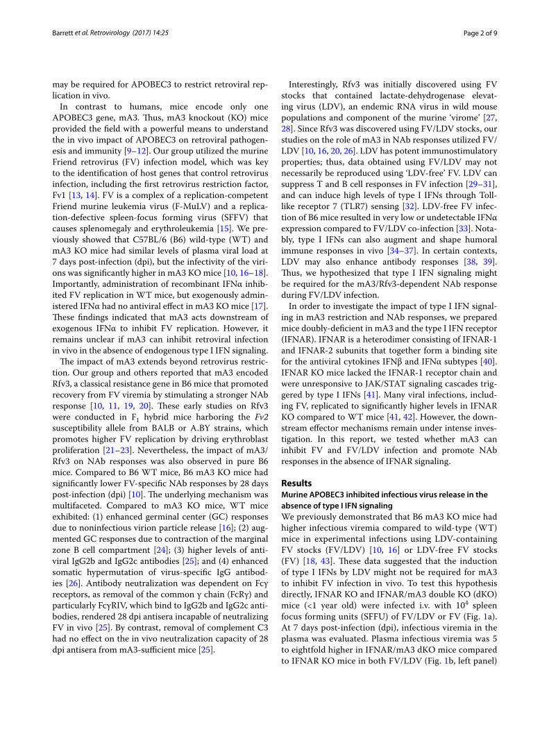
IFNAR signaling is not required for mA3 restriction of acute FV/LDV or FV infectionTo complement the plasma viremia results, we evaluated cellular F-MuLV infection levels in splenocytes. To detect F-MuLV infected cells, we utilized a previously described flow cytometry method using MAb 34, which is specific for the F-MuLV glyco-gag protein [16, 44] (Fig. 2a). Flow cytometry-based methods to detect virus infection gen-erally have lower dynamic range than quantitative PCR-based methods. Nevertheless, consistent with the plasma infectious viremia data, we observed that in both FV/LDV (Fig. 2b) and FV (Fig. 2c) infections, mA3 deficiency in IFNAR KO mice resulted in significantly higher sple-nocyte infection levels (1.6-fold) compared to IFNAR KO mice. Altogether, the data in Figs. 1 and 2 indicate that
Fig. 1 APOBEC3/Rfv3 inhibits infectious virus release in an IFNAR KO background. a Experimental design. B6 IFNAR KO and IFNAR/mA3 dKO mice were infected with FV and samples analyzed at the indicated time points. b Infectious viremia was measured by incubating plasma onto suscepti-ble Mus dunni cells for 2 days and determining F-MuLV proviral DNA levels. Infectious viremia was determined for both (left) FV/LDV and (right) LDV-free FV infection. The same samples in (b) were used to determine c plasma viral RNA loads by qPCR. The ratio of the log-transformed infectious titer in (b) and plasma viral loads in (c) were used to estimate d virion infectivity. Each dot corresponds to a mouse and lines correspond to mean values. The total number of mice analyzed was combined from 2 to 3 independent experiments. Data were analyzed using a 2-tailed unpaired Student’s t test, with exact p values shown. Fold-change values in statistically-significant comparisons were based on average non-log-transformed values
Page 4 of 9Barrett et al. Retrovirology (2017) 14:25
mA3 restricted acute FV replication independently of type I IFN signaling and LDV co-infection.
LDV co‑infection is required for the mA3/Rfv3 neutralizing antibody phenotypeThe Rfv3 gene was identified using FV/LDV stocks [21]. LDV is a potent stimulator of type I IFN responses [32], and type I IFNs can stimulate B cell responses [34–37, 45]. To test if mA3/Rfv3-mediated enhancement of NAb responses occurs in LDV-free FV infection, WT and mA3 KO mice were infected with 104 SFFU of FV/LDV or FV and plasma NAb levels were evaluated at 28 dpi. Consistent with our previously published data [10, 25], mA3 KO mice had significantly lower NAb titers
(3.4-fold) compared to WT mice during FV/LDV co-infection (Fig. 3a). By contrast, the difference in NAb responses between WT and mA3 KO mice infected with LDV-free FV was not statistically significant (Fig. 3b). Thus, LDV co-infection is required for mA3 to promote the FV-specific NAb response.
IFNAR signaling is required for the mA3/Rfv3‑dependent neutralizing antibody responseSince type I IFNs can influence antibody responses and LDV is a potent inducer of type I IFNs, we next inves-tigated if type I IFN signaling is required for the mA3/Rfv3-dependent NAb response. IFNAR KO and IFNAR/mA3 KO mice were infected with 104 SFFU of FV/LDV
Fig. 2 APOBEC3/Rfv3 inhibits acute FV infection of splenocytes independent of type I IFN signaling. Splenocyte FV infection levels were measured by flow cytometry using a glyco-gag specific monoclonal antibody. a Representative flow plots showing glyco-gag+ splenocytes from FV/LDV infected mice at 7 dpi. The percentage of live splenocytes that expressed glyco-gag was evaluated in b FV/LDV and c LDV-free FV infections. Each dot corresponds to a mouse and lines correspond to mean values. The total number of mice analyzed was combined from 2 to 3 independent experiments. Fold-change of mean values per cohort are shown. Data were analyzed using a 2-tailed unpaired Student’s t test, with exact p values shown

Page 5 of 9Barrett et al. Retrovirology (2017) 14:25
and NAb titers evaluated from 28 dpi plasma. In con-trast to FV/LDV infections showing that mA3 influences NAb responses in the B6 background (Fig. 3a) [10, 25], the NAb titers between IFNAR KO and IFNAR/mA3 KO mice were not significantly different from each other (Fig. 4a).
We previously showed that the mA3-dependent NAb response correlated with higher titers of FV-specific IgG2b and IgG2c, both of which signal through FcγRIV [25, 46]. We therefore quantified FV-specific end-point IgG2 titers at 28 dpi plasma of FV/LDV-infected IFNAR KO versus IFNAR/mA3 KO mice. In contrast to mA3 influencing antiretroviral IgG2b and IgG2c titers, mA3 deficiency had no impact on the FV-specific IgG2 response in an IFNAR KO background (Fig. 4b). These data suggest that type I IFN signaling modulates the APOBEC3/Rfv3-dependent, FV-specific IgG2b/c response.
IFNAR KO mice are more susceptible to FV infec-tion compared to normal B6 mice [42]. IFNAR KO mice infected with 104 SFFU of FV had viral loads that were 250-fold higher than B6 WT mice infected with the same inoculum dose. Thus, the observation that mA3 can influence NAb responses in the B6 background (Fig. 3a) but not in an IFNAR KO background (Fig. 4a) may be due to a difference in infection levels and/or antigen load. Specifically, higher FV titers may result in greater immu-nosuppression in the IFNAR KO background. To address this caveat, we infected IFNAR KO mice with fivefold
lower FV inoculum dose (2000 SFFU) and evaluated NAb titers at 28 dpi. A 2000 SFFU inoculum resulted in FV viral loads in IFNAR KO mice that were comparable to that of B6 WT mice infected with 104 SFFU at 7 dpi (≤103 copies/ml of F-MuLV). At this lower inoculum dose, we did not observe a significant difference in NAb titers (Fig. 4c) and FV-specific IgG2b/c titers (Fig. 4d) between IFNAR KO and IFNAR/mA3 double-KO mice. Combining the results from Figs. 3 and 4c in a four-way comparison, removal of IFNAR signaling in mA3 KO mice did not lead to a further decrease in NAb responses (Additional file 1: Fig. S1). This data suggested that IFNAR and mA3 function in a linked pathway controlling NAb responses. Altogether, the data demonstrates that the mA3-dependent antibody response against FV/LDV infection requires type I IFN signaling in vivo.
DiscussionType I IFN signaling is required for controlling many viral infections, but the downstream mechanisms remain less understood. Recently, major efforts have been undertaken to determine which of the hundreds of ISGs act as antiviral effectors in vivo. Several APOBEC3 genes are considered as ISGs. Thus, we hypothesized that APOBEC3 may be a critical effector of endogenous type I IFN response against FV infection. Surprisingly, we found that mA3 acts as a type I IFN-independent restriction factor that limits acute FV infection. A likely explanation is that B6 mice already express high baseline expression levels of mA3 [47–49], potentially mitigat-ing the need for type I IFN-mediated induction of mA3 to achieve restriction. However, the result is intrigu-ing given that mA3 is critical for the inhibitory activity of recombinant IFNα treatment against FV infection in vivo [17]. Collectively, our findings suggest that the effector mechanisms mobilized by exogenous IFNα administration (primarily APOBEC3) might be differ-ent from that of an endogenous type I IFN response (APOBEC3 + other ISGs) during retrovirus infec-tion. We speculate that the difference in ISG effector(s) mobilized during exogenous IFNα treatment versus the endogenous type I IFN response may be due to the nature of the type I IFNs involved. Our previous study involving exogenous IFNα treatment utilized only one type of IFNα (universal) [17], but an endogenous type I IFN response likely stimulates a combination of multiple IFNα subtypes. The IFNα subtypes demonstrate diverse biological properties, including antiviral activities, both in vitro and in vivo [50, 51]. To date, it remains unclear if diverse IFNα subtypes may be stimulating distinct anti-viral effectors in vivo. The antiviral ISG effectors that act downstream of IFNAR signaling to reduce FV replica-tion in vivo remains to be determined.
Fig. 3 LDV co-infection is critical for the APOBEC3/Rfv3-dependent NAb response. B6 WT and mA3 KO mice were infected with 104 SFFU of a FV/LDV or b LDV-free FV. Plasma samples at 28 dpi were heat-inactivated and the reciprocal plasma dilution that conferred 50% neutralization was computed. Log4-transformed data are shown and used for statistical analyses. Each dot corresponds to a mouse and lines correspond to mean values. The total number of mice analyzed was combined from 2 independent experiments. Fold-change values in statistically-significant comparisons were based on median non-log-transformed values. Data were analyzed using 2-tailed unpaired Student’s t test, with p values indicated; ns, not significant (p > 0.05)

Page 6 of 9Barrett et al. Retrovirology (2017) 14:25
In contrast to mA3 functioning as a type I IFN-inde-pendent restriction factor that limits acute infection, mA3 functioned as a type I IFN-dependent innate resist-ance factor that promotes virus-specific NAb and IgG2 responses. The reason for why mA3 requires type I IFN signaling to modulate antibody responses but not retro-viral restriction may be due to differences in the com-plexity of these processes. Retrovirus restriction requires only the APOBEC3 enzyme, whereas orchestrating a NAb response would require the mobilization of multi-ple pathways. For example, type I IFNs can modulate the contribution of follicular B cells to the antibody response, resulting in higher antigen-specific IgG2c titers [45].
LDV, a potent type I IFN inducer, was also required to reveal the Rfv3 phenotype. At first glance, this finding appears to be at odds with data showing that LDV can suppress B cell responses during FV infection [31]. FV/LDV infections exhibited a delay in NAb development compared to LDV-free FV infections in B6 mice [31]. However, this previous study utilized fivefold to tenfold lower levels of viral inoculum compared to our current study. In fact, early studies suggested that under some conditions, LDV may also enhance antibody responses [38, 39]. Thus, we speculate that at low inoculum doses, LDV may suppress adaptive immune responses. By con-trast, at high inoculum doses (such as the one we used in this study), LDV’s immunosuppressive effects may have been overcome. Interestingly, FV on its own can suppress B cell responses through PTEN-mediated inhibition of the PI3K pathway [52]. Further studies should help shed insight on how FV and LDV inoculum dose affects the balance between B cell stimulation and suppression in the FV/LDV coinfection model. Overall, the IFNAR-depend-ence of the mA3/Rfv3 NAb response in the current study suggested that the type I IFN response induced by LDV also activated mA3. This raises the possibility that mim-icking the ‘adjuvant’ properties of LDV through type I IFN-inducing agents may augment antiretroviral NAb responses by modulating APOBEC3 activity.
MethodsMiceC57BL/6 (B6) and IFNAR KO mice [41], backcrossed for over 15 generations in the B6 background, were pur-chased from the Jackson Laboratory. mA3 KO mice, gen-erously provided by Dr. Warner Greene, were initially derived from a 129P2 embryonic stem cell gene trap library [10] and backcrossed for 10 generations into B6. The IFNAR and mA3 genes are located on chromosomes 16 and 15, respectively. To generate IFNAR/mA3 dKO mice, IFNAR KO and mA3 KO mice were crossed and the progeny genotyped to generate IFNAR+/− mA3+/− mice. These heterozygous mice were further crossed to obtain IFNAR−/− mA3−/− mice (~6.25% by Mendelian genetics). The cohorts described in this study specifically compared IFNAR KO versus IFNAR/mA3 dKO mice less than 6 months of age.
FV infectionsTwo FV stocks were used in this study. FV/LDV was in vivo passaged from the FV stock that was used to ini-tially describe Rfv3 [21]. LDV-free FV (or simply FV) was in vitro passaged, and confirmed to have no contami-nating LDV [30]. Both stocks were prepared in BALB/c mice and titered as previously described. FV (2000 to 10,000 SFFU) was inoculated intravenously through
Fig. 4 APOBEC3/Rfv3-dependent NAb response requires type I IFN signaling. Mice were infected with FV/LDV at two different inoculum doses: a, b 10,000 SFFU and c, d 2000 SFFU. a, c Plasma samples at 28 dpi were heat-inactivated and the reciprocal plasma dilution that conferred 50% neutralization was computed. Log4-transformed data are shown and used for statistical analyses. b, d FV-specific IgG2b/c titers were determined by endpoint-titration ELISA for mice infected with 104 SFFU of FV/LDV. Native FV virions were coated into ELISA plates and twofold dilutions of plasma were added. IgG2b/c antibod-ies were detected using a combination of anti-IgG2b and anti-IgG2c conjugates. In all panels, each dot corresponds to a mouse and lines correspond to mean values. The total number of mice analyzed was combined from 2 independent experiments. Data were analyzed using 2-tailed unpaired Student’s t test; ns not significant (p > 0.05)
Page 7 of 9Barrett et al. Retrovirology (2017) 14:25
the retroorbital sinus in 300 µl RPMI and bleeds or ter-minal harvests were obtained at either 7 or 28 days post-infection.
Plasma virus infectious titersAs previously described [17, 18], 5 µl of plasma were incubated with Mus dunni cells in a 48-well plate, and after 2 days, F-MuLV DNA copies were measured by quantitative PCR normalized to 100 ng total DNA input.
Plasma viral load quantificationViral RNA was extracted from 50 µl of plasma using the RNAEasy kit (Qiagen). The isolated RNA was then sub-jected to a one-step TaqMan reverse transcriptase PCR reaction using FV-specific primers as previously described [17, 18]. FV copy numbers were determined against a standard curve using T7-transcribed RNA standards.
Flow cytometrySplenocytes were stained for the F-MuLV glyco-gag pro-tein using the mAb 34 antibody for 1 h, then stained with anti-mouse IgG1-APC (Columbia Biosciences) for 30 min. An LSR-II flow cytometer (BD Biosciences) was used to capture up to 250,000 events per sample, and Flowjo software (Treestar) was used to analyze the data. Glyco-gag+ cells were gated based on biological controls using uninfected splenocytes (Fig. 2a).
Neutralizing antibody assaySerial dilutions of heat-inactivated plasma were com-bined with a standard amount (50–100 infectious units) of F-MuLV for 1 h at 37 °C, then the mixture was added onto Mus dunni cells in 48-well plates. After 2 days, F-MuLV titers were detected using an F-MuLV Env-spe-cific monoclonal antibody, mAb 720 [10, 53]. Inhibition curves were constructed by nonlinear regression using the one-site total equation in GraphPad Prism 5.0 [16]. NAb titers corresponded to the concentration of plasma that resulted in 50% neutralization compared to the con-trol samples with F-MuLV alone.
FV‑specific IgG2b/c titersEndpoint titration ELISAs were performed as previ-ously described [16, 25]. Briefly, serial twofold dilutions of plasma were incubated for 1 h on 96-well Immulon-4 HBX plates pre-coated with 200 ng native FV virions. After 6 washes with PBS-Tween 10, an equimolar com-bination of anti-IgG2b and anti-IgG2c antibodies (1:4000; Southern Biotechnology) conjugated to HRP were added, washed, then incubated with TMB substrate. Endpoint titers were calculated as the plasma dilution that corre-sponded to twice the mean background of wells without plasma added.
Statistical analysesInfection and antibody values were log-transformed to normalize the data for analysis using a 2-tailed unpaired Student’s t test (GraphPad Prism 5.0).
Authors’ contributionsMLS, BSB, MSH and KJHasenkrug designed the research; BSB, STJ, MSH, KG AND KJHeilman performed the experiments; RMK contributed reagents and expertise; MLS, BSB, STJ, MSH, KG and KJHeilman analyzed data; MLS, KJHasen-krug, BSB and MSH wrote the paper; MLS supervised the work. All authors read and approved the final manuscript.
Author details1 Department of Medicine, University of Colorado Denver, Aurora, CO, USA. 2 Department of Immunology and Microbiology, University of Colorado Den-ver, Aurora, CO, USA. 3 Rocky Mountain Laboratories, NIAID, NIH, Hamilton, MT, USA. 4 Division of Infectious Diseases, University of Colorado Denver, Mail Stop B-168, 12700 E 19th Avenue, Aurora, CO 80045, USA.
Competing interestsThe authors declare that they have no competing interests.
Ethics approval and consent to participateMice were handled in accordance with the recommendations of the NIH Guide for the Care and Use of Laboratory Animals and approved by the UCD IACUC committee [permit number B-89715(07)1E].
FundingThis work was supported by National Institutes of Health (NIH) Grant R01 AI116603 (MLS) the University of Colorado Department of Medicine Early Career Scholar Program (MLS), The RNA Bioscience Initiative (MLS) and the Intramural Research Program at the National Institutes of Allergy and Infec-tious Diseases (KJHasenkrug). MSH was supported in part through the Tim Gill Foundation. STJ is a recipient of the T32 AI007405 University of Colorado Training Program in Immunology Predoctoral Award.
Publisher’s NoteSpringer Nature remains neutral with regard to jurisdictional claims in pub-lished maps and institutional affiliations.
Received: 26 January 2017 Accepted: 6 April 2017
References 1. Meager A. The interferons: characterization and application. Germany:
Wiley-VCH; 2006. 2. Santiago ML, Greene WC. The role of the Apobec3 family of cytidine
deaminases in innate immunity, G-to-A hypermutation and evolution of retroviruses. In: Domingo E, Parrish CR, Holland JJ, editors. Origin and evolution of viruses. London: Academic Press; 2008. p. 183–206.
3. Harris RS, Dudley JP. APOBECs and virus restriction. Virology. 2015;479–480:131–45.
4. Stopak KS, Chiu YL, Kropp J, Grant RM, Greene WC. Distinct pat-terns of cytokine regulation of APOBEC3G expression and activity in
Additional file
Additional file 1: Fig. S1. Four-way comparison of NAb responses. NAb data from WT versus mA3 KO mice from Fig. 3 and IFNAR KO versus IFNAR/mA3 dKO mice from Fig. 4c were analyzed. Inoculum doses were lower in the IFNAR KO background to account for the higher susceptibility of these mouse strains to FV infection. Pairwise analyses were performed using a 2-tailed Student’s t test. *p < 0.05; **p < 0.01; NS not significant.
Page 8 of 9Barrett et al. Retrovirology (2017) 14:25
primary lymphocytes, macrophages, and dendritic cells. J Biol Chem. 2007;282:3539–46.
5. Refsland EW, Stenglein MD, Shindo K, Albin JS, Brown WL, Harris RS. Quantitative profiling of the full APOBEC3 mRNA repertoire in lympho-cytes and tissues: implications for HIV-1 restriction. Nucleic Acids Res. 2010;38:4274–84.
6. Goujon C, Malim MH. Characterization of the alpha interferon-induced postentry block to HIV-1 infection in primary human macrophages and T cells. J Virol. 2010;84:9254–66.
7. Sarkis PT, Ying S, Xu R, Yu XF. STAT1-independent cell type-specific regula-tion of antiviral APOBEC3G by IFN-alpha. J Immunol. 2006;177:4530–40.
8. Malim MH. APOBEC proteins and intrinsic resistance to HIV-1 infection. Philos Trans R Soc Lond B Biol Sci. 2009;364:675–87.
9. Okeoma CM, Lovsin N, Peterlin BM, Ross SR. APOBEC3 inhibits mouse mammary tumour virus replication in vivo. Nature. 2007;445:927–30.
10. Santiago ML, Montano M, Benitez R, Messer RJ, Yonemoto W, Chesebro B, Hasenkrug KJ, Greene WC. Apobec3 encodes Rfv3, a gene influencing neu-tralizing antibody control of retrovirus infection. Science. 2008;321:1343–6.
11. Takeda E, Tsuji-Kawahara S, Sakamoto M, Langlois MA, Neuberger MS, Rada C, Miyazawa M. Mouse APOBEC3 restricts friend leukemia virus infection and pathogenesis in vivo. J Virol. 2008;82:10998–1008.
12. Low A, Okeoma CM, Lovsin N, de las Heras M, Taylor TH, Peterlin BM, Ross SR, Fan H. Enhanced replication and pathogenesis of Moloney murine leukemia virus in mice defective in the murine APOBEC3 gene. Virology. 2009;385:455–63.
13. Hasenkrug KJ, Dittmer U. Immune control and prevention of chronic Friend retrovirus infection. Front Biosci. 2007;12:1544–51.
14. Best S, Le Tissier P, Towers G, Stoye JP. Positional cloning of the mouse retrovirus restriction gene Fv1. Nature. 1996;382:826–9.
15. Halemano K, Harper MS, Guo K, Li SX, Heilman KJ, Barrett BS, Santiago ML. Humoral immunity in the Friend retrovirus infection model. Immunol Res. 2013;55:249–60.
16. Smith DS, Guo K, Barrett BS, Heilman KJ, Evans LH, Hasenkrug KJ, Greene WC, Santiago ML. Noninfectious retrovirus particles drive the APOBEC3/Rfv3 dependent neutralizing antibody response. PLoS Pathog. 2011;7:e1002284.
17. Harper MS, Barrett BS, Smith DS, Li SX, Gibbert K, Dittmer U, Hasenkrug KJ, Santiago ML. IFN-alpha treatment inhibits acute Friend retrovirus replication primarily through the antiviral effector molecule Apobec3. J Immunol. 2013;190:1583–90.
18. Li SX, Barrett BS, Harper MS, Heilman KJ, Halemano K, Steele AK, Guo K, Silverman RH, Santiago ML. Ribonuclease L is not critical for innate restric-tion and adaptive immunity against Friend retrovirus infection. Virology. 2013;443:134–42.
19. Doig D, Chesebro B. Anti-Friend virus antibody is associated with recov-ery from viremia and loss of viral leukemia cell-surface antigens in leu-kemic mice. Identification of Rfv-3 as a gene locus influencing antibody production. J Exp Med. 1979;150:10–9.
20. Santiago ML, Smith DS, Barrett BS, Montano M, Benitez RL, Pelanda R, Hasenkrug KJ, Greene WC. Persistent Friend virus replication and disease in Apobec3-deficient mice expressing functional B-cell-activating factor receptor. J Virol. 2011;85:189–99.
21. Chesebro B, Wehrly K. Identification of a non-H-2 gene (Rfv-3) influencing recovery from viremia and leukemia induced by Friend virus complex. Proc Natl Acad Sci USA. 1979;76:425–9.
22. Persons DA, Paulson RF, Loyd MR, Herley MT, Bodner SM, Bernstein A, Cor-rell PH, Ney PA. Fv2 encodes a truncated form of the Stk receptor tyrosine kinase. Nat Genet. 1999;23:159–65.
23. Jelacic TM, Thompson D, Hanson C, Cmarik JL, Nishigaki K, Ruscetti S. The tyrosine kinase sf-Stk and its downstream signals are required for maintenance of friend spleen focus-forming virus-induced fibroblast transformation. J Virol. 2008;82:419–27.
24. Beck-Engeser GB, Winkelmann R, Wheeler ML, Shansab M, Yu P, Wunsche S, Walchhutter A, Metzner M, Vettermann C, Eilat D, et al. APOBEC3 enzymes restrict marginal zone B cells. Eur J Immunol. 2015;45:695–704.
25. Halemano K, Barrett BS, Heilman KJ, Morrison TE, Santiago ML. Require-ment for Fc effector mechanisms in the APOBEC3/Rfv3-dependent neutralizing antibody response. J Virol. 2015;89:4011–4.
26. Halemano K, Guo K, Heilman KJ, Barrett BS, Smith DS, Hasenkrug KJ, Santiago ML. Immunoglobulin somatic hypermutation by APOBEC3/Rfv3 during retroviral infection. Proc Natl Acad Sci USA. 2014;111:7759–64.
27. Li K, Schuler T, Chen Z, Glass GE, Childs JE, Plagemann PG. Isolation of lactate dehydrogenase-elevating viruses from wild house mice and their biological and molecular characterization. Virus Res. 2000;67:153–62.
28. Virgin HW. The virome in mammalian physiology and disease. Cell. 2014;157:142–50.
29. Duley AK, Ploquin MJ, Eksmond U, Ammann CG, Messer RJ, Myers L, Hasenkrug KJ, Kassiotis G. Negative impact of IFN-gamma on early host immune responses to retroviral infection. J Immunol. 2012;189:2521–9.
30. Robertson SJ, Ammann CG, Messer RJ, Carmody AB, Myers L, Dittmer U, Nair S, Gerlach N, Evans LH, Cafruny WA, Hasenkrug KJ. Suppression of acute anti-friend virus CD8+ T-cell responses by coinfection with lactate dehydrogenase-elevating virus. J Virol. 2008;82:408–18.
31. Marques R, Antunes I, Eksmond U, Stoye J, Hasenkrug K, Kassiotis G. B lymphocyte activation by coinfection prevents immune control of friend virus infection. J Immunol. 2008;181:3432–40.
32. Ammann CG, Messer RJ, Peterson KE, Hasenkrug KJ. Lactate dehydroge-nase-elevating virus induces systemic lymphocyte activation via TLR7-dependent IFNalpha responses by plasmacytoid dendritic cells. PLoS One. 2009;4:e6105.
33. Gerlach N, Schimmer S, Weiss S, Kalinke U, Dittmer U. Effects of Type I Interferons on Friend retrovirus infection [Author’s correction]. J Virol. 2007;81:6160.
34. Fink K, Lang KS, Manjarrez-Orduno N, Junt T, Senn BM, Holdener M, Akira S, Zinkernagel RM, Hengartner H. Early type I interferon-mediated signals on B cells specifically enhance antiviral humoral responses. Eur J Immu-nol. 2006;36:2094–105.
35. Finkelman FD, Svetic A, Gresser I, Snapper C, Holmes J, Trotta PP, Katona IM, Gause WC. Regulation by interferon alpha of immunoglobulin isotype selection and lymphokine production in mice. J Exp Med. 1991;174:1179–88.
36. Bayer W, Lietz R, Ontikatze T, Johrden L, Tenbusch M, Nabi G, Schimmer S, Groitl P, Wolf H, Berry CM, et al. Improved vaccine protection against retrovirus infection after co-administration of adenoviral vectors encod-ing viral antigens and type I interferon subtypes. Retrovirology. 2011;8:75.
37. Le Bon A, Schiavoni G, D’Agostino G, Gresser I, Belardelli F, Tough DF. Type i interferons potently enhance humoral immunity and can promote isotype switching by stimulating dendritic cells in vivo. Immunity. 2001;14:461–70.
38. Michaelides MC, Simms ES. Immune responses in mice infected with lactic dehydrogenase virus. I. Antibody response to DNP-BGG and hyper-globulinaemia in BALB/c mice. Immunology. 1977;32:981–8.
39. Isakov N, Feldman M, Segal S. The mechanism of modulation of humoral immune responses after infection of mice with lactic dehydrogenase virus. J Immunol. 1982;128:969–75.
40. Lavoie TB, Kalie E, Crisafulli-Cabatu S, Abramovich R, DiGioia G, Moolchan K, Pestka S, Schreiber G. Binding and activity of all human alpha inter-feron subtypes. Cytokine. 2011;56:282–9.
41. Muller U, Steinhoff U, Reis LF, Hemmi S, Pavlovic J, Zinkernagel RM, Aguet M. Functional role of type I and type II interferons in antiviral defense. Science. 1994;264:1918–21.
42. Gerlach N, Schimmer S, Weiss S, Kalinke U, Dittmer U. Effects of type I interferons on Friend retrovirus infection. J Virol. 2006;80:3438–44.
43. Li SX, Barrett BS, Guo K, Kassiotis G, Hasenkrug KJ, Dittmer U, Gibbert K, Santiago ML. Tetherin/BST-2 promotes dendritic cell activation and func-tion during acute retrovirus infection. Sci Rep. 2016;6:20425.
44. Dittmer U, Race B, Peterson KE, Stromnes IM, Messer RJ, Hasenkrug KJ. Essential roles for CD8 + T cells and gamma interferon in protec-tion of mice against retrovirus-induced immunosuppression. J Virol. 2002;76:450–4.
45. Swanson CL, Wilson TJ, Strauch P, Colonna M, Pelanda R, Torres RM. Type I IFN enhances follicular B cell contribution to the T cell-independent antibody response. J Exp Med. 2010;207:1485–500.
46. Nimmerjahn F, Lux A, Albert H, Woigk M, Lehmann C, Dudziak D, Smith P, Ravetch JV. FcgammaRIV deletion reveals its central role for IgG2a and IgG2b activity in vivo. Proc Natl Acad Sci USA. 2010;107:19396–401.
47. Santiago ML, Benitez RL, Montano M, Hasenkrug KJ, Greene WC. Innate retroviral restriction by Apobec3 promotes antibody affinity maturation in vivo. J Immunol. 2010;185:1114–23.
48. Okeoma CM, Petersen J, Ross SR. Expression of murine APOBEC3 alleles in different mouse strains and their effect on mouse mammary tumor virus infection. J Virol. 2009;83:3029–38.
Page 9 of 9Barrett et al. Retrovirology (2017) 14:25
• We accept pre-submission inquiries
• Our selector tool helps you to find the most relevant journal
• We provide round the clock customer support
• Convenient online submission
• Thorough peer review
• Inclusion in PubMed and all major indexing services
• Maximum visibility for your research
Submit your manuscript atwww.biomedcentral.com/submit
Submit your next manuscript to BioMed Central and we will help you at every step:
49. Sanville B, Dolan MA, Wollenberg K, Yan Y, Martin C, Yeung ML, Strebel K, Buckler-White A, Kozak CA. Adaptive evolution of Mus Apobec3 includes retroviral insertion and positive selection at two clusters of residues flank-ing the substrate groove. PLoS Pathog. 2010;6:e1000974.
50. Gibbert K, Schlaak JF, Yang D, Dittmer U. IFN-alpha subtypes: distinct bio-logical activities in anti-viral therapy. Br J Pharmacol. 2013;168:1048–58.
51. Gerlach N, Gibbert K, Alter C, Nair S, Zelinskyy G, James CM, Dittmer U. Anti-retroviral effects of type I IFN subtypes in vivo. Eur J Immunol. 2009;39:136–46.
52. Getahun A, Wemlinger SM, Pratyaydipta R, Santiago ML, van Dyk LF, Cambier JC. Impaired B cell function during viral infections due to PTEN-mediated inhibition of the PI3K pathway. J Exp Med. 2017;214:931–41.
53. Robertson MN, Miyazawa M, Mori S, Caughey B, Evans LH, Hayes SF, Chesebro B. Production of monoclonal antibodies reactive with a dena-tured form of the Friend murine leukemia virus gp70 envelope protein: use in a focal infectivity assay, immunohistochemical studies, electron microscopy and western blotting. J Virol Methods. 1991;34:255–71.