Embed Size (px)
Citation preview

Type 1 interferon-dependent repression of NLRC4 andiPLA2 licenses down-regulation of Salmonella flagellininside macrophagesAjay Suresh Akhadea, Shaikh M. Atifb,1, Bhavana S. Lakshmia, Neha Dikshitb, Kelly T. Hughesc, Ayub Qadrib,2,and Naeha Subramaniana,d,e,2
aInstitute for Systems Biology, Seattle, WA 98109; bHybridoma Laboratory, National Institute of Immunology, 110067 New Delhi, India; cDepartment ofBiology, University of Utah, Salt Lake City, UT 84112; dDepartment of Immunology, University of Washington, Seattle, WA 98109; and eDepartment ofGlobal Health, University of Washington, Seattle, WA 98109
Edited by Vishva M. Dixit, Genentech, San Francisco, CA, and approved October 14, 2020 (received for review February 13, 2020)
Inflammasomes have been implicated in the detection and clear-ance of a variety of bacterial pathogens, but little is known aboutwhether this innate sensing mechanism has any regulatory effecton the expression of stimulatory ligands by the pathogen. Duringinfection with Salmonella and many other pathogens, flagellin is amajor activator of NLRC4 inflammasome-mediated macrophagepyroptosis and pathogen eradication. Salmonella switches to aflagellin-low phenotype as infection progresses to avoid thismechanism of clearance by the host. However, the host cues thatSalmonella perceives to undergo this switch remain unclear. Here,we report an unexpected role of the NLRC4 inflammasome in pro-moting expression of its microbial ligand, flagellin, and identify arole for type 1 IFN signaling in switching of Salmonella to aflagellin-low phenotype. Early in infection, activation of NLRC4 byflagellin initiates pyroptosis and concomitant release of lysophos-pholipids which in turn enhance expression of flagellin by Salmonellathereby amplifying its ability to elicit cell death. TRIF-dependent pro-duction of type 1 IFN, however, later represses NLRC4 and the lyso-phospholipid biosynthetic enzyme iPLA2, causing a decline inintracellular lysophospholipids that results in down-regulation of fla-gellin expression by Salmonella. These findings reveal a previouslyunrecognized immune-modulating regulatory cross-talk betweenendosomal TLR signaling and cytosolic NLR activation with signifi-cant implications for the establishment of infection with Salmonella.
Salmonella | inflammasome | flagellin | type 1 interferon | NLRC4
The innate immune system senses microbial pathogens throughrecognition of conserved entities collectively referred to as
pathogen/microbe-associated molecular patterns (PAMPs/MAMPs).These entities interact with conserved pattern recognition receptors(PRRs), including Toll-like receptors (TLRs), Nod-like receptors(NLRs), retinoic acid-inducible gene (RIG)-I-like receptors (RLRs),and C-type lectin receptors (CLRs) that are expressed by immunecells and other cell types. Activation of PRRs by PAMPs is dictatedby the availability and expression levels of PAMPs at different stagesof infection and results in host responses which are vital for in-flammation and immunity against pathogens (1). However, somepathogens, including Salmonella spp., a facultative intracellularpathogen, have evolved the ability to use these host responses fortheir own replication and establishment of infection (2).Flagellin, the monomeric protein constituting bacterial fla-
gella, is one of the key Salmonella effector molecules which bindsand activates membrane-bound TLR-5 as well as the cytosolicsensor NLRC4 and plays a major role in generating inflamma-tory responses (3–5). In macrophages, flagellin as well as the rodprotein PrgJ, which are inadvertently released into the host cy-tosol by the type III secretion system (T3SS), are detected by theNAIPs. In mice, seven NAIPs are present of which NAIP1 sensesthe T3SS needle protein, NAIP2 detects the T3SS inner rodprotein, and NAIP5 and NAIP6 recognize flagellin (6–9). Hu-mans however encode a single functional NAIP which has been
recently shown to broadly detect multiple T3SS proteins andflagellin (10). Ligand binding to the NAIPs leads to recruitmentand oligomerization of NLRC4 (11, 12). Activation of the NAIP-NLRC4 inflammasome by these effectors and activation of theNLRP3 inflammasome by an as yet unidentified aconitase-regulated Salmonella effector (13–15) results in caspase-1-depen-dent pyroptosis and production of active IL-1β which promotesclearance of the bacterium and protects the host against Salmo-nella (13, 16, 17). It is believed that as infection progresses, Sal-monella circumvents this host-protective response by suppressingthe expression of flagellin to lower than the resting levels usuallyexpressed by bacteria in culture (18). Down-regulation of flagellinis essential for the bacterium to establish successful infection.Previous work has shown that a Salmonella Typhimurium strainmodified to constitutively express flagellin (ST-FliCON) andtherefore unable to naturally down-regulate flagellin expression isavirulent and cleared successfully from the host compared to itswild-type (WT) counterpart (17). Despite this central role of fla-gellin in Salmonella pathogenesis, the molecular mechanisms thatregulate the physiological switch of Salmonella from a flagellin-high
Significance
While the effect of bacterial molecules on the host immunesystem is well studied, how host factors affect the expression ofbacterial molecules is less appreciated. Here we uncover theimpact of inflammasome activation and type 1 interferon onthe expression of bacterial flagellin. Flagellin induces NLRC4inflammasome-mediated pyroptosis causing clearance of Sal-monella. We show that inflammasome activation also produceslysophospholipids which increase flagellin expression by Salmo-nella early in infection. We further demonstrate that as infectionprogresses, type 1 IFN inhibits NLRC4 and lysophospholipid syn-thesis, resulting in down-regulation of flagellin expression, aphenotype that the pathogen switches to during establishment ofin vivo infection. These findings unravel pathways for biphasicregulation of expression of flagellin, a key Salmonella effector.
Author contributions: A.S.A., A.Q., and N.S. designed research; A.S.A., S.M.A., B.S.L., N.D.,and N.S. performed research; K.T.H. contributed new reagents/analytic tools; A.S.A.,S.M.A., B.S.L., N.D., A.Q., and N.S. analyzed data; and A.S.A., A.Q., and N.S. wrotethe paper.
The authors declare no competing interest.
This article is a PNAS Direct Submission.
Published under the PNAS license.1Present address: Department of Medicine, University of Colorado Anschutz Medical Cam-pus, Aurora, CO 80045.
2To whom correspondence may be addressed. Email: [email protected] or [email protected].
This article contains supporting information online at https://www.pnas.org/lookup/suppl/doi:10.1073/pnas.2002747117/-/DCSupplemental.
www.pnas.org/cgi/doi/10.1073/pnas.2002747117 PNAS Latest Articles | 1 of 12
IMMUNOLO
GYAND
INFLAMMATION
Dow
nloa
ded
at U
niv
of W
ashi
ngto
n Li
brar
ies
on N
ovem
ber
11, 2
020

to a flagellin-low phenotype and aid in establishment of an in-tracellular niche within macrophages in vivo are incompletelyunderstood.Upon entry into macrophages, Salmonella resides in a vacuole
called the Salmonella-containing vacuole (SCV) where it shutsdown expression of the Salmonella pathogenicity island 1 (SPI-1)and concomitantly switches on expression of Salmonella patho-genicity island 2 (SPI-2), which is activated by the PhoP/PhoQtwo-component system (19) and encodes genes required for in-tracellular replication. Prior work has shown that shutdown ofSPI-1 in growth media that mimic conditions associated with theSCV such as acidic pH and low Mg2+ is also accompanied byrepression of flagellin (20, 21). This is because low pH and lowMg2+ activate the PhoP/PhoQ system (20, 22, 23) and activatedPhoP is believed to suppress expression of flagellin (21). Anoteworthy issue relating to these early studies is that effects onPhoP/PhoQ-regulated genes were examined only during in vitroculture of bacteria in growth medium and not in the context of S.Typhimurium residence within macrophages. Therefore, thephysiological contribution of these mechanisms to flagellin re-pression of intracellular Salmonella remains debatable. For ex-ample, contrary studies have shown that the effect of low pH onflagellin protein expression is observed only at a very low pH(pH = 3) and not at pH 5 (20) which is close to the physiologi-cally relevant pH of the SCV (24, 25). Likewise, neither variationof extracellular Mg2+ nor reduced Mg2+ in the SCV was found toplay a role in PhoP activation by Salmonella inside macrophages(26). Consequently, the regulatory mechanisms conventionallythought to repress flagellin expression by Salmonella remaincontroversial and there is scarce evidence to suggest that thesefactors are responsible for down-regulation of flagellin by bacteriaresiding within macrophages. Moreover, the physiological mech-anisms that regulate repression of flagellin in vivo are unknown.In this study we describe a host innate immune circuit that
regulates expression of Salmonella flagellin during both the early/extracellular and the later/intracellular phases of macrophage in-fection with this pathogen. We find that during early infection ofmacrophages with S. Typhimurium, rapid NLRC4 inflammasome-dependent macrophage pyroptosis is necessary and sufficient forreleasing a host lysophospholipid stimulus that promotes synthesisand release of flagellin from Salmonella. Unexpectedly, these hostfactors regulate not only the initial increase in flagellin productionbut also the later down-regulation of flagellin by Salmonella insidemacrophages. This later effect is mediated by a natural type 1 IFN-dependent host negative feedback response that represses ex-pression of NLRC4 and the lysophospholipid biosynthetic enzymecalcium-independent phospholipase A2 (iPLA2) within cells,causing a decline in intracellular lysophospholipids over time,which promotes eventual down-regulation of flagellin by intra-cellular bacteria. Our data identify host NLRC4 inflammasomeactivity as a temporal and biphasic regulator of expression of itsown bacterial ligand, flagellin. We also describe a physiologicallyrelevant type 1 IFN-mediated host mechanism that controlsswitching of Salmonella from a flagellin-high to a flagellin-lowphenotype within macrophages in vivo. These findings have im-portant implications for understanding the intricate evolutionaryadaptations that shape host–pathogen cross-talk.
ResultsIFNAR-Dependent Gradual Down-Regulation of Flagellin by IntracellularSalmonella. To understand how flagellin expression might be regu-lated during infection with Salmonella, we assessed time-dependentexpression of flagellin by intracellular Salmonella in a gentamicinprotection assay. Unless specified otherwise, experiments weredone using the S. Typhimurium SL1344 strain. Ex vivo differenti-ated murine WT bone marrow-derived macrophages (BMDMs)were infected with log phase S. Typhimurium and then maintainedin 100 μg/mL gentamicin, a bactericidal concentration that kills
extracellular bacteria and blocks any possible reinfection of surviv-ing BMDMs. Cells were lysed at different time points (6, 24, and 48h) and intracellular bacteria were harvested and subjected to im-munoblot or gene expression analysis. DnaK served as a control.The results showed that flagellin protein expression by intracellularbacteria declines gradually starting at 24 h with a considerablereduction by 48 h postinfection (Fig. 1 A, Left). This was accom-panied by a reduction in expression of fliC, the gene encodingflagellin (Fig. 1 A, Right), suggesting that intracellular residence ofSalmonella is associated with a gradual transcriptional repression offlagellin. These data are consistent with previous work showing thepresence of motile, flagellated Salmonella inside macrophages 4 to6 h after infection (27), and indicate that Salmonella does not loseexpression of flagellin immediately upon internalization by macro-phages but rather displays a progressive loss of flagellin expressionover time.It has been reported that type 1 IFN signaling dampens
inflammasome activation (28). Increased ASC specks, caspase-1activation, and IL-1β production are observed in IFNAR-deficientmacrophages infected with Salmonella (29). Given that flagellin isone of the main triggers of inflammasome activation by Salmonella(4), we asked whether sustenance of flagellin expression due tolack of IFNAR signaling may be one of the reasons for increasedinflammasome activity in IFNAR−/− cells. BMDMs conditionedwith 0.22-μm filter-sterilized supernatants from infected cellsshowed greater down-regulation of flagellin by intracellular bac-teria with nearly complete abrogation by 48 h when compared toBMDMs conditioned with cell supernatant from uninfected cells(Fig. 1B), suggesting that a soluble factor released by infected cellspromotes down-regulation of flagellin by intracellular bacteria. Totest if this factor may be type 1 IFN, we infected WT or IFNAR−/−
BMDMs with S. Typhimurium and analyzed expression of fla-gellin. Compared to intracellular bacteria recovered from WTBMDMs which down-regulated flagellin, bacteria isolated fromIFNAR−/− BMDMs showed a significant reversal of flagellindown-regulation both at the mRNA (Fig. 1 C, Right) and theprotein (Fig. 1 C, Left) level. Both WT and IFNAR−/− BMDMsshowed equivalent induction of IFN-β (SI Appendix, Fig. S3D).We also examined the effect of IFNAR signaling on flagellindown-regulation by Salmonella in situ. WT and IFNAR−/−
BMDMs were infected with a reporter strain of S. Typhimurium14028 in which the fliC promoter drives expression of GFP (S.Typhimurium 14028 fliC-GFP), and intracellular bacteria werevisualized by imaging at 6, 24, and 48 h after infection. Consistentwith our population-level immunoblot data, a decline in fliC ex-pression was also seen at the level of single bacteria and singlecells in WT BMDMs (Fig. 1D). This was reversed in IFNAR−/−
BMDMs (Fig. 1D), confirming that IFNAR signaling promotesdown-regulation of flagellin by intracellular bacteria. Importantly,similar results were obtained during in vivo infection. WT andIFNAR−/− mice were infected intraperitoneally (i.p.) with S.Typhimurium, and expression of flagellin by intracellular bacteriarecovered from splenic monocytes at days 2 and 5 postinfection wasanalyzed. The results showed that bacteria recovered from WTspleens down-regulated flagellin by day 5. However, bacteria re-covered from IFNAR−/− spleens showed minimal down-regulationof flagellin, suggesting that IFNAR signaling is required for Sal-monella to switch from a flagellin-high to a flagellin-low statein vivo (Fig. 1E).
NLRC4 Inflammasome-Dependent Pyroptosis Promotes Early Increasein Flagellin Production by Salmonella. To understand the mechanismof down-regulation of flagellin expression by intracellular Salmo-nella, we reasoned that an intracellular stimulus maintains flagellinexpression, and IFNAR-mediated depletion of that stimulus mightcause time-dependent down-regulation of flagellin expression byintracellular Salmonella. To investigate the nature of the stimu-lus, we infected macrophages briefly with WT S. Typhimurium
2 of 12 | www.pnas.org/cgi/doi/10.1073/pnas.2002747117 Akhade et al.
Dow
nloa
ded
at U
niv
of W
ashi
ngto
n Li
brar
ies
on N
ovem
ber
11, 2
020

(KK1004; SI Appendix, Table S1) (50 multiplicity of infection [MOI]for 30 min; this MOI was chosen so as to liberate sufficient amountsof the intracellular host stimulus into the supernatant) and analyzedsecretion of flagellin in the supernatants of Salmonella–macrophagecocultures. We measured flagellin released in the supernatant be-cause at this early stage of infection, flagellin is contributed pre-dominantly by extracellular Salmonella present in the culturemedium (we refer to this as flagellin release by extracellular Sal-monella) and translation of flagellin in Salmonella is coupled to itssecretion (30). Flagellin was readily detected in the supernatantsderived from bacteria–macrophage coculture at levels much higherthan those obtained from Salmonella grown in cell culture mediumwithout macrophages. Similar amounts of flagellin were detectedregardless of whether macrophages were infected with WT S.Typhimurium KK1004 or a fliD mutant strain that expresses fla-gellin but lacks the ability to polymerize it into filaments andtherefore does not express surface flagella (S. TyphimuriumKK1004 fliD:Tn10; Fig. 2 A, Left). This suggests that early in in-fection, interaction with macrophages triggers release of flagellinfrom Salmonella. Moreover, the secreted flagellin is monomeric in
nature and not the result of degradation or depolymerization ofsurface flagella.Macrophages undergo rapid caspase-1-dependent pyroptotic
cell death upon infection with Salmonella which is primarilydependent on activation of the NLRC4 inflammasome by fla-gellin (13, 31) (Fig. 2 A, Right). Therefore, we examined if therewas any relationship between infection-induced pyroptosis andrelease of flagellin by the bacterium. Inhibition of cell death withzVADfmk (a pan-caspase inhibitor) or zYVAD (a caspase-1-specific inhibitor) during S. Typhimurium infection of macro-phages almost completely abrogated flagellin release fromthe pathogen (Fig. 2B). Consistent with these findings, infectionof NLRC4−/− or Caspase-1/11−/− but not NLRP3−/− macro-phages with S. Typhimurium induced release of markedly loweramounts of flagellin from the pathogen compared to WT mac-rophages (Fig. 2B). These results suggest that sensing of amacrophage pyroptosis-derived stimulus triggers secretion offlagellin from Salmonella. Filter-sterilized supernatants from S.Typhimurium-infected WT macrophages led to increasedβ-galactosidase activity in a S. Typhimurium KK1004 reporter
A
B
C
E
DFig. 1. IFNAR signaling promotes down-regulationof flagellin by Salmonella. (A) Immunoblot (Left) andqRT-PCR (Right) for flagellin expression by intracel-lular Salmonella obtained from infected mCSF-differentiated BMDMs at the indicated times. WTBMDMs were infected with 25 MOI of log phase S.Typhimurium for 30 min. Extracellular bacteria wereremoved and cells were incubated in complete me-dium containing gentamicin (100 μg/mL) for 6, 24,and 48 h. At each time point, intracellular bacteriawere harvested. DnaK was used as a loading controlfor immunoblots and as a housekeeping gene towhich fliC expression was normalized in qPCR. fliCgene expression at 6 h was set to 1. Hpi, hourspostinfection. (B) Immunoblot for flagellin and DnaKshowing that a soluble factor from infected cellspromotes down-regulation of flagellin by intracel-lular Salmonella. Cell-free supernatant from WTBMDMs uninfected or infected with log phase S.Typhimurium (25 MOI) was used to condition freshBMDMs for 5 h prior to infection of these cells with S.Typhimurium. Intracellular bacteria were harvestedat the indicated times and expression of flagellin andDnaK was analyzed by immunoblot. (C) Immunoblot(Left) and qPCR (Right) for flagellin expression byintracellular S. Typhimurium obtained from in-fected WT and IFNAR−/− BMDMs (25 MOI) at the in-dicated times postinfection. Expression of fliC wasnormalized to dnaK. (D) Representative confocalimmunofluorescence images (Top and Center) andquantification (Bottom) showing fliC expression by S.Typhimurium in situ. WT and IFNAR−/− BMDMs wereinfected with log phase fliC-GFP reporter strain of S.Typhimurium 14028 (25 MOI or 30 min) followed bygentamicin protection (100 μg/mL) for the indicatedtimes. Cells were stained with antibody to S. Typhi-murium LPS. Intracellular Salmonella were visualizedas LPS-positive (red), flagellin-positive (green), anddouble positive (yellow). The percentage of fliC-positive bacteria per condition was enumerated asa fraction of total LPS-positive bacteria. At least 25cells were analyzed per condition. (E) Immunoblot(Top) and corresponding densitometric quantifi-cation (Bottom) for flagellin and DnaK expressionby intracellular S. Typhimurium obtained fromspleens of infected WT and IFNAR−/− mice (200 col-ony forming unit [CFU] i.p.). Intracellular Salmonellawere harvested from adherence-purified monocytes on days 2 and 5 postinfection, and protein expression was analyzed by immunoblotting. Band intensitiesof flagellin were normalized to DnaK using ImageJ software. Data are representative of three (A–C) or two (D and E) independent experiments. Error bars ongraphs are mean ± SD (A, C, and E) or mean ± SEM (D). **P < 0.01, ***P < 0.001. ns, not significant.
Akhade et al. PNAS Latest Articles | 3 of 12
IMMUNOLO
GYAND
INFLAMMATION
Dow
nloa
ded
at U
niv
of W
ashi
ngto
n Li
brar
ies
on N
ovem
ber
11, 2
020

strain carrying a promoterless Lac operon fused to the fliCpromoter (S.Tym fliC-lacZ; KK1110). This increased reporteractivity was not observed with supernatants from pyroptosis-defective macrophages, suggesting that a stimulus releasedupon macrophage pyroptosis activates de novo transcriptionfrom the fliC promoter (Fig. 2C). Furthermore, SipB-deficient S.Typhimurium, or PrgJ-deficient S. Typhimurium which do notefficiently induce caspase-1-dependent pyroptosis (32, 33) (SIAppendix, Fig. S1A) produced substantially lower amounts offlagellin upon coculture with macrophages compared to their re-spective parent WT S. Typhimurium strain (Fig. 2D). The stimu-latory effect of pyroptosis was specific to flagellin as there was nodifference in the amount of LPS present in supernatants ofmacrophage–Salmonella cocultures irrespective of whether SipB-deficient, PrgJ-deficient or their respective congenic parental WTS. Typhimurium strain was used to infect macrophages (Fig. 2D).Together, these results suggest that NLRC4 and caspase-1-de-pendent pyroptosis of macrophages liberates a stimulus whichactivates transcription and release of flagellin from Salmonella.Furthermore, NLRP3 is not involved in early pyroptosis upon
Salmonella infection (31) and the consequent release of flagellinfrom the pathogen (Fig. 2 B and C).In macrophages, the NLRC4 inflammasome is activated by fla-
gellin and the rod protein PrgJ (33). We therefore asked if inflam-masome activation by flagellin alone may be sufficient for furtherenhancing production of flagellin from bacteria in a feedforward,autoamplifying circuit. To test this, we infected macrophages with ei-ther WT or flagellin-deficient S. Typhimurium and examined theability of filter-sterilized culture supernatants from these cocul-tures to trigger flagellin from S. Typhi. A highly specific anti-S.Typhi flagellin monoclonal antibody enabled us to discriminateflagellin molecules derived from these two closely relatedSalmonella species (34). The results showed that flagellin releasefrom S. Typhi was much less when it was incubated with supernatantfrom macrophages infected with aflagellar S. Typhimurium(S. Typhimurium KK1004 flhD::Tn10, which activatescaspase-1 poorly) (4), even at a MOI 10 times higher than ofWT S. Typhimurium KK1004. There was however no differencein the amount of LPS present in S. Typhi supernatants underthese conditions (SI Appendix, Fig. S1 B, Left). As expected,
A B
C D E
F
Fig. 2. NLRC4 inflammasome-mediated pyroptosispromotes increase in flagellin production by Sal-monella. (A) Immunoblot for flagellin and LPS (Left)and assay for LDH (Right) in filter-sterilized super-natants of peritoneal macrophages infected withlog phase WT S. Typhimurium KK1004 or its con-genic aflagellar mutant (fliD::Tn10) (50 MOI for 1 h).Flagellin and LPS released by bacteria were ana-lyzed by immunoblot and macrophage cell deathwas assessed by spectrophotometric detection ofLDH. (B) Immunoblot for flagellin and LPS (Left) andassay for LDH (Right) in filter-sterilized supernatantsof peritoneal macrophages of the indicated geno-types infected with log phase S. Typhimurium (50MOI for 1 h). WT macrophages were infected inpresence or absence of the pan-caspase inhibitorzVADfmk (100 μM) and caspase-1-specific inhibitorzYVADfmk (100 μM). (C) β-Galactosidase (β-gal)reporter assay for transcriptional induction of fla-gellin (fliC ) in response to supernatants from mac-rophages treated as in B. S. Typhimurium KK1004with the fliC promoter region fused to a promo-terless lacZ (S.Tym fliC-lacZ; KK1110) was treatedwith filter-sterilized supernatants from macro-phages in B (horizontal axes) and β-gal activity wasdetermined colorimetrically. (D) Immunoblot forflagellin and LPS in supernatants of macrophagesinfected with log phase WT S. Typhimurium SL1344or its SipB-deficient derivative and WT S. Typhimu-rium KK1004 or its PrgJ-deficient derivative for 1 h.(E) Immunoblot for flagellin released by S. Typhiupon treatment with supernatants from macro-phages transfected with S. Typhimurium flagellin.LPS-primed peritoneal macrophages were trans-fected with 200 ng or 1,000 ng S. Typhimuriumflagellin for 3 h. Cell-free supernatants were ana-lyzed for IL-1β and LDH (Middle and Lower), andtheir ability to trigger release of flagellin fromS. Typhi using monoclonal antibodies specific toS. Typhi flagellin (Upper). (F, Left) Schematic of theexperimental setup. (F, Right) Assay for IL-1β (Top)and LDH (Bottom) released from macrophages uponinfection with S. Typhimurium previously exposedto peritoneal macrophages or plain cell culturemedium. Log phase S. Typhimurium was either ex-posed to cell culture medium (RPMI) or peritonealmacrophages for 1 h at 37 °C. Bacteria were washedand used to infect LPS-primed macrophages at theindicated MOI for 1 h. Cell-free supernatants were analyzed for IL-1β and LDH. Data in A–F are representative of three independent experiments. Error barsare mean ± SD of triplicates. *P < 0.05, **P < 0.01, ***P < 0.001. ns, not significant.
4 of 12 | www.pnas.org/cgi/doi/10.1073/pnas.2002747117 Akhade et al.
Dow
nloa
ded
at U
niv
of W
ashi
ngto
n Li
brar
ies
on N
ovem
ber
11, 2
020

macrophages infected with WT S. Typhimurium KK1004 producedgreater IL-1β compared to those infected with its aflagellar coun-terpart and the presence of IL-1β correlated with the presence of aflagellin-inducing stimulus in these supernatants (SI Appendix, Fig.S1 B, Right). These results suggest that flagellin-dependentinflammasome activation is sufficient to generate a host stimuluscapable of activating flagellin production from Salmonella. Toconfirm this, we delivered S. Typhimurium flagellin (endotoxin-free, ultrapure) intracellularly into LPS-primed macrophages andanalyzed cell supernatants for IL-1β, lactate dehydrogenase (LDH)and their ability to activate release of flagellin from S. Typhi. Asexpected, cells transfected with flagellin showed IL-1β and LDHrelease in a dose-dependent fashion (Fig. 2 E,Middle and Bottom).More importantly, supernatants derived from these cells also trig-gered flagellin release from S. Typhi (Fig. 2 E, Top). Taken to-gether, these results demonstrate that NLRC4 inflammasome-dependent pyroptosis following intracellular delivery of flagellinis sufficient to liberate a host stimulus that can induce secretion offlagellin from pathogenic Salmonella.We next asked if increased flagellin expression by bacteria
exposed to macrophages may confer on them an increased abilityto induce pyroptosis in healthy cells. To test this, we infectedmacrophages with S. Typhimurium which was previously eitherincubated with RPMI or cocultured with macrophages andmeasured LDH and IL-1β release in the supernatant. Comparedto bacteria that had not been previously cocultured with mac-rophages, Salmonella previously exposed to macrophages had anincreased ability to trigger cell death and IL-1β secretion in freshmacrophages (Fig. 2F). These results indicate that early inter-action of Salmonella with macrophages leads to generation ofbacteria with higher pyroptotic capacity, a modulation that isreported to promote clearance of the pathogen from the system(17) (SI Appendix, Fig. S9 A and B).
Lysophospholipids from Pyroptotic Macrophages Enhance FlagellinProduction by Salmonella. We next examined the identity of theflagellin-inducing host stimulus released upon macrophagepyroptosis. The ability of supernatants derived from infectedmacrophages to trigger flagellin release from S. Typhi was notaffected by Proteinase K digestion, indicating that the stimuluswas nonproteinaceous in nature (SI Appendix, Fig. S1C). How-ever, delipidification of supernatant from S. Typhimurium-infected macrophages using chloroform–methanol extraction ab-rogated its ability to trigger flagellin release from S. Typhi(Fig. 3A), indicating that the stimulus was lipid in nature. We havepreviously shown that lysophosphatidylcholine (LPC) produced byintestinal epithelial cells triggers release of monomeric flagellinfrom Salmonella (35). To test if lysophospholipids are also releasedby pyroptotic macrophages and may be the active components inmacrophage supernatants, we infected RAW 264.7 macrophagecells with S. Typhimurium and analyzed culture supernatants for thepresence of lysophospholipids by thin layer chromatography (TLC)and mass spectrometry, as well as for their ability to trigger releaseof flagellin from S. Typhi. The results showed that supernatant frominfected macrophages could readily induce release of flagellin fromS. Typhi (SI Appendix, Fig. S1D). TLC analysis revealed that thesupernatant contained LPC as one of its lipid components (Fig. 3B),which was confirmed by liquid chromatography-mass spectrometry(LC-MS) (SI Appendix, Fig. S1E). Inhibition of the lysophospholipidbiosynthetic enzyme, iPLA2, but not calcium-sensitive cytosolicPLA2 (cPLA2) in macrophages caused a significant reduction incellular levels of LPC and in the amounts of LPC released uponinfection with Salmonella (Fig. 3C), without any effect on macro-phage cell death as measured by LDH release in the supernatant(Fig. 3 D, Right). This reduction correlated with reduced flagellinrelease from Salmonella cultured with macrophages in presenceof the iPLA2 inhibitor (Fig. 3 D, Left). The levels of extracellularLPC and consequently flagellin release from Salmonella were also
reduced if cell death was inhibited with zVADfmk or zYVAD orinfections were performed with macrophages lacking NLRC4 orCaspase-1/11, which do not undergo early cell death with Salmo-nella (Figs. 2B and 3E). In contrast, cells lacking NLRP3 readilyreleased LPC upon infection with Salmonella and consequentlyinduced release of flagellin from bacteria (Figs. 2 B, Left and 3E).These results indicate that lysophospholipids including LPC pro-duced by iPLA2 and released upon pyroptosis of Salmonella-infected macrophages activate release of flagellin from thispathogen. As expected, purified lysophospholipids triggeredrelease of flagellin from Salmonella. Moreover, SipB-deficientor PrgJ-deficient bacteria were as competent as their respectiveparental WT S. Typhimurium strain at producing flagellin inresponse to lysophospholipids (SI Appendix, Fig. S1F), con-firming that reduced flagellin secretion by SipB-deficient orPrgJ-deficient Salmonella (Fig. 2D) was because of reducedavailability of lysophospholipids due to inefficient induction ofmacrophage pyroptosis by these bacteria (SI Appendix, Fig.S1A) (32, 33) and not due to an intrinsic defect in flagellinsecretion or secondary effects of SipB/PrgJ deficiency.Cytosolic LPS activates Caspase-11 (36, 37). To determine if
Caspase-11 is involved in modulating release of flagellin bySalmonella, we infected WT and Caspase-11−/− macrophageswith Salmonella and analyzed release of extracellular flagellin.Infection-induced caspase-1 activation (SI Appendix, Fig. S1G),cell death (Fig. 3 F, Right), and release of flagellin by Salmonella(Fig. 3 F, Left) were not affected by Caspase-11 deficiency. Ac-cordingly, WT and Caspase-11−/− macrophages released similaramounts of LPC into the supernatant upon infection (Fig. 3G).Together these results indicate that release of intracellular LPCand consequent release of flagellin by Salmonella is dependent onpyroptosis mediated by NLRC4 and Caspase-1 and independentof Caspase-11.We next investigated the mechanism by which LPC regulates
flagellin expression in Salmonella. We have previously shown thatLPC can increase transcription of flagellin from the fliC promoter(35). More recently, LPC was also found to promote productionof Salmonella invasion-promoting molecules, SipA and SipC,through sustained induction of the SPI-1 transcriptional regulator,hilA (38), which raised the possibility that LPC sensing by Sal-monella might modulate a regulator common to the SPI-1 andflagellar regulons. The DNA binding protein HilD is a dominantregulator of hilA transcription (39) and also directly activatestranscription of the flagellar master operon flhDC (40). Wetherefore analyzed HilD protein levels and flhDC transcription inSalmonella using FLAG-tagged HilD and flhDC-luciferase re-porter strains respectively of S. Typhimurium 14028. Stimulationwith LPC led to a marked increase in HilD amounts (SI Appendix,Fig. S1H), which was accompanied by an increase in flhDC tran-scription (SI Appendix, Fig. S1I). These results suggest that LPCenhances flagellin expression in a HilD-flhDC-dependent manner.
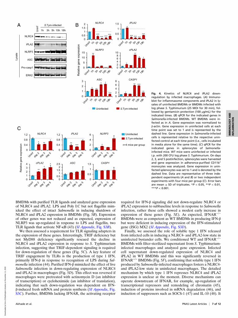
Temporal Down-Regulation of NLRC4 and iPLA2 Expression inSalmonella-Infected Macrophages. Our data (Fig. 1A) and thoseof others (21) suggest that Salmonella eventually switches to aflagellin-low phenotype inside macrophages. We reasoned thatthe feedforward circuit that amplifies flagellin production inresponse to macrophage pyroptosis and lysophospholipids, asdescribed in Figs. 2 and 3, must be arrested for this to occur. Totest this, we analyzed expression of iPLA2 and inflammasomecomponents at varying times post S. Typhimurium infection ofWT BMDMs. The results showed that NLRC4 and iPLA2 pro-tein expression was substantially down-regulated to subbaselinelevels post Salmonella infection (Fig. 4A). The decrease inNLRC4 and iPLA2 expression was also seen at the mRNA levelin BMDMs (Fig. 4B) and in vivo in adherence-purified mono-cytes from spleens of S. Typhimurium-infected mice (Fig. 4C). Incontrast, the expression of NLRP3, ASC, Caspase-1, and cPLA2
Akhade et al. PNAS Latest Articles | 5 of 12
IMMUNOLO
GYAND
INFLAMMATION
Dow
nloa
ded
at U
niv
of W
ashi
ngto
n Li
brar
ies
on N
ovem
ber
11, 2
020

did not decrease below their level of expression in uninfectedcells, indicating that such down-regulation was seeminglyspecific to NLRC4 and iPLA2 (Fig. 4 A–C). These resultsindicate that Salmonella infection leads to decreased NLRC4and iPLA2 expression in macrophages both in vitro andin vivo.Because a significant proportion of Salmonella-infected mac-
rophages undergo rapid NLRC4-dependent cell death (Fig. 2B),we wondered if the observed down-regulation of NLRC4 overtime could simply be due to a preferential selection of preex-isting NLRC4-low cells in the macrophage population which areresistant to cell death, rather than an active infection-inducedrepression of NLRC4. To test this, we analyzed gene expressionin Caspase-1/11−/− and Gasdermin D−/− BMDMs which are re-fractory to Salmonella-induced pyroptosis (41, 42). Time-dependentdown-regulation of NLRC4 and iPLA2 was also observed in in-fected Caspase-1/11−/− and Gasdermin D−/− BMDMs (SI Appendix,Fig. S2 A and B), indicating that repression of NLRC4 and iPLA2expression is independent of macrophage cell death.
A Type 1 IFN-Dependent Host Response Fosters a NLRC4 and iPLA2-Low State in Macrophages. We next sought to investigate themechanism by which NLRC4 and iPLA2 expression is down-regulated in Salmonella-infected macrophages. We first askedif reduction in NLRC4 and iPLA2 levels was due to an activetargeting of these genes by the pathogen. We infected BMDMseither with viable S. Typhimurium or bacteria that had beenrendered nonviable by exposure to heat or gentamicin andassessed expression of NLRC4, iPLA2, cPLA2, and otherinflammasome components (Fig. 5A and SI Appendix, Fig. S3A).Killed bacteria were as efficient as live bacteria in promotingdown-regulation of NLRC4 and iPLA2 (Fig. 5A). None of theother inflammasome components or cPLA2 were down-regulated(SI Appendix, Fig. S3A). Similar results were obtained with Pseu-domonas aeruginosa, another flagellated Gram-negative bacterium(Fig. 5A and SI Appendix, Fig. S3A). These results suggest thatsuppression of NLRC4 and iPLA2 expression in macrophagesdoes not require metabolically active or invasive bacteria and maybe a natural host response to bacterial recognition by cell surface-associated or endosomal TLRs. To test this possibility, we treated
A B C
D E
F G
S.Tym-inf
S.Tym-inf
S.Tym-inf
No
Mɸ
No
Mɸ
Fig. 3. Pyroptosis-derived lysophospholipids am-plify production of flagellin by Salmonella. (A) Im-munoblot for flagellin and LPS released by S. Typhiin response to delipidified supernatant from S.Typhimurium-infected macrophages. Serum-free cellculture supernatant from log phase S. Typhimurium-infected macrophages (50 MOI) was extracted withchloroform/methanol (1:1). The aqueous phase wasincubated with S. Typhi. (B) Analysis of lipids in su-pernatant of S. Typhimurium-infected macrophagesby TLC. Cell-free supernatant from RAW 264.7 cellseither left uninfected or cocultured with log phase S.Typhimurium (50 MOI) was extracted with chloro-form/methanol (1:1) and subjected to TLC. Lipidswere visualized with iodine. LPA, purified LPA (lyso-phosphatidic acid); LPC, purified LPC (lysophosphati-dylcholine). (C) Estimation of LPC released byinfected macrophages upon PLA2 inhibition. Perito-neal macrophages from WT mice were treated withvehicle (dimethylsulfoxide [DMSO]) or with inhibitorsspecific for iPLA2 (FKGK-11, 30 μM) or cPLA2 (pyrro-phenone, 1 μM) for 12 h. Cells were infected with 50MOI log phase S. Typhimurium for 1 h and super-natants were collected. For 100% lysis control, un-infected cells were lysed in equal volume of lysisbuffer. LPC in infected cell supernatants and the100% lysis control was estimated by ELISA. (D) Im-munoblot for flagellin and LPS (Left), and assay forLDH (Right) in filter-sterilized supernatants of mac-rophages infected with S. Typhimurium. Peritonealmacrophages from WT mice were treated with ve-hicle or with inhibitors specific for iPLA2 or cPLA2 asdescribed in C, followed by infection with 50 MOI logphase S. Typhimurium for 1 h. (E) Estimation of LPCin supernatants of peritoneal macrophages of theindicated genotypes infected with 50 MOI log phaseS. Typhimurium for 1 h. WT macrophages were in-fected in presence or absence of the pan-caspaseinhibitor zVADfmk (100 μM) and caspase-1-specificinhibitor zYVADfmk (100 μM). LPC in infected cellsupernatants and the 100% lysis control was quan-tified by ELISA. (F) Immunoblot for flagellin and LPS(Left) and LDH assay for macrophage cell death(Right) in filter-sterilized supernatants of WT,Caspase-11−/−, and Caspase-1/11−/− peritoneal mac-rophages infected with 50 MOI log phase S. Typhi-murium for 1 h. (G) Estimation of LPC in supernatantsof WT, Caspase-11−/−, and Caspase-1/11−/− peritoneal macrophages infected with S. Typhimurium as in F. LPC in the supernatants and the 100% lysis controlwas estimated by ELISA. Data are representative of two independent experiments. Error bars are mean ± SD of triplicates. ND, not detected. *P < 0.05, **P <0.01, ***P < 0.001. ns, not significant.
6 of 12 | www.pnas.org/cgi/doi/10.1073/pnas.2002747117 Akhade et al.
Dow
nloa
ded
at U
niv
of W
ashi
ngto
n Li
brar
ies
on N
ovem
ber
11, 2
020
BMDMs with purified TLR ligands and analyzed gene expressionof NLRC4 and iPLA2. LPS and Poly I:C but not flagellin mim-icked the effect of intact Salmonella in inducing shutdown ofNLRC4 and iPLA2 expression in BMDMs (Fig. 5B). Expressionof other genes was not reduced and as expected, expression ofNLRP3 was up-regulated in response to LPS and flagellin, twoTLR ligands that activate NF-κB (43) (SI Appendix, Fig. S3B).We then assessed a requirement for TLR signaling adapters in
the expression of these genes. Interestingly, TRIF deficiency butnot MyD88 deficiency significantly rescued the decline inNLRC4 and iPLA2 expression in response to S. Typhimuriuminfection, suggesting that TRIF-dependent signaling is requiredfor down-regulation of these genes (Fig. 5C). A key feature ofTRIF engagement by TLRs is the production of type 1 IFN,primarily IFN-β in response to recognition of LPS during Sal-monella infection (44). Purified IFN-βmimicked the effect of liveSalmonella infection in down-regulating expression of NLRC4and iPLA2 in macrophages (Fig. 5D). This effect was reversed ifmacrophages were pretreated with actinomycin D (an inhibitorof transcription) or cycloheximide (an inhibitor of translation),indicating that such down-regulation was dependent on IFN-β-induced fresh mRNA and protein synthesis (SI Appendix, Fig.S3C). Further, BMDMs lacking IFNAR, the activating receptor
required for IFN-β signaling did not down-regulate NLRC4 oriPLA2 expression to subbaseline levels in response to Salmonellainfection, rather these cells showed a modest early increase inexpression of these genes (Fig. 5E). As expected, IFNAR−/−
BMDMs were as competent as WT BMDMs in producing IFN-βbut were deficient in inducing expression of the IFN-stimulatedgene (ISG) MX2 (SI Appendix, Fig. S3D).Finally, we assessed the role of soluble type 1 IFN released
from infected cells in inducing a NLRC4- and iPLA2-low state inuninfected bystander cells. We conditioned WT and IFNAR−/−
BMDMs with filter-sterilized supernatant from S. Typhimurium-infected macrophages and analyzed gene expression. Infectedcell supernatant down-regulated expression of NLRC4 andiPLA2 in WT BMDMs and this was significantly reversed inIFNAR−/− BMDMs (Fig. 5F), confirming that soluble type 1 IFNproduced by Salmonella-infected macrophages fosters a NLRC4-and iPLA2-low state in uninfected macrophages. The detailedmechanism by which type 1 IFN represses NLRC4 and iPLA2expression is unclear at the moment. Diverse mechanisms canoperate downstream of IFNAR, for example, up-regulation oftranscriptional repressors and remodeling of chromatin (45),induction of proteins involved in mRNA degradation (46), andinduction of suppressors such as SOCS-1 (47) and IL-10 (48). It
A B
C
Fig. 4. Kinetics of NLRC4 and iPLA2 down-regulation by infected macrophages. (A) Immuno-blot for inflammasome components and iPLA2 in ly-sates of uninfected BMDMs or BMDMs infected withlog phase S. Typhimurium (25 MOI for 30 min), fol-lowed by gentamicin protection (100 μg/mL) for theindicated times. (B) qPCR for the indicated genes inSalmonella-infected BMDMs. WT BMDMs were in-fected as in A. Gene expression was normalized toβ-actin. Gene expression in uninfected cells at eachtime point was set to 1 and is represented by thedashed line. Gene expression in Salmonella-infectedcells is represented relative to the respective unin-fected control at each time point (i.e., cells incubatedin media alone for the same time). (C) qPCR for theindicated genes in splenocytes of Salmonella-infected mice. WT mice were uninfected or infectedi.p. with 200 CFU log phase S. Typhimurium. On days2, 3, and 5 postinfection, splenocytes were harvestedand gene expression in adherence-purified CD11b+
monocytes was analyzed. Gene expression in unin-fected splenocytes was set to 1 and is denoted by thedashed line. Data are representative of three inde-pendent experiments (A and B) or two independentexperiments with four mice per group (C). Error barsare mean ± SD of triplicates. *P < 0.05, **P < 0.01,***P < 0.001.
Akhade et al. PNAS Latest Articles | 7 of 12
IMMUNOLO
GYAND
INFLAMMATION
Dow
nloa
ded
at U
niv
of W
ashi
ngto
n Li
brar
ies
on N
ovem
ber
11, 2
020

is possible that through one or more of these or yet unidentifiedmechanisms, factors induced by IFN-β down-regulate expressionof NLRC4 and iPLA2.Our findings indicate that type 1 IFN represses expression of
NLRC4 and iPLA2. Therefore, we assessed the effect of IFNARsignaling on caspase-1 activation and LPC which lie directlydownstream of NLRC4 and iPLA2, respectively. Consistent withreduced NLRC4 expression, IFN-β-treated cells showed reducedcell death as measured by LDH release (SI Appendix, Fig. S4A)and reduced caspase-1 activation as evidenced by reducedamounts of cleaved caspase-1 (p20) relative to that of procaspase-1 (p46) in response to S. Typhimurium infection (SI Appendix, Fig.S4B). Measurement of intracellular LPC showed that consistentwith the kinetics of flagellin down-regulation, intracellular LPClevels decline over time in S. Typhimurium-infected WT BMDMswith a significant decrease by 48 h (Fig. 5G). In contrast,IFNAR−/− BMDMs do not show a decline in intracellular LPCover time (Fig. 5G). Interestingly and unexpectedly, Caspase-1/11−/− BMDMs showed a faster decline in intracellular LPC levelscompared to WT BMDMs with a significant decrease in LPC inCaspase-1/11−/− cells by 24 h postinfection and a further reductionat 48 h, suggesting that caspase-1 activity is upstream of LPC andmaintains intracellular LPC expression. Together these data
suggest that IFNAR signaling leads to a decrease in intracellularlevels of LPC, likely due to repression of either a NLRC4/Cas-pase-1/LPC axis and/or direct repression of iPLA2 expressiondownstream of IFNAR (SI Appendix, Fig. S9C).
NLRC4 and iPLA2 Regulate Expression of Flagellin by IntracellularSalmonella. We next examined if inhibition of a NLRC4/Cas-pase-1 and/or iPLA2/LPC axis downstream of IFNAR contrib-utes to down-regulation of flagellin by intracellular Salmonella.The reversal of flagellin down-regulation in IFNAR−/− BMDMswas abrogated and flagellin was down-regulated to an extentsimilar to that seen in WT BMDMs if either caspase-1 or iPLA2activity was inhibited in IFNAR−/− BMDMs (Fig. 6 A and B).These results indicate that the effect of IFNAR signaling onSalmonella flagellin expression is mediated through repression ofcaspase-1 and iPLA2 activity and these factors are the execu-tioners of flagellin down-regulation downstream of IFNAR.Consistent with a role for NLRC4 and caspase-1, intracellularSalmonella recovered from NLRC4−/− and Caspase-1/11−/−
BMDMs showed nearly complete abrogation of flagellin ex-pression both at the mRNA and the protein level at 24 h post-infection compared to those obtained from WT and ASC−/−
BMDMs (Fig. 6C). NLRP3 activation is dependent on the
A B C
D E F
G
Fig. 5. Type 1 IFN signaling fosters a NLRC4-, iPLA2-,and LPC-low state in macrophages. (A) qPCR for NLRC4and iPLA2 showing down-regulation of these genes ininfected BMDMs over time. WT BMDMs were infectedwith 25 MOI of live, heat-killed (90 °C, 30 min) orgentamicin-killed (100 μg/mL, 30 min) S. Typhimuriumor P. aeruginosa for 30 min. Infection was synchronizedat 1,500 rpm for 5 min. Extracellular bacteria were re-moved followed by gentamicin protection for the indi-cated times. Gene expression was normalized to β-actinand the uninfected control at that time point (dashedline, set to 1) as described in Fig. 4B. (B) qPCR for NLRC4and iPLA2 in BMDMs infected with log phase S. Typhi-murium or treated with TLR ligands. WT BMDMs wereinfected as in A or treated with TLR agonists LPS (1 μg/mL), flagellin (1 μg/mL), or poly I:C (5 μg/mL) for theindicated times. (C) qPCR for NLRC4 and iPLA2 in in-fected BMDMs of the indicated genotypes showing re-versal of NLRC4 and iPLA2 down-regulation in TRIF−/−
BMDMs. BMDMs were infected with 25 MOI log phaseS. Typhimurium for 30 min followed by gentamicinprotection as in A. (D) qPCR for NLRC4 and iPLA2 inBMDMs infected with log phase S. Typhimurium ortreated with purified recombinant IFN-β (1,000 U/mL)for the indicated times. (E) qPCR showing that NLRC4and iPLA2 gene expression is not down-regulated belowbaseline (dashed line set to 1) in IFNAR−/− BMDMs. WTand IFNAR−/− BMDMs were infected with log phase S.Typhimurium as in A. (F) qPCR for NLRC4 and iPLA2 inBMDMs exposed to supernatant (i.e., secreted factors)derived from Salmonella-infected BMDMs. WT andIFNAR−/− BMDMs were treated with filter-sterilizedsupernatants from log phase S. Typhimurium-infectedWT BMDMs for the indicated times and expression ofNLRC4 and iPLA2 was analyzed. (G) Estimation of in-tracellular LPC in BMDMs of the indicated genotypesinfected with 20 MOI log phase S. Typhimurium for30 min followed by gentamicin protection for the indi-cated times. LPC concentration in cell lysates was mea-sured by ELISA. The level of LPC in uninfected BMDMsof each genotypewas set at 100% (dashed line) and LPClevels postinfection are represented as % change in LPCcompared to uninfected BMDMs of the respective ge-notype. Hpi, hours postinfection. Data are representa-tive of three (A–F) or two independent experiments (G).Error bars are mean ± SD of triplicates. *P < 0.05, **P <0.01, ***P < 0.001. ns, not significant.
8 of 12 | www.pnas.org/cgi/doi/10.1073/pnas.2002747117 Akhade et al.
Dow
nloa
ded
at U
niv
of W
ashi
ngto
n Li
brar
ies
on N
ovem
ber
11, 2
020

adapter protein ASC; therefore, these results suggest thatNLRP3 does not play a role in control of flagellin expression,and caspase-1 activation by NLRC4 independent of ASC main-tains flagellin expression by intracellular Salmonella. A similardependence on NLRC4 and caspase-1 was observed in situ usinga S. Typhimurium 14028 fliC-GFP reporter strain (SI Appendix,Fig. S5 A–D). In the absence of caspase-1 and caspase-11, Sal-monella showed a faster time-dependent down-regulation of in-tracellular flagellin expression both in primary BMDMs andimmortalized BMDMs (iBMDMs) (Fig. 6C and SI Appendix, Fig.S6A). Whereas the kinetics of flagellin down-regulation by Sal-monella in WT iBMDMs was delayed (SI Appendix, Fig. S6A;note that flagellin in WT iBMDMs is not down-regulated even at48 h) compared to primary BMDMs (Fig. 1A), clear down-regulation at 24 and 48 h was observed in Caspase-1/11−/−
iBMDMs compared to WT iBMDMs (SI Appendix, Fig. S6A).Consistent with a requirement for caspase-1 in sustaining fla-gellin expression by intracellular Salmonella, SipB-deficient orPrgJ-deficient bacteria, which are unable to activate caspase-1efficiently (SI Appendix, Fig. S1A) (32, 33), showed a decrease inflagellin expression at 24 h and 48 h compared to their respective
parent WT S. Typhimurium strain (SI Appendix, Fig. S6B). TheseNLRC4/Caspase-1-dependent effects on flagellin down-regulationare independent of intracellular phagosomal pH because there areno differences in acidification of phagosomes between WT andCaspase-1/11−/− macrophages during Gram-negative bacterial in-fection (49). These effects were also independent of macrophagepyroptosis because intracellular Salmonella recovered from Gas-dermin D−/− BMDMs which activate caspase-1 normally but areprotected from pyroptotic membrane rupture (41, 42), showedflagellin down-regulation kinetics identical to bacteria residing inWT BMDMs (SI Appendix, Fig. S6C). Further, growth phase ofinfecting Salmonella did not affect the kinetics of flagellin down-regulation in BMDMs, even though stationary phase bacteria, asexpected, induced lower cell death (SI Appendix, Fig. S7 A–D).Finally, flagellin down-regulation was not dependent on NLRP3or Caspase-11 with identical kinetics of down-regulation beingobserved in WT, NLRP3−/−, and Caspase-11−/− BMDMs (SIAppendix, Fig. S7 D and E). Together these results indicate thata NLRC4/Caspase-1 axis sustains expression of flagellin byintracellular Salmonella.
A C
B
D E
F G
Fig. 6. NLRC4 inflammasome and iPLA2 activitycontrols expression of flagellin by intracellular Sal-monella. (A and B) Immunoblot for flagellin andDnaK expressed by intracellular Salmonella obtainedfrom BMDMs treated with caspase-1-specific (A) oriPLA2-specific (B) inhibitors. WT or IFNAR−/− BMDMswere treated with either vehicle (DMSO), the cas-pase-1-specific inhibitor zYVADfmk (100 μM) (A), orthe iPLA2-specific inhibitor FKGK-11 (50 μM) (B) for30 min prior to infection with 25 MOI log phase S.Typhimurium for 30 min. Intracellular Salmonellawere harvested at 6, 24, and 48 h after infection. (C)Immunoblot (Top) and qPCR (Bottom) for flagellinexpressed by intracellular Salmonella obtained fromBMDMs of the indicated genotypes. BMDMs wereinfected with 25 MOI log phase S. Typhimurium for30 min followed by gentamicin protection. Intracel-lular Salmonella were recovered at 6 and 24 h post-infection. (D) Immunoblot (Left) and qPCR (Right) forflagellin expressed by intracellular Salmonellaobtained from BMDMs treated with PLA2 inhibitors.WT BMDMs were treated with either vehicle (DMSO)or inhibitors selective for cPLA2 (pyrrophenone,1 μM) and iPLA2 (FKGK-11, 50 μM) for 30 min prior toinfection with 25 MOI log phase S. Typhimurium for30 min. Intracellular Salmonella were harvested at 6and 24 h after infection. fliC gene expression wasnormalized to dnaK. (E and F) Immunoblot (Top) andcorresponding densitometric quantification (Bottom)for flagellin expressed by intracellular Salmonellaobtained from spleens of mice of the indicatedgenotypes. WT and NLRC4−/− mice (E) or WT andcaspase-1/11−/− mice (F) were infected i.p. with 200CFU log phase S. Typhimurium. On days 2 and 5postinfection, flagellin and DnaK expression by in-tracellular Salmonella harvested from adherence-purified monocytes was analyzed by immunoblot-ting. Band intensities of flagellin were normalized toDnaK using ImageJ. (G) Immunoblot (Top) and cor-responding densitometric quantification (Bottom)for flagellin expressed by intracellular Salmonellaobtained from spleens of mice treated with iPLA2-specific inhibitor (FKGK-18; 20 mg/kg body weight)or vehicle (DMSO) and infected i.p. with 200 CFU logphase S. Typhimurium. On days 2 and 5 postinfec-tion, intracellular Salmonella were recovered fromadherent splenocytes and flagellin and DnaK expression were analyzed. Band intensities of flagellin were normalized to DnaK using ImageJ. Data arerepresentative of two (A and B) or three (C–G) independent experiments. Error bars are mean ± SD of triplicates. *P < 0.05, **P < 0.01, ***P < 0.001. ns, notsignificant.
Akhade et al. PNAS Latest Articles | 9 of 12
IMMUNOLO
GYAND
INFLAMMATION
Dow
nloa
ded
at U
niv
of W
ashi
ngto
n Li
brar
ies
on N
ovem
ber
11, 2
020

We then further examined the relationship between lyso-phospholipids and expression of flagellin by intracellular Sal-monella. Primary BMDMs were infected with Salmonella in thepresence or absence of inhibitors selective for cPLA2 or iPLA2to inhibit the biogenesis of lysophospholipids generated by theseenzymes. Treatment with the iPLA2-selective inhibitor FKGK-11, which specifically inhibits iPLA2 without affecting caspase-1activation (50) or macrophage cell death (Fig. 3 D, Right), ab-rogated expression of flagellin by intracellular bacteria both atthe mRNA and the protein levels at 24 h (Fig. 6D). Treatmentwith the cPLA2-selective inhibitor pyrrophenone (51) had noeffect (Fig. 6D). Similar results were obtained in iBMDMs (SIAppendix, Fig. S6D), suggesting that iPLA2-generated lyso-phospholipids support expression of flagellin by intracellularSalmonella. The intracellular levels of LPC also correlated wellwith flagellin expression by Salmonella isolated from WT mac-rophages and macrophages lacking different inflammasomecomponents. While the levels of LPC decreased only at 48 hpostinfection in WT, NLRP3−/−, and Caspase-11−/− macro-phages, NLRC4−/− and Caspase-1/11−/− macrophages showed afaster decline in intracellular LPC with a significant reduction by24 h postinfection (SI Appendix, Fig. S8 A and B). Consistentwith the faster reduction in LPC, intracellular Salmonella down-regulated flagellin faster in NLRC4−/− and Caspase-1/11−/−
macrophages as compared to WT, NLRP3−/−, and Caspase-11−/−
macrophages (SI Appendix, Fig. S7 D and E).Salmonella populations can be stochastic and heterogeneous
in nature (52), so we next asked if flagellin down-regulation byintracellular Salmonella in WT macrophages over time (Fig. 1A)could simply be due to a preferential selection of cells infectedwith flagellin-low or perhaps PrgJ-low Salmonella in the invadingpopulation of bacteria as these cells are likely to not die bypyroptosis (see schematic in SI Appendix, Fig. S6E), rather thanan adaptation by Salmonella to an infection-induced decrease inhost NLRC4 and iPLA2. Our collective data argued against thispossibility. Firstly, inhibition of iPLA2 in WT BMDMs did notaffect Salmonella-induced cell death (Fig. 3 D, Right), but still ledto faster down-regulation of flagellin expression by intracellularSalmonella (Fig. 6D), suggesting that flagellin expression is de-pendent on iPLA2-generated lipids and not on pyroptosis. Sec-ondly, as shown in SI Appendix, Fig. S6C, intracellular bacteriafrom Gasdermin D−/− BMDMs which are protected frompyroptotic membrane rupture show flagellin down-regulationkinetics identical to bacteria from WT BMDMs. Thirdly, sta-tionary phase bacteria induced lower cell death but showed fla-gellin down-regulation kinetics identical to that of log phasebacteria (SI Appendix, Fig. S7 A–D). Fourthly, PrgJ-deficientSalmonella down-regulated flagellin intracellularly with kineticsidentical to SipB-deficient bacteria when compared to their re-spective parental congenic WT counterparts, strongly suggestingthat lack of caspase-1 activation due to PrgJ deficiency leads toflagellin down-regulation and is independent of PrgJ expressionper se (SI Appendix, Fig. S6B). Finally, if selection bias was atplay, then we would expect NLRC4−/− and Caspase-1/11−/−
macrophages to retain more flagellin-high bacteria intracellularlyover time because of the resistance of these genotypes to pyroptosisand consequently show a slower down-regulation of flagellin com-pared toWTmacrophages (see schematic in SI Appendix, Fig. S6E),an outcome opposite to the observed outcome (Fig. 6C and SIAppendix, Fig. S5). Of note, although NLRC4 and iPLA2 expres-sion in WT BMDMs was down-regulated early on (i.e., maximallyby 10 h postinfection at the mRNA level; Fig. 4 A and B), flagellindown-regulation peaked at 48 h postinfection (Fig. 1A). Theabundance of cellular lysophospholipids and the time it takes todeplete the preexisting intracellular lipid pool in macrophages(Fig. 5G and SI Appendix, Fig. S8 A and B) may contribute to thedelayed kinetics of flagellin down-regulation.
Finally, we examined the role of the NLRC4/Caspase-1 andiPLA2 axes in regulating flagellin expression by intracellular bac-teria during in vivo infection. WT, NLRC4−/−, Caspase-1/11−/−
mice or mice treated with the iPLA2-specific in vivo inhibitor,FKGK-18 (53), were infected i.p. with S. Typhimurium and killedat days 2 and 5 postinfection. Bacteria were recovered from thespleen and flagellin expression was analyzed. Intracellular bacteriaobtained from NLRC4−/− and Caspase-1/11−/− spleens showedmarkedly greater down-regulation of flagellin compared to thoserecovered from WT spleens (Fig. 6 E and F). Reduced flagellinexpression on day 5 in NLRC4−/− and Caspase-1/11−/− mice wasassociated with increased bacterial burden in the spleens of thesemice (SI Appendix, Fig. S8C). Similarly, intracellular bacteria re-covered from spleens of iPLA2 inhibitor-treated mice showedsignificantly lower expression of flagellin at day 5 compared tothose treated with vehicle (Fig. 6G). These results indicate thatNLRC4/Caspase-1 and iPLA2 activity sustains expression of fla-gellin by intracellular Salmonella both in vitro and in vivo.
DiscussionCoevolution of pathogens and the host immune system hasresulted in complex evolutionary adaptations that contribute tohost defense and an array of immune evasion mechanisms in themicrobes. Pyroptosis, a caspase-1-mediated form of inflamma-tory cell death induced upon sensing of flagellin by NLRC4, isessential for clearance of pathogenic Salmonella from the hostand is circumvented by the pathogen by down-regulating theexpression of flagellin at later stages of infection. Although thisset of linked events is well described, the molecular mechanismsat play in prompting the pathogen to switch to a flagellin-lowphenotype in vivo and establish an intracellular survival nichewithin macrophages are incompletely understood. It is believedbased on in vitro studies of bacteria exposed to various pertur-bations in growth medium, that conditions associated with theSCV such as acidic pH and low Mg2+ suppress expression offlagellin (20, 21); however, the contribution of these factors incontrolling flagellin expression by intracellular Salmonella insidemacrophages is controversial (20, 24–26). Here, we provide ev-idence that a natural type 1 IFN-dependent host negative feed-back response to bacterial infection enables down-regulation offlagellin by Salmonella inside macrophages (SI Appendix, Fig.S9C). Interestingly, during early infection of macrophages withSalmonella, NLRC4 inflammasome-dependent pyroptosis isnecessary and sufficient for generating a host lysophospholipidstimulus that increases expression of biologically active mono-meric flagellin by extracellular bacteria in a feedforward manner(SI Appendix, Fig. S9B). The increased flagellin can confer anearly advantage to the pathogen by promoting TLR-5-dependentsystemic dissemination of Salmonella (54); however, our datashow that the feedforward circuit generates bacteria which aremore potent at inducing pyroptosis of healthy macrophages(Fig. 2F), a modulation that is known to result in neutrophil-mediated killing and clearance of Salmonella from the host(17) (SI Appendix, Fig. S9B). This positive feedback loop is,however, later kept in check by type 1 IFN-dependent shutdownof NLRC4 and iPLA2 expression in macrophages, creating anintracellular environment low in caspase-1 activity and lyso-phospholipids that Salmonella adapts to for flagellin down-regulation inside macrophages (SI Appendix, Fig. S9C). Over-all, our findings suggest that Salmonella has evolved to cooptNLRC4 activation to initially enhance production of extracellu-lar flagellin that activates TLR-5, which along with other TLRspromotes systemic spread of the pathogen (54, 55) at the risk ofNLRC4-mediated clearance, and later on benefit from the IFN-induced decrease in NLRC4 and lysophospholipid production todown-regulate flagellin intracellularly within macrophages.These data also identify a role for NLRC4 inflammasome ac-tivity, which is classically implicated in the detection and
10 of 12 | www.pnas.org/cgi/doi/10.1073/pnas.2002747117 Akhade et al.
Dow
nloa
ded
at U
niv
of W
ashi
ngto
n Li
brar
ies
on N
ovem
ber
11, 2
020

clearance of a variety of bacterial pathogens, as a regulator oftemporal and biphasic expression of its own bacterial ligand.One consequence of the IFN-induced decrease in NLRC4 for the
host is that it limits infection-induced pyroptosis (SI Appendix, Fig.S4). Down-regulation of NLRC4 expression may therefore be apreemptive host measure to restrain cell death and preserve amacrophage pool for subsequent priming of an adaptive immuneresponse (56). It may also, along with direct down-regulation ofiPLA2, be a host strategy to limit generation of LPC and preventovert inflammation. This is because LPC released by dying cells actsas a chemoattractant for macrophages, which is believed to bebeneficial for effective removal of cell corpses (57); however, un-checked inflammatory cell recruitment can exacerbate inflamma-tion and cause tissue-damaging effects. LPC can also amplify TLR-activated inflammatory responses (58), an excess of which can bedetrimental to the host. A recent study demonstrated reducedNLRC4 expression in LPS-treated human monocytes (59) and in athree-dimensional organotypic model of human intestinal mucosastimulated with S. Typhi (60), suggesting that the IFN-dependentNLRC4lo state described here might also be relevant in human cells.Although our data in macrophages show that IFNAR signalingpotently down-regulates NLRC4 and iPLA2 to subbaseline levels, itis important to note that in vivo, in addition to type 1 IFN, otherinterferons derived from various cellular sources could also possiblycontribute to establishment of a NLRC4-, iPLA2-, and LPC-lowenvironment. Future studies are required to identify the molecu-lar details of type 1 IFN-induced repression of NLRC4 and iPLA2and possible involvement of other interferons in this process.For the pathogen, an important outcome of a NLRC4- and
iPLA2-low intracellular environment depleted of lysophospho-lipids is its switching to a flagellin-low phenotype. Given thatflagellin is a potent mediator of innate and inflammatory re-sponses (5), a target of antibodies and T cells during infection(61, 62), and can even affect the suppressive function of T reg-ulatory cells (63), down-regulation of flagellin could allow thepathogen to escape immune control. Consistent with this notion,it had been shown that Salmonella designed to persistently ex-press flagellin is cleared in a NLRC4/Caspase-1-dependentmanner and is hence attenuated in vivo (17). The data providedhere show that flagellin down-regulation by intracellular Sal-monella is accelerated in NLRC4−/− as well as Caspase-1/11−/−
mice (Fig. 6 E and F) and is reversed in IFNAR−/− mice(Fig. 1E). It is notable that flagellin expression by intracellularSalmonella during the systemic phase of infection in these micecorrelates inversely with the susceptibility of these mice to Sal-monella infection. NLRC4−/− as well as Caspase-1/11−/− mice aremore susceptible and are reported to display higher bacterialburdens than WT mice (64, 65) while IFNAR−/− mice are lesssusceptible and display lower bacterial burdens than WT mice(29). In addition, reduced serum levels of lysophospholipids havebeen reported in patients with gastroenteritis caused by Salmonella(66), implying that establishment of successful infection by Salmo-nellamight be strongly linked to its ability to down-regulate flagellinexpression under conditions of lowered iPLA2 expression.Given that macrophages undergo rapid caspase-1-dependent
pyroptosis in response to Salmonella infection, an interestingissue relates to the nature of cells that survive the initial on-slaught of infection by extracellular bacteria and subsequentlyprovide a niche for intracellular residence and flagellin down-regulation by Salmonella. Although caspase-1 activation is be-lieved to be an all-or-none digital response at the single cell levelusing current methods (67), all prevailing methods for measuringcaspase-1 activity are insensitive to weak or local activations ofcaspase-1. Therefore, it is possible that infected cells with a weakor subthreshold level of caspase-1 activation that is insufficient tocommit macrophages to pyroptosis survive the initial onslaught ofinfection. Moreover, our data suggest that type 1 IFN produced by
macrophages early during infection likely fosters a NLRC4loi-PLA2lo state in the surviving cells which promotes flagellin down-regulation by the pathogen (Figs. 1B and 5F). Given that levels ofintracellular flagellin correlate inversely with systemic bacterialloads, it is possible that these NLRC4- and iPLA2-low cells mightserve as an ideal reservoir for stealthily replicating Salmonella andultimately potentiate dissemination of the pathogen to secondarylymphoid organs. Recent single-cell RNA-Seq studies suggest thatvariable gene expression in infected host cells shapes differentfunctional cellular states, some of which favor the establishment ofa long-term intracellular niche, and others that allow Salmonellato escape intracellular immune activity and proliferate (68). Al-though current single-cell RNA-Seq methods are restricted toprofiling eukaryotic transcripts, future technical improvements ofthe dual RNA-Seq technique which profiles gene expression inhost and bacteria simultaneously (69) may permit the correlationof flagellin and other bacterial virulence factors with host cellheterogeneity and reprogramming.Another interesting aspect of the intracellular flagellin regulatory
circuit we describe here is that caspase-1 is upstream of LPC and cancontrol the levels of intracellular LPC (Fig. 5G). Because caspase-1activation is traditionally believed to induce pyroptotic cell death ofmacrophages, an outstanding question concerns the mechanism bywhich caspase-1 regulates the levels of intracellular LPC indepen-dent of pyroptosis. In this regard, it is noteworthy that a branch oflipid synthesis during innate immune activation relies on caspase-1whereby caspase-1 induces the activation of sterol regulatory ele-ment binding proteins (SREBPs) which in turn promote expressionof genes involved in lipid synthesis (70). SREBP-1 activation leads toproduction of phospholipids (71) which are substrates for productionof lysophospholipids. Alternatively, SREBP-2 can regulate the ex-pression of iPLA2. The 5′ flanking region of the iPLA2 gene isreported to contain a sterol regulatory element (SRE) that can bindSREBP-2, and mutant cells that constitutively generate matureSREBP proteins exhibit increased iPLA2 expression and activity(72). Through these or a yet unidentified mechanism(s), caspase-1activity could control intracellular lysophospholipid levels.Taken together, our data identify a type 1 IFN-dependent host
mechanism that fosters a NLRC4- and iPLA2-low cellular statewhich is coopted by Salmonella to switch to a flagellin-low phe-notype inside macrophages. Our findings also identify a mode ofinnate immune regulation whereby an innate sensing mecha-nism, that is, NLRC4 inflammasome activity, regulates temporalexpression of its own microbial ligand, and add to our under-standing of the complex evolutionary adaptations that shapehost–pathogen cross-talk.
Materials and MethodsDetailed information on experimental models (bacterial strains, cell lines,mice), reagents, assays, and statistical analysis is presented in SI Appendix, SIMaterials and Methods. S. Typhimurium SL1344 grown to log phase in LBwere used in all experiments unless mentioned otherwise. All animal ex-periments were approved by the Institute for Systems Biology’s InstitutionalAnimal Care and Use Committee.
Data Availability. All study data are included in the article and SI Appendix.
ACKNOWLEDGMENTS. This work was supported by the Institute for SystemsBiology, and in part by the National Institute of Immunology (funded by theDepartment of Biotechnology, Government of India) and funds from the Stevenand Alexandra Cohen Foundation to N.S. We thank Prof. Kazuhiro Kutsukakefor S. Typhimurium strain KK1004 and its flhD::Tn10 derivative; Prof. Emma-nuelle Charpentier for S. Typhimurium SL1344 and its SipB-negative derivative;Dr. Brad Cookson for S. Typhimurium 14028 expressing fliC-GFP; Dr. Eicke Latzfor WT and Caspase-1/11-deficient bone marrow-derived macrophage cell lines;Dr. Russell Vance for bones from Gasdermin D−/− mice; Dr. Vishva Dixit forNLRC4−/− and Caspase 1/11−/− mice; and Drs. Teresa Thurston and David Holdenfor critical reading of the manuscript.
Akhade et al. PNAS Latest Articles | 11 of 12
IMMUNOLO
GYAND
INFLAMMATION
Dow
nloa
ded
at U
niv
of W
ashi
ngto
n Li
brar
ies
on N
ovem
ber
11, 2
020
1. S. W. Brubaker, K. S. Bonham, I. Zanoni, J. C. Kagan, Innate immune pattern recog-nition: A cell biological perspective. Annu. Rev. Immunol. 33, 257–290 (2015).
2. N. Arpaia et al., TLR signaling is required for Salmonella typhimurium virulence. Cell144, 675–688 (2011).
3. K. D. Smith et al., Toll-like receptor 5 recognizes a conserved site on flagellin required forprotofilament formation and bacterial motility. Nat. Immunol. 4, 1247–1253 (2003).
4. E. A. Miao et al., Cytoplasmic flagellin activates caspase-1 and secretion of interleukin1beta via Ipaf. Nat. Immunol. 7, 569–575 (2006).
5. H. Zeng et al., Flagellin is the major proinflammatory determinant of enteropatho-genic Salmonella. J. Immunol. 171, 3668–3674 (2003).
6. E. M. Kofoed, R. E. Vance, Innate immune recognition of bacterial ligands by NAIPsdetermines inflammasome specificity. Nature 477, 592–595 (2011).
7. Y. Zhao et al., The NLRC4 inflammasome receptors for bacterial flagellin and type IIIsecretion apparatus. Nature 477, 596–600 (2011).
8. M. Rayamajhi, D. E. Zak, J. Chavarria-Smith, R. E. Vance, E. A. Miao, Cutting edge:Mouse NAIP1 detects the type III secretion system needle protein. J. Immunol. 191,3986–3989 (2013).
9. J. Yang, Y. Zhao, J. Shi, F. Shao, Human NAIP and mouse NAIP1 recognize bacterialtype III secretion needle protein for inflammasome activation. Proc. Natl. Acad. Sci.U.S.A. 110, 14408–14413 (2013).
10. V. M. Reyes Ruiz et al., Broad detection of bacterial type III secretion system andflagellin proteins by the human NAIP/NLRC4 inflammasome. Proc. Natl. Acad. Sci.U.S.A. 114, 13242–13247 (2017).
11. Z. Hu et al., Structural and biochemical basis for induced self-propagation of NLRC4.Science 350, 399–404 (2015).
12. L. Zhang et al., Cryo-EM structure of the activated NAIP2-NLRC4 inflammasome re-veals nucleated polymerization. Science 350, 404–409 (2015).
13. P. Broz et al., Redundant roles for inflammasome receptors NLRP3 and NLRC4 in hostdefense against Salmonella. J. Exp. Med. 207, 1745–1755 (2010).
14. M. A. Wynosky-Dolfi et al., Oxidative metabolism enables Salmonella evasion of theNLRP3 inflammasome. J. Exp. Med. 211, 653–668 (2014).
15. L. Franchi et al., Cytosolic flagellin requires Ipaf for activation of caspase-1 and inter-leukin 1beta in salmonella-infected macrophages. Nat. Immunol. 7, 576–582 (2006).
16. L. A. Knodler et al., Noncanonical inflammasome activation of caspase-4/caspase-11mediates epithelial defenses against enteric bacterial pathogens. Cell Host Microbe16, 249–256 (2014).
17. E. A. Miao et al., Caspase-1-induced pyroptosis is an innate immune effector mech-anism against intracellular bacteria. Nat. Immunol. 11, 1136–1142 (2010).
18. L. A. Cummings, S. L. Barrett, W. D. Wilkerson, I. Fellnerova, B. T. Cookson, FliC-specificCD4+ T cell responses are restricted by bacterial regulation of antigen expression.J. Immunol. 174, 7929–7938 (2005).
19. J. J. Bijlsma, E. A. Groisman, The PhoP/PhoQ system controls the intramacrophagetype three secretion system of Salmonella enterica. Mol. Microbiol. 57, 85–96 (2005).
20. P. Adams et al., Proteomic detection of PhoPQ- and acid-mediated repression ofSalmonella motility. Proteomics 1, 597–607 (2001).
21. L. A. Cummings, W. D. Wilkerson, T. Bergsbaken, B. T. Cookson, In vivo, fliC expressionby Salmonella enterica serovar Typhimurium is heterogeneous, regulated by ClpX,and anatomically restricted. Mol. Microbiol. 61, 795–809 (2006).
22. E. García Véscovi, F. C. Soncini, E. A. Groisman, Mg2+ as an extracellular signal: En-vironmental regulation of Salmonella virulence. Cell 84, 165–174 (1996).
23. F. C. Soncini, E. García Véscovi, F. Solomon, E. A. Groisman, Molecular basis of themagnesium deprivation response in Salmonella typhimurium: Identification of PhoP-regulated genes. J. Bacteriol. 178, 5092–5099 (1996).
24. D. Drecktrah, L. A. Knodler, R. Ireland, O. Steele-Mortimer, The mechanism of Sal-monella entry determines the vacuolar environment and intracellular gene expres-sion. Traffic 7, 39–51 (2006).
25. M. Rathman, M. D. Sjaastad, S. Falkow, Acidification of phagosomes containing Sal-monella typhimurium in murine macrophages. Infect. Immun. 64, 2765–2773 (1996).
26. N. Martin-Orozco et al., Visualization of vacuolar acidification-induced transcriptionof genes of pathogens inside macrophages. Mol. Biol. Cell 17, 498–510 (2006).
27. G. Sano et al., Flagella facilitate escape of Salmonella from oncotic macrophages.J. Bacteriol. 189, 8224–8232 (2007).
28. G. Guarda et al., Type I interferon inhibits interleukin-1 production and in-flammasome activation. Immunity 34, 213–223 (2011).
29. N. Robinson et al., Type I interferon induces necroptosis inmacrophages during infectionwith Salmonella enterica serovar Typhimurium. Nat. Immunol. 13, 954–962 (2012).
30. J. E. Karlinsey, J. Lonner, K. L. Brown, K. T. Hughes, Translation/secretion coupling bytype III secretion systems. Cell 102, 487–497 (2000).
31. J. L. Tenthorey et al., NLRC4 inflammasome activation is NLRP3- and phosphorylation-independent during infection and does not protect frommelanoma. J. Exp. Med. 217,e20191736 (2020).
32. D. Hersh et al., The Salmonella invasin SipB induces macrophage apoptosis by bindingto caspase-1. Proc. Natl. Acad. Sci. U.S.A. 96, 2396–2401 (1999).
33. E. A. Miao et al., Innate immune detection of the type III secretion apparatus throughthe NLRC4 inflammasome. Proc. Natl. Acad. Sci. U.S.A. 107, 3076–3080 (2010).
34. A. Qadri, S. Ghosh, S. Upadhyay, G. P. Talwar, Monoclonal antibodies against flagellarantigen of Salmonella typhi. Hybridoma 8, 353–360 (1989).
35. N. Subramanian, A. Qadri, Lysophospholipid sensing triggers secretion of flagellinfrom pathogenic salmonella. Nat. Immunol. 7, 583–589 (2006).
36. J. A. Hagar, D. A. Powell, Y. Aachoui, R. K. Ernst, E. A. Miao, Cytoplasmic LPS activates cas-pase-11: Implications in TLR4-independent endotoxic shock. Science 341, 1250–1253 (2013).
37. N. Kayagaki et al., Noncanonical inflammasome activation by intracellular LPS inde-pendent of TLR4. Science 341, 1246–1249 (2013).
38. S. Shivcharan, J. Yadav, A. Qadri, Host lipid sensing promotes invasion of cells withpathogenic Salmonella. Sci. Rep. 8, 15501 (2018).
39. C. D. Ellermeier, J. R. Ellermeier, J. M. Slauch, HilD, HilC and RtsA constitute a feedforward loop that controls expression of the SPI1 type three secretion system regulatorhilA in Salmonella enterica serovar Typhimurium. Mol. Microbiol. 57, 691–705 (2005).
40. H. M. Singer, C. Kühne, J. A. Deditius, K. T. Hughes, M. Erhardt, The Salmonella Spi1virulence regulatory protein HilD directly activates transcription of the flagellarmaster operon flhDC. J. Bacteriol. 196, 1448–1457 (2014).
41. N. Kayagaki et al., Caspase-11 cleaves gasdermin D for non-canonical inflammasomesignalling. Nature 526, 666–671 (2015).
42. J. Shi et al., Cleavage of GSDMD by inflammatory caspases determines pyroptotic celldeath. Nature 526, 660–665 (2015).
43. F. G. Bauernfeind et al., Cutting edge: NF-kappaB activating pattern recognition andcytokine receptors license NLRP3 inflammasome activation by regulating NLRP3 ex-pression. J. Immunol. 183, 787–791 (2009).
44. A. Sing et al., Bacterial induction of beta interferon in mice is a function of the li-popolysaccharide component. Infect. Immun. 68, 1600–1607 (2000).
45. D. C. Otero, N. J. Fares-Frederickson, M. Xiao, D. P. Baker, M. David, IFN-β selectivelyinhibits IL-2 production through CREM-mediated chromatin remodeling. J. Immunol.194, 5120–5128 (2015).
46. D. Sarkar, E. S. Park, P. B. Fisher, Defining the mechanism by which IFN-beta dowre-gulates c-myc expression in human melanoma cells: Pivotal role for human polynu-cleotide phosphorylase (hPNPaseold-35). Cell Death Differ. 13, 1541–1553 (2006).
47. Z. Ma, H. Qin, E. N. Benveniste, Transcriptional suppression of matrix metalloproteinase-9 gene expression by IFN-gamma and IFN-beta: Critical role of STAT-1alpha. J. Immunol.167, 5150–5159 (2001).
48. F. W. McNab et al., Type I IFN induces IL-10 production in an IL-27-independentmanner and blocks responsiveness to IFN-γ for production of IL-12 and bacterialkilling in Mycobacterium tuberculosis-infected macrophages. J. Immunol. 193,3600–3612 (2014).
49. A. Sokolovska et al., Activation of caspase-1 by the NLRP3 inflammasome regulates theNADPH oxidase NOX2 to control phagosome function. Nat. Immunol. 14, 543–553 (2013).
50. C. Baskakis et al., Synthesis of polyfluoro ketones for selective inhibition of humanphospholipase A2 enzymes. J. Med. Chem. 51, 8027–8037 (2008).
51. K. Seno et al., Pyrrolidine inhibitors of human cytosolic phospholipase A2. Part 2:Synthesis of potent and crystallized 4-triphenylmethylthio derivative ‘pyrrophenone’.Bioorg. Med. Chem. Lett. 11, 587–590 (2001).
52. M. K. Stewart, L. A. Cummings, M. L. Johnson, A. B. Berezow, B. T. Cookson, Regu-lation of phenotypic heterogeneity permits Salmonella evasion of the host caspase-1inflammatory response. Proc. Natl. Acad. Sci. U.S.A. 108, 20742–20747 (2011).
53. T. Ali et al., Characterization of FKGK18 as inhibitor of group VIA Ca2+-independentphospholipase A2 (iPLA2β): Candidate drug for preventing beta-cell apoptosis anddiabetes. PLoS One 8, e71748 (2013).
54. S. Uematsu et al., Detection of pathogenic intestinal bacteria by toll-like receptor 5 onintestinal CD11c+ lamina propria cells. Nat. Immunol. 7, 868–874 (2006).
55. S. Talbot et al., Toll-like receptor 4 signalling through MyD88 is essential to controlSalmonella enterica serovar typhimurium infection, but not for the initiation ofbacterial clearance. Immunology 128, 472–483 (2009).
56. M. M. McDaniel, L. C. Kottyan, H. Singh, C. Pasare, Suppression of inflammasome acti-vation by IRF8 and IRF4 in cDCs is critical for T cell priming. Cell Rep. 31, 107604 (2020).
57. K. Lauber et al., Apoptotic cells induce migration of phagocytes via caspase-3-medi-ated release of a lipid attraction signal. Cell 113, 717–730 (2003).
58. N. Sharma, A. S. Akhade, S. Ismaeel, A. Qadri, Serum-borne lipids amplify TLR-activatedinflammatory responses. J. Leukoc. Biol., 10.1002/JLB.3AB0720-241RR (2020).
59. F. Awad et al., Impact of human monocyte and macrophage polarization on NLRexpression and NLRP3 inflammasome activation. PLoS One 12, e0175336 (2017).
60. R. Salerno-Gonçalves, J. E. Galen, M. M. Levine, A. Fasano, M. B. Sztein, Manipulationof Salmonella typhi gene expression impacts innate cell responses in the human in-testinal mucosa. Front. Immunol. 9, 2543 (2018).
61. A. Brown, C. E. Hormaeche, The antibody response to salmonellae in mice and hu-mans studied by immunoblots and ELISA. Microb. Pathog. 6, 445–454 (1989).
62. S. J. McSorley, B. T. Cookson, M. K. Jenkins, Characterization of CD4+ T cell responsesduring natural infection with Salmonella typhimurium. J. Immunol. 164, 986–993 (2000).
63. N. K. Crellin et al., Human CD4+ T cells express TLR5 and its ligand flagellin enhancesthe suppressive capacity and expression of FOXP3 in CD4+CD25+ T regulatory cells.J. Immunol. 175, 8051–8059 (2005).
64. F. A. Carvalho et al., Cytosolic flagellin receptor NLRC4 protects mice against mucosaland systemic challenges. Mucosal Immunol. 5, 288–298 (2012).
65. M. Lara-Tejero et al., Role of the caspase-1 inflammasome in Salmonella typhimuriumpathogenesis. J. Exp. Med. 203, 1407–1412 (2006).
66. V. K. Makarov, A. E. Leventsova, [The lipid composition of bloodserum in patients withsalmonella infection and suffering of alcohol abuse]. Klin. Lab. Diagn., 14–17 (2012).
67. T. Liu et al., Single-cell imaging of caspase-1 dynamics reveals an all-or-none in-flammasome signaling response. Cell Rep. 8, 974–982 (2014).
68. A. E. Saliba et al., Single-cell RNA-seq ties macrophage polarization to growth rate ofintracellular Salmonella. Nat. Microbiol. 2, 16206 (2016).
69. A. J. Westermann et al., Dual RNA-seq unveils noncoding RNA functions in host-pathogen interactions. Nature 529, 496–501 (2016).
70. L. Gurcel, L. Abrami, S. Girardin, J. Tschopp, F. G. van der Goot, Caspase-1 activation oflipid metabolic pathways in response to bacterial pore-forming toxins promotes cellsurvival. Cell 126, 1135–1145 (2006).
71. J. D. Horton, J. L. Goldstein, M. S. Brown, SREBPs: Activators of the complete programof cholesterol and fatty acid synthesis in the liver. J. Clin. Invest. 109, 1125–1131(2002).
72. S. J. Seashols, A. del Castillo Olivares, G. Gil, S. E. Barbour, Regulation of group VIAphospholipase A2 expression by sterol availability. Biochim. Biophys. Acta 1684,29–37 (2004).
12 of 12 | www.pnas.org/cgi/doi/10.1073/pnas.2002747117 Akhade et al.
Dow
nloa
ded
at U
niv
of W
ashi
ngto
n Li
brar
ies
on N
ovem
ber
11, 2
020

1
Supplementary Information for
Type 1 interferon-dependent repression of NLRC4 and iPLA2 licenses downregulation of Salmonella flagellin inside macrophages 5 Ajay S. Akhade1, Shaikh M. Atif2, Bhavana S. Lakshmi1, Neha Dikshit2, Kelly T. Hughes3, Ayub
Qadri2,5, Naeha Subramanian1,4,5,6
Naeha Subramanian
Email: [email protected] 10
Ayub Qadri
Email: [email protected]
This PDF file includes: 15
SI Materials and Methods
Figures S1 to S9
SI References 20 25 30 35 40
www.pnas.org/cgi/doi/10.1073/pnas.2002747117

2
SI Materials and Methods 45 Contact for Reagent and Resource Sharing All data is available in the main text or the supplementary materials. This study did not generate
any unique datasets or code. Reagents described in this study are available upon request to
N.S. ([email protected]).
50 Experimental Model Details Bacterial strains
Details are provided in Table S1. Bacteria were grown overnight with shaking in Luria-Bertani
(LB) medium. Experiments were done using the WT S.Typhimurium SL1344 strain unless
mentioned otherwise. In all experiments involving use of bacterial mutants, relevant congenic 55 parental WT strains were used as a control.
Cell lines and mice
WT C57BL6 mice, TRIF-/- and Caspase-11-/- mice were obtained from Jackson laboratories.
NLRC4-/- and Caspase-1/11-/- mice on the C57BL6 background were provided by Dr. Vishva 60 Dixit (Genentech). MyD88-/- mice and IFNAR-/- mice on the C57BL6 background were obtained
from Dr. Michael Gale Jr (University of Washington, Seattle). Bones from Gasdermin D-/- mice
were provided by Dr. Russell Vance (University of California, Berkeley). All animal experiments
were approved by the Institute for Systems Biology’s Institutional Animal Care and Use
Committee. WT and Caspase-1-deficient immortalized BMDM lines (C57BL/6 background) were 65 provided by Dr. Eicke Latz, Institute of Innate Immunity, Bonn. Cells were grown in RPMI-1640
medium supplemented with 10% heat-inactivated FCS in a humidified atmosphere of 5% CO2 at
37°C.
Antibodies and other reagents 70 Pancaspase inhibitor (zVADfmk, Cat# 219007) and caspase-1 specific inhibitor (zYVADfmk
Cat# 218746) were obtained from Calbiochem. iPLA2 inhibitor (FKGK-11, Cat# 13179) and
cPLA2 inhibitor (Pyrrophenone, Cat# 13294) were from Cayman Chemicals. iPLA2 inhibitor for
in vivo use (FKGK-18, Cat# AOB4846) was acquired from Aobious. S.Typhimurium LPS was
from Sigma (Cat# L6143). Lysophospholipids (18:1 LPA Cat# 857130, 18:1 LPC Cat# 855773) 75 were obtained from Avanti Polar Lipids. Monoclonal antibodies to S.Typhi flagellin and S.Typhi
LPS were generated in the Qadri laboratory and have been described previously (1, 2). The

3
following commercially available antibodies were used for immunoblotting: Rabbit antiserum to
S.Typhimurium flagellin (Cat# 228421) and S.Typhimurium LPS (Cat# 227791 BD
Laboratories), anti-DnaK antibody (Enzo Life Sciences, Cat# ADI-SPA-880), anti–NLRC4 80 (Calbiochem, Cat# 06-1125), anti–iPLA2 (Cayman, Cat# 160507), anti–NLRP3 (Adipogen, Cat#
AG-20B-0014) and anti–ASC (Cat# 271054), anti–caspase–1(Cat# 56036) and anti–ERK2
(Cat# 1647, Santacruz).
Method Details 85 Generation of mouse macrophages
Splenic, bone marrow–derived and peritoneal macrophages from C57BL/6 mice were isolated
by standard protocols (3). Peritoneal macrophages were elicited by injection of 4% thioglycollate
solution and were collected by peritoneal lavage 3 days later. Adherent macrophages were
prepared by culturing cells at a density of 3x10⁶ cells/well in a 6-well plate in serum-free RPMI 90 for 2 h and removing non-adherent cells.
For isolating splenic macrophages, a single cell suspension of splenocytes was prepared, RBCs
were lysed using Ack lysing buffer and cells were left for adherence in a tissue culture dish for 1
h. Non-adherent cells were removed by washing and adherent cells were scraped off and
seeded in a 6 well plate at a density of 2x10⁶ cells/well. 95 For BMDMs, mice were euthanized, bones (femur and tibia) were harvested and flushed with
ice-cold cell culture medium. RBCs were lysed using Ack lysing buffer and cells were plated in a
150 mm tissue culture dish in medium containing 50 ng/ml m-CSF. On day 3, cells were fed with
an equal volume of fresh medium containing 50 ng/ml m-CSF. BMDMs were harvested on day 5
by scraping in ice-cold PBS. Cells were seeded in cell culture medium with 50 ng/ml m-CSF at a 100 density of 2x10⁶ cells per well in a 6 well plate.
S.Typhimurium infection of macrophages and analysis of extracellular flagellin release
For all experiments, S.Typhimurium were grown to mid to late log phase (8-10 h) in LB broth at
37°C with shaking at 220rpm unless mentioned otherwise. Bacteria were washed with and 105 resuspended in serum-free RPMI-1640 medium. Bacteria were incubated with macrophages at
a pathogen to cell ratio of 50:1 or as indicated in the figure legends in 1 ml serum–free RPMI-
1640 for 1 h at 37°C with 5% CO2. A high MOI (50) was chosen so as to liberate sufficient
amounts of the cell death-associated intracellular host stimulus responsible for enhancing
flagellin expression by Salmonella. In some experiments, peritoneal macrophages were treated 110 with 100 µM zVADfmk or 100 µM zYVADfmk for 30 min prior to infection. After inhibitor
4
treatment, macrophages were infected in presence of inhibitor for 1 h. Macrophages were also
infected with S.Typhimurium strains lacking SipB or carrying transposon Tn10 insertion
mutations in fliD (fliD::Tn10), flhD (flhD::Tn10) and prgJ (prgJ::Tn10) genes. When mutant
strains were included in the assay, infections were synchronized by centrifuging plates at 1500 115 rpm for 10 min. Following incubation, bacteria were pelleted at 13,000 rpm for 5 min and
supernatants were filtered through a 0.22 µm membrane to remove bacteria and/or any cellular
debris. Filtered supernatants were analyzed for flagellin by immunoblotting with S.Typhimurium
flagellin-specific polyclonal antibody (BD Laboratories). Reactive bands were visualized with
Enhanced Chemiluminescence Reagent (Pierce). 120 Analysis of flagellin release by S.Typhi
Filter-sterilized supernatants from S.Typhimurium-infected or flagellin-transfected macrophages
were incubated with S.Typhi (2×10⁸ bacteria/ml) for 1 h at 37°C. Supernatants from uninfected
macrophages were used as controls. Following incubation, bacterial suspensions were 125 centrifuged at 13,000 rpm and filtered supernatants were analyzed for S.Typhi flagellin by
immunoblotting with S.Typhi flagellin-specific monoclonal antibody as described previously (1).
For detection of LPS, blots were stripped with a solution of low pH (0.15 M NaCl containing
acetic acid, pH 3) and reprobed with monoclonal antibody to S.Typhi LPS as described
previously (1, 2). 130
IL-1b ELISA
IL-1b levels in cell culture supernatants were measured using a commercially available ELISA
kit (Opt EIA Mouse IL-1bSet; BD Pharmingen) according to the manufacturers’ instructions.
135 β-galactosidase assay for fliC promoter activity
A S.Typhimurium strain containing the fliC promoter region fused to a promoterless lacZ (S.Tym
fliC-lacZ) was used for assessing fliC promoter activity. 4X10⁸ S.Tym fliC-lacZ were treated for
30 min at 37oC with 1 ml filter-sterilized supernatants from macrophages previously infected with
WT S.Typhimurium. Bacterial suspensions were centrifuged at 13,000 rpm for 5 min. Pellets 140 were resuspended in 0.5 ml PBS, sonicated, and sonicates were centrifuged at 13,000 rpm to
remove debris. β-galactosidase activity in the supernatants was assayed by a colorimetric assay
with chlorophenol red β-D-galactoside as the substrate and measuring absorbance at 570 nm.
Transfections 145

5
Endotoxin-free flagellin was purified from S.Typhimurium as described (4) or obtained
commercially (Invivogen). Macrophages from C57BL/6 mice were seeded in 6-well plates and
stimulated for 2 h with LPS (100 ng/ml). Cells were washed 2-3 times with serum-free medium
and transfected with 200 ng or 1000 ng flagellin for 3 h using FuGENE 6 transfection reagent
(Promega). Cell-free supernatants were analyzed for IL-1b, LDH and for their ability to trigger 150 flagellin release from S.Typhi. LDH activity in supernatants was determined using the CytoTox
96 assay (Promega), and IL-1b was measured by ELISA.
Proteinase K treatment
Supernatants from S.Typhimurium–infected macrophages were digested with proteinase K (1 155 µg/ml) for 1 h at 37°C, followed by heat treatment at 80°C for 45 min to inactivate proteinase K.
Untreated and proteinase K–treated supernatants were used to stimulate S.Typhi. Flagellin
released into the supernatants was analyzed by immunoblotting.
Delipidification 160 Supernatant from S.Typhimurium–infected macrophages was extracted with a chloroform /
methanol mixture (1:1). The lipid-free aqueous phase was collected, incubated with S.Typhi for
1 h and flagellin released by bacteria was analyzed by immunoblotting.
TLC and MS analysis 165 The mouse macrophage cell line, RAW 264.7, was cultured overnight in serum-free medium.
Cells were infected with S.Typhimurium at a bacteria to cell ratio of 50:1 for 30 min. Cells were
washed with medium containing gentamycin (100 µg/ml) to remove extracellular bacteria. Fresh
gentamycin-containing medium was added to cells and supernatant collected after 1 h was
processed for lipid extraction according to the procedure of Bligh and Dyer (5). Lipids were 170 separated by TLC and spots were developed using iodine vapors. Lipids were extracted from
the spots with a chloroform / methanol mixture (1:1) and analyzed by LCMS (Waters 1525,
Micromass LCT).
Isolation of intracellular Salmonella 175 Ex vivo-differentiated BMDMs were infected with S.Typhimurium at a MOI of 25 for 30 min.
Immortalized BMDMs or RAW 264.7 cells were infected at a MOI of 20 for 1 h. These MOIs
were chosen so as to ensure lower cell death which enabled assessment of cells for flagellin
expression by intracellular Salmonella over an extended period of infection. In some

6
experiments, WT BMDMs were pre-treated with iPLA2–specific inhibitor (FKGK-11) or cPLA2–180 specific inhibitor (Pyrrophenone) for 30 min prior to infection. Extracellular bacteria were
removed by washing and macrophages were incubated in cell culture medium containing
gentamycin (100 µg/ml). At different time points up to 48 h post infection, cells were washed to
remove gentamycin and lysed in RPMI-1640 containing 0.2% Triton X-100. Intracellular bacteria
were expanded in serum-free RPMI-1640 at 37ºC with shaking at 200 rpm. Bacteria were 185 washed and bacterial numbers were normalized across all samples. Flagellin expression was
analyzed by immunoblot or qPCR.
Immunoblot analysis
For analysis of extracellular flagellin release, filter-sterilized supernatants from S.Typhimurium-190 infected macrophages were boiled in Laemelli buffer and run on a 12% SDS-polyacrylamide gel.
Filter-sterilized supernatants from S.Typhi incubated with supernatants from S.Typhimurium-
infected or flagellin-transfected macrophages were also run similarly. For analysis of flagellin
expression by intracellular Salmonella, bacteria obtained from infected macrophages were
enumerated spectrophotometrically by measuring absorbance at 630nm. Equal numbers of 195 bacteria were boiled in Laemelli buffer and run on a 12% SDS-polyacrylamide gel (106
bacteria/lane). Proteins were transferred to nitrocellulose membranes. Flagellin, LPS and DnaK
expression was analyzed by immunoblotting with specific antibodies. Reactive bands were
visualized with Enhanced Chemiluminescence Reagent (Pierce).
For analysis of protein expression in macrophages, ex vivo differentiated BMDMs were seeded 200 in a 6-well plate at a density of 2x10⁶ cells per well. Cells were infected with Salmonella at a
MOI of 25 for 30 min. Cells were washed with RPMI to remove extracellular bacteria and
incubated in RPMI containing 100 µg/ml gentamycin. At 1, 3, 5, 10 and 18 h post infection, cells
were washed with RPMI and lysed in RIPA lysis buffer. Protein concentration in cell lysates was
estimated colorimetrically using BCA protein assay kit (Pierce) and 40 µg cell lysate per sample 205 was run on a 12% SDS-polyacrylamide gel. Proteins were transferred to nitrocellulose
membranes. Expression of NLRC4, iPLA2, NLRP3, ASC, caspase-1 and ERK2 (loading control)
was analyzed by immunoblotting with specific antibodies.
Quantitative RT-PCR 210 For analysis of macrophage gene expression, cells were infected as described above. At
different times post infection, cells were lysed with TRIzol reagent (Invitrogen) and RNA was
extracted using RNeasy kit (Qiagen). Splenocytes harvested from infected or uninfected mice

7
on days 2, 3 and 5 were adherence-purified and incubated in medium containing 100 µg/ml
gentamycin for 1 h followed by RNA extraction. cDNA was synthesized using Superscript III first 215 strand kit, according to the instructions provided by the manufacturer (Invitrogen) and
expression of various genes such as NLRC4, iPLA2, cPLA2, NLRP3, ASC, caspase-1, IFN-β
and MX2 was determined by quantitative RT-PCR. Gene expression in each sample was
normalized to that of β-actin which served as an internal control, and represented as fold
change relative to that of uninfected cells i.e. cells incubated in media alone for the same time. 220 For analysis of fliC gene expression by intracellular Salmonella, bacterial RNA was isolated as
described previously (6) and cDNA was synthesized with random hexamers using RevertAid
first strand cDNA synthesis kit (Thermo). qPCR analysis was performed using primers specific
for fliC or dnaK. Levels of fliC mRNA were normalized to that of dnaK and fold change in fliC
gene expression at different time points post infection was plotted relative to that of intracellular 225 Salmonella at 6 h post infection.
Confocal microscopy
For analysis of flagellin expression by confocal microscopy, BMDMs were infected with
S.Typhimurium fliC-GFP (S.Typhimurium 14028::pkas32::pfliC::gfp, kindly provided by Dr. Brad 230 Cookson, University of Washington, Seattle) at a MOI of 25 for 30 min. Extracellular bacteria
were removed by washing and macrophages were incubated in cell culture medium
supplemented with gentamycin (100 µg/ml). At different time points post infection, macrophages
were permeabilized on ice using chilled methanol for 5 min followed by staining for Salmonella
LPS. Intracellular Salmonella were visualized as LPS-positive and scored for flagellin 235 expression based on GFP positivity. The number of fliC-positive bacteria per condition was
quantified as a fraction of the total number of LPS-positive bacteria imaged.
Estimation of intracellular LPC
BMDMs were lysed in lysis buffer containing NP-40 (10 mM Tris-HCl pH 7.2, 140 mM NaCl, 2 240 mM EDTA, 1% NP-40). LPC levels in the lysate were estimated using a competitive ELISA
according to the manufacturer’s instructions (MyBioSource Inc, Cat# MBS2031889). Protein
concentration in cell lysates was estimated using BCA protein assay kit (Pierce). Levels of LPC
were then normalized to that of total protein in the cell lysates.
245 In vivo experiments

8
S.Typhimurium grown to mid to late log phase (8-10h) in LB broth at 37°C with shaking at
220rpm were washed with serum-free RPMI and bacteria were enumerated
spectrophotometrically by recording absorbance at 630 nm. WT, NLRC4-/-, Caspase-1/11-/- and
IFNAR-/- C57BL6 mice were infected i.p. with 200 CFU of S.Typhimurium. For experiments with 250 the in vivo iPLA2 inhibitor, FKGK-18, mice were treated with inhibitor (20 mg/kg body weight
i.p.) (7) or with vehicle (DMSO) in PBS daily starting from one day prior to infection until day 5
post infection. Mice were euthanized on days 2 and day 5 post infection and spleens were
harvested. A single cell suspension of splenocytes was prepared, RBCs were lysed using Ack
lysing buffer and macrophages were allowed to adhere to plastic in a tissue culture dish for 1 h 255 in RPMI containing 5% serum and gentamycin (100 µg/ml). Non-adherent cells were removed
by washing and adherent cells were lysed in RPMI-1640 containing 0.2% Triton X-100 for 5 min
to harvest intracellular bacteria.
Quantification and Statistical Analysis 260 Statistical analysis
Student's t-test was used to calculate p-values. Data are expressed as mean ± SD (or as
indicated in the figure legends). Statistical analyses were performed using Student’s t test in
Microsoft Excel. p-values less than 0.05 were considered statistically significant. Statistical
parameters such as the value of n, the number of replicates, precision measures and statistical 265 significance are reported in the figures and figure legends.

9
Fig. S1. Macrophage pyroptosis-derived lysophospholipids activate release of flagellin from 270 Salmonella. Related to Figure 2 and Figure 3. (A) Estimation of cell death by lactate dehydrogenase (LDH) release in filter-sterilized supernatants of peritoneal macrophages infected with 50 MOI of log phase WT S.Typhimurium SL1344 and its SipB-deficient derivative or WT S.Typhimurium KK1004 and its PrgJ-deficient derivative for 1 h. (B) Immunoblot for flagellin and LPS (left) released by S.Typhi in response to filter-sterilized supernatants of 275
A B
Untreated
Proteinase K-treated
CS RPMI
IB:Flagellin
C1 2
S.Typhi flagellin
D E 18:1 LPC standard extracted from the TLC plate
(Figure 3B, Lane 2 from left)
Spot 2 extracted from the TLC plate
(Figure 3B, Lane 4 from left)
0
100
200m/z
0
100
200 250 300 350 450400 500 550 600 650 700 750 800 850 900 950 1000
%
%
250 300 350 450400 500 550 600 650 700 750 800 850 900 950 1000
m/z
226.9159301.0956
377.1813
485.1554
544.1465
688.0239
339.2409454.2090
485.1554
544.2339
688.1221
LPC extract (Methanol)
SPOT2 (Methanol)
0
20
40
60
80
RPMI 200µM LPC
% c
hang
e in
flhDC
Lum
ines
cenc
e
IH
RPM
I
LPC
200
µM
DnaK
HilD-FLAG
***
(Supernatant)
S.Typhiflagellin
S.TyphiLPS
S.Typhi + + + + +
macrophage sup + + + + +
0.5 5 5 50
S.Tym
WT
S.Tym
flhD::Tn10
0MOI
0 0.5 5 5 50 0
400
800
MOI
IL-1
β(p
g/ m
l)
WT flhD::Tn10S.Tym
***
0
400
800
Sup
erna
tant
Sup
erna
tant
Lysa
te
S.Tym SL1344
LPA + - - + - -
LPC - + - - + -
Flagellin
WT SipB(-)
(Supernatant)
WT PrgJ(-)
- + - - + -
- - + - - +
S.Tym KK1004
F
G
Casp1-p20
Casp1 p46
ERK2
S.Tym - + - + - +
WT
Cas
p1/1
1-/-
Cas
p11-/-
Lysa
teS
up
0
20
40
60
80%
cel
l dea
th
WT
SipB
(-)
PrgJ
(-)
Uninf
WT
S.TymSL1344
S.TymKK1004
******

10
S.Typhimurium-infected macrophages. LPS-primed peritoneal macrophages were infected with log phase WT (KK1004) or an aflagellar mutant (flhD::Tn10) of S.Typhimurium KK1004 at the indicated MOIs for 30 min. Extracellular bacteria were removed and cells were incubated in fresh medium for 2 h. IL-1b in supernatants was measured by ELISA (right), and filter-sterilized supernatants were analyzed for their ability to induce flagellin secretion from S.Typhi by immunoblot (left). (C) Immunoblot for flagellin released 280 by S.Typhi in response to proteinase K-treated supernatant from S.Typhimurium–infected macrophages. Serum-free cell culture-supernatant from log phase S.Typhimurium-infected macrophages was treated with Proteinase K (1 µg/ml) for 1 h prior to incubation with S.Typhi. Flagellin secreted by S.Typhi was analyzed by immunoblot. (D) Immunoblot for flagellin released by S.Typhi in response to supernatants of S.Typhimurium-infected macrophages. Cell-free supernatant from S.Typhimurium–infected RAW 264.7 285 cells was incubated with S.Typhi and flagellin in the supernatant was analyzed by immunoblot. (E) Analysis of LPC in supernatant of S.Typhimurium-infected RAW 264.7 cells by LCMS. The product-ion spectra of the LPC standard (18:1 LPC) and the corresponding spot in S.Typhimurium-infected macrophage supernatant (Spot 2) in Figure 3B extracted from silica are shown. LPC was identified as an active component. (F) Immunoblot for flagellin in filter-sterilized supernatants of WT S.Typhimurium 290 SL1344 and its SipB-deficient derivative or WT S.Typhimurium KK1004 and its PrgJ-deficient derivative stimulated with 200 µM lysophospholipids (LPA and LPC) for 1 h. (G) Immunoblot for caspase-1 activation in macrophages infected with S.Typhimurium. Peritoneal macrophages from WT, Caspase-11-/- and Caspase-1/11-/- mice were infected with 20 MOI of log phase S.Typhimurium for 30 min. Caspase-1 p20 in the supernatant and Procaspase-1 (p46) in the lysate was assayed by immunoblotting. ERK2 was 295 used as a loading control. (H) Immunoblot showing increased expression of HilD upon treatment with LPC. A reporter strain of S.Typhimurium 14028 expressing FLAG-tagged HilD (8) was grown to log phase followed by treatment with 200 µM LPC or RPMI as a control for 30 min. Expression of HilD was assessed by immunoblotting. DnaK served as a loading control. (I) Luciferase reporter assay showing increased flhDC promoter activity upon treatment with LPC. A reporter strain of S.Typhimurium 14028 300 harboring transcriptional fusion of the flhDC promoter region to the luxCDBAE luciferase operon of Photorhabdus luminescens (PflhDC-luxCDABE) (8) was grown to log phase followed by treatment with 200 µM LPC or RPMI as a control for 30 min. Luminescence was measured using a plate reader and normalized for cell density by recording absorbance at 595 nm. % increase in luciferase activity upon LPC treatment was determined relative to that of cells treated with RPMI as a control. Data in A-D and F are 305 representative of three independent experiments. Data in E and G-I are representative of two independent experiments. Error bars on graphs are mean±SD of triplicates. ** p<0.01, *** p<0.001.

11
310 Fig. S2. Repression of NLRC4 and iPLA2 gene expression in Salmonella-infected macrophages is independent of pyroptosis. Related to Figure 4. (A, B) Quantitative RT-PCR for the indicated genes in infected BMDMs showing that Salmonella infection downregulates NLRC4 and iPLA2 in Caspase-1/11-/- BMDMs (A) and Gasdermin D-/- BMDMs (B) similar
Casp1/11-/-BMDMs, S.Tym–infected
WT BMDMs, S.Tym–infected Gasdermin D-/-BMDMs, S.Tym–infected
A
B
0
0.4
0.8
1.2
0
0.5
1
1.5
0
5
10NLRC4 iPLA2 NLRP3
0
2
4
6
0
0.5
1
1.5
0
1
2
3
cPLA2 ASC Caspase-1
Time (h)1 3 5 10 1 3 5 10
Time (h)1 3 5 10 1 3 5 10
Time (h)1 3 5 10 1 3 5 10
Time (h)1 3 5 10 1 3 5 10
Time (h)1 3 5 10 1 3 5 10
Time (h)1 3 5 10 1 3 5 10
Rel
ativ
e ge
ne e
xpre
ssio
n ***
**** ** *** **
*** ****** *** ***
****
*** ******
******
***
*** ***
***
***
***
******
WT BMDMs, S.Tym–infected
0
0.5
1
1.5
0
0.4
0.8
1.2
0
2
4
6NLRC4 iPLA2 NLRP3
0
5
10
15
0
0.5
1
1.5
cPLA2 ASC
1 3 5 10 18 1 3 5 10 18
Time (h)
1 3 5 10 18 1 3 5 10 18 1 3 5 10 18 1 3 5 10 18
1 3 5 10 18 1 3 5 10 181 3 5 10 18 1 3 5 10 18
Time (h) Time (h)
Time (h) Time (h)
Rel
ativ
e ge
ne e
xpre
ssio
n
*********
******
****** ** *** ** *** ** *** ** ** **
** **
** ****
**
***
*****
**
***
*
**
***
***
*
**

12
to WT BMDMs. Gene expression at each time point was normalized to the uninfected control at that time 315 point (dashed line, set to 1) as described in Figure 4B. Data are representative of two independent experiments. Error bars on graphs are mean±SD of triplicates. * p<0.05, ** p<0.01, *** p<0.001.

13
320 Fig. S3. Type 1 IFN signaling represses NLRC4 and iPLA2 expression. Related to Figure 5. (A) Quantitative RT-PCR for the indicated genes in infected BMDMs. WT BMDMs were infected with live or killed S.Typhimurium (upper panel) or Pseudomonas aeruginosa (lower panel) as described in Figure 5A followed by gentamycin protection for varying times. RNA was extracted from BMDMs and expression of the indicated genes was analyzed by qPCR. (B) Quantitative RT-PCR for the indicated genes in 325 S.Typhimurium-infected BMDMs or BMDMs activated with TLR ligands. WT BMDMs were infected with
0
2
4
6
0
5
10
15
0
1
2
3
0
2
4
6
0
2
4
0
5
10
0
1
2
0
1
2
3
Rel
ativ
e ge
ne e
xpre
ssio
n
NLRP3 cPLA2 ASC CASP1
NLRP3 cPLA2 ASC CASP1Live Gentamycin – killed Heat – killed
A
Time (h)
C
- 1 3 5 10 - 1 3 5 10 - 1 3 5 10 - 1 3 5 10
- 1 3 5 10 - 1 3 5 10 - 1 3 5 10 - 1 3 5 10
Time (h) post Salmonella infection
Time (h) post Pseudomonas infection
0
1000
2000
0
50
100
150
WT IFNAR-/-
IFN-β
MX2
Rel
ativ
e ge
ne e
xpre
ssio
n
0
4
8
12NLRP3
06
121824
00.5
11.5
2
0
2
4cPLA2
ASC
CASP1
- 1 3 5 10 - 1 3 5 10 - 1 3 5 10
- 1 3 5 10 - 1 3 5 10 - 1 3 5 10
Time (h) post Salmonella infection
IFN-βIFN-β + ActDIFN-β + CHX
0
0.5
1
1.5
0
0.5
1
1.5 iPLA2
NLRC4
- 1 3 5Time (h)
Rel
ativ
e ge
ne e
xpre
ssio
n
D
B
0
5
10
15
0
2
4
6
0
2
4
02468
Rel
ativ
e ge
ne
expr
essi
on
NLRP3 cPLA2 ASC CASP1Live S.TymLPSPoly I:CFlagellin
- 1 3 5 10 - 1 3 5 10 - 1 3 5 10 - 1 3 5 10
14
log phase S.Typhimurium as in Figure 5A followed by gentamycin protection or treated with TLR agonists LPS (1 µg/ml), flagellin (1 µg/ml) or poly I:C (5 µg/ml) for the indicated times. (C) Quantitative RT-PCR for NLRC4 and iPLA2 in BMDMs stimulated with IFN-β. WT BMDMs were either left untreated or treated with Actinomycin D, an inhibitor of de novo transcription (10 µg/ml) or Cycloheximide, an inhibitor of de novo 330 translation (100 µg/ml) for 30 min. Cells were then treated with IFN-β (1000 U/ml) for designated times. Expression of NLRC4 and iPLA2 was analyzed by qPCR. (D) Quantitative RT-PCR for the indicated genes in infected WT and IFNAR-/- BMDMs. BMDMs were infected with 25 MOI of log phase S.Typhimurium for 30 min as described in Figure 5A followed by gentamycin protection for the designated times. Expression of the indicated genes was analyzed by qPCR. A and C show one representative of 335 three independent experiments. Data in B and D are one representative of two independent experiments. Error bars are mean±SD of triplicates.
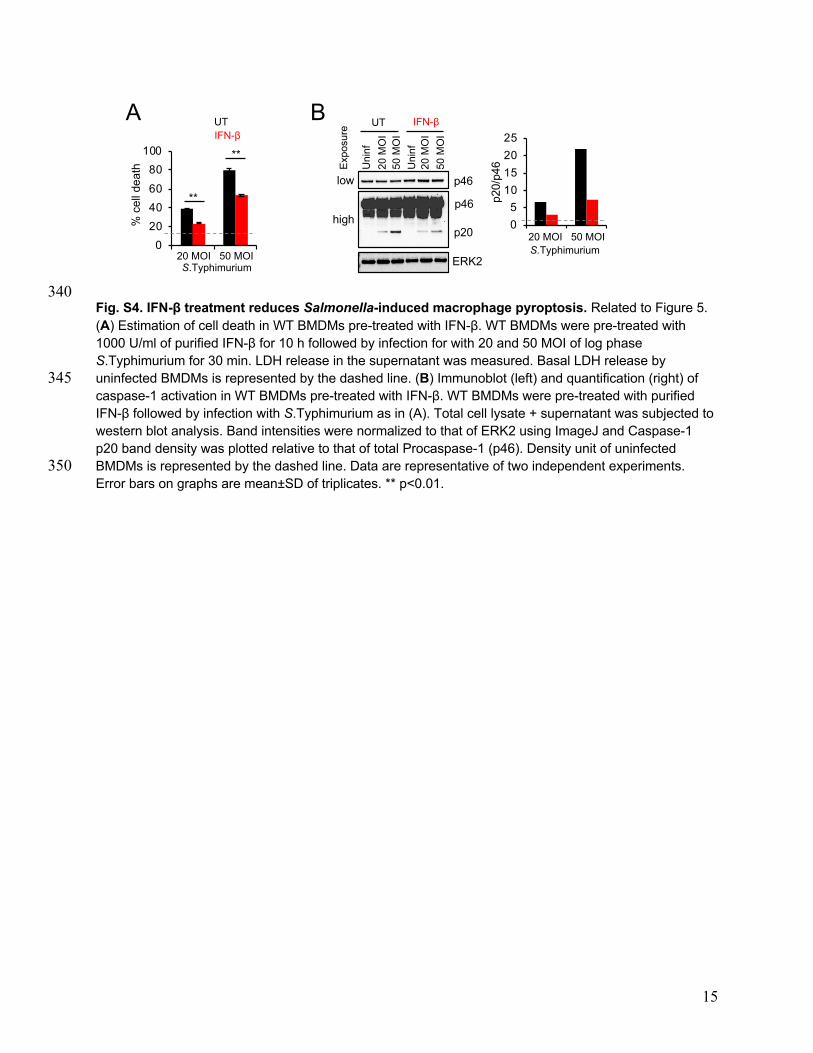
15
340 Fig. S4. IFN-β treatment reduces Salmonella-induced macrophage pyroptosis. Related to Figure 5. (A) Estimation of cell death in WT BMDMs pre-treated with IFN-β. WT BMDMs were pre-treated with 1000 U/ml of purified IFN-β for 10 h followed by infection for with 20 and 50 MOI of log phase S.Typhimurium for 30 min. LDH release in the supernatant was measured. Basal LDH release by uninfected BMDMs is represented by the dashed line. (B) Immunoblot (left) and quantification (right) of 345 caspase-1 activation in WT BMDMs pre-treated with IFN-β. WT BMDMs were pre-treated with purified IFN-β followed by infection with S.Typhimurium as in (A). Total cell lysate + supernatant was subjected to western blot analysis. Band intensities were normalized to that of ERK2 using ImageJ and Caspase-1 p20 band density was plotted relative to that of total Procaspase-1 (p46). Density unit of uninfected BMDMs is represented by the dashed line. Data are representative of two independent experiments. 350 Error bars on graphs are mean±SD of triplicates. ** p<0.01.
A B
05
10152025
S.Typhimuriump20
ERK2
p46
p46
low
high
20 M
OI
50 M
OI
Uni
nf
20 M
OI
50 M
OI
Uni
nf
UT IFN-β
Expo
sure
p20/
p46
20 MOI 50 MOI0
20
40
60
80
100%
cel
l dea
th
S.Typhimurium20 MOI 50 MOI
**
**
UTIFN-β

16
Fig. S5. Kinetics of flagellin downregulation by Salmonella in situ in NLRC4-/- and Caspase-1/11-/- 355 BMDMs. Related to Figure 6. (A-D) Representative confocal immunofluorescence images (A-C) and quantification (D) showing fliC expression by S.Typhimurium in macrophages of the indicated genotypes. BMDMs of the indicated genotypes were infected with log phase fliC-GFP reporter strain of S.Typhimurium 14028 at a MOI of 25 for 30 min. Extracellular bacteria were removed and cells were incubated for the indicated times in 360 complete medium containing gentamycin (100 µg/ml). At indicated time points post infection, cells were stained with antibody to S.Typhimurium LPS. Intracellular Salmonella were visualized as LPS-positive (red), flagellin-positive (green) and double positive (yellow). The percentage of fliC-positive bacteria per condition were enumerated as a fraction of total LPS-positive bacteria. At least 25 cells were imaged per condition. Data are representative of two independent experiments. Error bars on graphs are mean±SEM. 365 ** p<0.01, *** p<0.001. ns: not significant.
fliC
LPS
Merge
6h 24h 48h
NLRC4-/-
fliC
LPS
Merge
6h 24h 48h
Casp1/11-/-
fliC
LPS
Merge
6h 24h 48h
WTA B
C D
0
20
40
60
80
100
6h 24h 48h
% fliC
posi
tive
bact
eria
ns
***
*** ***
ns
**
WTNLRC4-/-Caspase-1/11-/-
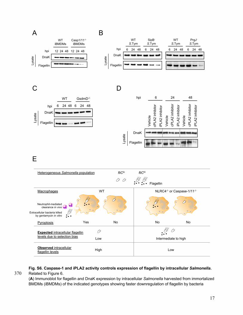
17
Fig. S6. Caspase-1 and iPLA2 activity controls expression of flagellin by intracellular Salmonella. Related to Figure 6. 370 (A) Immunoblot for flagellin and DnaK expression by intracellular Salmonella harvested from immortalized BMDMs (iBMDMs) of the indicated genotypes showing faster downregulation of flagellin by bacteria
A
C
Heterogeneous Salmonella population
Yes No
WT
Expected intracellular flagellin levels due to selection bias
Observed intracellular flagellin levels
NLRC4-/- or Caspase-1/11-/-Macrophages
NoNo
fliClo fliChi
Flagellin
Neutrophil-mediated clearance in vivo
Low Intermediate to high
Pyroptosis
LowHigh
Extracellular bacteria killed by gentamycin in vitro
E
Flagellin
6 24 48 6 24 48hpi
DnaK
WT GsdmD-/-
WT iBMDMs
Casp1/11-/-iBMDMs
DnaK
Flagellin
12 24 48 12 24 48hpi
B
iPLA
2 in
hibi
tor
cPLA
2 in
hibi
tor
iPLA
2 in
hibi
tor
cPLA
2 in
hibi
tor
Flagellin
Veh
icle
Veh
icle
Veh
icle
6 24 48
iPLA
2 in
hibi
tor
cPLA
2 in
hibi
tor
DnaK
hpi
D
6 24 48 6 24 48
WT S.Tym
DnaK
Flagellin
SipB-S.Tym
hpi 6 24 48 6 24 48
WT S.Tym
PrgJ-S.Tym
Lysa
te
Lysa
te
Lysa
te
Lysa
te

18
recovered from caspase1/11-/- BMDMs compared to those recovered from WT BMDMs. WT and caspase-1/11-/- iBMDMs were infected with S.Typhimurium at a MOI of 20 for 30 min. Extracellular bacteria were removed and cells were incubated for the indicated times in complete medium containing gentamycin 375 (100 µg/ml). Intracellular bacteria were harvested and flagellin and DnaK were analyzed by immunoblot. hpi: hours post infection. (B) Immunoblot for flagellin and DnaK expression by intracellular Salmonella isolated from iBMDMs showing faster downregulation of flagellin by SipB–deficient or PrgJ–deficient bacteria compared to WT bacteria. iBMDMs were infected with log phase WT S.Typhimurium SL1344 or its SipB-deficient derivative and WT S.Typhimurium KK1004 or its PrgJ-deficient derivative as in (A). (C) 380 Immunoblot for flagellin and DnaK expression by intracellular Salmonella recovered from WT and Gasdermin D-/- BMDMs. Cells were infected with 25 MOI of log phase S.Typhimurium for 30 min followed by gentamycin protection for the indicated times. Flagellin and DnaK expression by intracellular bacteria were assessed by immunoblot. (D) Immunoblot for flagellin and DnaK expressed by intracellular Salmonella in iBMDMs treated with PLA2 inhibitors or vehicle. iBMDMs were treated with inhibitors of 385 cPLA2 or iPLA2 followed by infection with Salmonella as in Figure 6D. At 6, 24 and 48 h post infection, intracellular Salmonella were recovered and flagellin and DnaK expression was analyzed by immunoblot. (E) Observed outcomes of flagellin expression by intracellular bacteria in NLRC4 and Caspase-1/11-/- genotypes are not explained by selection bias or pyroptosis. Data are representative of two (B and C) or three (A and D) independent experiments. 390

19
Fig. S7. Downregulation of flagellin expression by intracellular Salmonella is independent of 395 growth phase of infecting Salmonella, NLRP3 and Caspase-11. Related to Figure 6. (A) Growth curve analysis of S.Typhimurium. S.Typhimurium were grown in LB broth at 37°C with shaking at 220rpm and absorbance was measured at 630nm at the indicated time intervals. Bacteria were in mid to late log phase after 8-10 h and in stationary phase after 15 h of growth in LB. (B) Effect of growth phase of Salmonella on cell death in WT BMDMs. WT BMDMs were infected with 20 MOI of log 400 phase (10 h culture) or stationary phase (15 h culture) S.Typhimurium for 30 min. LDH release in the supernatant was measured. Basal LDH release by uninfected BMDMs is represented by the dashed line. (C) Effect of growth phase of Salmonella on caspase-1 activation. WT BMDMs were infected as in (B). Caspase-1 p20 in the supernatant and Procaspase-1 (p46) in the lysate was assayed by immunoblotting. ERK2 was used as a loading control. (D) Immunoblot for flagellin and DnaK expression by intracellular 405 Salmonella showing similar kinetics of flagellin downregulation irrespective of the growth phase of infecting Salmonella. WT, NLRP3-/-, NLRC4-/- and Caspase-1/11-/- BMDMs were infected with 20 MOI of log phase (10 h culture) or stationary phase (15 h culture) S.Typhimurium for 30 min followed by
0
0.2
0.4
0.6
0.8
1
1.2
0 3 6 9 12 15
A630
Time (h)
0
10
20
30
40
50
10h culture 15h culture
% c
ell d
eath
WT
Casp1/11-/-NLRC4-/-NLRP3-/-
A B
Casp1-p20
WT NLRP3-/- NLRC4-/-
Uni
nf10
h c
ultu
re15
h c
ultu
re
Casp1 p46
ERK2
Uni
nf10
h c
ultu
re15
h c
ultu
re
Uni
nf10
h c
ultu
re15
h c
ultu
re
D
WT NLRP3-/- NLRC4-/-
6 24 48 6 24 48 6 24 48 6 24 48
Casp1/11-/-
Flagellin
DnaK
Log phase (10 h culture)
6 24 48 6 24 48 6 24 48 6 24 48
WT NLRP3-/- NLRC4-/- Casp1/11-/-
Stationary phase (15 h culture)
hpi
Flagellin
DnaK
hpi
C
E
Flagellin
DnaK
hpi
WT
6 24 48 6 24 48 6 24 48
Casp1/11-/-Casp11-/-
******
Lysa
teLy
sate
Lysa
teLy
sate
Sup

20
gentamycin protection for the indicated times. Flagellin and DnaK expression by intracellular bacteria were assessed by immunoblot. (E) Immunoblot for flagellin and DnaK expression by intracellular 410 Salmonella harvested at indicated time points showing similar kinetics of flagellin downregulation in WT and Caspase-11-/- BMDMs and faster downregulation in Caspase-1/11-/- BMDMs. BMDMs were infected as in (D). hpi: hours post infection. Data are representative of two independent experiments. Error bars on graphs are mean±SD of triplicates. *** p<0.001. 415

21
Fig. S8. NLRC4/Caspase-1 axis sustains intracellular levels of LPC in infected BMDMs. Related to Figure 6. (A, B) Estimation of intracellular LPC in Salmonella-infected BMDMs. BMDMs of the indicated genotypes 420 were infected with 20 MOI of log phase S.Typhimurium for 30 min followed by gentamycin protection for the indicated times. LPC concentration in cell lysates was measured by ELISA. The level of LPC in uninfected BMDMs of each genotype was set at 100% (dashed line) and LPC levels post infection are represented as % change in LPC compared to uninfected BMDMs of the respective genotype. hpi: hours post infection. (C) Bacterial load in spleens of infected WT, NLRC4-/- and Caspase-1/11-/- mice. WT, 425 NLRC4-/- and Caspase-1/11-/- mice were infected with 200 CFU of log phase S.Typhimurium i.p. On day 5 post infection, spleens were homogenized and serial dilutions were plated onto Salmonella–Shigella (SS) agar. Bacterial colonies were enumerated and expressed as total colony forming units (CFU) per gram of the tissue. n=4 mice per group. Data are representative of two independent experiments. Error bars on graphs are mean±SD of triplicates. * p<0.05, ** p<0.01, *** p<0.001. ns: not significant. 430
A B
0
1
2
3
4
WT NLRC4-/- Casp1/11-/-
CFU
/g ti
ssue
(108
)
*
*C
0
50
100
150
Intra
cellu
lar L
PC
(% c
hang
e ov
er u
ninf
)
* ****
*** **
**
- 24 48 - 24 48 - 24 48 - 24 48hpi
WT Casp1/11-/-NLRP3-/- NLRC4-/-
**
0
50
100
150
- 24 48 - 24 48 - 24 48 hpi
WT Casp1/11-/-Casp11-/-
Intra
cellu
lar L
PC
(%
chan
ge o
ver u
ninf
)
*
**
** *
*
*ns
ns

22
Fig. S9. Proposed model of NLRC4, iPLA2 and IFNAR-dependent switching of Salmonella from a 435 flagellin-high to a flagellin-low phenotype inside macrophages. (A) Initial infection of macrophages with Salmonella leads to robust early activation of caspase-1 through the NLRC4 inflammasome. Caspase-1 activation leads to pyroptosis with concomitant release of lysophospholipids such as LPC. LPC in turn promotes synthesis and release of flagellin from extracellular Salmonella (9). In addition, activation of TRIF likely by Salmonella LPS, leads to secretion of IFN-β from 440 macrophages. (B) Increased flagellin expression by extracellular bacteria increases their ability to induce pyroptosis upon internalization by healthy macrophages, which leads to increased activation of IL-1b and further release of LPC. The released LPC can in turn trigger de novo synthesis and release of flagellin from
A
B C?
?
LPC
Early / flagellin-high Late / flagellin-low

23
extracellular bacteria, which sets up a feedforward, auto-amplifying circuit. In this manner, a flagellinhi 445 phenotype makes the pathogen more vulnerable to innate immune detection eventually leading to its clearance by neutrophils (10). (C) IFNAR activation by IFN-β downregulates expression of NLRC4 and iPLA2 in the surviving macrophage population. This eventually creates an intracellular state that is depleted of lysophospholipids and refractory to NLRC4-dependent caspase-1 activation and pyroptosis. Salmonella 450 adapts to this LPC-low intracellular environment and downregulates flagellin expression thereby transforming the bacterium to a flagellinlo phenotype inside macrophages. The exact mechanisms by which IFNAR signaling downregulates NLRC4 and iPLA2 expression, and caspase-1 controls intracellular LPC levels are unknown. 455
460 465 470 475 480 485
490

24
Table S1. List of strains used in this study* 495
* The LT2 strains include a deletion of the fljBA operon and hin recombinase that is a flagellar phase 1 locked (FliCON) background designated fljBenx vh2 (17), which was found to be deleted for the phase 2 flagellar region (D(hin-fljBA)) (18). The DprgJ60 allele is an in-frame nonpolar deletion of prgJ described previously (19). The PflhDC8093(PflhDC-luxCDABE-Km-PflhDC
+ allele is a duplication of the flhDC promoter region with one copy controlling wild-type flhDC operon expression and the other controlling lux operon 500 expression and constructed as described previously (8). The construction of hilD227::3xFLAG(HilD-FLAG) allele is described (8). SI References 505
1. A. Qadri, S. Ghosh, S. Upadhyay, G. P. Talwar, Monoclonal antibodies against flagellar
antigen of Salmonella typhi. Hybridoma 8, 353-360 (1989). 2. A. Qadri, S. K. Gupta, G. P. Talwar, Monoclonal antibodies delineate multiple epitopes
on the O antigens of Salmonella typhi lipopolysaccharide. J Clin Microbiol 26, 2292-2296 510 (1988).
3. A. H. Fortier, L. A. Falk, Isolation of murine macrophages. Curr Protoc Immunol Chapter 14, Unit 14 11 (2001).
4. K. D. Smith et al., Toll-like receptor 5 recognizes a conserved site on flagellin required for protofilament formation and bacterial motility. Nature immunology 4, 1247-1253 515 (2003).
5. E. G. Bligh, W. J. Dyer, A rapid method of total lipid extraction and purification. Can J Biochem Physiol 37, 911-917 (1959).
6. R. J. Nichols et al., Phenotypic landscape of a bacterial cell. Cell 144, 143-156 (2011).
Strain Description Origin Used in Figs:
SL1344 S.Typhimurium SL1344 WT E. Charpentier (11) All unless specified otherwise
EC428 (SB169)
sipB- S.Typhimurium SL1344 sipB::aphT-KmR
E. Charpentier (12) 2D, S1A, S1F, S6B
KK1004
S.Typhimurium strain LT2 fljBenx vh2 phase 1 locked derivative of LT2
K. Kutsukake (13) 2A, 2D, S1A, S1B, S1F, S6B
KK1110 LT2 fliC:: Mud1(AmpR lac) fljBenx vh2 (KK1004 fliC-lac operon fusion)
K. Kutsukake (14) 2C
KK2040
LT2 flhD3425::Tn10 fljBenx vh2
K. Kutsukake (13) S1B
TH25748
LT2 zec-7080::Tn10dTc ∆prgJ60 fljBenx vh2 This study (K. Hughes) 2D, S1A, S1F, S6B
TH2949
LT2 fliD68::Tn10 fljBenx vh2
This study (K. Hughes) 2A
BC1091 ST14028 fliCWT/pKAS32::fliC::gfp (ST14028 fliC-GFP)
B. Cookson (15) 1D, S5A-D
TH25823
ST14028 zec-7080::Tn10dTc hilD227::3xFLAG(HilD-FLAG) (ST14028-hilD227-3xFLAG)
This study (K. Hughes) S1H
TH25679
ST14028 zec-7080::Tn10dTc PflhDC8093(PflhDC-luxCDABE-Km-PflhDC+)
(ST14028 flhDC-luciferase)
This study (K. Hughes) S1I
S.Typhi S.Typhi Vi-negative clinical isolate G. Mehta (16) 2E, 3A, S1B, S1C, S1D

25
7. R. N. Bone et al., Inhibition of Ca2+-independent phospholipase A2beta (iPLA2beta) 520 ameliorates islet infiltration and incidence of diabetes in NOD mice. Diabetes 64, 541-554 (2015).
8. C. Mouslim, K. T. Hughes, The effect of cell growth phase on the regulatory cross-talk between flagellar and Spi1 virulence gene expression. PLoS Pathog 10, e1003987 (2014). 525
9. N. Subramanian, A. Qadri, Lysophospholipid sensing triggers secretion of flagellin from pathogenic salmonella. Nature immunology 7, 583-589 (2006).
10. E. A. Miao et al., Caspase-1-induced pyroptosis is an innate immune effector mechanism against intracellular bacteria. Nature immunology 11, 1136-1142 (2010).
11. S. K. Hoiseth, B. A. Stocker, Aromatic-dependent Salmonella typhimurium are non-530 virulent and effective as live vaccines. Nature 291, 238-239 (1981).
12. K. Kaniga, S. Tucker, D. Trollinger, J. E. Galan, Homologs of the Shigella IpaB and IpaC invasins are required for Salmonella typhimurium entry into cultured epithelial cells. J Bacteriol 177, 3965-3971 (1995).
13. K. Kutsukake, Y. Ohya, S. Yamaguchi, T. Iino, Operon structure of flagellar genes in 535 Salmonella typhimurium. Mol Gen Genet 214, 11-15 (1988).
14. K. Kutsukake, T. Iino, Role of the FliA-FlgM regulatory system on the transcriptional control of the flagellar regulon and flagellar formation in Salmonella typhimurium. J Bacteriol 176, 3598-3605 (1994).
15. L. A. Cummings, W. D. Wilkerson, T. Bergsbaken, B. T. Cookson, In vivo, fliC 540 expression by Salmonella enterica serovar Typhimurium is heterogeneous, regulated by ClpX, and anatomically restricted. Mol Microbiol 61, 795-809 (2006).
16. A. Sharma, A. Qadri, Vi polysaccharide of Salmonella typhi targets the prohibitin family of molecules in intestinal epithelial cells and suppresses early inflammatory responses. Proc Natl Acad Sci U S A 101, 17492-17497 (2004). 545
17. T. Iino, A Stabilizer of Antigenic Phases in Salmonella Abortus-Equi. Genetics 46, 1465-1469 (1961).
18. K. Kutsukake, H. Nakashima, A. Tominaga, T. Abo, Two DNA invertases contribute to flagellar phase variation in Salmonella enterica serovar Typhimurium strain LT2. J Bacteriol 188, 950-957 (2006). 550
19. D. H. Wee, K. T. Hughes, Molecular ruler determines needle length for the Salmonella Spi-1 injectisome. Proc Natl Acad Sci U S A 112, 4098-4103 (2015).