Embed Size (px)
Citation preview

Type 1 Fimbriae, a Colonization Factor of UropathogenicEscherichia coli, Are Controlled by the Metabolic SensorCRP-cAMPClaudia M. Muller1, Anna Aberg1, Jurate Straseviciene1, Levente Emody2,3, Bernt Eric Uhlin1*, Carlos
Balsalobre4*
1 Department of Molecular Biology and Laboratory for Molecular Infection Medicine Sweden (MIMS), Umea University, Umea, Sweden, 2 Institute of Medical Microbiology
and Immunology, University of Pecs Medical School, Budapest, Hungary, 3 Veterinary Research Institute, Hungarian Academy of Sciences, Budapest, Hungary,
4 Departament de Microbiologia, Universitat de Barcelona, Barcelona, Spain
Abstract
Type 1 fimbriae are a crucial factor for the virulence of uropathogenic Escherichia coli during the first steps of infection bymediating adhesion to epithelial cells. They are also required for the consequent colonization of the tissues and for invasionof the uroepithelium. Here, we studied the role of the specialized signal transduction system CRP-cAMP in the regulation oftype 1 fimbriation. Although initially discovered by regulating carbohydrate metabolism, the CRP-cAMP complex controls amajor regulatory network in Gram-negative bacteria, including a broad subset of genes spread into different functionalcategories of the cell. Our results indicate that CRP-cAMP plays a dual role in type 1 fimbriation, affecting both the phasevariation process and fimA promoter activity, with an overall repressive outcome on fimbriation. The dissection of theregulatory pathway let us conclude that CRP-cAMP negatively affects FimB-mediated recombination by an indirectmechanism that requires DNA gyrase activity. Moreover, the underlying studies revealed that CRP-cAMP controls theexpression of another global regulator in Gram-negative bacteria, the leucine-responsive protein Lrp. CRP-cAMP-mediatedrepression is limiting the switch from the non-fimbriated to the fimbriated state. Consistently, a drop in the intracellularconcentration of cAMP due to altered physiological conditions (e.g. growth in presence of glucose) increases thepercentage of fimbriated cells in the bacterial population. We also provide evidence that the repression of type 1 fimbriaeby CRP-cAMP occurs during fast growth conditions (logarithmic phase) and is alleviated during slow growth (stationaryphase), which is consistent with an involvement of type 1 fimbriae in the adaptation to stress conditions by promotingbiofilm growth or entry into host cells. Our work suggests that the metabolic sensor CRP-cAMP plays a role in coupling theexpression of type 1 fimbriae to environmental conditions, thereby also affecting subsequent attachment and colonizationof host tissues.
Citation: Muller CM, Aberg A, Straseviciene J, Emody L, Uhlin BE, et al. (2009) Type 1 Fimbriae, a Colonization Factor of Uropathogenic Escherichia coli, AreControlled by the Metabolic Sensor CRP-cAMP. PLoS Pathog 5(2): e1000303. doi:10.1371/journal.ppat.1000303
Editor: Pascale Cossart, Institut Pasteur, France
Received September 3, 2008; Accepted January 18, 2009; Published February 20, 2009
Copyright: � 2009 Muller et al. This is an open-access article distributed under the terms of the Creative Commons Attribution License, which permitsunrestricted use, distribution, and reproduction in any medium, provided the original author and source are credited.
Funding: This work was supported by grants from the Spanish Ministry of Education and Sciences (BIO2004-02747, BIO2007-64637 and the Ramon y CajalProgram), the Swedish Research Council, the Medical Faculty of Umea University, the Swedish Foundation for International Cooperation in Research and HigherEducation (STINT), the International Graduate College IGK 587/2, the EU FP6 EuroPathoGenomics Network of Excellence, the Hungarian Research Foundation(grant OTKA 62092), and was in part performed within the Umea Centre for Microbial Research (UCMR).
Competing Interests: The authors have declared that no competing interests exist.
* E-mail: [email protected] (BEU); [email protected] (CB)
Introduction
Bacteria have the ability to rapidly adapt to changes in the
environment, a feature that is important for survival and
multiplication both during colonization of host organisms and in
the environment. An efficient adaptation implies the ability to
sense external parameters and to transduce the perceived signals to
cellular regulators, which then incite adaptive changes in the
physiology of the cell. One form of signal transduction occurs via
cytoplasmatic secondary messenger systems, so-called alarmones,
which can mediate a rapid response. Alarmones are low molecular
mass, non-proteinaceous, enzymatically synthesized compounds.
Several modified nucleotides have been described to execute this
function in bacteria, among them the 39,59-cyclic adenosine
monophosphate (cAMP). cAMP is a ubiquitous molecule found in
both prokaryotes and eukaryotes. In bacteria, the activity of cAMP
was initially thought to be restricted to its role in catabolite
repression [1]. However, there is evidence for an extended role of
cAMP as sensory signal involved in global gene regulation in
bacteria [2–5]. The level of intracellular cAMP is modulated by
several environmental factors [6–8]. The cellular target for cAMP-
signaling is the cAMP receptor protein (CRP). Dimeric CRP in
complex with one molecule of cAMP exhibits DNA-binding
activity to sites located near promoter regions [9]. Thereby, CRP-
cAMP acts as a global regulator of gene expression by controlling
the expression of almost 200 operons in E. coli [10–12].
Type 1 fimbriae mediate attachment to both biotic and abiotic
surfaces and are involved in the early stages of biofilm formation
[13,14]. In E. coli, type 1 fimbriae play a crucial role during
urinary tract infections by mediating adhesion to mannose-
containing receptors on the uroepithelium and promoting the
formation of intracellular bacterial communities [15–17]. Those
PLoS Pathogens | www.plospathogens.org 1 February 2009 | Volume 5 | Issue 2 | e1000303
adhesins are encoded by the fim determinant composed of two
independent transcription units coding for the recombinases FimE
and FimB, and a polycistronic operon encoding the structural
components (FimA, FimF, FimG, and FimH) and a pilus assembly
system (FimC and FimD) [18,19]. Phase variable expression of the
fim operon is associated with the inversion of a 314-bp
chromosomal region, flanked by two 9-bp inverted repeats, that
contains the fimA promoter [20,21]. When the invertible element is
in the so-called ON orientation, the promoter is directed towards
the structural fim genes, thus allowing transcription, whereas
transcription is abolished in the inverted OFF orientation. The
inversion process is catalyzed by FimB and FimE, two members of
the tyrosine site-specific recombinase family [22,23].
Several regulators are involved in the fine modulation of the
expression of type 1 fimbriae by environmental conditions [24,25].
A proper supercoiling state of the DNA and the presence of
accessory proteins, such as the DNA binding proteins Lrp and
IHF, are essential features that affect the recombination process
and determine whether the cell is fimbriated or not [26–29]. Other
regulators such as RpoS, ppGpp, NanR and NagC modulate type
1 fimbriation mostly by altering the expression of the recombinases
that catalyze the recombination event [30–32]. Moreover, the
global regulator H-NS has been shown to affect type 1 fimbriation
both by regulating the expression of the recombinases and by
directly interacting with the fim invertible element [33,34]. Effects
on the expression of type 1 fimbriae in cya derivatives of E. coli K-
12, which are defective in cAMP synthesis, were reported earlier
[35]. However, different strains responded divergently upon
addition of exogenous cAMP in static cultures and it was not
clarified at what level the reported effects were operating. In this
work, we describe that CRP-cAMP represses type 1 fimbriation.
The dissection of the mechanism underlying the observed
phenomenon demonstrated that CRP-cAMP indirectly represses
FimB-mediated recombination during the phase variation process.
In contrast to many other regulators of the phase variation of type
1 fimbriae described, CRP-cAMP affects phase variation inde-
pendently of the levels of the recombinases. We propose a novel
model by which CRP-cAMP controls the type 1 fimbriation state
in the bacterial population by affecting DNA gyrase activity. In
addition, our studies led to the new discovery that Lrp expression
in E. coli is under the control of the CRP-cAMP complex.
Results/Discussion
Type 1 fimbriation in E. coli is enhanced by the lack of theCRP-cAMP regulatory complex
For a successful colonization of hosts by bacteria, it is crucial
that the expression of bacterial surface structures, which mediate
the interaction with the host tissues, is finely regulated. In E. coli,
the CRP-cAMP complex has been shown to regulate the
production of several of those surface structures, such as flagella
or P-fimbriae [36–39]. Using a crp deletion mutant derivative of
the extensively studied uropathogenic E. coli (UPEC) isolate J96,
we further characterized the role of CRP-cAMP in the modulation
of the expression of those colonization factors. Confirming
previous data, the CRP-cAMP deficient derivatives were non-
motile and had lost the ability to cause mannose-resistant
haemagglutination (MRHA) (data not shown). Agglutination tests
using specific antisera against the Pap and Prs fimbriae, adhesins
that mediate MRHA, confirmed that the expression of those
fimbriae is strictly dependent on the presence of functional CRP-
cAMP in the cell (data not shown).
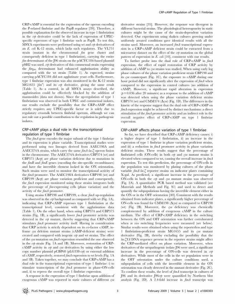
J96, as most of the UPEC isolates, also expresses type 1
fimbriae, which are essential for the adherence and invasion of the
bladder uroepithelium. The expression of type 1 fimbriae can be
detected by mannose-sensitive yeast agglutination (MSYA), which
attests the ability of type 1 fimbriated bacteria to bind to
mannosides-containing receptors on the surface of yeast cells. A
clear stimulation in the ability to cause MSYA was observed in the
J96crp strain as compared with wt when growing in various culture
media (LB, TBA, CFA, and TSA; data not shown). Semi-
quantitative MSYA, using serially diluted LB cultures, corrobo-
rated these results: agglutination of yeast cells was observed with a
higher dilution of the J96crp cell suspension (4-fold, i.e. containing
8-times less bacterial cells) as compared to wt (Table 1). These
results indicate that the deficiency in CRP-cAMP caused a
substantial increase in the expression of type 1 fimbriae on the cell
surface.
In UPEC, a regulatory crosstalk between fimbrial operons
occurs, which also affects the expression of type 1 fimbriae. It is
known that the UPEC-specific regulators PapB, SfaB, and FocB,
which are involved in the regulation of P-related, S-related, and
F1C-related fimbriae, respectively, have the ability to repress the
expression of type 1 fimbriae [40–42]. It has been described that
Table 1. Semi-quantitative MSYA in wt and crp derivatives ofdifferent bacterial strains
Genotype
Strain wt crp
J96 fimJ96 1/2 1/8
VL751/pACYC184 fim2 n.d. n.d.
VL751/pSH2 fimJ96 1/32 1/128
MG1655 fimMG1655 1/4 1/8
Bacterial cultures of the indicated strains (wt and crp) were grown in LB mediumovernight at 37uC with vigorous shaking. The origin of the fim determinantpresent in each strain is indicated. The numeric values indicate the highestdilution of the bacterial culture that agglutinated yeast cells. n.d. = no MSYAdetected.doi:10.1371/journal.ppat.1000303.t001
Author Summary
Attachment of bacteria to the surface of host tissues is acrucial initial step in the establishment of bacterialinfections. This process is mediated by adhesins, such asthe type 1 fimbriae of Escherichia coli, which play a key roleduring urinary tract infections by mediating adhesion tothe uroepithelium. The expression of type 1 fimbriae isfinely regulated attending to environmental signals and isunder phase variation control, which determines thepercentage of fimbriated cells in the population. In thisreport, we show that the expression of type 1 fimbriae isrepressed by a metabolic sensor of the cell, the globalregulatory complex CRP-cAMP. We demonstrate that CRP-cAMP affects the switching outcome by selectivelyinhibiting the recombination process in one directiononly, resulting in a lower percentage of fimbriated cells.Such a switch to the non-fimbriated state after successfuladhesion might be advantageous in the urinary tract,where the immune mechanisms of the host favor theremoval of bacteria expressing immunogenic surfacestructures. Understanding the regulatory networks thatgovern regulation of virulence and colonization factors isboth of basic interest and might help to develop novelstrategies to treat bacterial infections.
CRP-cAMP Regulation of Type 1 Fimbriae
PLoS Pathogens | www.plospathogens.org 2 February 2009 | Volume 5 | Issue 2 | e1000303
CRP-cAMP is essential for the expression of the operon encoding
the P-related fimbriae and the PapB regulator [39]. Therefore, a
possible explanation for the observed increase in type 1 fimbriation
in the crp derivative could be the lack of expression of UPEC-
specific repressors of type 1 fimbriae such as PapB. To test this,
MSYA experiments were performed using wt and crp derivatives of
an E. coli K-12 strain, which lacks such regulators. The VL751
strain (mutant in the chromosomal fim gene cluster and
consequently deficient in type 1 fimbriation), carrying the entire
fim determinant of the J96 strain on the pACYC184-based plasmid
pSH2 was used. crp derivatives of this commensal strain expressing
the fimJ96 determinant had enhanced agglutination ability as
compared with the wt strain (Table 1). As expected, strains
carrying pACYC184 did not agglutinate yeast cells. Furthermore,
type 1 fimbriae expression was also monitored in the K-12 strain
MG1655 (fim+) and its crp derivative, giving the same result
(Table 1). As a control, in all MSYA assays described, the
agglutination could be effectively blocked by the addition of
mannosides (data not shown). Since the up-regulation of type 1
fimbriation was observed in both UPEC and commensal isolates,
our results exclude the possibility that the CRP-cAMP effect
strictly requires any UPEC-specific factor or is solely due to
regulatory crosstalk between fimbrial operons, although we can
not rule out a possible contribution to the regulation in pathogenic
isolates.
CRP-cAMP plays a dual role in the transcriptionalregulation of type 1 fimbriae
The fimA gene encodes the major subunit of the type 1 fimbriae
and its expression is phase variable. Transcriptional studies were
performed using two lineages derived from AAEC198A and
AAEC374A strains, which carry the same fimA-lacZ operon fusion
in the chromosome. The AAEC374A derivatives CBP374 (wt) and
CBP375 (Dcrp) are phase variation deficient due to mutations in
the fimB and fimE genes (encoding the site-specific recombinases)
and have the invertible element locked in the ON orientation.
Such strains were used to monitor the transcriptional activity of
the fimA promoter. The AAEC198A derivatives CBP198 (wt) and
CBP199 (Dcrp) are phase variation proficient and consequently,
the fimA expression monitored using these strains integrates both
the percentage of fim-expressing cells (phase variation) and the
activity of the fimA promoter.
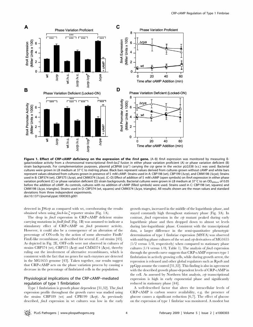
Using strains CBP198 and CBP199, a clear fimA up-regulation
was observed in the crp background as compared with wt (Fig. 1A),
indicating that CRP-cAMP represses type 1 fimbriation at the
transcriptional level, consistent with the agglutination data
(Table 1). On the other hand, when using CBP374 and CBP375
strains (Fig. 1B), a significantly lower fimA promoter activity was
detected in the crp mutant, thereby suggesting that CRP-cAMP
stimulates fimA promoter activity itself. Having in consideration
that CRP activity is strictly dependent on its co-factor cAMP, in-
frame cya deletion mutant strains (cAMP-deficient strains) were
created and compared with isogenic crp and wt strains. The effect
of the cya mutation on fimA expression resembled the one observed
in the crp strain (Fig. 1A and 1B). Moreover, restoration of CRP-
cAMP activity in crp and cya derivatives by using either the low
copy number plasmid pCBP68 (pLG338-crp) or external addition
of cAMP, respectively, restored fimA expression to wt levels (Fig. 1A
and 1B). Taken together, we may conclude that CRP-cAMP has a
dual role in the transcriptional expression of type 1 fimbriae: i) to
stimulate transcription of the fimA promoter in phase-ON-cells
and, ii) to repress the overall type 1 fimbriae expression.
A response in the expression of type 1 fimbriae upon addition of
exogenous cAMP was reported in static cultures of different cya
derivative strains [35]. However, the response was divergent in
different bacterial strains. The physiological heterogeneity in static
cultures might be the cause of the strain-dependent variation
detected. Our experiments using shaken cultures growing under
uniformly aerated conditions gave identical results with all the
strains used. Moreover, an increased fimA transcriptional expres-
sion in a CRP-cAMP deficient strain could be extracted from a
microarray dataset on the effect of the crp mutation on the global
pattern of expression in E. coli [10], consistent with our results.
To further probe into the dual role of CRP-cAMP in fimA
expression, the effect of rapid restoration of CRP activity by
addition of cAMP to cya strains was studied. When using early log
phase cultures of the phase variation proficient strain CBP198 and
its cya counterpart (Fig. 1C), the exposure to cAMP during one
hour period did not significantly alter the expression of fimA when
compared to the expression in control cultures (no addition of
cAMP). However, a significant rapid alteration in expression
(p = 0.036 after 20 minutes) as a response to the addition of cAMP
was detected when using the phase variation deficient strains
CBP374 (wt) and CMM374 (Dcya) (Fig. 1D). The differences in the
kinetic of the response suggest that the dual role of CRP-cAMP on
fimA expression might be achieved by distinct mechanisms: a direct
stimulation of the fimA promoter activity and an indirect role in the
overall negative effect of CRP-cAMP on type 1 fimbriae
expression.
CRP-cAMP affects phase variation of type 1 fimbriaeSo far, we have described that CRP-cAMP deficiency causes: i)
a higher degree of type 1 fimbriation, ii) an increase in the
expression of type 1 fimbriae in phase variation proficient strains,
and iii) a reduction in fimA promoter activity in phase variation
deficient strains. These results suggest that the percentage of
fimbriated cells (ON-cells) in both crp and cya mutant strains is
elevated when compared to wt, causing the overall increase in fimA
expression. To test this prediction, the percentage of ON-cells in
the population was monitored by plating cultures of the phase
variable fimA-lacZ reporter strains on indicator plates containing
X-gal. As predicted, a significant increase in the percentage of
ON-cells in both the crp and cya mutant strains was observed
(Fig. 2A). A quantitative PCR based method was validated (see
Materials and Methods and Fig. S1) and used to detect and
quantify the subpopulations having the invertible element either in
the ON or in the OFF orientation [40]. Consistent with the results
obtained from indicator plates, a significantly higher percentage of
ON-cells was found for CMM198 (Dcya) as compared to CBP198
(wt) (Fig. 2B). Moreover, the cya deficiency was chemically
complemented by addition of exogenous cAMP in the culture
medium. The effect of CRP-cAMP deficiency in the switching
between the ON and OFF orientation was further corroborated
when in vivo switching frequencies were measured (see below).
Similar results were obtained when using the reporterless and type
1 fimbriation-proficient strain MG1655 and its cya mutant
derivative (Fig. 2B), thereby excluding the possibility that the
lacZYA DNA sequence present in the reporter strains might affect
the CRP-mediated effect on phase variation. Moreover, when
derivatives of the uropathogenic isolate J96 were used, a significant
increase in the percentage of ON-cells was detected in crp
derivatives. While most of the cells in the wt population were in
the OFF orientation under the culture conditions used, a
subpopulation of cells with the invertible element in the ON
orientation was clearly detected in the mutant derivative (Fig. 2C).
To confirm these results, the level of fimA transcript in cultures of
J96 and its derivative J96crp were quantified by Northern blot
analysis (Fig. 2D). A 2.4-fold increase in fimA transcript was
CRP-cAMP Regulation of Type 1 Fimbriae
PLoS Pathogens | www.plospathogens.org 3 February 2009 | Volume 5 | Issue 2 | e1000303
detected in J96crp as compared with wt, corroborating the results
obtained when using fimA-lacZ reporter strains (Fig. 1A).
The drop in fimA expression in CRP-cAMP deficient strains
carrying mutations in fimB fimE (Fig. 1B) was assumed to indicate a
stimulatory effect of CRP-cAMP on fimA promoter activity.
However, it could also be a consequence of an alteration of the
percentage of ON-cells by the action of some alternative FimB/
FimE-like recombinase, as described for several E. coli strains [43].
As depicted in Fig. 2E, OFF-cells were not observed in cultures of
strains CBP374 (wt), CBP375 (Dcrp) and CMM374 (Dcya), thereby
ruling out the involvement of alternative recombinases, which is
consistent with the fact that no genes for such enzymes are detected
in the MG1655 genome [43]. Taken together, our results suggest
that CRP-cAMP acts on the phase variation process by causing a
decrease in the percentage of fimbriated cells in the population.
Physiological implications of the CRP-cAMP–mediatedregulation of type 1 fimbriation
Type 1 fimbriation is growth phase dependent [31,32]. The fimA
expression profile throughout the growth curve was studied using
the strains CBP189 (wt) and CPB199 (Dcrp). As previously
described, fimA expression in wt cultures was low in the early
growth stages, increased in the middle of the logarithmic phase, and
stayed constantly high throughout stationary phase (Fig. 3A). In
contrast, fimA expression in the crp mutant peaked during early
logarithmic phase and then dropped down to almost wt levels
during late-logarithmic phase. Consistent with the transcriptional
data, a larger difference in the semi-quantitative phenotypic
determination of type 1 fimbriae expression (MSYA) was observed
with mid-log phase cultures of the wt and crp derivatives of MG1655
(1/2 versus 1/8, respectively) when compared to stationary phase
cultures (1/4 versus 1/8, Table 1). The analysis of fimA expression
through the growth curve suggests that CRP-cAMP represses type 1
fimbriation in actively growing cells, while during growth arrest, the
repression is released and other global regulators such as RpoS and
ppGpp assume the control [31,32]. This finding is also in agreement
with the described growth phase-dependent levels of CRP-cAMP in
the cell. As assessed by Northern blot analysis, crp transcriptional
expression is high in early exponential phase and significantly
reduced in stationary phase [44].
A well-described factor that alters the intracellular levels of
CRP-cAMP is carbon source availability, e.g. the presence of
glucose causes a significant reduction [6,7]. The effect of glucose
on the expression of type 1 fimbriae was monitored. A modest but
Figure 1. Effect of CRP-cAMP deficiency on the expression of the fimA gene. (A–B) fimA expression was monitored by measuring ß-galactosidase activity from a chromosomal transcriptional fimA-lacZ fusion in either phase variation proficient (A) or phase variation deficient (B)strain backgrounds. For complementation purposes, plasmid pCBP68 (crp+) carrying the crp gene in the vector pLG338 (v.c.) was used. Bacterialcultures were grown in LB medium at 37uC to mid-log phase. Black bars represent values derived from cultures grown without cAMP and white barsrepresent values obtained from cultures grown in presence of 5 mM cAMP. Strains used in A: CBP198 (wt), CBP199 (Dcrp), and CMM198 (Dcya); Strainsused in B: CBP374 (wt), CBP375 (Dcrp), and CMM374 (Dcya). (C–D) Effect of addition of 5 mM cAMP (open symbols) on fimA expression in either phasevariation proficient (C) or phase variation deficient (D) strain backgrounds. Bacterial cultures were grown in LB medium at 37uC to an OD600nm of 0.05before the addition of cAMP. As controls, cultures with no addition of cAMP (filled symbols) were used. Strains used in C: CBP198 (wt, squares) andCMM198 (Dcya, triangles). Strains used in D: CBP374 (wt, squares) and CMM374 (Dcya, triangles). All results shown are the mean values and standarddeviations from three independent experiments.doi:10.1371/journal.ppat.1000303.g001
CRP-cAMP Regulation of Type 1 Fimbriae
PLoS Pathogens | www.plospathogens.org 4 February 2009 | Volume 5 | Issue 2 | e1000303

significant increase in the percentage of fimA-expressing cells could
be observed when CBP198 (wt) cultures were grown in M9-
glucose medium compared with cultures grown in M9-glycerol
(Fig. 3B). The stimulatory effect of the presence of glucose on
transcriptional expression of type 1 fimbriae was also observed by
microarray analysis on the effect of glucose in the general
expression pattern in E. coli [45].
CRP-cAMP deficient strains have a significant growth defect
compared to the wt (i.e: 89 and 34 minutes generation time in LB
for CBP199 and CBP198, respectively), which might raise the
question whether the increased type 1 expression in the crp strains
is merely due to the growth alterations. However, growth in media
that significantly increases the growth rate of the crp strain, i.e. LB
medium containing glucose (32 and 48 minutes generation time
for CBP198 and CBP199, respectively), did not alter the difference
in the expression of type 1 fimbriae between the wt and the crp
strains (data not shown), suggesting that the CRP specific effect on
type 1 fimbriae expression is not coupled to the growth rate.
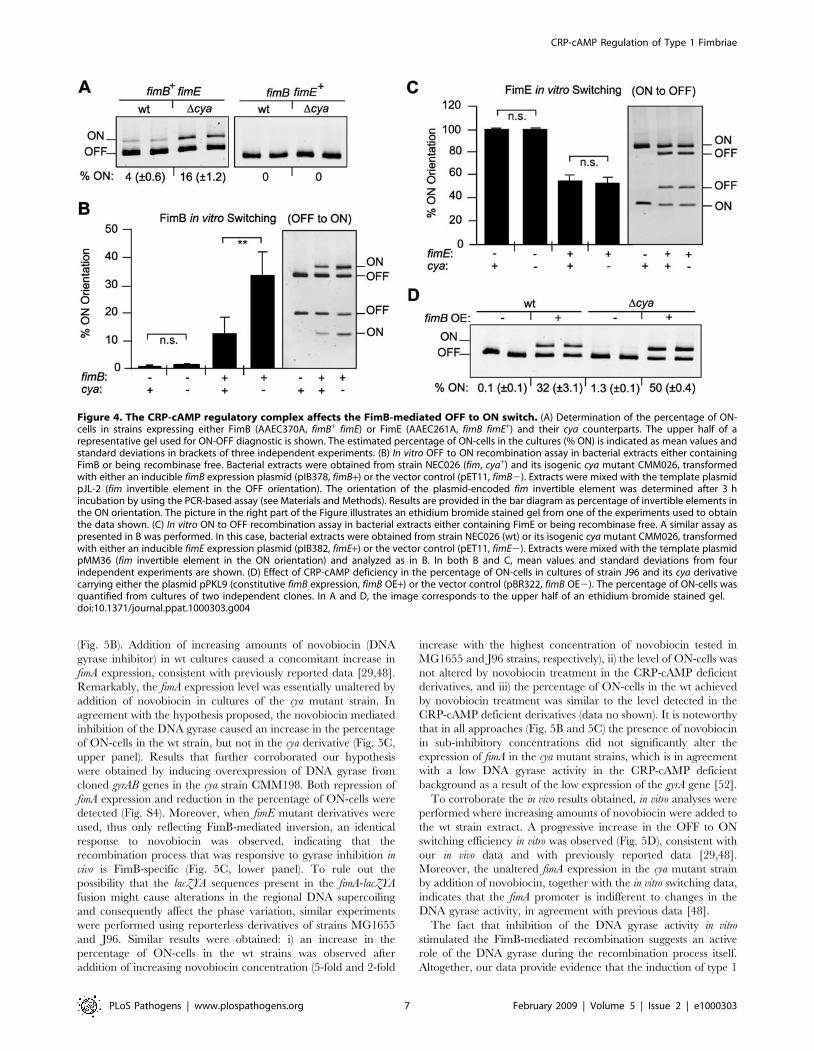
CRP-cAMP affects the FimB-mediated OFF to ON switchboth in vivo and in vitro
The reported increase in the percentage of ON-cells in the
CRP-cAMP deficient strains could be achieved either by
stimulating the OFF to ON inversion (exclusively catalyzed by
FimB) or by causing the opposite effect on the ON to OFF
inversion (mainly catalyzed by FimE). To further dissect the role of
CRP-cAMP in the recombination event, the percentage of ON-
cells in wt and cya derivative strains expressing either FimB
(AAEC370A, fimE) or FimE (AAEC261A, fimB) was determined
(Fig. 4A). In the FimB proficient strains (fimE), a significant
increase in the percentage of ON-cells was detected in the strain
lacking CRP-cAMP (16% in cya versus 4% in wt). However, in
FimE proficient strains (fimB), consistent with published results
[46], all cells were in the OFF orientation independently of the
presence or absence of the CRP-cAMP complex. These results
suggest that CRP-cAMP is directly or indirectly affecting the
FimB-mediated inversion. To corroborate these data, in vitro
recombination assays were performed using template plasmids as
recombination substrate in bacterial extracts of cya and cya+ strains
overexpressing either FimB or FimE. The induction of the
synthesis of the recombinases in cultures of the cya and cya+
strains provided apparently identical amounts of the enzymes in
the extracts of both strains as determined by Coomassie-stained
SDS-PAGE (Fig. S2). When FimB-mediated OFF to ON inversion
was monitored (Fig. 4B), recombination occurred with both cya
and wt extracts in the presence of FimB. However, a remarkable
3-fold higher percentage (p = 0.003) of invertible fragments in the
Figure 2. The percentage of fimbriated cells in the population is increased in crp and cya strains. (A) The percentage of fimA-expressingcells in presence (white bar) or absence (black bars) of 5 mM cAMP was determined by the indicator plate assay (see Materials and Methods) usingmid-log phase cultures of the strains CBP198 (wt), CBP199 (Dcrp) and CMM198 (Dcya). Mean values and standard deviations from three independentexperiments are shown. (B) Quantification of the percentage of ON-cells in bacterial populations by a PCR-based assay. Cultures of wt and cyaderivatives of strains CBP198 and MG1655 were grown to mid-log phase in presence (white bars) or absence (black bars) of 5 mM cAMP. Mean valuesand standard deviations of three independent experiments are shown. (C) ON-OFF diagnostic of mid-log phase cultures of the J96 strain and its crpderivative; the arrowhead highlights the fragment corresponding to ON-cells detected in the J96crp samples. (D) Northern hybridization of total RNAextracted from mid-log cultures from strains J96 (wt), J96crp (Dcrp), VL751 (Dfim), and AAG42 (Dlrp) with specific probes for fimA, fimB, lrp, and 16SrRNA as indicated. (E) ON-OFF diagnostic of duplicated cultures of the phase variation deficient strains CBP374 (wt), CBP375 (Dcrp), and CMM374(Dcya). A control showing the band pattern of an OFF population was included for comparison. The pictures in panels C and E are electronicallyinverted images of ethidium bromide stained acrylamide gels.doi:10.1371/journal.ppat.1000303.g002
CRP-cAMP Regulation of Type 1 Fimbriae
PLoS Pathogens | www.plospathogens.org 5 February 2009 | Volume 5 | Issue 2 | e1000303

ON orientation was detected in the extract from the cya strain
when compared with wt extracts. On the other hand, FimE-
mediated inversion from the ON to the OFF state did not seem to
be affected by a mutation in the cya gene (Fig. 4C). The FimB
recombinase can also catalyze the switch from ON to OFF.
However, no effect of CRP-cAMP on the FimB-mediated ON to
OFF inversion was detected when in vitro recombination assays
with DNA template in the ON orientation were performed (data
not shown). Altogether, our in vitro studies corroborate the results
obtained in vivo and suggest that the CRP-cAMP complex
specifically affects the FimB-mediated recombination event from
the OFF to the ON orientation. Supporting this conclusion, in vivo
switching frequency estimations indicated that the OFF to ON
switching rate was significantly increased in strain CBP199 (Dcrp)
as compared with CBP198 (wt) (1.161024 and 1.461022 per cell
and generation in wt and mutant, respectively), while no
significant effect was observed in the ON to OFF switching
(1.061026 and 1.661026 per cell and generation in wt and
mutant, respectively). Also supporting our results, it was reported
that the FimB-mediated switching frequency from OFF to ON is
3-fold higher in the presence of glucose (i.e. reduced intracellular
levels of CRP-cAMP) than in the presence of glycerol [24].
Although higher expression of fimB was observed in crp
derivatives as compared to wt counterparts in both J96 and
MG1655 strains (Fig. 2D and data not shown), the in vitro data,
where the recombinases were overexpressed to the same degree in
both extracts, suggest that the enhanced OFF to ON switching in
absence of CRP-cAMP is not strictly dependent on the levels of the
FimB recombinase. To further test this hypothesis, in vivo
experiments were performed under conditions of constitutive fimB
expression using plasmid pPKL9, which contains the fimB gene
under the control of the tet promoter (Fig. 4E). Control
experiments by Northern blot analyses verified that the fimB
expression levels from plasmid pPKL9 were essentially identical in
CRP-cAMP proficient and deficient genetic backgrounds (data not
shown). The percentage of ON-cells in cultures of J96 derivatives
constitutively expressing fimB (carrying plasmid pPKL9) was
significantly elevated in the CRP-cAMP deficient strain as
compared with wt, yielding a 50% higher percentage of ON-cells.
Comparable results were obtained when using MG1655 derivative
strains (data not shown). Altogether, our results both in vivo and in
vitro indicate that the CRP-cAMP complex has a negative effect on
the switching process independently of the intracellular concen-
tration of the FimB recombinase.
CRP-cAMP represses type 1 fimbriation by an indirectmechanism
Two possible mechanisms by which CRP-cAMP affects the
FimB-mediated switch should be considered: either CRP-cAMP
can directly interact with the invertible DNA fragment repressing
the OFF to ON switch, or the effect of CRP-cAMP may be
indirect.
The slow response when adding exogenous cAMP to CMM198
cultures (Fig. 1C) suggested that the role of CRP-cAMP in the
regulation of the phase variation occurs by an indirect mechanism.
Nevertheless, to establish whether CRP-cAMP might also be
directly involved in the switching process, in vitro recombination
assays were performed using extracts of the cya strain while restoring
CRP-cAMP activity by addition of increasing amounts of cAMP
(Fig. 5A). No obvious alteration in the FimB-mediated switch was
detected, strongly suggesting that CRP-cAMP does not directly
interact with the nucleoprotein complex that is the substrate for the
FimB recombinase. Accordingly, no effect was observed in the
outcome of in vitro recombination assays when purified CRP was
added to extracts obtained from a crp strain (data not shown).
Simultaneously, the possible binding of CRP-cAMP to various
DNA fragments spanning different regions of the fim determinant
was tested (Fig. S3A). No strong CRP binding was detected to any
of the DNA fragments tested. At most, a low affinity binding was
detected in case of the fragment containing the fimA promoter
(PCR7; Fig. S3B). However, when DNase I footprinting analysis of
this putative CRP binding site was performed, no binding was
observed (data not shown). It has been reported that CRP-cAMP
might bind to many low affinity binding sites along the E. coli
chromosome [47]. Although it is possible that such low affinity
CRP binding site(s) may exist in the fimA promoter region, our
experimental evidence (Fig. 5A) suggested that binding is not
required for the phase variation control. A possible involvement of
the putative CRP binding site(s) in the positive control of the fimA
promoter activity (Fig. 1B) will be further studied.
Inhibition of DNA gyrase activity mimics the effect ofCRP-cAMP on type 1 fimbrial phase variation
Recently, it has been shown that inhibiting the DNA gyrase
promotes the FimB-mediated inversion from OFF to ON and
therefore it was concluded that DNA supercoiling determines the
directionality of the FimB-mediated recombination [29,48]. DNA
gyrase is an enzyme that catalyses ATP-dependent DNA breakage,
strand passage and rejoining of double-stranded DNA (for a recent
review see Nollmann et al. [49]). DNA gyrase is involved in the
regulation of DNA topology, but also in other processes such as
replication or illegitimate recombination [50,51]. Remarkably, it
has been described that CRP-cAMP modulates the expression of
the gyrA gene encoding the DNA gyrase. In crp deficient strains,
low levels of gyrA expression and DNA gyrase activity, monitored
as alterations in the topology of plasmid DNA, were detected [52].
One may hypothesize that the CRP-cAMP mediated effect on the
FimB-recombination process could directly result from the low
levels of DNA gyrase activity detected in crp deficient strains. To
test this hypothesis, the effect of inhibiting the DNA gyrase in vivo
was analyzed in both wt (CBP198) and cya (CMM198) strains
Figure 3. Type 1 fimbriae expression profile in different growthconditions. (A) fimA expression was determined by measuring ß-galactosidase activity at various optical densities from cultures of thefimA-lacZ reporter strains CBP198 (wt, black bars) and CBP199 (Dcrp,white bars) in LB medium at 37uC. (B) Quantification of the percentageof fimA-expressing cells in the population of strain CBP198 (wt) onindicator plates. Cultures were grown to mid-log phase at 37uC in M9minimal medium containing either glycerol (glyc.) or glucose (gluc.) as acarbon source. Mean values and standard deviations from threeindependent experiments are shown.doi:10.1371/journal.ppat.1000303.g003
CRP-cAMP Regulation of Type 1 Fimbriae
PLoS Pathogens | www.plospathogens.org 6 February 2009 | Volume 5 | Issue 2 | e1000303
(Fig. 5B). Addition of increasing amounts of novobiocin (DNA
gyrase inhibitor) in wt cultures caused a concomitant increase in
fimA expression, consistent with previously reported data [29,48].
Remarkably, the fimA expression level was essentially unaltered by
addition of novobiocin in cultures of the cya mutant strain. In
agreement with the hypothesis proposed, the novobiocin mediated
inhibition of the DNA gyrase caused an increase in the percentage
of ON-cells in the wt strain, but not in the cya derivative (Fig. 5C,
upper panel). Results that further corroborated our hypothesis
were obtained by inducing overexpression of DNA gyrase from
cloned gyrAB genes in the cya strain CMM198. Both repression of
fimA expression and reduction in the percentage of ON-cells were
detected (Fig. S4). Moreover, when fimE mutant derivatives were
used, thus only reflecting FimB-mediated inversion, an identical
response to novobiocin was observed, indicating that the
recombination process that was responsive to gyrase inhibition in
vivo is FimB-specific (Fig. 5C, lower panel). To rule out the
possibility that the lacZYA sequences present in the fimA-lacZYA
fusion might cause alterations in the regional DNA supercoiling
and consequently affect the phase variation, similar experiments
were performed using reporterless derivatives of strains MG1655
and J96. Similar results were obtained: i) an increase in the
percentage of ON-cells in the wt strains was observed after
addition of increasing novobiocin concentration (5-fold and 2-fold
increase with the highest concentration of novobiocin tested in
MG1655 and J96 strains, respectively), ii) the level of ON-cells was
not altered by novobiocin treatment in the CRP-cAMP deficient
derivatives, and iii) the percentage of ON-cells in the wt achieved
by novobiocin treatment was similar to the level detected in the
CRP-cAMP deficient derivatives (data no shown). It is noteworthy
that in all approaches (Fig. 5B and 5C) the presence of novobiocin
in sub-inhibitory concentrations did not significantly alter the
expression of fimA in the cya mutant strains, which is in agreement
with a low DNA gyrase activity in the CRP-cAMP deficient
background as a result of the low expression of the gyrA gene [52].
To corroborate the in vivo results obtained, in vitro analyses were
performed where increasing amounts of novobiocin were added to
the wt strain extract. A progressive increase in the OFF to ON
switching efficiency in vitro was observed (Fig. 5D), consistent with
our in vivo data and with previously reported data [29,48].
Moreover, the unaltered fimA expression in the cya mutant strain
by addition of novobiocin, together with the in vitro switching data,
indicates that the fimA promoter is indifferent to changes in the
DNA gyrase activity, in agreement with previous data [48].
The fact that inhibition of the DNA gyrase activity in vitro
stimulated the FimB-mediated recombination suggests an active
role of the DNA gyrase during the recombination process itself.
Altogether, our data provide evidence that the induction of type 1
Figure 4. The CRP-cAMP regulatory complex affects the FimB-mediated OFF to ON switch. (A) Determination of the percentage of ON-cells in strains expressing either FimB (AAEC370A, fimB+ fimE) or FimE (AAEC261A, fimB fimE+) and their cya counterparts. The upper half of arepresentative gel used for ON-OFF diagnostic is shown. The estimated percentage of ON-cells in the cultures (% ON) is indicated as mean values andstandard deviations in brackets of three independent experiments. (B) In vitro OFF to ON recombination assay in bacterial extracts either containingFimB or being recombinase free. Bacterial extracts were obtained from strain NEC026 (fim, cya+) and its isogenic cya mutant CMM026, transformedwith either an inducible fimB expression plasmid (pIB378, fimB+) or the vector control (pET11, fimB2). Extracts were mixed with the template plasmidpJL-2 (fim invertible element in the OFF orientation). The orientation of the plasmid-encoded fim invertible element was determined after 3 hincubation by using the PCR-based assay (see Materials and Methods). Results are provided in the bar diagram as percentage of invertible elements inthe ON orientation. The picture in the right part of the Figure illustrates an ethidium bromide stained gel from one of the experiments used to obtainthe data shown. (C) In vitro ON to OFF recombination assay in bacterial extracts either containing FimE or being recombinase free. A similar assay aspresented in B was performed. In this case, bacterial extracts were obtained from strain NEC026 (wt) or its isogenic cya mutant CMM026, transformedwith either an inducible fimE expression plasmid (pIB382, fimE+) or the vector control (pET11, fimE2). Extracts were mixed with the template plasmidpMM36 (fim invertible element in the ON orientation) and analyzed as in B. In both B and C, mean values and standard deviations from fourindependent experiments are shown. (D) Effect of CRP-cAMP deficiency in the percentage of ON-cells in cultures of strain J96 and its cya derivativecarrying either the plasmid pPKL9 (constitutive fimB expression, fimB OE+) or the vector control (pBR322, fimB OE2). The percentage of ON-cells wasquantified from cultures of two independent clones. In A and D, the image corresponds to the upper half of an ethidium bromide stained gel.doi:10.1371/journal.ppat.1000303.g004
CRP-cAMP Regulation of Type 1 Fimbriae
PLoS Pathogens | www.plospathogens.org 7 February 2009 | Volume 5 | Issue 2 | e1000303

fimbriation detected in the CRP-cAMP deficient strains is a
process mediated by the alteration in DNA gyrase activity and
therefore can be mimicked by the specific inhibition of this
enzymatic activity by novobiocin. Moreover, our in vitro results
comparing OFF to ON switching between wt and cya mutant using
the same DNA template for both extracts (Fig. 4B and Fig. 5D)
demonstrated that the CRP-cAMP effect on phase variation is not
merely dependent on the initial supercoiling state of the fim
invertible element.
Lack of CRP-cAMP results in increased Lrp levelsRecombination at the fim invertible element requires Lrp, a
DNA bending protein that directly binds to specific sites within the
invertible element and stimulates DNA inversion [26]. Kelly et al.
[29] demonstrated that this binding activity of Lrp is required to
promote the FimB-mediated OFF to ON directionality observed
when the DNA gyrase was inhibited. The Lrp levels were
determined by immunoblot analysis of crp+ and crp strains (Fig. 6A)
and the Lrp content detected was several fold higher in the crp
strains than in the crp+ strains. These results suggest a possible link
between the CRP and Lrp regulons. Interestingly, a direct
demonstration of CRP dependent regulation of Lrp expression
has not been done, although two putative CRP sites have been
predicted in the promoter region of the lrp gene [53], suggesting a
possible direct regulation by CRP-cAMP. Additionally, CRP-
cAMP could act indirectly by positively regulating GadE, which
represses lrp expression [54,55]. Transcriptional studies have been
performed by Northern blot analysis of RNA from derivatives of
MG1655 and J96 (Fig. 2D). An increase in the level of the lrp
transcript in the crp derivatives was detected as compared with wt
(2.5 and 1.7- fold in MG1655 and J96 derivatives, respectively),
suggesting a role for CRP-cAMP in the control of lrp transcription,
Figure 5. Insights on the mechanism of action of the CRP-cAMP complex in vivo and in vitro. (A) Effect of the addition of cAMP during invitro OFF to ON recombination. Bacterial extracts were obtained from strains NEC026 (wt) or CMM026 (Dcya) transformed with the inducible fimBexpression plasmid (pIB378). Extracts were mixed with the template plasmid pJL-2 in absence or presence of increasing amounts of cAMP (1 to50 mM final concentration). Mean values and standard deviations of three independent experiments are shown. (B) Effect of increasing amounts ofthe gyrase inhibitor novobiocin on fimA expression. ß-galactosidase activity was measured from strains CBP198 (wt, black bars) and CMM198 (Dcya,white bars) grown to mid-log phase in LB medium supplemented with 0, 6.25, 12.5, and 25 mg ml21 novobiocin. Mean values and standarddeviations from two independent experiments are shown. (C) Effect of DNA gyrase inhibition on the orientation of the fim invertible element in vivo.Upper panel: ON-OFF diagnostic of the samples used in Fig. 5B, representing the strain CBP198 (fimB+ fimE+) and its cya derivative CMM198 grown inpresence of novobiocin (concentrations as indicated). Lower panel: ON-OFF diagnostic of the strain AAEC370A (fimB+ fimE) and its Dcya derivativeCMM370A subject to the same growth conditions as in the upper panel. Both panels depict electronically inverted images of the upper half ofacrylamide gels after ethidium bromide staining. (D) Effect of DNA gyrase inhibition on FimB-mediated OFF to ON switching in vitro. Increasingamounts of novobiocin (0, 50, 200 mg ml21) were added to the in vitro recombination reactions. Bacterial extracts from strains NEC026 (wt, left panel)or CMM026 (Dcya, right panel) transformed with the inducible fimB expression plasmid (pIB378) were used together with the template plasmid pJL-2.Mean values and standard deviations in brackets of the estimated percentage of invertible elements in the ON orientation from four independentexperiments are given as numbers below each lane. The images correspond to ethidium bromide stained gels from a representative experiment usedto obtain the data shown.doi:10.1371/journal.ppat.1000303.g005
CRP-cAMP Regulation of Type 1 Fimbriae
PLoS Pathogens | www.plospathogens.org 8 February 2009 | Volume 5 | Issue 2 | e1000303
although an additional regulation at the posttranscriptional level
can not be ruled out. Further studies would be required to
characterize the CRP-cAMP dependent regulation of Lrp
expression.
Interestingly, when the levels of intracellular Lrp were
monitored in the same cultures as in Fig. 5B, again a differential
effect of the inhibition of the DNA gyrase in wt and cya strains was
observed (Fig. 6B). In a wt strain, the Lrp levels were strongly
elevated at the highest concentration of novobiocin, where the
amount of Lrp was apparently identical to the amount detected in
the cya strain in absence of novobiocin, which again might be
explained by the low DNA gyrase activity detected in the CRP-
cAMP deficient background [52]. We tested whether increased
levels of Lrp by itself would cause an alteration in the FimB-
mediated switching process. Our results clearly indicate that Lrp
overexpression per se did not result in any significant changes in the
percentage of ON-cells when no alteration in DNA gyrase activity
was induced (Fig. 6C).
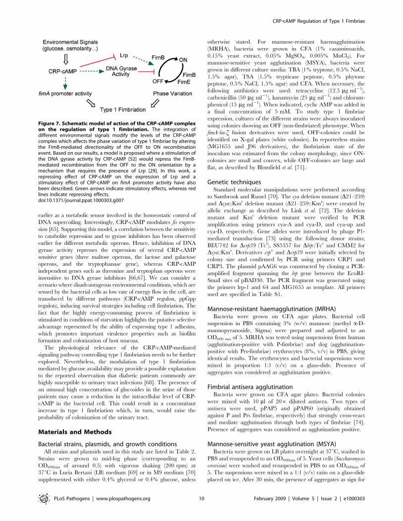
Concluding remarksThe expression of type 1 fimbriae implies the allocation of an
important part of the asset of the bacterial cell for the production of
those proteinaceous appendages, as considerable amounts of energy
and amino acids are needed for their synthesis. A tightly regulated
expression of such organelles is therefore expected. Considering the
important metabolic effort performed by the bacterial cells
committed to be fimbriated, regulation by phase variation can be
seen as a selective advantage for the bacterial population, in
addition of providing phenotypical heterogeneity in an otherwise
genetically homogeneous population. In previous works, we have
shown that the expression of type 1 fimbriae is stimulated when
intracellular levels of the stringent response alarmone ppGpp are
raised [32,56]. The level of ppGpp in the cell increases under amino
acid starvation and energy stress [57]. In this report, the role of
CRP-cAMP, a regulatory complex that is associated with the energy
state of the cell, has been included in the extensive list of regulatory
networks controlling type 1 fimbriation.
Interestingly, many of the global regulators that affect type 1
fimbriae expression, such as H-NS, RpoS, Lrp, and now CRP-
cAMP, have been shown to interplay among each other, thereby
orchestrating gene regulation cascades in response to the growth
conditions [58]. From our studies, we can conclude that CRP-
cAMP is a major regulator of fimbriation during the exponential
growth phase (Fig. 3A) and is required to maintain the growth
expression profile of type 1 fimbriae. We dissected the initial
observation that the crp derivatives of J96 showed an increased
ability to agglutinate yeast cells and conclude that CRP-cAMP
represses type 1 fimbriation, as schematically shown in Fig. 7, by
the recently described mechanism of switching directionality
established by the activity of the DNA gyrase and the presence
of Lrp [29], thereby affecting FimB-mediated OFF to ON
switching. Remarkably, CRP-cAMP inhibited FimB-mediated
recombination at a template plasmid isolated from a crp+
background, indicating that the regulatory effect does not merely
depend on the supercoiling state of the DNA and thereby
suggesting an active role of the DNA gyrase in the OFF to ON
recombination event. Interestingly, CRP-cAMP has a dual effect
on type 1 fimbriation by repressing phase variation and promoting
promoter activity. Further studies will be required for fully
understanding the underlying mechanisms by which CRP-cAMP
affects both levels of regulation of type 1 fimbriation.
In Salmonella, crp cya mutants are avirulent in a mouse model
[59] and it has been reported that the crp and the cya genes are
strongly repressed during infection of macrophages [60]. More-
over, it has been observed that DNA becomes more relaxed when
bacteria are growing in certain intracellular environments and
consequently the expression of those genes that are required for
intracellular survival is induced [61]. Therefore, CRP-cAMP
might be involved in controlling Salmonella virulence in a pathway
that includes DNA supercoiling and the sensing of environmental
conditions as previously proposed [62].
It is well described that CRP and cAMP levels are affected by
environmental conditions such as glucose availability and osmolarity
[6,7]. Interestingly, such environmental conditions also affect DNA
topology in E. coli in a DNA gyrase dependent manner [63,64]. The
link between CRP-cAMP mediated regulation of gene expression and
DNA gyrase activity might represent a specialized signal transduction
pathway that senses the metabolic and energetic status of the cell. It
can not be ruled out that in this regulatory pathway others factors
such as the FIS protein might be involved. FIS has been proposed
Figure 6. Induction of Lrp in a CRP-cAMP deficient background.(A) Lrp levels in different genetic backgrounds. Whole bacterial celllysates of the strains CBP198 (wt), CBP199 (Dcrp), CBP199/pLG339(Dcrp/v.c.), CBP199/pLG339-CRP (Dcrp/crp), and AAG42 (Dlrp) weresubjected to immunoblot analyses using Lrp-specific antiserum.Numbers below each lane represent the average of the signal intensityfrom three independent experiments relative to the corresponding wtvalue (set as one). (B) Analysis of Lrp levels in whole cell lysates of strainCBP198 (wt) and CMM198 (Dcya) grown in the presence of novobiocinat the indicated concentrations (same bacterial cultures as in Fig. 5B).(C) Effect of overexpression of lrp on the percentage of ON-cells.Cultures of strain CBP198 transformed with a plasmid that carries the lrpgene under the inducible Para-promoter (pAAG6) were grown to mid-log phase in presence of the indicated concentrations of arabinose. Theinduced levels of lrp were assessed by immunoblotting using Lrp-specific antiserum. Simultaneously, quantification of the percentage ofON-cells in the populations was performed by using the PCR-basedmethod. The lower image corresponds to the upper half of arepresentative gel used for ON-OFF diagnostic; the results are givenas numbers below each lane.doi:10.1371/journal.ppat.1000303.g006
CRP-cAMP Regulation of Type 1 Fimbriae
PLoS Pathogens | www.plospathogens.org 9 February 2009 | Volume 5 | Issue 2 | e1000303
earlier as a metabolic sensor involved in the homeostatic control of
DNA supercoiling. Interestingly, CRP-cAMP modulates fis expres-
sion [65]. Supporting this model, a correlation between the sensitivity
to catabolite repression and to gyrase inhibitors has been observed
earlier for different metabolic operons. Hence, inhibition of DNA
gyrase activity represses the expression of several CRP-cAMP
sensitive genes (three maltose operons, the lactose and galactose
operons, and the tryptophanase gene), whereas CRP-cAMP
independent genes such as threonine and tryptophan operons were
insensitive to DNA gyrase inhibitors [66,67]. We can consider a
scenario where disadvantageous environmental conditions, which are
sensed by the bacterial cells as low rate of energy flow in the cell, are
transduced by different pathways (CRP-cAMP regulon, ppGpp
regulon), inducing survival strategies including cell fimbriation. The
fact that the highly energy-consuming process of fimbriation is
stimulated in conditions of starvation highlights the putative selective
advantage represented by the ability of expressing type 1 adhesins,
which promotes important virulence properties such as biofilm
formation and colonization of host mucosa.
The physiological relevance of the CRP-cAMP-mediated
signaling pathway controlling type 1 fimbriation needs to be further
explored. Nevertheless, the modulation of type 1 fimbriation
mediated by glucose availability may provide a possible explanation
to the reported observation that diabetic patients commonly are
highly susceptible to urinary tract infections [68]. The presence of
an unusual high concentration of glucosides in the urine of those
patients may cause a reduction in the intracellular level of CRP-
cAMP in the bacterial cell. This could result in a concomitant
increase in type 1 fimbriation which, in turn, would raise the
probability of colonization of the urinary tract.
Materials and Methods
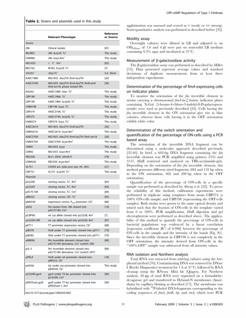
Bacterial strains, plasmids, and growth conditionsAll strains and plasmids used in this study are listed in Table 2.
Strains were grown to mid-log phase (corresponding to an
OD600nm of around 0.5) with vigorous shaking (200 rpm) at
37uC in Luria Bertani (LB) medium [69] or in M9 medium [70]
supplemented with either 0.4% glycerol or 0.4% glucose, unless
otherwise stated. For mannose-resistant haemagglutination
(MRHA), bacteria were grown in CFA (1% casaminoacids,
0.15% yeast extract, 0.05% MgSO4, 0.005% MnCl2). For
mannose-sensitive yeast agglutination (MSYA), bacteria were
grown in different culture media: TBA (1% tryptone, 0.5% NaCl,
1.5% agar), TSA (1.5% trypticase peptone, 0.5% phytone
peptone, 0.5% NaCl, 1.5% agar) and CFA. When necessary, the
following antibiotics were used: tetracycline (12.5 mg ml21),
carbenicillin (50 mg ml21), kanamycin (25 mg ml21) and chloram-
phenicol (15 mg ml21). When indicated, cyclic AMP was added in
a final concentration of 5 mM. To study type 1 fimbriae
expression, cultures of the different strains were always inoculated
using colonies showing an OFF (non-fimbriated) phenotype. When
fimA-lacZ fusion derivatives were used, OFF-colonies could be
identified on X-gal plates (white colonies). In reporterless strains
(MG1655 and J96 derivatives), the fimbriation state of the
inoculum was estimated from the colony morphology, since ON-
colonies are small and convex, while OFF-colonies are large and
flat, as described by Blomfield et al. [71].
Genetic techniquesStandard molecular manipulations were performed according
to Sambrook and Russel [70]. The cya deletion mutant (D21–259)
and Dcya::Kmr deletion mutant (D21–259::Kmr) were created by
allelic exchange as described by Link et al. [72]. The deletion
mutant and Kmr deletion mutant were verified by PCR
amplification using primers cya-A and cya-D, and cya-up and
cya-D, respectively. Gene alleles were introduced by phage P1-
mediated transduction [73] using the following donor strains;
BEU742 for Dcrp39 (Tcr), SS5357 for Dlrp::Tcr and CMM2 for
Dcya::Kmr. Derivatives crp+ and Dcrp39 were initially selected by
colony size and confirmed by PCR using primers CRP1 and
CRP3. The plasmid pAAG6 was constructed by cloning a PCR-
amplified fragment spanning the lrp gene between the EcoRI-
SmaI sites of pBAD30. The PCR fragment was generated using
the primers lrp-1 and 64 and MG1655 as template. All primers
used are specified in Table S1.
Mannose-resistant haemagglutination (MRHA)Bacteria were grown on CFA agar plates. Bacterial cell
suspension in PBS containing 3% (w/v) mannose (methyl a-D-
mannopyranoside, Sigma) were prepared and adjusted to an
OD600 nm of 5. MRHA was tested using suspensions from human
(agglutination-positive with P-fimbriae) and dog (agglutination-
positive with Prs-fimbriae) erythrocytes (8%, v/v) in PBS, giving
identical results. The erythrocytes and bacterial suspensions were
mixed in proportion 1:1 (v/v) on a glass-slide. Presence of
aggregates was considered as agglutination positive.
Fimbrial antisera agglutinationBacteria were grown on CFA agar plates. Bacterial colonies
were mixed with 10 ml of 206 diluted antisera. Two types of
antisera were used, pPAP5 and pPAP60 (originally obtained
against P and Prs fimbriae, respectively) that strongly cross-react
and mediate agglutination through both types of fimbriae [74].
Presence of aggregates was considered as agglutination positive.
Mannose-sensitive yeast agglutination (MSYA)Bacteria were grown on LB plates overnight at 37uC, washed in
PBS and resuspended to an OD600nm of 5. Yeast cells (Saccharomyces
cerevisiae) were washed and resuspended in PBS to an OD600nm of
5. The suspensions were mixed in a 1:1 (v/v) ratio on a glass-slide
placed on ice. After 30 min, the presence of aggregates as sign for
Figure 7. Schematic model of action of the CRP-cAMP complexon the regulation of type 1 fimbriation. The integration ofdifferent environmental signals modify the levels of the CRP-cAMPcomplex which affects the phase variation of type 1 fimbriae by alteringthe FimB-mediated directionality of the OFF to ON recombinationevent. Based on our results, a model is proposed where a stimulation ofthe DNA gyrase activity by CRP-cAMP [52] would repress the FimB-mediated recombination from the OFF to the ON orientation by amechanism that requires the presence of Lrp [29]. In this work, arepressing effect of CRP-cAMP on the expression of Lrp and astimulatory effect of CRP-cAMP on fimA promoter activity have alsobeen described. Green arrows indicate stimulatory effects, whereas redlines indicate repressing effects.doi:10.1371/journal.ppat.1000303.g007
CRP-cAMP Regulation of Type 1 Fimbriae
PLoS Pathogens | www.plospathogens.org 10 February 2009 | Volume 5 | Issue 2 | e1000303
agglutination was assessed and scored as + (weak) or ++ (strong).
Semi-quantitative analysis was performed as described before [32].
Motility assayOvernight cultures were diluted in LB and adjusted to an
OD600nm of 1.0 and 4 ml were put on semi-solid LB medium
containing 0.3% agar and incubated at 37uC.
Measurement of b-galactosidase activityThe b-galactosidase assay was performed as described by Miller
[75]. Data presented represent average values and standard
deviations of duplicate measurements from at least three
independent experiments.
Determination of the percentage of fimA-expressing cellson indicator plates
To monitor the orientation of the fim invertible element in
strains carrying a chromosomal fimA-lacZ fusion, indicator plates
containing X-Gal (5-bromo-4-chloro-3-indolyl-ß-D-galactopyra-
noside) were used as previously described [24]. Cells having the
fim invertible element in the ON orientation give rise to blue
colonies, whereas those cells having it in the OFF orientation
exhibit white color.
Determination of the switch orientation andquantification of the percentage of ON-cells using a PCRbased assay
The orientation of the invertible DNA fragment can be
determined using a molecular approach described previously
[32,40]. In brief, a 602-bp DNA fragment containing the fim
invertible element was PCR amplified using primers 2535 and
3137, HinfI restricted and analyzed on TBE-acrylamide-gels.
Depending on the orientation of the fim invertible element, this
method generates different sized fragments (484 and 118 bp when
in the ON orientation, 402 and 200 bp when in the OFF
orientation).
Quantification of the percentage of ON-cells in a specific
sample was performed as described by Aberg et al. [32]. To prove
the reliability of this method, calibration experiments were
performed in triplicate using templates from strain CBP374 (a
100% ON-cells sample) and CBP198 (representing the OFF-cells
samples). Both strains were grown to the same optical density and
mixed such that the fraction of ON-cells in the template varied
from 0 to 100%. PCR amplification, HinfI digestion and gel
electrophoresis were performed as described above. The applica-
bility of this method to quantify the percentage of ON-cells in
bacterial populations was evidenced by a linear correlation
[regression coefficient (R2) of 0.998] between the percentage of
ON-cells in the sample and the intensity of the bands (Fig. S1).
Since the invertible element in CBP198 is not completely in the
OFF orientation, the intensity derived from ON-cells in the
‘‘100%-OFF’’ sample was subtracted from all intensity values.
RNA isolation and Northern analysisTotal RNA was extracted from mid-log cultures using the hot-
phenol method [76]. Contaminating DNA was removed by DNase
I (Roche Diagnostics) treatment for 1 h at 37uC, followed by RNA
cleanup using the RNeasy Mini kit (Qiagen). For Northern
analysis, 20 mg of total RNA were separated on a formaldehy-
de:agarose gel and transferred to Hybond-N membranes (Amer-
sham) by capillary blotting as described [77]. The membrane was
hybridized with 32P-labeled DNA-fragments corresponding to the
coding sequences of fimA, fimB, lrp, and rrnA, which were PCR
Table 2. Strains and plasmids used in this study
Relevant PhenotypeReferenceor Source
Strains
J96 Clinical isolate [81]
BEU805 J96 Dcrp39, Tcr This study
CMM96 J96 Dcya::Kmr This study
MG1655 l2 F2 fim+ [82]
BEU742 M182 Dcrp39, Tcr [7]
SS5357 Dlrp::Tcr S.A. Short
AAEC198A MG1655 DlacZYA fimA-lacZYA [26]
AAEC374A MG1655 DlacZYA fimA-lacZYA fimB-am6fimE-am18, phase locked ON
[26]
AAG42 AAEC198A Dlrp:: Tcr This study
CBP198 AAEC198A, Tcr This study
CBP199 AAEC198A Dcrp39, Tcr This study
CMM198 CBP198 Dcya, Tcr This study
CBP374 AAEC374A, Tcr This study
CBP375 AAEC374A Dcrp39, Tcr This study
CMM374 CBP374 Dcya, Tcr This study
AAEC261A MG1655 DlacZYA fimB-lacZYA [26]
CMM261A AAEC261A Dcya::Kmr This study
AAEC370A MG1655 DlacZYA fimA-lacZYA fimE-am18 [26]
CMM370A AAEC370A Dcya::Kmr This study
CMM1 MG1655 Dcya This study
CMM2 MG1655 Dcya::Kmr This study
NEC026 BL21 (DE3) DfimB-H [79]
CMM026 NEC026 Dcya::Kmr This study
VL751 CSH50 ara D(lac-pro) rpsL thi Dfim [21]
CBP751 VL751 Dcrp39, Tcr This study
Plasmids
pLG338 cloning vector, Tcr, Kmr [83]
pLG339 cloning vector, Tcr, Kmr [83]
pACYC184 cloning vector, Tcr, Cmr [84]
pBR322 cloning vector, Cbr, Tcr [85]
pBAD30 expression vector, Para promoter, Cbr [86]
pSH2 fim-cluster from J96 cloned intopACYC184, Cmr
[19]
pCBP68 wt crp allele cloned into pLG338, Kmr [7]
pLG339-CRP wt crp allele cloned into pLG339, Kmr [87]
pET11 T7 promoter expression vector, Cbr [88]
pIB378 fimB under T7 promoter cloned into pET11 [79]
pIB382 fimE under T7 promoter cloned into pET11 [79]
pMM36 fim invertible element cloned intopACYC184 derivative, Cmr (switch ON)
[89]
pJL-2 fim invertible element cloned intopACYC184 derivative, Cmr (switch OFF)
[89]
pPKL9 fimB under tet promoter cloned intopBR322, Cbr
[18]
pAAG6 lrp under ara promoter cloned intopBAD30, Cbr
This study
pCA24N-gyrA gyrA under T5-lac promoter cloned intopCA24N, Cmr
[90]
pRSFDuet-gyrB gyrB under T7-lac promoter cloned intopRSFDuet-1, Kmr
[90]
doi:10.1371/journal.ppat.1000303.t002
CRP-cAMP Regulation of Type 1 Fimbriae
PLoS Pathogens | www.plospathogens.org 11 February 2009 | Volume 5 | Issue 2 | e1000303
generated using the primer pairs fimA-RT1&2, fimB-RT1&2, lrp-
RT1&2 and 16S-RT1&2, respectively. After hybridization
overnight at 52uC, membranes were subsequently washed in 16SSC-0.1% SDS for 15 min at room temperature and in 0.16SSC-0.1% SDS for 15 min at 52uC. Autoradiograms were
obtained using StoragePhosphor screens (Molecular Dynamics),
which were scanned using the Storm Imaging System (Molecular
Dynamics).
Determination of FimB and FimE switching frequencies invivo
FimB and FimE-promoted switching frequencies were mea-
sured as previously described [24].
In vitro recombination assayTo perform FimB in vitro recombination assays, bacterial
extracts were obtained from cultures of the fim mutant strain
NEC026 and its isogenic cya mutant strain harboring the plasmid
pIB378 (fimB gene under the control of an IPTG inducible
promoter in pET11). fimB expression was induced with 0.4 mM
IPTG after the cultures grown in minimal MOPS [78] at 28uChad reached an OD600nm of 0.15. Cells were harvested after 24 h
of induction at 28uC and processed as described [40,79]. As
control, extracts lacking FimB were obtained from cultures of the
same strains carrying the pET11 plasmid. To perform FimE in vitro
recombination assays, bacterial extracts were obtained from
cultures of the strain NEC026 and its isogenic cya mutant strain
transformed with the plasmid pIB382 (fimE gene under the control
of an IPTG inducible promoter in pET11). Cultures were
manipulated as described above. The in vitro recombination assay
was performed as described [40,79]. The resulting orientation of
the invertible element was analyzed after 3 h incubation at 37uCusing the PCR-based method described above.
ImmunoblottingWhole cell extracts from bacterial cultures were separated by
SDS-PAGE as described by Laemmli [80] using 15% polyacryl-
amide gels. Samples were transferred to PVDF membranes using a
semidry blotting apparatus. After blocking the membranes
overnight in Tris-buffered saline containing 0.1% Tween-20
(TBS-T) and 5% skimmed milk, membranes were incubated for
1 h at room temperature with 2,0006 diluted Lrp-specific
antiserum or 6,0006 diluted PapA-specific antiserum [38] in
TBS-T containing 5% skimmed milk. After 36 15 min washes in
TBS-T, membranes were incubated for 1 h with 20,0006 diluted
anti-rabbit immunoglobuline-horseradish peroxidase conjugate
(Dianova, Hamburg, Germany). After further washing, membranes
were developed using the enhanced chemiluminescence (ECL+) kit
(GE Healthcare) and analyzed on a Chemidoc System (BioRad)
equipped with the QuantityOneH Software for quantification.
Statistical analysisDifferences between average values were tested for significance
by performing an unpaired, two-sided Student’s t-test. The levels
of significance of the resulting p values are reported by the
following symbols: * = p,0.05; ** = p,0.01; *** = p,0.001 and
n.s. = non-significant.
Supporting Information
Figure S1 Validation of the PCR based assay used for
quantifying the percentage of cells in the population with the
invertible element in the ON-orientation. Suspensions of CBP198
(OFF-template) and CBP374 (ON-template) cells were mixed in
the indicated ratios (0–100% ON-template). The HinfI restriction
pattern of the PCR amplified fragment containing the fim
invertible element is shown. The size (in base pairs) of the
different diagnostic ON and OFF fragments is indicated. Below
each lane, the results of quantification and calculation of the
percentage of ON-cells in the population in each sample according
to the described method are presented as mean values6standard
deviation of three independent calibration experiments.
Found at: doi:10.1371/journal.ppat.1000303.s001 (0.32 MB TIF)
Figure S2 Electrophoretic analysis of protein extracts used for
the in vitro recombination assays. Extracts from NEC026 (wt) and
CMM026 (cya) strains carrying the plasmids pET11 (vector
control), pIB378 (pET11 carrying the fimB gene) and pIB382
(pET11 carrying fimE) were obtained as described in materials and
methods. Proteins were separated by SDS-15% PAGE and
Coomassie stained. The bands representing the induced FimB
and FimE (right margin) and the molecular mass of relevant
protein markers (left margin) are indicated.
Found at: doi:10.1371/journal.ppat.1000303.s002 (0.44 MB TIF)
Figure S3 DNA-binding pattern of CRP at the fim regulatory
regions. A. Schematic representation of the fim determinant and
position of the promoters (P) of fimB, fimE, and fimA. Black
arrowheads represent the inverted repeats flanking the invertible
element. The relative positions of the PCR fragments used for the
gel mobility shift are depicted below. B. Gel mobility shift assay of
purified CRP protein (3 mM) and various PCR-amplified DNA
fragments (PCR1-5, and PCR7, here shown using OFF-cells as
template for amplification). In all panels, samples correspond to:
lane 1: no protein (cAMP present); lane 2: CRP protein with
20 mM cAMP (active form); lane 3: CRP protein without cAMP
(inactive form). Identical result was obtained when PCR7 fragment
was obtained from ON-cells as template. Recombinant CRP
protein was purified from strain pp6/pHA7 essentially as described
(Zhang et al., 1991). The PCR amplification and the mobility shift
assay were performed as described (Xia et al., 2000). - Zhang, X.P.,
Gunasekera, A., Ebright, Y.W., and Ebright, R.H. (1991) J.
Biomol. Struct. Dyn. 9: 463-473. - Xia, Y., Gally, D., Forsman-
Semb, K., and Uhlin, B.E. (2000) EMBO J. 19: 1450-1457
Found at: doi:10.1371/journal.ppat.1000303.s003 (0.41 MB TIF)
Figure S4 Effect of gyrA and gyrB overexpression on the
expression of type 1 fimbriae in a CRP-cAMP deficient strain.
A. fimA expression from strain CMM198 (cya) carrying the
plasmids pCA24N-gyrA and pRSFDuet-gyrB, which carry the gyrA
and the gyrB genes, respectively, under IPTG-inducible promoters,
was monitored in either the absence (2) or the presence (+) of
IPTG. Bacterial cultures were grown in LB medium to mid-log
phase. IPTG was added at a final concentration of 0.015 mM
(condition that did not cause any deleterious effect on the bacterial
growth). Mean values and standard deviations from three
independent experiments are shown. B. ON-OFF diagnostic of
two of the bacterial cultures used in A. The panel depicts an
electronically inverted image of the upper half of acrylamide gel
after ethidium bromide staining; the arrowhead highlights the
fragment corresponding to ON-cells.
Found at: doi:10.1371/journal.ppat.1000303.s004 (0.10 MB TIF)
Table S1 Oligonucleotides used in this study.
Found at: doi:10.1371/journal.ppat.1000303.s005 (0.03 MB
DOC)
Acknowledgments
We thank Dr. Francisco J. Munoa from the University of Barcelona, Spain,
for scientific discussions and helpful suggestions. We are grateful to Prof.
CRP-cAMP Regulation of Type 1 Fimbriae
PLoS Pathogens | www.plospathogens.org 12 February 2009 | Volume 5 | Issue 2 | e1000303

Jorg Hacker from the Institute of Molecular Biology of Infectious Diseases
in Wurzburg, Germany, for donating the Lrp antiserum and to Dr.
Ryosuke Ohniwa from the University of Tsukuba, Japan, for providing the
plasmids pCA24N-gyrA and pRSFDuet-gyrB.
Author Contributions
Conceived and designed the experiments: CMM AA JS LE BEU CB.
Performed the experiments: CMM AA JS CB. Analyzed the data: CMM
AA JS LE BEU CB. Contributed reagents/materials/analysis tools: JS LE
BEU CB. Wrote the paper: CMM CB.
References
1. Ullmann A, Monod J (1968) Cyclic AMP as an antagonist of cataboliterepression in Escherichia coli. FEBS Lett 2: 57–60.
2. Kolb A, Busby S, Buc II, Garges S, Adhya S (1993) Transcriptional Regulation
by cAMP and its Receptor Protein. Ann Rev Biochem 62: 749–797.
3. Botsford JL, Harman JG (1992) Cyclic AMP in prokaryotes. Microbiol Mol BiolRev 56: 100–122.
4. Lory S, Wolfgang M, Lee V, Smith R (2004) The multi-talented bacterial
adenylate cyclases. Int J Med Microbiol 293: 479–482.
5. Baker DA, Kelly JM (2004) Structure, function and evolution of microbialadenylyl and guanylyl cyclases. Mol Microbiol 52: 1229–1242.
6. Ishizuka H, Hanamura A, Kunimura T, Aiba H (1993) A lowered concentration
of cAMP receptor protein caused by glucose is an important determinant forcatabolite repression in Escherichia coli. Mol Microbiol 10: 341–350.
7. Balsalobre C, Johansson J, Uhlin BE (2006) Cyclic AMP-Dependent
Osmoregulation of crp Gene Expression in Escherichia coli. J Bacteriol 188:5935–5944.
8. Hogema BM, Arents JC, Inada T, Aiba H, van Dam K, et al. (1997) Catabolite
repression by glucose 6-phosphate, gluconate and lactose in Escherichia coli. MolMicrobiol 24: 857–867.
9. Harman JG (2001) Allosteric regulation of the cAMP receptor protein.
Biochimica et Biophysica Acta (BBA) - Protein Structure and MolecularEnzymology 1547: 1–17.
10. Gosset G, Zhang Z, Nayyar S, Cuevas WA, Saier MH Jr (2004) Transcriptome
Analysis of Crp-Dependent Catabolite Control of Gene Expression in Escherichia
coli. J Bacteriol 186: 3516–3524.
11. Martinez-Antonio A, Collado-Vides J (2003) Identifying global regulators in
transcriptional regulatory networks in bacteria. Curr Opin Microbiol 6:482–489.
12. Zheng D, Constantinidou C, Hobman JL, Minchin SD (2004) Identification of
the CRP regulon using in vitro and in vivo transcriptional profiling. Nucl Acids Res
32: 5874–5893.
13. Pratt LA, Kolter R (1998) Genetic analysis of Escherichia coli biofilm formation:
roles of flagella, motility, chemotaxis and type I pili. Mol Microbiol 30: 285–293.
14. Schembri MA, Hjerrild L, Gjermansen M, Klemm P (2003) Differential
Expression of the Escherichia coli Autoaggregation Factor Antigen 43. J Bacteriol185: 2236–2242.
15. Connell H, Agace W, Klemm P, Schembri M, Marild S, et al. (1996) Type 1
fimbrial expression enhances Escherichia coli virulence for the urinary tract. ProcNatl Acad Sci USA 93: 9827–9832.
16. Hung C-S, Bouckaert J, Hung D, Pinkner J, Widberg C, et al. (2002) Structural
basis of tropism of Escherichia coli to the bladder during urinary tract infection.Mol Microbiol 44: 903–915.
17. Wright KJ, Seed PC, Hultgren SJ (2007) Development of intracellular bacterial
communities of uropathogenic Escherichia coli depends on type 1 pili. CellMicrobiol 9: 2230–2241.
18. Klemm P (1986) Two regulatory fim genes, fimB and fimE, control the phase
variation of type 1 fimbriae in Escherichia coli. EMBO J 5: 1389–1393.
19. Orndorff PE, Falkow S (1984) Organization and expression of genes responsiblefor type 1 piliation in Escherichia coli. J Bacteriol 159: 736–744.
20. Abraham JM, Freitag CS, Clements JR, Eisenstein BI (1985) An Invertible
Element of DNA Controls Phase Variation of Type 1 Fimbriae of Escherichia coli.Proc Natl Acad Sci USA 82: 5724–5727.
21. Freitag CS, Abraham JM, Clements JR, Eisenstein BI (1985) Genetic analysis of
the phase variation control of expression of type 1 fimbriae in Escherichia coli.J Bacteriol 162: 668–675.
22. Esposito D, Scocca J (1997) The integrase family of tyrosine recombinases:
evolution of a conserved active site domain. Nucl Acids Res 25: 3605–3614.
23. McCusker MP, Turner EC, Dorman CJ (2008) DNA sequence heterogeneity inFim tyrosine-integrase recombinase-binding elements and functional motif
asymmetries determine the directionality of the fim genetic switch in Escherichia
coli K-12. Mol Microbiol 67: 171–187.
24. Gally DL, Bogan JA, Eisenstein BI, Blomfield IC (1993) Environmental
regulation of the fim switch controlling type 1 fimbrial phase variation in
Escherichia coli K-12: effects of temperature and media. J Bacteriol 175:6186–6193.
25. Schwan WR, Lee JL, Lenard FA, Matthews BT, Beck MT (2002) Osmolarity
and pH Growth Conditions Regulate fim Gene Transcription and Type 1 PilusExpression in Uropathogenic Escherichia coli. Infect Immun 70: 1391–1402.
26. Blomfield IC, Calie PJ, Eberhardt KJ, McClain MS, Eisenstein BI (1993) Lrp
stimulates phase variation of type 1 fimbriation in Escherichia coli K-12. J Bacteriol175: 27–36.
27. Eisenstein BI, Sweet DS, Vaughn V, Friedman DI (1987) Integration Host
Factor is Required for the DNA Inversion that Controls Phase Variation inEscherichia coli. Proc Natl Acad Sci U S A 84: 6506–6510.
28. Gally DL, Rucker TJ, Blomfield IC (1994) The leucine-responsive regulatoryprotein binds to the fim switch to control phase variation of type 1 fimbrial
expression in Escherichia coli K-12. J Bacteriol 176: 5665–5672.
29. Kelly A, Conway C, O Croinin T, Smith SGJ, Dorman CJ (2006) DNA
Supercoiling and the Lrp Protein Determine the Directionality of fim SwitchDNA Inversion in Escherichia coli K-12. J Bacteriol 188: 5356–5363.
30. Sohanpal BK, El-Labany S, Lahooti M, Plumbridge JA, Blomfield IC (2004)
Integrated regulatory responses of fimB to N-acetylneuraminic (sialic) acid andGlcNAc in Escherichia coli K-12. Proc Natl Acad Sci USA 101: 16322–16327.
31. Dove SL, Smith SGJ, Dorman CJ (1997) Control of Escherichia coli type 1 fimbrial
gene expression in stationary phase: a negative role for RpoS. Mol Gen Genet
254: 13–20.
32. Aberg A, Shingler V, Balsalobre C (2006) (p)ppGpp regulates type 1 fimbriationof Escherichia coli by modulating the expression of the site-specific recombinase
FimB. Mol Microbiol 60: 1520–1533.
33. O’Gara JP, Dorman CJ (2000) Effects of local transcription and H-NS on
inversion of the fim switch of Escherichia coli. Mol Microbiol 36: 457–466.
34. Kawula TH, Orndorff PE (1991) Rapid site-specific DNA inversion in Escherichia
coli mutants lacking the histone-like protein H-NS. J Bacteriol 173: 4116–4123.
35. Eisenstein BI, Beachey EH, Solomon SS (1981) Divergent effects of cyclic
adenosine 39,59-monophosphate on formation of type 1 fimbriae in different K-12 strains of Escherichia coli. J Bacteriol 145: 620–623.
36. Yokota T, Gots JS (1970) Requirement of Adenosine 39, 59-Cyclic Phosphate forFlagella Formation in Escherichia coli and Salmonella typhimurium. J Bacteriol 103:
513–516.
37. Goransson M, Forsman P, Nilsson P, Uhlin BE (1989) Upstream activatingsequences that are shared by two divergently transcribed operons mediate
cAMP-CRP regulation of pilus-adhesin in Escherichia coli. Mol Microbiol 3:
1557–1565.
38. Forsman K, Sonden B, Goransson M, Uhlin B (1992) Antirepression Function inEscherichia coli for the cAMP-cAMP Receptor Protein Transcriptional Activator.
Proc Natl Acad Sci U S A 89: 9880–9884.
39. Baga M, Goransson M, Normark S, Uhlin BE (1985) Transcriptional activationof a pap pilus virulence operon from uropathogenic Escherichia coli. EMBO J 4:
3887–3893.
40. Xia Y, Gally D, Forsman-Semb K, Uhlin BE (2000) Regulatory cross-talk
between adhesin operons in Escherichia coli: inhibition of type 1 fimbriaeexpression by the PapB protein. EMBO J 19: 1450–1457.
41. Holden NJ, Uhlin BE, Gally DL (2001) PapB paralogues and their effect on the
phase variation of type 1 fimbriae in Escherichia coli. Mol Microbiol 42: 319–330.
42. Lindberg S, Xia Y, Sonden B, Goransson M, Hacker J, et al. (2008) Regulatory
Interactions among Adhesin Gene Systems of Uropathogenic Escherichia coli.
Infect Immun 76: 771–780.
43. Bryan A, Roesch P, Davis L, Moritz R, Pellett S, et al. (2006) Regulation of
Type 1 Fimbriae by Unlinked FimB- and FimE-Like Recombinases in
Uropathogenic Escherichia coli Strain CFT073. Infect Immun 74: 1072–1083.
44. Gonzalez-Gil G, Kahmann R, Muskhelishvili G (1998) Regulation of crp
transcription by oscillation between distinct nucleoprotein complexes. EMBO J
17: 2877–2885.
45. Gutierrez-Rios R, Freyre-Gonzalez J, Resendis O, Collado-Vides J, Saier M, etal. (2007) Identification of regulatory network topological units coordinating the
genome-wide transcriptional response to glucose in Escherichia coli. BMC
Microbiology 7: 53.
46. McClain MS, Blomfield IC, Eberhardt KJ, Eisenstein BI (1993) Inversion-independent phase variation of type 1 fimbriae in Escherichia coli. J Bacteriol 175:
4335–4344.
47. Grainger DC, Hurd D, Harrison M, Holdstock J, Busby SJW (2005) Studies of
the distribution of Escherichia coli cAMP-receptor protein and RNA polymerasealong the E. coli chromosome. Proc Natl Acad Sci USA 102: 17693–17698.
48. Dove SL, Dorman CJ (1994) The site-specific recombination system regulating
expression of the type 1 fimbrial subunit gene of Escherichia coli is sensitive tochanges in DNA supercoiling. Mol Microbiol 14: 975–988.
49. Nollmann M, Crisona NJ, Arimondo PB (2007) Thirty years of Escherichia coli
DNA gyrase: From in vivo function to single-molecule mechanism. Biochimie
89: 490–499.
50. Kreuzer KN, Cozzarelli NR (1979) Escherichia coli Mutants Thermosensitive forDeoxyribonucleic Acid Gyrase Subunit A: Effects on Deoxyribonucleic Acid
Replication, Transcription, and Bacteriophage Growth. J Bacteriol 140:424–435.
51. Ikeda H, Shiraishi K, Ogata Y (2004) Illegitimate recombination mediated bydouble-strand break and end-joining in Escherichia coli. Adv Biophys 38: 3–20.
52. Gomez-Gomez J, Baquero F, Blazquez J (1996) Cyclic AMP receptor protein
positively controls gyrA transcription and alters DNA topology after nutritionalupshift in Escherichia coli. J Bacteriol 178: 3331–3334.
CRP-cAMP Regulation of Type 1 Fimbriae
PLoS Pathogens | www.plospathogens.org 13 February 2009 | Volume 5 | Issue 2 | e1000303
53. Brown CT, Callan CG Jr (2004) Evolutionary comparisons suggest many novel
cAMP response protein binding sites in Escherichia coli. Proc Natl Acad Sci USA
101: 2404–2409.
54. Hommais F, Krin E, Coppee J-Y, Lacroix C, Yeramian E, et al. (2004) GadE
(YhiE): a novel activator involved in the response to acid environment in
Escherichia coli. Microbiology 150: 61–72.
55. Hirakawa H, Inazumi Y, Senda Y, Kobayashi A, Hirata T, et al. (2006) N-
Acetyl-D-Glucosamine Induces the Expression of Multidrug Exporter Genes,
mdtEF, via Catabolite Activation in Escherichia coli. J Bacteriol 188: 5851–5858.
56. Aberg A, Shingler V, Balsalobre C (2008) Regulation of the fimB promoter: a
case of differential regulation by ppGpp and DksA in vivo. Mol Microbiol 67:
1223–1241.
57. Cashel M, Gentry DR, Hernandez VJ, Vinella D (1996) The stringent response.
In Neidhardt FC, Curtiss R III, Ingraham JL, Lin EDC, Low KB, et al. (1996)
Escherichia coli and Salmonella: Cellular and Molecular Biology, Vol. 1, ASM Press
1458-1496.
58. Martınez-Antonio A, Janga SC, Thieffry D (2008) Functional organisation of
Escherichia coli transcriptional regulatory network. J Mol Biol 381: 238–247.
59. Curtiss III R, Kelly SM (1987) Salmonella typhimurium deletion mutants lacking
adenylate cyclase and cyclic AMP receptor protein are avirulent and
immunogenic. Infect Immun 55: 3035–3043.
60. Eriksson S, Lucchini S, Thompson A, Rhen M, Hinton JCD (2003) Unravelling
the biology of macrophage infection by gene expression profiling of intracellular
Salmonella enterica. Mol Microbiol 47: 103–118.
61. O Croinın T, Carroll RK, Kelly A, Dorman CJ (2006) Roles for DNA
supercoiling and the Fis protein in modulating expression of virulence genes
during intracellular growth of Salmonella enterica serovar Typhimurium. Mol
Microbiol 62: 869–882.
62. Dorman CJ (1991) DNA supercoiling and environmental regulation of gene
expression in pathogenic bacteria. Infect Immun 59: 745–749.
63. Balke VL, Gralla JD (1987) Changes in the linking number of supercoiled DNA
accompany growth transitions in Escherichia coli. J Bacteriol 169: 4499–4506.
64. Higgins C, Dorman C, Stirling D, Waddell L, Booth I, et al. (1988) A
physiological role for DNA supercoiling in the osmotic regulation of gene
expression in S. typhimurium and E. coli. Cell 52: 569–584.
65. Travers A, Schneider R, Muskhelishvili G (2001) DNA supercoiling and
transcription in Escherichia coli: The FIS connection. Biochimie 83: 213–217.
66. Sanzey B (1979) Modulation of gene expression by drugs affecting deoxyr-
ibonucleic acid gyrase. J Bacteriol 138: 40–47.
67. Shuman H, Schwartz M (1975) The effect of nalidixic acid on the expression of
some genes in Escherichia coli K-12. Biochem Biophys Res Commun 64: 204–209.
68. Hoepelman AIM, Meiland R, Geerlings SE (2003) Pathogenesis and
management of bacterial urinary tract infections in adult patients with diabetes
mellitus. Int J Antimicrob Agents 22: 35–43.
69. Bertani G (1951) Studies on Lysogenesis I.: The Mode of Phage Liberation by
Lysogenic Escherichia coli. J Bacteriol 62: 293–300.
70. Sambrook J, Russel DW (2001) Molecular Cloning - A Laboratory Manual. New York:
Cold Spring Harbor Laboratory Press.
71. Blomfield IC, McClain MS, Princ JA, Calie PJ, Eisenstein BI (1991) Type 1
fimbriation and fimE mutants of Escherichia coli K-12. J Bacteriol 173: 5298–5307.
72. Link A, Phillips D, Church G (1997) Methods for generating precise deletions
and insertions in the genome of wild-type Escherichia coli: application to openreading frame characterization. J Bacteriol 179: 6228–6237.
73. Willetts NS, Clark AJ, Low B (1969) Genetic Location of Certain Mutations
Conferring Recombination Deficiency in Escherichia coli. J Bacteriol 97: 244–249.74. Lund B, Marklund BI, Stromberg N, Lindberg F, Karlsson KA, et al. (1988)
Uropathogenic Escherichia coli can express serologically identical pili of differentreceptor binding specificities. Mol Microbiol 2: 255–263.
75. Miller JH (1992) A short course in bacterial genetics. Cold Spring Harbor
Laboratory Press. New York.76. Chuang SE, Daniels DL, Blattner FR (1993) Global regulation of gene
expression in Escherichia coli. J Bacteriol 175: 2026–2036.77. Sheehan B, Klarsfeld A, Msadek T, Cossart P (1995) Differential activation of
virulence gene expression by PrfA, the Listeria monocytogenes virulence regulator.J Bacteriol 177: 6469–6476.
78. Neidhardt FC, Bloch PL, Smith DF (1974) Culture Medium for Enterobacteria.
J Bacteriol 119: 736–747.79. Gally DL, Leathart J, Blomfield IC (1996) Interaction of FimB and FimE with
the fim switch that controls the phase variation of type 1 fimbriae in Escherichia coli
K-12. Mol Microbiol 21: 725–738.
80. Laemmli UK (1970) Cleavage of structural proteins during assembly of the head
of bacteriophage T4. Nature 227: 680–685.81. Hull RA, Gill RE, Hsu P, Minshew BH, Falkow S (1981) Construction and
expression of recombinant plasmids encoding type 1 or D-mannose-resistant pilifrom a urinary tract infection Escherichia coli isolate. Infect Immun 33: 933–938.
82. Guyer MS, Reed RR, Steitz JA, Low KB (1981) Identification of a sex-factor-affinity site in E. coli as gamma delta. Cold Spring Harb Symp Quant Biol 45:
135–140.
83. Stoker NG, Fairweather NF, Spratt BG (1982) Versatile low-copy-numberplasmid vectors for cloning in Escherichia coli. Gene 18: 335–341.
84. Chang AC, Cohen SN (1978) Construction and characterization of amplifiablemulticopy DNA cloning vehicles derived from the P15A cryptic miniplasmid.
J Bacteriol 134: 1141–1156.
85. Bolivar F, Rodriguez RL, Greene PJ, Betlach MC, Heyneker HL, et al. (1977)Construction and characterization of new cloning vehicles. II. A multipurpose
cloning system. Gene 2: 95–113.86. Guzman L, Belin D, Carson M, Beckwith J (1995) Tight regulation, modulation,
and high-level expression by vectors containing the arabinose pBAD promoter.J Bacteriol 177: 4121–4130.
87. Bell A, Gaston K, Williams R, Chapman K, Kolb A, et al. (1990) Mutations that
alter the ability of the Escherichia coli cyclic AMP receptor protein to activatetranscription. Nucl Acids Res 18: 7243–7250.
88. Linn T, St Pierre R (1990) Improved vector system for constructingtranscriptional fusions that ensures independent translation of lacZ. J Bacteriol
172: 1077–1084.
89. McClain MS, Blomfield IC, Eisenstein BI (1991) Roles of fimB and fimE in site-specific DNA inversion associated with phase variation of type 1 fimbriae in
Escherichia coli. J Bacteriol 173: 5308–5314.90. Ohniwa RL, Morikawa K, Kim J, Ohta T, Ishihama A, et al. (2006) Dynamic
state of DNA topology is essential for genome condensation in bacteria. EMBO J25: 5591–5602.
CRP-cAMP Regulation of Type 1 Fimbriae
PLoS Pathogens | www.plospathogens.org 14 February 2009 | Volume 5 | Issue 2 | e1000303