Embed Size (px)
Citation preview
Ann. N.Y. Acad. Sci. ISSN 0077-8923
ANNALS OF THE NEW YORK ACADEMY OF SCIENCESIssue: The Year in Diabetes and Obesity
Type 1 diabetes: role of intestinal microbiome in humansand mice
Brian P. Boerner1 and Nora E. Sarvetnick2,3
1Department of Internal Medicine, 2Department of Surgery, 3Nebraska Regenerative Medicine Project, University of NebraskaMedical Center, Omaha, Nebraska
Address for correspondence: Nora Sarvetnick, Ph.D., Department of Surgery, University of Nebraska Medical Center, 985965Nebraska Medical Center, Omaha, NE 68198-5965. [email protected]
Type 1 diabetes is a disease involving autoimmune destruction of pancreatic beta cells in genetically predisposedindividuals. Identifying factors that trigger initiation and progression of autoimmunity may provide opportunitiesfor directed prophylactic and therapeutic measures to prevent and/or treat type 1 diabetes. The human intestinalmicrobiome is a complex, symbiotic ecological community that influences human health and development, includ-ing the development and maintenance of the human immune system. The role of the intestinal microbiome inautoimmunity has garnered significant attention, and evidence suggests a particular role for intestinal microbiomealterations in autoimmune disease development, including type 1 diabetes. This review will examine the role of theintestinal microbiome in the development and function of the immune system and how this relates to the develop-ment of autoimmunity. Data from animal and human studies linking alterations in the intestinal microbiome andintestinal integrity with type 1 diabetes will be closely examined. Finally, we will examine the interactions betweenthe intestinal microbiome and dietary exposures and how these interactions may further influence autoimmunityand type 1 diabetes development.
Keywords: type 1 diabetes; intestine, microbiome; gliadin
Introduction
Type 1 diabetes is an autoimmune disease resultingin the destruction of insulin-secreting beta cells ofthe pancreas. The resultant lack of insulin resultsin hyperglycemia that is secondarily associated withmicro- and macrovascular complications. Lifelongexogenous insulin replacement remains the main-stay of therapy for the majority of patients withtype 1 diabetes. Despite improvements in insulintherapy, glucose monitoring, and glycemic control,life expectancy is shorter, and quality of life is sig-nificantly compromised, compared to the generalpopulation.1–3 The economic burden is also signifi-cant. Each year in the United States, type 1 diabetesaccounts for 14.4 billion dollars in medical costs andlost income.4 These factors have prompted substan-tial research efforts to define the pathophysiologyof type 1 diabetes in order to develop more effica-cious therapies or to prevent the disease all together.Recently, the intestinal microbiome, encompassing
the entire bacterial community of the intestine, hasgarnered significant attention for its role in nor-mal health and development and potential role indisease including type 1 diabetes. The following willreview type 1 diabetes pathogenesis, the study of theintestinal microbiome, and the potential role that al-terations in the microbiome and intestinal integritymay have in the development of type 1 diabetes.
Type 1 diabetes mellitus
IncidenceThe incidence of type 1 diabetes has increaseddramatically in developed countries over the pastseveral decades. The majority of data collected re-garding type 1 diabetes incidence comes from largeregistries, including a European registry, EURO-DIAB (Epidemiology and Prevention of Diabetes),and the DiaMond network, encompassing 57 coun-tries, including the United States, China, and sev-eral European countries. The EURODIAB registries
doi: 10.1111/j.1749-6632.2011.06340.xAnn. N.Y. Acad. Sci. 1243 (2011) 103–118 c© 2011 New York Academy of Sciences. 103
Type 1 diabetes and intestinal microbiome Boerner & Sarvetnick
revealed a 3.9% annual increase in the incidenceof type 1 diabetes from 1989 to 2003.5 Similarly,DiaMond revealed a 2.4% annual increase in inci-dence between 1990 and 1994 and 3.4% between1995 and 1999.6 In the United States specifically,data previously revealed a steady or modestly in-creasing incidence of type 1 diabetes.7 Recent datafrom the SEARCH for Diabetes in Youth study, how-ever, suggest that the incidence of type 1 diabetes inthe United States is higher than previously thoughtwith 24.3 cases per 100,000 person-years, rivalingthe incidence seen in other large, multinational reg-istries.8 The rapid increase in the incidence of type1 diabetes in developed countries suggests that non-genetic factors contribute to the pathogenesis of thedisease. Specifically, environmental factors includ-ing dietary changes, alterations in infectious diseaseexposures, and increased pharmaceutical use, espe-cially antibiotics, may contribute to the develop-ment of the disease.
PathophysiologyThe pathophysiology of type 1 diabetes is com-plex and still not entirely understood. The clini-cal manifestations of type 1 diabetes represent theend stage of several distinct pathogenic processes.Genetic predisposition combined with environ-mental factors initiate the process of autoimmunedestruction of the beta cells of the pancreas, aprocess that involves both the innate and adap-tive immune systems. The beta cell destruction re-mains subclinical until approximately 80% of thebeta cell mass is destroyed, at which time hyper-glycemia ensues.9 Eventually, near complete or com-plete loss of beta cells occurs resulting in signifi-cant insulin deficiency, worsening hyperglycemia,and the absolute necessity of exogenous insulintherapy.
Genetic susceptibility is a key factor in any in-dividual’s risk for developing type 1 diabetes. Todate, several specific genetic risk factors for type 1diabetes have been identified; and to further em-phasize the role of genetics in the pathogenesis oftype 1 diabetes, monozygotic twin siblings of in-dividuals with type 1 diabetes have been shown tohave a 50% risk of developing type 1 diabetes.10 Spe-cific polymorphisms in the major histocompatibil-ity complex (MHC), immune molecules normallypresent on the surface of antigen presenting cells(APC), were the first genetic factors noted to be
associated with an increased likelihood of type 1 di-abetes. Specifically, the presence of HLA alleles DQand DR significantly increases the likelihood of type1 diabetes, though the amount of risk is in largepart determined by environmental factors.11,12 Un-derscoring the significance of HLA polymorphisms,greater than 90% of Caucasians with type 1 diabeteshave a HLA DR3 or DR4 allele.13
Additional loci associated with, and thought toincrease the risk for, type 1 diabetes have beenidentified and include a variable nucleotide ter-minal repeat (VNTR) of the insulin gene, poly-morphisms of the lymphocyte-specific protein ty-rosine phosphatase (PTPN22) gene, and cytotoxicT lymphocyte-associated protein 4 (CTLA-4).14–16
The International Type 1 Diabetes Genetics Consor-tium (http://www.t1dgc.org), established in 2002,continues to expand the knowledge of the geneticfactors that predispose to type 1 diabetes, for exam-ple knowledge obtained from genome-wide associ-ation studies to identify novel loci that increase riskfor type 1 diabetes.17,18 To date more than 40 lociassociated with type 1 diabetes have been identi-fied, including interferon-induced helicase (IFIH1),interleukin 2 receptor alpha (ILR2A), and three re-cently identified loci: LIM domain only 7 (LMO7),protein EFR3 homolog B (EFR3B), and an inter-genic region on 6q27.18–21
The fact that the risk is not 100% for monozy-gotic twins of individuals with type 1 diabetes sug-gests an additional component in the pathogenesis.This prompted the hypothesis that environmentalfactors are required to trigger islet autoimmunityand initiate the process of islet destruction in ge-netically predisposed individuals. Specific triggersin human type 1 diabetes are relatively unknownbut proposed factors include viral infections, suchas coxsackievirus; certain dietary components in-cluding gliadin, cereal, and method of infant feed-ing (breastfeeding versus cow’s milk); and improvedsanitation and decreased childhood infections, theaptly named “hygiene hypothesis.” The exact im-munological events leading to insulitis (islet inflam-mation), beta cell loss, and subsequent diabetes arecomplex and not entirely understood. A full reviewof the current knowledge of these events is beyondthe scope of this paper. In general, however, boththe innate and adaptive immune systems are inap-propriately activated and recruited to the pancreasby a triggering event, initiating an immune cascade
104 Ann. N.Y. Acad. Sci. 1243 (2011) 103–118 c© 2011 New York Academy of Sciences.
Boerner & Sarvetnick Type 1 diabetes and intestinal microbiome
that ultimately results in loss of self-tolerance andislet destruction.
Murine models have provided much of the back-ground knowledge into the immunology of type 1diabetes, though data in humans are growing. Instudies of NOD mice, APC, such as macrophagesand dendritic cells (DC), are the first to infiltrate thepancreas, presumably returning to pancreatic lymphnodes to present beta cell antigens to naive CD4+ Tcells, “priming” these CD4+ T cells, and transform-ing them into a Th1 subtype.22,23 Once triggered,autoreactive T cells converge on the pancreas andinsulitis ensues. Damage to the islets produces ad-ditional self-antigens, which further amplifies T cellactivation. B cells are also involved in the patho-genesis of diabetes in the NOD mouse via antigenpresentation and production of proinflammatorycytokines as well as production of islet cell antibod-ies.24,25 Furthermore, impaired production and ac-tion of Foxp3+ regulatory T cells (Treg cells) resultsin abnormal Th1 and Th2 cell responses.26,27 Th1cells subsequently produce additional cytokines, at-tracting CD8+ cytotoxic T cells that function to ini-tiate cell death or apoptosis of beta cells, which leadsto loss of insulin secretion and clinical manifesta-tions of type 1 diabetes.28
Although studying type 1 diabetes immunologyin humans has proven more difficult, many of thefindings to date are similar to those in animal mod-els. Specifically, autoreactive T cells clearly play aprominent role in human type 1 disease develop-ment. Studying cadaveric pancreases of individualswith new-onset type 1 diabetes, Wilcox et al. re-vealed that CD8+ cells, along with macrophages,were the predominant cell types in islet infiltrates.29
Ineffective function of Treg cells is also thought tocontribute to disease development in humans.30,31
Deciphering the triggering and propagating event(s)in the autoimmune cascade could potentially allowfor targeted screening, prophylaxis, and therapy fortype 1 diabetes.
The intestinal microbiome
BackgroundHumans are colonized and live in a symbiotic rela-tionship with a vast number and variety of microor-ganisms, termed the “microbiome,” that influencedevelopment and general health. The majority ofthese organisms are bacteria, and it is estimated thatthe average human microbiome contains 1014 bacte-
ria. While these organisms colonize many epithelialsurfaces, including skin and upper airways, the in-testinal tract, especially the large intestine, harborsthe largest number of bacteria.
Early in the neonatal period the microbiome isestablished and continues to develop over severalmonths to a year toward an adult microbiome.The route of delivery (vaginal vs. cesarean section)and method of nutrition (breastfeeding vs. formula)strongly influence an infant’s core microbiota. Vagi-nal delivery exposes the neonate to the mother’svaginal and intestinal flora. The intestinal micro-biome of infants born by cesarean section, however,is initially dominated by skin flora, namely Staphylo-coccus, with delayed acquisition of Bacteroides, Bifi-dobacterium, Lactobacillus, and Escherichia coli.32–34
The alterations in flora between cesarean sectionand vaginal delivery persist well beyond infancy.34
Of particular relevance to this paper, risk of type1 diabetes onset in childhood is higher in childrendelivered by cesarean section.35 Adult microbiotaremain relatively stable over time, as reported byManichanh et al., who showed over two years that anindividuals microbiome will maintain phylotypeswith 60% similarity.36
External factors influence the composition andfunction of the intestinal microbiome in humans. Inparticular, dietary exposures likely affect the func-tional diversity by altering the proportion of var-ious members of the microbiome within the in-testine. A poignant example of this phenomenonwas presented in a study of twins that revealedobese individuals have reduced diversity of the in-testinal microbiome compared to their lean, twincontrols.37 Specifically, the intestinal microbiomeof obese individuals had a reduced abundance ofBacteroidetes and an enhanced abundance of Firmi-cutes and Actinobacteria associated with a reducedfunctional diversity compared to lean controls. Re-cently, Muegge et al. sampled fecal DNA from 33mammalian species and 18 humans to understandthe effects of diet on a wide range of species and di-etary habits.38 Differences in the structure and func-tion of the intestinal microbiome were influenced bywhether the host was an herbivore or carnivore. In-terestingly, in the human subjects, who were asked tomaintain meticulous food diaries, differences in thestructure and function of the intestinal microbiomecould be seen among these individuals based ontheir dietary intake. Taken together, these findings
Ann. N.Y. Acad. Sci. 1243 (2011) 103–118 c© 2011 New York Academy of Sciences. 105
Type 1 diabetes and intestinal microbiome Boerner & Sarvetnick
suggest that dietary exposures can directly influencethe diversity and function of the intestinal micro-biome that can further affect immune development.
The intestinal microbiome is vast and diverseand the bacterial 16S ribosomal RNA gene sequence(16S) provides a useful tool for analyzing the scopeand diversity of the intestinal microbiome. The nearubiquitous expression of 16S in bacteria and theease of use compared to DNA–DNA hybridizationexplain the widespread application of 16S gene se-quencing in studies of bacterial communities. Utiliz-ing 16S analysis, 395 phylotypes have been identifiedin the intestinal microbiome, and approximately80% of these species were not able to be culturedwith current methods.39 Though many phyla arerepresented, the human intestinal microbiome com-prises mainly four phyla: Firmicutes, Bacteroidetes,Actinobacteria, and Proteobacteria, with Firmicutesand Bacteroidetes being the two most prominentphyla.40,41 Between individuals, however, significantvariability exists in the bacterial composition of theintestinal microbiome.37
While 16S studies provide information on thenumber of known species in the microbiome, noinformation is provided on the function of thesebacteria. Metagenomics has evolved as a powerfulmethod to obtain and analyze the genetic diversityof the microbiome. Utilizing a “shotgun sequence”approach, massively parallel pyrosequencing, andcomplex computer software, metagenomics allowsfor evaluation of large samples of microbial genesincluding genes related directly to function. Initialstudies utilizing metagenomic analysis of the hu-man intestinal microbiome revealed multiple genesnot normally found in the human genome.42 Manyof these gene products were involved in processesof amino acid, glycan, xenobiotic, and vitaminmetabolism and biosynthesis. More recently, Qinet al. performed metagenomics on a larger cohortof 124 healthy Europeans, revealing the presenceof approximately 1,150 bacterial species within thecohort.43 Each individual’s intestinal microbiomewas estimated to comprise 160 bacterial species thatcontributed 150-fold more genes compared to thehuman gene set.
Despite the variability of microbiota between in-dividuals, a recent metagenomics study of 22 indi-viduals from four countries suggests the existence of“enterotypes,” distinct groups of human microbiotathat may respond differently to environmental stim-
uli.44 The ongoing Human Microbiome Project wasdeveloped to form a more concrete understandingof the composition of the human microbiome andthe role the microbiome plays in normal physiologyand development of disease.45
Though much knowledge is gained with metage-nomics studies, the methods whereby intestinal bac-terial samples are collected and analyzed must beemphasized and standardized, as variable meth-ods can yield inconsistent results. Colonic bacte-ria samples collected from the same individual viaa stool sample or intestinal biopsy may reveal dif-ferent bacteria.39 Methods used to extract samplesmay also influence the bacterial composition data.46
Momozawa et al. recently confirmed the differencesseen in bacterial yield between stool and biopsy sam-ples, and that methods of stool sample recovery, ei-ther from colonoscopy or fresh stool sample, canidentify different bacteria.47 The authors contendthat only biopsy specimens should be used for high-throughput analysis of human colonic bacteria.
Intestinal microbiome and autoimmunityThe intestinal microbiome is a complex system thatacts in a symbiotic relationship with the host to in-fluence development, nutrition, immunity, and dis-ease. The intestinal mucosa is a common entry sitefor pathogens and harbors a significant proportionof the cells of the immune system. An intact mucosaprovides the first line of defense against pathogensand other pathogenic antigens. Research in animalmodels and humans continues to define the vitalrole the intestinal flora plays in protecting the mu-cosa from invading pathogens and influencing thedevelopment and maintenance of both the systemicand innate immune systems.
Epidemiological data have revealed a dispropor-tionate prevalence of autoimmune diseases in de-veloped countries. This constitutes the original ba-sis for the “hygiene hypothesis,” the suggestionthat decreased exposure to microorganisms, bothpathogenic and symbiotic, in childhood alters nat-ural development of the immune system predispos-ing to the loss of self-tolerance.48 This hypothesishas fostered the concept that altered intestinal mi-crobiota may be one of the predisposing factors forthe development of autoimmunity. In addition tothe knowledge that intestinal microbiota contributesignificantly to the development and maintenanceof the immune system, recent animal model and
106 Ann. N.Y. Acad. Sci. 1243 (2011) 103–118 c© 2011 New York Academy of Sciences.

Boerner & Sarvetnick Type 1 diabetes and intestinal microbiome
human studies of several different autoimmune dis-eases lend additional credence to the claim that al-tered intestinal flora may be at the forefront of thepathogenesis of these diseases.
Crohn’s disease and ulcerative colitis, knownbroadly as inflammatory bowel disease (IBD), areautoimmune diseases of the intestinal tract that leadto mucosal inflammation and development of in-testinal lesions. IBD is also associated with severalextraintestinal manifestations. The exact etiology ofIBD is unclear but likely involves environmental,genetic, and immune factors. The role of the micro-biome in IBD development is becoming more estab-lished. Animal studies have revealed that the severityof experimentally-induced intestinal inflammationcan be modulated via introduction of anaerobic bac-teria.49 Interestingly, germ-free animals appear to beprotected from experimentally induced colitis butshow rapid development of disease upon coloniza-tion with enteric bacteria.50–52 Metagenomic eval-uation of fecal samples from patients with Crohn’sdisease has revealed a reduced diversity of the Fir-micutes phyla compared to healthy controls.53 Sim-ilarly, Frank et al. characterized subsets of patientswith Crohn’s disease and ulcerative colitis who hadreduced bacteria in the Firmicutes and Bacteroidetesphyla.54 Willing et al. provided further evidence ofan altered intestinal microbiome in Crohn’s diseasein their study of monozygotic twins.55 Small intes-tine biopsies of twins concordant or discordant forileal Crohn’s disease revealed a preponderance of E.coli and a reduced abundance of Faecalibacteriumprausnitzii, compared to individuals with colonicCrohn’s or healthy controls.
Alterations in intestinal flora have been hypoth-esized to contribute to the pathogenesis of severalother autoimmune diseases including celiac sprue,allergy, multiple sclerosis, rheumatoid arthritis, andankylosing spondylitis.56–60 Identifying specific in-testinal microorganisms that alter risk of a diseasewill not only assist in defining pathogenesis but alsoprovide a method of screening and the ability totailor therapy specifically.
The intestinal microbiome: effects onimmunity and risk of autoimmune diabetes
Much of the knowledge regarding the role that theintestinal microbiome may play in the developmentof autoimmune diabetes comes from animal studiesusing diabetes-prone and germ-free animals. These
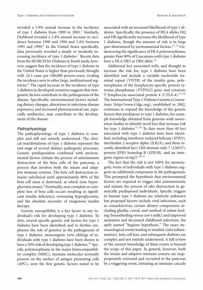
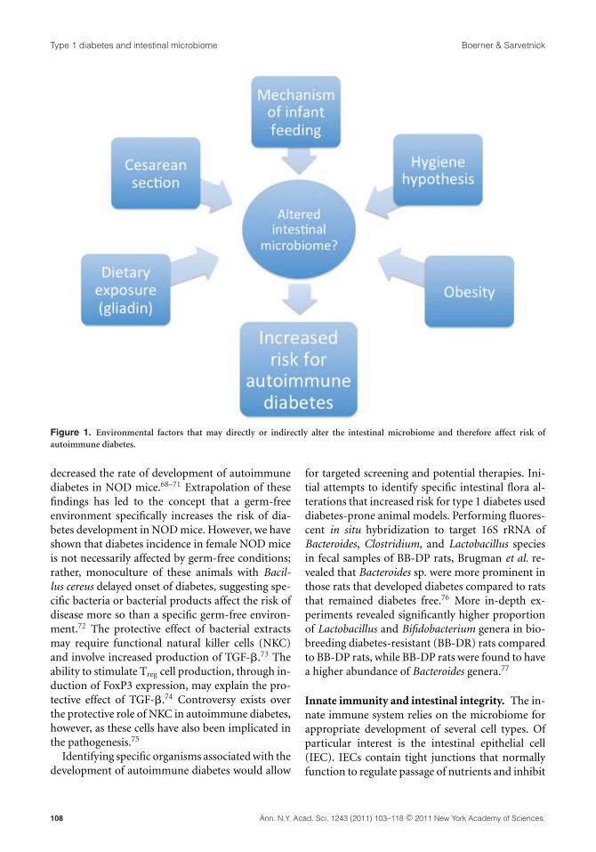
studies, combined with epidemiological data fromhumans, have begun to establish the many facets ofthe intestinal microbiome that directly affect the riskfor, and development of, autoimmune diabetes. Ad-ditionally, early therapeutic studies have also beenestablished directly targeting the intestinal micro-biome. As illustrated in Figure 1, several environ-mental factors thought to be triggers of autoim-mune diabetes may, at least in part, increase risk fordiabetes due to their effects on the composition ofthe intestinal microbiome.
Animal studiesAnimal models have provided a wealth of infor-mation regarding the influence of the intestinalflora on autoimmunity and autoimmune diabetesdevelopment.
Alterations in flora. Using mice raised in germ-free environments and subsequently exposed to mi-croorganisms of choice (gnotobiology), much hasbeen ascertained about the structural and functionaleffects of the intestinal flora on the immune sys-tem. Peyer’s patches, splenic germinal centers, andmesenteric lymph nodes of germ-free mice are sig-nificantly smaller and fewer in number comparedto mice raised in a normal environment.61 Defi-ciencies in the development of mucosal-associatedlymphoid tissue, specifically plasma cells and CD4+
T cells, are other consequences of a germ-free envi-ronment.62,63 In germ-free animals, reintroductionof normal gut flora can normalize size and cellularityof lymphoid structures and increase antibody pro-duction.64 In fact, colonization of germ-free micewith a single organism, segmented filamentous bac-terium (SFB), is sufficient to stimulate productionof IL-17–producing CD4+ T cells.65 The intestinalmicrobiome affects several other components of im-mune development and function, as will be outlinedthroughout this section.
The sentinel studies suggesting a role for mi-croorganism exposure in the pathogenesis of au-toimmune diabetes employed diabetes-prone ani-mals, NOD mice, and BioBreeding diabetes-prone(BB-DP) rats in particular, raised germ-free and/orexposed to a variety of infectious organisms orantigens.
Early studies revealed that in NOD mice, chronicviral infections were associated with a lowerincidence of diabetes.66,67 Active infection with my-cobacteria and stimulus with bacterial antigens also
Ann. N.Y. Acad. Sci. 1243 (2011) 103–118 c© 2011 New York Academy of Sciences. 107
Type 1 diabetes and intestinal microbiome Boerner & Sarvetnick
Figure 1. Environmental factors that may directly or indirectly alter the intestinal microbiome and therefore affect risk ofautoimmune diabetes.
decreased the rate of development of autoimmunediabetes in NOD mice.68–71 Extrapolation of thesefindings has led to the concept that a germ-freeenvironment specifically increases the risk of dia-betes development in NOD mice. However, we haveshown that diabetes incidence in female NOD miceis not necessarily affected by germ-free conditions;rather, monoculture of these animals with Bacil-lus cereus delayed onset of diabetes, suggesting spe-cific bacteria or bacterial products affect the risk ofdisease more so than a specific germ-free environ-ment.72 The protective effect of bacterial extractsmay require functional natural killer cells (NKC)and involve increased production of TGF-�.73 Theability to stimulate Treg cell production, through in-duction of FoxP3 expression, may explain the pro-tective effect of TGF-�.74 Controversy exists overthe protective role of NKC in autoimmune diabetes,however, as these cells have also been implicated inthe pathogenesis.75
Identifying specific organisms associated with thedevelopment of autoimmune diabetes would allow
for targeted screening and potential therapies. Ini-tial attempts to identify specific intestinal flora al-terations that increased risk for type 1 diabetes useddiabetes-prone animal models. Performing fluores-cent in situ hybridization to target 16S rRNA ofBacteroides, Clostridium, and Lactobacillus speciesin fecal samples of BB-DP rats, Brugman et al. re-vealed that Bacteroides sp. were more prominent inthose rats that developed diabetes compared to ratsthat remained diabetes free.76 More in-depth ex-periments revealed significantly higher proportionof Lactobacillus and Bifidobacterium genera in bio-breeding diabetes-resistant (BB-DR) rats comparedto BB-DP rats, while BB-DP rats were found to havea higher abundance of Bacteroides genera.77
Innate immunity and intestinal integrity. The in-nate immune system relies on the microbiome forappropriate development of several cell types. Ofparticular interest is the intestinal epithelial cell(IEC). IECs contain tight junctions that normallyfunction to regulate passage of nutrients and inhibit
108 Ann. N.Y. Acad. Sci. 1243 (2011) 103–118 c© 2011 New York Academy of Sciences.

Boerner & Sarvetnick Type 1 diabetes and intestinal microbiome
translocation of pathogenic organisms. IECs alsoexpress direct interaction with immune cells andproduce and respond to a variety of cytokine stim-uli. Many changes in IEC development and functionare observed when alterations are made to the in-testinal flora of animal models. For example, IECs ofgerm-free mice turn over at a slower rate than micewith normal intestinal microflora.78 Additionally,mice depleted of their normal flora by antibioticsalso show diminished IEC replication, significantalteration in IEC gene expression, and impaired pro-duction of antimicrobial factors including RegIII� ,a Gram-positive specific antimicrobial peptide.79
Reintroduction of colonic bacteria induces prolifer-ation of IECs in germ-free mice via toll-like receptor(TLR) recognition of the microbes.80
IECs play a prominent role in the developmentand regulation of the immune system. An intactintestinal epithelium serves to regulate passage ofantigens to DC and increased gut permeability dueto a compromised epithelium results in exposureto antigens that is a potential trigger for an autoim-mune response in predisposed individuals. IECs alsofunction to uptake, process, and present antigenand can promote activation of CD8+ T cells, as re-viewed.81 Additionally, IEC modulate function ofDC via production of factors such as IL-10 that pro-mote a tolerogenic DC response.82 IEC may also playa role in Treg cell expansion.83 Taken together, thisevidence suggests that IEC are critical to the properdevelopment of the innate immune system and dys-regulation of IEC by an altered intestinal flora mayresult in the development of autoimmunity.
Evidence in the BB-DP rat model has suggestedthat Lactobacillus johnsonii N6.2 is a specific or-ganism that may delay the onset of autoimmunediabetes via modulation of intestinal integrity.84 Intheir study, Valladares et al. also noted a significantincrease in the tight junction protein claudin-1 anddecreased oxidative stress in the ileal mucosa of theL. johnsonii treated animals. These findings suggestthat autoimmune diabetes may be the result of aninflammatory response initiated by a leaky gut ep-ithelium, which is the result of an altered intestinalflora.
Further evidence from animal studies suggeststhat the integrity of the gut epithelium plays aprominent role in autoimmune diabetes develop-ment. BB-DP rats express less of the tight junctionprotein claudin and display increased intestinal per-
meability compared to control Wistar rats.85 Trans-lating further, increased gut permeability was asso-ciated with a higher rate of diabetes development inBB-DP rats on a regular diet.86 Watts et al. addition-ally demonstrated that upregulation of zonulin—aprotein that serves to regulate tight junctions—and,subsequently, increased intestinal permeability sig-nificantly increased the rate of diabetes developmentin BB-DP rats.87
More recently, Wen et al. sought to establishhow the interaction between microorganisms andthe intestinal epithelium could trigger autoimmu-nity and diabetes development.88 The specific targetof this study was myeloid differentiation factor 88(MyD88), an adaptor molecule used by multipleTLR to regulate the innate immune response. Wild-type NOD mice developed diabetes quickly. NODmice with a MyD88 knockout (KO), however, wereprotected from diabetes development while germ-free, MyD88-KO NOD mice developed diabetes ina rapid fashion. Furthermore, recolonization of theMyD88-KO mice decreased diabetes development,suggesting protection from diabetes is microorgan-ism dependent. Knockdown of TLR2, TLR3, andTLR4 did not attenuate diabetes development, how-ever, suggesting that while some microorganismsprotect the host from developing diabetes, this pro-tection is independent of the TLR system.
Adaptive immune system. The development andfunction of the adaptive immune system is also di-rectly affected by the intestinal microbiota, a processthat may influence risk of disease. Treg cells secreteanti-inflammatory cytokines IL-10 and TGF-� andfunction to temper the immune response throughdown-regulation of both the innate and adaptiveimmune systems.89 Intestinal Treg cells in particu-lar play a prominent role in tolerance of oral anti-gens and microbes.90 Evidence from animal modelssuggests the presence of the intestinal microbiotais vital for the production of adequate numbers offunctional Treg cells from the intestine. In germ-freemice, Treg cells are less prominent and less effectivein suppressing T cell activity compared to Treg cellsin conventional mice.91 In an experimental model ofcolitis, IL-10 production from Treg cells of germ-freemice is also suppressed, as is the ability of these cellsto modulate disease.92 Conversion of CD4+ T cellsto Treg cells was stimulated by B. fragilis monocolo-nization of germ-free mice, a process dependent on
Ann. N.Y. Acad. Sci. 1243 (2011) 103–118 c© 2011 New York Academy of Sciences. 109
Type 1 diabetes and intestinal microbiome Boerner & Sarvetnick
the B. fragilis-produced polysachharide A (PSA).93
IL-10 production and T cell suppressive activity ofTreg cells were also enhanced in a PSA-dependentfashion.
T helper 17 cells (Th17) are a proinflammatorysubset of T cells that secrete interleukin 17 (IL-17),have antimicrobial activities, and have been impli-cated in many autoimmune diseases including mul-tiple sclerosis, inflammatory bowel disease, and pso-riasis.94 The intestine harbors the largest number ofTh17 cells, and the intestinal microbiota is likely re-quired for the production and expansion of thesecells.95–97
In a recent report, Lau et al. sought to un-derstand the mechanisms by which L. johnsoniiexposure can delay diabetes in BB-DP rats.98 In-terestingly, the study demonstrated enhanced Th17differentiation in the mesenteric lymph nodes ofanimals fed L. johnsonii. The authors suggested thatalthough Th17 cells are associated with insulitis theTh17 bias seen with L. johnsonii feeding limits thenecessary conversion to the diabetogenic Th1 celltype, thereby inhibiting or delaying onset of dia-betes. Others have suggested that a Th17 bias mayreduce the risk of autoimmune diabetes via mucosalprotection produced by IL-17 upregulation.99 Otherundefined mechanisms may also explain the effectseen with L. johnsonii exposure.
Other important regulatory cells of the immunesystem are directly influenced by the intestinal mi-crobiome, including DC, which do not develop inappropriate numbers in germ-free mice, and B cells,which produce IgA at a reduced amount in germ-free mice, compared to wild-type mice.100,101
Dietary factors. The intestinal microbiota’s influ-ence on nutrition and the potential role of thesefactors in autoimmune diabetes pathogenesis hasalso been explored in animal models. The initialevidence suggesting a role for nutrition in autoim-mune diabetes came from Hoorfar et al. in 1993.102
Using NOD mice exposed to wheat-flour diet or hy-drolyzed casein, this study revealed a significantlylower incidence of diabetes in mice receiving thehydrolyzed diet. Expanding further, Brugman et al.completely prevented diabetes by providing antibi-otics and a hydrolyzed casein diet to BB-DP rats.76
Additional studies have expanded on the dietaryhypothesis to more precisely pinpoint the compo-nents and associated mechanisms of diet that may
increase risk for autoimmune diabetes. Gliadin, aglycoprotein implicated in the intestinal damage ofceliac sprue, is the most extensively studied dietarycomponent in the pathogenesis of type 1 diabetes.NOD mice that lack exposure to dietary gluten de-velop diabetes at a significantly lower rate than micefed a standard diet.103–105 BB-DP rats fed a cereal-based diet develop diabetes associated with a pan-creatic Th1 cytokine pattern, compared to BB-DPrats on a protein-based diet who had less insulitis,a Th2 cytokine pattern, and overall lower incidenceof diabetes.106 Investigation into the mechanismsleading to development of insulitis and diabetes inanimals on a gliadin diet suggests that these proteinsincrease small intestine inflammation and intestinalpermeability.86,107 Furthermore, BB-DP rats have asignificantly higher proportion of Th1 cells in themesenteric lymph nodes upon exposure to wheatproteins.108 Pancreatic lymph node dendritic cellssample gut antigens and, upon recognizing proteinproducts, stimulate production of activated T cells,a process that leads to increased beta cell apopto-sis.109 Finally, gliadin exposure also suppresses Treg
cell production in NOD mice, which is another po-tential mechanism by which dietary exposure mayenhance genetic diabetes risk.110
The known association of alterations in diet andintestinal flora with diabetes development suggeststhat these two entities work in concert to affect dis-ease risk. With this in mind, the interactions be-tween intestinal bacteria and gliadin peptides arebeing closely examined. Hansen et al. examined theeffect of a gluten-free versus standard diet on bac-terial composition in NOD mice.111 Diabetes devel-oped in 47% of the standard-fed mice compared toonly 5% in the mice fed a gluten-free diet. Exami-nation of intestinal bacteria revealed a significantlylower prevalence of aerobic, microaerophilic, andanaerobic bacteria in the mice fed a gluten-free diet,compared to mice on a standard diet. Much of thedifference in bacterial composition was directly at-tributable to Gram-positive bacteria.
In vitro studies have provided additional infor-mation regarding the interactions between gliadinand intestinal flora. As shown by Laparra et al., in-testinal epithelial cells (Caco-2) in culture exposedto gliadin-derived peptides produce inflammatorycytokines, a process that was downregulated whenthese cell preparations were inoculated with Bi-fidiobacteria, an intestinal bacteria that has been
110 Ann. N.Y. Acad. Sci. 1243 (2011) 103–118 c© 2011 New York Academy of Sciences.

Boerner & Sarvetnick Type 1 diabetes and intestinal microbiome
associated with lower incidence of diabetes in an-imal models.112 The effect was most pronouncedwith Bifidobacterium longum. Gliadin peptide se-quences were modified upon exposure to B. longum,suggesting a mechanism by which “protective” in-testinal bacteria can indirectly maintain intestinalintegrity. The same research group recently exam-ined the proteome of Caco-2 cells after exposureto gliadin peptides in the presence or absence of B.longum.113 Protein expression was altered in Caco-2cells treated with gliadin peptides in the absenceof B. longum; specifically, expression of proteinsinvolved in cytoskeletion formation and apoptosiswas altered. These effects were ameliorated when B.longum was added to the cell culture.
Whereas B. longum may protect the intestinal ep-ithelium from gliadin-induced structural changes,other species of bacteria have been shown to actwith gliadin to synergistically alter the integrity ofthe intestinal epithelium. Germ-free rats exposedto gliadin were found to have fewer goblet cells inthe small intestine, a sign of early enteropathy, andthis finding was more pronounced in animals inocu-lated with Escherichia coli CBL2 or Shigella CBL2.114
Shigella CBL2 also augmented interferon-�–induced impairment of tight junctions, allowingenhanced translocation of gliadin into the laminapropria.
The results from the above studies indicate thatgliadin exposure increases the risk for diabetesthrough mechanisms involving both the integrityon the intestinal epithelium and the composition ofthe intestinal flora.
Probiotics. Expanding further on the concept of aprotective intestinal flora, modification of the in-testinal flora by probiotics has been investigated asa method to modulate the risk for diabetes. Lac-tobacillus casei-treated NOD mice were protectedfrom diabetes onset and were found to have reducednumbers of splenic CD8+ T cells and systemic in-flammatory markers.115 IL-2 levels were also higherin probiotic-treated mice, and the enhanced expres-sion of this cytokine may serve to stabilize FoxP3+
Treg cells, as recently described by Chen et al.116
NOD mice administered the probiotic compoundVSL#3 showed a reduced severity of insulitis, re-duced beta cell destruction, and lower rates of dia-betes development compared to control.117 In thisstudy, IL-10 expression was significantly increased
in the Peyer’s patches, spleen, and pancreas, suggest-ing an immunomodulatory effect of the probiotic.Another potential mechanism behind the protectiveeffect of probiotics may include these organisms’ability to inhibit adherence of enteropathogenic bac-teria by binding to intestinal epithelial cells and up-regulating mucin production.118,119
Human studiesUsing discoveries in animal models, studies are be-ginning to emerge establishing the role of intestinalflora and integrity in the development of autoim-mune diabetes in humans.
Alterations in flora. Attempts have been made todefine an understanding of the underlying intesti-nal flora alterations underlying autoimmune di-abetes development in humans. Using 16S RNAamplification techniques, Giongo et al. set out todefine specific taxa that differed between childrenwith type 1 diabetes and healthy controls.120 Overtime, the intestinal microbiota of children whodeveloped type 1 diabetes consisted of a higherproportion of the Bacteroidetes phyla and a lowerproportion of the Firmicutes phyla. Opposite find-ings were observed in control patients, in whomBacteroidetes phyla sequences decreased over timewhile Firmicutes sequences increased. Additionally,within the Bacteroidetes phyla, the Bacteroides ova-tus species represented 24% of the total increase incases. Though this study was small, comprising eightcase subjects and four controls, the findings repre-sented some of the first evidence of specific changesin the composition of intestinal bacteria in humanswith type 1 diabetes. Larger cohort studies such asThe Environmental Determinants of Diabetes in theYoung (TEDDY, currently in progress), are neededto further define alterations in the composition ofthe intestinal microbiome in humans and the under-lying mechanisms that lead to autoimmunity anddiabetes development.121
Intestinal integrity and immunity. Similar to ani-mal models of autoimmune diabetes, several studiesof intestinal integrity in human subjects with type 1diabetes have revealed evidence of increased intesti-nal permeability.122–124 Furthermore, the intestinalpermeability seen in individuals with type 1 diabetesis detectable prior to clinical onset of disease, sug-gesting that the intestine is directly involved in thedisease development.125 A recent study by Brown
Ann. N.Y. Acad. Sci. 1243 (2011) 103–118 c© 2011 New York Academy of Sciences. 111

Type 1 diabetes and intestinal microbiome Boerner & Sarvetnick
et al. used metagenomics to determine the poten-tial function of bacteria that differ between individ-uals with type 1 diabetes and healthy controls.126
Findings from this study may begin to explain thecontribution of the intestinal microbiome to main-tenance of intestinal integrity. Bacteria from indi-viduals with type 1 diabetes tended to have higherexpression of genes related to motility and adhesioncompared to controls. Additionally, evaluation of16S demonstrated a higher proportion of butyrate-producing and mucin-degrading bacteria in con-trols compared to cases. Both butyrate and mucinare thought to be directly involved in maintainingintestinal epithelial integrity. The authors suggesta hypothesis whereby healthy individuals harbor ahigher proportion of butyrate-producing bacteriathat help to maintain intestinal integrity and pre-vent autoimmunity. These preliminary findings mayhelp to advance the knowledge of the role of intesti-nal integrity in autoimmunity and type 1 diabetesdevelopment.
Inflammatory changes in the intestine of pa-tients with type 1 diabetes lend further evidencefor the involvement of the intestinal mucosa andaltered intestinal immunity in the pathogenesis oftype 1 diabetes. Though sometimes structurally nor-mal, small intestine biopsies of patients with type1 diabetes reveal increased expression of HLA-DR,HLA-DP, and intercellular adhesion molecule-1(ICAM-1).127 Frequently, microstructural intestinalchanges are seen in patients with type 1 diabetes,including changes to the microvilli and tight junc-tions.128 Higher densitites of IL-1�+ and IL-4+ cellsin these biopsies also point toward a heightened in-testinal inflammatory state in type 1 diabetes. Jeju-nal biopsies of patients with type 1 diabetes show ev-idence of mucosal inflammation and, upon in vitroexposure to gliadin, an exaggerated inflammatoryresponse compared to control.129 Finally, Treg cellproduction in the intestinal mucosa is suppressedin patients with type 1 diabetes, as demonstrated byreduced numbers of FoxP3+ cells in small intestinebiopsies of children with type 1 diabetes.130
Dietary factors. As in animal models, human in-testinal integrity appears to be compromised uponexposure to gliadin peptides. Human intestinal sam-ples exposed to gliadin ex vivo display amplified re-lease of zonulin and increased permeability.129 In-testinal samples from patients with celiac disease
had more robust and sustained permeability com-pared to nonceliac disease controls, consistent withthe theory of genetic predisposition enhancing riskin those with celiac disease. The role of gliadin in thepathogenesis of type 1 diabetes likely goes beyondaffecting intestinal integrity. Small bowel biopsies ofpatients with type 1 diabetes exposed to gliadin re-veal an exaggerated inflammatory response.129 Pro-duction of cytokines TNF-� and IL-8—both in-volved in the pathogenesis of type 1 diabetes—bymonocytes of susceptible humans is also stimu-lated by gliadin.131 Finally, islet cell autoimmunityappears to be exaggerated in individuals who areexposed to gliadin-containing foods prior to sixmonths of age.132 Given the association of type 1diabetes and celiac disease, and the role of gliadinin both, it is likely that these disease processes sharespecific risk factors and etiologies. Similar geneticsusceptibility between the two diseases, as revealedby Smyth et al., further enhances the concept ofshared pathogenic mechanisms.133
Probiotics. Based on initial promising results inanimal models, the use of probiotics to delay or pre-vent type 1 diabetes in humans has become an areaof interest. Studies in healthy humans have demon-strated that exposure to Lactobacillus plantarum en-hanced the expression of tight junction proteins,
Table 1. Organisms and other factors that may alter riskfor autoimmune diabetes in diabetes-prone animal mod-els and humans
May promote
May delay or prevent autoimmune
autoimmune diabetes diabetes
Chronic viral infectionsa Bacteroides sp.a,b
Mycobacterial infectiona Bacteroides generaa,b
Bacterial antigensa Gliadina,b
Monoculture with B.
cereusa
Impaired intestinal
epithelial integritya,b
Lactobacillus generaa,b Cow’s milkb
Lactobacillus johnsonii
N6.2a
Delivery by cesarean
sectionb
Lactobacillus caseia Hygiene hypothesisb
Probiotic VSL #3a Acute viral infectionsb
(coxsackievirus)
Bifidobacterium generaa
aAnimal models, bhumans.
112 Ann. N.Y. Acad. Sci. 1243 (2011) 103–118 c© 2011 New York Academy of Sciences.

Boerner & Sarvetnick Type 1 diabetes and intestinal microbiome
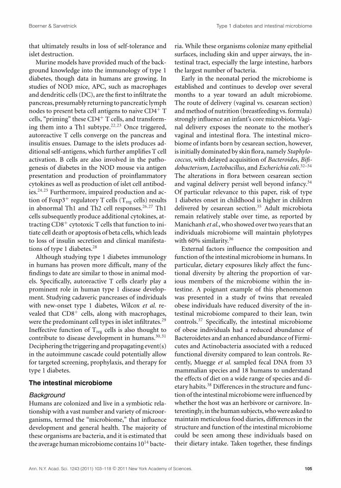
Figure 2. General schema outlining proposed relationships between intestinal flora/integrity and development of type 1 diabetesin genetically predisposed individuals.
suggesting the ability of these organisms to improveendothelial integrity.134 The PRODIA study, cur-rently ongoing in Finland, is investigating childrenwith genetic susceptibility to type 1 diabetes and theability of probiotics to decrease diabetes autoanti-bodies in this group.135
Conclusion
Type 1 diabetes is a complex series of events cul-minating in autoimmune destruction of pancre-atic beta cells, hyperglycemia, and risk for sub-sequent vascular complications. In those withgenetic predisposition, environmental factors affectdisease risk and pathogenesis. Table 1 briefly sum-marizes some of the specific organisms and factorsthat have been identified to potentially alter riskof autoimmune diabetes development in diabetes-prone animal models and humans. Evidence to datestrongly suggests that intestinal microbiota, throughtheir impact on immune development and intesti-nal structure and function, are vital to the patho-
genesis of type 1 diabetes, though the exact mech-anisms whereby intestinal bacteria are altered andhow those alterations influence type 1 diabetes de-velopment are still unclear. Figure 2 presents a gen-eral view of the knowledge to date regarding po-tential mechanisms in the development of type 1diabetes in genetically predisposed individuals andhow the intestinal microbiome may play a directrole in this process. Continued exploration into thespecific role of the intestinal microbiome in the de-velopment of type 1 diabetes will allow for targetedscreening and development of novel therapies totreat and ideally prevent the disease.
Acknowledgment
This work was supported by a generous grant fromthe J.W. Kieckhefer Foundation.
Conflicts of interest
The authors declare no conflicts of interest.
Ann. N.Y. Acad. Sci. 1243 (2011) 103–118 c© 2011 New York Academy of Sciences. 113
Type 1 diabetes and intestinal microbiome Boerner & Sarvetnick
References
1. Kalyva, E., E. Malakonaki, C. Eiser & D. Mamoulakis. 2011.Health-related quality of life (HRQoL) of children with type1 diabetes mellitus (T1DM): self and parental perceptions.Pediatr. Diabetes 12: 34–40.
2. Cameron, F.J., C. Clarke, K. Hesketh, et al. 2002. Regionaland urban Victorian diabetic youth: clinical and quality-of-life outcomes. J. Paediatr. Child. Health 38: 593–596.
3. Miller R.G., Secrest A.M., Sharma R.K., et al. 2011. Im-provements in life expectancy of type 1 diabetes: The Pitts-burgh Epidemiology of Diabetes Complications Study. 71stScientific Sessions of the American Diabetes AssociationAbstract Number 0078-OR.
4. Tao, B., M. Pietropaolo, M. Atkinson, et al. 2010. Estimatingthe cost of type 1 diabetes in the U.S.: a propensity scorematching method. PLoS One 5: e11501–e11512.
5. Green, A. & C.C. Patterson. 2001. Trends in the incidence ofchildhood-onset diabetes in Europe 1989–1998. Diabetolo-gia 44(Suppl 3): B3–B8.
6. DIAMOND Project Group. 2006. Incidence and trends ofchildhood type 1 diabetes worldwide 1990–1999. Diabet.Med. 23: 857–866.
7. Libman, I.M. & R.E. LaPorte. 2005. Changing trends inepidemiology of type 1 diabetes mellitus throughout theworld: how far have we come and where do we go fromhere. Pediatr. Diabetes 6: 119–121.
8. Dabelea, D., R.A. Bell, R.B. D’Agostino, Jr, et al. 2007.Incidence of diabetes in youth in the United States. JAMA297: 2716–2724.
9. Notkins, A.L. & A. Lernmark. 2001. Autoimmune type 1diabetes: resolved and unresolved issues. J. Clin. Invest. 108:1247–1252.
10. Redondo, M.J., P.R. Fain & G.S. Eisenbarth. 2001. Geneticsof type 1A diabetes. Recent Prog. Horm. Res. 56: 69–89.
11. Altobelli, E., A. Blasetti, R. Petrocelli, et al. 2005. HLADR/DQ alleles and risk of type I diabetes in childhood:a population-based case–control study. Clin. Exp. Med. 5:72–79.
12. Sanjeevi, C.B., S.K. Sedimbi, M. Landin-Olsson, et al. 2008.Risk conferred by HLA-DR and DQ for type 1 diabetes in0- to 35-year age group in Sweden. Ann. N.Y. Acad. Sci.1150: 106–111.
13. Thomson, G., W.P. Robinson, M.K. Kuhner, et al. 1988.Genetic heterogeneity, modes of inheritance, and riskestimates for a joint study of Caucasians with insulin-dependent diabetes mellitus. Am. J. Hum. Genet. 43: 799–816.
14. Bell, G.I., S. Horita & J.H. Karam. 1984. A polymorphic lo-cus near the human insulin gene is associated with insulin-dependent diabetes mellitus. Diabetes 33: 176–183.
15. Bottini, N., L. Musumeci, A. Alonso, et al. 2004. A func-tional variant of lymphoid tyrosine phosphatase is associ-ated with type I diabetes. Nat. Genet. 36: 337–338.
16. Ueda, H., J.M. Howson, L. Esposito, et al. 2003. Associationof the T-cell regulatory gene CTLA4 with susceptibility toautoimmune disease. Nature 423: 506–511.
17. Cooper, J.D., D.J. Smyth, A.M. Smiles, et al. 2008. Meta-analysis of genome-wide association study data identifies
additional type 1 diabetes risk loci. Nat. Genet. 40: 1399–1401.
18. Barrett, J.C., D.G. Clayton, P. Concannon, et al. 2009.Genome-wide association study and meta-analysis findthat over 40 loci affect risk of type 1 diabetes. Nat. Genet.41: 703–707.
19. Smyth, D.J., J.D. Cooper, R. Bailey, et al. 2006. A genome-wide association study of nonsynonymous SNPs identifiesa type 1 diabetes locus in the interferon-induced helicase(IFIH1) region. Nat. Genet. 38: 617–619.
20. Lowe, C.E., J.D. Cooper, T. Brusko, et al. 2007. Large-scalegenetic fine mapping and genotype-phenotype associationsimplicate polymorphism in the IL2RA region in type 1diabetes. Nat. Genet. 39: 1074–1082.
21. Bradfield, J.P., H.Q. Qu, K. Wang, et al. 2011. A genome-wide meta-analysis of six type 1 diabetes cohorts iden-tifies multiple associated Loci. PLoS Genet. 7: e1002293–e1002301.
22. Gagnerault, M.C., J.J. Luan, C. Lotton & F. Lepault. 2002.Pancreatic lymph nodes are required for priming of betacell reactive T cells in NOD mice. J. Exp. Med. 196: 369–377.
23. Turley, S., L. Poirot, M. Hattori, et al. 2003. Physiologicalbeta cell death triggers priming of self-reactive T cells bydendritic cells in a type-1 diabetes model. J. Exp. Med. 198:1527–1537.
24. Wong, F.S., L. Wen, M. Tang, et al. 2004. Investigation ofthe role of B-cells in type 1 diabetes in the NOD mouse.Diabetes 53: 2581–2587.
25. Serreze, D.V., S.A. Fleming, H.D. Chapman, et al. 1998.B lymphocytes are critical antigen-presenting cells forthe initiation of T cell-mediated autoimmune diabetes innonobese diabetic mice. J. Immunol. 161: 3912–3918.
26. Sgouroudis, E. & C.A. Piccirillo. 2009. Control of type 1diabetes by CD4+Foxp3 +regulatory T cells: lessons frommouse models and implications for human disease. Dia-betes Metab. Res. Rev. 25: 208–218.
27. Tang, Q., J.Y. Adams, C. Penaranda, et al. 2008. Central roleof defective interleukin-2 production in the triggering ofislet autoimmune destruction. Immunity 28: 687–697.
28. Wang, B., A. Gonzalez, C. Benoist & D. Mathis. 1996. Therole of CD8+ T cells in the initiation of insulin-dependentdiabetes mellitus. Eur. J. Immunol. 26: 1762–1769.
29. Willcox, A., S.J. Richardson, A.J. Bone, et al. 2009. Analysisof islet inflammation in human type 1 diabetes. Clin. Exp.Immunol. 155: 173–181.
30. Lindley, S., C.M. Dayan, A. Bishop, et al. 2005. Defectivesuppressor function in CD4(+)CD25(+) T-cells from pa-tients with type 1 diabetes. Diabetes 54: 92–99.
31. Tree, T.I., B.O. Roep & M. Peakman. 2006. A mini meta-analysis of studies on CD4+CD25+ T cells in human type1 diabetes: report of the Immunology of Diabetes SocietyT Cell Workshop. Ann. N.Y. Acad. Sci. 1079: 9–18.
32. Dominguez-Bello, M.G., E.K. Costello, M. Contreras, et al.2010. Delivery mode shapes the acquisition and structureof the initial microbiota across multiple body habitats innewborns. Proc. Natl. Acad. Sci. USA 107: 11971–11975.
33. Gronlund, M.M., O.P. Lehtonen, E. Eerola & P. Kero.1999. Fecal microflora in healthy infants born by differentmethods of delivery: permanent changes in intestinal flora
114 Ann. N.Y. Acad. Sci. 1243 (2011) 103–118 c© 2011 New York Academy of Sciences.

Boerner & Sarvetnick Type 1 diabetes and intestinal microbiome
after cesarean delivery. J. Pediatr. Gastroenterol. Nutr. 28:19–25.
34. Salminen, S., G.R. Gibson, A.L. McCartney & E. Isolauri.2004. Influence of mode of delivery on gut microbiotacomposition in seven year old children. Gut 53: 1388–1389.
35. Cardwell, C.R., L.C. Stene, G. Joner, et al. 2008. Caesareansection is associated with an increased risk of childhood-onset type 1 diabetes mellitus: a meta-analysis of observa-tional studies. Diabetologia 51: 726–735.
36. Manichanh, C., E. Varela, C. Martinez, et al. 2008. Thegut microbiota predispose to the pathophysiology of acutepostradiotherapy diarrhea. Am. J. Gastroenterol. 103: 1754–1761.
37. Turnbaugh, P.J., M. Hamady, T. Yatsunenko, et al. 2009. Acore gut microbiome in obese and lean twins. Nature 457:480–484.
38. Muegge, B.D., J. Kuczynski, D. Knights, et al. 2011. Dietdrives convergence in gut microbiome functions acrossmammalian phylogeny and within humans. Science 332:970–974.
39. Eckburg, P.B., E.M. Bik, C.N. Bernstein, et al. 2005. Diver-sity of the human intestinal microbial flora. Science 308:1635–1638.
40. Frank, D.N., A.L. St Amand, R.A. Feldman, et al.2007. Molecular-phylogenetic characterization of micro-bial community imbalances in human inflammatory boweldiseases. Proc. Natl. Acad. Sci. USA 104: 13780–13785.
41. Ley, R.E., C.A. Lozupone, M. Hamady, et al. 2008. Worldswithin worlds: evolution of the vertebrate gut microbiota.Nature reviews. Microbiology 6: 776–788.
42. Gill, S.R., M. Pop, R.T. Deboy, et al. 2006. Metagenomicanalysis of the human distal gut microbiome. Science 312:1355–1359.
43. Qin, J., R. Li, J. Raes, et al. 2010. A human gut microbialgene catalogue established by metagenomic sequencing.Nature 464: 59–65.
44. Arumugam, M., J. Raes, E. Pelletier, et al. 2011. Enterotypesof the human gut microbiome. Nature 473: 174–180.
45. Turnbaugh, P.J., R.E. Ley, M. Hamady, et al. 2007. Thehuman microbiome project. Nature 449: 804–810.
46. Wu, G.D., J.D. Lewis, C. Hoffmann, et al. 2010. Samplingand pyrosequencing methods for characterizing bacterialcommunities in the human gut using 16S sequence tags.BMC Microbiol. 10: 206–210.
47. Momozawa, Y., V. Deffontaine, E. Louis & J.F. Medrano.2011. Characterization of bacteria in biopsies of colon andstools by high throughput sequencing of the V2 region ofbacterial 16S rRNA gene in human. PLoS One 6: e16952–e16962.
48. Okada, H., C. Kuhn, H. Feillet & J.F. Bach. 2010. The ‘hy-giene hypothesis’ for autoimmune and allergic diseases: anupdate. Clin. Exp. Immunol. 160: 1–9.
49. Verdu, E.F., P. Bercik, B. Cukrowska, et al. 2000. Oral ad-ministration of antigens from intestinal flora anaerobicbacteria reduces the severity of experimental acute colitisin BALB/c mice. Clin. Exp. Immunol. 120: 46–50.
50. Dieleman, L.A., F. Hoentjen, B.F. Qian, et al. 2004. Re-duced ratio of protective versus proinflammatory cytokine
responses to commensal bacteria in HLA-B27 transgenicrats. Clin. Exp. Immunol. 136: 30–39.
51. Stepankova, R., F. Powrie, O. Kofronova, et al. 2007. Seg-mented filamentous bacteria in a defined bacterial cocktailinduce intestinal inflammation in SCID mice reconstitutedwith CD45RBhigh CD4 +T cells. Inflam. Bowl Dis. 13:1202–1211.
52. Veltkamp, C., S.L. Tonkonogy, Y.P. De Jong, et al. 2001.Continuous stimulation by normal luminal bacteria is es-sential for the development and perpetuation of colitis inTg(epsilon26) mice. Gastroenterology 120: 900–913.
53. Manichanh, C., L. Rigottier-Gois, E. Bonnaud, et al. 2006.Reduced diversity of faecal microbiota in Crohn’s dis-ease revealed by a metagenomic approach. Gut 55: 205–211.
54. Frank, D.N., A.L. St Amand, R.A. Feldman, et al.2007. Molecular-phylogenetic characterization of micro-bial community imbalances in human inflammatory boweldiseases. Proc. Natl. Acad. Sci. USA 104: 13780–13785.
55. Willing, B., J. Halfvarson, J. Dicksved, et al. 2009. Twinstudies reveal specific imbalances in the mucosa-associatedmicrobiota of patients with ileal Crohn’s disease. Inflam.Bowl Dis. 15: 653–660.
56. Ou, G., M. Hedberg, P. Horstedt, et al. 2009. Proximal smallintestinal microbiota and identification of rod-shaped bac-teria associated with childhood celiac disease. Am. J. Gas-troenterol. 104: 3058–3067.
57. Adlerberth, I., D.P. Strachan, P.M. Matricardi, et al. 2007.Gut microbiota and development of atopic eczema in 3European birth cohorts. J. Allergy Clin. Immunol. 120: 343–350.
58. Ochoa-Reparaz, J., D.W. Mielcarz, L.E. Ditrio, et al. 2009.Role of gut commensal microflora in the development ofexperimental autoimmune encephalomyelitis. J. Immunol.183: 6041–6050.
59. Toivanen, P. 2003. Normal intestinal microbiota in the ae-tiopathogenesis of rheumatoid arthritis. Ann. Rheum. Dis.62: 807–811.
60. Rehakova, Z., J. Capkova, R. Stepankova, et al. 2000. Germ-free mice do not develop ankylosing enthesopathy, a spon-taneous joint disease. Hum. Immunol. 61: 555–558.
61. Macpherson, A.J. & N.L. Harris. 2004. Interactions betweencommensal intestinal bacteria and the immune system. Nat.Rev. Immunol. 4: 478–485.
62. Macpherson, A.J., L. Hunziker, K. McCoy & A. Lamarre.2001. IgA responses in the intestinal mucosa againstpathogenic and non-pathogenic microorganisms. MicrobesInfection/Institut Pasteur 3: 1021–1035.
63. Macpherson, A.J., M.M. Martinic & N. Harris. 2002. Thefunctions of mucosal T cells in containing the indigenouscommensal flora of the intestine. Cell. Mol. Life Sci. 59:2088–2096.
64. Pabst, O., H. Herbrand, M. Friedrichsen, et al. 2006. Adap-tation of solitary intestinal lymphoid tissue in responseto microbiota and chemokine receptor CCR7 signaling. J.Immunol. 177: 6824–6832.
65. Ivanov, II, K. Atarashi, N. Manel, et al. 2009. Inductionof intestinal Th17 cells by segmented filamentous bacteria.Cell 139: 485–498.
Ann. N.Y. Acad. Sci. 1243 (2011) 103–118 c© 2011 New York Academy of Sciences. 115
Type 1 diabetes and intestinal microbiome Boerner & Sarvetnick
66. Oldstone, M.B. 1988. Prevention of type I diabetes innonobese diabetic mice by virus infection. Science 239:500–502.
67. Wilberz, S., H.J. Partke, F. Dagnaes-Hansen & L. Herberg.1991. Persistent MHV (mouse hepatitis virus) infectionreduces the incidence of diabetes mellitus in non-obesediabetic mice. Diabetologia 34: 2–5.
68. Martins, T.C. & A.P. Aguas. 1996. Changes in B and T lym-phocytes associated with mycobacteria-induced protectionof NOD mice from diabetes. J. Autoimmunity 9: 501–507.
69. Satoh, J., S. Shintani, K. Oya, et al. 1988. Treatment withstreptococcal preparation (OK-432) suppresses anti-isletautoimmunity and prevents diabetes in BB rats. Diabetes37: 1188–1194.
70. Sai, P. & A.S. Rivereau. 1996. Prevention of diabetes inthe nonobese diabetic mouse by oral immunological treat-ments. Comparative efficiency of human insulin and twobacterial antigens, lipopolysacharide from Escherichia coliand glycoprotein extract from Klebsiella pneumoniae. Dia-betes Metab. 22: 341–348.
71. Qin, H.Y. & B. Singh. 1997. BCG vaccination preventsinsulin-dependent diabetes mellitus (IDDM) in NOD miceafter disease acceleration with cyclophosphamide. J. Au-toimmun. 10: 271–278.
72. King, C. & N. Sarvetnick. 2011. The incidence of type-1diabetes in NOD mice is modulated by restricted flora notgerm-free conditions. PLoS One 6: e17049–e17052.
73. Alyanakian, M.A., F. Grela, A. Aumeunier, et al. 2006.Transforming growth factor-beta and natural killer T-cellsare involved in the protective effect of a bacterial extract ontype 1 diabetes. Diabetes 55: 179–185.
74. Fu, S., N. Zhang, A.C. Yopp, et al. 2004. TGF-beta in-duces Foxp3 + T-regulatory cells from CD4 + CD25—precursors. Am. J. Transplant. 4: 1614–1627.
75. Shi, F.D., M. Flodstrom, B. Balasa, et al. 2001. Germ linedeletion of the CD1 locus exacerbates diabetes in the NODmouse. Proc. Natl. Acad. Sci. USA 98: 6777–6782.
76. Brugman, S., F.A. Klatter, J.T. Visser, et al. 2006. Antibiotictreatment partially protects against type 1 diabetes in theBio-Breeding diabetes-prone rat. Is the gut flora involvedin the development of type 1 diabetes? Diabetologia 49:2105–2108.
77. Roesch, L.F., G.L. Lorca, G. Casella, et al. 2009. Culture-independent identification of gut bacteria correlated withthe onset of diabetes in a rat model. ISME J. 3: 536–548.
78. Pull, S.L., J.M. Doherty, J.C. Mills, et al. 2005. Activatedmacrophages are an adaptive element of the colonic epithe-lial progenitor niche necessary for regenerative responsesto injury. Proc. Natl. Acad. Sci. USA 102: 99–104.
79. Reikvam, D.H., A. Erofeev, A. Sandvik, et al. 2011. Deple-tion of murine intestinal microbiota: effects on gut mucosaand epithelial gene expression. PLoS One 6: e17996–e18009.
80. Rakoff-Nahoum, S., J. Paglino, F. Eslami-Varzaneh, et al.2004. Recognition of commensal microflora by toll-likereceptors is required for intestinal homeostasis. Cell 118:229–241.
81. Campbell, N., X.Y. Yio, L.P. So, et al. 1999. The intestinalepithelial cell: processing and presentation of antigen to themucosal immune system. Immunol. Rev. 172: 315–324.
82. Jarry, A., C. Bossard, C. Bou-Hanna, et al. 2008. MucosalIL-10 and TGF-beta play crucial roles in preventing LPS-driven, IFN-gamma-mediated epithelial damage in humancolon explants. J. Clin. Invest. 118: 1132–1142.
83. Westendorf, A.M., D. Fleissner, L. Groebe, et al. 2009.CD4+Foxp3+ regulatory T cell expansion induced byantigen-driven interaction with intestinal epithelial cellsindependent of local dendritic cells. Gut 58: 211–219.
84. Valladares, R., D. Sankar, N. Li, et al. 2010. Lactobacillusjohnsonii N6.2 mitigates the development of type 1 diabetesin BB-DP rats. PLoS One 5: e10507–e10516.
85. Neu, J., C.M. Reverte, A.D. Mackey, et al. 2005. Changes inintestinal morphology and permeability in the biobreedingrat before the onset of type 1 diabetes. J. Pediatr. Gastroen-terol. Nutr. 40: 589–595.
86. Meddings, J.B., J. Jarand, S.J. Urbanski, et al. 1999. In-creased gastrointestinal permeability is an early lesion in thespontaneously diabetic BB rat. Am. J. Physiol. 276: G951–G957.
87. Watts, T., I. Berti, A. Sapone, et al. 2005. Role of the intesti-nal tight junction modulator zonulin in the pathogenesis oftype I diabetes in BB diabetic-prone rats. Proc. Natl. Acad.Sci. USA 102: 2916–2921.
88. Wen, L., R.E. Ley, P.Y. Volchkov, et al. 2008. Innate immu-nity and intestinal microbiota in the development of Type1 diabetes. Nature 455: 1109–1113.
89. Campbell, D.J. & M.A. Koch. 2011. Phenotypical and func-tional specialization of FOXP3 +regulatory T cells. Naturereviews. Immunology 11: 119–130.
90. Dubois, B., G. Joubert, M. Gomez de Aguero, et al. 2009.Sequential role of plasmacytoid dendritic cells and regula-tory T cells in oral tolerance. Gastroenterology 137: 1019–1028.
91. Ostman, S., C. Rask, A.E. Wold, et al. 2006. Impaired reg-ulatory T cell function in germ-free mice. Eur. J. Immunol.36: 2336–2346.
92. Strauch, U.G., F. Obermeier, N. Grunwald, et al. 2005.Influence of intestinal bacteria on induction of regulatoryT cells: lessons from a transfer model of colitis. Gut 54:1546–1552.
93. Round, J.L. & S.K. Mazmanian. 2010. Inducible Foxp3+regulatory T-cell development by a commensal bacteriumof the intestinal microbiota. Proc. Natl. Acad. Sci. USA 107:12204–12209.
94. Hu, Y., F. Shen, N.K. Crellin & W. Ouyang. 2011. The IL-17 pathway as a major therapeutic target in autoimmunediseases. Ann. N.Y. Acad. Sci. 1217: 60–76.
95. Ivanov, II, L. Frutos Rde, N. Manel, et al. 2008. Specificmicrobiota direct the differentiation of IL-17-producingT-helper cells in the mucosa of the small intestine. CellHost Microbe 4: 337–349.
96. Zaph, C., Y. Du, S.A. Saenz, et al. 2008. Commensal-dependent expression of IL-25 regulates the IL-23-IL-17axis in the intestine. J. Exp. Med. 205: 2191–2198.
97. Gaboriau-Routhiau, V., S. Rakotobe, E. Lecuyer, et al. 2009.The key role of segmented filamentous bacteria in the co-ordinated maturation of gut helper T cell responses. Im-munity 31: 677–689.
116 Ann. N.Y. Acad. Sci. 1243 (2011) 103–118 c© 2011 New York Academy of Sciences.

Boerner & Sarvetnick Type 1 diabetes and intestinal microbiome
98. Lau, K., P. Benitez, A. Ardissone, et al. 2011. Inhibitionof type 1 diabetes correlated to a Lactobacillus john-sonii N6.2-mediated Th17 bias. J. Immunol. 186: 3538–3546.
99. Vaarala, O. 2011. The gut as a regulator of early inflam-mation in type 1 diabetes. Curr. Opin. Endocrinol. DiabetesObes. 18: 241–247.
100. Williams, A.M., C.S. Probert, R. Stepankova, et al. 2006.Effects of microflora on the neonatal development of gutmucosal T cells and myeloid cells in the mouse. Immunology119: 470–478.
101. Hapfelmeier, S., M.A. Lawson, E. Slack, et al. 2010. Re-versible microbial colonization of germ-free mice revealsthe dynamics of IgA immune responses. Science 328: 1705–1709.
102. Hoorfar, J., K. Buschard & F. Dagnaes-Hansen. 1993. Pro-phylactic nutritional modification of the incidence of dia-betes in autoimmune non-obese diabetic (NOD) mice. Br.J. Nutr. 69: 597–607.
103. Funda, D.P., A. Kaas, H. Tlaskalova-Hogenova & K.Buschard. 2008. Gluten-free but also gluten-enriched(gluten+) diet prevent diabetes in NOD mice; the glutenenigma in type 1 diabetes. Diabetes Metab. Res. Rev. 24:59–63.
104. Schmid, S., K. Koczwara, S. Schwinghammer, et al. 2004.Delayed exposure to wheat and barley proteins reduces dia-betes incidence in non-obese diabetic mice. Clin. Immunol.111: 108–118.
105. Locke, N.R., S. Stankovic, D.P. Funda & L.C. Harri-son. 2006. TCR gamma delta intraepithelial lymphocytesare required for self-tolerance. J. Immunol. 176: 6553–6559.
106. Scott, F.W., H.E. Cloutier, R. Kleemann, et al. 1997. Poten-tial mechanisms by which certain foods promote or inhibitthe development of spontaneous diabetes in BB rats: dose,timing, early effect on islet area, and switch in infiltratefrom Th1 to Th2 cells. Diabetes 46: 589–598.
107. Maurano, F., G. Mazzarella, D. Luongo, et al. 2005. Smallintestinal enteropathy in non-obese diabetic mice fed a dietcontaining wheat. Diabetologia 48: 931–937.
108. Chakir, H., D.E. Lefebvre, H. Wang, et al. 2005. Wheatprotein-induced proinflammatory T helper 1 bias inmesenteric lymph nodes of young diabetes-prone rats. Di-abetologia 48: 1576–1584.
109. Turley, S.J., J.W. Lee, N. Dutton-Swain, et al. 2005. En-docrine self and gut non-self intersect in the pancreaticlymph nodes. Proc. Natl. Acad. Sci. USA 102: 17729–17733.
110. Ejsing-Duun, M., J. Josephsen, B. Aasted, et al. 2008. Di-etary gluten reduces the number of intestinal regulatory Tcells in mice. Scandinavian J. Immunol. 67: 553–559.
111. Hansen, A.K., F. Ling, A. Kaas, et al. 2006. Diabetes preven-tive gluten-free diet decreases the number of caecal bacteriain non-obese diabetic mice. Diabetes Metab. Res. Rev. 22:220–225.
112. Laparra, J.M. & Y. Sanz. 2010. Bifidobacteria inhibit theinflammatory response induced by gliadins in intestinalepithelial cells via modifications of toxic peptide generationduring digestion. J. Cell Biochem. 109: 801–807.
113. Oliares, M., M. Laparra & Y. Sanz. 2011. Influence of Bifi-dobacterium longum CECT 7347 and gliadin peptides onintestinal epithelial cell proteome. J. Agric. Food Chem. 59:7666–7671.
114. Cinova, J., G. De Palma, R. Stepankova, et al. 2011. Roleof intestinal bacteria in gliadin-induced changes in intesti-nal mucosa: study in germ-free rats. PLoS One 6: e16169–e16179.
115. Matsuzaki, T., Y. Nagata, S. Kado, et al. 1997. Preventionof onset in an insulin-dependent diabetes mellitus model,NOD mice, by oral feeding of Lactobacillus casei. APMIS105: 643–649.
116. Chen, Q., Y.C. Kim, A. Laurence, et al. 2011. IL-2 controlsthe stability of Foxp3 expression in TGF-{beta}-inducedFoxp3+ T cells in vivo. J. Immunol. 186: 6329–6337.
117. Calcinaro, F., S. Dionisi, M. Marinaro, et al. 2005. Oralprobiotic administration induces interleukin-10 produc-tion and prevents spontaneous autoimmune diabetes inthe non-obese diabetic mouse. Diabetologia 48: 1565–1575.
118. Johansson, M.L., G. Molin, B. Jeppsson, et al. 1993. Ad-ministration of different Lactobacillus strains in fermentedoatmeal soup: in vivo colonization of human intestinalmucosa and effect on the indigenous flora. Appl. Environ.Microbiol. 59: 15–20.
119. Mack, D.R., S. Michail, S. Wei, et al. 1999. Probiotics inhibitenteropathogenic E. coli adherence in vitro by inducingintestinal mucin gene expression. Am. J. Physiol. 276: G941–G950.
120. Giongo, A., K.A. Gano, D.B. Crabb, et al. 2010. Towarddefining the autoimmune microbiome for type 1 diabetes.ISME J. 5: 82–91.
121. TEDDY Study Group. 2008. The Environmental Determi-nants of Diabetes in the Young (TEDDY) Study. Ann. N.Y.Acad. Sci. 1150: 1–13.
122. Carratu, R., M. Secondulfo, L. de Magistris, et al. 1999. Al-tered intestinal permeability to mannitol in diabetes melli-tus type I. J. Pediatr. Gastroenterol. Nutr. 28: 264–269.
123. Kuitunen, M., T. Saukkonen, J. Ilonen, et al. 2002. Intesti-nal permeability to mannitol and lactulose in children withtype 1 diabetes with the HLA-DQB1∗02 allele. Autoimmu-nity 35: 365–368.
124. Sapone, A., L. de Magistris, M. Pietzak, et al. 2006. Zonulinupregulation is associated with increased gut permeabilityin subjects with type 1 diabetes and their relatives. Diabetes55: 1443–1449.
125. Bosi, E., L. Molteni, M.G. Radaelli, et al. 2006. Increasedintestinal permeability precedes clinical onset of type 1diabetes. Diabetologia 49: 2824–2827.
126. Brown, C.T., A.G. Davis-Richardson, A. Giongo, et al. 2011.Gut microbiome metagenomics analysis suggests a func-tional model for the development of autoimmunity fortype 1 diabetes. PLoS One 6: e25792–e25801.
127. Westerholm-Ormio, M., O. Vaarala, P. Pihkala, et al. 2003.Immunologic activity in the small intestinal mucosa ofpediatric patients with type 1 diabetes. Diabetes 52: 2287–2295.
128. Secondulfo, M., D. Iafusco, R. Carratu, et al. 2004. Ul-trastructural mucosal alterations and increased intestinal
Ann. N.Y. Acad. Sci. 1243 (2011) 103–118 c© 2011 New York Academy of Sciences. 117
Type 1 diabetes and intestinal microbiome Boerner & Sarvetnick
permeability in non-celiac, type I diabetic patients. Digest.Liver Dis. 36: 35–45.
129. Auricchio, R., F. Paparo, M. Maglio, et al. 2004. In vitro-deranged intestinal immune response to gliadin in type 1diabetes. Diabetes 53: 1680–1683.
130. Tiittanen, M., M. Westerholm-Ormio, M. Verkasalo, et al.2008. Infiltration of forkhead box P3-expressing cells insmall intestinal mucosa in coeliac disease but not in type 1diabetes. Clin. Exp. Immunol. 152: 498–507.
131. Cinova, J., L. Palova-Jelinkova, L.E. Smythies, et al. 2007.Gliadin peptides activate blood monocytes from pa-tients with celiac disease. J. Clin. Immunol. 27: 201–209.
132. Ziegler, A.G., S. Schmid, D. Huber, et al. 2003. Early infant
feeding and risk of developing type 1 diabetes-associatedautoantibodies. JAMA 290: 1721–1728.
133. Smyth, D.J., V. Plagnol, N.M. Walker, et al. 2008. Sharedand distinct genetic variants in type 1 diabetes and celiacdisease. N. Engl. J. Med. 359: 2767–2777.
134. Karczewski, J., F.J. Troost, I. Konings, et al. 2010. Regula-tion of human epithelial tight junction proteins by Lac-tobacillus plantarum in vivo and protective effects on theepithelial barrier. Am. J. Physiol. Gastrointest. Liver Physiol.298: G851–G859.
135. Ljungberg, M., R. Korpela, J. Ilonen, et al. 2006. Probioticsfor the prevention of beta cell autoimmunity in children atgenetic risk of type 1 diabetes—the PRODIA study. Ann.N.Y. Acad. Sci. 1079: 360–364.
118 Ann. N.Y. Acad. Sci. 1243 (2011) 103–118 c© 2011 New York Academy of Sciences.


![The Gut-Diabetes Connection w VU slides 9-25-18 [Read-Only] · Describe the microbiome development and maturation 2. Recognize the microbiome relationship to human body metabolism,](https://img.pdfslide.us/doc/110x75/5f53f479b61c2213e5688667/the-gut-diabetes-connection-w-vu-slides-9-25-18-read-only-describe-the-microbiome.jpg)