Embed Size (px)
Citation preview

2
Two-Stage Urethroplasty for Severe Primary Hypospadias
Yun-Man Tang and Shao-Ji Chen Sichuan Provincial People’s Hospital
China
1. Introduction
Surgical repair of hypospadias is challenging and problematic even for the most experienced specialists, and this is especially true when severe and complicated case is confronted. In the history of hypospadiology, surgeons have developed a vast armamentarium of hypospadias repairs that are still in use, with no procedure able to deal with varied cases universally. Debates persist, though the focus of controversies alter. Sometimes we found us involved in a regeneration of old era, an example occasion is that many of us nowadays are faced upon the aroused enthusiasm of two-stage repair for severe hypospadias. In the early era of hypospadias surgery, two-stage correction with orthoplasty and urethra bed substitution only or as stage I repair, followed by urethroplasty in stage II is a widely accepted strategy in hypospadias repair. The representitive techniques include Denis-Brown procedure and its varied modifications. These techniques are still in use in many centers nowadays (Bar et al, 2005; Brown, 1949; Gearhart & Witherington, 1979; Patel & Caldamone, 2010). The reason of wide acceptance of such techniques is more for technical simplicity tnan for satisfactory function or cosmesis. In that days, single-stage accomplishment of hyopspadias repair for surgeons was a dream but nightmare concerning the technical poverty and disappointing outcome. It was largely the contribution of John Duckett that the concept and technique of single-stage repair gained worldwide acceptance. The classic transverse prepucial island tube technique became the norm in numerous centers and is still the mainstay for correction of severe hypospadias in many centers currently (Duckett, 2002a, 2002b; MacGillivray et al, 2002; Soutis et al, 2003). With attention paid to long term outcome and pursuit on cosmesis as time goes on, pitfalls of such island and tube flap urethroplasty in one stage have been seriously concerned by more and more authors. Meatal configuration, glans appearance, limited local tissue for repair, indeed high complication rate in long-term follow-up, the problems such as fistula, glans/neourethra dihiscence, stricture, urethral diverticulum, are all included in criticism and reevaluation. (Castanon et al, 2000; Dermibilek et al, 2001; Dewan et al, 1991; Lam et al, 2005; Powell et al, 2000). Nowadays, a technique preponderance is obvious in management of mild to moderate hypospadias, ie, application of Snodgrass technique. But for those extremely severe cases, the popular single-stage strategies are still highly problematic. Inspired by free graft techniques used in plastic surgery, some specilists are back to the almost aridit field of two-
www.intechopen.com
Current Concepts of Urethroplasty
10
stage repairs with introducing a free graft as urethra bed for urethroplasty in hypospadias management. The grafts were harvested from extragenital skin, inner prepuce, and evolved to oral mucosa. The latter becomes a new gold standard as urethra substitute in two-stage repair advocated by some authors today (Goyal et al, 2010; Markiewicz et al, 2007; Mokhless et al, 2007; Simonato et al, 2008). Notwithstanding the dispute on single-stage vs two-stage repairs is ardent, agreement exists that procedure assignment in hypospadias repair is highly based on the patient’s individual anomaly status and on the surgeon’s individual experience and preference. And, surgical goals are in common, ie, to reconstruct a normal or near-normal appearance mimiking that of a post-circumcision one, to place a water-proof neourethra with adequate and even caliber extending to the apex of the glans for stand voiding and normal urine stream, to create a straight penis adequate for sexual intercorse, and to control the risk of complications (Baskin & Ebbers, 2006).
2. Fundamental requirements in two-stage repair
General process of staged surgery includes adequate orthoplasty, urethra bed preparation and glans/grove augmentation in stage I, and in stage II accomplishment of tubular urethroplasty, meatoplasty, glansplasty, penoplasty and scrotoplasty.
2.1 Stage I Common requirements in stage I surgery include the follows : Satisfactory orthoplasty should be achieved in stage I. In most of the severe cases, penile curvature is related to urethral tethering and categorized to group IV chordee (Donnahoo et al, 1998; Mingin & Baskin, 2002; Tang et al, 2007). Plate transection and removal of ventral fibrous tissue is mandatory for orthoplasty in most cases to obtain a straight and length-spared penis, instead of dorsal plication alone. When plate transection and orthoplasty have been done, usually a large defect between the urethrostomy opening or plate incised margin and subcoronal margin is created. A well urethra bed should be prepared for future tubularization. Neourethral bed is desired to be with appropriate epithelial histological properties withstanding exposure to air until stage II tube urethroplasty, with flat than lumpy surface, with adequate axial length extending to the apex of the glans, with width enough for future retrieval as reconstruction of even and full circumferencial caliber, with good vascularity avoiding acute/chronic ischemia and contracture. Small glans and shallow groove present in a high proportion of severe hypospadias cases, thus augmentation in stage I is warranted.
2.2 Stage II In stage II, the aim of surgery is to accomplish the whole correction of the deformities. In most cases, orthoplasy was satisfactory and the urethal reconstruction becomes the fosus problem. If chordee present as incomplete correction or as recurrence, further management is required. Urethroplasty is the central problem resolved in stage II correction. With good preparation of urethra bed in stage I, tubularization of the urethra bed for long segment urethroplasty is out of chanllenging. The caliber of the neourethra should be adequate, uniform and similar to the proximal normal urethra. When free graft is the choice of urethra bed substitute in
www.intechopen.com

Two-Stage Urethroplasty for Severe Primary Hypospadias
11
stage I, graft take should be unquestionable, or else additional tissue should be involved in urethral reconstruction. To guarantee a water proofing neourethra, an additional vascularized coverage is of great importance. Relocation of meatus is optimally made on the apex of the glans and be slit-like. In stage II, a cornical and plump glans is realigned following meatoplasty. Skin coverage of the penile shaft is required to be smooth in surface, even in axial view, and adequate in length. Bifid scrotum and penoscrotal transposition are frequently associated with severe hypospadias. Such associated deformities desire correction, usually accomplished in stage II hypospadias surgery. Elemination of scrotal bifida is usually out of problem. While satisfactory correction of severe translocation necessitating radical dissection and may threaten reliability of urethral replacement, thus to leave penoscrotal correction to a future surgery is rational in some cases.
2.3 New concept of two-stage repair Indeed that the aim of sgate I surgery should be to prepare optimal local anatomic status for stage II other than doctrinal orthoplasty, urethra bed preparation and glans/groove augmentation. Shall we do more in stage I than leave behind so many problems awaiting future resolution? In popular two-stage repair procedures, stage II surgery still bears high risk of complications (Bracka , 2011). This can be explained with the facts that: (1) long segment urethral reconstruction is associated with high risk of fistula, stenosis/stricture, diverticulum/dilatation, and curvature recurrence; and (2) extensive coverage of neourethra as well as correction of bifid scrotum and penoscrotal transposition denotes radical trauma, underlying infection, fascia tethering curvature, and threatened vascularity. Another usual problem of such radical reconstruction, concerning complicated plastic contents vs limited local tissue, is the sacrfice of cosmesis, which has profound psychological disturbance. Some authors tried modifications of the stage I procedure with partial urethroplasty especially in cases owning a relatively long and healthy urethral plate (Schumacher et al, 1997; Cheng et al, 2003). Partial urethroplasty and additional correction of anatomical alterations achieved in stage I surgery might aleviate the reconstructive burden in stage II. We further advocate a new concept of two-stage hypospadias repair, that two-stage strategy can be either more radical or more conservative (article in press). With technical development and individual experience gathering, one-stage repair of severe hypospadias is advocated by quite a number of specialists, let alone by patients. Our concept of “conservative” denotes that two-stage repair be indicated in those who could be repaired in single-stage, while complicated correction is anticipated with high risk of complications and poor appearance. The concept of “radical” denotes that when two-stage is indicated, much more can be done in stage I to diminish the risks in stage II surgery. In addition to sufficient orthoplasty, partial urethroplasty is achieved distal to the penoscrotal junction, and in some cases extended to the subcoronal area. The defect between neomeatus and the glans tip is substituted with skin flap as urethra bed, meanwhile glans/groove augmentation is accomplished. Stand voiding can be expected with such management. With partial urethroplasty carried out, scrotal bifida is eliminated, penoscrotal transposition resolved in a degree, and penoscrotal angle created. At completion of these steps in stage I surgery, the anomaly becames a much milder variant of hypospadias. Thus reconstruction of stage II surgery is much more easier and much less risky. With twice chances of cosmetic tailor, good appearance can be expected.
www.intechopen.com

Current Concepts of Urethroplasty
12
2.4 Indications of two-stage repair in severe primary hypospadias Two-stage repair can be indicated in severe primary hypospadias when: (1) severe
chordee (greater than 45 degree) is related to urethral tethering (type IV chodee), and
sufficient plate transection warrants long segment urethral substitute; (2) genital skin
tissue is insufficient for reconstruction; (3) development of the penis is poor with
microphallus and underdeveloped glans/groove, and orthotopic meatus is not likely to be
created; (4) severe penoscrotal transposition is associated, with its correction the
urethroplasty may be threatened; (5) dorsal hood is not suitable for flap transfer and
tubularization in that tissue volume is inadequate, unfolding of prepuce hood endangers
vascularity or is dissatisfactory and resulted in a highly irregular surface, or vasculature
of dorsal skin is of network type (Yucel et al, 2004); (6) poor cosmesis is expected with
single-stage repair; (7) surgeon experience of hypospadias correction is immature
(Manzoni & Bracka, 2004; Price et al, 2003; Titley & Bracka,1998). The prevalence of
hypospadias is high. The surgical reconstruction is complicated, problematic and this
anomaly should be referred to experienced pediatric urologist, pediatric surgeon, general
urologist, or plastic surgeon, but indeed a large number of surgeons with less experience
are involved in treatment of this disease. Experienced specialists are able to handle varied
procedures to deal with individual cases, but for surgeons who manage hypospadias less
than 40-60 cases per year, decision-making is difficult. When the less experienced
surgeons are confronted with severe hypospadias, a strategy of relatively easy-handled
two-stage procedure is a wise choice.
3. Detail concerns in surgery
Practical procedures used in hypospadias varies so greatly that more than 300 procedures
are available. To decide which procedure be optimal in individual patient is without
consensus, while some common concerns in methodology exist when hypospadias
anormaly is disassociated into several deformity parts.
3.1 The nature and severity of chordee With the aid of intraoperative artifical erection test, the nature and severity of chordee is
defined. Chordee, with or without hypospadias, is categorized into 4 groups, namely skin
tethering as type I, fibrotic fascia as II, corporal disproportion as III, and urethral tethering
as IV (Donnahoo et al, 1998; Mingin & Baskin, 2002; Tang et al, 2007). In most cases of
severe hypospadias, the ventral curvature is of type IV or III. Correction of urethral
tethering warrants plate transection, chordee resection and urethral substitute. Multiform
procedures are used for correction of penile disproportion. Both dorsal and ventral
approaches are applicable (Bologna et al, 1999), and currently the most popular procedure
is dorsal albuginea plication in the midline area (Bar Yosef et al, 2004; Baskin et al, 2000;
Soygur et al, 2004; Yucel et al, 2006). Concerning often poorly developed penis in severe
hypospadias, ventral graft is beneficial for shaft lengthening. More radical and
complicated procedures including corporeal rotation and penile disassembly are
advocated by few authors to correct extreme curvature (Decter, 1999; Mitchell & Bagli,
1996). Additional dorsal plication is nowadays a popular treatment in severe hypospadias
with urethral tethering to achieve satisfactory orthoplasty and to save tissue mobilized for
urethroplasty.
www.intechopen.com
Two-Stage Urethroplasty for Severe Primary Hypospadias
13
3.2 Meatus location and morphology Usually a natural location of the hypospadiac meatus can not represent the severity of the
anomaly. This is extremely true in severe hypospadias. When the length of urethral
replacement is assessed based on a meatus location, preceding sufficient orthoplasty and
urethrotomy on the distal thin urethral are needed. In pinhole meatus, meatotomy is desired.
3.3 Quality of the urethral plate The urethral plate of severe hypospadias can be fibrotic or relatively healthy. When
transected and released, the fibrotic plate contracts dramatically, and removal of the fibrous
tissue to the level of healthy urethal is rational. If the plate is healthy and with proper
length, it can be preserved as part of urethral substitute. Dense fibrotic band resection also
can be resected with elevation of the overlying plate and distal urethra to satisfy orthoplasty
and preservation of plate.
3.4 Size and configuration of the glans/groove A wide groove on flat glans may be preserved intact for stage II urethroplasty, meatoplasty and glansplasty. But in most occasions of severe hypospadias, the glans/groove is underdeveloped with small glans and narrow or shallow groove. Thus glans/groove augmentation is required in stage I. For augmentation, wing dissection of the glans is carried out with either a midline incision or two para-groove incisions. Resurfacing of the raw area with flap or graft leads to effective glans augmentation. When glans/groove size is still inadequate for full circumferential tube urethroplasty in stage II, the strategies of choice may be midline incision as in Snodgrass procedure or, in extreme cases, in stage I to prepare more axial length of urethra bed in the distal shaft area allowing stage II mobilization and advancement, and another wing dissection made to utilize the glans/groove tissue all for meatoplasty and glansplasty.
3.5 Tissue volume, configuration, and vascularity of the dorsal prepuce As an anatomic feature, preputial skin is defect ventrally and accomodated dorsally to form a hood appearance. The dorsal hood skin is known as the best for urethral reconstruction. Abundant hood tissue may be used to accomplish urethra substitute and penile body skin cover, thus to complete a single-stage repair is possible. But in most severe cases, the tissue obtainable for urethral substiture is limited and a two-stage procedure becomes considerable. Radojicic & Perovic (Radojicic & Perovic, 2004) evaluated the prepuce in hypospadias and categorized the prepuce into 6 groups, namely group A—“monk’s hood” or “1 humped” (43 cases, 24.7%), B—“cobra eyes” (80, 45.9%), C—“normal” (intact) (4, 2.3%), D—“flat” (24, 13.8%), E—“v”-shaped (16, 9.2%), and F—“collar-scarf” (7, 4.0%). Cobra eyes and monk’s hood prepuces had the most favorable vascular pattern for the creation of flaps, while the “flat” and “v”-shaped prepuces had the most unfavorable vascular pattern. They noted that the flat (group D) and v-shaped (group E) prepuces occur often in severe hypospadias, and the v-shaped prepuce is usually present with penoscrotal transposition. In such cases surgery for hypospadias is even more complicated because the underdeveloped preputium is not appropriate to correct the ventral deficiency. Through a transillumination technique, the prepuce vasculature is recognized and this is of importance in deciding by what manner the prepuce utilized in urethroplasty. Yucel et al (Yucel et al, 2004) classified the arterial vascular anatomy of normal and hypospadiac prepuce as 1
www.intechopen.com

Current Concepts of Urethroplasty
14
artery predominant, 2 arteries predominant, H-type arching artery and net-like arterial system. They noted that hypospadiac prepuce revealed a net-like arterial system more frequently (50%) and the frequency was higher in more severe hypospadiac prepuce. Such net-like vasculature is not suitable for island tube urethral replacement, and attempt of two-stage repair should be seriously considered.
3.6 Development status of the penis and scrotum Microphallus is common in severe hypospadias. When this deformity presents, gender assignment, endocrinal mesurement, and the possibility of proper endocrinal treatment should be evaluated prior to surgical correction, especially when bilateral dispalpable cryptorchidism is associated. Only when the gender is assigned as male the correction of hypospadias warranted. Underdeveloped scrotum with bilateral cryptorchidism limits hypospadias reconstruction, then orchidopexy and staged reconstruction of hypospadias are of choice. Preceding endocrinal treatment and orchidopexy are helpful in local tissue augmentation and decrease of complication risk. Sometimes webbed penis can be noted in severe hypo-spadias. Here the absence of penoscrotal angle appearance is usually resulted from urethral tethering, warranting plate transection. Penoscrotal transpositon is common, and it’s presency makes surgical correction of hypospadias more complicated. While scrotal skin with transposition is abundant in many cases, and is useful to supplement the skin coverage of the penile shaft through rotation or advancement flap transfer technique. Properly used flap transfer technique in repair of hypospadias is accompanied with correction of transposition in varied degrees. Dramatical residual transposition after urethroplasty should be left for future correction. Skin in the midline area of bifid scrotum is mostly with mucosa appearance, ie smooth, pinky, and hairless. Such property makes skin in this area a proper candidate to urethal replacement. Apart from midline region utilized in urethral reconstruction, bilateral incision margins should be tailored to eleminate bifida and result in a wound with thick wall to cover the scrotal segment of neourethra and diminish the risk of fistula.
3.7 Neourethral coverage Neourethral coverage is a guarantee to diminishing risk of fistula. This can be achieved with dartos flap or tunica vaginalis flap (Chatterjee et al, 2004; Snodgrass & Bush, 2011; Tavakkoli Tabassi & Mohammadi, 2010). In free graft two-stage surgery, the dorsal tissue can be set aside for future urethra and skin coverage, then degloving in stage I is unnecessary.
3.8 Skin coverage To accomplish skin coverage is more of a cosmetic problem than functional one. When this objective is not likely to be succeeded in single stage, staged repairs is desired. To add more tissue in reconstruction for complicated cases, free graft could be involved as well. After stage I correction, the dorsal redundant hood skin is preserved in varied degrees. Thus redundant skin apart from that used in urethroplasty is unfolded and rotated or transposed to cover the penile skin defect. When this is insufficient, usually in the proximal ventral area, mobilization and advancement of scrotal skin may be beneficial.
3.9 Source of free grafts There is renewed interest in contemporary hypospadiology that the urethra bed be replaced with free grafts. Inner prepuce is the most popular free graft of genitalia origin (Bracka,
www.intechopen.com

Two-Stage Urethroplasty for Severe Primary Hypospadias
15
1995; Ferro et al, 2002; Johal et al, 2006). In recent years, more and more specilists have recommended utility of extragenital substitute when limited local tissue for repair in severe hypospadias as well as in hypospadias cripple is concerned. The extragenital donors include oral mucosa (buccal, lingual, or labial mucosa), postauricular skin, and skin graft from other donor sites such as abdominal wall and thigh. In general, oral mucosa is the first choice, followed by postauricular skin when oral pathology exists. It is proposed that use of free graft from extragenital origin results in good cosmesis and diminished complication rate (Markiewicz et al, 2007; Mokhless et al, 2007; Tahmeedullah et al, 2003). Some authors propose that all other extra-genital skin graft sites such as groin or inner arm should be avoided altogether. Even when these sites are intrinsically non-hairy, it has been the experience of the authors that skin from these areas when transferred to a highly vascular glans sponge may subsequently start to grow coarse hairs (Bracka, 2008).
4. Surgical techniques of stage I repair
Procedures applied in two-stage hypospadias varies, while two of them are representative and derive multiple modifications. Those two typical procedures are Byars technique and Bracka technique. The procedures vary in stage I, and are similar in stage II with Duplay, Snodgrass, or Snodgraft technique (stage II procedures not illustrated in this chapter).
4.1 Byars technique Byars technique (Fig. 1) requires orthoplasty through chodee resection and degloving, glans augmentation and urethra bed preparation with Byars flap (bilateral penile skin in continuity with dorsal prepuce) in stage I. The advantage is technical simplicity, and this technique is largely with disadvantage of suboptimal cosmesis.
Fig. 1. Byars stage I procedure. A (left): Subcoronal circumferencial incision. B (middle): Penile degloving and orthoplasty, paraplate incisions with glans wing dissection, and dorsal Byars disseciton on the prepuce. C (right): Dorsal Byars flap translocated ventrally to cover the raw area and to prepare urethra bed
4.2 Bracka technique Bracka previously proposed the use of inner prepuce as free graft for urethra bed substitute, later on he altered his choice to harvest buccal mucosa (Bracka, 2008) (Fig. 2.1 and Fig. 2.2).
www.intechopen.com

Current Concepts of Urethroplasty
16
With introduction of extragenital graft, room for reconstruction of optimal function and appearance enlarges significantly. The special disadvantages are concerns of donor site mobidities, graft contracture, unsound recipient wound bed, surface keratinization, and absence of long-term evidence for growth synchronism between graft and genital tissue.
Fig. 2.1 Bracka stage I procedure. A (left): A subcoronal circumferencial incision is made. B (middle): Plate transection, penile degloving and orthoplasty are achieved. With midline incisions on the ventral glans, wing dissection is performed. The axial length and width of urethra bed substitute is measured for preparation of free graft. C (right): The free graft is secured to the ventral aspect of the corpora. Multiple stiches are placed through the graft in order to reduce the risk of hematoma and graft loss
Fig. 2.2 Preparation of free graft. The graft can be obtained from oral mucosa, or other donor sites such as postauricular skin and inner prepuce. The size of graft may be larger than required in case of graft contracture and loss. The harvested graft is placed on a board. Subcutaneous fibrofatty tissue is removed and the graft should be prepared as thin as possible
4.3 Plate reconstruction and tubularization urethroplasty (PRTU) as stage I surgery in two-stage repair According to the authors’ experience, much more can be achieved in stage I. In our team, a two-stage technique in which stage I procedure named as plate reconstruction and tubularization urethroplasty (PRTU) is applied on severe primary hypospadias repair (Chen, et al, 2010; Tang et al, 2010) (Fig. 3.1 to Fig. 3.6). The content of stage I PRTU includes orthoplasty, glans/groove augmentation, partial urethral substitute, urethra bed preparation, correction of scrotal bifida, and correction of penoscrotal transposition in varied degrees
www.intechopen.com
Two-Stage Urethroplasty for Severe Primary Hypospadias
17
(Fig. 3.1 to Fig. 3.4). Stage II surgery is either a Duplay or Snodgrass procedure. Within a 2 year period, 21 cases with severe hypospadias underwent PRTU surgery as stage I of two-stage repair, and in 14 of them stage II repair accomplished. Followup after stage II repair ranged from 4 to 25 months, with an average of 13.8 months. No fistula, dehiscence, stricture, residual curvature, and dilatation was noted after stage I repair. After stage II repair, distal stricture with urethral dilatation (diverticulum) were noted in 1 case and fistula in 2 (with 1 of the 2 healed spontaneously after 3 weeks saline bath). The patients were otherwise normal in urine stream and cosmesis. The PRTU technique is versatile in that surgical strategy can be variable with operation proceding. After plate transection and orthoplasty being succeeded, plate reconstruction is performed starting from the meatomy or proximal plate cutting end. Plate reconstruction extends forward and can be stopped anywhere. If the dorsal prepucce is supple and suitable for tube urethroplasty, a combination of island prepucial flap and reconstructed plate tubularization may achieve full circumferencial and full length urethroplasty, ie., single-stage repair is able to be completed. Alternatively, if the dorsal prepuce is moderately deficient, or the configuration and vasculature is not suitable for island tube urethral replacement, reconstruction of plate can be extended to the glans tip and then tubularized, the remaining tissue of Byars flap is utilized for skin cover. Thus a single-stage reconstruction
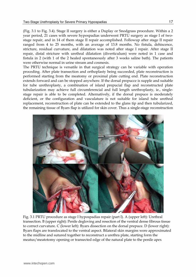
Fig. 3.1 PRTU procedure as stage I hypospadias repair (part I). A (upper left): Urethral transection. B (upper right): Penile degloving and resection of the ventral dense fibrous tissue to correct curvature. C (lower left): Byars dissection on the dorsal prepuce. D (lower right): Byars flaps are translocated to the ventral aspect. Bilateral skin margins were approximated to the midline and sutured together to reconstruct a urethra plate, starting form the meatus/meatotomy opening or transected edge of the natural plate to the penile apex
www.intechopen.com

Current Concepts of Urethroplasty
18
Fig. 3.2 PRTU procedure as stage I hypospadias repair (part II). E (upper left): A U-shaped incision with proper width is made starting form 2 mm proximal to the meatus to the level of anticipated neomeatus, the pecicle vasculature of neoplate is preserved through lateral subcutaneous dissection. The design of neomeatus location is based on evaluation of remaining tissue for skin coverage after urethroplasty (see Fig. 3.3). An alternative strategy to decide the neomeatus location is to extend the incision distally along with tubularization process. F (upper right): Tubularization of the U flap. G (lower left): Midline incision and glans wing dissection. H (lower right): The distal end of ventrally transposed Byars flap is filled in the raw area of wing dissection. The filling flap is trimmed to size for adequate glans/groove augmentation and for future urethroplasty
Fig. 3.3 In another case the tube urethroplasty extended to the midshaft and neomeatus created there
www.intechopen.com

Two-Stage Urethroplasty for Severe Primary Hypospadias
19
is available, too. When the preputial tissue is highly limited and inadequate for both full length urethral replacement and skin coverage, tubularization of the reconstructed plate stops at an appropriate site to allow optimal skin coverage. Thereafter the untubularized part of reconstructed plate as urethra bed is let alone for latter stage urethroplasty. Advantages of this PRTU procedure include: (1) tissue sparing; (2) good blood supply for neourethra and urethra bed; (3) good cosmesis; (4) well indicated for adult primary repair; (5) relatively simple technique; (6) low complication rate. When genital tissue is extremely limited for reconstruction, disadvantages of this technique may be visualized, ie., PRTU is contraindicated. Introduction of a free graft may be better in this occasion.
Fig. 3.4 PRTU procedure as stage I hypospadias repair (part III). I ( left): The distal end of tube urethroplasty is sutured with adjacent skin to achieve meatoplasty. J (right): Skin cover including penoplasty, scrotoplasty, perineoplasty if needed. Scrotal bifida is eleminated and penoscrotal transposition corrected in a degree
Fig. 3.5 Stage I surgery with PRTU partial urethroplasty in a 2-year-old boy with perineal hypospadias. A. Preoperative front view. B. Preoperative lateral view. C. Preoperative view shows the penis. D. Immediately after stage I surgery
www.intechopen.com
Current Concepts of Urethroplasty
20
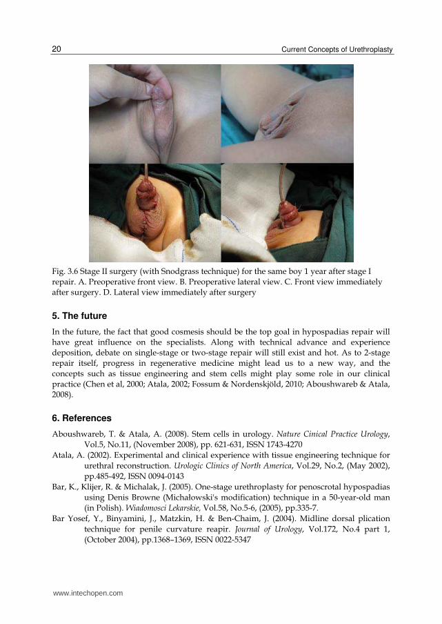
Fig. 3.6 Stage II surgery (with Snodgrass technique) for the same boy 1 year after stage I
repair. A. Preoperative front view. B. Preoperative lateral view. C. Front view immediately
after surgery. D. Lateral view immediately after surgery
5. The future
In the future, the fact that good cosmesis should be the top goal in hypospadias repair will
have great influence on the specialists. Along with technical advance and experience
deposition, debate on single-stage or two-stage repair will still exist and hot. As to 2-stage
repair itself, progress in regenerative medicine might lead us to a new way, and the
concepts such as tissue engineering and stem cells might play some role in our clinical
practice (Chen et al, 2000; Atala, 2002; Fossum & Nordenskjöld, 2010; Aboushwareb & Atala,
2008).
6. References
Aboushwareb, T. & Atala, A. (2008). Stem cells in urology. Nature Cinical Practice Urology,
Vol.5, No.11, (November 2008), pp. 621-631, ISSN 1743-4270
Atala, A. (2002). Experimental and clinical experience with tissue engineering technique for
urethral reconstruction. Urologic Clinics of North America, Vol.29, No.2, (May 2002),
pp.485-492, ISSN 0094-0143
Bar, K., Klijer, R. & Michalak, J. (2005). One-stage urethroplasty for penoscrotal hypospadias
using Denis Browne (Michałowski's modification) technique in a 50-year-old man
(in Polish). Wiadomosci Lekarskie, Vol.58, No.5-6, (2005), pp.335-7.
Bar Yosef, Y., Binyamini, J., Matzkin, H. & Ben-Chaim, J. (2004). Midline dorsal plication
technique for penile curvature reapir. Journal of Urology, Vol.172, No.4 part 1,
(October 2004), pp.1368–1369, ISSN 0022-5347
www.intechopen.com
Two-Stage Urethroplasty for Severe Primary Hypospadias
21
Baskin, L.S., Erol, A., Li, Y.W. & Liu, W. (2000). Anatomy of the neurovascular bundle: is
safe mobilization possible? Journal of Urology, Vol.164, No.3 part 2, (September
2000), pp. 977–980, ISSN 0022-5347
Baskin, L.S. & Ebbers, M.B. (2006). Hypospadias: Anatomy, etiology and technique. Journal
of Pediatric Surgery, Vol.41, No.3, (March 2006), pp. 463-472, ISSN 0022-3468
Bologna, R.A., Noah, T.A., Nasrallah, P.F. & McMahon, D.R. (1999). Chordee: varied
opinions and treatments as documented in a survey of the American Academy of
Pediatics, Section of Urology. Urology, Vol.53, No.3, (March 1999), pp.608-612, ISSN
0090-4295
Bracka, A. (1995). A versatile two-stage hypospadias repair. British Journal of Plastic Surgery,
Vol.48, No.6, (1995), pp. 345-352, ISSN 0007-1226
Bracka, A. (2008). The role of two-stage repair in modern hypospadiology.Indian Journal of
Urology, Vol.24, No.2, (April 2008), pp.210-218
Bracka, A. (2011). Buccal mucosa: good but not perfect. Journal of Urology, Vol.185, No.3,
(March 2011), pp. 777–778, ISSN 0022-5347
Brown, D. (1949). An operation for hypospadias. Proceedings of the Royal Society of Medicine,
Vol.42, No.7, (July 1949), pp.466-468
Castanon, M., Munoz, E., Carrasco, R., Rodò, J. & Morales L. (2000). Treatment of proximal
hypospadias with a tubularized island flap uretroplasty and the onlay technique: A
comparative study. Journal of Pediatric Surgery, Vol.35, No.10, (October 2000), pp.
1453-1455, ISSN 0022-3468
Chatterjee, U.S., Mandal, M.K., Basu, S., Da,s R. & Majhi T. (2004). Comparative study of
dartos fascia and tunica vaginalis pedicle wrap for the tubularized incised plate in
primary hypospadias repair. BJU International, Vol.94, No.7, (November 2004), pp.
1102-1104, ISSN 1464-4096
Chen, F., Yoo, J.J. & Atala, A. (2000). Experimental and clinical experience using tissue
regeneration for urethral reconstruction. World Journal of Urology, Vol.18, No.1,
(Febrary 2000), pp.67-70, ISSN 0724-4983
Chen, S., Tang, Y. & Mao, Y. (2010). Staged repair for severe hypospadias: Choicelessness?
Devolution? Evolution? 3rd World Congress of Pediatric Surgery Abstract Book, pp. 83,
New Delhi, India, October 21-24, 2010
Cheng, E.Y., Kropp, B.P., Pope ,J.C. 4th & Brock, J.W. 3rd. (2003). Proximal division of the
urethral plate in staged hypospadias repair. Journal of Urology, Vol.170, No.4 part 2,
(October 2003), pp. 1580-1583, ISSN 0022-5347
Decter, R.M. (1999). Chordee correction by corporal rotation: the split and roll technique.
Journal of Urology, Vol.162, No.3 part 2, (September 1999), pp.1152-1154, ISSN 0022-
5347
Dermibilek, S., Kanmaz, T., Aydin, G. & Yucesan, S. (2001). Outcome of one-stage
techniques for proximal hypospadias repair. Urology, Vol.58, No.2, (August 2001),
pp.267-270, ISSN 0090-4295
Dewan, P.A., Dineen, M.D., Winkle, D. & Duffy, P.G., Ransley P. (1991). Hypospadias:
Duckett pedicle tube urethroplasty. European Urology, Vol.20, No.1, (1991), pp.39–
42, ISSN 0302-2838
Donnahoo, K.K., Cain, M.P., Pope, J.C., Casale, A.J., Keating, M.A., Adams, M.C. & Rink,
R.C. (1998). Etiology, management and surgical complications of congenital
www.intechopen.com

Current Concepts of Urethroplasty
22
chordee without hypospadias. Journal of Urology, Vol.160, No.3 part 2, (September
1998), pp.1120-1122, ISSN 0022-5347
Duckett, J.W. Jr. (2002a). Transverse preputial island flap technique for repair of severe
hypospadias. 1980. Journal of Urology, Vol.167, No.2 part 2, (Febrary 2002), pp.1179-
1182, ISSN 0022-5347
Duckett, J.W. (2002b). The island flap technique for hypospadias repair. 1981. Journal of
Urology, Vol.167, No.5, (May 2002), pp.2148-2152, ISSN 0022-5347
Ferro, F., Zaccara, A., Spagnoli, A., Lucchetti, M.C., Capitanucci, M.L. & Villa, M. (2002).
Skin graft for 2-stage treatment of severe hypospadias: back to the future?
Journal of Urology, Vol.168, No.4 part 2, (October 2002), pp.1730-1733, ISSN 0022-
5347
Fossum, M. & Nordenskjöld, A. (2010).Tissue-engineered transplants for the treatment of
severe hypospadias. Hormone Research in Paediatrics, Vol.73, No.2, (2010), pp.148-
52
Gearhart, J.P. & Witherington, R. (1979). The Denis-Browne hypospadias repair revisited.
Journal of Urology, Vol.122, No.1, (July 1979), pp.66-67, ISSN 0022-5347
Goyal, A., Singh, M.V. & Dickson, A.P. (2010). Oral mucosa graft for repair of hypospadias:
outcomes at puberty. Journal of Urology, Vol.184, No.6, (December 2010), pp.2504-
2508, ISSN 0022-5347
Johal, N.S., Nitkunan, T., O'Malley, K. & Cuckow, P.M. (2006). The two-stage repair for
severe primary hypospadias. European Urology, Vol.50, No.2, (August 2006), pp.366-
371, ISSN 0302-2838
Lam, P.N., Greenfield, S.P. & Williot, P. (2005). 2-stage repair in infancy for severe
hypospadias with chordee: long-term results after puberty. Journal of Urology,
Vol.174, No.4 part 2, (October 2005), pp.1567-1572, ISSN 0022-5347
MacGillivray, D., Shankar, K.R. & Rickwood, A.M. (2002). Management of severe
hypospadias using Glassberg's modification of the Duckett repair. BJU International,
Vol.89, No.1, (January 2002), pp. 101-102, ISSN 1464-4096
Manzoni, G., Bracka, A., Palminteri, E. & Marrocco, G. (2004). Hypospadias surgery: When,
what and by whom. BJU International, Vol.94, No.8, (November 2004), pp. 1188-
1195, ISSN 1464-4096
Markiewicz, M.R., Lukose, M.A., Margarone, J.E. 3rd, Barbagli, G., Miller, K.S. & Chuang
SK. (2007). The oral mucosa graft: a systematic review. Journal of Urology, Vol.178,
No.2, (August 2007), pp.387-394, ISSN 0022-5347
Mingin, G., Baskin, L.S. (2002). Management of chordee in children and young adults.
Urologic Clinics of North America, Vol.29, No.2, (May 2002), pp.277-284, ISSN 0094-
0143
Mitchell, M.E. & Bagli, D.J.(1996). Complete penile disassembly for epispadias repair: the
Mitchell technique. Journal of Urology, Vol.155, No.1, (January 1996), pp.300-304,
ISSN 0022-5347
Mokhless, I.A., Kader, M.A., Fahmy, N. & Youssef, M. (2007). The multistage use of buccal
mucosa grafts for complex hypospadias: histological changes. Journal of Urology,
Vol.177, No.4, (April 2007), pp.1496-1499, ISSN 0022-5347
Patel, S.R. & Caldamone, A.A. (2010). Sir Denis Browne: contributions to pediatric urology.
Journal of Pediatric Urology, Vol.6, No.5, (October 2010), pp.496-500
www.intechopen.com
Two-Stage Urethroplasty for Severe Primary Hypospadias
23
Powell, C.R., McAleer, I., Alagiri, M. & Kaplan GW. (2000). Comparison of flap versus grafts
in proximal hypospadias surgery. Journal of Urology, Vol.163, No.4, (April 2000),
pp.1286-1288, ISSN 0022-5347
Price, R.D., Lambe, G.F. & Jones, R.P. (2003). Two-stage hypospadias repair: Audit in a
district general hospital. British Journal of Plastic Surgery, Vol.56, No.8, (December
2003), pp. 752-758, ISSN 0007-1226
Radojicic, Z.I. & Perovic, S.V. (2004). Classification of prepuce in hypospadias according to
morphological abnormalities and their impact on hypospadias repair. Journal of
Urology, Vol.172, No.1, (July 2004), pp.301-304, ISSN 0022-5347
Schumacher, S., Yabumoto, H., Salge, S., Shrestha, G. & Ikoma F. (1997). Surgical results of
complete hypospadias repair in two stages. International Urology and Nephrology,
Vol.29, No.3, (1997), pp.333-40
Simonato, A., Gregori, A., Ambruosi. C., Venzano, F., Varca, V., Romagnoli, A. &
Carmignani, G. (2008). Lingual mucosal graft urethroplasty for anterior urethral
reconstruction. European Urology, Vol.54, No.1, (July 2008), pp.79-85, ISSN 0302-
2838
Snodgrass, W. & Bush, N. (2011). Tubularized incised plate proximal hypospadias repair:
Continued evolution and extended applications. Journal of Pediatric Urology, Vol.7,
No.1, (Febrary 2011), pp.2-9
Soutis, M., Papandreou, E., Mavridis, G. & Keramidas, D. (2003). Multiple failed
urethroplasties: definitive repair with the Duckett island-flap technique. Journal
of Pediatric Surgery, Vol.38, No.11, (November 2003), pp. 1163-1166, ISSN 0022-
3468
Soygur, T., Filiz, E., Zumrutbas, A.E. & Arikan, N. (2004). Results of dorsal midline plication
in children with penile curvature and hypospadias. Urology, Vol.64, No.4, (October
2004), pp.795-798, ISSN 0090-4295
Tahmeedullah, Khan, A. & Obaidullah MA. (2003). Comparison of prepucial skin,
postauricular skin and buccal mucosal graft results in hypospadias repair. Journal of
the College of Physicians and Surgeons—Pakistan, Vol.13, No.9, (September 2003),
pp.515-518
Tang, Y.M., Chen, S.J., Huang, L.G. & Wang, M.H. (2007). Chordee without hypospadias:
report of 79 Chinese prepubertal patients. Journal of Andrology, Vol.28, No.4, (July-
August 2007), pp.630-633, ISSN 0196-3635
Tang, Y., Chen, S., Mao, Y., et al. (2010). Plate reconstruction and tubularization for
correction of severe hypospadias, 3rd World Congress of Pediatric Surgery Abstract
Book, pp. 84, New Delhi, India, October 21-24, 2010
Tavakkoli Tabassi, K. & Mohammadi, S. (2010). Tunica vaginalis flap as a second layer for
tubularized incised plate urethroplasty.Urology Journal, Vol.7, No.4, (Fall 2010),
pp.254-257
Titley, O.G. & Bracka, A. (1998). A 5-year audit of trainees experience and outcomes with
two-stage hypospadias surgery. British Journal of Plastic Surgery, Vol.51, No.5, (July
1998), pp.370-375, ISSN 0007-1226
Yucel, S., Guntekin, E., Kukul, E., Karaguzel, G., Ciftcioglu, A., Melikoglu, M. & Baykara,
M. (2004). Comparison of hypospadiac and normal preputial vascular anatomy.
www.intechopen.com

Current Concepts of Urethroplasty
24
Journal of Urology, Vol.172, No.5 part 1, (November 2004), pp.1973-1976, ISSN
0022-5347
Yucel, S., Sanli, A., Kukul, E., Karaguzel, G., Melikoglu, M. & Guntekin, E. (2006). Midline
dorsal plication to repair recurrent chordee at reoperation for hypospadias surgery
complication. Journal of Urology, Vol.175, No.2, (Febrary 2006), pp.699-702, ISSN
0022-5347
www.intechopen.com

Current Concepts of UrethroplastyEdited by Dr Ivo Donkov
ISBN 978-953-307-392-7Hard cover, 152 pagesPublisher InTechPublished online 19, July, 2011Published in print edition July, 2011
InTech EuropeUniversity Campus STeP Ri Slavka Krautzeka 83/A 51000 Rijeka, Croatia Phone: +385 (51) 770 447 Fax: +385 (51) 686 166www.intechopen.com
InTech ChinaUnit 405, Office Block, Hotel Equatorial Shanghai No.65, Yan An Road (West), Shanghai, 200040, China
Phone: +86-21-62489820 Fax: +86-21-62489821
Urethral reconstructive surgery has always been a challenging part for urologist since the dawn of ourspeciality. In this book leading experts in lower urinary reconstructions from all over the world present theirviews and experience in that field, together with practical tips and tricks. The book is an excellent source ofinformation for those who are already dealing with urethral surgery, and also an invaluable companion forurologists in training or those who want to dedicate themselves to this great sub-specialty. This book is anexcellent reference guide and companion on the way to operating and consulting room, or when writing anarticle and reviewing the current practices. The abundance of methods and continuing development of newapproaches to the problem prove the complexity of it.
How to referenceIn order to correctly reference this scholarly work, feel free to copy and paste the following:
Yun-Man Tang and Shao-Ji Chen (2011). Two-Stage Urethroplasty for Severe Primary Hypospadias, CurrentConcepts of Urethroplasty, Dr Ivo Donkov (Ed.), ISBN: 978-953-307-392-7, InTech, Available from:http://www.intechopen.com/books/current-concepts-of-urethroplasty/two-stage-urethroplasty-for-severe-primary-hypospadias

© 2011 The Author(s). Licensee IntechOpen. This is an open access articledistributed under the terms of the Creative Commons Attribution 3.0License, which permits unrestricted use, distribution, and reproduction inany medium, provided the original work is properly cited.