Embed Size (px)
Citation preview
The EMBO Journal vol.4 no.l3A pp.3519-3524, 1985
Two genes encoding the bovine mitochondrial ATP synthaseproteolipid specify precursors with different import sequences andare expressed in a tissue-specific manner
Nicholas J.Gay1 and John E.Walker
Medical Research Council Laboratory of Molecular Biology, Hills Road,Cambridge CB2 2QH, UK'Present address: Department of Biochemistry and Biophysics, UCSFMedical School, San Francisco, CA 94143, USA
Communicated by J.E.Walker
Two cDNAs encoding different precursor proteins of the samemature proteolipid subunit of mitochondrial ATP synthasehave been cloned from a bovine cDNA library. The hybridisa-tion probe was a mixture of 17-mer oligonucleotides contain-ing 256 discrete sequences. The coding sequences of the twocDNAs differ in 25 silent positions of codons and the 3' non-coding sequences are only weakly related. The precursor se-quences, which direct the import of the proteolipid into themitochondrion, are 61 and 68 amino acids long. They arerelated to each other in regions which probably are recogni-tion signals for the processing protease. The correspondinggenes are expressed differently in various tissues in a way thatreflects their embryonic origin.Key words: ATPase/proteolipid/mitochondrial import/tissuespecificity
IntroductionThe mitochondrial ATP synthase complex (F1FO-ATPase; review-ed by Senior, 1979) is anchored in the inner membrane of theorganelle by an intrinsic membrane sector, Fo. The proteolipid[or dicyclohexylcarbodiimide (DCCD) binding protein] is anessential element for the function of the proton channel in thismembrane enzyme complex (Sebald and Hoppe, 1981). As itsname implies it is a hydrophobic protein; in bovine mitochon-dria it is 75 amino acids in length (Sebald and Hoppe, 1981).In mammals (Anderson et al., 1981, 1982), Aspergillus nidulans(Turner et al., 1979), Neurospora crassa (Jackl and Sebald, 1975)(but not Saccharomyces cerevisiae; Macino and Tzagoloff, 1979)it is a nuclear gene product. It is presumed that it is synthesisedon free ribosomes as a longer precursor, and imported into themitochondrion in a post-translational process that involvesremoval of a segment from the N-terminal region of the precur-sor (Schatz and Butow, 1983; Hay et al., 1984). In the N. crassapre-proteolipid this import sequence is 66 amino acids long(Viebrock et al., 1982).
In the course of studies of genes for subunits of bovinemitochondrial ATP synthase, we have cloned cDNAs correspond-ing to precursors of the bovine mitochondrial proteolipid. Weemployed as hybridisation probe a mixture of 256 oligonucleo-tides, 17 bases long, with sequences predicted from a segmentof the known protein sequence of the mature proteolipid (Sebaldand Hoppe, 1981). Thereby we have characterised two differentcDNA species. DNA sequence analysis reveals that they encodeexactly the same mature proteolipid (with 25 changes in silentfirst and third positions of the codons) but differ substantiallyboth in the DNA sequence encoding the processed import se-
© IRL Press Limited, Oxford, England
quence and in the 3' non-coding regions. The import sequencesare sufficiently conserved, however, to suggest that they bothdirect the protein into mitochondria.By hybridisation of diverged regions of the two cDNAs with
RNA derived from different tissues, it appears that the two genesare expressed in a tissue specific manner.
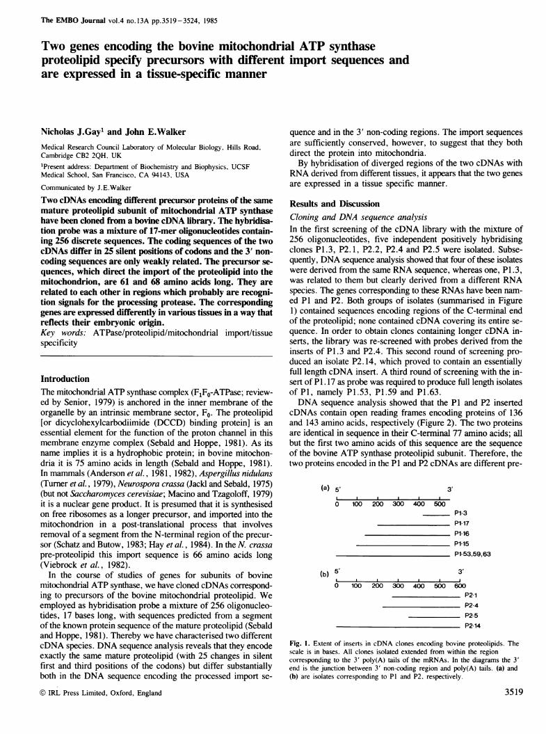
Results and DiscussionCloning and DNA sequence analysisIn the first screening of the cDNA library with the mixture of256 oligonucleotides, five independent positively hybridisingclones P1.3, P2.1, P2.2, P2.4 and P2.5 were isolated. Subse-quently, DNA sequence analysis showed that four of these isolateswere derived from the same RNA sequence, whereas one, P1.3,was related to them but clearly derived from a different RNAspecies. The genes corresponding to these RNAs have been nam-ed P1 and P2. Both groups of isolates (summarised in Figure1) contained sequences encoding regions of the C-terminal endof the proteolipid; none contained cDNA covering its entire se-quence. In order to obtain clones containing longer cDNA in-serts, the library was re-screened with probes derived from theinserts of P1.3 and P2.4. This second round of screening pro-duced an isolate P2.14, which proved to contain an essentiallyfull length cDNA insert. A third round of screening with the in-sert of P1.17 as probe was required to produce full length isolatesof P1, namely P1.53, P1.59 and P1.63.DNA sequence analysis showed that the P1 and P2 inserted
cDNAs contain open reading frames encoding proteins of 136and 143 amino acids, respectively (Figure 2). The two proteinsare identical in sequence in their C-terminal 77 amino acids; allbut the first two amino acids of this sequence are the sequenceof the bovine ATP synthase proteolipid subunit. Therefore, thetwo proteins encoded in the P1 and P2 cDNAs are different pre-
(a) 5' 3'
O 100 200 300 400 500P1-3
P1l17P1-16P1-5P1 53,59,63
(b) 5' 3'
0 100 200 300 400 500 600P21P24P25P2 14
Fig. 1. Extent of inserts in cDNA clones encoding bovine proteolipids. Thescale is in bases. All clones isolated extended from within the regioncorresponding to the 3' poly(A) tails of the mRNAs. In the diagrams the 3'end is the junction between 3' non-coding region and poly(A) tails. (a) and(b) are isolates corresponding to P1 and P2, respectively.
3519

N.H.Gay and J.E.Walker
,j0 ,i,) Precursor
|OT IrGA I .'. F' As 1..G AF' 1 CCAA r j(:lFITT T 1iC(7:lS.l.CC'CCCTlCI'GCALiAC T1f *6Ai(- Al ISCAGC:t,CEAt;.CiC6L,CF,utC 1 l Ch71rlI'l'Cl(l'tc,r TlCGAl (CCTIC rT
F: ''' rSCc l','iC'(::C,C.'t'l'C.AI'C CCC.A(A 1'"IHACIi' t'CUt.C[AAC l 1 (C,GFCbCCt ACt GCtTCC' ITGAl'CAGGACiAALffi4T C'A IK F V T F S I.. I F F N
Precursor 4 60
1 k 12_ :L -0 1 .01" tC T R G L. I Ri :: V S A F L ]:I OS V O F S Y S S6 F:'
F:lt GTACCAGGGS(IJTC,GATCAG:G rlC IGTGCTGCCiC I (1C (TUt,I AG AGGC CAAcAG[ACCAATCl CAAGCC'TTC CiTA CAGCAGTGGC C'CAC
F2 CCFCTA'ACAAGTATGAGtCiC GhAT C AfC T'i'C,GC,A L'T1(.tG1GAG tCAIC GAA, AC A CA CGGATGAGAGC CACACi CAGCTTIG GCAGTAG*r S r V l S S L 'iA V V IR'F E- r L T f E 'S H C. S L A V
(1*.;) t0010i0 10 140O 160
Mature00e22 r- 240
0 V A R R- F 1i1 S V V S R LIE [Ii T A AK FF.t ITGCA(.FCjlIGG(*'C,C,t(.U----. ------ ---- - -C-- G-GAAT"I'C'CAGACCA,fiAGTT1TG TCTC'CLCGGfA[C"A rl GAC.ACALGCGGCC'AAGT TTA:: '.'. *:.::.:. . . . ..I. . . . . . . . . . . . . .':+:':'.^:'::::::
F(21 TTCCCCGC CC'CCCTCAC'Al CTIChCl ACI CC. 1ATAGU CIGC:tiGC1GI iTC.CAA CAC,1 LG.C'AlTT CChAAGGACAII,AT AC AGC,AGCCAAGTTC'AF: F: F: l r I I.. T F: D T A A K F
Mature
60 -80sJ 30 3 0It G A 6 A A r V G6V A (i s AG U J: G 1 v F:' c S i.. I :1 G Y A R
F:: 1. 'IT'GCl: CGC 'GCfi'G CT G.'CC;h(,C AGTG(TCC).TGf(iGCO, fGG'TCAGCG C-TC''lCGl''TTGG AA ChCA G TG T 'T cAGcC rGAGC'T( AT"A TrGGCTATG CCAGf GGA.. + + + + . . . . . . . . * . . . . . . . . . .
PF- T C G3rl 3i.c-f.1:l.:C(;(C l(5T ffi^(sr 3 f:r. 3il -Gi FCr Gr A^.(G C If AA -fI sT GCiC,AC G' rfiT G ;GAf3 T C A T-C:fTTG( FFA TGIC A (3GAI G A 1- r-'sAF' G S L I I G Y A R
A.6ejki ) (} [3.O C1 (3 3 2)0 340
,401i36(-3 A(D0 400 420N F S l. K L. F S AA]: L G F' A L S E: A GL6 F' CLl i Y A
F1L ALCLC"16'ILGT ITGAAGCAG(CA C'T'l IT rIt T ATUIC"C Al'THCT1(G1G1C,T1TTtF'CC' CTFGTTCh AGGCA'TA'T'GFGurJFCCT CT G rT T rGA-TGGTCGCC r
F: 2) AC",('C. T' T Cl'C:'T'G AA GCAs iC,t" G C-':'1'G'll'-C'CT'IA :GC C'f TTIC: 7 G-,(:C.6CTTTCrrCr:C" C T c -TCCG C. GC A TSl(G GC"iTC'G'f'''TTTGCCTGAT G GGC CTIKF' SY.K t.) 1 L F S 't A :[ L G F ALl IS E A 61l L.. F C L M Y A
':3(.") O3 '^.R3a44 '2'
^a + .1" .^.,*160i 4 0S3Ft:' LAp1 r CA I, Hph I[I 11 1CLA11T(1 1 1 A11l1GA;C(:(i-TGAGAC C AA1C.(( CcC':f'GCCA(C:A(CCCTCCrGGCTtACUcCCTG1AGl'G61
TAj2 TTC'F('T'C'Al'(:'C''(:1''11'Gf('6 C i AGA GGA6 C(`- GTT'-'I .......................- C' C AfC 'CTC' C'+--sA CTA ;rTCTrc T-C C GGTG7 r AT C"ATTGCCC T'G 'AI'C TTF l: F
44118 460i 160{ 5f00
.)re :- 4r0F- 1 GCI/:,AACC(T''llfAC'C'A'T'iAAACACAAT(l611I:'lCFCIAA^AAAA
-. , .3
..0 Rsa I ii 810 600
F:.? AAo AA AA('4A
Fig. 2. DNA sequences of bovine cDNAs and derived protein sequences of P1 and P2 pre-proteolipids. The nucleotide sequences were aligned with thecomputer program NUCALN (Wilbur and Lipman, 1983). The numbers refer to the nucleotide sequences and colons denote identities. Dashes indicateinsertions in both nucleotide and protein sequences. The sites of initiation of translation and of processing of the pre-proteolipids are shown. The underlinedsequences are likely to be signals for addition of poly(A) to the 3' end of the message (Proudfoot and Brownlee, 1976). The Hphl site in P1 and the RsaI sitein P2 were employed to produce specific prime cut probes from the diverged 3' ends of the two cDNAs (see Figure 6).
cursors of this subunit. To produce the mature subunit, 61 and68 amino acids respectively are removed from the P1 and P2precursors (see Figure 3).As the conservation of the mature protein sequence implies,
the corresponding coding sequences are also highly conserved,differing from each other only in 25 silent first and third posi-tions of codons (Figure 2). In contrast, the 3' non-coding regionsof the P1 and P2 cDNAs are much less related, 39 bases beingconserved. The P2 mRNA is somewhat longer than P1 in thisregion [157 and 109 bases respectively, excluding poly(A) tails].It should be noted that the six consecutive A residues at the 3'end of P1 are specified in the gene sequence (N.J. Gay and J.E.Walker, unpublished results). In two out of seven isolates ex-amined, the three terminal A residues are replaced by the se-
quence CTC; it is assumed that this is an allelic polymorphism.The sequence of P1 and P2 cDNAs both contain polyadenyla-tion signals near to their 3' ends (see Figure 2) (Proudfoot andBrownlee, 1976) and given that the mRNAs are polyadenylatedit seems to be reasonable to assume that they will be translatedand the protein products produced. However, direct proof forthis is at present lacking. The coding regions corresponding tothe pre-sequences are much less conserved than those for themature protein (see Figure 2); 82 bases out of 202 are identical,and conserved bases are clustered. The extent of the 5' non-codingregion has not yet been established with absolute certainty. How-ever, it is clear from size estimates of 570 i 15 bp and 630± 15 bp for the P1 and P2 mRNAs made by Northern hybridisa-tions, that the sequences of the cDNAs cannot extend much
3520
Two genes for bovine mitochondrial ATP synthase
10 20 30 4 I
P 1 MQT7 .ALLISPALIRSCITRGLIRPVSAS FLSRPEIQSVOPS YSSGPLQVA. .~ ~ . e .. . . .. ..
P2 MYTCAKFVSTPSL IRRISTVLSRSLSAVVVRRPEThTDESHSSLAVVPRP:10 20 30 40
P1 R--
P2 LTLF_S
P1
P2
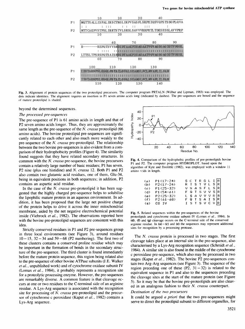
Fig. 3. Alignment of protein sequences of the two proteolipid precursors. The computer program PRTALN (Wilbur and Lipman, 1983) was employed. Thedots indicate identities. The alignment requires an insertion in P1 seven amino acids long (indicated by dashes). The pre-sequences are boxed and the sequenceof mature proteolipid is shaded.
beyond the determined sequences.The processed pre-sequencesThe pre-sequence of P1 is 61 amino acids in length and that ofP2 seven amino acids longer. Thus, they are approximately thesame length as the pre-sequence of the N. crassa proteolipid (66amino acids). The bovine proteolipid pre-sequences are signifi-cantly related to each other and also much more weakly to thepre-sequence of the N. crassa pre-proteolipid. The relationshipbetween the two bovine pre-sequences is also evident from a com-parison of their hydrophobicity profiles (Figure 4). The similarityfound suggests that they have related secondary structures. Incommon with the N. crassa pre-sequence, the bovine precursorscontain a relatively large number of basic residues; P1 has seven,P2 nine (plus one histidine) and N. crassa 12. Both P1 and P2also contain two glutamic acid residues, one of them, Glu-34,being in equivalent positions in both sequences; in addition, P2contains an aspartic acid residue.
In the case of the N. crassa pre-proteolipid it has been sug-gested that the highly charged pre-sequence helps to solubilisethe lipophilic mature protein in an aqueous environment. In ad-dition, it has been proposed that the large net positive chargeof the protein helps to drive it across the inner mitochondrialmembrane, aided by the net negative electrochemical potentialinside (Viebrock et al., 1982). The observations reported herewith the bovine pre-proteolipid sequences are consistent with thisidea.
Strictly conserved residues in P1 and P2 pre-sequences groupin three local environments (see Figure 3), around residues10- 15, 32-34 and 59-68 (P2 numbering). The first two ofthese clusters contains a conserved proline residue which maybe important in the formation of bends in the secondary struc-ture of the pre-sequence. The third cluster is found immediatelybefore the mature protein sequence, this region being related alsoto the pre-sequence of other bovine ATPase subunits (J.E. Walkeret al., unpublished work) and of cytochrome oxidase subunit IV(Lomax et al., 1984), it probably represents a recognition sitefor a proteolytic processing enzyme. However, the pre-sequencesare remarkably diverse. A common feature is that cleavage oc-curs at one or two residues to the C-terminal side of an arginineresidue. A Lys-Arg sequence is associated with the recognitionsite for processing of N. crassa pre-proteolipid and the precur-sor of cytochrome c peroxidase (Kaput et al., 1982) contains aLys-Arg sequence.
xLSott. /PiC: 20 (
0CZ
-20 P2
40 -
20 40 60 80 100 120 14Residue No.
Fig. 4. Comparison of the hydrophobic profiles of pre-proteolipids bovineP1 and P2. The computer program HYDROPLOT, based upon the*algorithm of Kyte and Doolittle (1982), was employed with a window 11amino nids in length.
(a) F'1 ( 1 7- 4 )(b) P2(17 2 4)(c) P1 (25-32)(d) F'1 (54 -6 1 )(e) F'2' ( '25 --32 )(f) P2 (61-68)([) CO IV
S (' T R G L I RR T S 1 V L. S RiV S A S F L S RIF 0 T S V V S R'l.. S A V V V 1: RIF Q T S A I S UI S T S V C V K
Fig. 5. Related sequences within the pre-sequences of the bovineproteolipids and cytochrome oxidase subunit IV (Lomax et al.. 1984). In(d). (f) and (g) cleavage occurs on the C-terminal side of the conservedarginine residue. In (a)-(c) and (e) the sequences may represent additionalsites for recognition by a processing protease.
The N. crassa protein is processed in two stages. The firstcleavage takes place at an internal site in the pre-sequence, alsocharacterised by a Lys-Arg recognition sequence (Schmidt et al.,1984). A similar site is also found in the middle of the cytochromec peroxidase pre-sequence, which also may be processed in twostages (Kaput et al., 1982). The bovine P2 pre-sequences con-tain two Arg-Arg sequences (see Figure 3). The sequence of theregion preceding one of these (P2, 31 -32) is related to theequivalent sequence in P1 and also to the sequences precedingthe cleavage sites at the start of the mature protein (see Figure5). So it may be that the bovine pre-proteolipids are also cleav-ed in an analogous fashion to their N. crassa counterpart.Destination of the two precursorsIt could be argued a priori that the two pre-sequences mightserve to direct the proteolipid subunit to different organelles, for
3521
N.H.Gay and J.E.Walker
as m0 WA
i ...4
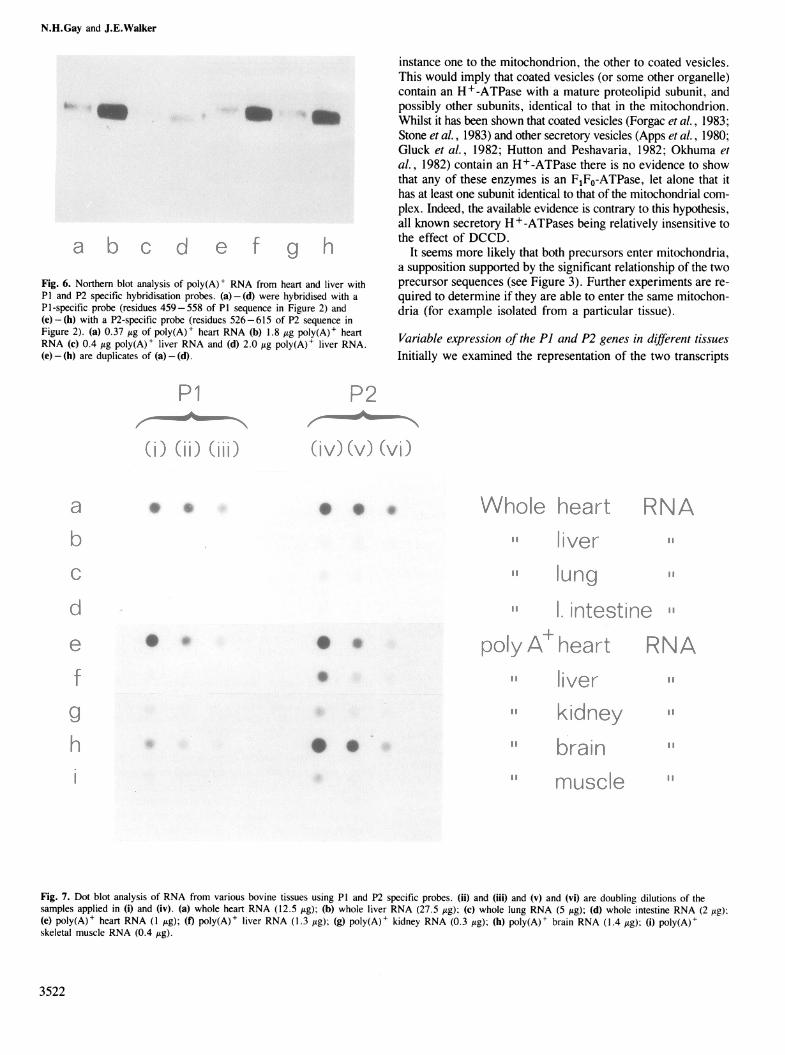
Fig. 6. Northern blot analysis of poly(A)+ RNA from heart and liver withP1 and P2 specific hybridisation probes. (a) - (d) were hybridised with aP1-specific probe (residues 459-558 of P1 sequence in Figure 2) and(e) - (h) with a P2-specific probe (residues 526-615 of P2 sequence inFigure 2). (a) 0.37 itg of poly(A)+ heart RNA (b) 1.8 ug poly(A)+ heartRNA (c) 0.4 Ag poly(A)+ liver RNA and (d) 2.0 Ag poly(A)+ liver RNA.(e)-(h) are duplicates of (a)-(d).
instance one to the mitochondrion, the other to coated vesicles.This would imply that coated vesicles (or some other organelle)contain an H+-ATPase with a mature proteolipid subunit, andpossibly other subunits, identical to that in the mitochondrion.Whilst it has been shown that coated vesicles (Forgac et al., 1983;Stone et al., 1983) and other secretory vesicles (Apps et al., 1980;Gluck et al., 1982; Hutton and Peshavaria, 1982; Okhuma etal., 1982) contain an H+-ATPase there is no evidence to showthat any of these enzymes is an FIF0-ATPase, let alone that ithas at least one subunit identical to that of the mitochondrial com-plex. Indeed, the available evidence is contrary to this hypothesis,all known secretory H+-ATPases being relatively insensitive tothe effect of DCCD.
It seems more likely that both precursors enter mitochondria,a supposition supported by the significant relationship of the twoprecursor sequences (see Figure 3). Further experiments are re-quired to determine if they are able to enter the same mitochon-dria (for example isolated from a particular tissue).
Variable expression of the P1 and P2 genes in different tissuesInitially we examined the representation of the two transcripts
i *v/
*s
0 oLv)OI A > it
r idta e'gsWE.qika .. .s
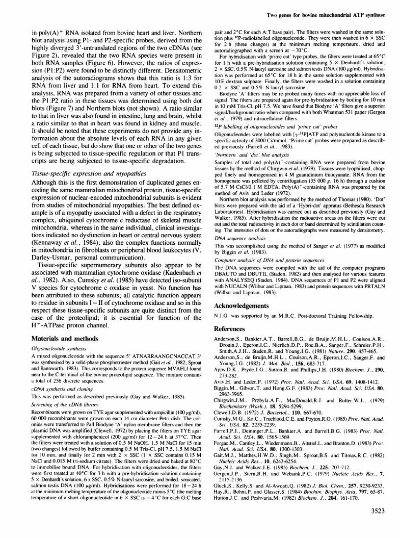
Fig. 7. Dot blot analysis of RNA from various bovine tissues using P1 and P2 specific probes. (ii) and (iii) and (v) and (vi) are doubling dilutions of thesamples applied in (i) and (iv). (a) whole heart RNA (12.5 /ig); (b) whole liver RNA (27.5 Itg); (c) whole lung RNA (5 .tg); (d) whole intestine RNA (2 lAg);(e) poly(A)+ heart RNA (1 jug); (f) poly(A)+ liver RNA (1.3 ag); (g) poly(A)+ kidney RNA (0.3 lzg); (h) poly(A)+ brain RNA (1.4 /tg); (i) poly(A)+skeletal muscle RNA (0.4 tg).
3522
{.I
.... 0, ):, "I1
;1 tjp
II
I,p
Two genes for bovine mitochondrial ATP synthase
in poly(A)+ RNA isolated from bovine heart and liver. Northernblot analysis using P1- and P2-specific probes, derived from thehighly diverged 3'-untranslated regions of the two cDNAs (seeFigure 2), revealed that the two RNA species were present inboth RNA samples (Figure 6). However, the ratios of expres-sion (P1 P2) were found to be distinctly different. Densitometricanalysis of the autoradiograms shows that this ratio is 1:3 forRNA from liver and 1:1 for RNA from heart. To extend thisanalysis, RNA was prepared from a variety of other tissues andthe P1:P2 ratio in these tissues was determined using both dotblots (Figure 7) and Northern blots (not shown). A ratio similarto that in liver was also found in intestine, lung and brain, whilsta ratio similar to that in heart was found in kidney and muscle.It should be noted that these experiments do not provide any in-formation about the absolute levels of each RNA in any givencell of each tissue, but do show that one or other of the two genesis being subjected to tissue-specific regulation or that P1 trans-cripts are being subjected to tissue-specific degradation.
Tissue-specific expression and myopathiesAlthough this is the first demonstration of duplicated genes en-coding the same mammalian mitochondrial protein, tissue-specificexpression of nuclear-encoded mitochondrial subunits is evidentfrom studies of mitochondrial myopathies. The best defined ex-ample is of a myopathy associated with a defect in the respiratorycomplex, ubiquinol cytochrome c reductase of skeletal musclemitochondria, whereas in the same individual, clinical investiga-tions indicated no dysfunction in heart or central nervous system(Kennaway et al., 1984); also the complex functions normallyin mitochondria in fibroblasts or peripheral blood leukocytes (V.Darley-Usmar, personal communication).
Tissue-specific supernumerary subunits also appear to beassociated with mammalian cytochrome oxidase (Kadenbach etal., 1982). Also, Cumsky et al. (1985) have detected iso-subunitV species for cytochrome c oxidase in yeast. No function hasbeen attributed to these subunits; all catalytic function appearsto residue in subunits I-II of cytochrome oxidase and so in thisrespect these tissue-specific subunits are quite distinct from thecase of the proteolipid; it is essential for function of theH + -ATPase proton channel.
Materials and methodsOligonucleotide synthesisA mixed oligonucleotide with the sequence 5' ATNARRAANGCNACCAT 3'was synthesised by a solid-phase phosphotriester method (Gait et al., 1982: Sproatand Bannwarth, 1983). This corresponds to the protein sequence MVAFLI foundnear to the C-terminal of the bovine proteolipid sequence. The mixture containsa total of 256 discrete sequences.cDNA s.ynthesis anid cloningThis was performed as described previously (Gay and Walker, 1985).
Screenzing of the eDNA libraryRecombinants were grown on TYE agar supplemented with ampicillin (100 ,g/ml).60 000 recombinants were grown on each 14 cm diameter Petri dish. The col-onies were transferred to Pall Biodyne 'A' nylon membrane filters and then theplasmid DNA was amplified (Clewell, 1972) by placing the filters on TYE agarsupplemented with chloramphenicol (200 Atg/ml) for 12-24 h at 37°C. Thenthe filters were treated with a solution of 0.5 M NaOH, 1.5 M NaCI for 15 min(two changes) followed by buffer containing 0.5 M Tris-CI, pH 7.5, 1.5 M NaCIfor 10 min, and finally for 2 min with 2 x SSC (1 x SSC contains 0.15 MNaCI and 0.015 M tri-sodium citrate). The filters were dried and baked at 80°Cto immobilise bound DNA. For hybridisation with oligonucleotides, the filterswere first treated at 40°C for 3 h with a pre-hybridisation solution containingS x Denhardt's solution, 6 x SSC, 0.5% N-lauryl sarcosine, and boiled, sonicated.salmon testis DNA (100 zg/ml). Hybridisations were performed for 18-24 hat the minimum melting temperature of the oligonucleotide minus 5°C (the meltingtemperature of a short oligonucleotide in 6 x SSC is -4°C for each G:C base
pair and 2°C for each A:T base pair). The filters were washed in the same solu-tion plus 32P-radiolabelled oligonucleotide. They were then washed in 6 x SSCfor 2 h (three changes) at the minimum melting temperature, dried andautoradiographed with a screen at -70°C.
For hybridisation with 'prime cut' type probes, the filters were treated at 65°Cfor 1 h with a pre-hybridisation solution containing 5 x Denhardt's solution,2 x SSC, 0.5% N-lauryl sarcosine and salmon testis DNA (100 ig/mJ). Hybridisa-tion was performed at 65°C for 18 h in the same solution supplemented with10% dextran sulphate. Finally, the filters were washed in a solution containing0.2 x SSC and 0.5% N-lauryl sarcosine.
Biodyne 'A' filters may be re-probed many times with no appreciable loss ofsignal. The filters are prepared again for pre-hybridisation by boiling for 10 minin 10 mM Tris-CI, pH 7.5. We have found that Biodyne 'A' filters give a superiorsignal/background ratio when compared with both Whatman 531 paper (Gergenet al., 1979) and nitrocellulose filters.32p labelling of oligonucleotides and 'primle cut' probesOligonucleotides were labelled with ['y-32P]ATP and polynucleotide kinase to aspecific activity of 3000 Ci/mmol. 'Prime cut' probes were prepared as describ-ed previously (Farrell et al., 1983).'Northern and 'dot' blot analysisSamples of total and poly(A)+-containing RNA were prepared from bovinetissues by the method of Chirgwin et al. (1979). Tissues were lyophilised, chop-ped finely and homogenised in 4 M guanidinium thiocyanate. RNA from thehomogenate was pelleted by centrifugation (35 000 g, 16 h) through a cushionof 5.7 M CsCl/0.1 M EDTA. Poly(A)+-containing RNA was prepared by themethod of Aviv and Leder (1972).
Northern blot analysis was performed by the method of Thomas (1980). 'Dot'blots were prepared with the aid of a 'Hybri-dot' appratus (Bethesda ResearchLaboratories). Hybridisation was carried out as described previously (Gay andWalker, 1985). After hybridisation the radioactive areas on the filters were cutout and the total radioactivity in each dot or band determined by scintillation count-ing. The intensities of dots on the autoradiographs were measured by densitometry.DNA sequence analysisThis was accomplished using the method of Sanger et al. (1977) as modifiedby Biggin et al. (1983).
Computer analysis of DNA anid protein sequencesThe DNA sequences were compiled with the aid of the computer programsDBAUTO and DBUTIL (Staden, 1982) and then analysed for various featureswith ANALYSEQ (Staden, 1984). DNA sequences of P1 and P2 were alignedwith NUCALN (Wilbur and Lipman, 1983) and protein sequences with PRTALN(Wilbur and Lipman, 1983).
AcknowledgementsN.J.G. was supported by an M.R.C. Post-doctoral Training Fellowship.
ReferencesAnderson,S., Bankier,A.T., Barrell,B.G., de Bruijn,M.H.L., Coulson,A.R.,
Drouin,J., Eperon,I.C., Nierlich,D.P., Roe,B.A., Sanger,F., Schreier,P.H.,Smith.A.J.H., Staden,R. and Young,I.G. (1981) Nature, 290, 457-465.
Anderson,S., de Bruijn,M.H.L., Coulson,A.R., Eperon,I.C., Sanger,F. andYoung,I.G. (1982) J. Mol. Biol., 156, 683-717.
Apps,D.K., Pryde,J.G., Sutton,R. and Phillips,J.H. (1980) Biochem. J., 190,273-282.
Aviv,H. and Leder,P. (1972) Proc. Natl. Acad. Sci. USA, 69, 1408-1412.Biggin,M., Gibson,T. and Hong,G.F. (1983) Proc. Natl. Acad. Sci. USA, 80,
2963-3965.Chirgwin,J.M., Przbyla,A.F., MacDonald,R.J. and Rutter,W.J., (1979)
Biochemistry (Wash.), 18, 5294-5299.Clewell,D.B. (1972) J. Bacteriol., 110. 667-670.Cumsky,M.G., Ko,C.. Trueblood,C.E. and Poyton,R.O. (1985) Proc. Natl. Acad.
Sci. USA, 82, 2235-2239.Farrell,P.J., Deininger,P.L., Bankier,A. and Barrell,B.G. (1983) Proc. Natl.
Acad. Sci. USA, 80, 1565-1569.Forgac,M., Cantley,L.. Wiedenmann,B., Altstiel,L. and Branton.D. (1983) Proc.
Natl. Acad. Sci. USA, 80, 1300-1303.Gait,M.J., Matthes,H.W.D., Singh,M., Sproat,B.S. and Titmas,R.C. (1982)
Nucleic Acids Res., 10, 6243-6254.Gay,N.J. and Walker,J.E. (1985) Biochemn. J., 225. 707-712.Gergen,J.P., Stern,R.H. and Websink,P.C. (1979) Nucleic Acids Res., 7,
2115-2136.Gluck,S., Kelly,S. and Al-Awqati,Q. (1982) J. Biol. Chem., 257, 9230-9233.Hay,R.. Bohni,P. and Glasser,S. (1984) Biochim. Biophvs. Actai, 797, 65-87.Hutton,J.C. and Peshvaria,M. (1982) Biochemn. J., 204, 161-170.
3523

N.H.Gay and J.E.Walker
Jack1,G. and Sebald,W. (1975) Eur. J. Biochem., 54, 97-106.Kadenbach,B., Hartmann,R., Glanville,R. and Buse,G. (1982) FEBS Lett.. 138.
236-238.Kaput,J., Goltz,S. and Blobel,G. (1982) J. Biol. Chem.. 257, 15054-15058.Kennaway,N.G., Buist,N.R.M., Darley-Usmar,V.M., Papadimitrou,A.,
Dimauro,S., Kelley,R.I., Capaldi,R.A., Blank,N.K. and D'Agostino,A. (1984)Pediatr. Res., 18, 991-999.
Kyte,J. and Doolittle,R.F. (1982) J. Mol. Biol., 157, 105-132.Lomax,M.I., Bachman,N.J., Nasoff,M.S., Caruthers,M.H. and Grossman,L.I.
(1984) Proc. Natl. Acad. Sci. USA, 81, 6259-6299.Macino,G. and Tzagoloff,A. (1979) J. Biol. Chem., 254, 4617-5623.Okhuma,S., Moriyama,Y. and Takano,T. (1982) Proc. Natl. Acad. Sci. USA.
79, 2758-2762.Proudfoot,N.J. and Brownlee,G.G. (1976) Nature, 263, 211-214.Sadler,I., Suda,K., Schatz,G., Kaudewitz,F. and Haid,A. (1984) EMBO J., 3,
2137-2143.Sanger,F., Nicklen,S. and Coulson,A.R. (1977) Proc. Natl. Acad. Sci. USA,
74, 5463-5467.Schatz,G. and Butow,R.A. (1983) Cell, 32, 316-318.Schmidt,B., Wachter,E., Sebald,W. and Neupert,W. (1984) Eur. J. Biochem.,
144, 581-588.Sebald,W. and Hoppe,J. (1981) Curr. Top. Bioenerg., 12, 1-64.Senior,A.E. (1979) in Capaldi,R.A. (ed.), Membrane Proteins in Energy Transduc-
tion, Dekker Inc., New York and Basel, pp. 233-276.Sproat,B.S. and Bannwarth,W. (1983) Tetrahedron Lett.1, 24, 5771-5774.Staden,R. (1982) Nucleic Acids Res., 10, 4731-4751.Staden,R. (1984) Nucleic Acids Res., 12, 521-538.Stone,D.K., Xie,X.S. and Racker,E. (1983) J. Biol. Chein., 258, 4059-4062.Thomas,P.S. (1980) Proc. Natl. Acad. Sci. USA, 77, 5201-5205.Turner,G., Imam,G. and Kuntzel,H. (1979) Eur. J. Biochem., 97, 565-571.Viebrock,A., Perz,A. and Sebald,W. (1982) EMBO J., 5, 565-571.Wilbur,W.J. and Lipman,D.J. (1983) Proc. Natl. Acad. Sci. USA, 80, 726-730.
Received on 5 September 1985; revised on 7 October 1985
3524


![ATP Synthase Subunit a Supports Permeability Transition in ...Mitochondrial ATP synthase, an enzyme that provides cellular energy in the form of ATP, is composed of 17 subunits [1]](https://img.pdfslide.us/doc/110x75/5f101bf57e708231d4477d9e/atp-synthase-subunit-a-supports-permeability-transition-in-mitochondrial-atp.jpg)