Embed Size (px)
Citation preview

Two Doses of Bovine Viral Diarrhea Virus DNA Vaccine Delivered byElectroporation Induce Long-Term Protective Immune Responses
Sylvia van Drunen Littel-van den Hurk,a,b Zoe Lawman,a Marlene Snider,a Don Wilson,a Jan V. van den Hurk,a Barry Ellefsen,c
Drew Hannamanc
Vaccine and Infectious Disease Organizationa and Microbiology and Immunology,b University of Saskatchewan, Saskatoon, Saskatchewan, Canada; Ichor Medical Systems,San Diego, California, USAc
Bovine viral diarrhea virus (BVDV) is a pathogen of major importance in cattle, so there is a need for new effective vaccines. DNA vac-cines induce balanced immune responses and are relatively inexpensive and thus promising for both human and veterinary applica-tions. In this study, newborn calves with maternal antibodies were vaccinated intramuscularly (i.m.) with a BVDV E2 DNA vaccinewith the TriGrid Delivery System for i.m. delivery (TDS-IM). Two doses of this vaccine spaced 6 or 12 weeks apart were sufficient toinduce significant virus-neutralizing antibody titers, numbers of activated T cells, and reduction in viral shedding and clinical presen-tations after BVDV-2 challenge. In contrast to the placebo-treated animals, the vaccinated calves did not lose any weight, which is anexcellent indicator of the well-being of an animal and has a significant economic impact. Furthermore, the interval between the twovaccinations did not influence the magnitude of the immune responses or degree of clinical protection, and a third immunization wasnot necessary or beneficial. Since electroporation may enhance not only the magnitude but also the duration of immunity after DNAimmunization, the interval between vaccination and challenge was extended in a second trial, which showed that two doses of this E2DNA vaccine again significantly reduced clinical disease against BVDV for several months. These results are promising and supportthis technology for use against infectious diseases in cattle and large species, including humans, in general.
Bovine viral diarrhea virus (BVDV) is a Pestivirus in the familyFlaviviridae and a pathogen of major importance in beef and
dairy herds worldwide. BVDV strains have been classified intogenotypes 1 and 2. BVDV-1 infections are common and result infever, increased respiratory rate, diarrhea, and a reduction inwhite blood cells. Infections with type 2 BVDV strains may lead tohigh fever, hemorrhaging, diarrhea, reduction of white blood cellsand platelets, and death, although only a minority of BVDV-2strains are highly virulent. BVDV can also infect several types ofimmune cells, which results in functional impairment and down-regulation of the immune response not only to BVDV infectionbut also to secondary infections. As such, BVDV is a contributingfactor to bovine respiratory disease (shipping fever). Each BVDVgenotype contains cytopathic (CP) and noncytopathic (NCP) bio-types based on cell culture (1). NCP BVDV strains can cause per-sistent infections in fetuses of cows infected during early stages ofgestation, while infection of cows after day �120 leads to transientinfection (2). Persistently infected calves shed virus for life andthus act as a reservoir of infection (3). If persistently infectedcalves become superinfected with a CP BVDV strain or the origi-nal virus mutates, they may develop mucosal disease. Althoughpregnant cows are vaccinated prior to breeding to prevent persis-tent infections (4), there still is a high incidence of BVDV in dairyand beef calves during the first 9 months of age. Consequently, it isimportant to be able to effectively vaccinate newborn calvesagainst BVDV, even in the presence of maternal antibodies, whichmay be achieved by DNA immunization.
BVDV encodes a single polyprotein precursor, which, throughco- and posttranslational processing by host and viral proteases,produces 12 structural and nonstructural proteins (5–8). BVDVvirions are composed of the capsid protein C and the glycopro-teins ERNS, E1, and E2 (9). Since the envelope protein E2 plays amajor role in viral attachment and penetration (10) and neutral-
izing antibodies are mostly E2 specific (11), the E2 protein is themost appropriate antigen in a subunit protein or DNA vaccine.
Due to the endogenous production of proteins expressed in thehost, DNA vaccines have the advantage of generally inducing bal-anced immune responses, as well as not being inactivated by ma-ternal antibodies. Furthermore, DNA vaccines are stable, safe, andrelatively inexpensive. One of the bottlenecks of DNA immuniza-tion was the relatively low protein expression in vaccinated largeanimals. In order to optimize the transfection efficiency in cattle,we previously adapted electroporation-mediated delivery forcalves using the TriGrid Delivery System for intramuscular (i.m.)delivery (TDS-IM) (Ichor Medical Systems, San Diego, CA). Us-ing the TDS-IM, we subsequently demonstrated significantly en-hanced gene expression and improved immune responses to aplasmid encoding a model antigen (12) or BVDV E2 (13) afterthree immunizations.
In the current study, we first determined the frequency andinterval of vaccinations required to induce protective immunitywith the E2 DNA vaccine in newborn calves. This trial showed thattwo doses spaced 6 or 12 weeks apart are sufficient, while the thirddose is not beneficial. Since electroporation enhances not only themagnitude but also the duration of immunity by DNA immuni-zation (14, 15), we then performed a second trial in which the
Received 25 September 2012 Returned for modification 16 October 2012Accepted 27 November 2012
Published ahead of print 5 December 2012
Address correspondence to Sylvia van Drunen Littel-van den Hurk,[email protected].
This article is VIDO manuscript number 654.
Copyright © 2013, American Society for Microbiology. All Rights Reserved.
doi:10.1128/CVI.00565-12
166 cvi.asm.org Clinical and Vaccine Immunology p. 166–173 February 2013 Volume 20 Number 2
on March 10, 2019 by guest
http://cvi.asm.org/
Dow
nloaded from

interval between vaccination and challenge was extended. Thisdemonstrated that two doses of the E2 DNA vaccine can induceprotective immunity in newborn calves against BVDV challengefor several months.
MATERIALS AND METHODSCells and virus. Madin-Darby bovine kidney (MDBK) cells were grown inEagle’s minimal essential medium (MEM) with 1.0 mM sodium pyruvate,0.1 mM nonessential amino acids, 10 mM HEPES, 50 �g/ml gentamicin,and 10% BVDV-free fetal bovine serum (FBS) at 37°C in a CO2 incubator.All cell culture reagents were purchased from Gibco/Invitrogen (Carls-bad, CA). The BVDV type 1a strain NADL and BVDV type 2 strain 1373were grown in MDBK cells.
Construction, purification, and expression of plasmids. PlasmidspMASIA-tPAs-�E2.1 and pMASIA-tPAs-�E2.2, which encode trun-cated, secreted versions of BVDV type 1 (NADL) and type 2 (Q140) E2(�E2.1 and �E2.2) were created as described previously (16). The codonbias of the E2 genes was optimized in favor of expression in bovine cells(http://www.kazuka.or.jp/codon/) (17, 18). The plasmids were grown inEscherichia coli DH5� on kanamycin-selective antibiotic plates, purifiedusing Qiagen Endofree Plasmid Giga Kits (Qiagen, Mississauga, ON, Can-ada), and dissolved in calcium- and magnesium-free phosphate-bufferedsaline (PBS) (Gibco/Invitrogen). Expression of type 1 and type 2 E2 wasconfirmed by transient transfection of COS-7 cells and analysis by West-ern blotting (13).
Immunization and challenge of cattle. Two vaccination-challengetrials were performed. For both trials, �6-week-old Angus cross calves bornto cows that are routinely vaccinated with BVDV vaccine (Breed Back 9/Som-nugen; Boehringer Ingelheim, Burlington, ON, Canada) were randomly al-located to treatment groups. The calves weighed approximately 60 kg andwere housed at a local ranch during the first 6 weeks of the trial and at theresearch station of the Vaccine and Infectious Disease Organization duringthe remaining weeks. The trials were carried out according to the guidelinesprovided by the Canadian Council for Animal Care.
In the first trial, 28 calves were selected and randomly allocated to fourgroups of seven animals each. Calves in groups A, B, and C were injectedi.m. in the right gluteus maximus muscle with TDS-IM (Ichor MedicalSystems). The TriGrid is composed of four stainless steel electrodes posi-tioned around a central injection port. The TriGrid array was adapted foruse in cattle as described in detail previously (12). The electrodes wereinserted, and the automatic injection device was activated to initiate IMinjection of the plasmids. Subsequently, a 250-V/cm electrical field waslocally applied for a total duration of 400 ms at a 10% duty cycle. Theelectroporation procedure was well tolerated, not requiring anesthesia.
Each calf received a DNA vaccine consisting of 1.5 mg of pMASIA-tPAs-�E2.1 and 1.5 mg pMASIA-tPAs-�E2.2 in 1 ml. Group D was in-jected with the diluent. Group A was vaccinated on days 0, 42, and 84,group B on days 0 and 84, and group C on days 42 and 84. Four weeks afterthe last vaccination, on day 112, the calves were challenged with BVDVstrain 1373 (6 � 106.2 50% tissue culture infective dose [TCID50] in 4 mlPBS; 2 ml into each nostril) using an intranasal cannula (Pfizer CanadaInc.). On the day of challenge and for 14 days afterwards, the calves’temperatures were measured. Sera were collected at the start of the trial, aswell as on days 28, 42, 70, 84, 112, 116, 120, and 124. Peripheral blood wascollected on days 70, 112, and 124. Nasal swabs and blood for isolation ofwhite blood cells (WBCs) were collected 3 days prior to challenge, on theday of challenge, and on days 2, 4, 6, 8, 10, and 12 postchallenge. Temper-atures and weights were recorded daily prior to and for 14 days afterchallenge.
In the second trial, 12 calves were selected and randomly allocated totwo groups of 6 animals each. Group A was vaccinated with the sameBVDV DNA vaccine (1.5 mg of pMASIA-tPAs-�E2.1 and 1.5 mgpMASIA-tPAs-�E2.2), and group B was injected with the diluent, both in1 ml in the right gluteus maximus muscle with the TDS-IM (Ichor Med-ical Systems). All calves were revaccinated after 58 days. The calves were
challenged with BVDV strain 1373 on day 167 and monitored for 14 daysafterwards as described for the previous trial. Sera were collected at thestart of the trial, as well as on days 28, 58, 70, 103, 137, 160, 167, 171, 175,and 179. Peripheral blood was collected on days 70, 160, and 175. Nasalswabs and blood were collected 3 days prior to challenge, on the day ofchallenge, and on days 2, 4, 6, 8, 10, and 12 postchallenge. Temperaturesand weights were recorded daily prior to and for 14 days after challenge.
Enzyme-linked immunosorbent assay (ELISA) and virus neutral-ization (VN) assay. BVDV E2-specfic IgG titers were determined byELISA as described in detail previously (16, 19). Immulon 2 HB U-bottompolystyrene microtiter plates (Dynatech Laboratories, Gaithersburg, MD)were coated overnight with culture supernatant containing �E2.1 or�E2.2 at 4 ng per well and incubated for 1.5 h at room temperature withserially diluted bovine sera. E2-specific antibodies were detected with al-kaline phosphatase (AP)-conjugated goat anti-bovine IgG or anti-bovineIgA (Kirkegaard and Perry Laboratories, Gaithersburg, MD), and the re-action was visualized with p-nitrophenyl phosphate (PNPP) (Sigma-Aldrich, Oakville, ON, Canada). Absorbance was read on a model Spec-tramax 340 PC384 Microplate Spectrophotometer (Molecular DevicesCorp.) at 405 nm, with a reference wavelength of 490 nm. ELISA titerswere calculated as the highest dilution resulting in a reading of 2 standarddeviations above the value of a negative-control serum.
Virus neutralization titers were determined as previously described(16, 19). Sera were inactivated for 30 min. at 56°C. Briefly, BVDV strainNADL or 1373 was incubated for 1.5 h at 37°C with serially diluted bovinesera. These samples were added to duplicate microtiter plates containing80 to 90% confluent MDBK cells. The plates were incubated for 1.5 h at37°C in a CO2 incubator, MEM with 2% FBS was added, and the plateswere further incubated at 37°C for 4 days (NADL) or 6 days (1373). Forstrain 1373, the cells were fixed with 80% acetone in 0.85% saline at roomtemperature (RT), permeabilized with 0.5% Triton X-100 in PBS for 10min, washed, and blocked with PBS containing 10% FBS. Subsequently,the cells were incubated with E2.2-specific rabbit antibody generated in-house, followed by AP-conjugated goat anti-rabbit IgG (Kirkegaard &Perry Laboratories). BCIP/NBT (5-bromo-4-chloro-indolylphosphate/nitroblue tetrazolium) was used for detection. The VN titer was reportedas the reciprocal of the highest dilution that completely inhibited viralinfection in the two replicate samples.
IFN-� ELISPOT assay. Multiscreen-HA enzyme-linked immunosor-bent spot assay (ELISPOT) plates (Millipore, Bedford, MA) were coatedovernight with a bovine gamma interferon (IFN-�)-specific monoclonalantibody (20) and blocked with 1% bovine serum albumin (BSA) in PBS.Peripheral blood mononuclear cells (PBMCs) were isolated as describedpreviously (16, 19) and dispensed at 106 cells/well in triplicate wells in theabsence or presence of 2 �g/ml of �E2.1 or �E2.2 protein. The plates wereincubated at 37°C, and after 24 h bovine IFN-�-specific rabbit serum (20)was added. AP-conjugated goat anti-rabbit IgG (Kirkegaard & Perry Labora-tories) followed by BCIP/NBT (Sigma-Aldrich Inc.) was used to visualizeIFN-�-secreting cells. The number of IFN-�-secreting cells per 106 PBMCswas expressed as the difference between the number of spots in the BVDVE2-stimulated wells and the number of spots in the medium control wells.
Virus isolation. Nasal secretions were collected with cotton swabs inMEM supplemented with antibiotic-antimycotic (Gibco/Invitrogen),and white blood cells were isolated from blood with 0.83% ammoniumchloride (Sigma-Aldrich), and resuspended in 1 ml Eagle’s MEM (Gibco/Invitrogen). The nasal swabs and WBCs were serially diluted and added toduplicate wells of a microtiter plate with MDBK cells. After 1.5 h of incu-bation at 37°C, MEM with 2% FBS was added, and the plates were furtherincubated. After 6 days, infected cells were fixed and identified by stainingwith a BVDV-2 E2-specific rabbit antibody as described for virus neutral-ization. The reciprocal of the highest dilution still showing virus in repli-cate wells was reported as the virus titer. The sera were considered positiveif the titer was more than 1:10 and negative if less than 1:10.
Hematological analysis. Blood samples were analyzed on the day ofchallenge and on days 2, 4, 6, 8, 10, and 12 postchallenge by Prairie Diag-
Protective BVDV DNA Vaccine in Newborn Calves
February 2013 Volume 20 Number 2 cvi.asm.org 167
on March 10, 2019 by guest
http://cvi.asm.org/
Dow
nloaded from

nostic Services (Saskatoon, SK, Canada). Total WBC counts were quan-tified and expressed as counts per liter.
Statistical analysis. Data were analyzed with the aid of a softwareprogram (GraphPad Prism 5.0, San Diego, CA). As sample sizes weresmall, outcome variables were assumed not to be normally distributed.Therefore, in the first trial differences among all groups were examinedusing the Kruskal-Wallis test. If a significant difference was found amongthe groups, median ranks between pairs of groups were compared usingthe Mann-Whitney U test. In the second trial, differences between groupsA and B were analyzed using the Mann-Whitney U test. Differences wereconsidered significant if P was �0.05.
RESULTSEffects of frequency and interval between immunizations on themagnitude of the immune responses induced by a BVDV E2DNA vaccine. The goal of the first trial was to determine the ef-fects of the number of the E2 DNA vaccine doses and of the inter-
val between immunizations on the induction of protection againstBVDV-2 challenge. To cover both BVDV genotypes, the calveswere immunized with plasmids encoding E2.1 and E2.2. However,in view of the fact that BVDV-2 was used to evaluate the ability ofthe DNA vaccine to induce protective immunity, all immunolog-ical assays are reported for BVDV-2. To determine the influence ofthe different immunization regimens on the humoral immuneresponses, the BVDV-2-neutralizing antibody titers were deter-mined. Prior to the first immunization, all calves had low levels ofmaternal antibodies, and there was no difference between the fourexperimental groups (Fig. 1A and B). Groups A and B were im-munized on day 0 of the trial at a time when VN titers were de-tectable in the animals. The VN level persisted in vaccinated calvesbut soon declined 2- to 3-fold among the unvaccinated animals(groups C and D) (Fig. 1C). The VN titers increased in group A
FIG 1 BVDV-2-specific VN antibody titers of calves vaccinated with BVDV E2 DNA vaccine. Group A was vaccinated with BVDV E2 DNA vaccine on days 0,42, and 84, group B on days 0 and 84, and group C on days 42 and 84. Group D was injected with the diluent. (A) Kinetics of BVDV-2-specific serum VN titers.(B to F) BVDV-2-specific serum VN titers immediately prior to vaccination (B), on day 42 (after the first immunization of groups A and B) (C), on day 70 (afterthe second immunization of group A and the first immunization of group C) (D), on day 112 (after the third immunization of group A and the secondimmunization of groups B and C) (E), and on day 124 (day 12 postchallenge) (F). In panel A, median values are shown. In panels B through F, each data pointrepresents an individual animal and median values are indicated with the bars. The significance of differences is shown by asterisks: *, P � 0.05; **, P � 0.01.
van Drunen Littel-van den Hurk et al.
168 cvi.asm.org Clinical and Vaccine Immunology
on March 10, 2019 by guest
http://cvi.asm.org/
Dow
nloaded from

after the second immunization as expected, while they remainedat the same level after the first immunization of group C; in con-trast, there was a decline in titers in group B, as well as a furtherdecrease in group D (Fig. 1D). Interestingly, after the third immu-nization the titers in group A did not further increase, whilegroups B and C, which were immunized for the second time,showed an increase in VN titer such that there were no differencesbetween the three vaccinated groups at the time of BVDV chal-lenge or afterwards (Fig. 1E and F). The kinetics and levels of IgGproduction were very similar to those of the VN responses, con-firming that the humoral responses induced by the three immu-nization regimens were equivalent in both magnitude and quality.The IgG and VN responses to BVDV-1 were also measured andfound to be similar to those specific for BVDV-2 (data not shown).
The numbers of IFN-�-secreting cells in the peripheral bloodwere also determined. The calves were not accessible until 6 weeksafter the first immunization, at which time no increase in IFN-�-secreting cells was observed in the two vaccinated groups (data notshown). This may be due to the fact that based on characteristickinetics of activated T cells, this time point is not optimal fordetection of BVDV-induced cell-mediated immune responsesafter primary immunization. However, significantly increasednumbers of IFN-�-secreting cells were observed after group A wasreimmunized and group C was immunized for the first time, incomparison with groups B and D (Fig. 2). After the last immuni-zation, groups A, B, and C all showed enhanced IFN-� productioncompared to group D. Two weeks after BVDV-2 challenge, therewere no differences between groups, although group D tended tohave lower numbers of IFN-�-secreting cells. However, due to thevariable response in this group the difference was not significant.Again, the level of E2.1-induced T cell activation was very similarto that elicited by E2.2 (data not shown).
These data show that newborn calves with maternal BVDV-specific antibodies responded to vaccination with a BVDV E2DNA vaccine. Furthermore, the interval between the two vaccina-tions did not influence the VN antibody titers or numbers of ac-tivated T cells achieved, and a third immunization administered 6weeks after the second one was not necessary or beneficial.
Influence of frequency and interval between immunizationswith BVDV DNA vaccine on the temperatures, weights, and he-matological profiles after BVDV-2 challenge. Rectal tempera-tures were measured for 14 days after BVDV-2 challenge. Regard-less of the frequency of, and interval between, immunizations, themean temperatures of the vaccinated animals (groups A to C)were increased only on days 7 and 8, while the placebo-treatedcalves (group D) had elevated temperatures between days 7 and14. The differences between groups A, B, and C and group D weresignificant from day 8 through days 14, 12, and 11, respectively(Fig. 3A). As another objective parameter for the disease severity,body weights were assessed. All calves in groups A, B, and C main-tained their weights during the first 9 days and then gained weightduring the remainder of the trial. In contrast, the calves in group Dlost weight from day 9 onwards, such that they were significantlylighter than the animals in groups A, B, and C (Fig. 3B). As leuko-penia is characteristic of BVDV infection, the number of white
FIG 2 BVDV-2 E2-induced T cell responses of calves vaccinated with BVDVE2 DNA vaccine. Group A was vaccinated with BVDV E2 DNA vaccine on days0, 42, and 84, group B on days 0 and 84, and group C on days 42 and 84. GroupD was injected with the diluent. PBMCs were collected on day 70 (after thesecond immunization of group A and the first immunization of groups B andC), on day 112 (after the third immunization of group A and the secondimmunization of groups B and C), and on day 124 (day 12 postchallenge). Thenumber of IFN-�-secreting cells/106 PBMCs is expressed as the differencebetween the numbers of spots in the E2.2-stimulated and medium controlwells. Bars represent median values and interquartile ranges. The significanceof differences is shown by asterisks: *, P � 0.05; **, P � 0.01.
FIG 3 Changes in temperatures, weights, and white blood cell counts afterchallenge of calves with BVDV-2. Group A was vaccinated with BVDV E2DNA vaccine on days 0, 42, and 84, group B on days 0 and 84, and group C ondays 42 and 84. Group D was injected with the diluent. (A) Temperaturechange in °C; (B) weight change in kg; (C) change in WBC counts expressed asn � 109/liter. Data are shown as the medians for seven calves. a, b, c, days withsignificant differences (P � 0.05 or P � 0.01) between groups A and D, B andD, and C and D, respectively.
Protective BVDV DNA Vaccine in Newborn Calves
February 2013 Volume 20 Number 2 cvi.asm.org 169
on March 10, 2019 by guest
http://cvi.asm.org/
Dow
nloaded from
blood cells was determined. While group D showed a significantdecline in the number of WBCs from days 4 through 14, the threevaccinated groups (A, B, and C) had lower numbers only fromdays 4 through 6. The differences between groups A and B andgroup D were significant on days 8, 10, and 14 and between groupsC and D on days 8 and 10 (Fig. 3C).
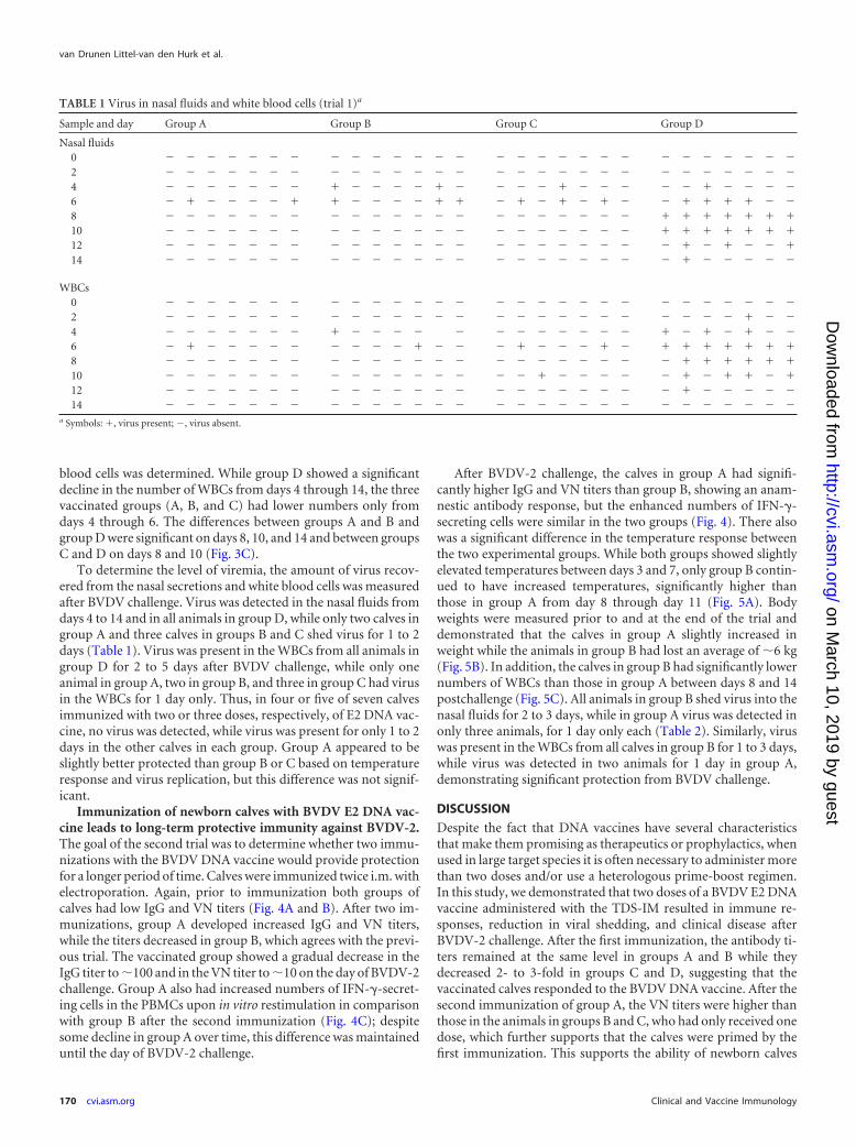
To determine the level of viremia, the amount of virus recov-ered from the nasal secretions and white blood cells was measuredafter BVDV challenge. Virus was detected in the nasal fluids fromdays 4 to 14 and in all animals in group D, while only two calves ingroup A and three calves in groups B and C shed virus for 1 to 2days (Table 1). Virus was present in the WBCs from all animals ingroup D for 2 to 5 days after BVDV challenge, while only oneanimal in group A, two in group B, and three in group C had virusin the WBCs for 1 day only. Thus, in four or five of seven calvesimmunized with two or three doses, respectively, of E2 DNA vac-cine, no virus was detected, while virus was present for only 1 to 2days in the other calves in each group. Group A appeared to beslightly better protected than group B or C based on temperatureresponse and virus replication, but this difference was not signif-icant.
Immunization of newborn calves with BVDV E2 DNA vac-cine leads to long-term protective immunity against BVDV-2.The goal of the second trial was to determine whether two immu-nizations with the BVDV DNA vaccine would provide protectionfor a longer period of time. Calves were immunized twice i.m. withelectroporation. Again, prior to immunization both groups ofcalves had low IgG and VN titers (Fig. 4A and B). After two im-munizations, group A developed increased IgG and VN titers,while the titers decreased in group B, which agrees with the previ-ous trial. The vaccinated group showed a gradual decrease in theIgG titer to �100 and in the VN titer to �10 on the day of BVDV-2challenge. Group A also had increased numbers of IFN-�-secret-ing cells in the PBMCs upon in vitro restimulation in comparisonwith group B after the second immunization (Fig. 4C); despitesome decline in group A over time, this difference was maintaineduntil the day of BVDV-2 challenge.
After BVDV-2 challenge, the calves in group A had signifi-cantly higher IgG and VN titers than group B, showing an anam-nestic antibody response, but the enhanced numbers of IFN-�-secreting cells were similar in the two groups (Fig. 4). There alsowas a significant difference in the temperature response betweenthe two experimental groups. While both groups showed slightlyelevated temperatures between days 3 and 7, only group B contin-ued to have increased temperatures, significantly higher thanthose in group A from day 8 through day 11 (Fig. 5A). Bodyweights were measured prior to and at the end of the trial anddemonstrated that the calves in group A slightly increased inweight while the animals in group B had lost an average of �6 kg(Fig. 5B). In addition, the calves in group B had significantly lowernumbers of WBCs than those in group A between days 8 and 14postchallenge (Fig. 5C). All animals in group B shed virus into thenasal fluids for 2 to 3 days, while in group A virus was detected inonly three animals, for 1 day only each (Table 2). Similarly, viruswas present in the WBCs from all calves in group B for 1 to 3 days,while virus was detected in two animals for 1 day in group A,demonstrating significant protection from BVDV challenge.
DISCUSSION
Despite the fact that DNA vaccines have several characteristicsthat make them promising as therapeutics or prophylactics, whenused in large target species it is often necessary to administer morethan two doses and/or use a heterologous prime-boost regimen.In this study, we demonstrated that two doses of a BVDV E2 DNAvaccine administered with the TDS-IM resulted in immune re-sponses, reduction in viral shedding, and clinical disease afterBVDV-2 challenge. After the first immunization, the antibody ti-ters remained at the same level in groups A and B while theydecreased 2- to 3-fold in groups C and D, suggesting that thevaccinated calves responded to the BVDV DNA vaccine. After thesecond immunization of group A, the VN titers were higher thanthose in the animals in groups B and C, who had only received onedose, which further supports that the calves were primed by thefirst immunization. This supports the ability of newborn calves
TABLE 1 Virus in nasal fluids and white blood cells (trial 1)a
Sample and day Group A Group B Group C Group D
Nasal fluids0 � � � � � � � � � � � � � � � � � � � � � � � � � � � �2 � � � � � � � � � � � � � � � � � � � � � � � � � � � �4 � � � � � � � � � � � � � � � � � � � � � � � �6 � � � � � � � � � � � � � � � �8 � � � � � � � � � � � � � � � � � � � � � 10 � � � � � � � � � � � � � � � � � � � � � 12 � � � � � � � � � � � � � � � � � � � � � � � � � 14 � � � � � � � � � � � � � � � � � � � � � � � � � � �
WBCs0 � � � � � � � � � � � � � � � � � � � � � � � � � � � �2 � � � � � � � � � � � � � � � � � � � � � � � � � � �4 � � � � � � � � � � � � � � � � � � � � � � �6 � � � � � � � � � � � � � � � � � 8 � � � � � � � � � � � � � � � � � � � � � � 10 � � � � � � � � � � � � � � � � � � � � � � � 12 � � � � � � � � � � � � � � � � � � � � � � � � � � �14 � � � � � � � � � � � � � � � � � � � � � � � � � � � �
a Symbols: , virus present; �, virus absent.
van Drunen Littel-van den Hurk et al.
170 cvi.asm.org Clinical and Vaccine Immunology
on March 10, 2019 by guest
http://cvi.asm.org/
Dow
nloaded from

with maternal antibodies to respond to vaccination. The intervalbetween vaccinations did not influence the magnitude of the im-mune responses or protection, which suggests that calves could bevaccinated at branding and then again at entry into the feedlot.Interestingly, once groups B and C had received their second dose,both the VN titers and the numbers of IFN-�-secreting T cellswere equivalent to those of group A after three doses. No virus wasdetected from at least four of seven calves in each of the groups A,B, and C, while the other animals showed reduced durations ofvirus shedding, indicating significant protection from infection.
Importantly, the vaccinated groups experienced only transienttemperature responses and did not lose any weight, which is prob-ably the best indicator of the well-being of an animal and has asignificant economic impact. The fact that there was no significantdifference in the level of protection between groups A, B, and Csuggests that in the context of the immunization regimen tested,the third dose is not needed or beneficial. However, it is possiblethat, as reported for other species (D. Hannaman, unpublisheddata), a third dose spaced further apart from the second one mighthave been beneficial or aided in the duration of the immune re-sponse.
The protective efficacy of this DNA vaccine was confirmed in asecond trial, in which again three of six animals did not haveclinical signs, while the other three showed significantly less andshorter disease, even 3 months after the second vaccination. How-ever, in this trial the animals recovered from leukopenia closer tothe termination of the trial, although at that time their weightswere stable and they did not show evidence of subsequent infec-
FIG 4 BVDV-2-specific immune responses of calves vaccinated with BVDVE2 DNA vaccine. Group A was vaccinated with BVDV E2 DNA vaccine, andgroup B was injected with the diluent. (A) Kinetics of E2.2-specific serum IgGtiters. (B) Kinetics of BVDV-2-specific serum VN titers. (C) E2.2-induced Tcell activation on day 70 (after the second immunization [Imm 2]), day 160 (7days prior to challenge), and day 175 (day 11 postchallenge). The number ofIFN-�-secreting cells/106 PBMCs is expressed as the difference between thenumbers of spots in the E2.2-stimulated and medium control wells. Bars rep-resent median values and interquartile ranges. The significance of differences isshown by asterisks: *, P � 0.05; **, P � 0.01.
FIG 5 Changes in temperatures, weights, and white blood cell counts afterchallenge of calves with BVDV-2. Group A was vaccinated with BVDV E2DNA vaccine, and group B was injected with the diluent. (A) Temperaturechange in °C; (B) weight change in kg; (C) change in WBC counts expressed asn � 109/liter. Data are shown as the medians for six calves. The significance ofdifferences is shown by asterisks: *, P � 0.05; **, P � 0.01; ***, P � 0.001.
Protective BVDV DNA Vaccine in Newborn Calves
February 2013 Volume 20 Number 2 cvi.asm.org 171
on March 10, 2019 by guest
http://cvi.asm.org/
Dow
nloaded from

tion. The fact that there was no weight gain in the vaccinatedgroup may be attributed to the older age of the animals comparedto those in the first trial. In contrast, although virus was detected inall animals on days 8 and 10 in the nasal fluids and on days 6 and8 in the WBCs in both trials, in trial 1 virus was detected earlier inthe nasal fluids and for a longer time in the WBCs. The calves camefrom the same source, and the conditions were comparable envi-ronmentally and nutritionally. However, the calves in the secondtrial were older, which may explain this slight difference in re-sponse, although it is not clear why a shorter virus shedding du-ration did not translate to faster recovery from leukopenia. Whilethe DNA vaccine clearly induced protection for a longer period oftime, the levels appeared to wane in the second trial. Anotherstudy would be needed to determine the limit, possibly with alower challenge dose in addition to the current one, to moreclosely mimic a field situation, in which the viral dose can beexpected to be not quite as high.
A number of approaches to enhance DNA vaccine efficacy havebeen reported including vector optimization, formulation of plas-mids, inclusion of immune modulators, and improved deliverymethods using needle-free devices. Electroporation has proven tobe one of the most successful strategies to overcome the limita-tions of DNA vaccines in large species. This has been demon-strated for a number of species, including nonhuman primates,pigs, and cattle (reviewed in reference 21). In addition, as electro-poration has been well tolerated by human volunteers (22), sev-eral clinical studies have been completed or are under way, al-though most of them involve cancer patients and few targetinfectious disease (21). Recently, a placebo-controlled compara-tive study of electroporation versus injection with a multigenicHIV-1 DNA vaccine in human volunteers was completed. In thisphase I study, electroporation with the TriGrid was well toleratedand significantly improved the magnitude and breadth of the cell-mediated immune responses compared with i.m. injection (23).Electroporation has been used in only a few studies involving cat-tle. Enhanced, but transient, T cell responses to plasmid-encoded
mycobacterial antigens were found in cattle by using separatedouble-needle electrodes; however, humoral responses were notinvestigated (24). Expression of growth hormone-releasing hor-mone (GHRH) in the trapezius muscle of heifers was reportedfrom a plasmid delivered by electroporation (25). We previouslydemonstrated enhanced gene expression, as well as improved im-mune responses, when plasmids encoding reporter antigens wereadministered with the TDS-IM (12). In addition, three doses ofBVDV E2 DNA vaccine administered with the TDS-IM resulted inthe induction of enhanced immune responses and protectionfrom BVDV-2 challenge (13). The TDS-IM was also recently usedin a heterologous prime-boost regimen with a foot-and-mouthdisease virus (FMDV) DNA vaccine. In this study, electroporationalso enhanced both humoral and cell-mediated immune re-sponses, as well as clinical protection from FMDV challenge (26).However, these cattle received two doses of DNA vaccine followedby a protein boost, which is less favorable from a cost-benefitaspect.
To our knowledge, our present report represents one of the fewdemonstrating significant protective immunity with two doses ofDNA vaccine in cattle. With the potential for development of bat-tery-operated electroporation devices, this technology might befeasible for veterinary use in future. In addition, the fact that theE2 DNA vaccine in combination with electroporation is promis-ing against BVDV supports the use of this technology against in-fectious disease in humans.
ACKNOWLEDGMENTS
We thank Laura Latimer for technical assistance and Brock Evans andSherry Tetland for care and handling of the calves. We also thank AntonioUbach and Ryan Betts of Ichor Medical Systems for their assistance withthe production of the electroporation devices.
Financial support was provided by the Alberta Beef Producers, AlbertaLivestock Industry Development Fund, Saskatchewan Agricultural Devel-opment Fund, Alberta Livestock and Meat Agency, and Agriculture andFood Council of Alberta.
REFERENCES1. Ridpath JF. 2003. BVDV genotypes and biotypes: practical implications
for diagnosis and control. Biologicals 31:127–131.2. Harding MJ, Cao X, Shams H, Johnson AF, Vassilev VB, Gil LH,
Wheeler DW, Haines D, Sibert GJ, Nelson LD, Campos M, Donis RO.2002. Role of bovine viral diarrhea virus biotype in the establishment offetal infections. Am. J. Vet. Res. 63:1455–1463.
3. Stokstad M, Loken T. 2002. Pestivirus in cattle: experimentally inducedpersistent infection in calves. J. Vet. Med. B Infect. Dis. Vet. Public Health49:494 –501.
4. Brock KV. 2004. Strategies for the control and prevention of bovine viraldiarrhea virus. Vet. Clin. North Am. Food Anim. Pract. 20:171–180.
5. Bolin SR, Ridpath JF. 1990. Frequency of association of noncytopathicbovine viral diarrhea virus with bovine neutrophils and mononuclear leu-kocytes before and after treatment with trypsin. Am. J. Vet. Res. 51:1847–1851.
6. Collett MS, Larson R, Belzer SK, Retzel E. 1988. Proteins encoded bybovine viral diarrhea virus: the genomic organization of a pestivirus. Vi-rology 165:200 –208.
7. Collett MS, Wiskerchen M, Welniak E, Belzer SK. 1991. Bovine viraldiarrhea virus genomic organization. Arch. Virol. Suppl. 3:19 –27.
8. Gil LH, Ansari IH, Vassilev V, Liang D, Lai VC, Zhong W, Hong Z,Dubovi EJ, Donis RO. 2006. The amino-terminal domain of bovine viraldiarrhea virus Npro protein is necessary for alpha/beta interferon antag-onism. J. Virol. 80:900 –911.
9. Rumenapf T, Unger G, Strauss JH, Thiel HJ. 1993. Processing of theenvelope glycoproteins of pestiviruses. J. Virol. 67:3288 –3294.
10. Donis RO. 1995. Molecular biology of bovine viral diarrhea virus and its
TABLE 2 Virus in nasal fluids and white blood cells (trial 2)a
Sample andday Group A Group B
Nasal fluids0 � � � � � � � � � � � �2 � � � � � � � � � � � �4 � � � � � � � � � �6 � � � � � � � 8 � � � � � � 10 � � � � � � 12 � � � � � � � � � � �14 � � � � � � � � � � � �
WBCs0 � � � � � � � � � � � �2 � � � � � � � � � � � �4 � � � � � � � � � � � �6 � � � � � �8 � � � � � � 10 � � � � � � � � � � �12 � � � � � � � � � � � �14 � � � � � � � � � � � �
a Symbols: , virus present; �, virus absent.
van Drunen Littel-van den Hurk et al.
172 cvi.asm.org Clinical and Vaccine Immunology
on March 10, 2019 by guest
http://cvi.asm.org/
Dow
nloaded from

interactions with the host. Vet. Clin. North Am. Food Anim. Pract. 11:393– 423.
11. Bolin SR. 1993. Immunogens of bovine viral diarrhea virus. Vet. Micro-biol. 37:263–271.
12. van Drunen Littel-van den Hurk S, Luxembourg A, Ellefsen B, WilsonD, Ubach A, Hannaman D, van den Hurk JV. 2008. Electroporation-based DNA transfer enhances gene expression and immune responses toDNA vaccines in cattle. Vaccine 26:5503–5509.
13. van Drunen Littel-van den Hurk S, Lawman Z, Wilson D, LuxembourgA, Ellefsen B, van den Hurk JV, Hannaman D. 2010. Electroporationenhances immune responses and protection induced by a bovine viraldiarrhea virus DNA vaccine in newborn calves with maternal antibodies.Vaccine 28:6445– 6454.
14. Babiuk S, Tsang C, van Drunen Littel-van den Hurk S, Babiuk LA,Griebel PJ. 2007. A single HBsAg DNA vaccination in combination withelectroporation elicits long-term antibody responses in sheep. Bioelectro-chemistry 70:269 –274.
15. Tsang C, Babiuk S, van Drunen Littel-van den Hurk S, Babiuk LA,Griebel P. 2007. A single DNA immunization in combination with elec-troporation prolongs the primary immune response and the duration ofimmune memory. Vaccine 25:5485–5494.
16. Liang R, van den Hurk JV, Landi A, Lawman Z, Deregt D, TownsendH, Babiuk LA, van Drunen Littel-van den Hurk S. 2008. DNA primeprotein boost strategies protect cattle from bovine viral diarrhea virus type2 challenge. J. Gen. Virol. 89:453– 466.
17. Nakamura Y, Gojobori T, Ikemura T. 2000. Codon usage tabulated frominternational DNA sequence databases: status for the year 2000. NucleicAcids Res. 28:292.
18. Pontarollo RA, Babiuk LA, Hecker R, Van Drunen Littel-Van DenHurk S. 2002. Augmentation of cellular immune responses to bovineherpesvirus-1 glycoprotein D by vaccination with CpG-enhanced plasmidvectors. J. Gen. Virol. 83:2973–2981.
19. Liang R, van den Hurk JV, Babiuk LA, van Drunen Littel-van den HurkS. 2006. Priming with DNA encoding E2 and boosting with E2 protein
formulated with CpG oligodeoxynucleotides induces strong immune re-sponses and protection from Bovine viral diarrhea virus in cattle. J. Gen.Virol. 87:2971–2982.
20. Raggo C, Habermehl M, Babiuk LA, Griebel P. 2000. The in vivo effectsof recombinant bovine herpesvirus-1 expressing bovine interferon-gamma. J. Gen. Virol. 81(Part 11):2665–2673.
21. van Drunen Littel-van den Hurk S, Hannaman D. 2010. Electroporationfor DNA immunization: clinical application. Expert Rev. Vaccines 9:503–517.
22. Wallace M, Evans B, Woods S, Mogg R, Zhang L, Finnefrock AC,Rabussay D, Fons M, Mallee J, Mehrotra D, Schodel F, Musey L. 2009.Tolerability of two sequential electroporation treatments usingMedPulser DNA delivery system (DDS) in healthy adults. Mol. Ther. 17:922–928.
23. Vasan S, Hurley A, Schlesinger SJ, Hannaman D, Gardiner DF, DuginDP, Boente-Carrera M, Vittorino R, Caskey M, Andersen J, Huang Y,Cox JH, Tarragona-Fiol T, Gill DK, Cheeseman H, Clark L, Dally L,Smith C, Schmidt C, Park HH, Kopycinski JT, Gilmour J, Fast P,Bernard R, Ho DD. 2011. In vivo electroporation enhances the immu-nogenicity of an HIV-1 DNA vaccine candidate in healthy volunteers.PLoS One 6:e19252. doi:10.1371/journal.pone.0019252.
24. Tollefsen S, Vordermeier M, Olsen I, Storset AK, Reitan LJ, Clifford D,Lowrie DB, Wiker HG, Huygen K, Hewinson G, Mathiesen I, Tjelle TE.2003. DNA injection in combination with electroporation: a novelmethod for vaccination of farmed ruminants. Scand. J. Immunol. 57:229 –238.
25. Brown PA, Davis WC, Draghia-Akli R. 2004. Immune-enhancing effectsof growth hormone-releasing hormone delivered by plasmid injectionand electroporation. Mol. Ther. 10:644 – 651.
26. Fowler V, Robinson L, Bankowski B, Cox S, Parida S, Lawlor C, GibsonD, O’Brien F, Ellefsen B, Hannaman D, Takamatsu HH, Barnett PV. ADNA vaccination regime including protein boost and electroporationprotects cattle against foot-and-mouth disease. Antiviral Res. 94:25–34.
Protective BVDV DNA Vaccine in Newborn Calves
February 2013 Volume 20 Number 2 cvi.asm.org 173
on March 10, 2019 by guest
http://cvi.asm.org/
Dow
nloaded from