Embed Size (px)
Citation preview
The Plant Cell, Vol. 9, 1559-1572, September 1997 O 1997 American Society of Plant Physiologists
Two Distinct Sources of Elicited Reactive Oxygen Species in Tobacco Epidermal Cells
Andrew C. Allan and Robert Fluhr’ Department of Plant Genetics, Weizmann lnstitute of Science, P.O. Box 26, Rehovot 761 00, Israel
Reactive oxygen species (ROS) play a prominent role in early and later stages of the plant pathogenesis response, pu- tatively acting as both cellular signaling molecules and direct antipathogen agents. A single-cell assay, based on the fluorescent probe dichlorofluorescein, was used to scrutinize the generation and movement of ROS in tobacco epider- mal tissue. ROS, generated within cells, quickly moved apoplastically as H202 into neighboring cells. Two classes of rapidly elicited intracellular ROS, originating from distinct sources, were distinguished. Cryptogein, the funga1 elicitor from Phytophthora cryptogea, induced ROS from a flavin-containing oxidase source. ROS accumulation could be inhib- ited by a number of pharmacological agents, suggesting induction through an active signal transduction pathway. The insensitivity of the increase in ROS to the externa1 addition of enzymes that dissipate ROS suggests that this oxidative increase is primarily intracellular. In contrast, amines and polyamines, compounds that form during wounding and pathogenesis, induced ROS at an apoplastic site from peroxidase- or amine oxidase-type enzyme(s). Salicylic acid, a putative inhibitor of cellular catalases and peroxidases, did not induce cellular ROS, as measured by dichlorofluores- cein fluorescence. The physiological relevance of ROS-generated signals was indicated by the rapid alteration of the epidermal cell glutathione pool and the cellular redox state. In addition, induction of ROS by all elicitors was correlated with subsequent cell death.
INTRODUCTION
Pathogen-induced oxidative bursts are harnessed by the plant cell both as a means of “poisoning” an invading patho- gen and as an intracellular and intercellular messenger of this invasion. Reactive oxygen species (ROS), including hy- drogen peroxide (H2O2) and superoxide (Oz-), are the agents of this burst ‘(reviewed in Sutherland, 1991; Mehdy, 1994; Baker and Orlandi, 1995; Bowell et al., 1995; lnzé and Van Montagu, 1995). Although moderately reactive themselves, much of the cellular damage caused by HzOz and Oz- is the result of conversion to even more reactive species; for ex- ample, H202 is converted in the presence of Fez+ to the ex- tremely toxic hydroxyl free radical (OH.) via the Fenton reaction. However, it is HzO2 that is the most attractive can- didate for signaling via ROS because of its relatively long life and high permeability across membranes.
Severa1 enzymatic sources of ROS appear to exist in the plant cell. Evidence is accumulating for the activity of a plasma membrane NAD(P)H oxidase analogous to the mam- malian enzyme producing Oz-. Production of ROS during pathogen response can be prevented by diphenyleneiodo- nium (DPI), an inhibitor of flavin-containing oxidases, includ- ing NAD(P)H oxidase (Levine et al., 1994) and xanthine oxidase (Doussiére and Vignais, 1992). Components of a pu-
’To whom correspondence should be addressed. E-mail IpfluhrQ wiccmail.weizmann.ac.il; fax 972-8-93441 81.
tative plant NAD(P)H oxidase have been identified (Desikan et al., 1996; Groom et al., 1996; Murphy and Auh, 1996). An- other source of ROS, elicited during pathogen attack, is a cell wall-located peroxidase. Although peroxidases usually function to dissipate H2O2, it appears that fast production of HZOz from this enzyme occurs during alkalization of the apo- plast (Bowell et al., 1995), which has been shown to accom- pany pathogen recognition (Viard et al., 1994; Salzer et al., 1996). It is postulated that cell wall peroxidase functions to cross-link walls and toxify the apoplast. Whether one or more of these systems produce the oxidative burst has yet to be determined; it is possible that they operate in tandem or have different kinetics. For example, Otte and Barz (1996) have recently suggested that ROS produced by NAD(P)H oxidase drives peroxidase-catalyzed oxidative processes. An additional source of ROS may emanate intracellularly from xanthine or aldehyde oxidase activity, as seen in mammalian systems (Hille and Massey, 1985). The molecular character- ization of plant xanthine/aldehyde oxidase has been de- scribed recently (Ori et al., 1997).
A wide range of factors appears to elicit an oxidative re- sponse in plant cells. Abiotic inputs, such as UV-B (280 to 320 nm), temperature extremes, pollutants, and osmotic and mechanical stress, all appear to produce increases in ROS (Green and Fluhr, 1995; lnzé and Van Montagu, 1995; Yahraus et al., 1995). Pathogen-related ROS elicitors mainly include
1560 The Plant Cell
pathogen-derived macromolecules that appear to bind plantreceptors to induce ROS (Chandra et al., 1996; Scofield etal., 1996; Tang et al., 1996). Two phases of ROS inductionby fungal or bacterial elicitors have been measured in plantcell suspension cultures. Very rapid responses (within min-utes) have been termed phase I (Baker and Orlandi, 1995)and have been shown to be specifically inhibited by DPI,calcium influx inhibitors, and kinase inhibitors (Baker andOrlandi, 1995; Hammond-Kosack and Jones, 1996). Althoughphase I production of ROS appears to involve an elicitor-receptor interaction, the responses are not always correlatedwith plant disease resistance. Later ROS production (manyhours) is termed phase II and correlates with the resistanceor susceptibility of the plant to the pathogen. Phase I and IIbursts differ kinetically; however, they may also differ as tothe source of ROS and/or the type of ROS produced.
Previous studies on the oxidative burst have concentratedon the rapid sampling of extracellular ROS produced by cellsuspensions. In this study, we used the oxidatively sensitivefluorophore dichlorofluorescein (DCF) to directly measurefast intracellular increases in ROS. In addition to cellularloading, the optical properties of DCF make it amenable toimaging by laser scanning confocal microscopy of leaf tis-sue. DCF has been used previously in plant cell cultures tovisualize oxidative processes in response to mechanicalstress (Yahraus et al., 1995) and long-term accumulation ofROS induced by fungal infection of parsley cells, before theoccurrence of cell death (Naton et al., 1996). In this study,we describe the use of DCF to monitor fast oxidative cyto-solic responses to a number of known elicitors of plant-pathogen responses. Our study differentiates the intracellu-lar oxidative response to two principal sources of ROS: acell wall peroxidase-like activity and a flavin-containing oxi-dase system. These responses are measured in fully differ-entiated epidermal cells rather than in cell culture, whichallows the imaging of ROS transients in single cells and theirsubsequent movement between cells.
RESULTS
DCF Measures Oxidative Potential in EpidermalPeel Cells
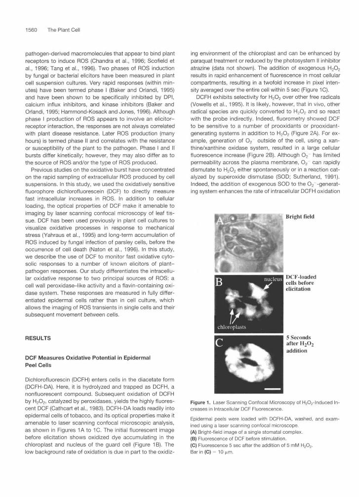
Dichlorofluorescin (DCFH) enters cells in the diacetate form(DCFH-DA). Here, it is hydrolyzed and trapped as DCFH, anonfluorescent compound. Subsequent oxidation of DCFHby H2O2, catalyzed by peroxidases, yields the highly fluores-cent DCF (Cathcart et al., 1983). DCFH-DA loads readily intoepidermal cells of tobacco, and its optical properties make itamenable to laser scanning confocal microscopic analysis,as shown in Figures 1A to 1C. The initial fluorescent imagebefore elicitation shows oxidized dye accumulating in thechloroplast and nucleus of the guard cell (Figure 1 B). Thelow background rate of oxidation is due in part to the oxidiz-
ing environment of the chloroplast and can be enhanced byparaquat treatment or reduced by the photosystem II inhibitoratrazine (data not shown). The addition of exogenous H2O2
results in rapid enhancement of fluorescence in most cellularcompartments, resulting in a twofold increase in pixel inten-sity averaged over the entire cell within 5 sec (Figure 1C).
DCFH exhibits selectivity for H2O2 over other free radicals(Vowells et al., 1995). It is likely, however, that in vivo, otherradical species are quickly converted to H2O2 and so reactwith the probe indirectly. Indeed, fluorometry showed DCFto be sensitive to a number of prooxidants or prooxidant-generating systems in addition to H2O2 (Figure 2A). For ex-ample, generation of O2 outside of the cell, using a xan-thine/xanthine oxidase system, resulted in a large cellularfluorescence increase (Figure 2B). Although O2 has limitedpermeability across the plasma membrane, O2 can rapidlydismutate to H2O2 either spontaneously or in a reaction cat-alyzed by superoxide dismutase (SOD; Sutherland, 1991).Indeed, the addition of exogenous SOD to the O2~-generat-ing system enhances the rate of intracellular DCFH oxidation
Bright field
IKT-loadedcells beforeelicitation
5 Secondsafter H,O2
addition
Figure 1. Laser Scanning Confocal Microscopy of H2O2-lnduced In-creases in Intracellular DCF Fluorescence.
Epidermal peels were loaded with DCFH-DA, washed, and exam-ined using a laser scanning confocal microscope.(A) Bright-field image of a single stomatal complex.(B) Fluorescence of DCF before stimulation.(C) Fluorescence 5 sec after the addition of 5 mM H202.Bar in (C) = 10 (j.m.

Sources of Elicited ROS in Tobacco 1561
(Figure 2B), whereas catalase abrogates the response (data not shown), indicating that H202 formed in this manner freely enters the cell. Rose bengal (4,5,6,7-tetrachloro-2’,4‘,5’,7’- tetraiodofluoroscein) is a water-soluble xanthene dye that forms singlet. oxygen (IO2) when irradiated with white light (Knox and Dodge, 1984). In the plant, ’0, is rapidly con- verted to 0,- and H202 (Foyer et al., 1994). Tobacco peel tissue treated with rose bengal showed a rapid light-depen- dent increase in DCF fluorescence (Figure 2C), suggesting fast conversion of this ROS to H202. UV-B radiation directly elicits multilevel oxidative processes in plants (Hideg and Vass, 1996) and induces the accumulation of free radical scavenging enzymes (Rao et al., 1996), flavonoids (Li et al., 1993), and pathogenesis-related proteins (Green and Fluhr, 1995). Tissues loaded with DCFH show rapid UV-B-dependent accumulation of fluorescence (Figure 2D). DCFH is not directly oxidized by UV-B because exposure of DCFH in vitro to the same levels of UV-B (5 pmol m-2 sec-l) did not induce fluorescence (data not shown).
DCFH oxidation by H,Op requires the presence of peroxi- dase activity (Cathcart et al., 1983). Thus, intracellular re- sponses of DCFH to exogenously added ROS are limited by the availability of cytosolic peroxidases. This dependency makes the calibration of fluorescent responses to meaningful intracellular H202 concentrations difficult. However, intracellu- lar DCF fluorescence increases after addition of exogenous concentrations of H202 from 1 pM to 5 mM (Figure 2E). Thus, within this range of H202, peroxidase activities are not limit- ing, and treatments with the other prooxidants and ROS- generating systems (Figure 2E; xanthine/xanthine oxidase, rose bengal, and UV-B) can result in local concentrations of ROS that are equivalent to the addition of micromolar and millimolar amounts of exogenous H202.
lntercellular ROS Movement 1s Apoplastic
H202 diffusion from elicited tobacco cells activated gene ex- pression in unelicited cells (Levine et al., 1994). We exam- ined the movement of ROS between cells by generating a local ROS signal and examining the response in adjoining cells using the DCF assay. Tissue preloaded with rose ben- gal and DCFH-DA was exposed to 30 sec of white light pre- cisely focused using the microscope condenser (circled areas; Figures 3A and 3D). lncreases in fluorescence were detected during and immediately after white light treatment both in exposed cells and in neighboring cells (Figure 3B). Scanning by using the laser (488 nm excitation) alone was ineffective in inducing measurable activation of rose bengal, and no increase in fluorescence occurred after light expo- sure in cells loaded with DCFH only &e., without rose bengal; data not shown). Changes in the pixel intensity over time in selected cytosolic areas within the exposed cells (area l ) , adjacent cells (area 2), and cells not directly adjacent (area 3) are shown in Figure 3C. ROS generated by light exposure quickly traversed to neighboring cells as well as to cells
Z ‘ 6 T /
40 UV-B on UV-B on
I I 20
d d s i 2 1 k 2 b h h 3 b Time (min)
T a I
Figure 2. Tissue Fluorescence of DCF in the Presence of Prooxidants.
Epidermal peels were loaded with DCFH-DA, washed, and affixed to a holder in a fluorometer cuvette. Relative fluorescence was moni- tored during a time course in which in-flight additions of the indi- cated prooxidants were made. (A) HDz (5 mM). (B) Xanthine oxidase (XO; 0.2 units [U] per mL) and xanthine (0.5 mM), followed by the addition of SOD (25 units per ml). (C) White light exposure of tissue (70 pmol m-2 sec-l) treated with 50 pM rose bengal. (D) Tissue exposed to a supplementary UV-B light source (5 pmol m2 sec-l). (E) Mean fluorescence changes over 10 min expressed as a per- centage of the maximal response possible, as established by treat- ment with 5 mM HzOz for 30 min (performed for all fluorometry experiments). The results of three replicate fluorometric experiments (?SE) are shown. RB, rose bengal; X, xanthine; XO, xanthine oxidase.

1562 The Plant Cell
i- CM CO CO
Time (sec)Figure 3. Movement of ROS in Epidermal Tissue.
Epidermal tissue was loaded with DCFH-DA and rose bengal, washed, and examined by laser scanning confocal microscopy. During a timecourse of image acquisition, bright white light was focused through the microscope condenser on the circled areas, as shown in (A) and (D).(A) DCFH-DA- and rose bengal-treated cells before exposure to light.(B) Cells shown in (A) 180 sec after a white light exposure (30 sec).(C) Time course of change in pixel intensities of selected cytosolic regions, as represented by the boxes in (A).(D) DCFH-DA- and rose bengal-loaded cells in the presence of catalase (100 units per ml).(E) Cells shown in (D) 180 sec after a white light exposure (30 sec). The pseudocolor key is shown within the bar, which was applied to pixel in-tensity values for all of the four fluorescence images.(F) Time course of change in pixel intensities of selected cytosolic regions as represented by the boxes in (D).Bar in (E) = 80 (j.m for all images.
without direct symplastic connection (more than one cellaway). Little or no increase in ROS was observed in areasthat were five or six cells removed from the site of light ex-posure (up to 15 min after exposure; data not shown), indi-cating that the generated ROS were dissipated rather thanpropagated through the tissue.
The addition of catalase before white light exposure abro-gated the fluorescence increase in adjacent cells but not indirectly exposed cells (Figures 3D to 3F). Because the exog-enously added catalase remains in the apoplast, one facetof the induced intercellular movement of ROS apparently in-
volves H2O2. Catalase sensitivity rules out the possibility thatit is the dye, and not ROS, that is moving between cells. Italso indicates that activation of rose bengal by low levels ofreflected and/or detracted light outside of the area of fo-cused light exposure was negligible. We suggest that cyto-solically generated ROS can leave the cell as H2O2 and enterother cells via an apoplastic route, illustrating the potentialfor H2O2 as an intercellular signal. Consistent with this con-clusion is the observation that unexposed stomatal guardcells, which have no symplastic connection to their epider-mal neighbors, also showed increases in intracellular ROS

Sources of Elicited ROS in Tobacco 1563
when nearby rose bengal-loaded cells were activated to produce ROS (data not shown).
Distinct Sources of Elicitor-lnduced Oxidative Bursts
We examined the intracellular accumulation of ROS induced by various types of elicitors. Cryptogein, a small extracellu- lar funga1 protein produced by fhytophthora cryptogea, in- duces on its non-host tobacco rapid changes in ion fluxes, the accumulation of ROS, ethylene, and phytoalexins, and finally, hypersensitive cell death (Ricci et al., 1989; Milat et ai., 1990; Viard et al., 1994). The application of 25 nM cryp- togein to DCFH-loaded tissue resulted in a rapid burst of flu- orescence, followed by a steady accumulation of ROS (Figure 4A). This increase in fluorescence was completely in- hibited by the antioxidant N-acetyl-L-cysteine (at 0.5 mM; see next section).
Salicylic acid (SA) is implicated inystemic pathogen re- sistance and has been postulated to enhance H202 accumu- lation via inhibition of a cytosolic catalase (Chen et al., 1993).
.- c .A ~ 1 mM \
i I]B u 5mM I
Salicylicacid
4
L-Arginine \
C
4 5 mM H202 / ImM
20
I I I , , , I O 4 8 12 16 20 zh 28 32 36
Time (min)
Figure 4. Tissue Fluorescence of DCF in the Presence of Exoge- nous Elicitors.
Epidermal peels were loaded with DCFH-DA, washed, and affixed to a holder in a fluorometer cuvette. Fluorescence was then monitored during a time course in which in-flight additions were made that were followed by the addition of 5 mM H202. (A) Cryptogein (25 nM). (6) Salicylic acid (1 mM). (C) L-Arginine (1 mM). (D) Putrescine (1 mM).
Application of SA to DCFH-loaded tissue caused little or no detectable change in fluorescence (Figure 48). The tissue's sensitivity to the subsequent addition of exogenous H 202 was slightly reduced, indicating that SA may inhibit peroxi- dases required for the DCF assay (Figure 48). lnhibition of ascorbate peroxidase by SA has been observed (Durner and Klessig, 1995). However, it is unlikely that cytosolic peroxi- dases are inhibited by SA to the extent that DCFH cannot be converted to DCF by SA-generated ROS, because L-argi- nine continued to stimulate DCFH oxidation when L-arginine and SA were added simultaneously (data not shown).
Another possible source of ROS in plants is the produc- tion of nitric oxide (NO). In animal systems, NO synthase uses L-arginine'as a substrate to produce NO and L-citrul- line. NO synthase activity has recently been demonstrated in plants (Ninnemann and Maier, 1996). Application of NO to DCFH-loaded tissue via the NO donor (5)-S-nitroso-N-acetyl- penicillamine resulted in a large increase in fluorescence (data not shown), indicating that NO or its ROS derivatives were effectively reported by DCF, as shown in human neu- trophil cells (Rao et ai., 1992). Therefore, we tested whether NO synthase activity could be detected in tobacco cells. Ap- plication of L-arginine to the DCFH-loaded cells resulted in a large increase in DCF fluorescence (Figure 4C). This was spe- cific for the L-isomer and was inhibited by the NO synthase inhibitor N-monomethyl-L-arginine monoacetate (L-NMMA; see next section). However, other amines produced a similar burst, including L-histidine, L-citrulline, L-glutamine, canava- nine, and the polyamines spermidine, spermine, and pu- trescine (data shown for putrescine; Figure 4D). L-Arginine and putrescine had no in vivo effects on the pH indicators SNARF-1 and fluorescein (data not shown).
Amines can induce ROS production by acting as sub- strates for amine oxidases. These are a ubiquitous group of plant enzymes that catalyze the oxidation of a variety of monoamines, diamines, and polyamines to the correspond- ing aldehyde and release NH, and H202 (Tipping and McPherson, 1995). Peroxidase can also produce H202 when supplied with an appropriate reductant (Bowell et al., 1995). We examined ROS production in vitro with commercially available type I horseradish peroxidase by using the luminol assay and readily detected a catalase-sensitive ROS bincrease with L-arginine and putrescine (data not shown). This ROS production shows both L-isomer selectivity and inhibition by L-NMMA similar to that detected in vivo with tobacco tissue. These observations are consistent with the amine oxidase activity associated with peroxidase.
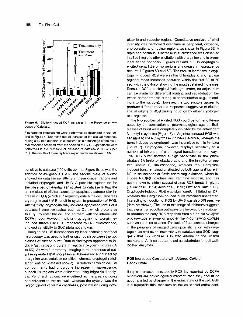
Catalase Sensitivity Differentiates Responses into Two Classes
Fluorometric experiments performed with epidermal cells, using the elicitors of oxidative bursts, in the presence or ab- sence of catalase, distinguished two types of response. Bursts induced by L-arginine and putrescine were highly
1564 The Plant Cell
h u B
80
.i 60
E o 40 * e 20
d -
Ir
Y
ii O
T Treatment only
T
Figure 5. Elicitor-lnduced DCF lncreases in the Presence or Ab- sence of Catalase.
Fluorometric experiments were performed as described in the leg- end to Figure 4. The mean rate of increase of the elicited response, during a 10-min duration, is expressed as a percentage of the maxi- mal response obtained after the addition of H,O,. Experiments were performed in the presence or absence of catalase (100 units per mL). The results of three replicate experiments are shown (?SE).
sensitive to catalase (100 units per mL; Figure 5), as was the addition of exogenous H202. The second class of elicitor showed no catalase sensitivity at these concentrations and included cryptogein and UV-B. A possible explanation for the observed differential sensitivities to catalase is that the amine class of elicitor causes an apoplastic extracellular in- crease in HzOz (which subsequently enters the cell), whereas cryptogein and UV-B result in cytosolic production of ROS. Alternatively, cryptogein may increase apoplastic levels of a catalase-insensitive radical such as 02-, which protonates to H0,- to enter the cell and so react with the intracellular DCFH probe. However, neither cryptogein- nor L-arginine- induced intracellular ROS, monitored by DCF fluorescence, showed sensitivity to SOD (data not shown).
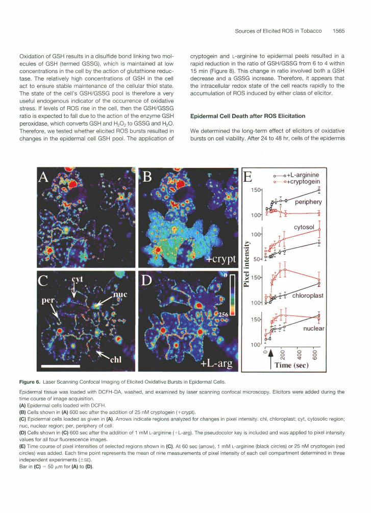
lmaging of DCF fluorescence by laser scanning confocal microscopy was used to further distinguish between the two classes of elicited burst. Both elicitor types appeared to in- duce fast cytosolic bursts in reactive oxygen (Figures 6A to 6D). As with fluorometry, imaging in the presence of cat- alase revealed that increases in fluorescence induced by L-arginine were catalase sensitive, whereas cryptogein elici- tation was not (data not shown). To determine which cellular compartments had undergone increases in fluorescence, subcellular regions were delineated using bright-field analy- sis. Peripheral regions were defined as the area including and adjacent to the cell wall, whereas the cytosol was the region devoid of visible organelles, possibly including cyto-
plasmic and vacuolar regions. Quantitative analysis of pixel intensity was performed over time in peripheral, cytosolic, choroplastic, and nuclear regions, as shown in Figure 6E. A rapid and continuous increase in fluorescence was observed in all cell regions after elicitation with L-arginine and is prom- inent at the periphery (Figures 6D and 6E). In cryptogein- elicited cells, little or no peripheral increase in fluorescence occurred (Figures 6B and 6E). The earliest increases in cryp- togein-induced ROS were in the chloroplastic and nuclear regions; these increases occurred within the first 30 to 60 sec, with the cytosol showing the most sustained increases. Because DCF is a single-wavelength probe, no adjustment can be made for differential loading and redistribution be- tween compartments during experimentation (e.g., reload- ing into the vacuole). However, the two elicitors appear to produce different recorded responses suggestive of distinct spatial origins of ROS during induction by either cryptogein or L-arginine.
The two sources of elicited ROS could be further differen- tiated by the application of pharmacological agents. Both classes of burst were completely inhibited by the antioxidant N-acetyl-L-cysteine (Figure 7). L-Arginine-induced ROS was sensitive to the NO synthase inhibitor L-NMMA, whereas the burst induced by cryptogein was insensitive to this inhibitor (Figure 7). Cryptogein, however, displays sensitivity to a number of inhibitors of plant signal transduction pathways. The ROS burst showed a high sensitivity to the phos- phatase 2A inhibitor okadaic acid and the inhibitor of pro- tein kinase C, staurosporine, whereas the L-arginine- induced burst remained unaffected by both agents (Figure 7). DPI is an inhibitor of flavin-containing oxidases, which in- cludes NAD(P)H oxidase and xanthine oxidase, and has been shown to inhibit severa1 elicited ROS bursts in plants (Levine et al., 1994; Jabs et al., 1996; Otte and Barz, 1996). Cryptogein-induced ROS was significantly inhibited by DPI, whereas the L-arginine-induced burst remained unaffected. Interestingly, induction of ROS by UV-B was also DPI sensitive (data not shown). The use of this range of inhibitors suggests that signal transduction pathways are invoked by cryptogein to produce the early ROS response from a putative NAD(P)H oxidase-type enzyme or another flavin-containing oxidase such as xanthine oxidase. The lack of fluorescence changes in the periphery of imaged cells upon elicitation with cryp- togein, as well as an insensitivity to catalase and SOD, sug- gests that this oxidase is located interna1 to the plasma membrane. Amines appear to act as substrates for cell wall- located enzymes.
ROS lncreases Correlate with Altered Cellular Redox State
If rapid increases in cytosolic ROS (as reported by DCFH oxidation) are physiologically relevant, then they should be accompanied by changes in the redox state of the cell. GSH is a tripeptide thiol that acts as the cell’s thiol antioxidant.
Sources of Elicited ROS in Tobacco 1565
Oxidation of GSH results in a disulfide bond linking two mol-ecules of GSH (termed GSSG), which is maintained at lowconcentrations in the cell by the action of glutathione reduc-tase. The relatively high concentrations of GSH in the cellact to ensure stable maintenance of the cellular thiol state.The state of the cell's GSH/GSSG pool is therefore a veryuseful endogenous indicator of the occurrence of oxidativestress. If levels of ROS rise in the cell, then the GSH/GSSGratio is expected to fall due to the action of the enzyme GSHperoxidase, which converts GSH and H2O2 to GSSG and H2O.Therefore, we tested whether elicited ROS bursts resulted inchanges in the epidermal cell GSH pool. The application of
cryptogein and L-arginine to epidermal peels resulted in arapid reduction in the ratio of GSH/GSSG from 6 to 4 within15 min (Figure 8). This change in ratio involved both a GSHdecrease and a GSSG increase. Therefore, it appears thatthe intracellular redox state of the cell reacts rapidly to theaccumulation of ROS induced by either class of elicitor.
Epidermal Cell Death after ROS Elicitation
We determined the long-term effect of elicitors of oxidativebursts on cell viability. After 24 to 48 hr, cells of the epidermis
-o+L-arginineo+cryptogein
150H
100 x
'tooCM
oo ooID
Time (sec)
Figure 6. Laser Scanning Confocal Imaging of Elicited Oxidative Bursts in Epidermal Cells.
Epidermal tissue was loaded with DCFH-DA, washed, and examined by laser scanning confocal microscopy. Elicitors were added during thetime course of image acquisition.(A) Epidermal cells loaded with DCFH.(B) Cells shown in (A) 600 sec after the addition of 25 nM cryptogein (+crypt).(C) Epidermal cells loaded as given in (A). Arrows indicate regions analyzed for changes in pixel intensity, chl. chloroplast; cyt, cytosolic region;nuc, nuclear region; per, periphery of cell.(D) Cells shown in (C) 600 sec after the addition of 1 mM L-arginine (+L-arg). The pseudocolor key is included and was applied to pixel intensityvalues for all four fluorescence images.(E) Time course of pixel intensities of selected regions shown in (C). At 60 sec (arrow), 1 mM L-arginine (black circles) or 25 nM cryptogein (redcircles) was added. Each time point represents the mean of nine measurements of pixel intensity of each cell compartment determined in threeindependent experiments (±SE).Bar in (C) = 50 M.ITI for (A) to (D).

1566 The Plant Cell
' o'
d i601 T 0 25 nM Cryptogein
1 mM L-Arginine
Figure 7. Effect of Pharmacological Agents on lnduction of ROS by Cryptogein and L-Arginine.
lncreases in DCF fluorescence in tobacco epidermal cells are shown as the percentage of maximal response after the addition of cryp- togein (25 nM) or L-arginine (1 mM) in the presence (+) of the indi- cated pharmacological agents. Results (?SE) are shown for three replicate experiments. NAC, N-acetyl-L-cysteine; L-NMMA, N-mono- methyl-L-arginine monoacetate; DPI, diphenyleneiodonium.
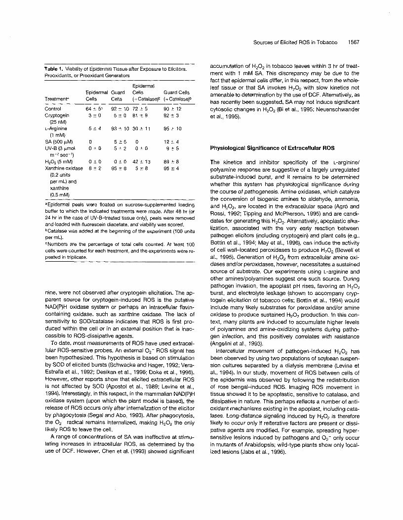
showed good viability (Table 1). However, inclusion of cryp- togein, L-arginine, and SA or exposure to UV-B resulted in epidermal cell death (Table 1). Interestingly, guard cells were generally more viable than epidermal cells and were strik- ingly insensitive to the addition of L-arginine. The addition of catalase to tissues treated with L-arginine or cryptogein had a protective effect, even though the rapid burst induced by cryptogein is catalase insensitive (Figure 5). Production of 02- from a xanthine/xanthine oxidase system caused epi- dermal cell death that could not be prevented by catalase. In addition, guard cells were insensitive to 02-. SA (500 pM) causes death of all cell types by 48 hr, even though similar concentrations do not result in short-term changes in oxida- tive potential (Figures 4 and 5). The cell death promoted by SA cannot be prevented by catalase. Thus, elicitation of ROS is associated with subsequent cell death; however, there are other pathways implicated in plant pathogen re- sponse that do not appear to invoke ROS but nevertheless result in cell death.
,t
DISCUSSION
Source of lntracellular ROS
The development of technologies for precise ROS measure- ment will further our understanding of stress-related oxida- tive responses during plant photosynthetic and pathogenic processes. The chemical properties of the probe DCFH-DA, coupled with laser scanning confocal microscopy and fluo- rornetric quantification, can be one such tool if the relevant caveats are kept in mind. DCFH preferably uses H202 in a peroxidase-based activation (Vowells et al., 1995). However, the ultimate formation of H20, from various species of reac- tive oxygen and other free radicals, by both spontaneous and enzyme-catalyzed processes, facilitates DCFH report- ing of increases in 02-, 'O2, and NO. The discrimination of which primary species are induced during bursts invoked by biotic elicitors is then based on differential sensitivity to ex- ternally applied agents. The catalase sensitivity of amine- elicited cytosolic ROS suggests that amines induce H202 in- flux into the cell after generation'in the apoplast. In contrast, the other class of ROS elicitors, typified by cryptogein and UV-B, was insensitive to extraceliular catalase and SOD. A comparison using confocal imaging of ROS induction by the two classes of elicitor supports different localization of amine- and cryptogein-induced ROS. The peripheral increases in DCF fluorescence, which were readily observed with L-argi-
Control l 0 r
O 15 30 45 60 Time (min)
Figure 8. Effect of Elicitor Addition on the Reduced-to-Oxidized GSH Ratio in Epidermal Peel Tissue.
Epidermal peels were floated on sucrose-containing loading buffer, and cryptogein (25 nM) or L-arginine (1 mM) was added. Samples of peel tissue were removed at the indicated time points, and an analy- sis of glutathione content was made. Each time point represents the mean (?SE) of three separate experiments.
Sources of Elicited ROS in Tobacco 1567
Table I. Viability of Epidermal Tissue after Exposure to Elicitors, Prooxidants, or Prooxidant Generators
Epidermal Epidermal Guard Cells Guard Cells
Treatmenta Cells Cells (+Catalase)b (+Catalase)b
Control 64 t 5 C 92 t 10 72?5 90 5 12 Cryptogein 3?0 5 ' 1 0 8 1 2 9 92 5 3
L-Arginine 5 2 4 93% 10 30 f 11 95 2 10 (25 nM)
(1 mM) SA (500 pM) O 5 ' 1 5 o 12 2 4 UV-B(3pmol 0 5 0 5 - 2 O t O 9 t 5
m-2 sec-I) H202 (5 mM) 0 2 0 O - + O 42-13 8 9 t 8 Xanthine oxidase 8 t 2 95 t 6 5 '1 8 95 r 4
(0.2 units per mL) and xanthine (0.5 mM)
aEpidermal peels were floated on sucrose-supplemented loading buffer to which the indicated treatments were made. After 48 hr (or 24 hr in the case of UV-B-treated tissue only), peels were removed and loaded with fluorescein diacetate, and viability was scored. bCatalase was added at the beginning of the experiment (100 units per mL). CNumbers are the percentage of total cells counted. At least 100 cells were counted for each treatment, and the experiments were re- oeated in triolicate.
nine, were not observed after cryptogein elicitation. The ap- parent source for cryptogein-induced ROS is the putative NAD(P)H oxidase system or perhaps an intracellular flavin- containing oxidase, such as xanthine oxidase. The lack of sensitivity to SOD/catalase indicates that ROS is first pro- duced within the cell or in an external position that is inac- cessible to ROS-dissipative agents.
To date, most measurements of ROS have used extracel- lular ROS-sensitive probes. An external 02- ROS signal has been hypothesized. This hypothesis is based on stimulation by SOD of elicited bursts (Schwacke and Hager, 1992; Vera- Estrella et al., 1992; Desikan et al., 1996; Doke et al., 1996). However, other reports show that elicited extracellular ROS is not affected by SOD (Aposto1 et al., 1989; Levine et al., 1994). Interestingly, in this respect, in the mammalian NAD(P)H oxidase system (upon which the plant model is based), the release of ROS occurs only after internalization of the elicitor by phagocytosis (Sega1 and Abo, 1993). After phagocytosis, the 02- radical remains internalized, making H202 the only likely ROS to leave the cell.
A range of concentrations of SA was ineffective at stimu- lating increases in intracellular ROS, as determined by the use of DCF. However, Chen et al. (1993) showed significant
accumulation of H 2 0 p in tobacco leaves within 3 hr of treat- ment with 1 mM SA. This discrepancy may be due to the fact that epidermal cells differ, in this respect, from the whole- leaf tissue or that SA invokes H202 with slow kinetics not amenable to determination by the use of DCF. Alternatively, as has recently been suggested, SA may not induce significant cytosolic changes in H 2 0 2 (Bi et al., 1995; Neuenschwander et al., 1995).
Physiological Significance of Extracellular ROS
The kinetics and inhibitor specificity of the L-arginine/ polyamine response are suggestive of a largely unregulated substrate-induced burst, and it remains to be determined whether this system has physiological significance during the course of pathogenesis. Amine oxidases, which catalyze the conversion of biogenic amines to aldehyde, ammonia, and H,02, are located in the extracellular space (Agr6 and Rossi, 1992; Tipping and McPherson, 1995) and are candi- dates for generating this H,O,. Alternatively, apoplastic alka- lization, associated with the very early reaction between pathogen elicitors (including cryptogein) and plant cells (e.g., Bottin et al., 1994; May et al., 1996), can induce the activity of cell wall-located peroxidases to produce H202 (Bowell et al., 1995). Generation of H202 from extracellular amine oxi- dases and/or peroxidases, however, necessitates a sustained source of substrate. Our experiments using L-arginine and other amines/polyamines suggest one such source. During pathogen invasion, the apoplast pH rises, favoring an H,Op burst, and electrolyte leakage (shown to accompany cryp- togein elicitation of tobacco cells; Bottin et al., 1994) would include many likely substrates for peroxidase and/or amine oxidase to produce sustained H,O, production. In this con- text, many plants are induced to accumulate higher levels of polyamines and amine-oxidizing systems during patho- gen infection, and this positively correlates with resistance (Angelini et al., 1993).
lntercellular movement of pathogen-induced H202 has been observed by using two populations of soybean suspen- sion cultures separated by a dialysis membrane (Levine et al., 1994). In our study, movement of ROS between cells of the epidermis was observed by following the redistribution of rose bengal-induced ROS. lmaging ROS movement in tissue showed it to be apoplastic, sensitive to catalase, and dissipative in nature. This perhaps reflects a number of anti- oxidant mechanisms existing in the apoplast, including cata- lases. Long-distance signaling induced by H20, is therefore likely to occur only if reiterative factors are present or dissi- pative agents are modified. For example, spreading hyper- sensitive lesions induced by pathogens and 02- only occur in mutants of Arabidopsis; wild-type plants show only local- ized lesions (Jabs et al., 1996).

1568 The Plant Cell
Regulation of ROS by Signal Transduction Pathways
The two sources of ROS appear to be distinct in both their location and sensitivity to pharmacological agents. Only the catalase-insensitive burst induced by cryptogein and UV-B showed sensitivity to inhibitors of plant signal transduction. The sensitivity of the cryptogein response to the protein ki- nase C inhibitor staurosporine has been shown (Viard et al., 1994). Cryptogein-induced ROS also shows a high sensitiv- ity to the phosphatase 2A inhibitor okadaic acid. This com- pound inhibits phagocytotic induction of NAD(P)H oxidase by phorbol myristate acetate (Sega1 and Abo, 1993), a com- pound recently shown to induce ROS from Arabidopsis protoplasts (Desikan et al., 1996). Previous studies have im- plicated NAD(P)H oxidase activity during bursts elicited by pathogen-derived macromolecules by showing sensitivity to DPI (Levine et al., 1994; Jabs et al., 1996; Otte and Barz, 1996). However, the mammalian xanthine oxidase also shows sen- sitivity to DPI (Doussiére and Vignais, 1992). Taken together with evidence for rapid cryptogein-induced changes in ion flux (Viard et al., 1994) and calcium influx (Tavernier et al., 1995), these results suggest that cryptogein invokes a very rapid and complex signal transduction pathway leading to ROS production from a flavin-containing oxidase, such as NAD(P)H oxidase or xanthine oxidase. The temporal sequence of these events should be amenable to simultaneous imag- ing of calcium and ROS transients.
UV-B radiation induces ROS intracellularly, and its effects are inhibited by DPI. This indicates that UV-B may be stimu- lating processes that induce an NAD(P)H oxidase or flavin ox- idase activity as well as having direct ionizing effects (Hideg and Vass, 1996). UV-B has been shown to induce pathogen responses in tobacco by using signal transduction pathways similar to elicitor-induced responses (Green and Fluhr, 1995). In mammalian systems, UV light rapidly (5 min) invokes kinase activity (Devary et al., 1992) via increases in ROS (Huang et al., 1996). To our knowledge, however, induction of flavin- containing oxidases has not been implicated.
ROS as a Message for Cellular Response
The DCF-based fluorescence assay portrays the potential of ROS as a rapid, all-pervasive signal within the plant cell. The accompanying swift adjustment of the cell’s redox state, as determined by the GSH/GSSG ratio, suggests a direct role for ROS as an intracellular message. Changes in the GSH/ GSSG ratio have also been observed during the treatment of tomato cells with race-specific elicitors of Cladosporium ful- vum (May et al., 1996).
In mammalian cells, stress transcription factors, such as nuclear factor-KB and activator protein-I , are redox regulated (Schreck et al., 1992; Meyer et al., 1993; Sen and Packer, 1996). Indeed, epidermal growth factor stimulation of tyrosine phosphotylation in carcinoma cells was shown to be medi- ated by cytosolic production of H202 (Bae et al., 1997). In
plants, gene activation by ROS was shown during the rapid induction of pathogenesis-related (PR) proteins, which was abrogated by ROS scavengers (Green and Fluhr, 1995). Suppression of catalase activity in tobacco under high light intensity (Chamnongpol et al., 1996), or direct application of H202 (€3 et al., 1995), also results in PR protein induction, suggesting H202 as an upstream messenger for redox regu- lation of PR genes. A tobacco Mybl oncoprotein homolog, induced within 15 min of SA treatment, has been shown to specifically bind the promoter of PR-1, suggesting the in- triguing possibility that this early interaction is redox sensi- tive because of the presence of a redox-sensitive cysteine in Mybl (Yang and Klessig, 1996). Direct redox regulation by prooxidants of sensitive cysteine sulfhydryl groups in zinc finger DNA binding proteins may serve to modulate gene ex- pression (Wu et al., 1996). In addition, a zinc finger-contain- ing protein mediates an 0,--initiated programmed cell death response in Arabidopsis (Dietrich et al., 1997). In this respect, the relatively high elicited ROS levels detected around the nucleus may be relevant.
Post-transcriptional and respiratory processes, such as mitochondrial phosphorylation events (Hbkansson and Allen, 1995) and initiation of translation in the chloroplast (Danon and Mayfield, 1994), have also been shown to be redox reg- ulated. This can be via direct effects on protein thiol state, changing the activity of key enzymes. The ubiquity of poten- tia1 redox-sensitive systems, which includes such varied re- sponses as sensing light-dark transitions, high irradiance stress, and pathogen response, raises the question of how specificity is achieved. The presence or induction of specific dissipative agents suggests one mechanism.
ROS and Cell Death
Elicitors of ROS described in this study hastened cell death, which could be abated by the presence of catalase, except in the case of UV-6. SA did not induce changes in ROS at con- centrations that were very effective in causing cell death. SA- induced cell death was not attenuated by catalase, indicating that alternative pathways are initiated by these elicitors of plant pathogenesis. ln whole tissues, millimolar application of SA generally does not cause cell death (e.g., 1 mM; Chen et al., 1993). The enhanced sensitivity of the epidermis may be dueto an absence of SA secondary metabolism or additional signals emanating from wounding in combination with SA. It is therefore possible that the toxicity of SA to these cells has nothing to do with its role in disease resistance.
Guard cells were generally more resistant to cell death in- duced by 02-, H202, and L-arginine. This may be a result of the abundance of chloroplasts in this cell type and their ac- companying ROS detoxification systems, which are an inher- ent part of the photosynthetic process. The lack of resistance of guard cells to cryptogein-induced death may indicate that their ROS dissipative capacity is overloaded or that additional factors are induced by cryptogein that enhance cell death. In

Sources of Elicited ROS in Tobacco 1569
addition, ctyptogein-induced death was catalase sensitive, yet early ctyptogein-induced intracellular ROS increases, as measured by using DCF, were catalase insensitive (Figure 5). It is likely that the extracellular dissipation of ROS can prevent the large buildup of H202 required to drive cell death. The generation of 02- by xanthine oxidase promoted cell death, which could not be reversed by catalase, although catalase abrogated xanthine oxidase-induced DCF fluorescence in- creases. This indicates that the damaging effect is the likely result of 0,- and not H,02, as has also been shown by Jabs et al. (1996). Moreover, the lethal damage done by O*- hap- pens outside of the cell membrane.
The time sequence of DCF-reported reactivity corre- sponds to early phase I responses between bacterial and plant cells (Baker and Orlandi, 1995). This phase invokes active signal transduction pathways but does not specify resistance or susceptibility to the pathogen. Phase II re- sponses (4 to 8 hr) are associated with subsequent hyper- sensitive cell death and resistance to the pathogen (Baker and Orlandi, 1995). Characterizing ROS during these two phases will help to delineate the signal transduction path- ways of pathogenesis.
METHODS
Chemicals and Treatments
Dichlorofluorescin diacetate (DCFH-DA) (Molecular Probes, Eugene, OR) was dissolved in DMSO to produce 100 mM stock, which was frozen as aliquots. Ctyptogein was a kind gift of P. Ricci and H. Keller (INRA, Antibes, France). The signal transduction inhibitors stauro- sporine, K-252a, mastoparan, and N-monomethyl-L-arginine monoac- etate (L-NMMA) and the nitric oxide (NO) donor (2)-S-nitroso- N-acetyl-penicillamine were purchased from Calbiochem (La Jolla, CA), HZ02 from Merck (Darmstadt, Germany), and SNARF-1 from Molecular Probes (Eugene, OR). UV-B irradiation was provided by a Rayonet RPR-3000A lamp (Southern New England Ultraviolet Com- pany, Hamden, CT). Horseradish peroxidase (type I; P-8125) and cat- alase (bovine liver; C-30) were from Sigma. Unless stated otherwise, other chemicals were of analytical grade and were purchased from Sigma.
Laser Scanning Confocal Microscopy and Fluorometry
The first fully expanded leaves were removed from greenhouse- grown tobacco plants (Nicotiana tabacum cv Xanthi). Epidermal peels were then removed from the abaxial surface of each leaf and placed into a small Petri dish containing 10 mL of loading buffer (Tris-KCI at 1 O and 50 mM, respectively, pH 7.2) and 5 pL of DCFH-DA from a 1 O0 mM stock in DMSO. Peels were maintained in the dark for 1 O min to obtain low basal levels of reactive oxygen species (ROS). For some experiments, peel tissue was also loaded in the presence of 50 pM rose bengal. The peels were then removed, floated on a dish of fresh buffer to wash off excess dye, and affixed to a glass coverslip with silicon grease (high vacuum, heavy; Merck) on which the peel remained immersed in 0.5 mL of loading buffer. Examination
of peels was performed using a Bio-Rad MRC-1024 laser scanning confocal microscope. A green argon-ion laser (488 nm) set on 3% power was used for excitation, with 525 nm emission. The viability of the cells within the epidermis under these media conditions was >95% for guard cells and 80% for epidermal cells, as tested by flu- orescein diacetate staining. lmages were captured over a time course, with laser scanning at set time points to avoid photoactivation of the dye. Elicitors or enzymes (no greater than a 50-pL volume) were added directly to the buffer during the time course. Analysis of im- ages was performed on a Power Macintosh 7200 computer (Apple Computer, Inc., Cupertino, CA), using the public domain National In- stitutes of Health image program (National Technical lnformation Service, Springfield, VA).
For fluorometry of whole tissue, a single peel loaded as described above was placed flat onto a polyacrylate plastic holder and affixed at both ends with silicon grease. The holder was inserted into a 3-mL polyacrylate fluorometer cuvette containing 2 mL of aerated loading buffer. The fluorometer (model LS-5B; Perkin-Elmer, Beaconsfield, UK) was set to an excitation of 488 nm and an emission of 525 nm, with slit widths at 5 nm. The cuvette was then placed into the fluorometer and, after establishing a stable baseline, elicitors, enzymes, or phar- macological agents were added. Exposure to supplementary light (UV-B at 5 pmol m-2 sec-I or white light at 70 pmol m-2 sec-l) was achieved by using a liquid light guide (Lumatec, Munich, Germany). For statistical purposes, fluorometry experiments were performed in triplicate, and fluorescence increases (over 1 O min) were expressed as a percentage of the maximal increase possible from the tissue (determined by exposing the tissue to 5 mM H2O2 for 30 min at the end of each experiment).
Luminol-Based Chemiluminescence Assay of H202
Assays were performed in 80 pL of loading buffer containing 1 mM amino acid or polyamine and 10 pL of a 0.2 mg/mL stock of luminol (dissolved in DMSO) within a well of a Microiite plate (Dynatech Lab- oratories, Chantilly, VA). Reactions were initiated by the addition of 10 pL of a 20 unit per mL stock of horseradish peroxidase, and lumi- nescence counts were measured in an ML 3000 Microtiter Plate Lu- minometer (Dynatech Laboratories). Each compound was tested at least in triplicate in the presence or absence of catalase (100 units per mL).
Measurement of GSH and GSSG
Reduced and oxidized glutathione (GSH and GSSG) was measured using a modified method for GSH measurement in microtiter plates (Baker et al., 1990). Briefly, epidermal tissue was peeled and floated on buffers with or without elicitors for the designated time. For each replicate, 20 peels ( 4 0 0 mg of tissue) were gently blotted, weighed, and then immediately frozen in liquid NP. The tissue was homogenized in 300 pL of 5% sulfosalicylic acid, the homogenate was centrifuged at 15,OOOg for 1 O min, and the supernatant was divided into two aliquots. 2-Vinylpyridine (0.35 M final) was added to one aliquot to conjugate GSH, neutralized to pH 6.5 with triethanolamine (10 pL), and allowed to stand at room temperature for 30 min. Oxidized glutathione concen- trations in these neutralized extracts were determined using the en- zymatic recycling assay (Baker et al., 1990) involving the color development at 412 nm of 5,5-dithiobis 2-nitro-benzoic acid in the presence of NADPH (0.25 mM) and GSH reductase (1.25 units per mL). Total GSH plus GSSG levels were determined using the

1570 The Plant Cell
same assay from aliquots without added 2-vinylpyridine. GSH stan- dards treated in the same way were used to calibrate the assay.
Cell Viability Assay
Peels were floated on 10 mL of loading buffer supplemented with 100 mM sucrose in covered Petri dishes. Dishes were placed on an orbital shaker at 30 rpm to ensure aeration. At set time points of up to 48 hr, peels were removed and loaded with the viability stain fluores- cein diacetate (5 pL of a 1 O0 mM stock in acetone added to 1 O mL of loading buffer) for 10 min, washed, and examined using an epifluo- rescence microscope (450- to 490-nm bandpass excitation filter, 515-nm longpass emission filter). Viability was scored for both guard cells and epidermal cells.
ACKNOWLEDGMENTS
We thank Drs. Pierre Ricci and Harald Keller for supplying us with cryp- togein. We also thank Shlomit Bleichman for her excellent technical assistance and Drs. Hillel Fromm, Avihai Danon, Ron Mitler, and Carlos Gitler for helpful discussion and critical reading of the manuscript. This work was supported by a European Union Grant for Biotechnology and a grant from the lsraeli Ministry of Science. A.C.A. was supported by a long-term EMBO postdoctoral fellowship. R.F. is a recipient of the Jack and Florence Goodman Career Development Chair.
Received March 24,1997; accepted July 15,1997.
REFERENCES
Agró, A.F., and Rossi, A. (1 992). Copper-containing plant oxidases. Biochem. SOC. Trans. 20, 369-373.
Angelini, R., Bragaloni, M., Federico, R., Infantino, A., and Porta- Puglia, A. (1 993). lnvolvement of polyamines, diamine oxidase and peroxidase in resistance of chickpea to Ascochyta rabiei. J. Plant Physiol. 142, 704-709.
Apostol, I., Heinstein, P.F., and Low, P.S. (1989). Rapid stimulation of an oxidative burst during elicitation of cultured plant cells. Plant Physiol. 90, 109-1 16.
Bae, Y.S., Kang, S.W., Seo, M.S., Baines, I.C., Tekle, E., Chock, P.B., and Rhee, S.G. (1997). Epidermal growth factor (EGF)- induced generation of hydrogen peroxide. J. Biol. Chem. 272,
Baker, C.J., and Orlandi, E.W. (1995). Active oxygen in plant patho- genesis. Annu. Rev. Phytopathol. 33, 299-321.
Baker, M.A., Cerniglia, G.J., and Zaman, A. (1990). Microtiter plate assay for the measurement of glutathione and glutathione disul- fide in large numbers of biological samples. Anal. Biochem. 190, 360-365.
Bi, Y.-M., Kenton, P., Mur, L., Darby, R., and Draper, J. (1995). Hydrogen peroxide does not function downstream of salicylic acid in the induction of PR protein expression. Plant J. 8, 235-245.
217-221.
Bottin, A., Véronési, D., Pontier, D., Esquerré-Tugayé, M.-T., Blein, J.-P., Rusterucci, C., and Ricci, P. (1994). Differential responses of tobacco cells to elicitors from two Phytophthora species. Plant Physiol. Biochem. 32,373-378.
Bowell, G.P., Butt, V.S., Davies, D.R., and Zimmerlin, A. (1995). The origin of the oxidative burst in plants. Free Radical Res. 23,
Cathcart, R., Schwiers, E., and Ames, B.N. (1983). Detection of picomole levels of hydroperoxides using a fluorescent dichloroflu- orescein assay. Anal. Biochem. 134,111-116.
Chamnongpol, S., Willekens, H., Langebartels, C., Van Montagu, M., Inzé, D., and Van Camp, W. (1996). Transgenic tobacco with a reduced catalase activity develops necrotic lesions and induces pathogenesis-related expression under high light. Plant J. 10,
Chandra, S., Martin, G.B., and Low, P.S. (1996). The Pto kinase mediates a signaling pathway leading to the oxidative burst in tomato. Proc. Natl. Acad. Sci. USA 93, 13393-13397.
Chen, Z., Silva, H., and Klessig, D.F. (1993). Active oxygen species in the induction of plant systemic acquired resistance by salicylic acid. Science 262, 1883-1 886.
Danon, A., and Mayfield, S.P. (1 994). Light-regulated translation of chloroplast messenger RNAs through redox potential. Science
Desikan, R., Hancock, J.T., Coffey, M.J., and Neill, S.J. (1996). Generation of active oxygen in elicited cells of Arabidopsis thaliana is mediated by a NADPH oxidase-like enzyme. FEBS Lett. 382,
Devary, Y., Gottlieb, R.A., Smeal, T., and Karin, M. (1992). The mammalian utraviolet response is triggered by activation of Src tyrosine kinases. Cell71, 1081-1 091.
Dietrich, R.J., Richberg, M.H., Schmidt, R., Dean, C., and Dangl, J.L. (1997). A nove1 zinc finger protein is encoded by the Arabi- dopsis LSD7 gene and functions as a negative regulator of plant cell death. Cell 88, 685-694.
Doke, N., Miura, Y., Sanchez, L.M., Park, H.-J., Noritake, T., Yoshioka, H., and Kawakita, K. (1996). The oxidative burst protects plants against pathogen attack: Mechanism and role as an emer- gency signal for plant bio-defense-A review. Gene 179,45-51.
Doussiére, J., and Vignais, P.V. (1992). Diphenylene iodonium as an inhibitor of the NADPH oxidase complex of bovine neutrophils. Eur. J. Biochem. 208,61-71.
Durner, J., and Klessig, D.F. (1995). lnhibition of ascorbate peroxi- dase by salicylic acid and 2,6-dichloroisonicotinic acid, two inducers of plant defense responses. Proc. Natl. Acad. Sci. USA
Foyer, C.H., Lelandais, M., and Kunert, K.J. (1994). Photoxidative stress in plants. Physiol. Plant. 92, 696-717.
Green, R., and Fluhr, R. (1995). UV-B-induced PR-1 accumulation is mediated by active oxygen species. Plant Cell 7, 203-212.
Groom, Q.J., Torres, M.A., Fordham-Skelton, A.P., Hammond- Kosack, K.E., and Jones, J.D.G. (1996). RbohA, a rice homo- logue of the mammalian gp97phox respiratory burst oxidase gene. Plant J. 10, 515-522.
HBkansson, G., and Allen, J.F. (1995). Histidine and tyrosine phos- phorylation in pea mitochondria: Evidence for protein phosphory- lation in respiratory redox signaling. FEBS Lett. 372, 238-242.
51 7-532.
491-503.
266, 171 7-1 71 9.
213-21 7.
92,11312-11316.

Sources of Elicited ROS in Tobacco 1571
Hammond-Kosack, K.E., and Jones, J.D.G. (1 996). Resistance gene-dependent plant defense responses. Plant Cell8, 1773-1 791.
Hideg, É., and Vass, 1. (1996). UV-B induced free radical production in plant leaves and isolated thylakoid membranes. Plant Sci. 115,
Hille, R., and Massey, V. (1 985). Molybdenum-containing hydroxy- lases: Xanthine oxidase, aldehyde oxidase, and sulfite oxidase. In Molybdenum Enzymes, T.G. Spiro, ed (New York: John Wiley and
Huang, R.-P., Wu, J.-X., Fan, Y., and Adamson, E.D. (1996). UV activates growth factor receptors via reactive oxygen intermedi- ates. J. Cell Biol. 133, 21 1-220.
Inzé, D., and Van Montagu, M. (1995). Oxidative stress in plants. Curr. Opin. Biotechnol. 6, 153-158.
Jabs, T., Dietrich, R.A., and Dangl, J.L. (1996). lnitiation of run- away cell death in an Arabidopsis mutant by extracellular super- oxide. Science 273, 1853-1 856.
Knox, P., and Dodge, A.D. (1984). Photodynamic damage to plant leaf tissue by rose bengal. Plant Sci. Lett. 37, 3-7.
Levine, A., Tenhaken, R., Dixon, R., and Lamb, C. (1994). H,O, from the oxidative burst orchestrates the plant hypersensitive dis- ease resistance response. Cell 79, 582-593.
Li, J., Ou-Lee, T.-M., Raba, R., Amundson, R.G., and Last, R.L. (1 993). Arabidopsis flavonoid mutants are hypersensitive to UV-B irradiation. Plant Cell5, 171-1 79.
May, M.J., Hammond-Kosack, K.E., and Jones, J.D.G. (1996). lnvolvement of reactive oxygen species, glutathione metabolism, and lipid peroxidation in the Cf-gendependent defense response of tomato cotyledons induced by race-specific elicitors of Cla- dosporium fulvum. Plant Physiol. 110, 1367-1 379.
Mehdy, M.C. (1994). Active oxygen species in plant defense against pathogens. Plant Physiol. 105, 467-472.
Meyer, M., Schreck, R., and Baeuerle, P.A. (1993). H,O, and anti- oxidants have opposite effects on activation of NF-KB and AP-1 in intact cells: AP-1 as a secondary antioxidant-responsive factor.
Milat, M.-L., Ricci, P., Bonnet, P., and Blein, J.-P. (1990). Capsid- iol and ethylene production by tobacco cells in response to cryp- togein, an elicitor from fbytophtbora cryptogea. Phytochemistry
Murphy, T.M., and Auh, C.-K. (1996). The superoxide synthases of plasma membrane preparations from cultured rose cells. Plant Physiol. 110, 621-629.
Naton, B., Hahlbrock, K., and Schmelzer, E. (1996). Correlation of rapid cell death with metabolic changes in fungus-infected, cul- tured parsley cells. Plant Physiol. 112,433-444.
Neuenschwander, U., Vernooij, B., Friedrich, L., Uknes, S., Kessmann, H., and Ryals, J. (1995). 1s hydrogen peroxide a sec- ond messenger of salicylic acid in systemic acquired resistance? Plant J. 8, 227-233.
Ninnemann, H., and Maier, J. (1996). lndications for the occurrence of nitric-oxide synthases in fungi and plants and the involvement in photoconidiation of Neurospora crassa. Photochem. Photobiol.
Ori, N., Eshed, Y., Pinto, P., Paran, I., Zamir, D., and Fluhr, R. (1997). TA01 -A representative of the molybdenum cofactor con- taining hydroxylases from tomato. J. Biol. Chem. 272, 1019-1025.
251 -260.
Sons), pp. 443-517.
EMBO J. 12,2005-2015.
30,2171-21 73.
64,393-398.
Otte, O., and Barz, W. (1996). The elicitor-induced oxidative burst in cultured chickpea cells drives the rapid insolubilization of two cell wall structural proteins. Planta 200, 238-246.
Rao, K.M., Padmanabhan, J., Kilby, D.L., Cohen, H.J., Currie, M.S., and Weinberg, J.B. (1992). Fiow cytometric analysis of nitric oxide production in human neutrophils using dichlorofluo- rescein diacetate in the presence of a calmodulin inhibitor. J. Leu- kocyte Biol. 51,496-500.'
Rao, M.V., Paliyath, G., and Ormrod, D.P. (1996). Ultraviolet-B- and ozone-induced biochemical changes in antioxidant ezymes of Arabidopsis thaliana. Plant Physiol. 110, 125-136.
Ricci, P., Bonnet, P., Huet, J.-C., Sallantin, M., Beauvais-Cante, F., Bruneteau, M., Billard, V., Michel, G., and Pernollet, J.-C. (1989). Structure and activity of proteins from pathogenic fungi fbytopbtbora eliciting necrosis and acquired resistance in tobacco. Eur. J. Biochem. 183,555-563.
Salzer, P., Hebe, G., Reith, A., Zittereii-kaid, B., Stransky, H., Gaschler, K., and Hager, A. (1996). Rapid reactions of spruce cells to elicitors released from the ectomycorrhizal fungus Hebe- loma crustuliniforme, and inactivation of these elicitors by extra- cellular spruce enzymes. Planta 198, 118-126.
Schreck, R., Albermann, K., and Bauerle, P.A. (1 992). Nuclear factor KB: An oxidative stress-responsive transcription factor of eukaryotic cells (a review). Free Radical Res. Commun. 17,221-237.
Schwacke, R., and Hager, A. (1992). Funga1 elicitors induce a tran- sient release of active oxygen species from cultured spruce cells that is dependent on Ca2+ and protein kinase activity. Planta 187,
Scofield, S.R., Tobias, C.M., Rathjen, J.P., Chang, J.H., Lavelle, D.T., Michelmore, R.W., and Staskawicz, B.J. (1 996). Molecular basis for gene-for-gene specificity in bacterial speck disease of tomato. Science 274,2063-2065.
Segal, A.W., and Abo, A. (1993). The biochemical basis of the NADPH oxidase of phagocytes. Trends Biochem. Sci. 18,43-47.
Sen, C.K., and Packer, L. (1996). Antioxidant and redox regulation of gene transcription. FASEB J. 10, 709-720.
Sutherland, M.W. (1 991). The generation of oxygen radicals during host plant responses to infection. Physiol. MOI. Plant Pathol. 39, 79-93.
Tang, X., Frederick, R.D., Zhou, J., Halterman, D.A., Jia, Y., and Martin, G.B. (1996). lnitiation of plant disease resistance by physical interaction of AvrPto and Pto kinase. Science 274,
Tavernier, E., Wendehenne, D., Blein, J.-P., and Pugin, A. (1 995). lnvolvement of free calcium in action of cryptogein, a proteina- ceous elicitor of hypersensitive reaction in tobacco cells. Plant Physiol. 109,1025-1031.
Tipping, A.J., and McPherson, M.J. (1995). Cloning and molecular analysis of the pea seedling copper amine oxidase. J. Biol. Chem.
Vera-Estrelia, R., Blumwald, E., and Higgins, V.J. (1992). Effect of specific elicitors of Cladosporium fulvum on tomato suspension cells. Plant Physiol. 99, 1208-1 21 5.
Viard, M.-P., Martin, F., Pugin, A., Ricci, P., and Blein, J.-P. (1994). Protein phosphorylation is induced in tobacco cells by the elicitor cryptogein. Plant Physiol. 104, 1245-1249.
136-1 41.
2060-2063.
270,16939-1 6946.

1572 The Plant Cell
Vowells, S.J., Sekhsaria, S., Malech, H.L., Shalit, M., and Yahraus, T., Chandra, S., Legendre, L., and Low, P.S. (1995). Evi- dence for a mechanically induced oxidative burst. Plant Physiol. Fleisher, T.A. (1995). Flow cytometric analysis of the granulocyte
Immunol. Methods 178,89-97.
Wu, X., Bishopric, N.H., Discher, D.J., Murphy, B.J., and Webster, K.A. (1996). Physical and functional sensitivity of zinc finger tran- scription factors to redox change. MOI. Cell. Biol. 16, 1035-1046.
respiratory burst: A comparison study of fluorescent probes. J. 109,1259-1 266.
Yang, Y., and Klessig, D.F. (1996). lsolation and characterization of a tobacco mosaic virus-inducible myb oncogene homolog from tobacco. Proc. Natl. Acad. Sci. USA 93, 14972-14977.

DOI 10.1105/tpc.9.9.1559 1997;9;1559-1572Plant Cell
A. C. Allan and R. FluhrTwo Distinct Sources of Elicited Reactive Oxygen Species in Tobacco Epidermal Cells.
This information is current as of July 28, 2018
Permissions X
https://www.copyright.com/ccc/openurl.do?sid=pd_hw1532298X&issn=1532298X&WT.mc_id=pd_hw1532298
eTOCs http://www.plantcell.org/cgi/alerts/ctmain
Sign up for eTOCs at:
CiteTrack Alerts http://www.plantcell.org/cgi/alerts/ctmain
Sign up for CiteTrack Alerts at:
Subscription Information http://www.aspb.org/publications/subscriptions.cfm
is available at:Plant Physiology and The Plant CellSubscription Information for
ADVANCING THE SCIENCE OF PLANT BIOLOGY © American Society of Plant Biologists