Embed Size (px)
Citation preview
Eur. J. Biochem. 214, 819-827 (1993) 0 FEBS 1993
Two different genes coding for fibronectin-binding proteins from Streptococcus dysgalactiae The complete nucleotide sequences and characterization of the binding domains
Per-Eric LINDGREN', Martin J. McGAVIN', Christer SIGNAS', Bengt GUSS', Sivashankarappa GURUSIDDAPPA', Magnus HOOKZ and Martin LINDBERG' ' Department of Microbiology, Swedish University of Agricultural Sciences, Uppsala, Sweden
Department of Biochemistry, University of Alabama at Birmingham, USA
(Received January 29/March 22, 1993) - EJB 93 0153/1
The binding of Streptococcus dysgalactiae to fibronectin involves fibronetcin-binding protein(s) present on the bacterial surface. Previously, we reported the cloning of two different genes coding for cell-wall-associated fibronectin-binding proteins from S. dysgalactiae strain S2 [Lindgren, P.-E., Speziale, P., McGavin, M. J., Monstein, H.-J., Hook, M., Visai, L., Kostiainen, T., Bozzini, S. & Lindberg, M. (1992) J. Biol. Chem. 267, 1924-19311. The two genes, fibA and fnbB, have now been sequenced and the primary amino acid sequences of the two fibronectin-binding proteins, FnBA and FnBB, have been deduced. The two proteins have predicted molecular masses of 117 kDa and 122 kDa, respectively, and are organized in a similar way. The fibronectin-binding activities are localized in repeated motifs, 32-37 amino acids long, in the COOH-terminal regions of the proteins. The two fibronectin-binding proteins have heterologous amino acid sequences, except for the COOH-terminal ends which include the fibronectin-binding repeats. The fibronectin-binding regions of the genes have been fused to IgG-binding domains of protein A, utilizing the IgG-binding ca- pacity of the resulting fusion proteins, to facilitate isolation of the fibronectin-binding domains.
Fibronectin (Fn) is a large dimeric glycoprotein (molecu- lar mass 430-470 kDa) which is found in plasma and other body fluids in a soluble form, and is incorporated as micro- fibrils into the extracellular matrix of most tissues as an inso- luble form. The main function of Fn is presumably related to its ability to mediate the substrate adhesion of eucaryotic cells, which involves the binding of specific cell-surface receptors of the integrin family to certain domains in the Fn molecule. The first characterized binding site in Fn for eucaryotic cells contains the amino acid sequence RGD and is located in the central region of the protein (Pierschbacher and Ruoslahti, 1984). Recently, additional recognition sites have been identified (Komoriya et al., 1991). Fn also in- teracts with other matrix components ; binding sites for colla- gen, fibrin and sulfated glycosaminoglycans are located in discrete domains of the protein. The present knowledge of the structure and function of Fn has recently been summa- rized by Hynes (1990).
Correspondence to M. Lindberg, Department of Microbiology, Swedish University of Agricultural Sciences, Box 7025, $75007 Uppsala, Sweden
Fan: +46 18 673392. Abbreviations. Fn, fibronectin; FnBA, Fn-binding protein en-
coded by fnbA; FnBB, Fn-binding protein encoded by fibB ; Sfb, fibronectin-binding protein.
Note. The novel nucleotide sequence data published here has been deposited with the EMBL sequence data banks and is available under the accession numbers 222150 and 222151.
Several types of microorganisms also bind to Fn, includ- ing Gram-positive bacteria, such as Staphylococcus aureus, coagulase-negative staphylococci, groups A, C and G strep- tococci (Kuusela, 1978; Switalski et al., 1982; Switalski et al., 1983; Hanski et al., 1992; Talay et al., 1992) and myco- bacteria (Abou-Zeid et al., 1988). Also, some Gram-negative bacteria, e.g. Escherichia coli (Froman et al., 1984; Arnqvist et al., 1992) and Porphyromonas gingivalis (Lantz et al., 1991), as well as the spirochetes Treponema pallidum (Pe- terson et al., 1983) and Treponema denticola (Dawson and Ellen, 1990), have been reported to bind Fn.
Many of the bacterial species which bind Fn are common wound pathogens, and it has been suggested that Fn serves as a substratum for bacterial adherence in the wound. Bind- ing of bacteria to Fn may, in this case, represent the first step in the development of an infection (Hook et al., 1989, Hanski et al., 1992). In most cases, bacteria appear to bind to site(s) located in the NH,-terminal domain of Fn (Mosher and Proc- tor, 1980; Speziale et al., 1984). In previous studes, we have characterized two different genes encoding Fn-binding pro- teins from s. aureus (Flock et al., 1987; Signas et al., 1989; Jonsson et al., 1991). Streptococcus dysgalactiae, a group C streptococcus, is like S. aureus, frequently associated with bovine mastitis (Watts, 1988). Most strains of S. dysgalactiae bind Fn and a dissociation constant of 26 nM was determined for binding of the 29-kDa NH,-terminal fragment of Fn to strain S2 (Mamo et al., 1987). We previously reported (Lind- gren et al., 1992) the cloning of two different genes encoding Fn-binding proteins from S. dysgalactiae S2. Cell extracts
820
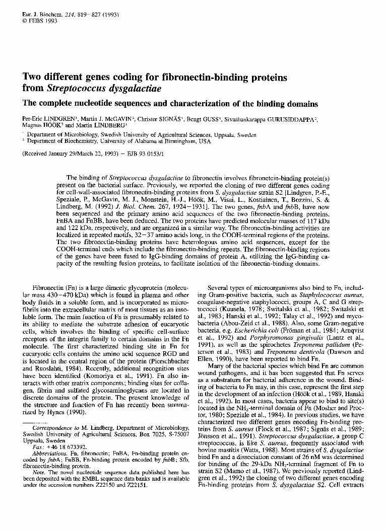
BamH I BstX I Hind 111 BStx I EcoR Xba I BamH I Xba I BstX I Xba I Sau3A I I I I f 1 I i I I I I I I I I 0 1 2 3 4 5 kb
Gene A
Expression pSDFlOO pSDF102 + pSDF102~10 + pSDF102c13 + pSDF102~9 + pSDF102cll + pSDF102~14 +
C +
pSDF102~18 -
d Cla I Pst I Bgl II Hind 111 Xho I Sau3A I Hind 111 Hind Ill
I I I I I I I I I 1 I
D
0 1 2 3 4 kb
E b 2 w3 Gene fnh B I , I
B X B RE3 MB
F Expression pSDF201 + pSDF202 + pSDF203 + pSDF250 + pSDF203c3 + pSDF203c6 (+) pSDF203~8 (+) pSDF203cll - pSDF203c9 -
Fig. 1. Restriction maps of inserts and schematic diagrams of proteins, clones and subclones. (A) Restriction map for genefnbA. The arrow indicates the direction of transcription of the gene. (B) Schematic diagram of the protein encoded by the gene fnbA. S, signal sequence; X, spacer region; X,, proline-rich sequence of the spacer region; R, domain consisting of repeated motifs; W, cell-wall-spanning domain; M, membrane-spanning domain. (C) Different clones and subclones. The Fn-binding activities of lysates from the different subclones, expressed as inhibition of binding of radiolabeled Fn to S. dysgaluctiae S2 are indicated. Mgre than 85% inhibition, + ; less than 15% inhibition, -. (D) Restriction map for fnbB. (E) Schematic diagram of the protein encoded by gene fnbB. (F) Clones and subclones forfnbB. Explanation as in A-C forfnbA but (+) indicates 30%-40% inhibition.
prepared from E. coli strain TG1, in which the cloned genes MATERIALS AND METHODS - - were expressed, efficiently blocked the binding of Fn to S. dysgalactiae. Monospecific antibodies to the purified re- combinant Fn-binding proteins were prepared. Using these antibodies in Western-blot analyses of S. dysgalactiae ly- sates, it appeared that only one of the genes (fnbB) was ex- pressed in the streptococcal cells under the culture conditions used.
We now report the nucleotide sequence of the two genes from S. dysgalactiae S2. The features of the nucleotide se- quences and the deduced amino acid sequences are dis- cussed. As with the previously characterized staphylococcal Fn-binding proteins, the Fn-binding domains are located in segments of the proteins consisting of repeated motifs. Corre- sponding gene domains were fused to the expression vector pEZZ (Nilsson et al., 1987). This vector is based on the IgG- binding domains of protein A from S. aureus which allows isolation and purification of fusion proteins using an IgG- Sepharose column.
Bacterial strains, plasmids and growth media E. coli strain TG1 and the plasmid vector pUC18 (Nor-
rander et al., 1983) were used in gene cloning. The pEZZ vector plasmid was used for expression of extracellular fu- sion proteins in E. coli TG1 (Nilsson et al., 1987). The E. coli clones containing pSDFlOO and pSDF200 harbouring the genes fnbA and fnbB, were described earlier (Lindgren et al., 1992). S. dysgalactiae strain S2 (Lindgren et al., 1992) was grown in Todd-Hewitt broth (Difco) and E. coli was grown in Luria-Bertani medium, both at 37°C.
Assay for Fn-binding activity Isolation, purification and radiolabelling of the 29-kDa
NH,-terminal fragment of bovine Fn was performed as de- scribed earlier (Lindgren et al., 1992). The preparation of lysates from E. coli clones and subclones, and analysis of Fn-binding activity, was as described (Lindgren et al., 1992).

821
DNA preparation and nucleotide sequencing
Methods for preparing DNA were essentially as de- scribed (Maniatis et al., 1982). Oligonucleotide primers for DNA sequencing were purchased from the Departments of Microbiology and Immunology at the Biomedical Center, University of Uppsala. Streptococcal DNA fragments, in plasmid vector pUC18, were sequenced by the method of Sanger et al. (1977). The sequencing reactions were per- formed using a Sequenase-version-2.0 kit (United States Bio- chemical Corporation). The DNA samples for sequencing were analyzed by PAGE using wedge-shaped 6% acrylamide gels containing 7.0M urea (Sanger et al., 1978). The se- quence data were recorded and analyzed by computer pro- grams (Devereux et a]., 1984).
SDSRAGE and Western blotting
SDSPAGE (Laemmli, 1970) was performed using stack- ing and running gels with 4% and 12% acrylamide, respec- tively. Proteins, separated on the SDS/polyacrylamide gel, were either stained with Coomassie brilliant blue or electro- phoretically transferred to a nitrocellulose filter (Schlei- cher & Schiill) and probed with a ‘251-labelled NH,-terminal fragment of Fn, essentially as described in Jonsson et al. (1991).
Restriction endonucleases and other enzymes
Restriction enzymes and T4 DNA ligase were purchased from International Biotechnologies Inc., New England Bio- labs, Promega or Boehringer Biochemicals and used accord- ing to the suppliers’ recommendations. For polymerase- chain-reaction amplification the Gene Ampm kit, obtained from Perkin Elmer Cetus, was used.
RESULTS
Subcloning of genes and sequencing strategy
We reported earlier the cloning and isolation of two dif- ferent genes encoding Fn-binding proteins from S. dysgalac- tiae S2 using the plasmid pUC18 (Lindgren et al., 1992). The genes of the original clones, containing plasmids called pSDFlOO and pSDF200, were further subcloned using appro- priate restriction endonuclease cleavages and digestion with exonuclease III for different time periods. A series of over- lapping subclones were constructed (Fig. 1). Both strands of the inserts in the plasmids were sequenced. The whole gene, fnbA, coding for the Fn-binding protein A (FnBA), was pre- sent within the insert of streptococcal DNA in pSDFlOO (Fig. 1A-C). The second gene,fnbB, coding for Fn-binding protein B (FnBB), was incomplete in the original insert in pSDF200. Thus, to obtain the sequence of the last 3‘ end of the gene (Fig. 1D-F), it was necessary to screen a library prepared by cleaving the chromosomal DNA with the restric- tion enzymes XhoI and HindIII, and the pUCl8 vector with HindIII and Sun. The library was screened with a 32P-la- belled 20-residue synthetic oligonucleotide of a unique se- quence derived from a site near the 3’ end of the insert in pSDF200. [y-32P]ATP was used to radiolabel the oligonucleo- tide. One positive clone, containing a plasmid called pSDF250, was chosen for further studies (Fig. 1F).
Nucleotide sequences of fnbA and fnbB and the deduced amino acid sequences of encoded proteins
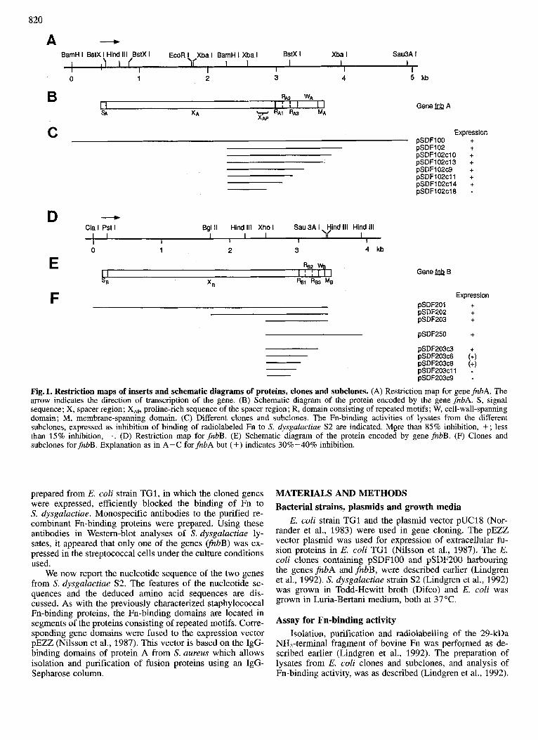
The DNA sequence of the insert in pSDFlOO (Fig. 2) containing fnbA has an open reading frame, starting with an ATG codon at nucleotide 437 and terminating in a TAA stop codon at nucleotide 3710, encoding a polypeptide of 1091 amino acids. Downstream of the coding region, there is a hairpin-loop structure that may act as a transcriptional termi- nator. Fifteen nucleotides upstream of the initiation codon, there is a potential ribosome-binding site, GAAGGA, and further upstream possible promoter signals are indicated, re- sembling those found in other Gram-positive bacteria (Mor- rison and Jaurin, 1990; Moran et al., 1982). As the encoded protein is a cell-surface protein in S. dysguluctiue, a removal of signal peptide should precede mature protein formation. Three possible cleavage sites for signal peptidase are indi- cated in Fig. 2, resulting in signal peptides of 24, 32 or 37 residues.
One major region of repetitive units was identified by computer analysis of the gene sequence. The repetitive re- gion, called RA, is located near the 3’ end of the gene (Fig. 1B) and consists of a 32-37-amino-acid motif repeated three times (Fig. 2 and Fig. 4). Furthermore, starting up- stream of RAT, at residue 829, there is a region of 12 amino acids very similar to the COOH-terminal regions of the R, repeats. Flanking the R, region there are domains rich in prolines. The upstream region is a unique sequence of ap- proximately 50 amino acids called X,, (starting at residue 763) of which 16 amino acids are prolines. Downstream of the R, region, the proline residues are again more frequent than in the rest of the protein, which is typical for the cell- wall-spanning domain (W,) of cell-surface proteins in Gram- positive cocci (Pancholi and Fischetti, 1988).
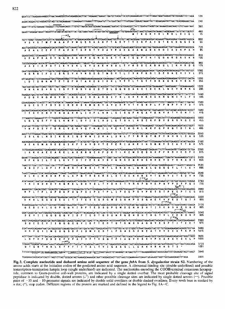
The insert in pSDF201 contains an open-reading frame encoding most of fnbB, while the insert in pSDF250, which partly overlaps pSDF201, contains an open reading frame encoding the 3‘ end of fnbB (Fig. 1E and F). The combined sequences of the inserts in pSDF201 and pSDF250 give the nucleotide sequence of the completefnbB gene (Fig. 3). 13 nucleotides upstream of the putative start codon TTG there is a sequence, GGAGGAG, which is a probable ribosome- binding site. Upstream of the ribosome-binding site, there are some possible promoter signals, as indicated in Fig. 3. The insert contains an open reading frame, coding for a polypep- tide of 1117 amino acids, beginning with a TTG start codon at nucleotide 140 and terminating in a TAA stop codon at nucleotide 3491 (Fig. 3). One possible cleavage site for a signal peptidase is indicated, resulting in a signal peptide (S,) of 27 amino acids. Downstream of the coding region, there is a loop structure, which probably acts as a transcrip- tion terminator. As in gene jhbA, a repetitive domain, called R,, is present in the 3’ end of the gene. This part of the gene encodes a region containing a unit of 36 amino acids repeated twice and partly a third time (Fig. 3 and Fig. 4). Downstream of the repeat sequence the gene encodes a proline-rich region, W,, probably spanning the cell wall.
The COOH-terminal ends of FnBA and FnBB proteins have similar general features, including hydrophobic trans- membrane domains (MA and M,) followed by a short region of charged, mainly basic, amino acids. These features are characteristic of cell surface proteins in Gram-positive cocci.
The Fn-binding regions In a previous study (Lindgren et al., 1992), smaller frag-
ments of the inserts in pSDFlOO and pSDF200 were sub-
822
: . - f i A
TGT T TCcoT;GGAACCATGiTTATGGCA~CACCTGT TATGGGAGAGGACGCkCTCAACCAACTGC~CiGTTACTACGGAATCCCCAGCGATACAAACiGAAGAGGATCAAGGTAGCCA ~ S V G T Y F Y A A P V M G E D A S ~ P T A ~ ~ ~ ~ E ~ P A ~ ~ ~ E E D ~ ~ ~ ~
CGAGCCACCiCCAAAAGCT iCTGTTCMGCOOAAGCCGCiACCCCCGCTGGTAMGCTGAAGCTACTACiAACACTGGTCMCCGACCAACACAGAGCAAGCACGT TCCCGCAGCAAGCG E P A P K A S V ~ A E A A S P A G K A E A ~ T N T G ~ P ~ N ~ E ~ A R S R S K R
TAATAAT C G i AAT TCACTTCGTTTMTTO*CTTTTATAGGAAGGTACG*~AATCTACCGATTTATCTGCATCCCATGCCMAA~TC~TGAMAACT~MCCMGTTiGGAMAAAGC N N R N S V R L I D F Y R K V G E S T D L S G U D A K K I D E K L M E V W K K A
TAAGGATGACTATAATCOAiG~GCGTAGATTTACAGGG~GCCATTCATMAGCAAGAGAAATTTTTAA~TTAGATAAAGMAAGAGGTCGGGTAAACGACAACATATTGTTTTATTTTC K D D Y N G W C V D L ~ G A I H K A R E I F N L D K E K R S G K R ~ H I V L F S
T AACTCT T CAT TTCTTT C A i TCAT T 1 T CT C M G T T C M C A AATGA A ACT AT AACT TCGACACT T TCA AAAPAT A AACTGCAM AGGCT C i AC A AT CAGGGGA AACT TTAACCT T ACiAGT A N S S F L S F I F S S S T N E S I T ~ T L S K D K L ~ K A L ~ S ~ E T L T L E Y
' P X A P . ATATAAGAT~AATGATAAAGAAGTGAAAGGTAAAGAACTAGACGATGTC~ CTTTAACTT ACAGT AAACAAACCGTTCGT AAGCCACAGGTCGAACCAAAiGTT CCTGAT ACACCI CAGGA
Y K I N D K E V K G K E L D D V S L T Y S K E T V R K P ~ V E P N V P D T P ~ E . x 4 P p F 7 .
AAAACCATTGACACCGCTTGCACCGTCAGAACCTTCACAACCATCTATTCCAGAGACACCACTGATACCGTCAGAACCT~CAGTTCCAGAGACATCAACACCAGAAGGTCCAACAGAGCG K P L T P L A P S E P S ~ P S I P E T P L I P S E P S V P E T S T P E ~ P T E G
. P R A I . E N N L C C ~ S E E I T I T E O S P S G M S ~ ~ N P ~ S ~ N E T V V E D T ~ T S . ~ R A Z . P E D I V L G G P C P V I O F T E D S ~ P G M S ~ N N S H T I T E D S K ~ S ~ E
AGAAAATAA;CTTGGTGGTCAGAGTGAAGAGATAACGATiACAGAAGAT~CTCAATCAGGGATGl CTGG;CAAAATCCTCCTTCTGGAAATGAAACAGTGGTTGAAGACACTCAAACAAG
TCAAGAGGAiATTGTACTTGGTGGTCCAGGTCAAGTGATiCACTTTACAGAACATAGCCAACCGGGTATGTCTGGTAATMTAGCCATACTATTACAGAAGATTCTAAACCAAGTCAACA
. r K A 3 . GGATGAGGTGATAATCCGCGCTCAAGGTCAGGTCATTGACTTTACAGAAGATACTCAATCTGGTATGTCTGCGGATAATACCCATACAGATCGGACACTCCTTCAAGAACACTCTAAACC
D E V I I G ~ P C P V I D F T E O T P S G M S ~ D N S H ~ D ~ ~ V L E E D S K P ' P w A .
AAGTCAAGAGGATGAGGTGAT AATCGGCGGTCAAGGT CAAGTGATTGAC 111 Ai AGAAGAT ACCCAAACCGGT ATGT CTGCGGCTCGACAAGTAGAGAGTCCAACAATCACCGAAGAAAC S ~ E O E V I I G ~ ~ ~ ~ V I D F T E O T ~ T ~ Y S ~ A ~ ~ V E S P T I T E E T
CCATAAACCAGAAATAATCATGGGCGGTCAAAGTGACCCTATTGATATGGTTGAGGACACTCTTCCTGG~ATGTCTGGC~CTAATGAAGCTACTGTTGTGGAAGAAGACACACGTCCTAA H K P E I I U G G ~ S D P I D Y V E D T L P G Y S G S N E A T V V E E D T R P K
L ~ F H F D N E E P V P A T V P T V S ~ T P I A ~ V E S K V P H A K A E S A L P
~ T G O T N K L E T F F T I T A L T V I G A A G L L G K K R R N N ~ T O
ACTTCAATTCCATTTTCATAATGAAGAGCCCGTTCCTGCAACGGTTCCAACCGTTTCTCAAACTCCTATiGCTCAGGTAGAAAGTAAAGiGCCTCATGCCAAAGCAGAGAGTGCGi~ACC
MA ~~AAAciG~~~~~ACAAATAAACTAGAAACGTTCTTTACCATTACAGCACTAACTGTTAiTGGAGCGGCAGGATTACTAGGCAAAAAACGTCGTAATAAiCAAACTGAT it ATCAGCAGA
T T T C A T C ~ G C ~ A A A C A A G G C T A A C A T T ~ T A G C C T i G T T T T ~ T l ~ C A C T G A C C T C T A A A A G T T A T G A C T G i T T T A A A G G G G G G G T A G G C C A A T C C T C A A A A G T A G T T A A G ~
TGAGAAACACCACATCACT i T AGTCTT ACiGCGCAT ACT AAAAGCAAAAGAT AAT TAGGAPCACT TGCTAACTGGAAAAAATCAAPTtC;AAAGCTAGTTGCCAAAGAACiCTAGA
120
240
360
480 15
600 55
720 95
840 135
960 175
1080 215
1200 255
1320 295
1440 335
1560 375
1680 415
1800 455
1920 495
2040 535
2160 575
2280 61 5
2400 655
2520 895
2640 135
2760 775
2880 81 5
3000 855
31 20 895
3240 935
3360 97 5
3480 1015
3600 1055
3720 1091
3840
3955
Fig.2. Complete nucleotide and deduced amino acid sequence of the gene fnbA from S. dysgahctiae strain S2. Numbering of the amino acids starts at the initiation codon of the predicted amino acid sequence. A ribosomal-binding site (double underlined) and possible transcription-termination hairpin loop (single underlined) are indicated. The nucleotides encoding the COOH-terminal consensus hexapep- tide, common to Gram-positive cell-wall proteins, are indicated by a single dotted overbar. The most probable cleavage site of signal peptidase is indicated by double, dotted arrows (1') and other possible cleavage sites are indicated by single dotted arrows (--+). Possible pairs of -35 and -10 promoter signals are indicated by double solid overlines or double dashed overlines. Every tenth base is marked by a dot; (*), stop codon. Different regions of the protein are marked and defined in the legend to Fig. 1A-C.
823
120
240 34
360 74
480 114
600 154
720 194
840 234
960 274
1080 31 4
1200 354
1320 394
1440 434
1560 474
1660 514
1800 554
1920 594
2040 634
2160 674
2280 71 4
2400 754
2520 794
2640 834
2760 874
2880 91 4
3000 954
31 20 994
3240 1034
3360 1074
3480 1114
3600 1117
3661
Fig. 3. Complete nucleotide sequence and deduced amino acid sequence of the gene fnbB from S. dysgalactiae strain S2. Explanations as in Fig. 2.
824 STREPTOCOCCUS DYSGALACTIAE S2, PnBA
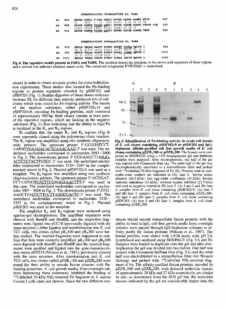
887 A1 850 EDTQT SQEDI V-LGG PGQVI DPTED SQPG!A SGNNS HTIT
A2 888 EDSKP SQEDE VIIGG L W V I DPTED TQSGM SGDNS HTDGT VLE 930
969 A3 931 EDSKP SQEDE VIIQQ QOQVI DPTED TQTGM SGAGQ VESP
II I I I I I I I I I I I I I I I I I I I I I I I I I
I I I I I I I I I T - I I I I I I I I I I I I I I I I
STREPTOCOCCUS DYSGALACTIAE S2. FnBB
994 B1 959 EETLP TEQGQ SGSTT EVEDT KQPEV IIGGQ GEIVD I
1030 B2 995 EENLP TEQGQ SGSTT EVEDT KGPEV IIGGQ G E W D I
B3 1031 EESLP TEQGQ SGSTT EVEDS KPKLS IHPDN FWPKE D 1066
I I I I I I I I I I I I I I I I I I I I I I I I I I I I I I I I I I
I I I I I I I I I I I I I I I I I I I I
Fig. 4. The repetitive motifs present in FnBA and FnBB. The numbers denote the positions in the amino acid sequences of these regions and a vertical line indicates identical amino acids. The conserved octapeptide EVIIGGQG is underlined.
cloned in order to obtain accurate probes for cross-hybridiza- tion experiments. These studies also located the Fn-binding regions to protein segments encoded by pSDF102 and pSDF203 (Fig. 1). Further digestion of these clones with exo- nuclease 111, for different time periods, produced sets of sub- clones which were tested for Fn-binding activity. The inserts of the smallest subclones, called pSDF102c14 and pSDF203 c8, encoding Fn-binding peptides, each consisted of approximately 500 bp. Both clones contain at least parts of the repetitive regions, which are lacking in the negative subclones (Fig. l) , thus indicating that the ability to bind Fn is localized in the R, and RB regions.
To confirm this, the entire R, and R, regions (Fig.4) were separately cloned using the polymerase chain reaction. The R, region was amplified using two synthetic oligonucle- otide primers. The upstream primer 5’-GCGGATCCT- AATGGTGAAGACACTCAAACAAG-3’ was used. The un- derlined nucleotides correspond to nucleotides 2983 - 3000 in Fig. 2. The downstream primer 5’-CCGAAGCTTATB ACTCTCTACTTGTCC-3’ was used. The underlined nucleo- tides correspond to nucleotides 3326-3343 in the comple- mentary strand in Fig. 2. Plasmid pSDF102c9 was used as a template. The R, region was amplified using two synthetic oligo-nucleotide primers. The upstream primer 5’-GCGGAT- CCTAATGGTGATTTCGAGGAAACTTT-3’ was used in this case. The underlined nucleotides correspond to nucleo- tides 3007-3024 in Fig. 3. The downstream primer 5’-CCG- AAGCTTAGTCTTCCTTAGGCCA CTC-3’ was used. The underlined nucleotides correspond to nucleotides 3320 - 3337 in the complementary strand in Fig. 3. Plasmid pSDF203 was used as the template.
The amplified R, and R, regions were analysed using agarose-gel electrophoresis. The amplified sequences were cleaved with BamHI and HindIII, and the respective. frag- ments were ligated into pUCl8 (previously digested with the same enzymes.) After ligation and transformation into E. coli TG1 cells, two clones called pR,100 and pRB200 were fur- ther studied. The inserted fragments were sequenced to con- firm that they were correctly amplified. pR,lOO and pRB100 were digested with BamHI and Hind111 and the inserted frag- ments were purified and ligated into the gene-fusiodsecre- tion vector pEZZl8 (Nilsson et al., 1987), previously cleaved with the same enzymes. After transformation into E. coli TG1 cells, two clones called pZZR,100 and pZZRB200 were tested for their ability to encode fusion proteins with Fn- binding properties. E. coli growth media, from overnight cul- tures harbouring these constructs, inhibited the binding of ‘251-labelled 29-kDa NH,-terminal Fn fragment to 5’. uureus Cowan I cells (data not shown). Since the two different con-
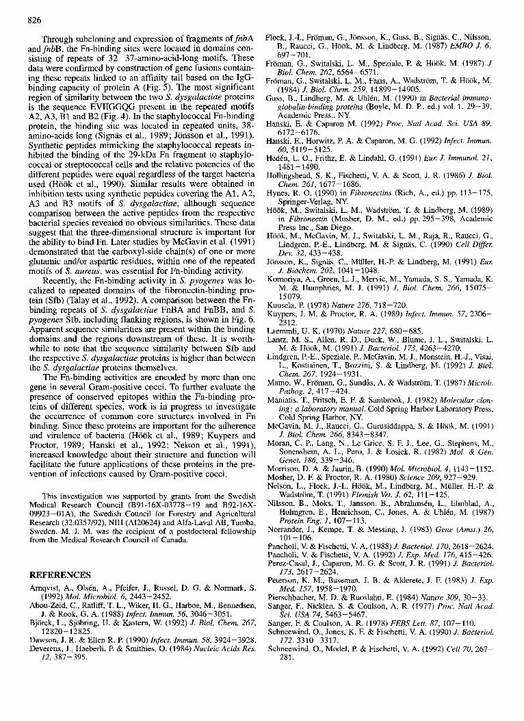
Fig. 5. Identification of Fn-binding activity in crude cell lysates of E. coli clones containing pSDF102c9 or pSDF203 and IgG- Sepharose affinity-purified cell free growth media of E. coli clones containing pZZRAIOO or pZZRB200. The lysates were sub- jected to SDSPAGE using a 12% homogeneous gel and duplicate samples were analyzed. After electrophoresis, one half of the gel was stained with Coomassie blue (A). The other half of the gel was electrophoretically transfered to a nitrocellulose filter and probed with ‘251-labelled 29-kDa fragment of Fn (B). Proteins used as mol- ecular-mass markers are indicated in (A), lane l : bovine serum albumin (66.2 m a ) ; hen egg-white ovalbumin (45 m a ) ; bovine carbonic anhydrase (31 m a ) ; soybean trypsin inhibitor (21.5 kDa) and used as negative control in (B) lane 5. (A) Lane 2 and (B) lane 4, samples from E. coli clone containing pSDF102c9; (A) lane 3 and (B) lane 3, samples from E. coli clone containing pZZR,100; (A) lane 4 and (B) lane 2, samples from E. coli clone containing pSDF203; (A) lane 5 and (B) lane 1, samples from E. coli clone containing pZZR,200.
structs should encode extracellular fusion proteins with the ability to bind to IgG, cell-free growth media from overnight cultures were passed through IgG-Sepharose columns to af- finity purify the fusion proteins (Nilsson et al., 1987). The bound proteins were eluted with 1.0 M acetic acid, pH 2.8, lyophilized and analysed using SDSPAGE (Fig. 5A and B). Samples were loaded in duplicate onto the gel and after elec- trophoresis the gel was divided into two halves. One half was stained with Coomassie brilliant blue (Fig. 5A) and the other half was electroblotted to a nitrocellulose filter (for Western blotting) and probed with ‘Z51-labelled NH,-terminal frag- ment of Fn. The affinity-purified fusion proteins, encoded by pZZR,100 and pZZR,200, with deduced molecular masses of approximately 28 kDa and 27 kDa respectively, are similar in size, as determined from the SDS gel but, the molecular masses indicated by the gel are considerably higher than the

825 FnBA PSEPSVPETS TPEGPTBGEN NLGGQgEEIT Sfb DPRYEFNNKD QSPLAGSSGE TEYITEWGN PneB THVKFSKRDI NGKELAGAMI EBRNLSGQTI
+A2 FnBA Sfb FnBB
FPBA Sf b PneB
FnBA QVIDrPTEDTQ lG??SGAGQVi? SPTITEETHK PEIIMoGQSD PIDMVEDTLP Sfb ESVEF?XDW %MS..GQTI: PQVETEDTKE P G V W Q S E SVEFTKDTQT
TEQGQS@STP ... EVEDTKG PEWIIGGQGE WDIEESLPT F ~ B B ~IIEDIEENLP +B2 +R4 +B3
+Aw FnBA GMSGSN'EATV VEEKPRPKLQ EHPDNEEPVP ATVPT'JSQTP IAQVESKVPH Sfb GWQFgSTVT IVED'i'RPRLV FblFDNI\WPKV BENREKPTKN PTPI . . . . . . FnBB EQ.GQSGSVT EVEDSKPKLS IHFDNEWPK. EDKPQLP . . . .A.VE.K.PK
+Rw *Bw
FLlBA Sfb PPBB
848 51 902
897 98 948
947 142 989
997 190 1036
1047 234 1077
1091 268 1117
Fig. 6. Comparison between the amino acid sequences of the COOH-terminal regions of FnBA and FnBB from S. dysgalactiae and the corresponding region, Sfb, from S. pyogenes (Talay et al., 1992). The LPXTGX consensus sequence is enclosed by a box. Numbering of the amino acids of FnBA and FnBB is according to Fig. 2 and Fig. 3. Sfb is numbered according to Fig. 2 in Talay et al. (1992). The ends of the proteins are indicated by (*). Identical amino acids are indicated by a shaded background. Dots indicate gaps obtained as a result of the alignment. The start of the regions discussed in this study are indicated by arrows.
deduced ones (Fig. 5A). The reason for this discrepancy is not clear but similar results have been reported for other bac- terial cell-wall proteins (Hollingshead et al., 1986; Signas et al., 1989). In Western blots, the fusion proteins bound the radiolabelled fragment of Fn (Fig. 5B). The respective puri- fied fusion proteins also inhibited the binding of radiola- belled 29-kDa NH,-terminal fragment of Fn to S. dysgalac- tiae strain S2 and to S. aureus strain Cowan-I cells when tested in an inhibition assay (data not shown). E. coli cell lysates from clones containing the plasmids pSDF102c9 and pSDF203, which were used as templates in the polymerase- chain-reaction amplifications, also contained peptides with Fn-binding activity (Fig. 5A and B).
DISCUSSION
We reported earlier the isolation of two genes encoding Fn-binding proteins from a library of genomic S. dysgalac- tiae strain S2 DNA (Lindgren et al., 1992). Restriction map- ping and cross-hybridization experiments suggested no rela- tionship between these genes and immunological data indi- cated that in the native streptococcal strain only one of the genes @bB) was expressed under the culture conditions em- ployed. In the present study, we report the complete nucleo- tide sequences of the two genes, fnbA and fibB, and their corresponding deduced amino acid sequences. The presence of two genes encoding Fn-binding activity is not unique to strain S2 since Southern-hybridization experiments showed that bothfnbA andfizbB were present in all 20 tested clinical isolates of S. dysgalactiae (data not shown), Recently pub- lished data indicate that in Streptococcus pyogenes there are also at least two different genes coding for Fn-binding activ- ity (Talay et al., 1991; Hansh et al., 1992). Furthermore, Pancholi and Fischetti (1992) have identified and purified a surface protein from group A-type-6 streptococci which is apparently distinct from other reported Fn-binding proteins in bacteria. This protein has both an enzymic activity (glycer-
aldehyde-3-phosphate dehydrogenase) and a binding capacity for a variety of eukaryotic proteins including Fn.
The two Fn-binding proteins of S. dysgalactiae have characteristics typical of cell-wall-associated proteins of Gram-positive cocci (Guss et al., 1990; Hollingshead et al., 1986; Pancholi and Fischetti, 1988). Sequences characteristic of signal peptides are present and both proteins have similar hydrophobic membrane spanning domains and COOH-termi- nal basic residues. Upstream of the membrane spanning domain there is, in all proteins of this type obtained from Gram-positive bacteria, a hexapeptide consensus sequence LPXTGX involved in cell-wall anchoring (Schneewind et al., 1992). This consensus sequence is also present in FnBA and FnBB of S. dysgalactiae but in FnBB there is an alanine instead of a threonine at position four, which is similar to protein L from Peptostreptococcus magnus (Bjorck et al., 1992). An interesting feature of FnBA is the presence of a 50-residue region, XAP, in which every third amino acid is a proline, located NH,-terminal to the RA region. A similar sequence has been observed in an IgA-binding protein from group B streptococci, called Bac, in which there is a 90-residue segment where every third residue is a proline (HedCn et al., 1991). In this protein, the proline residues are surrounded by amino acids with low hydrophilicity on one side and by mainly charged amino acids on the other side. However, in the X,, region of FnBA there is no such obvious pattern.
The overall organisation of the Fn-binding proteins from S. dysgatactiae resembles that of the staphylococcal Fn-bind- ing proteins (Signas et al., 1989; Jonsson et al., 1991). How- ever, the nucleotide and deduced amino acid sequences from the two species are different. The nucleotide sequences of the two S. aureus genes are similar throughout the entire coding sequence. This suggests that the presence of two Fn receptors in S. aureus may be a result of gene duplication, whereas the low sequence similarity between the two S. dys- galactiae Fn receptors suggests that the two genes have evolved independently.
826
Through subcloning and expression of fragments offnbA andfnbB, the Fn-binding sites were located in domains con- sisting of repeats of 32- 37-amino-acid-long motifs. These data were confirmed by construction of gene fusions contain- ing these repeats linked to an affinity tail based on the IgG- binding capacity of protein A (Fig. 5). The most significant region of similarity between the two S. dysgalactiae proteins is the sequence EVIIGGQG present in the repeated motifs A2, A3, B1 and B2 (Fig. 4). In the staphylococcal Fn-binding protein, the binding site was located in repeated units, 38- amino-acids long (Signas et al., 1989; Jonsson et al., 1991). Synthetic peptides mimicking the staphylococcal repeats in- hibited the binding of the 29-kDa Fn fragment to staphylo- coccal or streptococcal cells and the relative potencies of the different peptides were equal regardless of the target bacteria used (Hook et al., 1990). Similar results were obtained in inhibition tests using synthetic peptides covering the A1 , A2, A3 and B3 motifs of S. dysgalactiae, although sequence comparison between the active peptides from the respective bacterial species revealed no obvious similarities. These data suggest that the three-dimensional structure is important for the ability to bind Fn. Later studies by McGavin et al. (1991) demonstrated that the carboxyl-side chain(s) of one or more glutamic and/or aspartic residues, within one of the repeated motifs of S. aureus, was essential for Fn-binding activity.
Recently, the Fn-binding activity in S. pyogenes was lo- calized to repeated domains of the fibronectin-binding pro- tein (Sfb) (Talay et al., 1992). A comparison between the Fn- binding repeats of S. dysgalactiae FnBA and FnBB, and S. pyogenes Sfb, including flanking regions, is shown in Fig. 6. Apparent sequence similarities are present within the binding domains and the regions downstream of these. It is worth- while to note that the sequence similarity between Sfb and the respective S. dysgalactiae proteins is higher than between the S. dysgalactiae proteins themselves.
The Fn-binding activities are encoded by more than one gene in several Gram-positive cocci. To further evaluate the presence of conserved epitopes within the Fn-binding pro- teins of different species, work is in progress to investigate the occurrence of common core structures involved in Fn binding. Since these proteins are important for the adherence and virulence of bacteria (Hook et al., 1989; Kuypers and Proctor, 1989; Hanski et al., 1992; Nelson et al., 1991), increased knowledge about their structure and function will facilitate the future applications of these proteins in the pre- vention of infections caused by Gram-positive cocci.
This investigation was supported by grants from the Swedish Medical Research Council (B91-16X-03778-19 and B92-16X- 09923-01A), the Swedish Council for Forestry and Agrichltural Research (32.0357/92), NIH (AI20624) and Alfa-Lava1 AB, Tumba, Sweden. M. J. M. was the recipient of a postdoctoral fellowship from the Medical Research Council of Canada.
REFERENCES Amqvist, A., Olstn, A., Pfeifer, J., Russel, D. G. & Normark, S.
Abou-Zeid, C., Ratliff, T. L., Wiker, H. G., Harboe, M., Bennedsen,
Bjorck, L., Sjobring, U. & Kastern, W. (1992) J. Biol. Chem. 267,
Dawson, J. R. & Ellen R. P. (1990) Infect. Immun. 58, 3924-3928. Devereux, J., Haeberli, P. & Smithies, 0. (1984) Nucleic Acids Res.
(1992) Mol. Microbiol. 6, 2443-2452.
J. & Rook, G. A. (1988) Infect. Immun. 56, 3046-3051.
12820- 12825.
12. 387-395.
Flock, J.4, Froman, G., Jonsson, K., Guss, B., Signas, C., Nilsson, B., Raucci, G., Hook, M. & Lindberg, M. (1987) EMBO J. 6, 697-701.
Froman, G., Switalski, L. M., Speziale, P. & Hook, M. (1987) J Biol. Chem. 262, 6564-6571.
Froman, G., Switalski, L. M., Faris, A., Wadstrom, T. & Hook, M. (1984) J. Biol. Chem. 259, 14899-14905.
Guss, B., Lindberg, M. & Uhltn, M. (1990) in Bacterial immuno- globulin-binding proteins (Boyle, M. D. P., ed.) vol. 1, 29-39, Academic Press., NY.
Hanski, E. & Caparon M. (1992) Proc. Nut1 Acad. Sci. USA 89, 6172-6176.
Hanski, E., Horwitz, P. A. & Caparon, M. G. (1992) Infect. Immun. 60,5119-5125.
Hedtn, L. O., Frithz, E. & Lindahl, G. (1991) Euer. J. Immunol. 21, 1481 -1490.
Hollingshead, S. K., Fischetti, V. A. & Scott, J. R. (1986) J. Biol. Chem. 261, 1677-1686.
Hynes, R. 0. (1990) in Fibronectins (Rich, A., ed.) pp. 113-175, Springer-Verlag, NY.
Hook, M., Switalski, L. M., Wadstrom, T. & Lindberg, M. (1989) in Fibronectin (Mosher, D. M., ed.) pp. 295-398, Academic Press Inc., San Diego.
Hook, M., McGavin, M. J., Switalski, L. M., Raja, R., Raucci, G., Lindgren, P,-E., Lindberg, M. & Signas, C. (1990) Cell Diffeer. Dev. 32, 433-438.
Jonsson, K., Signas, C., Miiller, H.-P. & Lindberg, M. (1991) Eur: J. Biochem. 202, 1041 -1048.
Komoriya, A., Green, L. J., Mervic, M., Yamada, S. S., Yamada, K. M. & Hurnphries, M. J. (1991) J. Biol. Chem. 266, 15075- 15079.
Kuusela, P. (1978) Nature 276, 718-720. Kuypers, J. M. & Proctor, R. A. (1989) Infect. lmmun. 57, 2306-
Laemmli, U. K. (1970) Nature 227, 680-685. Lantz, M. S., Allen, R. D., Duck, W., Blume, J. L., Switalski, L.
M. & Hook, M. (1991) J. Bacteriol. 173, 4263-4270. Lindgren, P.-E., Speziale, P., McGavin, M. J., Monstein, H.-J., Visai,
L., Kostiainen, T., Bozzini, S. & Lindberg, M. (1992) J. Biol. Chem. 267, 1924-1931.
Mamo, W., Froman, G., Sundls, A. & Wadstrom, T. (1987) Microb. Pathog. 2, 417-424.
Maniatis, T., Fritsch, E. F. & Sambrook, J. (1982) Molecular clon- ing: a laboratory manual. Cold Spring Harbor Laboratory Press, Cold Spring Harbor, NY.
McGavin, M. J., Raucci, G., Gurusiddappa, S. & Hook, M. (1991) J. Biol. Chem. 266, 8343-8347.
Moran, C. P., Lang, N., Le Grice, S. F. J., Lee, G., Stephens, M., Sonenshein, A. L., Pero, J. & Losick, R. (1982) Mol. & Gen. Genet. 186, 339-346.
Morrison, D. A. & Jaurin, B. (1990) Mol. Microbiol. 4, 1143-1152. Mosher, D. F. & Proctor, R. A. (1980) Science 209, 927-929. Nelson, L., Flock, J . 4 , Hook, M., Lindberg, M., Miiller, H.-P. &
Wadstrom, T. (1991) Flemish Vet. J. 62, 111-125. Nilsson, B., Moks, T., Jansson, B., AbrahmsCn, L., Elmblad, A,,
Holmgren, E., Henrichson, C., Jones, A. & UhlCn, M. (1987) Protein Eng. I, 107-113.
Norrander, J., Kempe, T. & Messing, J. (1983) Gene (Amst.) 26,
Pancholi, V. & Fischetti, V. A. (1988) J. Bacteriol. 170,2618-2624. Pancholi, V. & Fischetti, V. A. (1992) J. Exp. Med. 176, 415-426. Perez-Casal, J., Caparon, M. G. & Scott, J. R. (1991) J. Bacteriol.
Peterson, K. M., Baseman, J. B. & Alderete, J. F. (1983) J . Exp.
Pierschbacher, M. D. & Ruoslahti, E. (1984) Nature 309, 30-33. Sanger, F., Nicklen, S . & Coulson, A. R. (1977) Proc. Nut1 Acud.
Sanger, F. & Coulson, A. R. (1978) FEBS Lett. 87, 107-110. Schneewind, O., Jones, K. F. & Fischetti, V. A. (1990) J. Bacteriol.
Schneewind, O., Model, P. & Fischetti, V. A. (1992) Cell 70, 267-
2312.
101-106.
173, 2617-2624.
Med. 157,1958-1970.
Sci. USA 74, 5463-5467.
172, 3310-3317.
281.
827
Signas, C., Raucci, G., Jonsson, K., Lindgren, P.-E., Ananthara- ma& G. M., Hook, M. & Lindberg, M. (1989) Proc. Nut1 Acad. Sci. USA 86, 699-703.
Speziale, P., Hook, M., Switalski, L. M. & Wadstrom, T. (1984) J. Bacteriol. 157, 420-427.
Switalski, L. M., Ljungh, A., RydCn, C., Rubin, K., Hook, M. & Wadstrom, T. (1982) Eul: J. Clin. Microbiol. 1 , 381-387.
Switalski, L. M., RydCn, C., Rubin, K., Ljungh, A., Hook, M. & Wadstrom, T. (1983) Infect. Immun. 42, 628-633.
Talay, S. R., Ehrenfeld, E., Chhatwal, G. S. & Timmis, K. N. (1991) Mol. Microbiol. 5, 1121-1734.
Talay, S. R., Valentin-Weigand, P., Jerlstrom, P. G., Timmis, K. N. & Chhatwal, G. S. (1992) Infect. Immun. 60, 3837-3844.
Thole, J. E. R., Schoningh, R., Janson, A. A. M., Garbe, T., Cornel- isse, Y. E., Clarke-Curtiss, J. E., Kolk, A. H. J., Ottenhoff, T. H. M., DeVries, R. R. P. & Abou-Zeid, C. (1992) Mol. Microbiol. 6, 153-163.
Watts, J. (1988) Vet. Micmbiol. 16, 41-66.