Embed Size (px)
Citation preview
Case Report
Conflicts ofFunding: T
ing agencies in1Departme
Genoa, Italy.2Departme
Pavia, Pavia,3Vascular a
Martino, Univ
Correspondlar Surgery UGenoa, Largogiovanni.spine
Ann Vasc Surghttps://doi.org/� 2018 PublisManuscript re2018; publishe
Twelve-year Follow-up PosteThoracicEndovascular Repair in Type B AorticDissection Shown by Three-dimensionalPrinting
Alice Finotello,1 Stefania Marconi,2 Bianca Pane,3 Michele Conti,2 Valerio Gazzola,3
Simone Mambrini,3 Ferdinando Auricchio,2 Domenico Palombo,3 and Giovanni Spinella,3
Genoa and Pavia, Italy
Background: Thoracic endovascular repair (TEVAR) is currently considered the therapy ofchoice for complicated type B acute aortic dissection (TBAAD). Although several papers havereported good outcomes at short- and medium-term follow-up, some questions still remainregarding the long-term durability and re-intervention rate during follow-up.Methods: We describe a case of a patient originally treated with TEVAR for TBAAD compli-cated by impending aortic rupture.Results: Endovascular repair successfully excluded the flow through the primary entry tear butduring the 12-year follow-up period the patient experienced several complications and re-inter-ventions. Various full-size three-dimensional (3D) models of the patient-specific vasculaturewere printed to better explain the different interventional interventions over the 12 years offollow-up and as a hands-on tool for medical education.Conclusions: The present case report, involving long-term follow-up, provides an example ofthe effectiveness and the safety of TEVAR for the treatment of complicated TBAAD shown atshort and medium-term follow-up. However, the long-term complications that were observedin this patient during follow-up support the importance of lifelong CTA surveillance. Furthermore,this study confirms the capability of 3D printing technology as a powerful tool to support commu-nication with patients and residents’ education through the physical analysis of the real cases.
interest: none.his research did not receive any specific grant from fund-the public, commercial, or not-for-profit sectors.
nt of Experimental Medicine, University of Genoa,
nt of Civil Engineering and Architecture, University ofItaly.
nd Endovascular Surgery Unit, Ospedale Policlinico Sanersity of Genoa, Genoa, Italy.
ence to: Giovanni Spinella, Vascular and Endovascu-nit, Ospedale Policlinico San Martino, University ofRosanna Benzi 10, 16132 Genoa, Italy; E-mail:
2018; -: 1.e1–1.e710.1016/j.avsg.2018.07.057hed by Elsevier Inc.ceived: April 21, 2018; manuscript accepted: July 7,d online: - - -
Recent guidelines recommend thoracic endovascu-
lar repair (TEVAR) as the first-choice treatment for
patients affected by complicated type B acute aortic
dissection (TBAAD).1 Although this treatment has
reduced morbidity and mortality rates considerably
compared to open surgery, the re-intervention rate
still remains high.2 However, little is known about
the long-term outcome of patients with TBAAD
treated by TEVAR.3 In addition, given the fact that
aortic dissections usually present at a younger age
than aneurysms or penetrating aortic ulcers,4 estab-
lishing a lifelong imaging follow-up protocol
following TEVAR is mandatory. We herein present
the complications and re-intervention modalities
that were faced during the 12-year follow-up after
1.e1
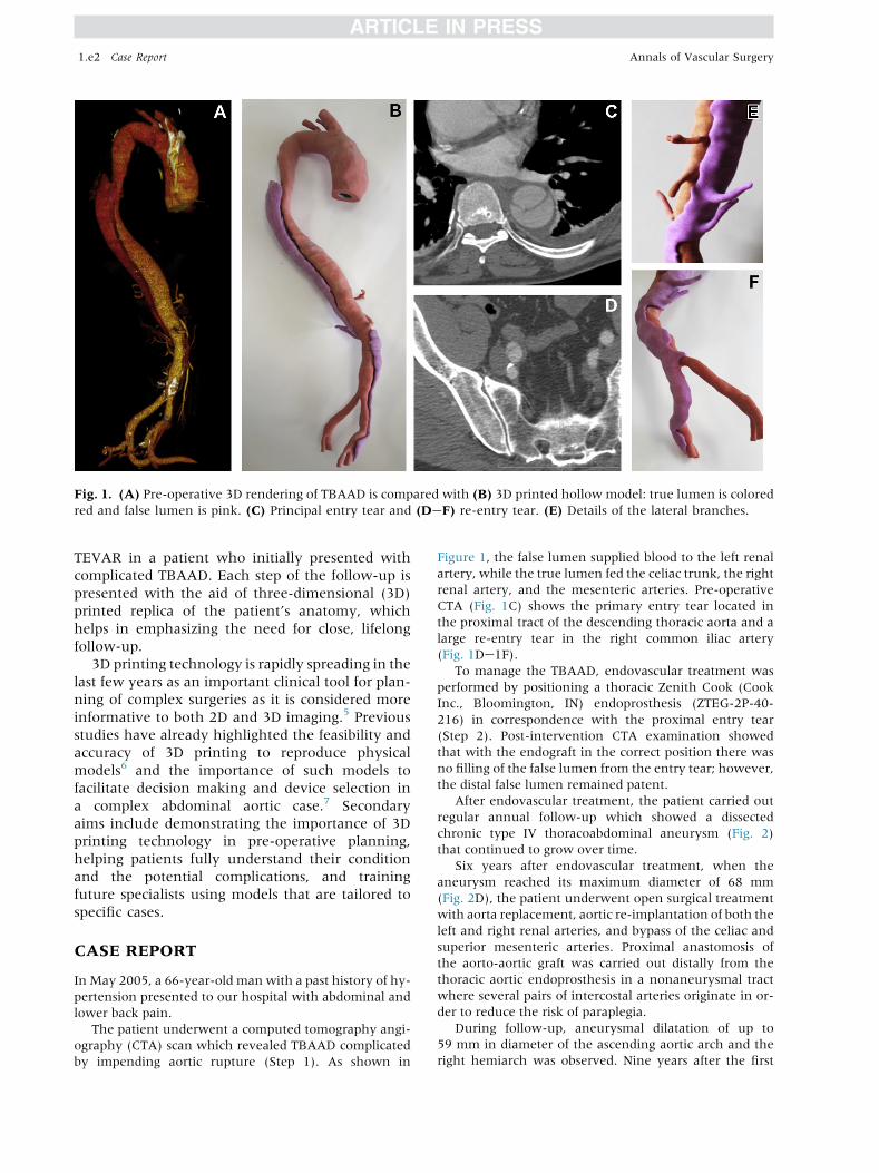
Fig. 1. (A) Pre-operative 3D rendering of TBAAD is compared with (B) 3D printed hollow model: true lumen is colored
red and false lumen is pink. (C) Principal entry tear and (DeF) re-entry tear. (E) Details of the lateral branches.
1.e2 Case Report Annals of Vascular Surgery
TEVAR in a patient who initially presented with
complicated TBAAD. Each step of the follow-up is
presented with the aid of three-dimensional (3D)
printed replica of the patient’s anatomy, which
helps in emphasizing the need for close, lifelong
follow-up.
3D printing technology is rapidly spreading in the
last few years as an important clinical tool for plan-
ning of complex surgeries as it is considered more
informative to both 2D and 3D imaging.5 Previous
studies have already highlighted the feasibility and
accuracy of 3D printing to reproduce physical
models6 and the importance of such models to
facilitate decision making and device selection in
a complex abdominal aortic case.7 Secondary
aims include demonstrating the importance of 3D
printing technology in pre-operative planning,
helping patients fully understand their condition
and the potential complications, and training
future specialists using models that are tailored to
specific cases.
CASE REPORT
In May 2005, a 66-year-old man with a past history of hy-
pertension presented to our hospital with abdominal and
lower back pain.
The patient underwent a computed tomography angi-
ography (CTA) scan which revealed TBAAD complicated
by impending aortic rupture (Step 1). As shown in
Figure 1, the false lumen supplied blood to the left renal
artery, while the true lumen fed the celiac trunk, the right
renal artery, and the mesenteric arteries. Pre-operative
CTA (Fig. 1C) shows the primary entry tear located in
the proximal tract of the descending thoracic aorta and a
large re-entry tear in the right common iliac artery
(Fig. 1De1F).To manage the TBAAD, endovascular treatment was
performed by positioning a thoracic Zenith Cook (Cook
Inc., Bloomington, IN) endoprosthesis (ZTEG-2P-40-
216) in correspondence with the proximal entry tear
(Step 2). Post-intervention CTA examination showed
that with the endograft in the correct position there was
no filling of the false lumen from the entry tear; however,
the distal false lumen remained patent.
After endovascular treatment, the patient carried out
regular annual follow-up which showed a dissected
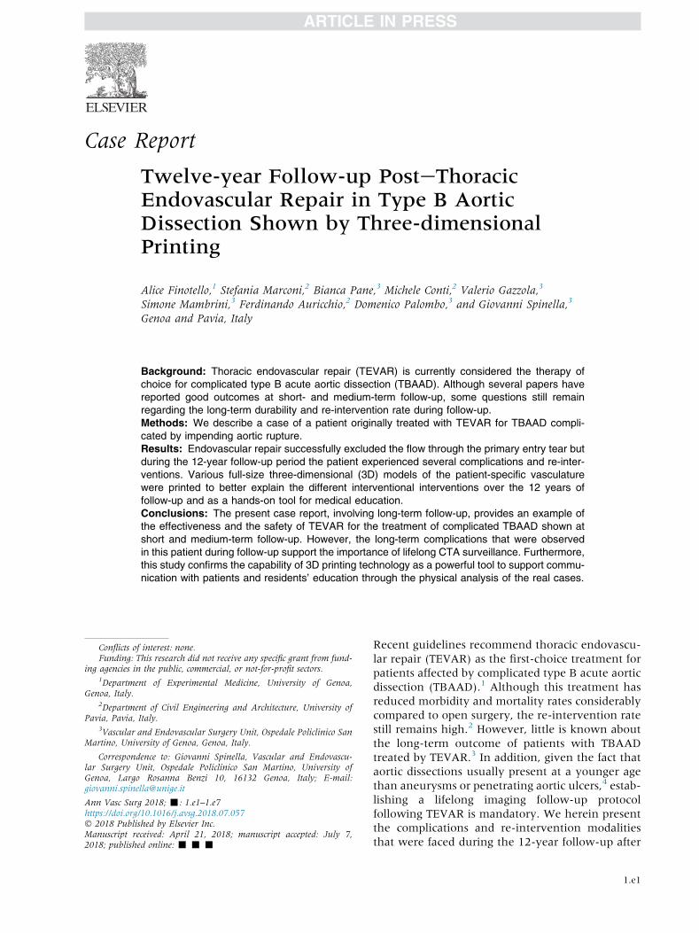
chronic type IV thoracoabdominal aneurysm (Fig. 2)
that continued to grow over time.
Six years after endovascular treatment, when the
aneurysm reached its maximum diameter of 68 mm
(Fig. 2D), the patient underwent open surgical treatment
with aorta replacement, aortic re-implantation of both the
left and right renal arteries, and bypass of the celiac and
superior mesenteric arteries. Proximal anastomosis of
the aorto-aortic graft was carried out distally from the
thoracic aortic endoprosthesis in a nonaneurysmal tract
where several pairs of intercostal arteries originate in or-
der to reduce the risk of paraplegia.
During follow-up, aneurysmal dilatation of up to
59 mm in diameter of the ascending aortic arch and the
right hemiarch was observed. Nine years after the first
Fig. 2. (A) 3D rendering and (B) 3D printed model show a
chronic dissected thoracoabdominal aortic aneurysm. True
(T) and false (F) lumen are highlighted both in the 3D
models (AeB) and in the CTA (C). (D) Maximum diam-
eter reached 6 years after the first TEVAR intervention.
Volume -, - 2018 Case Report 1.e3
endovascular treatment, the patient underwent ascending
aorta replacement with transposition of the anonymous
trunk and the left common carotid artery.
Eleven years after the first endovascular treatment,
during follow-up CTA, gradual enlargement of the
descending thoracic aorta between the thoracic endopros-
thesis and the surgical graft was detected (Step 3). As
depicted in Figure 3, type IB endoleakwas diagnosed. Tak-
ing into account the patient’s age and the previous inter-
ventions, a totally endovascular treatment using
fenestrated branched stent grafts was planned. The inter-
vention was carried out in two stages in order to reduce
the risk of paraplegia.
In the first stage (Step 4), a proximal side-branched
custom-made Zenith Cook endograft 46-30-230 was posi-
tioned to preserve intercostal branch flow with partial
overlapping on the previously implanted thoracic endo-
prosthesis (Fig. 4). A distal custom-made fenestrated
Zenith Cook 32-32-113 endograft was also implanted
with fenestration to allow for superior mesenteric artery,
celiac artery, and right renal artery bypass (two fenestra-
tions). During the same session, treatment of the renal ar-
tery was completed by positioning an Advanta (Atrium
Europe, Mijdrecht, The Netherlands) 7 � 20 mm covered
graft. The 3D printed model shown in Figure 4 allowed us
to observe the patency of the secondary branch arising
from the descending thoracic aorta.
Three months later, in the second stage, an Advanta
9 � 36 mm covered stent was positioned with bypass of
the superior mesenteric artery and the celiac trunk with
left humeral access. Finally, the side branch was
covered by positioning a Zenith Cook thoracic endograft
(Step 5). The patient was discharged on the sixth
post-operative day with no post-procedural complica-
tions, under antiplatelet therapy. The 1-month post-
operative CTA showed complete exclusion of the
aneurysm with patency of all the vessels that underwent
endovascular treatment. This was also confirmed by the
12-month follow-up CTA scan. Figure 5 shows the
outcome of the endovascular procedure.
The entire follow-up sequence and the interventional
procedures (Steps 1e5) were well documented by 3D
printed models.
Starting from CTA DICOM images, digital preparation
of the virtual 3D models is performed using the Vascular
Modeling Toolkit open source library. The 3D reconstruc-
tion procedure is repeated for each structure of interest
(i.e., true lumen, false lumen, endograft).
The full-size physical models are then manufactured
by binder jetting technology (Project 460+; 3D Sys-
tems). First, the 3DEdit (3D Systems) software is used
to assign different colors to each structure of interest.
Then the model is prototyped using a chalk-like pow-
der that is glued together by means of a liquid binder,
along with colors deployed through an inkjet cartridge.
After the 3D printing, the physical model is post-
processed using a cyanoacrylate infiltrating binder to
strengthen the mechanical properties of the 3D printed
model.
DISCUSSION
We report the case of a patient treated for compli-
cated TBAAD. Endovascular repair successfully
excluded the entry tear but during follow-up the
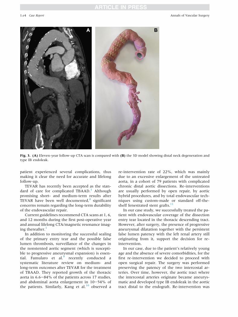
Fig. 3. (A) Eleven-year follow-up CTA scan is compared with (B) the 3D model showing distal neck degeneration and
type IB endoleak.
1.e4 Case Report Annals of Vascular Surgery
patient experienced several complications, thus
making it clear the need for accurate and lifelong
follow-up.
TEVAR has recently been accepted as the stan-
dard of care for complicated TBAAD.1 Although
promising short- and medium-term results after
TEVAR have been well documented,8 significant
concerns remain regarding the long-term durability
of the endovascular repair.
Current guidelines recommend CTA scans at 1, 6,
and 12 months during the first post-operative year
and annual lifelong CTA/magnetic resonance imag-
ing thereafter.1
In addition to monitoring the successful sealing
of the primary entry tear and the possible false
lumen thrombosis, surveillance of the changes in
the nonstented aortic segment (which is suscepti-
ble to progressive aneurysmal expansion) is essen-
tial. Famularo et al.9 recently conducted a
systematic literature review on medium- and
long-term outcomes after TEVAR for the treatment
of TBAAD. They reported growth of the thoracic
aorta in 6.6e84% of the patients across 17 studies,
and abdominal aorta enlargement in 10e54% of
the patients. Similarly, Kang et al.10 observed a
re-intervention rate of 22%, which was mainly
due to an excessive enlargement of the untreated
aorta, in a cohort of 79 patients with complicated
chronic distal aortic dissections. Re-interventions
are usually performed by open repair, by aortic
hybrid procedures, and by total endovascular tech-
niques using custom-made or standard off-the-
shelf fenestrated stent grafts.11
In our case study, we successfully treated the pa-
tient with endovascular coverage of the dissection
entry tear located in the thoracic descending tract.
However, after surgery, the presence of progressive
aneurysmal dilatation together with the persistent
false lumen patency with the left renal artery still
originating from it, support the decision for re-
intervention.
In our case, due to the patient’s relatively young
age and the absence of severe comorbidities, for the
first re-intervention we decided to proceed with
open surgical repair. The surgery was performed
preserving the patency of the two intercostal ar-
teries. Over time, however, the aortic tract where
the intercostal arteries originate became aneurys-
matic and developed type IB endoleak in the aortic
tract distal to the endograft. Re-intervention was
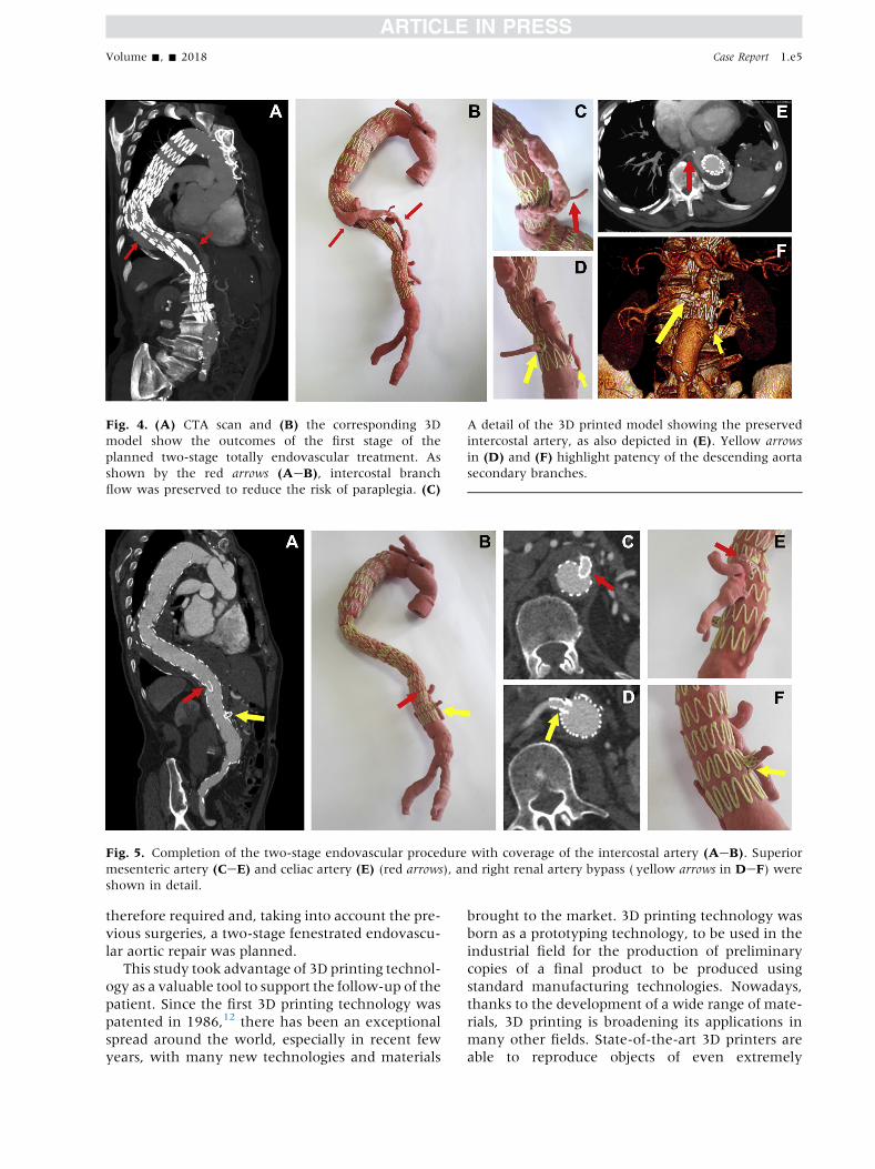
Fig. 4. (A) CTA scan and (B) the corresponding 3D
model show the outcomes of the first stage of the
planned two-stage totally endovascular treatment. As
shown by the red arrows (AeB), intercostal branch
flow was preserved to reduce the risk of paraplegia. (C)
A detail of the 3D printed model showing the preserved
intercostal artery, as also depicted in (E). Yellow arrows
in (D) and (F) highlight patency of the descending aorta
secondary branches.
Fig. 5. Completion of the two-stage endovascular procedure with coverage of the intercostal artery (AeB). Superior
mesenteric artery (CeE) and celiac artery (E) (red arrows), and right renal artery bypass ( yellow arrows in DeF) were
shown in detail.
Volume -, - 2018 Case Report 1.e5
therefore required and, taking into account the pre-
vious surgeries, a two-stage fenestrated endovascu-
lar aortic repair was planned.
This study took advantage of 3D printing technol-
ogy as a valuable tool to support the follow-up of the
patient. Since the first 3D printing technology was
patented in 1986,12 there has been an exceptional
spread around the world, especially in recent few
years, with many new technologies and materials
brought to the market. 3D printing technology was
born as a prototyping technology, to be used in the
industrial field for the production of preliminary
copies of a final product to be produced using
standard manufacturing technologies. Nowadays,
thanks to the development of a wide range of mate-
rials, 3D printing is broadening its applications in
many other fields. State-of-the-art 3D printers are
able to reproduce objects of even extremely
1.e6 Case Report Annals of Vascular Surgery
complex geometries: relying on a layer-by-layer
approach, 3D printers are able to face even shapes
impossible to realize with other manufacturing
technologies. Moreover, this technology is particu-
larly suitable for the production of single copies or
small batches of an object, since the price per copy
does not reduce increasing the number of products,
as it happens, for example, in injection molding.
Thanks to these characteristics, 3D printers appear
to be particularly suitable for the production of
personalized products: accordingly, they started to
be investigated in the medical field for the produc-
tion of patient-specific devices.
In recent years, the number of applications of 3D
printing in the field of medicine has exponentially
increased. Existing and potential medical uses range
from orthopedic reconstruction and implants13 to
living tissue fabrication.14 Although it was initially
only used as a tool for pre-operative planning,15
3D printing technology is now emerging as a power-
ful tool for patient education, device optimization,
and clinical training.16
As concerns vascular and cardiovascular surgery,
3D printing has been used to better understand the
unique pathological anatomy, to plan the best treat-
ment and to assist in the device selection.7 However,
at the same time, the use of 3D full size objects to
help medical students and residents understand
the course of vascular pathologies is currently not
still very widespread in the field of vascular
surgery.5
3D printed life-sized models have been success-
fully employed during follow-up by our group to
better evaluate both the evolution of the pathology
related to the onset of complications and the out-
comes of re-interventions. As already demonstrated
by Marconi et al.5 for laparoscopic surgery, the
tactile inspection of the printed models allows a bet-
ter understanding of the patient’s 3D real anatomy,
increasing the understanding of the sense of depth
and the anatomical relationship between the
different anatomical structures. In particular, the
aorta has a complex anatomy with a great number
of secondary branches originating from it, making
the mutual relationship between pathological sec-
tion and the arterial branches not trivial to under-
stand with standard visualization of CTA images
by means of flat screens.
Our experience allows confirming the literature
data about the superiority of 3D printed models
with respect to standard imaging and also for
follow-up monitoring, an application not yet inves-
tigated. For example, the 3D printed models
depicted in Figures 1 and 2 were beneficial to
show the remodeling of true and false lumen after
endovascular treatment, and, in particular, to better
understand the real 3D relationship between true
and false lumen and arterial secondary branches
which originate from the 2 channels, and its
changes before (Fig. 1) and after (Fig. 2) endovascu-
lar repair.
Similarly, the 3D printed model of Step 3 (Fig. 3)
allowed to observe the dislocation of the endograft
leading to endoleak development and the metallic
rings distortion which were hardly visible on CTA
images.
Importance of visual inspection of 3D printed
models is also confirmed by the 3D printed replica
of the last two surgical steps. In particular, the 3D
printed models allow to clearly observe the patency
of the intercostal artery after Step 4 and completion
of the endovascular procedure with coverage of
intercostal artery and flow exclusion from the aneu-
rysmal sac (Fig. 5). Moreover, both 3D printed
models offer a clear view about the spatial relation-
ship between the endograft distal landing zone and
the just-below re-implanted celiac trunk. 3D printed
models enable an immediate and effective compari-
son of the clinical situations at different times,
without the need of going through the analysis of
different image datasets. This aspect is particularly
valuable for complex anatomies to be investigated
in detail. Another interesting perspective confirmed
by this work concerns the use of 3D printed models
as a valuable teaching tool for both skilled surgeons
and for residents, medical students, and other pro-
fessional figures in the medical field. Moreover,
they could help the patient understand his/her clin-
ical situation, the therapeutic strategies proposed,
and the related risks. All these applications can be
translated into a clinical outcome improvement as
well as into a money saving for the hospital, espe-
cially as concerns the prevention of possible legal is-
sues for malpractice.
In this case report, we make use of 3D printing
technology to accurately reproduce 3D printed color
models of patient-specific life-size vasculature.
Among the current available options, we opted for
a binder jetting 3D printer which offers, with respect
to the specific application, the best compromise
among the level of accuracy (100 mm of layer thick-
ness), construction speed (23 mm/hr, resulting in a
total of 10 hr for the printing plus 2 hr for the post-
processing), and lower fabrication costs (about 200
euros for printing and post-processing) if compared
with other techniques. Moreover, its intrinsic func-
tioning enables to reproduce very complex struc-
tures without the need of supports’ placement and
theirmechanical removal, since themodel is contin-
uously self-supported by the uncured powder
Volume -, - 2018 Case Report 1.e7
during the printing. Production time and cost are
the key points to evaluate the effective introduction
of 3D printing into the daily clinical practice. At the
present state, 3D printing is mostly applied in iso-
lated cases or few numbers of cases, often
demanding the production to producers external
to the hospital, thus increasing costs and delivery
time. The time required to process the images and
produce themodel limits the application of 3D print-
ing for the production of patient-specific models
only to elective surgery. In this study, each model
took about 24 hr to move from CTA images to the
final 3D printed model. Image elaboration time is
reduced for bone models, thanks to the easier seg-
mentation process, while is extended to up to
1 day for abdominal parenchymal organs. The
application to emergency surgery is accordingly
prevented.
Considering the benefit brought by the technol-
ogy at different levels, as proved by this study, the
introduction of 3D printing technology as an addi-
tional support for surgeons and clinicians should
be promoted. Even if the choice of the most suitable
technology should consider the specific fields of
application, all the medical specialties could take
advantage from the 3D printing.
The high innovation rate of 3D printing technol-
ogies and materials will probably lead to a cost
reduction of both machines and supplies, hopefully
boosting the use of 3D printing technologies for the
daily clinical practice.
CONCLUSION
Our experience demonstrates the effectiveness of
TEVAR in the short- and medium-term follow-up
as a treatment option for complicated TBAAD.
However, the long-term complications that were
observed in this patient during long-term
follow-up support the importance of lifelong CTA
surveillance.
Furthermore, the study highlighted the effective-
ness of 3D printed supports. Besides being especially
useful in both pre-operative planning and device se-
lection, the use of 3D printed patient-specificmodels
could also be beneficial in follow-up monitoring, in
teaching residents andmedical students how to deal
with complicated cases.
REFERENCES
1. ESC Committee for Practice Guidelines. 2014 ESC Guide-
lines on the diagnosis and treatment of aortic diseases: docu-
ment covering acute and chronic aortic diseases of the
thoracic and abdominal aorta of the adult. The Task Force
for the Diagnosis and Treatment of Aortic Diseases of the Eu-
ropean Society of Cardiology (ESC). Eur Heart J 2014;35:
2873e926.
2. Kret MR, Azarbal AF, Mitchell EL, et al. Compliance with
long-term surveillance recommendations following endo-
vascular aneurysm repair or type B aortic dissection. J
Vasc Surg 2013;58:25e32.
3. Lou X, Chen EP, Duwayri YM, et al. The impact of thoracic
endovascular aortic repair on long-term survival in type B
aortic dissection. Ann Thorac Surg 2018;105:31e8.4. Mussa FF, Horton JD, Moridzadeh R, et al. Acute aortic
dissection and intramural hematoma: a systematic review.
JAMA 2016;316:754e63.5. Marconi S, Pugliese L, Botti M, et al. Value of 3D-printing
for the comprehension of surgical anatomy. Surg Endosc
2017;31:4102e10.
6. Ho D, Squelch A, Sun Z. Modelling of aortic aneurysm and
aortic dissection through 3D printing. J Med Radiat Sci
2017;64:10e7.
7. Tam MD, Laycock SD, Brown JR, et al. 3D printing of an
aortic aneurysm to facilitate decision making and device se-
lection for endovascular aneurysm repair in complex neck
anatomy. J Endovascular Ther 2013;20:863e7.
8. Zhang MH, Du X, GuoW, et al. Early and midterm outcomes
of thoracic endovascular aortic repair (TEVAR) for acute and
chronic complicated type B aortic dissection. Medicine
2017;96:e7183.
9. Famularo M, Meyermann K, Lombardi JV. Aneurysmal
degeneration of type B aortic dissections after thoracic endo-
vascular aortic repair: a systematic review. J Vasc Surg
2017;66:924e30.
10. Kang WC, Greenberg RK, Mastracci TM, et al. Endovascular
repair of complicated chronic distal aortic dissections: inter-
mediate outcomes and complications. J Thorac Cardiovasc
Surg 2011;142:1074e83.
11. Weber TF, B€ockler D, M€uller-Eschner M, et al. Frequency of
abdominal aortic expansion after thoracic endovascular
repair of type B aortic dissection. Vascular 2016;24:567e79.
12. Hull CW. Apparatus for production of three-dimensional ob-
jects by stereolithography. Patent number US 4575330.
Washington, DC: U.S. Patent and Trademark Office, 1986.
13. Auricchio F, Marconi S. 3D printing: clinical applications in
orthopaedics and traumatology. EFORT Open Rev 2016;1:
121e7.
14. Murphy SV, Atala A. 3D bioprinting of tissues and organs.
Nat Biotechnol 2014;32:773e85.
15. Itagaki MW. Using 3D printed models for planning and guid-
ance during endovascular intervention: a technical advance.
Diagn Interv Radiol 2015;21:338.
16. Marro A, Bandukwala T, Mak W. Three-dimensional print-
ing and medical imaging: a review of the methods and appli-
cations. Curr Probl Diagn Radiol 2016;45:2e9.