Embed Size (px)
Citation preview
Tutorial 1, The CellsTutorial 1, The Cells
IPAM Cells and Materials: IPAM Cells and Materials: At the Interface between Mathematics, Biology and EngineeringAt the Interface between Mathematics, Biology and Engineering
Dr. Toshikazu HamasakiDr. Toshikazu HamasakiDept. Bioengineering, UCLADept. Bioengineering, UCLA
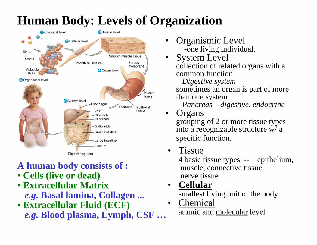
• Organismic Level-one living individual.
• System Levelcollection of related organs with a common function
Digestive systemsometimes an organ is part of more than one system
Pancreas – digestive, endocrine• Organs
grouping of 2 or more tissue types into a recognizable structure w/ a specific function.
Human Body: Levels of Organization
• Tissue4 basic tissue types -- epithelium,muscle, connective tissue, nerve tissue
• Cellularsmallest living unit of the body
• Chemicalatomic and molecular level
A human body consists of :• Cells (live or dead)• Extracellular Matrix
e.g. Basal lamina, Collagen ... • Extracellular Fluid (ECF)
e.g. Blood plasma, Lymph, CSF …
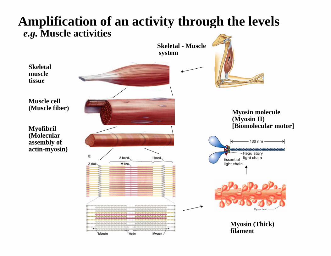
Amplification of an activity through the levelse.g. Muscle activities
Skeletalmuscletissue
Muscle cell(Muscle fiber)
Myofibril(Molecularassembly of actin-myosin)
Myosin (Thick)filament
Myosin molecule(Myosin II)[Biomolecular motor]
Skeletal - Musclesystem
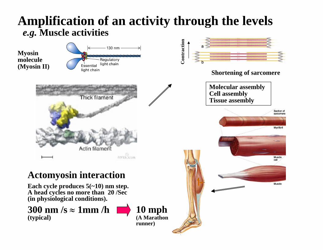
Amplification of an activity through the levelse.g. Muscle activities
Con
trac
tion
Myosinmolecule(Myosin II)
Actomyosin interactionEach cycle produces 5(~10) nm step. A head cycles no more than 20 /Sec (in physiological conditions).
300 nm /s ≈ 1mm /h (typical)
Molecular assemblyCell assemblyTissue assembly
10 mph(A Marathon runner)
Shortening of sarcomere
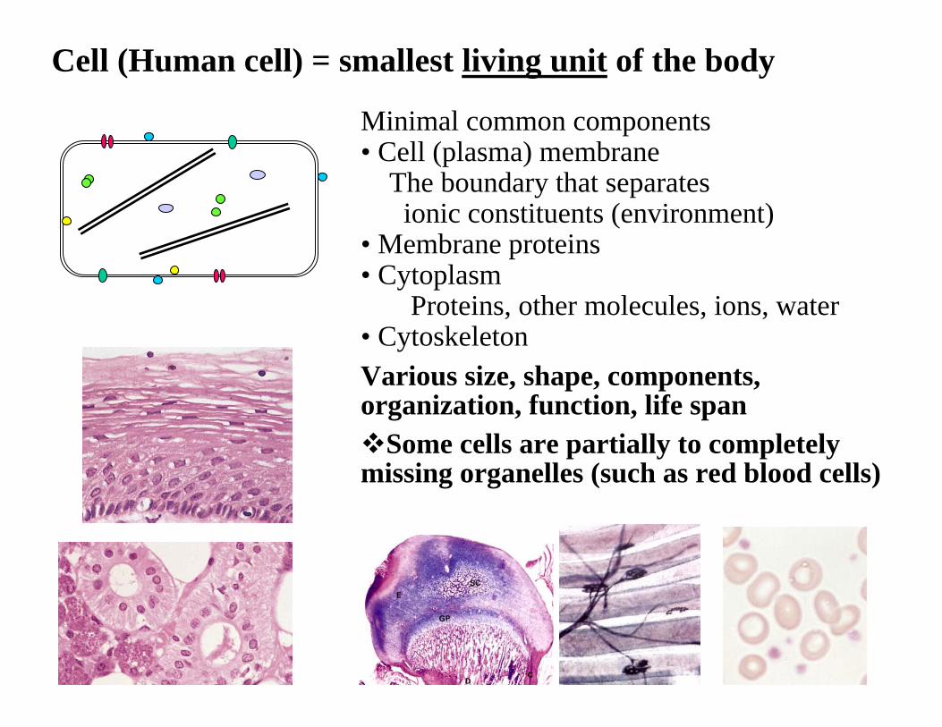
Cell (Human cell) = smallest living unit of the body
Minimal common components• Cell (plasma) membrane
The boundary that separatesionic constituents (environment)
• Membrane proteins• Cytoplasm
Proteins, other molecules, ions, water• CytoskeletonVarious size, shape, components, organization, function, life span
Some cells are partially to completely missing organelles (such as red blood cells)
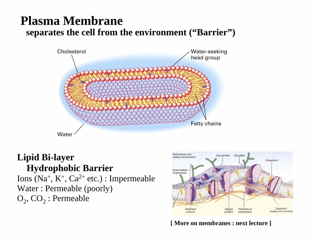
Plasma Membraneseparates the cell from the environment (“Barrier”)
Lipid Bi-layerHydrophobic Barrier
Ions (Na+, K+, Ca2+ etc.) : Impermeable Water : Permeable (poorly)O2, CO2 : Permeable
[ More on membranes : next lecture ]
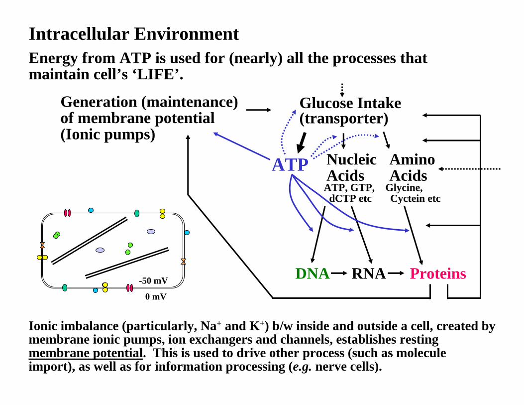
Intracellular Environment
Generation (maintenance) of membrane potential(Ionic pumps)
Glucose Intake(transporter)
ATP AminoAcids
NucleicAcids
DNA RNA Proteins-50 mV
0 mV
Energy from ATP is used for (nearly) all the processes that maintain cell’s ‘LIFE’.
Ionic imbalance (particularly, Na+ and K+) b/w inside and outside a cell, created by membrane ionic pumps, ion exchangers and channels, establishes resting membrane potential. This is used to drive other process (such as molecule import), as well as for information processing (e.g. nerve cells).
ATP, GTP, Glycine, dCTP etc Cyctein etc
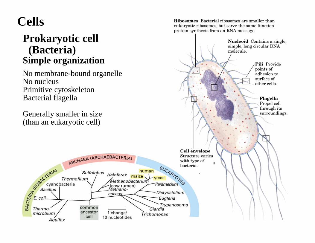
Prokaryotic cell (Bacteria)
Simple organizationNo membrane-bound organelleNo nucleusPrimitive cytoskeletonBacterial flagella
Generally smaller in size (than an eukaryotic cell)
Cells
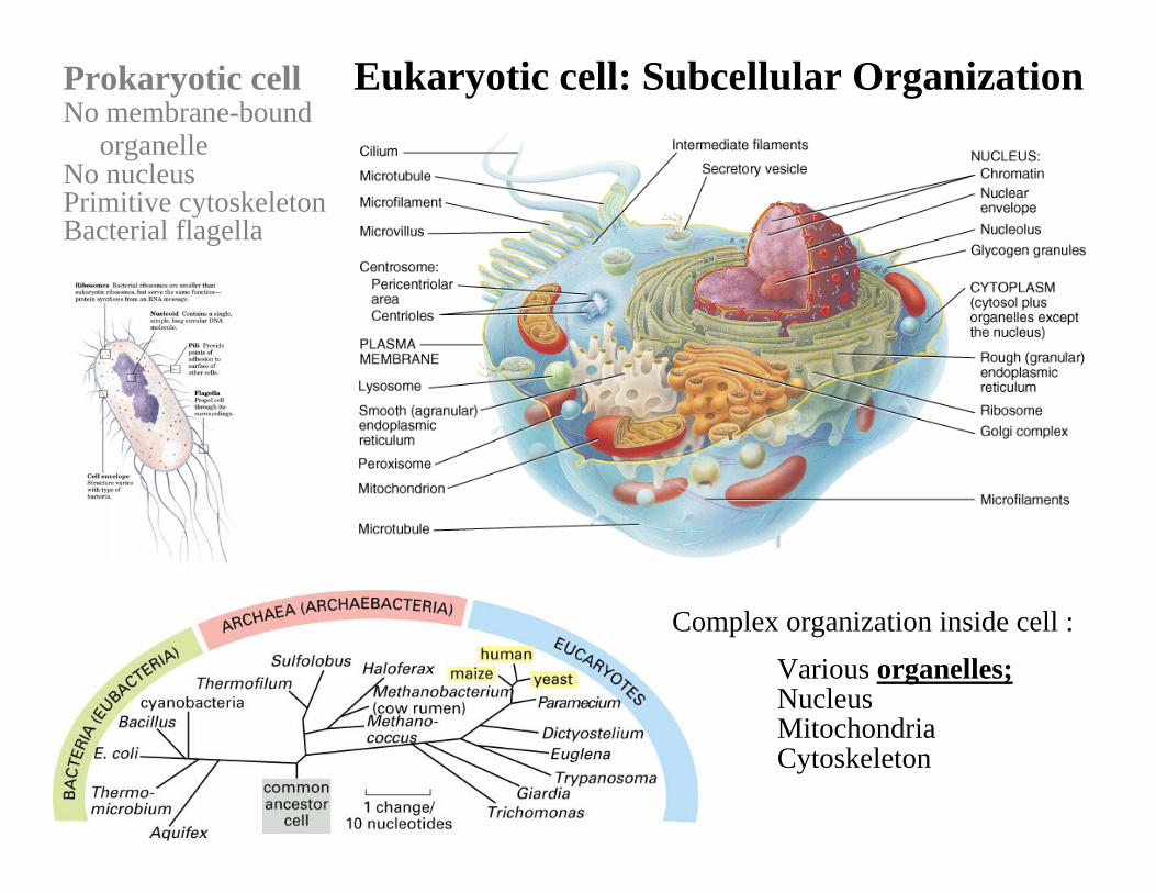
Prokaryotic cell Eukaryotic cell: Subcellular Organization
Complex organization inside cell :Various organelles;Nucleus MitochondriaCytoskeleton
No membrane-boundorganelle
No nucleusPrimitive cytoskeletonBacterial flagella
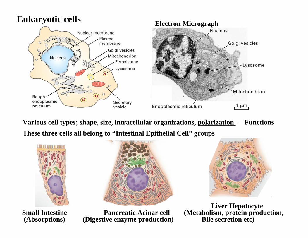
Eukaryotic cells Electron Micrograph
Liver HepatocyteSmall Intestine Pancreatic Acinar cell (Metabolism, protein production, (Absorptions) (Digestive enzyme production) Bile secretion etc)
Various cell types; shape, size, intracellular organizations, polarization – FunctionsThese three cells all belong to “Intestinal Epithelial Cell” groups
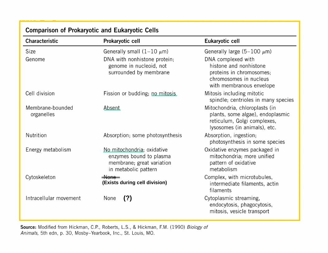
(?)
(Exists during cell division)
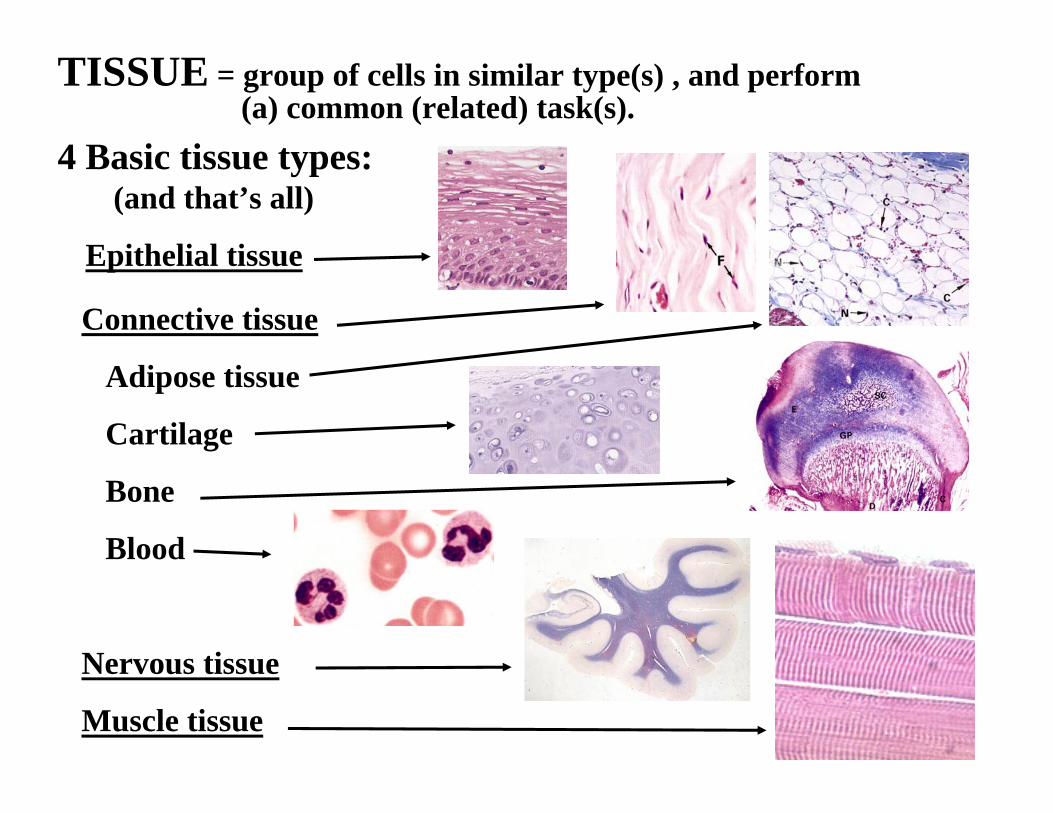
TISSUE = group of cells in similar type(s) , and perform(a) common (related) task(s).
4 Basic tissue types:(and that’s all)
Epithelial tissue
Connective tissue
Adipose tissue
Cartilage
Bone
Blood
Nervous tissue
Muscle tissue
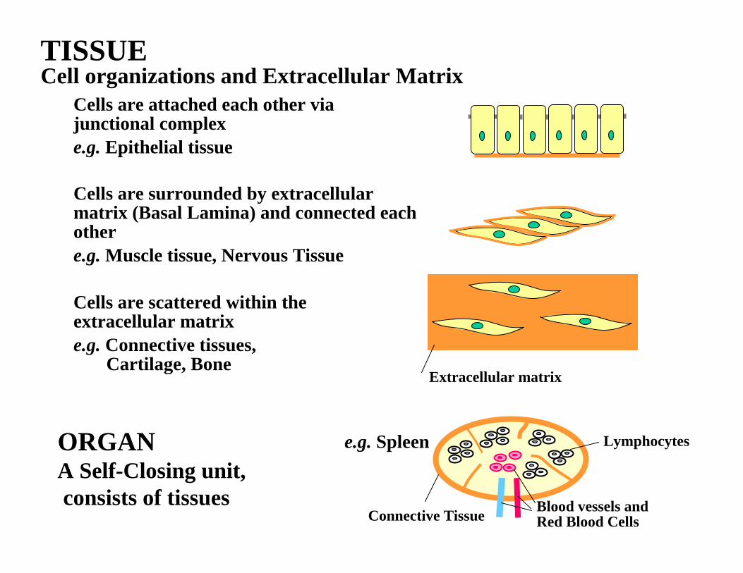
TISSUECell organizations and Extracellular Matrix
Cells are attached each other via junctional complexe.g. Epithelial tissue
Cells are surrounded by extracellularmatrix (Basal Lamina) and connected each othere.g. Muscle tissue, Nervous Tissue
Cells are scattered within the extracellular matrix e.g. Connective tissues,
Cartilage, Bone Extracellular matrix
ORGANA Self-Closing unit,consists of tissues
Connective Tissue Blood vessels and Red Blood Cells
Lymphocytese.g. Spleen
Basal lamina
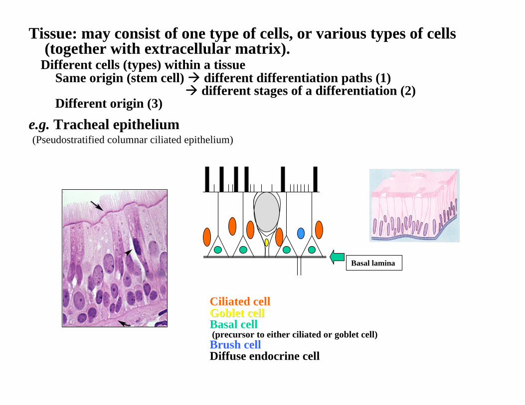
Tissue: may consist of one type of cells, or various types of cells(together with extracellular matrix).
Different cells (types) within a tissueSame origin (stem cell) different differentiation paths (1)
different stages of a differentiation (2)Different origin (3)
e.g. Tracheal epithelium(Pseudostratified columnar ciliated epithelium)
Ciliated cellGoblet cellGoblet cellBasal cell(precursor to either ciliated or goblet cell)Brush cellDiffuse endocrine cell
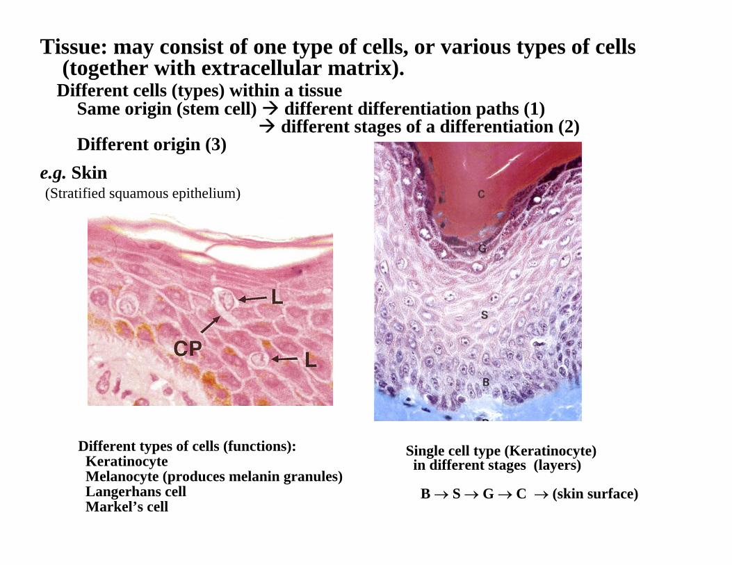
Tissue: may consist of one type of cells, or various types of cells(together with extracellular matrix).
Different cells (types) within a tissueSame origin (stem cell) different differentiation paths (1)
different stages of a differentiation (2)Different origin (3)
e.g. Skin(Stratified squamous epithelium)
Single cell type (Keratinocyte)in different stages (layers)
B → S → G → C → (skin surface)
Different types of cells (functions):KeratinocyteMelanocyte (produces melanin granules)Langerhans cell Markel’s cell
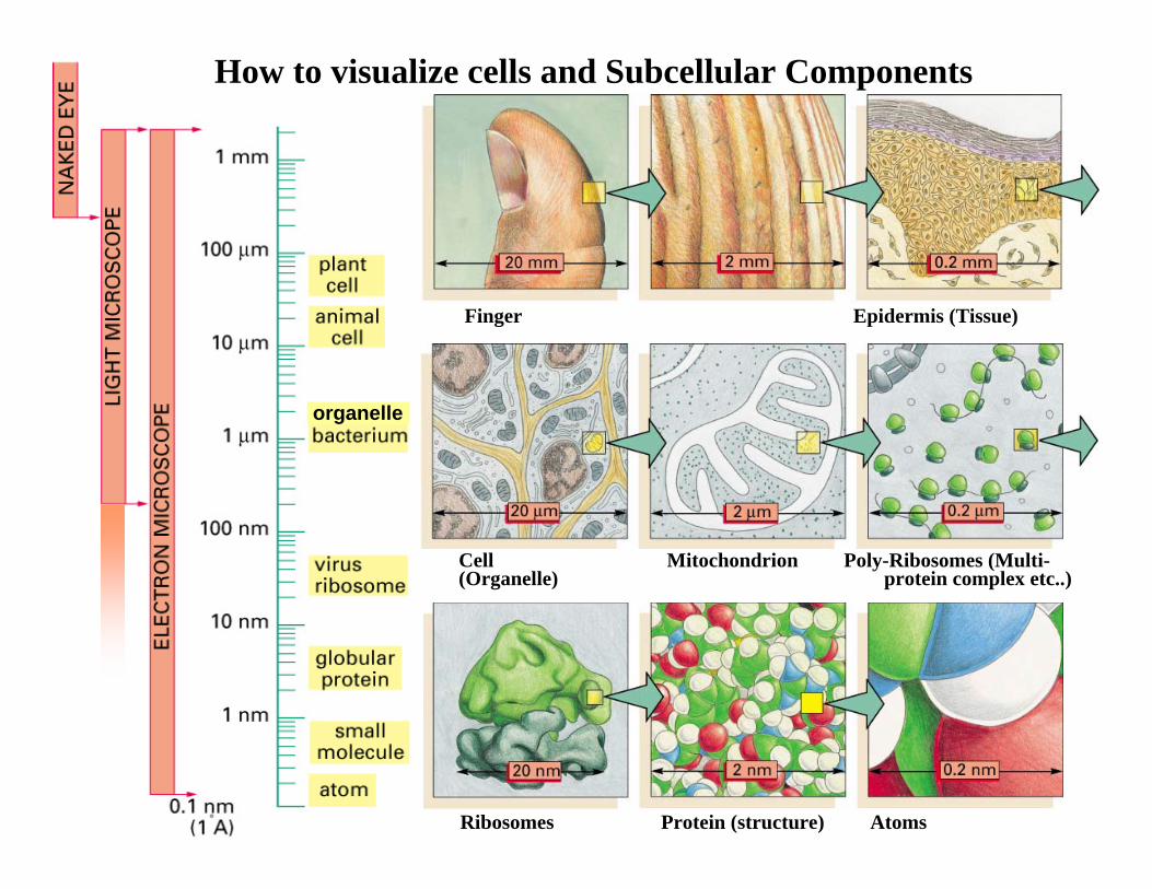
Finger Epidermis (Tissue)
Cell Mitochondrion Poly-Ribosomes (Multi-(Organelle) protein complex etc..)
Ribosomes Protein (structure) Atoms
organelle
How to visualize cells and Subcellular Components
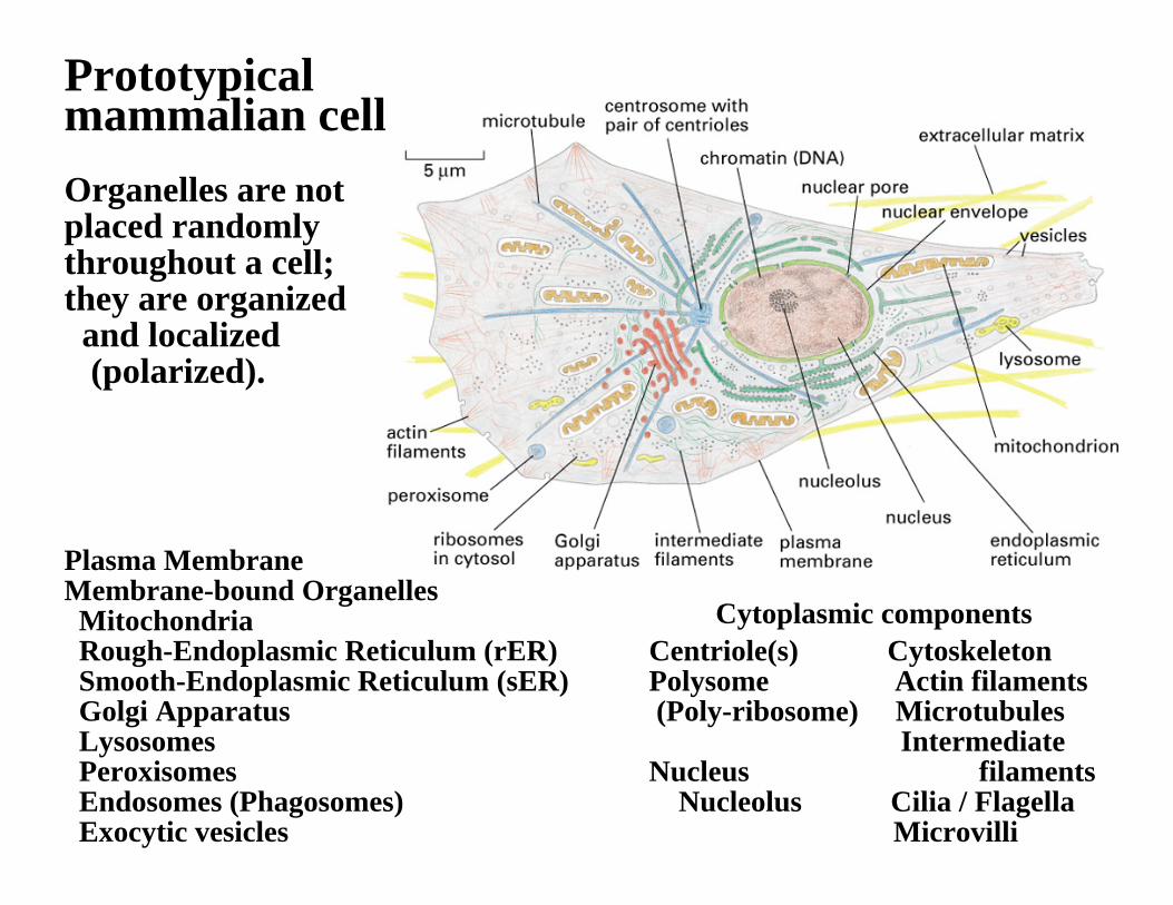
Plasma MembraneMembrane-bound OrganellesMitochondriaRough-Endoplasmic Reticulum (rER)Smooth-Endoplasmic Reticulum (sER)Golgi ApparatusLysosomesPeroxisomesEndosomes (Phagosomes)Exocytic vesicles
Prototypical mammalian cell:
Cytoplasmic componentsCentriole(s) CytoskeletonPolysome Actin filaments(Poly-ribosome) Microtubules
Intermediate Nucleus filaments
Nucleolus Cilia / FlagellaMicrovilli
Organelles are not placed randomly throughout a cell;they are organizedand localized (polarized).
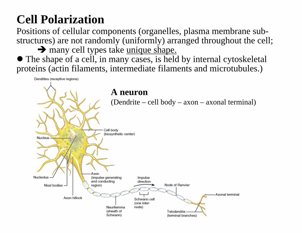
A neuron(Dendrite – cell body – axon – axonal terminal)
Cell PolarizationPositions of cellular components (organelles, plasma membrane sub-structures) are not randomly (uniformly) arranged throughout the cell;
many cell types take unique shape.The shape of a cell, in many cases, is held by internal cytoskeletal
proteins (actin filaments, intermediate filaments and microtubules.)
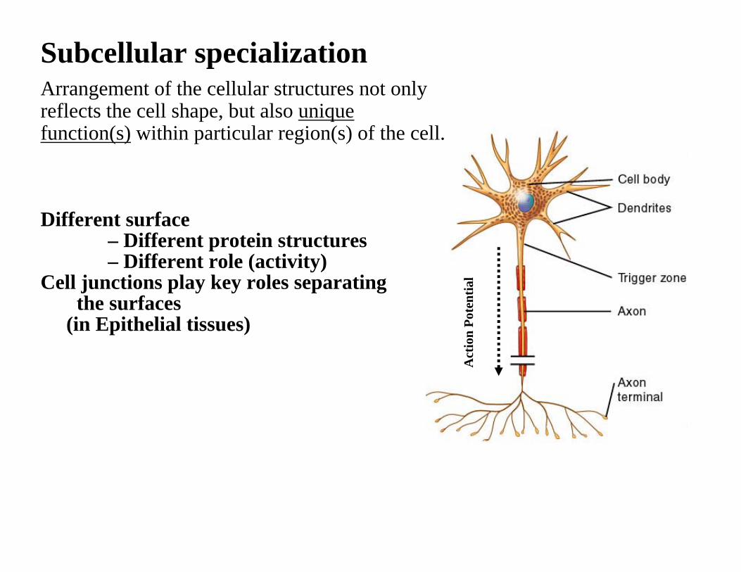
Subcellular specialization
Different surface – Different protein structures – Different role (activity)
Cell junctions play key roles separatingthe surfaces
(in Epithelial tissues)
Act
ion
Pote
ntia
l
Arrangement of the cellular structures not only reflects the cell shape, but also unique function(s) within particular region(s) of the cell.
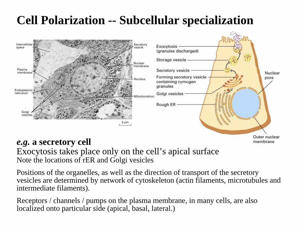
Cell Polarization -- Subcellular specialization
e.g. a secretory cellExocytosis takes place only on the cell’s apical surfaceNote the locations of rER and Golgi vesiclesPositions of the organelles, as well as the direction of transport of the secretoryvesicles are determined by network of cytoskeleton (actin filaments, microtubules and intermediate filaments).Receptors / channels / pumps on the plasma membrane, in many cells, are alsolocalized onto particular side (apical, basal, lateral.)
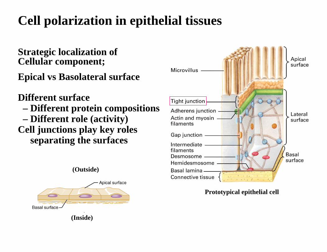
Cell polarization in epithelial tissues
Strategic localization ofCellular component;Epical vs Basolateral surface
Different surface – Different protein compositions – Different role (activity)
Cell junctions play key rolesseparating the surfaces
Prototypical epithelial cell
(Outside)
(Inside)
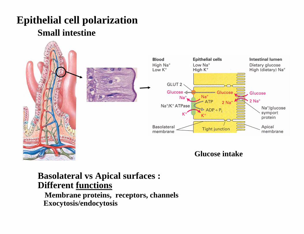
Epithelial cell polarizationSmall intestine
Basolateral vs Apical surfaces :Different functions
Membrane proteins, receptors, channelsExocytosis/endocytosis
Glucose intake
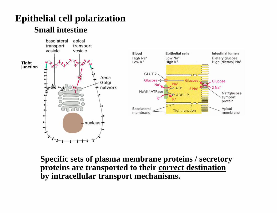
Epithelial cell polarizationSmall intestine
Tight junction
Specific sets of plasma membrane proteins / secretoryproteins are transported to their correct destinationby intracellular transport mechanisms.
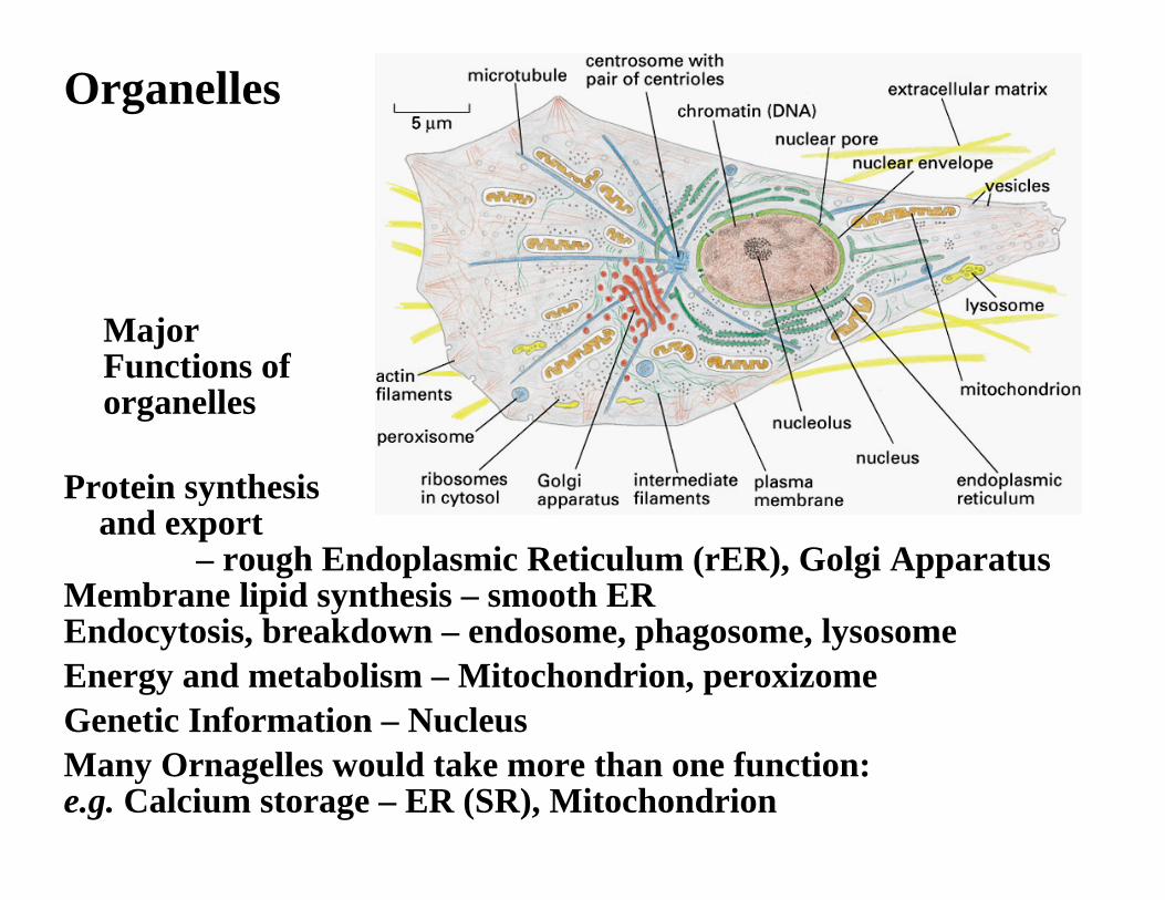
Organelles
Protein synthesisand export
– rough Endoplasmic Reticulum (rER), Golgi Apparatus Membrane lipid synthesis – smooth EREndocytosis, breakdown – endosome, phagosome, lysosomeEnergy and metabolism – Mitochondrion, peroxizomeGenetic Information – NucleusMany Ornagelles would take more than one function:e.g. Calcium storage – ER (SR), Mitochondrion
Major Functions of organelles
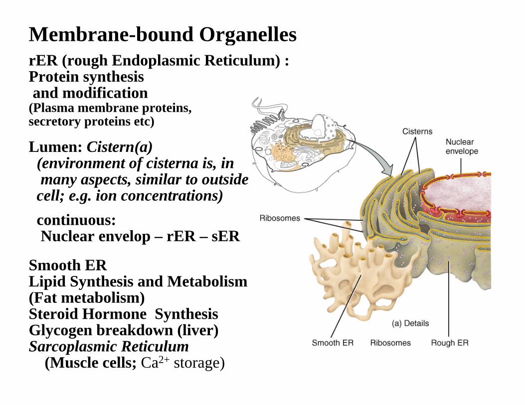
Smooth ERLipid Synthesis and Metabolism(Fat metabolism)Steroid Hormone SynthesisGlycogen breakdown (liver)Sarcoplasmic Reticulum
(Muscle cells; Ca2+ storage)
Lumen: Cistern(a)(environment of cisterna is, inmany aspects, similar to outside cell; e.g. ion concentrations) continuous:Nuclear envelop – rER – sER
Membrane-bound OrganellesrER (rough Endoplasmic Reticulum) : Protein synthesis and modification
(Plasma membrane proteins, secretory proteins etc)
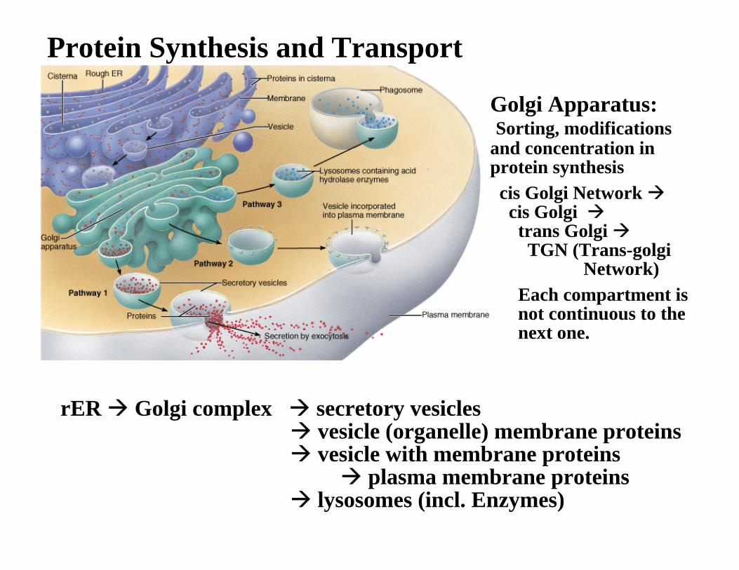
Protein Synthesis and Transport
rER Golgi complex secretory vesiclesvesicle (organelle) membrane proteins vesicle with membrane proteins
plasma membrane proteins lysosomes (incl. Enzymes)
Golgi Apparatus:Sorting, modifications
and concentration inprotein synthesiscis Golgi Network cis Golgi
trans GolgiTGN (Trans-golgi
Network)Each compartment isnot continuous to thenext one.
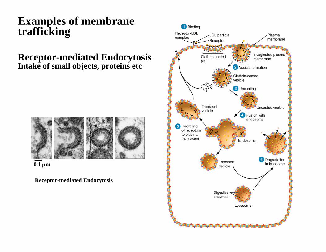
Examples of membranetrafficking
Receptor-mediated EndocytosisIntake of small objects, proteins etc
Receptor-mediated Endocytosis
0.1 µm
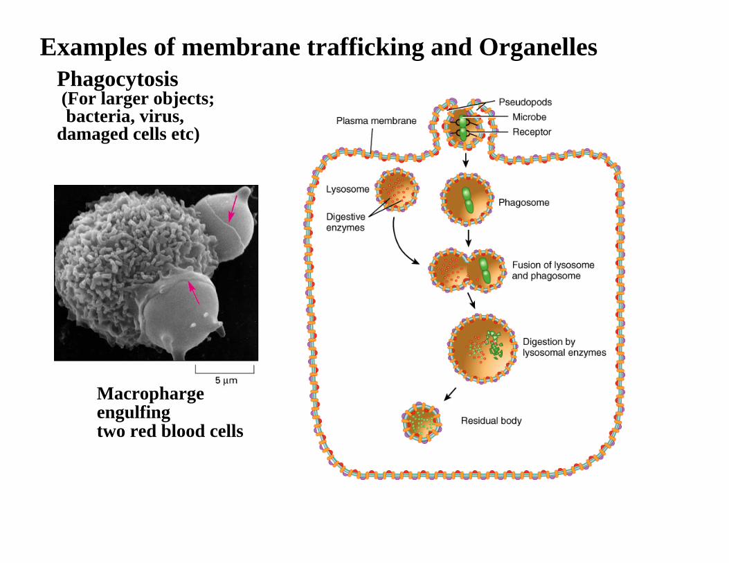
Examples of membrane trafficking and OrganellesPhagocytosis(For larger objects;bacteria, virus,
damaged cells etc)
Macrophargeengulfing two red blood cells
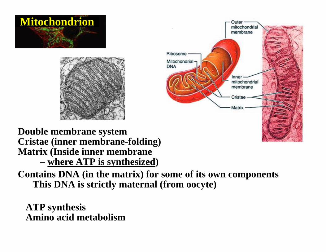
Mitochondrion
Double membrane systemCristae (inner membrane-folding)Matrix (Inside inner membrane
– where ATP is synthesized) Contains DNA (in the matrix) for some of its own components
This DNA is strictly maternal (from oocyte)
ATP synthesisAmino acid metabolism
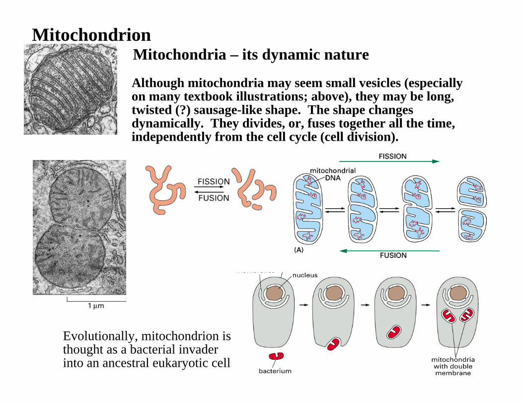
Mitochondrion
Although mitochondria may seem small vesicles (especially on many textbook illustrations; above), they may be long, twisted (?) sausage-like shape. The shape changes dynamically. They divides, or, fuses together all the time, independently from the cell cycle (cell division).
Mitochondria – its dynamic nature
Evolutionally, mitochondrion is thought as a bacterial invader into an ancestral eukaryotic cell
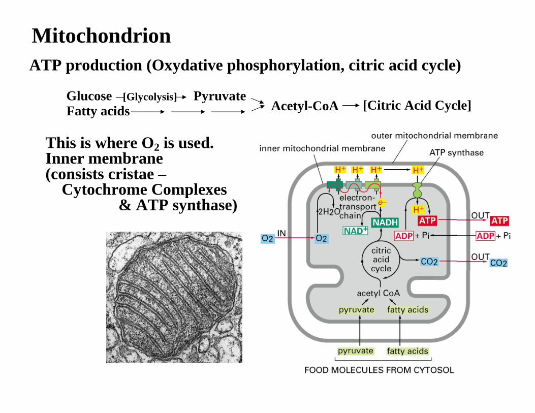
MitochondrionATP production (Oxydative phosphorylation, citric acid cycle)
This is where O2 is used.Inner membrane (consists cristae –
Cytochrome Complexes & ATP synthase)
Fatty acids Acetyl-CoAPyruvate[Glycolysis]Glucose
[Citric Acid Cycle]
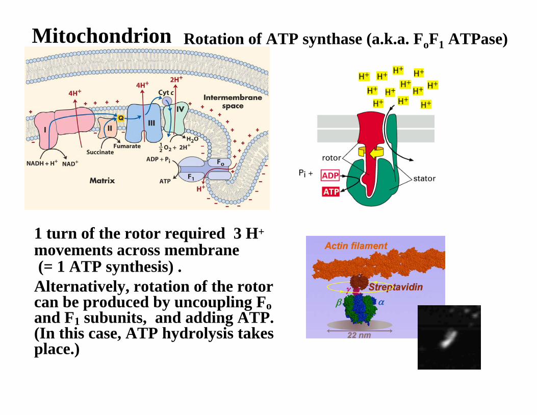
Mitochondrion Rotation of ATP synthase (a.k.a. FoF1 ATPase)
1 turn of the rotor required 3 H+
movements across membrane(= 1 ATP synthesis) .Alternatively, rotation of the rotor can be produced by uncoupling Foand F1 subunits, and adding ATP. (In this case, ATP hydrolysis takes place.)
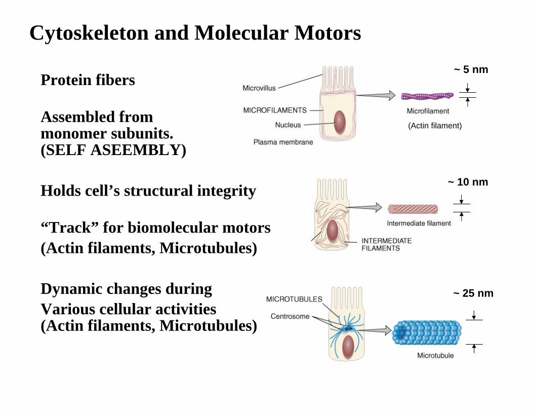
Cytoskeleton and Molecular Motors
Protein fibers
Assembled from monomer subunits.(SELF ASEEMBLY)
Holds cell’s structural integrity
“Track” for biomolecular motors (Actin filaments, Microtubules)
Dynamic changes during Various cellular activities(Actin filaments, Microtubules)
~ 5 nm
~ 10 nm
~ 25 nm
(Actin filament)
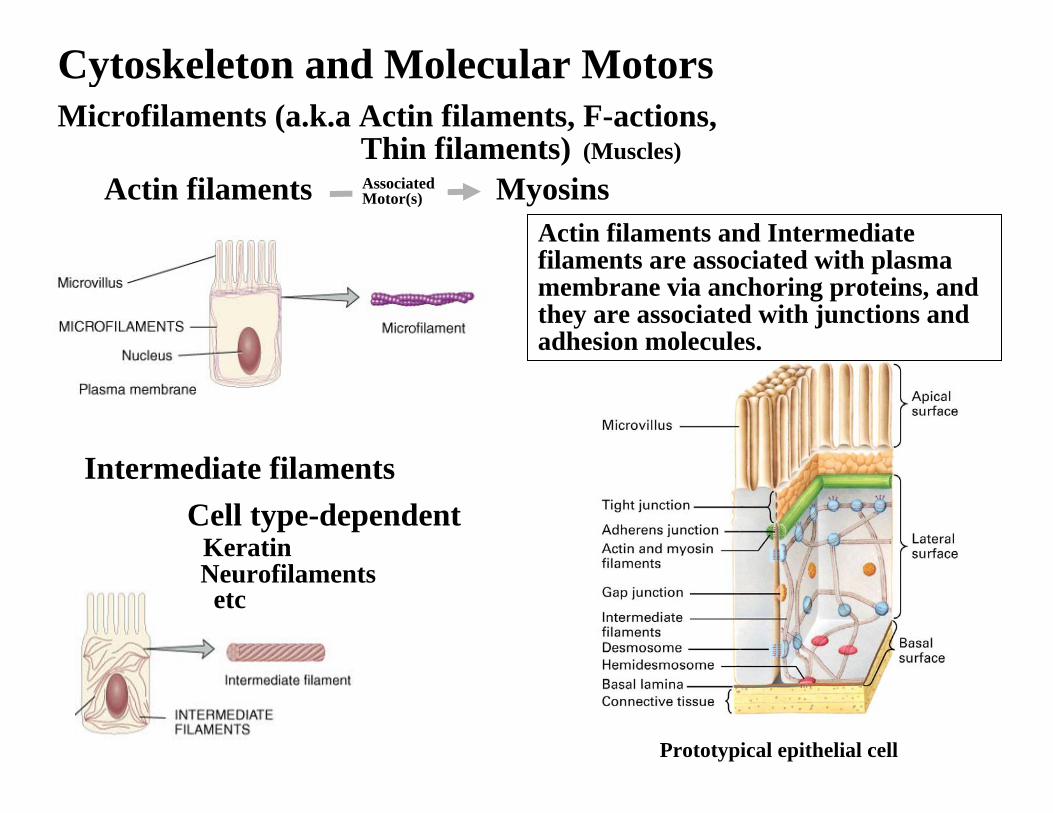
Cytoskeleton and Molecular Motors
Intermediate filaments
Actin filaments MyosinsAssociated Motor(s)
Cell type-dependentKeratinNeurofilamentsetc
Microfilaments (a.k.a Actin filaments, F-actions,Thin filaments) (Muscles)
Prototypical epithelial cell
Actin filaments and Intermediate filaments are associated with plasma membrane via anchoring proteins, and they are associated with junctions and adhesion molecules.
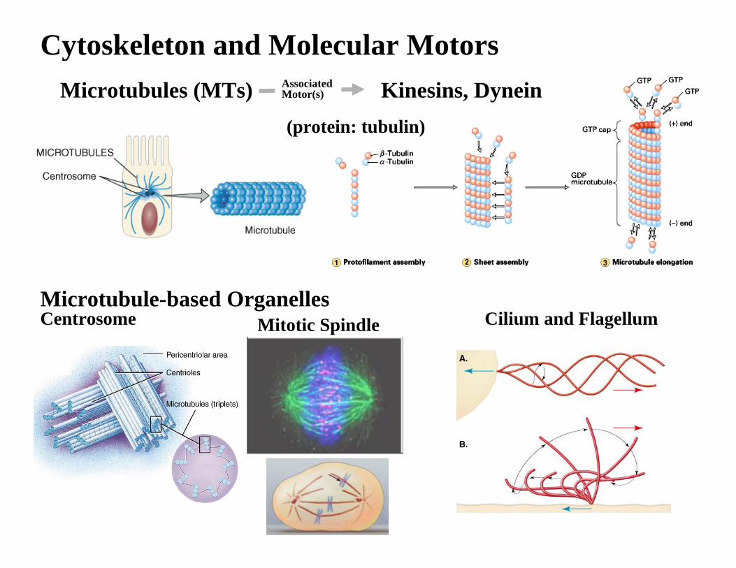
Cytoskeleton and Molecular Motors
Microtubule-based OrganellesCentrosome Cilium and Flagellum
Microtubules (MTs) Kinesins, DyneinAssociated Motor(s)
Mitotic Spindle
(protein: tubulin)
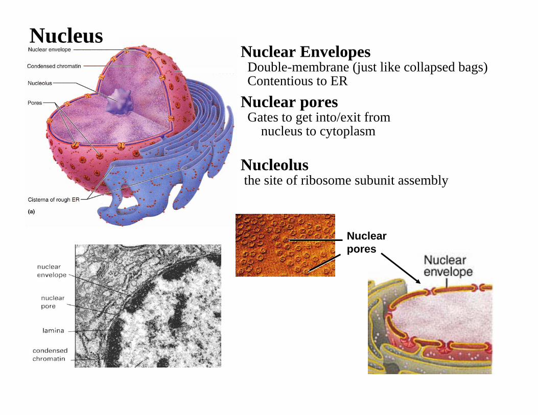
NucleusNuclear Envelopes
Double-membrane (just like collapsed bags)Contentious to ER
Nuclear poresGates to get into/exit from
nucleus to cytoplasm
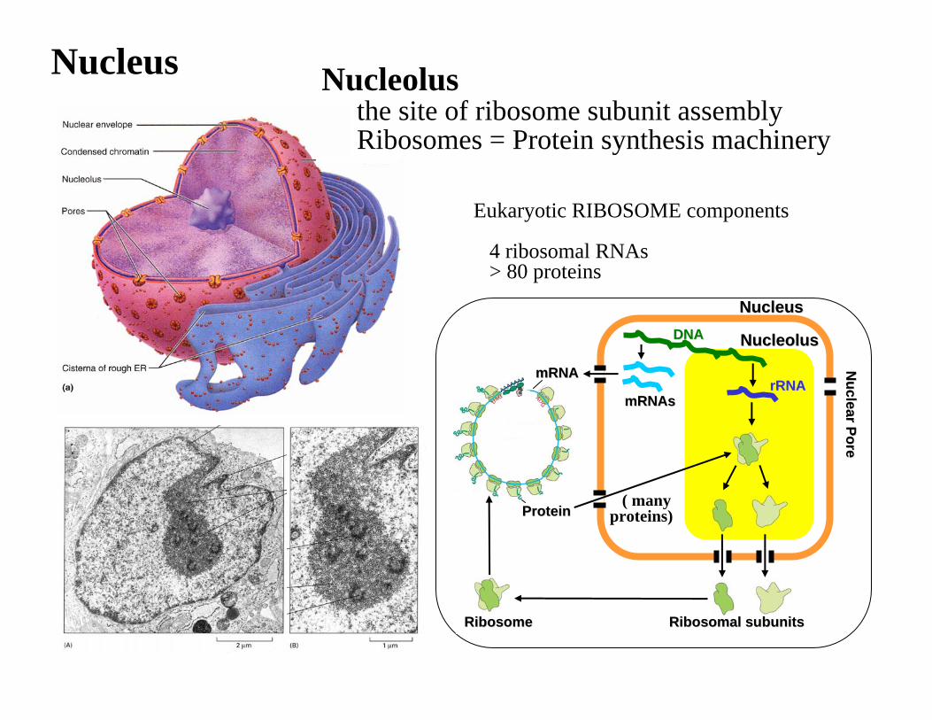
Nucleolusthe site of ribosome subunit assembly
Nuclear pores
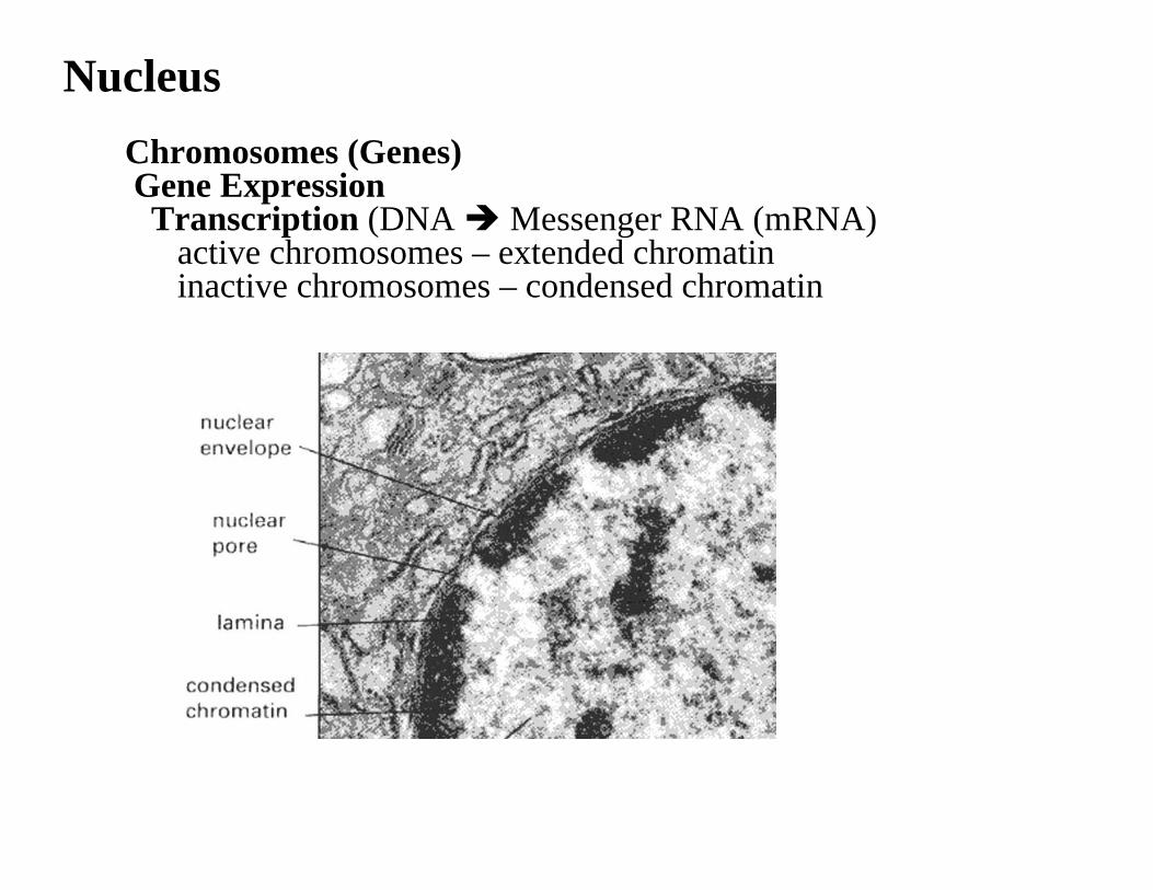
NucleusChromosomes (Genes)Gene ExpressionTranscription (DNA Messenger RNA (mRNA)
active chromosomes – extended chromatin inactive chromosomes – condensed chromatin
Nucleus
Eukaryotic RIBOSOME components
4 ribosomal RNAs> 80 proteins
DNADNA
rRNArRNAmRNAsmRNAs
Nuclear Pore
Nuclear Pore
NucleusNucleus
NucleolusNucleolus
ProteinProtein
mRNAmRNA
( many proteins)
Ribosomal subunitsRibosomal subunitsRibosomeRibosome
Nucleolusthe site of ribosome subunit assemblyRibosomes = Protein synthesis machinery