Embed Size (px)
Citation preview
Plant Virus Diagnostics: Comparison of classical and membrane-based techniques for immunoassay and coat protein sequence characterization for Cucumber mosaic virus and
three potyviruses
by
Peta-Gaye S. Chang
Dissertation submitted to the Faculty of the Virginia Polytechnic Institute and State University
in partial fulfillment of the requirements for the degree of
DOCTOR OF PHILOSOPHY in
Plant Pathology, Physiology and Weed Science
Dr. Sue A. Tolin (Chair) Dr. Erik L. Stromberg Dr. Boris A. Vinatzer
Mrs. Mary-Ann Hansen Dr. Richard E. Veilleux
May 29, 2009
Blacksburg, Virginia
Key words: Turnip mosaic virus, antibody, subgroup diversity, Tobacco etch virus, Soybean mosaic virus, TBIA
Copyright 2009, Peta-Gaye S. Chang
Plant Virus Diagnostics: Comparison of classical and membrane-based techniques for immunoassay and coat protein sequence characterization for Cucumber mosaic virus and
three potyviruses
Peta-Gaye S. Chang
Advisor: Dr. S. A. Tolin Department of Plant Pathology, Physiology and Weed Science
ABSTRACT
Diagnostics is important in the development and implementation of pest management strategies. The virus diagnostic capabilities of several plant pathology collaborators within the Integrated Pest Management Collaborative Research Support Program (IPM CRSP) host countries were evaluated with the aid of a survey. Very few plant disease diagnostic clinics had funds to cover daily operations despite over half of the responding clinics receiving an operational budget. Academically and government affiliated clinics within the developing host countries had little access to molecular tools and equipment, relying mostly on biological and serological methods. Clinics affiliated with private companies and within the USA relied more upon molecular assays. Ten CMV isolates identified by tissue blot immunoassay (TBIA) were collected from a garden at the Historic Smithfield Plantation on the Virginia Tech campus, and from Painter, Virginia on the Eastern Shore. Three CMV isolates from Smithfield were biologically compared to six early CMV isolates stored since the 1970s, while all isolates were compared serologically and molecularly. Sequences obtained after reverse transcription-polymerase chain reaction (RT-PCR) assigned the CMV isolates into subgroups, with eleven to subgroup 1A and three to subgroup 2. The subgroup assignments were confirmed by TBIA using CMV subgroup-specific monoclonal antibodies (Agdia Inc). At Smithfield Plantation, another virus, Turnip mosaic virus (TuMV) was identified from Dame’s Rocket (Hesperis matronalis L.). This is the first report of TuMV in Virginia. In TBIA virus-infected plant samples are blotted onto nitrocellulose membranes, dried, and processed. Membranes can be stored for long periods of time and transported safely across borders without risk of introducing viruses into new environments, but virus remains immunologically active for several months. Methods were developed with CMV and three potyviruses, using the same membranes, for detecting viral RNA by RT-PCR and direct sequencing of PCR products. Amplification by RT-PCR was possible after membrane storage for up to 15 months. The membranes also performed well with samples sent from IPM CRSP host countries and within the USA. This method should improve molecular diagnostic capabilities in developing countries, as samples can be blotted to membranes and sent to a centralized molecular laboratory for analysis.
iii
ACKNOWLEDGEMENTS
• I would firstly need to thank my advisor Dr. Sue A. Tolin for all her guidance,
patience and understanding throughout my studies. Without her I probably would
have never finished.
• My committee members: Drs. Boris Vinatzer, Erik Stromberg and Richard Veilleux,
and Mrs. Hansen, who have stuck by me these four years.
• Mrs. Moss Baldwin for helping with lab protocols and for her friendship and help in
many matters outside of school.
• Donna Ford, Patsy Neice and Judy Fielder for all their support within the department
and making my time within Blacksburg fun.
• Phil Keating for helping in the greenhouse and his fishing stories.
• Dr. Don Mullins and other members in Entomology who helped with my research.
• Other faculty and staff in PPWS for the friendly words and advice throughout the
years.
• All the great graduate students, both past and present, I have the pleasure of calling
my friends.
• Most especially to my family for their love and support.
• And last, but never least, GOD for putting up with me and allowing me to live this
long to meet all these people and for my accomplishments to date and in the future.
iv
DEDICATION
To my parents: my mom Nerissa Chang, my dad Dennis Chang, and
especially my sister Elise Chang for all the love and support they have
shown me my entire life. I love you all.
v
TABLE OF CONTENTS
ABSTRACT.......................................................................................................... ii
ACKNOWLEDGMENTS................................................................................... iii
DEDICATION..................................................................................................... iv
TABLE OF CONTENTS................................................................................... v
LIST OF TABLES.............................................................................................. viii
LIST OF FIGURES............................................................................................ ix
CHAPTER I – INTRODUCTION AND LITERATURE REVIEW............. 1
Diagnostics ……………………………………………………………... 2
Cucumber mosaic virus ………………………………………………… 10
Potyviruses ……………………………………………………………... 23
Objectives………………………………………………………………. 26
References ……………………………………………………………… 28
CHAPTER II – BIOLOGICAL AND MOLECULAR CHARACTERIZATION OF Cucumber mosaic virus (CMV) FROM VIRGINIA
Abstract ………………………………………………………………… 41
Introduction …………………………………………………………….. 42
Materials and Methods …………………………………………………. 43
Results ………………………………………………………………….. 52
Discussion ………………………………………………………………. 57
References ………………………………………………………………. 60
CHAPTER III – FIRST OCCURRENCE OF Turnip mosaic virus IN VIRGINIA, UNITED STATES ………………………………………….. 74
References………………………………………………………………... 75
CHAPTER IV – EVALUATION OF VIRUS DIAGNOSTIC CAPABILITIES OF COLLABORATORS IN IPM CRSP HOST COUNTRIES
Abstract ………………………………………………………………….. 79
Introduction ……………………………………………………………… 80
Methodology …………………………………………………………….. 82
vi
Results …………………………………………………………………… 84
Discussion ……………………………………………………………….. 92
References ……………………………………………………………….. 97
CHAPTER V – IMMUNOASSAY, RT-PCR AND DIRECT SEQUENCING OF
Cucumber mosaic virus AND POTYVIRUS COAT PROTEINS FROM THE SAME NITROPURE NITROCELLULOSE MEMBRANE
Abstract ………………………………………………………………….. 99
Introduction ……………………………………………………………… 100
Materials and Methods ………………………………………………….. 102
Results …………………………………………………………………… 108
Discussion ……………………………………………………………….. 115
References ……………………………………………………………….. 120
CHAPTER VI – GLOBAL APPLICATION OF NITROPURE
NITROCELLULOSE MEMBRANES FOR VIRUS DETECTION AND
IDENTIFICATION
Abstract ………………………………………………………………….. 134
Introduction ……………………………………………………………… 135
Materials and Methods ………………………………………………….. 135
Results …………………………………………………………………… 138
Discussion ……………………………………………………………….. 139
References ……………………………………………………………….. 141
CHAPTER VII - CONCLUSION AND RECOMMENDATIONS………….. 148
References .................................................................................................. 152
APPENDICES …………………………………………………………………. 153
A. Virus purification according to Lot et. al. (1972) …………………….. 154
B. Virus purification according to Lane et. al. (2003) ……………………. 156
C. Cucumber mosaic virus sequences used in primer design ……………... 157
D. Dissertation diagnostic survey in conjunction with the IPM-CRSP IPDN and
Insect-Transmitted Viruses Global Theme Projects ……………………… 159
vii
E. Approval letter from the International Review Board granting permission to
disseminate survey ………………………………………………………... 165
viii
LIST OF TABLES
Table 2.1 Hosts used for determining viability of stored Cucumber mosaic virus (CMV) isolates ………………........................................................................................ 64
Table 2.2 Plants tested for the presence of Cucumber mosaic virus with Tissue blot immunoassay ………………………………………………………………….. 65
Table 2.3 Host range and symptomatology of new and old isolates and strains of Cucumber mosaic virus ……............................................................................... 66
Table 4.1 Self-ratings of plant clinics participating in the study ……………… 88
Table 4.2 Education and total years of service of staff in participating plant clinics ………………………………………………………………………………….. 90
Table 4.3 Funding status as described by participating plant clinics ………….. 91
Table 5.1 Primers used for each virus in the polymerase chain reaction procedure and the expected amplicon size. ………………………………………………………… 126
Table 6.1 Total number of CMV and TEV samples collected from collaborating host countries ………………………………………………………………………... 143
ix
LIST OF FIGURES
Fig. 1.1 Cucumber mosaic virus genome …………………………………………. 15
Fig. 1.2 The global distribution of Cucumber mosaic virus ……………………… 20
Fig. 1.3 General Potyvirus genome ………………………………………………. 25
Fig. 2.1 Symptoms representative of different indicator plants inoculated with various Cucumber mosaic virus isolates and strains in host range and symptomatology tests. ……………………..........................................................................……………… 68
Fig. 2.2 Sucrose gradient showing Brome mosaic virus (BMV) and Cucumber mosaic virus (CMV) sedimentation bands ……………………………………………….. 70
Fig. 2.3a Dendrogram obtained from the alignment of CMV samples isolated in Virginia with selected sequences published in GenBank …………………………………. 71
Fig. 2.3b Percent identity and divergence of CMV samples isolated in Virginia compared to sequences published in GenBank …………………………………………….. 72
Fig. 2.4 Tissue blot immunoassay demonstrating specificity of Cucumber mosaic virus (CMV) monoclonal antibodies to subgroup 1 (top) and 2 (bottom) ……………… 73
Fig. 3.1 Mosaic (left) and petal color-breaking (right) symptoms on Dame’s Rocket naturally infected by Turnip mosaic virus ………………………………………… 77
Fig. 3.2 Systemic mosaic symptoms on a turnip leaf inoculated with Turnip mosaic virus ……………………………………………………………………………………… 77
Fig. 3.3 Petal color-breaking symptoms on Dame’s Rocket inoculated with Turnip mosaic virus (left) compared with a mock inoculated plant (right) ………………. 78
Fig. 4.1 Virus diagnostic approaches used by responding plant clinics…………… 85
Fig. 4.2 Percentages of samples received by each clinic …..................................... 87
Fig. 5.1 NitroPure nitrocellulose membranes with five viruses (Turnip mosaic virus (TuMV), Tobacco etch virus (TEV), Soybean mosaic virus (SMV), Cucumber mosaic virus (CMV), and Peanut stunt virus (PSV)) …………………………………….. 127
Fig. 5.2 Comparison of NPN membranes (A) and FTA® Plant Cards (B) in tissue blot immunosorbent assay processed with antibody to Tobacco etch virus (TEV).…… 128
Fig. 5.3 PCR products of Tobacco etch virus (TEV), Soybean mosaic virus (SMV) and Turnip mosaic virus (TuMV) transcribed from cDNA, and viewed on an ethidium bromide stained 2% agarose gel. …………………………………………………. 129
x
Fig. 5.4 Dendrogram obtained from the alignment of cucumovirus and potyvirus samples with reported sequences from GenBank representing each group. ……………….. 132
Fig. 5.5 Comparison of RNA template size with intensity of amplicon bands …… 133
Fig. 6.1 Sample cards for the collection of plant viruses from collaborators within the IPM CRSP host countries …………………………………………………………. 144
Fig. 6.2 Samples from Paya, Dominican Republic were blotted onto NitroPure nitrocellulose membranes, and strips processed with tissue blot immunoassay against antibodies to Cucumber mosaic virus and Tobacco etch virus……………………. 145
Fig. 6.3a Dendrogram obtained from the alignment of Cucumber mosaic virus samples amplified from samples collected through IPM CRSP collaborators and the Legume PIPE project with sequences published in GenBank ........................................................ 146
Fig. 6.3b Percent identity and divergence of Cucumber mosaic virus samples amplified from samples collected through IPM CRSP collaborators with sequences published in GenBank ………………………………….…........................................................ 147
1
CHAPTER I
Introduction and Literature Review
Plant viruses are one of the leading causes of plant diseases in the world. Viral
diseases result in billions of dollars lost per year by limiting plant produce quality and
quantity (reviewed by Thresh, 2006; van der Vlugt, 2006). Symptoms of plant viruses can
occur on leaves, stems, fruits or flowers and range from mild mosaic and mottling to
severe distortion, stunting and, in very rare cases, death of the plant (reviewed by Agrios,
2005). However, not all viruses are capable of replicating in all plants. Since plants lack
an immune system, they have developed defense mechanisms that detect and destroy a
majority of invading pathogens including viruses. The evolution of plant viruses through
genetic pressure and drift to infect particular crops has enabled them to overcome host
plant defenses (reviewed by Garcia-Arenal and Fraile, 2008). In plants resistant to
particular plant viruses, necrotic lesions are typically produced on inoculated leaves, and
are indicators of the plant’s defense response to virus replication. This response is called
the HR or “hypersensitive response” and leads to programmed cell death (PCD)
(Erickson et al., 1999).
Viral diagnostics is one of the most valuable tools for plant disease management.
The control of plant viruses relies heavily on phytosanitary applications, insect vector
control, and the use of cultivars resistant to specific plant viruses. To initiate effective
control practices, viruses must first be accurately identified. In the last few decades
diagnostic tools for virus identification and detection have become both available and
affordable to diagnostic laboratories, research centers and universities. Virologists have
2
moved from the more traditional use of biological indicator hosts to molecular
diagnostics and sequence data to establish relationships, groups, genera, and families
among the ever growing list of new viruses (Jordan et al., 2008) or viruses on new hosts
in new locations (Koike et al., 2008; Alfaro-Fernandez et al., 2008). In this review, the
major diagnostic methods used for virus identification and detection are discussed, with
special reference to one particularly important virus, Cucumber mosaic virus (CMV), the
type member of the genus Cucumovirus (Family Bromoviridae).
CMV has been one of the most studied viruses in the world. Its host range extends
to more than 1,200 plant species worldwide. The information pertaining to CMV is so
vast that it would be almost impossible to cover every detail. Instead, this review
highlights some of the important aspects as they relate to CMV and virus diagnostics.
CMV is often known to co-infect plant hosts with other plant viruses, particularly
members of the genus Potyvirus, as both are aphid-transmitted. Selected potyviruses and
their interactions with CMV will thus also be reviewed.
Diagnostics
The identification, detection and diagnosis of plant viruses rely on biological,
serological, and nucleic acid-based techniques, as well as determination of the physical
and chemical properties of the virus. In the fight against plant viral diseases, these
techniques have become the arsenals for many diagnosticians for aiding the production of
disease-free crops as well as for disease management strategies in the event of virus
introduction and detection. Plant virus diagnostics has grown steadily over the last few
decades with an increasing repertoire of user-friendly molecular tools for the rapid
detection of plant viruses that affect hundreds of plant crops. There are numerous reviews
3
and books on plant virus diagnostics (Cooper, 2006; van Regenmortel, 1992; Webster et
al., 2004) that examine methodologies available to diagnostic laboratories, a few of
which will be discussed here.
Symptomatology and Host Range
Diagnosing virus diseases begins with the proper identification of plant viruses associated
with the disease. One of the earliest methods in plant virus diagnosis, which is still
practiced today, is the differentiation of plant viruses using a range of symptom
expressions and biological activities on inoculated indicator test plants. Plant viruses
cause a wide variety of symptoms, including mosaic/mottling, stunting, leaf deformation,
petal-color breaking, chlorotic and necrotic lesions and spots, ringspots, reduction in
yield, wilting, and in many cases, combinations of these symptoms. The most commonly
used indicator plants to distinguish plant viruses are from the families Chenopodiaceae,
Solanaceae, Cucurbitaceae and Fabaceae. For instance, Cucumber mosaic virus (CMV)
subgroups 1 and 2 can be separated using cowpea (Vigna unguiculata L. (Walp) cv.
California Blackeye #5) based on lesion size. CMV subgroup 1 induces small necrotic
lesions on inoculated leaves, whereas subgroup 2 induces minute, gray lesions on the leaf
surface. However, not all CMV isolates produce lesions. The bean-infecting strain of
CMV, CMV-B, infects cowpea systemically and is the only CMV strain described that
does this. Virus strains can also react differently to different cultivars of a crop and vice
versa. Strains are often defined by the reaction of a set of differential cultivars of the
same crop species. For example, Soybean mosaic virus (SMV) strains can be
characterized based on severity and pathogenicity on different soybean cultivars (Cho
and Goodman, 1979; Ma et al., 2003). However, symptomatology and host range do not
4
give definitive answers on virus identification and must be used in conjunction with other
diagnostic procedures. However, these biological approaches play a significant role by
detecting differences between strains and pathotypes of plant viruses that may not be
detected by other methods.
Serology
Serological techniques, which include enzyme-linked immunosorbent assay
(ELISA) (Clark and Adams, 1977), tissue blot immunosorbent assay (TBIA) (Lin et al.,
1990) and lateral flow devices (Tsuda et al., 1992), are powerful tools for the detection of
plant viruses. These techniques are based on an antigen-antibody binding reaction
between epitopes on the surface of virus particles and the binding sites of specific anti-
virus antibodies (van Regenmortel, 1982). An antigen, which usually consists of purified
virus nucleoprotein particles, is injected into an appropriate animal to induce the
production of antibodies. Two types of antibodies can be made: polyclonal antibodies
that consist of a population of antibodies that bind to different regions of the antigen
protein (Ball et al. 1990), and monoclonal antibodies that consist of one type of antibody
that binds to one specific region on the antigen protein (Jordan, 1990).
One of the first serological tests used to show identity between antigens was the
Ouchterlony double diffusion test (Ball, 1990). The antigen-antibody binding reaction
forms a white precipitate visible within the gel, and the precipitin patterns determine the
relationship between adjacent antigen samples. Coalescence of the precipitin bands
without deviation or alterations suggests close identity between the two antigens, while
crossing of bands to form an ‘X’ suggests no identity and unrelatedness between the two
antigens. Intermediate reactions also occur in which a ‘spur’ is formed. In these cases
5
both antigens share a common antigenic protein, but not all, suggesting a relatedness, but
not identity (Ball, 1990; Walkey, 1991). The main drawback to this method is the high
concentrations of antigen and antibody that are required. For optimum band formation,
the concentration of virus should be between 1-2 mg/ml, a large amount if the virus titer
within the tissue is low (Ball, 1990). The amount of antibody required is at least 20 µl per
Ouchterlony well of a dilution in the range of 1:32, depending on the antibody titer,.
The sensitivity of the antigen-antibody reaction can be greatly increased with the
addition of a labeled probe. This was the premise for the enzyme-linked immunosorbent
assay (ELISA) (Clark and Adams, 1977; Converse and Martin, 1990). The assay is
conducted in microtiter plates, commonly with alkaline phosphatase and a substrate that
is catalyzed to a yellow color relative to the amount of antigen present. There are two
main types of ELISA, the ‘double-antibody sandwich’, in which the antigen is bound
between the specific antibody and the enzyme conjugated antibody, the ‘indirect ELISA’,
in which the antigen is bound only to the solid phase and rabbit antibody and the enzyme
conjugated antibody is bound to the rabbit antibody. Antibody-coated multiwell kits have
been developed against many different plant viruses and are commercially available
(Agdia® Inc., Bioreba® Ag.) for large volume processing of samples. The disadvantages
of ELISA are the incubation times required for samples and antibodies to adhere to
microtiter wells, as well as the tissue extraction time, which can take between one and
several hours.
The tissue blot immunosorbent assay (TBIA) was first used in the detection of
several plant viruses in plant tissue by Lin et al. (1990), and has since become a widely
used, sensitive, and reliable method for plant virus detection (Comstock and Miller, 2004;
6
Hsu and Lawson, 1991; Jonson et al., 2007). Virus particles are immobilized onto a
nitrocellulose membrane by squashing or blotting infected plant tissue onto the
membrane surface (Makkouk and Comeau, 1994), and the green pigment and other plant
debris removed by washing with 5% Triton X-100, a anionic detergent (Srinivasan and
Tolin, 1992) . Homologous antibodies then detect and bind to the recognized virus
particle. Alkaline phosphatase conjugated to the secondary antibody, which recognizes
the homologous antibody, catalyzes the production of an insoluble purple precipitate
upon the addition of a substrate, and is indicative of a positive reaction. TBIA has several
advantages over ELISA. TBIA requires no tissue extraction and membranes can be
blotted directly in the field. Additionally, samples blotted in the field can be processed at
a later date (Makkouk and Comeau, 1994), removing the need for transport and storage of
live plant specimens for serological analysis. The main disadvantage is that the test is
qualitative, rather than quantitative.
Immunostrips® (Agdia® Inc.) and Agrostrips® (Bioreba® Ag.) are lateral flow
devices (Tsuda et al., 1992) that give quick results and are simple to use. Infected tissue
is ground in an extraction buffer and the tip of the strip is placed into the buffer. As the
liquid moves up the wick, viral antigens are bound to gold flecks. As the infected sap
continues to rise, the antigen is bound at an antibody line through antigen-antibody
recognition binding. A positive result is displayed by the presence of two purple lines due
to the accumulation of gold flecks at this antibody line and at the control line. Only one
line, the control, is displayed for a negative result. The main advantage is time, as results
are usually attained within 5-10 min. The disadvantage is the cost, so this technique is not
recommended for large numbers of samples.
7
Nucleic acid-based techniques
Nucleic acid-based approaches are also used extensively for detection and
identification of plant viruses, particularly since the advent of the polymerase chain
reaction (PCR) (Saiki et al. 1988). Plant viruses with DNA genomes can be amplified
directly using generic or gene-specific primers to the region of amplification. Reverse
transcription (RT) of plant viral RNA genomes to a complementary DNA (cDNA)
template and amplification by cloning has been done since the early 1980’s.
Nucleic hybridization techniques are based on the recognition of target sequences,
specific sequences within the nucleic acid, using specific probes to each sequence. If the
target and probe are complementary in sequence, a duplex strand is formed. The
techniques were originally designed for the detection of viroids, and have since been used
for the detection of other virus-like pathogens and satellite RNAs, none of which can be
detected by serological means (Nikolaeva, 1995). Nylon membranes are spotted with
either sap from infected plants, treated with SDS to denature the virus particles (dot blot
hybridization), or blotted directly with infected tissue (tissue print hybridization). Probes,
which can be designed and purchased commercially (Agdia® Inc.), detect the specific
nucleic acid sequence.
Polymerase chain reaction (PCR) is an extremely sensitive in vitro method that
amplifies trace amounts of DNA to detectable levels using generic or gene-specific
primers to the region of amplification, and Taq DNA polymerase (Saiki et al., 1988).
PCR has numerous applications, including disease diagnosis, detection of plant pathogens
(Vincelli and Tisserat, 2008), molecular characterization (Alfaro-Fernandez et al., 2008),
DNA comparisons between related pathogen species (Kiss et al., 2008) and evolutionary
8
studies (Roossinck, 2001, 2002). PCR is used by many diagnostic and research
laboratories worldwide. PCR is not used just for DNA pathogens. For RNA viruses,
reverse primers to the RNA or poly(dT) oligonucleotides for RNA viruses with poly(A)
tails, such as potyviruses, are used to initiate transcription of complementary DNA
(cDNA). The cDNA is then used as a template in PCR reactions.
Initially, the source of viral RNA for cDNA synthesis and RT-PCR was from
purified virus particles or total RNA extracted from infected plant tissue. Burgoyne
(1996) patented the use of FTA® Cards for the collection and storage of DNA to be used
either directly or indirectly in PCR. FTA® Plant Cards were later produced and
commercialized by Whatman Inc. FTA® Cards are made from supported, cotton-based,
cellulose fiber membranes to which infected plant tissue is blotted. As claimed in US
Patent No. 6645717 (Smith et al., 2003), the fibers are “conditioned with chaotrophic and
other agents which lyse cells, and release and immobilize the genetic material while
inhibiting their degradation”.
FTA® Plant Cards, and the methodology utilized with these cards, have proven
useful for plant viruses (Ndunguru et al., 2005; Roy and Nassuth, 2005) and for plant
gene expression studies (Roy and Nassuth, 2005). Virus-infected plant samples or healthy
tissue are blotted onto the cellulose matrix and allowed to dry. The genomic DNA or
RNA remains safely stored on the FTA® Cards at room temperature. Elution of the
genomic DNA or RNA is accomplished simply by soaking a few discs removed from the
area of blotted tissue in an extraction buffer and adding the extract directly into a (RT)
PCR reaction mix. This method provides ample template for future reactions.
9
Diagnostics in developing countries
The role of plant clinics and diagnostic labs in any agricultural system is an
important one, particularly when a majority of the economy is dependent on the
exportation of its agricultural produce (Lawrence et al., 2005). In the United States, many
plant clinics regularly use advanced diagnostic procedures, many of which require
specific equipment and maintenance, and technical knowledge, in conjunction with the
more traditional diagnostic methods. Many are further equipped with reference material,
including computers to access the internet (Barnes, 1994), as well as access to scientific
journals through university library subscriptions. Ausher et al. (1996) reported similar
conditions in developing countries associated with international research centers or
institutes. Most diagnostic clinics and research laboratories were adequately equipped
with moderate to expensive equipment, qualified staff, scientific support, and in some
cases reference material, including computer access to the internet. However, in
laboratories not supported through international entities, the availability of equipment,
infrastructure, and funding was low.
Now, in 2009, several developing countries are reportedly in financial debt. The
demands for food crops are higher than ever, and some developing countries are on the
brink of starvation, despite relief aid (FAO, 2009). As funds required for diagnostic
laboratories are in short supply, many clinics rely on biological and serological
techniques for diagnosis, as the cost of equipment for molecular analysis is too high.
ELISA has been one of the most common serological techniques for virus detection since
the late 1970s. Some countries, ignorant to the purpose of some reagents, tend to
purchase entire kits to obtain a single buffer or antibody. This can become quite
10
expensive, as a simple 96-well plate ELISA kit can cost upwards of $170 without
shipping, while reagents alone would be upwards of $80. An alternative to ELISA is
TBIA. Srinivasan (1992) compared cost estimates for both TBIA and ELISA and found
that, while the calculated cost to run 50 samples was the same for both methods, the
initial start up cost for ELISA was much higher. Additionally, the antibodies used in
TBIA can be reused up to 4 times when stored at 4°C or -20°C. ELISA antibodies can
also be stored, but require individual removal from wells without damaging the well’s
surfaces. Finally, TBIA is not labor-intensive and results can be obtained within 3 hr,
unlike ELISA, which can take up to 2 days. In developing countries, TBIA is an ideal
method for virus diagnosis because it requires no expensive equipment.
Cucumber mosaic virus
CMV is an important plant virus, affecting hundreds of plant species and causing
numerous diseases. Many reviews on CMV have been written in the last few decades,
including those by Roossinck (2001, 2002), Palukaitis and Garcia-Arenal (2003),
Palukaitis et al., (1992), Perry (2001), and Kaper and Waterworth (1981). These reviews
cover a wide range of properties and characteristics of CMV, including genome and virus
structures, transmission, strains, hosts, diagnostic and purification methods, and control.
A brief description of several of these characteristics will be summarized herein.
CMV is one of the most economically important plant viruses in the world. CMV
was first reported in 1916 as a pathogen affecting cucumber and muskmelon in Michigan
(Doolittle, 1916). Since then, the recognized host range is one of the largest for any
known plant virus, covering more than 1,200 plant species in over 100 plant families
across monocotyledonous and dicotyledonous plants (reviewed by Edwardson and
11
Christie, 1997). CMV is the type member of the genus Cucumovirus, family
Bromoviridae. Other members in this genus are Peanut stunt virus (PSV) and Tomato
aspermy virus (TAV). In contrast to CMV, PSV and TAV have limited host ranges. PSV
has been reported primarily in leguminous crops, although several species in the plant
families Cucurbitaceae, Solanaceae and Chenopodiaceae are also susceptible (Miller and
Troutman, 1966; Mink, 1980; Xu, et al., 1986). TAV is the least widely distributed
species of the genus and is restricted to wherever Chrysanthemum spp. and some
cucurbits and tomatoes are grown (Blencowe and Caldwel, 1949; Fauquet et al., 2005).
Schmelzer (1971) and Phatak et al. (1976) identified two viruses, Robinia mosaic virus
(RoMV) isolated from Robinia spp., and Cowpea ringspot virus (CpRSV) isolated from
cowpea (Vigna unguiculata (L.) Walp.). The morphology and biology of both viruses
suggested placement in the Bromoviridae family. The addition of sequence data later
identified RoMV as a distinct strain of PSV (Kiss et al., 2008). CpRSV has remained a
member of the Bromoviridae family despite several differences. including its having only
3 RNAs instead of 4 (lacking the sub-genomic RNA 4), very weak to no serological
relationship to members of the family, and lack of transmission by aphids (reviewed by
Edwardson and Christie, 1997).
Cucumber mosaic virus structure – physical and biochemical properties
The CMV genome consists of three single-stranded ribonucleic acid (RNA)
molecules housed in three separate protein capsids. With uranyl acetate-negative staining
for electron microscopy, each capsid appears identical and icosahedral in shape, with
what appears to be a hollow center. The particles regularly appear to be flattened and
distorted (Tolin, 1977). The nucleoprotein capsids, approximately 28-31 nm in diameter,
12
are made up of 180 identical protein subunits in a T = 3 symmetry consistent with the
pentamer-hexamer subunit clustering (Smith et al., 2000). The molecular weight of each
protein subunit is between 24-25 kDa, depending on the strain. CMV has a sedimentation
rate of 98-104 S (Svedbergs) with a particle density of approximately 1.36 g/cm3 in CsCl
when stabilized with formaldehyde (Symons, 1985). All CMV particles have the same
density and sediment as single bands in sucrose gradients and are of equal density in
CsCl. It was later concluded, using nucleotide data, that the distribution of the RNAs
among the capsids balanced the particle weights (Kaper, 1975). The CMV capsid is
stabilized by RNA-protein interactions and readily dissociates into its components, 18%
RNA and 82% protein, in high alkaline pH and salts, and in low concentrations of the
anionic detergent, sodium dodecyl sulfate (SDS) (reviewed by Kaper and Waterworth,
1981). CMV is also RNAse-sensitive (Smith et al., 2000).
CMV is relatively unstable in extracted plant sap. The thermal inactivation point
for CMV is 70°C for 10 min. and the dilution end point is 10-4. Infectivity decreases and
can be lost completely when stored at room temperature (reviewed Palukaitis and Garcia-
Arenal, 2003). Stability of the virus in sap is greatly increased with the addition of
antioxidants and/or storing the sap at temperatures close to freezing. For storage, CMV
leaf samples must be frozen at temperatures at -70°C or dried and stored at 4°C, as
freezing at -20°C inactivates the virus.
Cucumber mosaic virus genome structure and organization
The tripartite genome of CMV consists of three single-stranded, positive sense
RNAs, two of which can be translated immediately by the host cell (Peden and Symons,
1973). The genome produces 5 messenger RNAs, 1, 2, 3, and 2 sub-genomic messengers
13
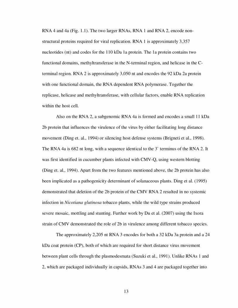
RNA 4 and 4a (Fig. 1.1). The two larger RNAs, RNA 1 and RNA 2, encode non-
structural proteins required for viral replication. RNA 1 is approximately 3,357
nucleotides (nt) and codes for the 110 kDa 1a protein. The 1a protein contains two
functional domains, methyltransferase in the N-terminal region, and helicase in the C-
terminal region. RNA 2 is approximately 3,050 nt and encodes the 92 kDa 2a protein
with one functional domain, the RNA dependent RNA polymerase. Together the
replicase, helicase and methyltransferase, with cellular factors, enable RNA replication
within the host cell.
Also on the RNA 2, a subgenomic RNA 4a is formed and encodes a small 11 kDa
2b protein that influences the virulence of the virus by either facilitating long distance
movement (Ding et. al., 1994) or silencing host defense systems (Brigneti et al., 1998).
The RNA 4a is 682 nt long, with a sequence identical to the 3` terminus of the RNA 2. It
was first identified in cucumber plants infected with CMV-Q, using western blotting
(Ding et. al., 1994). Apart from the two features mentioned above, the 2b protein has also
been implicated as a pathogenicity determinant of solanaceous plants. Ding et al. (1995)
demonstrated that deletion of the 2b protein of the CMV RNA 2 resulted in no systemic
infection in Nicotiana glutinosa tobacco plants, while the wild type strains produced
severe mosaic, mottling and stunting. Further work by Du et al. (2007) using the Ixora
strain of CMV demonstrated the role of 2b in virulence among different tobacco species.
The approximately 2,205 nt RNA 3 encodes for both a 32 kDa 3a protein and a 24
kDa coat protein (CP), both of which are required for short distance virus movement
between plant cells through the plasmodesmata (Suzuki et al., 1991). Unlike RNAs 1 and
2, which are packaged individually in capsids, RNAs 3 and 4 are packaged together into

14
one capsid. The 3a protein, also known as the movement protein (MP), functions to
regulate cell-to-cell movement of viral particles or ribonucleoproteins (positive sense
RNA bound to MP) through plasmodesmata (Ding et al., 1995). The MP targets the
plasmodesmata using tubules it forms through aggregates (Canto and Palukaitis, 1999;
Ding, et al., 1994; Suzuki et al., 1991). Once at the plasmodesmata, the MP modifies the
size exclusion limit to permit passage of the ribonucleoprotein (Vaquero et al., 1999) into
the next cell.
The capsid or coat protein (CP) is encoded by RNA 3 and translated from the
subgenomic coat protein messenger 4b, and is involved with short distance movement
between cells and aphid-mediated transmission (Chen and Francki, 1990; Ding et al.,
1995). The CP sequence, the most variable region of RNA 3, separates CMV into three
subgroups, 1A, 1B and 2. The difference within each subgroup is only 2-3%, but up to
15% between subgroups 1A and 1B, and as much as 25% between subgroups 1 and 2
(Palukaitis et al., 1992; Roossinck et al., 1999.
15
Fig. 1.1 Cucumber mosaic virus genome
Cucumber mosaic virus infection cycle
Similar to many other plant viruses, CMV enters a host through mechanical
wounds, or is introduced into the plant by insect or animal feeding. CMV is transmitted
by many species of aphids in a non-persistent manner. Once inside the cell, the coat
protein capsid disassociates, probably on cytoplasmic membranes, exposing viral RNAs.
The membrane-bound RNA is translated by ribosomes to non-structural proteins and
enzymes, the gene products of RNA 1 and 2, involved in virus replication (Nitta et al.,
1988). These enzymes, RNA dependent RNA polymerase, helicase and methytransferase,
then act with host factors to synthesize a negative strand, a complementary copy of the
viral RNA strand. At this point it is thought that both strands are temporarily bonded,
forming double-stranded RNA molecules. Once copying is complete, the negative strand
serves as a template for positive strand synthesis. With specific hybridization probes,
MMPP CCPP
CCPP
22bb
~3,300 nt
~3,000 nt
~1,000 nt
~2,200 nt
MMeetthhyyllttrraannssffeerraassee HHeelliiccaassee
RRNNAA ddeeppeennddeenntt RRNNAA ppoollyymmeerraassee
RNA 1
RNA 2
RNA 3
RNA 4
682 nt RNA 4a
Modified from Fauquet et al., 2005
16
appreciable levels of RNA were detected within 15 hours post-inoculation (Gonda and
Symons, 1979). When the copy number of positive-sense RNA reaches a threshold, a
value not exactly known, translation of some RNA to produce protein begins (Agrios,
2005). Two important proteins formed are the movement protein, required for short and
long distance movement within the plant, and the coat protein, used for short distance
movement and protection of the viral RNA in capsids. At this point viral RNAs can be
either encapsidated or form a ribonucleoprotein and move into adjacent cells through the
plasmodesmata. Encapsidation is a self-assembly process that occurs between the RNA
and the coat protein. There are no specific sites for binding, and instead, bonds are
formed between the protein subunits and RNA. The complete virion remains trapped
within the initial cell due to its size. Vaquero et al. (1994) demonstrated that, despite the
MP ability to modify the size of the plasmodesmata when expressed transgenically,
complete CMV particles, which are ~ 30 nm in diameter, could not enter the
plasmodesmata, but ribonucleoproteins, with diameters of only 1.5-2 nm, could easily
pass through.
The process of uncoating, replication, and encapsidation repeats in adjacent cells
until the viral nucleic acids reach the vascular bundles. Whole particles enter the phloem
and are transported with the photosynthates throughout the rest of the plant (Agrios,
2005).
CMV Diversity
Many RNA viruses have quasispecies, genetically diverse populations arising
from an initial host, and CMV is no exception. Many evolutionary studies have been
conducted on CMV (Bonnet et al., 2005; Roossinck, 2001, 2002), and all have observed
17
that the high rates of mutation, reassortment, and recombination of CMV have resulted in
its highly diverse nature. Schneider and Roossinck (2000) provided correlative data
between high rates of mutation of CMV and its increased host range. Initial passages into
new hosts resulted in significant changes in diversity, or quasispecies cloud size, even in
closely related species such as Nicotiana tabacum L. and N. benthamiana Domin
(Schneider and Roossinck, 2001). With further passages within the same host, the
number of mutations decreased rapidly, suggesting that CMV quickly attains and
maintains variations specific to that particular host. What was interesting to note was that
the mutations seen, even using identical CMV clones with the same host, were all unique
and not completely random (Schneider and Roossinck, 2000). Observed mutations were
distributed throughout the coat protein region on RNA 3 but had a higher bias towards
untranslated regions, while no mutations were observed within the ‘core’ region of the
coat protein, between nucleotide positions 1577 and 1846 of RNA 3.
The subgroup diversity of CMV can be determined molecularly by three methods.
The first and most commonly used method is RNA sequencing. The amplified sequence,
read from the original CMV RNA, can be compared to known strains and other isolate
sequences, both at a nucleotide and an amino acid level. Several phylogenetic studies
conducted with known strains of CMV displayed the same pattern of subgrouping using
different RNA segments, suggesting that subgroup diversity is not determined by one
RNA segment only (Bashir et al., 2006; Chen et al., 2007; Roossinck, 2002). In the
second method, primers specific for the different subgroups in CMV are used. These
specific primers are restricted only to the areas of the RNA that distinguish subgroups
(Yanming et al., 1997). The final method, restricto-typing, involves the use of restriction
18
enzymes designed to cut at specific locations in the DNA sequence. One such enzyme,
MspI, was highly successful in separating several CMV isolates into their respective
subgroups based on the number of restriction fragment patterns observed upon cleavage
of RNA 3. With the exception of the Ixora strain of CMV (subgroup IB), all of the CMV
isolates and strains tested yielded two fragments if the CMV isolate was subgroup IA,
four fragments if the CMV isolate was in subgroup IB, and five fragments of DNA for
subgroup II (Bashir et al., 2006; Chen et al., 2007). Restricto-typing was also successful
in determining reassortants of CMV (Chen et al., 2007). Reassortment is a natural
occurrence in viruses containing multipartite genomes and is important in genetic
variation and the development of new strains. In CMV, reassortment occurs more
frequently between subgroups IA and IB than between subgroups I and II, possibly due to
the higher percentage of similarity between IA and IB (Bonnet et al., 2005; Fraile et al.,
1997).
The subgroup diversity of CMV strains can also be determined serologically. Hsu
et al. (2000) developed monoclonal antibodies either for general CMV detection or
specific antibodies for subgroups 1 and 2 by ELISA. For further differentiation between
subgroups 1A and 1B, sequencing data must be obtained.
CMV Distribution
Every year, millions of dollars are lost because of severe diseases attributed to
CMV. Diseases in tropical regions are especially serious and widespread because of
constant warm temperatures, availability of crops year round, and ample rainfall resulting
in high humidity, all favorable conditions for the replication and distribution of CMV by
its aphid vector. Reoccurring epidemics of CMV (Albert et al., 1985; Grieco et al., 1997;
19
Kucharek et al., 1998) have prompted serological monitoring and development of several
CMV-resistant crop cultivars.
The wide distribution of CMV (Fig. 1.2) is primarily attributed to its aphid
vectors. Other methods of distribution include transportation of infected plant material
and seed transmission. Several species of aphids are capable of acquiring CMV, the most
common being the green peach aphid, Myzus persicae and the melon aphid, Aphis
gossypii (Blackman and Eastop, 2000). A brief feeding time of less than one minute
sufficiently insures acquisition of CMV and transmission to recipient plants by either
aphid. However, the ability to transmit the virus is only temporary and retention of the
virus is usually less than 1 hr from acquisition (Kucharek and Purcifull, 1997; Perry,
2001). Aphids are found worldwide, although a vast majority occurs in the Northern
temperate regions (Dixon, 1998). It is interesting to note that almost all major aphid
genera in the Northern temperate region are introduced Old World species, most likely
occurring during the movement of food crops, ornamentals and other plants between
countries (Blackman and Eastop, 2000).
Some of the more important crops affected by CMV include tomato, pepper,
cucurbit species including pumpkin and watermelon, root crops such as dasheen, and tree
crops such as banana. Several ornamental plants are also affected by CMV. In India,
Chrysanthemums are important cut-flowers that can be affected by CMV and TAV
through suckers, the primary method of propagation. Symptoms include stunting and
flower quality and can result in severe losses (Verma et al., 2004).
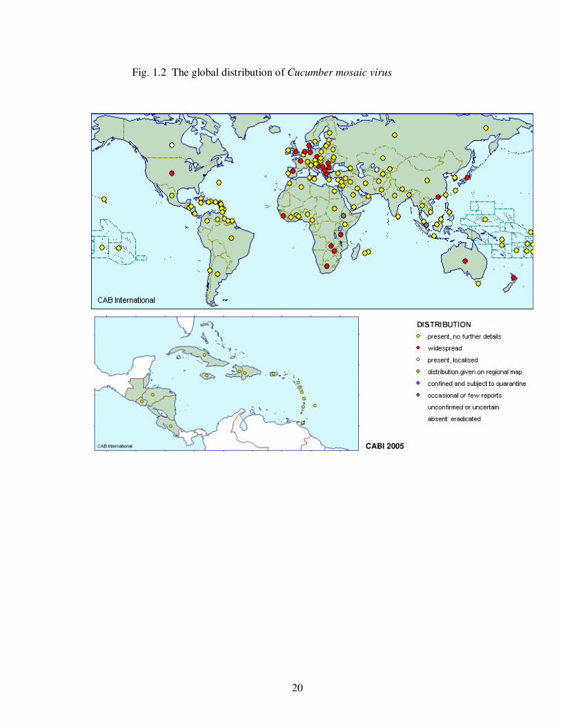
20
CABI 2005
Fig. 1.2 The global distribution of Cucumber mosaic virus
21
Control, Resistance and Management
Plant viruses cannot be treated using chemicals, and instead their control relies on
several preventive measures. The effective control of aphid vectors, removal of infected
plant material, and good cropping practices are just a few of the more commonly used
measures. Control and management practices are employed to reduce plant disease, and
include the use of pesticides for the control of aphid populations, barrier crops to protect
susceptible hosts, late or early planting of crops to avoid vector movement into cropping
area, and resistant cultivars (Gallitelli, 1998).
Breeding for Resistance
Breeding for natural resistance was the first method employed by farmers and
scientists for introducing resistance genes into favorable crops. The process involves
crossing of resistant and susceptible lines to introgress the resistant gene into the
susceptible variety, and back-crossing to the susceptible parental variety to achieve a
cultivar with both the resistance gene and the desired crop trait (Gallitelli, 1998). There
had not been much success in breeding resistant cultivars to CMV until as recently as the
last few decades when a CMV tolerant variety of pepper (Lapidot et. al., 1997), one of
tomato (Stoimenova and Sotirova, 2000), and one of cucumber (Kherebah et al., 2009)
were reported.
Induced Resistance
Cross-protection provided one of the early successes in the reduction of viral
disease spread, and involved the inoculation of plants with mild strains of viruses in
hopes of cross-protection against more virulent strains of the virus. Dodds (1982)
22
demonstrated that mixed mild and severe strains of CMV were antagonistic to each other,
resulting in lower accumulation of virus particles over time.
Transgenic technologies have played a large role in development of resistance in
plants. In transgenics, the plant itself is genetically modified by introducing a piece of a
viral gene into the plant’s genome (Gallitelli, 1998). In the mid 1980s, tobacco was the
first plant to be transformed using the model virus Tobacco mosaic virus (TMV) CP. A
delay in disease development was seen in the transformed tobacco when compared to the
control (Abel et al., 1986). Cuozzo et al. (1988) expressed CMV CP in transgenic tobacco
and tomato and found that delayed onset and fewer symptoms were seen on plants able to
synthesize the CP compared to those that could not (control). Use of the anti-sense RNA
for the CP also gave milder symptoms, but was easily overcome at higher concentrations
of the virus. Cultivars of squash and melon have both been transformed using coat
protein genes from three viruses, CMV (strain C), Watermelon mosaic virus 2 (WMV 2)
and Zucchini yellow mosaic virus (ZYMV), individually or together. Resistances to these
viruses have been observed both in the laboratory and in field trials (Fuchs et al., 1995).
Insertion of multiple coat protein genes into plant genomes conferred resistance to
multiple viruses (Fuchs et al., 1998). Fuchs et al. (1998) demonstrated that insertion of
one, two or all three CP genomes of CMV, ZYMV and WMV2 resulted in lower
incidence of symptoms and higher yields when compared to non-transformed controls.
Symptoms, when seen, were restricted to small dots on symptomatic leaves. It was
hypothesized that this method would aid in the reduction of acquisition of virus during
aphid feeding. The reduction in virus spread within a field supported this hypothesis
23
(Fuchs et al., 1998). Commercial crops have since been produced with resistance genes to
several plant viruses using this technology.
Synergism
In the tropics, plant viruses cause damaging diseases on fruit and vegetable crops
(Gallitelli, 2000; McLaughlin, 2003). In nature plants are usually bombarded by several
viruses at once, and some viruses are able to facilitate systemic infection of other viruses
that initially could not overcome the plant host’s defenses. The combination of several
viruses intensifies the reaction, enhancing disease severity with a greater virus
accumulation within cells (Poolpol and Inouye, 1986). Recent work has been done
involving cucurbits and the synergistic effects of more than one virus affecting a plant
species (Murphy and Bowen, 2006). There have been several reports of CMV co-
infecting plants with other viruses, most particularly with potyviruses (Murphy and
Bowen, 2006; Pinto et al., 2008; Wang et al., 2002; Zeng et al., 2007). Choi et al. (2002),
Guerini and Murphy (1999) and Wang et al. (2002, 2004) all reported enhanced
movement and resistance-breaking by CMV when plants were co-infected with WMV.
Because CMV and potyviruses are often found together in nature, it was important to
review the potyviruses, their genomes, and characteristics. Potyviruses used in this study
were Tobacco etch virus (TEV), Turnip mosaic virus (TuMV), and Soybean mosaic virus
(SMV).
Potyviruses
The genus Potyvirus has been discussed in several reviews (Hollings and Brunt,
1981; Shukla et al., 1994), so only a few of the major characteristics will be highlighted
24
in this section. Potyviruses comprise a large group of plant viruses that belong to the
genus Potyvirus, family Potyviridae. Several members are economically important plant
pathogens, causing millions of dollars in crop loss worldwide. The Potyvirus genus has
more than 400 members with a little over 50 tentative members (Fauquet et al., 2005).
Like cucumoviruses, potyviruses are disseminated by several species of aphids in a non-
persistent manner. But, unlike CMV, potyviruses require a helper component-proteinase
(HC-Pro) for successful vector transmission (Blanc et. al., 1998). This virus-encoded,
multi-functional protein has one motif that binds to the stylet of an aphid and another that
binds to the N-terminus of the potyvirus CP (Hull, 2002). Three members of particular
importance are Tobacco etch virus (TEV), Turnip mosaic virus (TuMV), and Soybean
mosaic virus (SMV) as they can cause significant damage to crops. Many potyviruses
have a narrow host range. SMV can only infect soybeans and other very closely related
beans. In contrast, TEV has a wide host range, affecting crops in families such as
Cucurbitaceae and Solanaceae, and is one of the leading causes in pepper decline in the
tropics (Myers, 1996). TuMV has a natural host range restricted primarily to the family
Brassicaceae, but affects several members within this family.
The Potyvirus genome and particle properties
Potyviruses are flexous rods ~750 nm in length with a genome that consists of a
positive-sense, single-stranded RNA of approximately 10,000 bases. Belonging to the
picorna-like supergroup of viruses, potyviruses have a VPg covalently bound to the 5`
end, and a poly(A) tail at the 3` end. During replication, the entire genome is translated to
produce a large polyprotein, which is cleaved by proteases to give several proteins. The
major, important proteins encoded include the P1-Protease, which cleaves the Tyr/Phe-
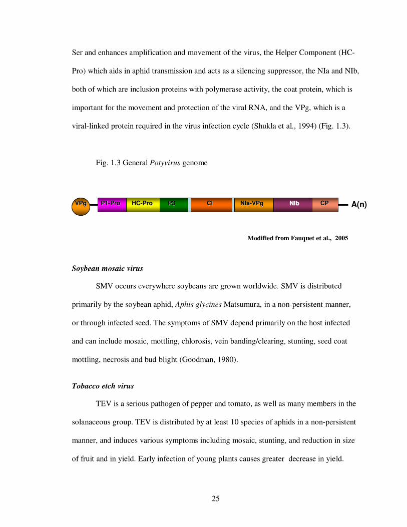
25
PP11--PPrroo HHCC--PPrroo PP33 CCll NNIIaa--VVPPgg NNIIbb CCPP VVPPgg A(n)
Ser and enhances amplification and movement of the virus, the Helper Component (HC-
Pro) which aids in aphid transmission and acts as a silencing suppressor, the NIa and NIb,
both of which are inclusion proteins with polymerase activity, the coat protein, which is
important for the movement and protection of the viral RNA, and the VPg, which is a
viral-linked protein required in the virus infection cycle (Shukla et al., 1994) (Fig. 1.3).
Fig. 1.3 General Potyvirus genome
Soybean mosaic virus
SMV occurs everywhere soybeans are grown worldwide. SMV is distributed
primarily by the soybean aphid, Aphis glycines Matsumura, in a non-persistent manner,
or through infected seed. The symptoms of SMV depend primarily on the host infected
and can include mosaic, mottling, chlorosis, vein banding/clearing, stunting, seed coat
mottling, necrosis and bud blight (Goodman, 1980).
Tobacco etch virus
TEV is a serious pathogen of pepper and tomato, as well as many members in the
solanaceous group. TEV is distributed by at least 10 species of aphids in a non-persistent
manner, and induces various symptoms including mosaic, stunting, and reduction in size
of fruit and in yield. Early infection of young plants causes greater decrease in yield.
Modified from Fauquet et al., 2005
26
Turnip mosaic virus
TuMV is transmitted by at least 89 species of aphids to over 318 plant species, a
majority of which belong to the family Brassicaceae. The virus causes mottling and
mosaic patterns, necrotic and ring spots, distortion, reduction in fruit yield, and color-
breaking on flowers. The distribution of TuMV has been reported worldwide particularly,
wherever vegetable crops are grown (Shukla et al., 1994).
Objectives
Originally, the dissertation was focused primarily on CMV and the diagnostic
techniques used for its detection and subgroup characterization, both locally in Virginia
and globally, using a single, solid matrix. This method allowed for the safe transport of
plant viruses across borders. Below are the original objectives of the dissertation.
i) To analyze the political, economical and trade constraints to viral diagnosis in
plant diagnostic and research laboratories in developed vs. developing
countries.
ii) To examine the diversity of CMV in Virginia and to validate the conventional
methods used in molecular analysis.
iii) To develop an affordable and rapid diagnostic and detection method for RNA
plant viruses using CMV and paper-based technology.
iv) To determine the diversity of CMV isolates in tropical and subtropical regions
and compare this diversity to CMV isolates and strains reported in temperate
regions.
27
The driving force behind the original dissertation – the application of diagnostic
techniques for the detection and subgrouping of CMV, has remained the same. However,
our discovery of the increased efficiency of NitroPure nitrocellulose membranes as
sources of plant viral nucleic acids led us to revise the objectives. The direction of the
dissertation is now focused specifically on diagnostics. The number of viruses was
increased to include three potyviruses, one of which is a first report in Virginia. The new
objectives are stated below:
i) To characterize three recently acquired isolates of Cucumber mosaic virus (CMV)
from Historic Smithfield Plantation using biological and molecular tools and
applications.
ii) To characterize Turnip mosaic virus isolated from Hesperis matronalis (L.) from
Historic Smithfield Plantation.
iii) To evaluate the virus diagnostic capabilities of collaborators in IPM-CRSP host
countries.
iv) To evaluate NitroPure nitrocellulose membranes for immunoassay, RT-PCR-based
amplification and direct sequencing of Cucumber mosaic virus and potyvirus coat
proteins
v) To globally apply NitroPure nitrocellulose membranes for virus detection and
identification.
28
References
Abbas, M., Khan, M. M., Mughal, S. M., and Khan, I. A. 2005. Prospects of classical
cross protection technique against Citrus tristeza closterovirus in Pakistan. Hort.
Sci. (PRAGUE) 32:74-83.
Abel, P. P., Nelson, R. S., De, B., Hoffmann, N., Rogers, S. G., Fraley, R. T., and
Beachy, R. N. 1986. Delay of disease development in transgenic plants that
express the Tobacco mosaic virus coat protein gene. Science 232:738-743.
Agrios, G. N., and Walker, M. E. 1985. Effect on Cucumber mosaic virus inoculation at
successive weekly intervals on growth and yield of pepper (Capsicum annuum)
plants. Plant Dis. 69:52-55.
Agrios, G. N. 2005. Plant diseases caused by viruses. Pages 723-824 in: Plant Pathology
Elsevier Academic Press, San Diego, California..
Albert, J., Hannay, J., and Randles, J. W. 1985. An epidemic of Cucumber mosaic virus
in South Australian Lupins. Aust. J. Agric. Res. 36:267-273.
Alfaro-Fernandez, A., Cebrian, M. C., Cordoba-Selles, C., Herrera-Vasquez, J. A., and
Jorda, C. 2008. First report of the US1 strain of Pepino mosaic virus in tomato in
the Canary Islands, Spain. Plant Dis. 92:1590-1590.
Ali, A., Natsuaki, T., and Okuda, S. 2004. Identification and molecular characterization
of viruses affecting cucurbits in Pakistan. J. Phytopath. 152:677-682.
Arneodo, J. D., de Breuil, S., Lenardon, S. L., Conci, V. C., and Conci, L. R. 2005.
Detection of Bean yellow mosaic virus and Cucumber mosaic virus infecting
gladiolus in Argentina. Agriscientia 22:87-89.
Ausher, R., Ben-Ze'ev, I. S., and Black, R. 1996. The role of plant clinics in plant disease
diagnosis and education in developing countries. Annu. Rev. Phytopathol. 34:51-
66.
Avgelis, A. 1987. Cucumber mosaic virus on Banana in Crete. J. Phytopathol. 120:20-24.
29
Ball, E. 1990. Agar double diffusion, plates (Ouchterlony): Viruses. Pages 111-120 in:
Serological methods for the detection and identification of viral and bacterial
plant pathogens: A laboratory manual, R. Hampton, E. Ball and S. De Boer, eds.
APS Press, St. Paul, Minnesota.
Ball, E., Hampton, R. O., De Boer, S. H., and Schaad, N. W. 1990. Polyclonal antibodies.
Pages 33-54 in: Serological methods for the detection and identification of viral
and bacterial plant pathogens: A laboratory manual, R. Hampton, E. Ball and S.
De Boer, eds. APS Press, St. Paul, Minnesota.
Bashir, N. S., Kalhor, M. R., and Zarghani, S. N. 2006. Detection, differentiation and
phylogenetic analysis of Cucumber mosaic virus isolates from cucurbits in the
northwest region of Iran. Virus Genes 32:277-288.
Barnes, L. W. 1994. The role of plant clinics in disease diagnosis and education: A North
American perspective. Annu. Rev. Phytopathol 32:601-609.
Blackman, R. L., and Eastop, V. F. 2000. Aphids on the World's Crops. John Wiley &
Sons Ltd., 2nd ed.
Blencowe, J. W., and Caldwel, J. 1949. Aspermy - A new virus disease of the tomato.
Ann. Appl. Biol. 36:320-326.
Bonnet, J., Fraile, A., Sacristan, S., Malpica, J. M., and Garcia-Arenal, F. 2005. Role of
recombination in the evolution of natural populations of Cucumber mosaic virus,
a tripartite RNA plant virus. Virology 332:359-368.
Brigneti, G., Voinnet, O., Li, W. X., and Ji, L. H. 1998. Viral pathogenicity determinants
are suppressors of transgene silencing in Nicotiana benthamiana. EMBO
17:6739-6746.
Burgoyne, L. A. Solid medium and method for DNA storage. US Patent 5,496,562. 1996.
March 5. http://www.patentstorm.us/patents/5496562.html
Canto, T., and Palukaitis, P. 1999. Are tubules generated by the 3a protein necessary for
Cucumber mosaic virus movement? Mol. Plant-Microbe Interact. 12:985-993.
30
Chen, Y., Chen, J., Zhang, H., Tang, X., and Du, Z. 2007. Molecular evidence and
sequence analysis of a natural reassortant between Cucumber mosaic virus
subgroup 1A and 2 strains. Virus Genes 35:405-413.
Chen, B., and Francki, R. I. B. 1990. Cucumovirus transmission by the aphid Myzus
persicae is determined solely by the viral coat protein. J. Gen. Virol. 71:939-944.
Cho, E. K., and Goodman, R. M. 1979. Strains of Soybean mosaic virus: classification
based on virulence in resistant soybean cultivars. Phytopathology 69:467-470.
Choi, S. K., Yoon, J. Y., Ryu, K. H., Choi, J. K., Palukaitis, P., and Park, W. M. 2002.
Systemic movement of a movement-deficient strain of Cucumber mosaic virus in
zucchini squash is facilitated by a cucurbit-infecting potyvirus. J. Gen. Virol.
83:3173-3178.
Clark, M. F., and Adams, A. N. 1977. Characteristics of the microplate method of
enzyme-linked immunosorbent assay for the detection of plant viruses. J. Gen.
Virol. 34:475-483.
Comstock, J., and Miller, J. 2004. Reliability of leaf-midrib tissue blot immunoassay to
detect Sugarcane yellow leaf virus and differences between cultivars in rate of
spread. Pages 40-41 in: International Society of Sugarcane Technologists
Pathology Workshop J. Amer. Society Sugarcane Technologists.
Converse, R. H., and Martin, R. R. 1990. ELISA methods for plant viruses. Pages 179-
196 in: Serological methods for the detection and identification of viral and
bacterial plant pathogens: A laboratory manual, R. Hampton, E. Ball and S. De
Boer, eds. APS Press, St. Paul, Minnesota.
Cuozzo, M., O'Connell, K. M., Kaniewski, W. K., Fang, R. X., Chua, N. H., and Tumer,
N. E. 1988. Viral protection in transgenic tobacco plants expressing the Cucumber
mosaic virus coat protein or its antisense RNA. Biotechnology 6:549-557.
Dale, P. J., Clarke, B., and Fontes, E. M. G. 2002. Potential for the environmental impact
of transgenic crops. Nature Biotech. 20:567-574.
31
Ding, S. W., Li, W. X., and Symons, R. H. 1995. A novel naturally occurring hybrid gene
encoded by a plant RNA virus facilitates long distance virus movement. EMBO
14:5762-5772.
Ding, S. W., Anderson, B. J., Haase, H. R., and Symons, R. H. 1994. New overlapping
gene encoded by the Cucumber mosaic virus genome. Virology 198:593-601.
Dixon, A. F. G. 1998. Aphid Ecology. Chapman & Hall, 2nd ed.
Dodds, J. A. 1982. Cross-protection and interference between electrophoretically distinct
strains of Cucumber mosaic virus in tomato. Virology 118:235-240.
Doolittle, S. P. 1916. A new infectious mosaic disease of cucumber. Phytopathology
6:145-147.
Du, Z.-Y., Chen, F.-F., Liao, Q.-S., Zhang, H.-R., Chen, Y.-F., and Chen, J.-S. 2007. 2b
ORFs encoded by subgroup IB strains of Cucumber mosaic virus induce
differential virulence on Nicotiana species. J. Gen. Virol. 88:2596-2604.
Edwardson, J. R., and Christie, R. G. 1997. Cucumber mosaic virus. Pages 133-143 in:
Viruses infecting pepper and other Solanaceous crops. University of Florida,
Agricultural Expt. Stat., IFAS, Gainesville.
FAO. 2009. Food prices remain high in developing countries. Food and Agriculture
Organization of the United Nations.
http://www.fao.org/news/story/en/item/12660/icode/
Fauquet, C. M., Mayo, M. A., Maniloff, J., Desselberger, U., and Ball, L. A., eds. 2005.:
Virus Taxonomy: 8th Report of the ICTV. Elsevier. 1259p.
Fraile, A., Alonso-Prados, J. L., Aranda, M. A., Bernal, J. J., Malpica, J. M., and Garcia-
Arenal, F. 1997. Genetic exchange by recombination or reassortment is infrequent
in natural populations of a tripartite RNA virus. J. Virol. 71 (2):934-940.
Fuchs, M., Klas, F., and Gonsalves, D. 1995. Risk assessment of virus spread using
transgenic plants containing viral coat protein genes. in: Proceedings and Papers
from the 1995 Risk Assessment Research Symposium .
32
Fuchs, M., Tricoli, D. M., Carney, K. J., Schesser, M., McFerson, J. R., and Gonsalves,
D. 1998. Comparative virus resistance and fruit yield of transgenic squash with
single and multiple coat protein genes. Plant Dis. 82:1350-1356.
Gallitelli, D. 1998. Present status of controlling Cucumber mosaic virus. Pages 507-523
in: Control of Plant Virus Diseases, A. Hadidi, R. K. Khetarpal and H.
Koganezawa, eds. APS Press, St. Paul, MN.
Gallitelli, D. 2000. The ecology of Cucumber mosaic virus and sustainable agriculture.
Virus Research 71:9-21.
Garcia-Arenal, F., and Fraile, A. 2008. Questions and concepts in plant virus evolution: a
historical perspective. Pages 1-14 in: Plant Virus Evolution, M. J. Roossinck, ed.
Springer-Verlag, Berlin.
Gonda, T. J., and Symons, R. H. 1979. Cucumber mosaic virus replication in cowpea
protoplasts: Time course of virus, coat protein and RNA synthesis. J. Gen. Virol.
45:723-736.
Goodman, R. 1980. Soybean mosaic potyvirus.
Grieco, F., Lanave, C., and Gallitelli, D. 1997. Evolutionary dynamics of Cucumber
mosaic virus satellite RNA during natural epidemics in Italy. Virology 229:166-
174.
Guerini, M. N., and Murphy, J. F. 1999. Resistance of Capsicum annuum `Avelar' to
pepper mottle potyvirus and alleviation of this resistance by co-infection with
Cucumber mosaic cucumovirus are associated with virus movement. J. Gen.
Virol. 80:2785-2792.
Hollings, M., and Brunt, A. A. 1981. Potyviruses. Pages 731-807 in: Handbook of Plant
Virus Infections and Comparative Diagnostics, E. Kurstak, ed. Elsevier/North
Holland Biomedical Press.
Hristova, D., Yordanova, A., and Mavrodieva, V. 2002. Differentiation of Bulgarian
isolates from Cucumber mosaic virus by serological methods. J. Phytopath.
150:334-339.
33
Hsu, H. T., Barzuna, L., Hsu, Y. H., Bliss, W., and Perry, K. L. 2000. Identification and
subgrouping of Cucumber mosaic virus with mouse monoclonal antibodies.
Virology 90:615-620.
Hsu, H. T., and Lawson, R. H. 1991. Direct tissue blotting for detection of Tomato
spotted wilt virus in Impatiens. Plant Dis. 75:292-295.
Hull, R. 2002. Matthews’ Plant Virology, 4th Edition. Academic Press, London. 1001 p.
Jonson, G., Park, J.-C., Kim, Y.-K., Lee, M.-J., Hyun, J.-N., and Kim, J.-G. 2007. Direct
stem blot immunoassay (DSBIA): A rapid, reliable and economical detection
technique suitable for testing large number of barley materials for field
monitoring and resistance screening to Barley mild mosaic virus and Barley
yellow mosaic virus. Plant Pathol. J. 23:260-265.
Jordan, R. L. 1990. Strategies and techniques for the production of monoclonal
antibodies; monoclonal antibody applications for viruses. Pages 55-86 in:
Serological methods for the detection and identification of viral and bacterial
plant pathogens: A laboratory manual, R. Hampton, E. Ball and S. De Boer, eds.
APS Press, St. Paul, Minnesota.
Jordan, R. L., Guaragna, M. A., Van Buren, T., and Putnam, M. L. 2008. First report of a
new Potyvirus, Tricyrtis virus Y, and Lily virus X, a Potexvirus, in Tricyrtis
formosana in the United States. Plant Dis. 92:648-648.
Kaper, J. M. 1975. The chemical basis of virus structure, dissociation and reassembly.
American Elsevier Publishing Co., New York.
Kaper, J. M., and Waterworth, H. E. 1981. Cucumoviruses. Pages 257-332 in: Handbook
of Plant Virus Interactions and Comparative Diagnosis, E. Kurstak, ed.
Elsevier/North Holland Biomedical Press.
Khereba, A. H., Abdallah, N. A., Hassan, H. A., Mohamed, M. A., Abdelkader, H. A.,
Abdrabou, A. M., and Zein, H. S. 2009. Evaluation of some cucumber inbred
lines and their hybrids for Cucumber mosaic virus (CMV) resistance. Arab J.
Biotech. 12:133-148.
34
Kiss, L., Sebestyén, E., László, E., Salamon, P., Balázs, E., and Salánki, K. 2008.
Nucleotide sequence analysis of Peanut stunt virus Rp strain suggests the role of
homologous recombination in cucumovirus evolution. Arch. Virol. 153:1373-
1377.
Koike, S. T., Kuo, Y.-W., Rojas, M. R., and Gilbertson, R. L. 2008. First report of
Impatiens necrotic spot virus infecting lettuce in California. Plant Dis. 92:1248-
1248.
Kucharek, T., and Purcifull, D. 1997. Aphid-transmitted viruses of cucurbits in Florida.
Report No. Circ-1184. Florida Cooperative Extension Service, Institute of Food
and Agricultural Sciences, University of Florida.
Kucharek, T. A., Purcifull, D. E., Christie, R. G., and Perkins, K. D. 1998. The
association of severe epidemics of Cucumber mosaic in commercial fields of
pepper and tobacco in North Florida with inoculum in Commelina benghalensis
and C. communis. Plant Dis. 82:1172-1172.
Lapidot, M., Paran, I., Ben-Joseph, R., Ben-Harush, S., Pilowsky, M., Cohen, S., and
Shifriss, C. 1997. Tolerance to Cucumber mosaic virus in pepper: Development of
advanced breeding lines and evaluation of virus level. Plant Dis. 81:185-188.
Lawrence, J., Tolin, S. A., Edwards, C., Fleischer, S. J., Jackson, D. M., Clarke-Harris,
D., McDonald, S. A., Dalip, K., and Chung, P. 2005. Developing IPM packages
in the Caribbean. pp. 95-120 in: Globalizing Integrated Pest Management: A
Participatory Research Process, G. W. Norton, E. A. Heinrichs, G. C. Luther and
M. E. Irwin, eds. Blackwell Publishing, Ames, Iowa.
Lin, N. S., Hsu, Y. H., and Hsu, H. T. 1990. Immunological detection of plant viruses and
a mycoplasma-like organism by direct tissue blotting on nitrocellulose
membranes. Phytopathology 80:824-828.
Ling, X. 1990. Isolation of Cucumber mosaic and Tobacco mosaic viruses from tomato in
Thailand. in: ARC Training. Asian vegetable Research and Development Center
- Asian Regional Center, Thailand.
35
Liu, S., He, X., Park, G., Josefsson, C., and Perry, K. L. 2002. A conserved capsid protein
surface domain of Cucumber mosaic virus is essential for efficient aphid vector
transmission. J. Virol. 76:9756-9762.
Ma, G., Chen, P., Buss, G. R., and Tolin, S. A. 2003. Genetic study of a lethal necrosis to
Soybean mosaic virus in PI 507389 soybean. J. Hered. 94:205-211.
Mahmood, T., and Rush, C. M. 1999. Evidence of cross-protection between Beet
soilborne mosaic virus and Beet necrotic yellow vein virus in sugar beet. Plant
Dis. 83:521-526.
Makkouk, K. M., and Comeau, A. 1994. Evaluation of various methods for the detection
of Barley yellow dwarf virus by the tissue-blot immunoassay and its use for virus
detection in cereals inoculated at different growth stages. Europ. J. Plant Pathol.
100:71-80.
McLaughlin, W. 2003. Viral diseases of hot pepper in the Caribbean: characterization,
vectors and management. in: Caribbean Hot Pepper Industry Workshop - Charting
the way forward. CARDI, Trinidad and Tobago.
Miller, L. E., and Troutman, J. L. 1966. Stunt disease of peanuts in Virginia. Plant Dis.
Rep. 50:139-143.
Mink, G. I. 1980. Peanut stunt virus. Revised by P. L. Guy, 1990.
http://image.fs.uidaho.edu/vide/descr585.htm
Murphy, J. F., and Bowen, K. L. 2006. Synergistic disease in pepper caused by the mixed
infection of Cucumber mosaic virus and Pepper mottle virus. Phytopathology
96:240-247.
Myers, L. R. S. 1996. The etiology of viruses affecting pepper (Capsicum spp.) in
Jamaica.
Masters Thesis, University of the West Indies, 142 p.
Ndunguru, J., Taylor, N. J., Yadav, J., Aly, H., Legg, J. P., Aveling, T., Thompson, G.,
and Fauquet, C. M. 2005. Application of FTA technology for sampling, recovery
36
and molecular characterization of viral pathogens and virus-derived transgenes
from plant tissues. Virol. J. 2:45.
Nikolaeva, O. V. 1995. Nucleic acid hybridization methods in diagnosis of plant viruses
and viriods. pp. xxx -xxx. in: Molecular methods in plant pathology, R. P. Singh
and U. S. Singh, eds. CRC Press Inc. xxx p.
Nitta, N., Takanami, Y., Kuwata, S., and Kubo, S. 1988. Inoculation with RNAs 1 and 2
of Cucumber mosaic virus induces viral RNA replicase activity in tobacco
mesophyll protoplasts. J. Gen. Virol. 69:2695-2700.
Palukaitis, P., and Garcia-Arenal, F. 2003. Cucumber mosaic virus.
http://www.dpvweb.net/dpv/showdpv.php?dpvno=400
Palukaitis, P., Roossinck, M. J., Dietzgen, R. G., and Francki, R. I. B. 1992. Cucumber
mosaic virus. Adv. Vir. Res. 41:281-348.
Paradies, F., Sialer, M. F., Gallitelli, D., Castellano, M. A., DiFranco, A., Digiaro, M.,
Martelli, G. P., and Yilmaz, M. A. 2000. Partial characterization of Cucumber
mosaic virus isolates from citrus and grapevine. J. Plant Path. 82:133-145.
Parrella, G., Gognalons, P., Gebre-Selassie, K., Vovlas, C., and Marchoux, G. 2003. An
update on the host range of Tomato spotted wilt virus. J. Plant Pathol. 85:227-264.
Peden, K. W. C., and Symons, R. H. 1973. Cucumber mosaic virus contains a
functionally divided genome. Virology 53:487-492.
Perry, K. L. 2001. Cucumoviruses. Pages 167-179 in: Virus-Insect-Plant Interactions, K.
F. Harris, O. P. Smith and J. E. Duffus, eds. Academic Press.
Phatak, H. C., Diaz-Ruiz, J. R., and Hull, R. 1976. Cowpea ringspot virus: A seed
transmitted Cucumovirus. Phytopathology 87:132-142.
Pinto, Z. V., Rezende, J. A. M., Yuki, V. A., and Piedade, S. M. d. S. 2008. Ability of
Aphis gossypii and Myzus persicae to transmit Cucumber mosaic virus in single
and mixed infection with two potyviruses to Zucchini squash. Summa
Phytopathologica 34:183-185.
37
Poolpal, P. and Inouye, T. 1986. Ultrastructure of plant cells doubly infected with
potyviruses and other unrelated viruses. Bull. Univ. Osaka Pref. Ser.B. 38:12-23.
Roossinck, M. J. 2001. Cucumber mosaic virus, a model for RNA virus evolution. Molec.
Plant Pathol. 2:59-63.
Roossinck, M. J. 2002. Evolutionary history of Cucumber mosaic virus deduced by
phylogenetic analysis. J. Virol. 76:3382-3387.
Roossinck, M. J., Zhang, L., and Hellwald, K.-H. 1999. Rearrangement in the 5' non-
translated region and phylogenetic analyses of Cucumber mosaic virus RNA 3
indicate radial evolution of the three subgroups. J. Virol. 73:6752-6758.
Roy, Y., and Nassuth, A. 2005. Detection of plant genes, gene expression and viral RNA
from tissue prints on FTA cards. Plant Mol. Biol. Rep. 23:383-395.
Rybicki, E. P., and von Wechmar, M. B. 1985. Serology and immunochemistry. Pages
207-244 in: The Plant Viruses: Polyhedral virions with tripartite genomes, R. I. B.
Francki, ed. Plenum Press, New York.
Saiki, R. K., Gelfand, D. H., Stoffel, S., Scharf, S., Higuchi, R., Horn, G. T., Mullis, K.
B., and Erlich, H. A. 1988. Primer-directed enzymatic amplification of DNA with
a thermostable DNA polymerase. Science 239:487-491.
Schmelzer, K. 1971. Robinia mosaic virus. CMI/AAB Descr. Plant Viruses 65:3
Schmelzer, K., and Schmidt, H. E. 1975. Systemic infection by Cucumber mosaic virus
on French bean Phaseolus vulgaris (L.) in the German Democratic Republic.
Zentralblatt fur Bakteriologie, Parasitenkunde, Infektionskrankheiten und
Hygiene, 2 130:234.
Schneider, W. L., and Roossinck, M. J. 2001. Genetic diversity in RNA virus
quasispecies is controlled by host-virus interactions. J. Virol. 75:6566-6571.
Schneider, W. L., and Roossinck, M. J. 2000. Evolutionarily related Sindbis-like plant
viruses maintain different levels of population diversity in a common host. J.
Virol. 74:3130-3134.
38
Shukla, D. D., Ward, C. W., and Brunt, A. A. 1994. The Potyviridae. CAB International,
Wallingford, UK. 515 p.
Smith, M.A., Qu, D., Iyer, M., and Davis, J. C. 2003. Solid medium and process for the
storage and rapid purification of nucleic acid. US patent 6,645,717. 2003. Nov.
11.
Smith, T. J., Chase, E., Schmidt, T., and Perry, K. L. 2000. The structure of Cucumber
mosaic virus and comparison to Cowpea chlorotic mottle virus. J. Virol. 74:7578-
7586.
Stobbs, L. W., and Barker, D. 1985. Rapid sample analysis with a simplified ELISA.
Phytopathology 75:492-495.
Srinivasan, I. 1992. Isolation and detection of Bean yellow mosaic virus, Clover yellow
mosaic virus and Peanut stunt virus from Trifolium L. spp. Virginia Polytechnic
Institute and State University, Blacksburg. Master of Science Thesis. 94p.
Srinivasan, I., and Tolin, S. A. 1992. Detection of three viruses of clovers by direct tissue
immunoblotting. Phytopathology 82:721.
Stoimenova, E., and Sotirova, V. 2000. Tomato lines segregation for resistance to
Cucumber mosaic virus. Acta Physiologiae Plantarum 22:353-355.
Suzuki, M., Kuwata, S., Kataoka, J., Masuta, C., Nitta, N., and Takanami, Y. 1991.
Functional analysis of deletion mutants of Cucumber mosaic virus RNA3 using
an in vitro transcription system. Virology 183:106-113.
Symons, R. H. 1985. Viral genome structure. Pages 57-82 in: The Plant Viruses:
Polyhedral virions with tripartite genomes, R. I. B. Francki, ed. Plenum Press,
New York.
Takahashi, H., Suzuki, M., Natsuaki, K., Shigyo, T., Hino, K., Teraoka, T., Hosokawa,
D., and Ehara, Y. 2001. Mapping the virus and host genes involved in the
resistance response in Cucumber mosaic virus-infected Arabidopsis thaliana.
Plant Cell Physiol. 42:340-347.
39
Thresh, J. M. 2006. Crop viruses and virus diseases: A global prospective. Pages 9-32 in:
Virus Diseases and Crop Biosecurity, J. I. Cooper, T. Kuhne and V. P. Polischuk,
eds. Springer. Dordrecht, The Netherlands. 148 p.
Tolin, S. A. 1977. Cucumovirus (Cucumber mosaic virus) group. Pages 303-309 in: The
Atlas of Insects and Plant Viruses, including Mycoplasma Viruses and Viroids, K.
Maramorosch and J. Albert, eds. Academic Press, New York.
Tolin, S. A. 1994. Cucumoviruses. Pages 278-285 in: Encyclopedia of Virology, R.
Webster and A. Granoff, eds. Academic Press Ltd. London.
Tsuda, S., Kameya-Iwaki, M., Hanada, K., and Tomaru, K. 1992. A novel detection and
identification technique for plant viruses: Rapid immunofilter paper assay (RIPA).
Plant Dis. 76:466-469.
van der Vlugt, R. A. A. 2006. Plant viruses in European agriculture: Current problems
and future aspects. Pages 33-44 in: Virus diseases and crop biosecurity, I. Cooper,
T. Kuhne and V. Polischuk, eds. Springer, Dordrecht, The Netherlands. 148 p.
van Regenmortel, M. H. V. 1982. Serology and immunochemistry of plant viruses.
Academic Press Inc. London. 302 p.
Vaquero, C., Turner, A. P., Demangeat, G., Sanz, A., Serra, M. T., Roberts, K., and
Garcia-Luque, I. 1994. The 3a protein from Cucumber mosaic virus increases the
gating capacity of plasmodesmata in transgenic tobacco plants. J. Gen. Virol.
75:3193-3197.
Vincelli, P., and Tisserat, N. 2008. Nucleic acid-based pathogen detection in Applied
Plant Pathology. Plant Dis. 92 :660-669.
Walkey, D. G. A. 1991. Virus Identification. Pages 133-167 in: Applied Plant Virology.
Chapman and Hall, New York.
Wang, Y., Gaba, V., Yang, J., Palukaitis, P., and Gal-On, A. 2002. Characterization of
synergy between Cucumber mosaic virus and Potyviruses in cucurbit hosts.
Phytopathology 92:51-58.
40
Wang, Y., Lee, K. C., Gaba, V., Wong, S. M., Palukaitis, P., and Gal-On, A. 2004.
Breakage of resistance to Cucumber mosaic virus by co-infection with Zucchini
yellow mosaic virus: enhancement of CMV accumulation independent of
symptom expression. Arch. Virol. 149:379-396.
Xu, Z., Barnett, O. W., and Gibson, P. B. 1986. Characterization of Peanut stunt virus
strains by host reactions, serology, and RNA patterns. Phytopathology 76:390-
395.
Yanming, Y., Kyung, K. S., and Anderson, E. J. 1997. Seed transmission of Cucumber
mosaic virus in spinach. Phytopathology 87:924-931.
Zeng, R., Liao, Q., Feng, J., Li, D., and Chen, J.-S. 2007. Synergy between Cucumber
mosaic virus and Zucchini yellow mosaic virus on Cucurbitaceae hosts tested by
real-time reverse transcription-polymerase chain reaction. Acta Biochimica et
Biophysica Sinica 39:431-437.
41
CHAPTER II
Biological and molecular characterization of Cucumber mosaic virus
(CMV) from Virginia.
Abstract
In Virginia, Cucumber mosaic virus (CMV) was detected in ten samples collected from
plants at Blacksburg and the Eastern Shore by tissue blot immunoassay (TBIA). Eight of
of the ten CMV isolates were examined further in this study. Three CMV isolates from
the Historic Smithfield Plantation, Blacksburg, from bowl squash (Cucurbita sp.),
flowering tobacco (Nicotiana sp.) and Vinca minor, common periwinkle, were cultured in
Xanthi tobacco (Nicotiana tabacum)and used for biological and molecular studies. Each
of these three isolates was compared with six CMV isolates collected and stored in the
1970’s. Symptoms induced were similar to those reported for other strains of CMV,
including a bean-infecting strain. There was very little change in a culture of CMV strain
Y cultured continuously in the greenhouse, relative to the same strain stored for over 30
years. For molecular comparisons, the RNA from the three Smithfield Plantation isolates
were obtained from purified virus dissociated with sodium dodecyl sulfate (SDS). For all
remaining new Virginia isolates, the RNA source was obtained from nitrocellulose
membranes from TBIA by a procedure described in Chapter V. The RNA of all CMV
isolates was reverse transcribed using the reverse primers specific to either of the two
subgroups of CMV: 1A/1B or 2. The cDNA was amplified by polymerase chain reaction
(PCR) using primer pairs specific to the coat protein (CP) of each subgroup. Analysis of
42
sequences aligned by Cluster W from cleaned PCR products assigned eleven of the
sixteen CMV samples to subgroup 1A, and three to subgroup 2. No samples aligned with
subgroup 1B. Sequence data were not obtained for one sample from Smithfield and for
one sample from the Eastern Shore that had tested positive by TBIA. The subgroup
assignment of CMV isolates by CP sequence was confirmed by TBIA using monoclonal
antibodies that reacted specifically with CMV subgroups 1 or 2.
Introduction
Cucumber mosaic virus (CMV; type species genus Cucumovirus, family
Bromoviridae) is one of the most economically important plant viruses because of its
enormous diversity and extensive host range. It infects more than 1,200 plant species in
over 100 plant families, including both monocots and dicots. Symptoms caused by CMV
vary in type and severity depending upon the host, strain, and environmental conditions,
and include, but are not limited to, mosaic, stunting, wilting, chlorosis, vein-
banding/clearing, fruit and leaf distortion, and reduction in yield.
CMV has a genome consisting of three single-stranded, positive-sense RNAs.
RNAs 1 and 2 encode non-structural proteins involved in viral replication in the host cell.
RNA 3 encodes both the 3a movement protein and the coat protein (CP), expressed from
a subgenomic messenger. Both 3a and CP are required for short distance virus
movement between plant cells through the plasmodesmata and vascular tissue (Canto et.
al., 1997; Suzuki et al., 1991). RNA 3 is also the most variable of the three RNA genomic
species, as it has been demonstrated to determine the diversity of CMV strains and
isolates. Based on serological relationships (Devergne and Cardin, 1973) and
43
comparative sequencing of RNA3, CMV strains are classified into three groups,
subgroup 1A, 1B and 2 (Rizos et. al., 1992; Roossinck et al., 1999; Roossinck, 2002).
The recent recognition of CMV in a garden of a historic landmark in Blacksburg,
Virginia prompted the re-examination of various CMV isolates from the Commonwealth.
In the laboratory were various CMV isolates that had been collected more than 30 years
previously during a project on Peanut stunt virus (PSV; genus Cucumovirus), and kept at
4°C in desiccated tissue. The objective of the current research was to compare the new
and old CMV isolates using indicator hosts and test plants (Palukaitis et. al., 1992;
Paradies et. al., 2000), using serology with polyclonal antibodies and subgroup specific
monoclonal antibodies (Hsu et. al., 2000), and by molecular methods. Primers were
designed from conserved regions identified near the coat protein encoding region of
RNA3 and used in reverse transcriptase polymerase chain reactions (RT-PCR) that
yielded high quality amplicons that were sequenced directly. Comparisons were made
between sources of viral RNA for RT, namely: purified virus (Lot et. al., 1972) or
partially purified virus (Lane, 2003) treated with a low concentration of detergent to
release RNA (Boatman and Kaper, 1976), and a new immunoassay-RT-PCR protocol
developed in this dissertation and described in Chapter V.
Materials and Methods
Collection and maintenance of field isolates
Leaves from plants displaying virus-like symptoms were collected from two
locations in Virginia. The first was a garden of heirloom plants at the Historic Smithfield
Plantation in Blacksburg. In this garden, symptoms included severe stunting, mosaic and
leaf curl on flowering tobacco (Nicotiana sp.); mosaic, leaf curl and stunting of the apical
44
meristem of a bowl gourd (Cucurbita sp.); and mosaic and deformation of leaves on
common periwinkle (Vinca minor L.). Leaves were collected and used to inoculate N.
tabacum cv. Xanthi in the greenhouse, establishing isolates designated T05, from
flowering tobacco, MO5 from bowl gourd, and V06 from Vinca, respectively.
The second location was the Eastern Shore Agricultural Research and Extension
Center (ESAREC) field station and surrounding farmers’ fields. The primary crops
located at the station were cucurbits and legumes. The samples were stored in labeled
plastic bags at 4°C. Representative leaves from each sample were blotted onto NitroPure
nitrocellulose (NPN) membranes and allowed to dry overnight at room temperature
(26°C ± 2°C). The remainder of the leaf samples were stored until the virus identities
could be ascertained by TBIA. Samples testing positive for CMV were stored at both -
80°C and desiccated over silica gel at 4°C.
CMV cultures were maintained on greenhouse plants by inoculating to new hosts
at six week intervals. Inoculum was prepared by grinding infected leaf tissue in 0.01M
sodium phosphate buffer, pH 7.0 (1:10, w:v) in a cooled, sterile mortar and pestle. The
pestle was used to rub the sap onto hosts previously dusted with silicon carbide powder
(carborundum). Excess carborundum and sap were removed from leaves by rinsing with
tap water. All new CMV isolates testing positive for CMV were maintained on N.
tabacum cv. Xanthi in the greenhouse with an average temperature of 28°C (± 2°C).
Viability and maintenance of stored samples
Seven CMV isolates (Table 2.1) were removed from storage and their viability
determined by inoculation to host plants. The plants were originally stored dried at 4°C.
Cultures CMV strain Y (# 108) was provided by J. L. Troutman (1967), CMV strain B (#
45
837) by R. Provvidenti (New York, 1977), CMV isolate 1/78 Wells Lupine (# 925) by
Wells and Demski (Georgia, 1978), CMV isolate BBS (# 220) isolated from squash
(Virginia, 1972), CMV strain S (# 926) isolated from squash (Virginia, 1976), CMV
strain N (# 961) and CMV isolate CVG (# 969) both isolated in Virginia 1978. For re-
activation, 0.1 – 0.2 gm of dried tissue was re-hydrated in 2 ml of 0.01M phosphate
buffer, pH 7.0, ground using a mortar and pestle, and rubbed onto healthy hosts dusted
with silicon carbide powder, grit 600 (Buehler®, Illinois USA). The development of
virus-like symptoms and newly infected hosts testing positive for CMV demonstrated
viability. All successfully re-activated stored samples were kept in the greenhouse on
hosts similar to those in which they had been stored.
Detection of Cucumber mosaic virus using Tissue blot immunoassay (TBIA)
Presence of CMV was confirmed using TBIA protocols modified from Lin et al.
(1990) and Srinivasan and Tolin (1992). The dried membrane was treated for 10 min with
5% Triton X-100 to remove the residual chlorophyll and other leaf debris. The membrane
was rinsed for 3 min in potassium phosphate buffered saline (KPS) (0.02 M K2HPO4 ,
0.15 M NaCl, pH 7.4) containing 0.05% Tween-20. The membrane was blocked for 20
min with 5% non-fat dry milk (Nestle Carnation, Nestle USA Inc., Solon OH) and 0.5%
bovine serum albumin (BSA) (Sigma-Aldrich® Inc., Missouri) in KPS. The membrane
was then placed for 90 min into either a combined anti-virus monoclonal antibody and
rabbit anti-mouse-IgG-alkaline phosphatase (RAM-AP), both at a dilution of 1:15,000 in
KPS, or a combined anti-virus polyclonal antibody and goat anti-rabbit (GAR-AP), both
at a dilution of 1:10,000 in KPS.
46
Three monoclonal antibodies were used: ID#: 10F10F9 which detected all CMV
subgroups, ID#: 44E9A7 which detected CMV subgroup 1, and ID#: 6D11D2 which
detected CMV subgroup 2 (Hsu et al., 2000). Monoclonal antisera were a gift from
Agdia® Inc. and the RAM-AP conjugate was purchased commercially (ImmunoResearch
Laboratories Inc., West Grove, PA). Polyclonal antisera, aCMV-S, was previously
prepared the Tolin laboratory, and the GAR-AP conjugate was purchased commercially
from Sigma-Aldrich®. After incubation, the membrane was rinsed in Tris buffered saline
(TBS) (0.05 M Tris base, 0.15 M NaCl, pH 7.6) containing 0.05% Tween-20 for 10 min,
followed by two additional 5 min rinses.
Finally, the membrane was immersed in substrate, which consisted of a
combination of nitroblue tetrazolium (NBT) and 5-bromo-4-chloro-3-indolyl phosphate
(BCIP) (Zymed Laboratories Inc, Invitrogen Corporation, Carlsbad CA), and incubated
for 5 – 10 min. The development of purple precipitate on blotted areas was indicative of
viral antigen presence. The membrane was rinsed thoroughly in de-ionized water and left
to air dry. All procedures were conducted at room temperature with constant, gentle
agitation using a MS1 Minishaker (IKA® Wilmington, NC) set at 200 rev/min. All
reagents used were obtained from Fisher Scientific Inc., USA unless otherwise stated.
Comparison of host range and symptomatology
All isolates of CMV were mechanically transmitted to the following indicator
host plants: Nicotiana tabacum L. cv. Xanthi, Nicotiana benthamiana L., Chenopodium
quinoa Willd., pumpkin (Cucurbita moschata Duchesne) cv. Small Sugar, zucchini (C.
pepo L.) cv. Black Beauty, cucumber (Cucumis sativus L.) cv. National Pickling, melon
(C. melo L.) cv. Hale’s Best Jumbo, tomato (Solanum lycopersicum L.) var. Rutgers,
47
cowpea (Vigna unguiculata (L.) Walp) cv. California Blackeye #5), pepper (Capsicum
annum (L.) cv. California Wonder, and bean (Phaseolus vulgaris L. cv. Bush Blue Lake).
Three or more plants of each host were inoculated with each CMV isolate. Symptoms
were recorded every three days for up to 8 weeks after inoculation. The experiment was
repeated with a second set of plants, inoculated 8-10 days after source virus transfer to
new hosts.
Virus Purification
Two purification methods were used. The purification procedure according to Lot
et al. (1972) produces a highly concentrated suspension of purified virus and uses no
NaCl, which has been found to inactivate certain strains of CMV. Minipurification is a
partial purification procedure designed, according to Lane et al. (2003), for the
identification of plant viruses based on coat protein size determined by polyacrylamide
gel electrophoresis. This procedure requires less time than virion purification. The final
suspension concentrations were determined by either the Bradford Reagent assay, a
colorimetric assay for protein, or ultraviolet absorption, using an extinction coefficient of
5 for CMV (Lot et. al., 1974) at 260 nm for a 1 mg/ml concentration of viral
nucleoprotein. In the Bradford assay, red Coomassie dye changes to a blue color when
bound to proteins. The absorbance of the dye at 595 nm was compared to that of the
Bovine Serum Albumin (BSA) standard. The absorbance is a linear function proportional
to the increase in protein (concentration) in the suspension (Bradford, 1976). Detailed
descriptions of both purification procedures are documented in Appendix A for the
procedure according to Lot et al. (1972), and Appendix B for the procedure according to
Lane et al. (2003).
48
Protein and virus concentration
For protein concentration using the Bradford Reagent, two fold serial dilutions of
a 2 mg/ml BSA (bovine serum albumin) were prepared in microtiter plate wells (Nunc,
Thermo Fisher Scientific, Rochester, NY USA) to produce the standard protein curve. A
similar two-fold dilution series of a 1:10 dilution of each purified or partially purified
virus particle suspension and healthy sap were placed in wells in line with the BSA serial
dilutions. The negative control was 100 µl of de-ionized water in plate wells. To each
well, 25 µl of Bradford Reagent was added and the plate incubated at 25°C for 30 min in
The Jitterbug (Boekel Scientific, Feasterville, PA USA). The absorbance at wavelength
595 nm was recorded with a Spectra Max Plus spectrophotometer (Molecular Devices,
Sunnyvale, CA USA). The concentration (mg/ml) of protein in the purified virus
preparations was determined from the BSA standard curve.
For nucleoprotein concentration, the absorbance of a 1:10 dilution of each virus
suspension was read directly with the spectrophotometer at wavelengths of 260 and 280
nm. The absorbance at 260 nm times the dilution factor and divided by the extension
coefficient for CMV gave the virus concentration in mg/ml. The virus suspension was
stored at 4°C.
Sucrose density gradients
To six centrifuge tubes, four concentrations of sucrose solution (Sigma, USA),
40%, 30%, 20% and 10%, in 0.005 M citrate buffer, pH 7, were layered from bottom to
top and allowed to diffuse overnight at 4°C. In the first tube, 1 ml of a 1:20 dilution of 8
mg/ml purified virus was layered onto the top of the sucrose gradient (Fig. 2.2A). In two
tubes, 1 ml of a second virus, Brome mosaic virus (BMV), concentration 2 mg/ml, also in
49
family Bromoviridae, was added as a sedimentation standard. In another tube, 1 ml of a
third virus, Tobacco mosaic virus (TMV), concentration 1.5 mg/ml, was added as the
second sedimentation constant. The tubes were placed in a SW28 swinging bucket rotor
and centrifuged (Beckman L8-80, Beckman Coulter, CA USA) at 24,000 rpm for 3 hr.
The tubes were removed and visualized with a bright beam of light shone vertically
downward through the gradient. The resulting virus band was extracted using a glass
pipette and stored at 4°C. The solution was diluted with an equal volume of 0.01 M
phosphate buffer and inoculated to healthy tobacco and Chenopodium quinoa plants
lightly dusted with carborundum to confirm virus presence.
RNA analysis
Primer design
Two sets of primers for amplification of the coat protein region of CMV were
designed using sequences obtained from the National Center for Biotechnology
Information (NCBI) database (see Appendix C). RNA 3 and coat protein sequences were
aligned using MegAlign, an alignment program in sequence software DNASTAR
Lasergene 8 (DNASTAR Inc., Madison, WI USA) and the general consensus was taken
just within the coat protein. The first set of primers: PfCMVCP4 forward, 5` -
GACAAATCTGAATCAACCAGTGC – 3`, and PrCMVCMVCP618 reverse, 5` -
CTCGACGTCAACATGAAGTA – 3`, were designed to amplify a DNA product 614 bp
in length within the coat protein genome. The second set of primers: PfCMVRNA31163
forward, 5` - ATGCTTCTCCRCGAGATT – 3` and PrCMVRNA32034 reverse, 5` -
GTAAGCTGGATGGACAAC – 3`, were designed to amplify the entire CP genome
including flanking 5` and 3` regions for a DNA product of 871 bp for subgroup 1.
50
A third set of primers: CMVSeo2FPr forward, 5` - TTCTCCGCGAGTTAGC – 3`
and CMVSero2RPr reverse, 5` - CGTAAGCTGGATGGAC – 3`, were from Yanming et.
al. (1997) and were designed to amplify the entire CP genome including flanking regions
for the specific detection of subgroup 2. These primers amplifed a DNA product of 860
bp.
RNA preparation and Reverse transcription
cDNA synthesis from purified virus was performed using the Reverse
Transcription System kit (Promega Corporation, Madison WI USA). To a 200 µl RT
tube, 1 µl of 8 mg/ml purified virus nucleoprotein (0.4 mg/ml final concentration) and 2
µl 0.1% sodium dodecyl sulfate (SDS) (0.01% final concentration) for protein
disassociation (Boatman and Kaper, 1976) were directly added to a 20µl reaction mix
containing the following reagents: 4 µl 25 mM MgCl2, 2 µL 10X Reverse Transcription
buffer, 2 µl 10 mM dNTP mix, 0.5 µl 40 U/µl Recombinant RNAsin® Ribonuclease
Inhibitor, 0.6 µl 25 U/µl AMV reverse transcriptase, 1 µl 10 mM CMV reverse primer
PrCMVCP618, and nuclease-free water up to 20µl. Optimization of SDS % in RT
reactions were performed using one RT reaction mix contained virus with no SDS and
another contained virus with 2 µl 1% SDS (final concentration of 0.1%: high
concentration). Healthy plant extracts were used as negative controls for the experiment.
Alternatively, RNA was obtained from NitroPure nitrocellulose membranes for
the CMV samples used in this study,. Following the procedure described in Chapter V, a
disc from infected tissue blotted onto a membrane was removed, cleaned and placed in a
20 µl RT mix.
51
For, RT was performed in a PCR Sprint Thermal Cycler (Thermo Electron
Corporation, Bioscience Technologies, Milford MA USA). The RT protocol for the two
RNA template preparation methods, using primer sets 1 and 2, was as follows: 25°C for
10 min, 42°C for 15 min, 95°C for 5 min, and 4°C for 5 min (Madhubala et. al., 2005).
The RT protocol for CMV subgroup 2, using only the membrane method, was as follows:
42°C for 60 min (Yanming et al. 1997). This protocol was also used for positive and
weakly positive samples by TBIA that could not be cultured
Polymerase chain reaction
The polymerase chain reaction step was performed using the PCR Core System II
kit (Promega Corporation, USA). To a 200µl PCR tube, 3 µl of cDNA template were
added to a 50 µl reaction mix containing the following reagents: 4 µl 25 mM MgCl2, 1 µl
10 mM dNTP mix, 10 µl 10X Colorless Flexi GoTaq® Reaction Buffer, 0.25 µl 5 U/µl
GoTaq® DNA Polymerase (Promega Corporation, USA), 1 µl 10 mM primer set, and
29.75 µl nuclease-free water. PCR was performed in a PCR Sprint Thermal Cycler. The
protocol for CMV primer set 1 was as follows: 95°C for 2 min, [94°C for 30 sec, 50°C
for 60 sec, and 72°C for 60 sec] for 35 cycles, and 72°C for 5 min (modified from
Madhubala et. al., 2005). The protocol for CMV subgroup 1 was as follows: 95°C for 2
min, [94°C for 30 sec, 46°C for 60 sec, and 72°C for 60 sec] for 35 cycles, and 72°C for
5 min (modified from Madhubala et. al., 2005). The protocol for CMV subgroup 2 was as
follows: 94°C for 2 min, [94°C for 45 sec, 45°C for 45 sec, and 72°C for 60 sec] for 35
cycles, and 72°C for 5 min (modified from Yanming et. al., 1997). The PCR products
were analyzed in a 2% agarose gel electrophoresis system using Tris-acetate-EDTA
52
(TAE) buffer (0.04M Tris-acetate, 0.1 mM EDTA, pH 8.0), stained with ethidium
bromide, and viewed using a UV transilluminator (UVP Inc., San Gabriel CA).
Preparation of PCR products and sequence analysis
PCR products were cleaned either from the 2% agarose gel with QIAquick Gel
Extraction Kit or directly with QIAquick PCR Purification Kit (Qiagen Inc., USA) as per
the manufacturer’s instructions. The concentration of the products was measured using a
NanoDrop Spectrophotometer (Thermo Fisher Scientific Inc., Wilmington, DE) and
diluted to a concentration of 10 ng/µl for sequencing at the Virginia Bioinformatics
Institute (VBI) at Virginia Tech (Blacksburg, VA).
The EditSeq program of DNASTAR Lasergene was used to remove any high
background signals at the starts and trim existing sequences obtained from GenBank. The
Cluster W program in MegAlign was used to align all sequences and produce a
phylogenetic tree comparing to CMV sequences submitted to GenBank.
Results
Collection of field isolates and tissue blot immunoassay
Three samples collected in Blacksburg, Virginia tested positive by TBIA for
CMV. The source plants were an ornamental tobacco, an heirloom gourd, and Vinca
minor L., all of which were located in the gardens and surrounding areas of the Historic
Smithfield Plantation. Symptoms on the naturally infected plants included vein banding,
leaf deformation, vein clearing, chlorosis and mosaic. CMV was weakly detected in hops
(Humulus lupulus L.) by TBIA, suggesting a different, but related, virus. However, the
virus could not be transferred to new hosts. Other plants at the Historical Plantation
exhibited virus-like symptoms but did not test positive for CMV. These included
53
Hesperis matronalis L. (Dame’s Rocket), ornamental tobacco, and Dipsacus sp.
(Teasel).The Dame’s Rocket tested positive for Turnip mosaic virus (TuMV), as
described in Chapter III, and the tobacco tested positive for TMV. CMV was also
detected in Magnolia stellata on the Virgina Tech campus in Blacksburg. The infected
leaf was collected as part of a disease notebook collection by an undergraduate student at
Virginia Tech. However, at the time of receipt from the teaching assistant of Pathology
course, the state of the tissue and the unavailability of tobacco Xanthi plants for transfer
did not permit culturing.
Crop trials at the Eastern Shore Agricultural Research and Extension Center
(AREC), Virginia, and four fields belonging to local farmers surrounding the main AREC
station were inspected for virus symptoms. Crops included watermelon, pumpkin, pepper,
tomato, soybean and snap beans. CMV tested weakly positive in pumpkin, pepper, snap
bean legumes and tomato, and, except the pumpkin and tomato, samples were not
transferred. Attempts at transferring the pumpkin and tomato samples to Xanthi tobacco
plants were unsuccessful, and the remaining leaf samples were discarded. Watermelons
tested positive for Watermelon mosaic virus 2 (WMV2) and had a high incidence of
downy mildew. A complete list of all plants, their symptoms, and presence of CMV is
shown in Table 2.2.
Stored samples
Only one of the seven isolates removed from storage, CMV isolate CVG
(accession # 969), was not viable, as repeated transfers to cucumber or tobacco were
unsuccessful. The seven isolates and their viability on new hosts are shown in Table 2.1.
CMV Blacksburg isolate, BBS (accession # 220), exhibited a low fitness level. At
54
temperatures 30-35°C and above, the virus was lost and the host plant fully recovered.
TBIA consistently failed to detect this CMV culture after the third transfer to new hosts
at these temperatures. Infection of other hosts was similarly not as robust. Symptoms, if
seen, developed within the first 2 weeks after inoculation. TBIA using tissue from
inoculated leaves gave relatively weak positive responses. CMV strain Y (accession #
108) displayed bright yellow mosaic patterns on newly emerging leaves, as typical for
that strain. CMV strains B and N (accession # 837 and # 961) and a Georgia isolate Lupin
(accession # 925) produced systemic mosaic and slight stunting on cowpea and Xanthi
tobacco respectively.
Comparison of host range and symptomatology
Symptoms induced by both field and re-activated isolates were similar to those
previously reported for CMV (Palukaitis and Garcia-Arenal, 2003). Table 2.3
summarizes the symptoms on 11 host plants covering three families: Solanaceae,
Cucurbitaceae and Leguminaceae. Symptoms of the three field isolates collected at
Smithfield induced very similar results, differing only slightly on melon, zucchini, tomato
and N. benthamiana. CMV-B completely differed in symptom responses from the other
isolates, particularly on legumes. Similar to the literature, CMV-B was systemic on all
legumes used, demonstrating that CMV-B was a legume-infecting strain. The remaining
isolates and strains produced small necrotic lesions on each legume. Comparison of the
continuously cultured CMV strain Y (CMV-Y-CC) and the stored CMV strain Y culture
(CMV-Y-108) showed very little difference in symptoms suggesting that strain Y had
changed little in the 30 years of culturing since storage. CMV isolate BBS induced very
little symptoms on any of the host plants in this study, and symptoms induced, for
55
instance on tobacco species, were very mild. It was noticed that this isolate was also
intolerant to wide changes in temperature. Temperatures falling below or going above 5°-
10°C from room temperature (26°C) partially inactivated the virus as no symptoms were
observed at these temperature ranges. The symptomatology of remaining isolates varied
among the different hosts, as shown in Table 2.3.
Virus Purification
The protein and nucleoprotein concentrations of each purified or partially purified
virus were obtained with the Bradford or ultraviolet absorption methods, respectively.
The concentration of each virus was adjusted to give a final concentration of 0.4 mg/ml in
the RT reaction mixture.
A sucrose gradient is a common procedure for the final purification step for plant
viruses and demonstration that purification has been successful. It can also be used to
estimate the sedimentation coefficient of a virus. Two viruses, BMV and TMV, with
known sedimentation values of 78-79 S and 194 S were used as comparisons against the
sedimentation of the new CMV isolates. CMV has a reported sedimentation value of 98-
104 S (Palukaitis and Gracia-Arenal, 2003). The CMV band was seen at a distance of 3.8
cm from the top of the solution, just below the band of BMV (Fig. 2.2B) at 2.9 cm, and
above the band for TMV at 4.5 cm (figure not shown). The band sedimented to within the
estimated depth for CMV, using BMV and TMV as reference points. To confirm that the
band observed was made by virus particles, it was extracted using a glass pipette, diluted
with an equal volume of 0.01M phosphate buffer, and inoculated to three plant hosts:
Xanthi tobacco, N. benthamiana and C. quinoa. Systemic mosaic, mottling and leaf
curling were observed on both tobacco plants (data not shown), while on C. quinoa 60-
56
100 chlorotic lesions (Fig. 2.2C) were observed. This confirmed that the band produced
in sucrose centrifugation contained virus particles. The tobacco plants, when processed
by TBIA, were positive for CMV.
RNA release and reverse transcription-polymerase chain reaction
The most common way of releasing nucleic acids from purified virus is the
phenol/chloroform extraction method (Logemann et. al., 1987). The hazards of this
procedure prompted research in alternative methods. Sodium dodecyl sulfate (SDS) is a
common anionic detergent which denatures secondary and tertiary structures. Boatman
and Kaper (1976) demonstrated that minute amounts of SDS resulted in the dissociation
of CMV particles. In the RT reaction, 0.01% SDS was sufficient to release RNA from 0.4
mg/ml of virus suspension into the reaction mix allowing for transcription, as shown by
the amplification of cDNA, viewed on a 2% agarose gel. Only PCR reactions for which
virus particles were present and had SDS added to the RT reaction showed amplicon
bands (data not shown). The concentration of SDS influenced the intensity of the
amplicons. In the same cDNA mixture, addition of virus with SDS at a higher
concentration (0.1% ), resulted in lower amplicon intensity than seen with 0.01% SDS,
Nitrocellulose membranes as sources of viral RNA
As described in Chapter V, discs from membranes positive for CMV were
removed, cleaned and subjected to RT-PCR. Weakly positive CMV samples from the
Eastern Shore and the Magnolia were also processed by this method. Depending upon the
set of primers used, the amplicons fell within the expected size range for the
corresponding CMV strain or isolate. Amplification from RNA transcribed from bound
57
virus particles from processed membranes was also observed from the four samples
(tomato, pepper, pumpkin and Magnolia) that could not be cultured (data not shown).
PCR purification and sequence analysis
Sequence analysis placed all but three samples within subgroup 1A (Fig. 2.3a)
with sequence identities between 93.5-94.8% at the nucleotide level with the three
reported subgroup 1A CMV strains Fny, Sny and C. All CMV isolates in this study had
over 97% sequence identity with each other. The three remaining samples: CMV-S
(#926), CMV-Lupin (#925), and CMV-BBS (#220) were placed with subgroup 2, with
identities between 96.4-97.5% with the two reported subgroup 2 strains, Kin and Trk.
Sequence identities remained within the same range when compared with each other (Fig.
2.3b). For the three CMV isolates from the Smithfield Plantation, sequence identities for
the gourd isolate were 93.9-94.5% identical to the Vinca minor and tobacco isolate,
which had 98.3% identity with each other. This suggested that, although all three isolates
were collected from the same location, the tobacco and Vinca isolate were possibly from
the same original source. The sequence data classification was confirmed using CMV
subgroup specific monoclonal antibodies (Fig. 2.4)
Discussion
CMV is one of the most diverse plant viruses in the world. Its economic
importance is reflected by its ability to affect more than 1,200 plant species. Over 50
strains and isolates of this virus have been reported from various temperate and tropical
regions (Palukaitis and Gracia-Arenal, 2003). In Virginia, CMV has been identified,
isolated and stored, but never characterized to subgroup. Strains from surrounding states
have also been collected and stored, but never characterized. The recent detection of three
58
CMV isolates at Virginia Tech’s Historic Smithfield Plantation in Blacksburg, VA, has
prompted renewed interest in strain characterization, particularly with previously stored
isolates. The newly detected isolates were from tobacco and winter gourd, both heirloom
varieties grown in the vegetable garden in back of the plantation house, and Vinca minor,
an evergreen, perennial, creeping plant surrounding the plantation were compared
biologically and molecularly, and assigned to subgroup.
Six of the seven strains of CMV removed from storage after 30 years were viable
(Table 2.1) and had originally been collected from Virginia, New York, and Georgia,
desiccated, and stored at 4°C. On hosts, the reactivated viruses had symptoms ranging
from mild mosaic to chlorotic spots to severe stunting and leaf deformation. Eleven
indicator hosts were used based on previous characterization studies for CMV (Raj et. al.,
2002; Valveri and Boutsika, 1999; Wahyuni et. al., 1992). CMV-Y/108 and CMV-Y-CC
(continuous culture) were from the same initial source. The similarities between
symptoms on hosts indicated that the continuously cultured CMV-Y had not changed
significantly in these properties since it was first put in storage 30 years ago.
Comparisons of all CMV isolates and strains used in this study are summarized in Table
2.3. CMV-B (accession # 837) was the only CMV strain to systemically infect bean. This
result is consistent with previous reports pertaining to CMV-B (Edwards et al., 1983)
Symptoms displayed were similar to those reported in literature. Bos and Maat (1974)
and Schmelzer and Schmidt (1975) first reported a CMV bean-infecting strain that
induced severe, systemic mosaic and mottling of bean and peas. In recent years, the
incidence of bean-infecting CMV has increased in midwestern and northeastern states of
USA, affecting snap bean production and yield (Shah et al., 2006). The remaining CMV
59
isolates induced necrotic lesions on both cowpea and Bush Blue Lake bean. Cowpea,
Vigna unguiculata, is an indicator host used to separate CMV isolates into subgroups 1 or
2 (Palukaitis and Garcia-Arenal, 2003).
Sequences obtained were clean and were adequate for use in the subgroup
characterization study. Sequence analysis of all samples are shown in Fig. 2.3a and b. All
but three CMV isolates: Lup-CMV, CMV-S and BBS-CMV, were aligned with reported
CMV subgroup 1A strains from GenBank with sequence identities between 93 and 95%
with subgroup 1A reported strains Fny, Sny and C, and over 97% sequence identity with
each other. The three CMV isolates that were not aligned to subgroup 1A, were
determined to be in subgroup 2. These isolates had sequence identities between 96 and
97.5% with the two reported subgroup 2 strains, Kin and Trk. Sequence identities
remained within the same range when compared with each other (Fig. 2.3b). For M05-
CMV, T05-CMV and V06-CMV, the three isolates from Smithfield Plantation, there was
a higher percentage identity between T05-CMV and V06-CMV than of either isolate with
the M05-CMV isolate. This suggests two possible explanations. The first is that the
tobacco and Vinca strains were initially from the same host, while the gourd was from a
different host, or that all three arrived from the same original host, but the rate of
mutation during initial passage through gourd was higher than that through tobacco or
Vinca minor (Schneider and Roossinck, 2001). It is also possible that the source of the
gourd isolate was from the seed of this heirloom variety, as there were no other plants
with virus symptoms testing positive for CMV with TBIA.
In this study, several isolates of CMV collected from two locations in Virginia
were characterized to subgroup. Of the samples collected, three were isolated from a
60
garden surrounded by relatively undisturbed woodland. Biological characterization
revealed the presence of one bean-infecting strain, strain B, and the significant difference
in symptoms for CMV Blacksburg isolate (accession # 220). Sequence data showed that
all CMV isolates obtained in Blacksburg between 2005 and 2006 were within subgroup
1A, while a 1970’s isolate from Blacksburg (220) belonged to subgroup 2. Four
additional CMV isolates were obtained in Virginia, but could not be transferred to
maintenance hosts. The method described in Chapter V allowed molecular analysis of
these isolates, all of which fell within subgroup 1A (Fig. 3 a and b). This new
methodology shows the significant contributions nitrocellulose membranes can make to
molecular characterizations. TBIA was sufficient sensitive to detect low levels of CMV
in some of the infected tissue collected from the crop plants growing on the Eastern
Shore. Previously, without fresh tissue, there was no possibility of obtaining nucleotide
sequence data. Membranes showing weakly positive TBIA reactions, however, yielded
PCR products and sequence data, permitting molecular characterization of CMV without
their isolation and culture. The methodology of using membranes as sources of RNA is
elaborated further in Chapter V.
References
Boatman, S., and Kaper, J. M. 1976. Molecular organization and stabilizing forces in
viruses IV: Selective interference with protein-RNA interactions by use of SDS.
Virology 71:1-11.
Bos, L., and Maat, D. Z. 1974. A strain of Cucumber mosaic virus, seed-transmitted in
beans. Europ. J. Plant Pathol. 80:113-123.
61
Bradford, M. M. 1976. A rapid and sensitive method for the quantitation of microgram
quantities of protein utilizing the principle of protein-dye binding. Anal. Biochem.
72: 248-254
Canto, T., Prior, D. A. M., Hellwald, K., Oparka, K. J., and Palukaitis, P. 1997.
Characterization of Cucumber mosaic virus: IV. Movement protein and coat
protein are both essential for cell-to-cell movement of Cucumber mosaic virus.
Virology 237:237-248.
Devergne, J. C., and Cardin, L. 1973. Contribution to the study of CMV IV.
Classification of several isolates on the basis of their antigenic structure.
Phytopathology 5:409-430.
Edwards, M. C., Gonsalves, D., and Provvidenti, R. 1983. Genetic analysis of Cucumber
mosaic virus in relation to host resistance: Location of determinants for
pathogenicity to certain legumes and Lactuca saligna. Phytopathology 73:269-
273.
Hsu, H. T., Barzuna, L., Hsu, Y. H., Bliss, W., and Perry, K. L. 2000. Identification and
subgrouping of Cucumber mosaic virus with mouse monoclonal antibodies.
Virology 90:615-620.
Lane, L. 2003. Overview of Minipurification. http://plantpath.unl.edu/llane/minipur.html
Longemann, J., Schell, J. and Willmitzer, L. 1987. Improved method for the isolation of
RNA from plant tissues. Anal. Biochemi. 163: 16-20
Lot, H., Marchoux, G., Marrou, J., Kaper, J. M., West, C. K., Van Vloten-Doting, L., and
Hull, R. 1974. Evidence for three functional RNA species in several strains of
Cucumber mosaic virus. J Gen Virol 22:81-93.
Lot, H., Marrou, J., Quiot, J. B., and Esvan, C. 1972. Contribution a l'etude de virus de la
mosaique du concombre (CMV). II. Methode de purification rapide du virus.
Annu. Phytopathol. 4:25-38.
Madhubala, R., Bhadramurthy, V., Bhat, A. I., Hareesh, P. S., Retheesh, S. T., and Bhai,
R. S. 2005. Occurrence of Cucumber mosaic virus on vanilla (Vanilla planifolia
Andrews) in India. J. Biosci. 30:339-350.
62
Palukaitis, P., and Garcia-Arenal, F. 2003. Cucumber mosaic virus.
http://www.dpvweb.net/dpv/showadpv.php?dpvno=400
Palukaitis, P., Roossinck, M. J., Dietzgen, R. G., and Francki, R. I. B. 1992. Cucumber
mosaic virus. Adv. Vir. Res. 41:281-348.
Paradies, F., Sialer, M. F., Gallitelli, D., Castellano, M. A., DiFranco, A., Digiaro, M.,
Martelli, G. P., and Yilmaz, M. A. 2000. Partial characterization of Cucumber
mosaic virus isolates from citrus and grapevine. J. Plant Path. 82:133-145.
Raj, S. K., Srivastava, A., Chandra, G., and Singh, B. P. 2002. Characterization of
Cucumber mosaic virus isolate infecting Gladiolus cultivars and comparative
evaluation of serological and molecular methods for sensitive diagnosis. Current
Science 83:1132-1137.
Rizos, H., Gunn, L. V., and Gillings, M. R. 1992. Differentiation of Cucumber mosaic
virus isolates using the polymerase chain reaction. J. Gen. Virol. 73:2099-2103.
Roossinck, M. J. 2002. Evolutionary history of Cucumber mosaic virus deduced by
phylogenetic analysis. J. Virol. 76:3382-3387.
Roossinck, M. J., Zhang, L., and Hellwald, K.-H. 1999. Rearrangement in the 5' non-
translated region and phylogenetic analyses of Cucumber mosaic virus RNA 3
indicate radial evolution of the three subgroups. J. Virol. 73:6752-6758.
Schmelzer, K. and Schmidt, H. E. 1975. Systemic infection by Cucumber mosaic virus
on French bean Phaseolus vulgaris L.) in the German Democratic Republic.
Zentralblatt fur Bakteriologie, Parasitenkunde, Infektionskrankheiten und
Hygiene 2 130: 234
Schneider, W. L., and Roossinck, M. J. 2001. Genetic diversity in RNA virus
quasispecies is controlled by host-virus interactions. J. Virol. 75:6566-6571.
Shah, D., Dillard, H., Mazumdar-Leighton, S., Gonsalves, D. and Nault, B. 2006.
Incidence, spatial patterns and association among viruses in snap bean and alfalfa
in New York. Plant Dis. 90: 203-210
63
Srinivasan, I., and Tolin, S. 1992. Detection of three viruses of clovers by direct tissue
immunoblotting. Phytopathology 82:721.
Suzuki, M., Kuwata, S., Kataoka, J., Masuta, C., Nitta, N., and Takanami, Y. 1991.
Functional analysis of deletion mutants of Cucumber mosaic virus RNA3 using
an in vitro transcription system. Virology 183:106-113.
Varveri, C., and Boutsika, K. 1999. Characterization of Cucumber mosaic cucumovirus
isolates in Greece. Plant Pathol. 48:95-100.
Wahyuni, W. S., Dietzen, R. G., Handa, K., and Francki, R. I. B. 1992. Serological and
biological variation between and within subgroup 1 and 2 strains of Cucumber
mosaic virus. Plant Pathol. 41:282-297.
Yanming, Y., Kyung, K. S. and Anderson, E. J. 1997. Seed transmission of Cucumber
mosaic virus in spinach. Phytopathology 87: 924-931
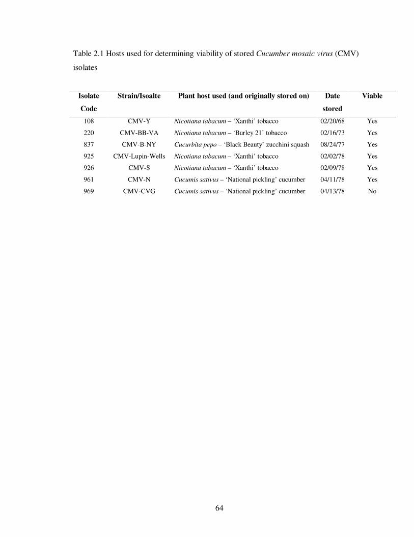
64
Table 2.1 Hosts used for determining viability of stored Cucumber mosaic virus (CMV)
isolates
Isolate
Code
Strain/Isoalte Plant host used (and originally stored on) Date
stored
Viable
108 CMV-Y Nicotiana tabacum – ‘Xanthi’ tobacco 02/20/68 Yes
220 CMV-BB-VA Nicotiana tabacum – ‘Burley 21’ tobacco 02/16/73 Yes
837 CMV-B-NY Cucurbita pepo – ‘Black Beauty’ zucchini squash 08/24/77 Yes
925 CMV-Lupin-Wells Nicotiana tabacum – ‘Xanthi’ tobacco 02/02/78 Yes
926 CMV-S Nicotiana tabacum – ‘Xanthi’ tobacco 02/09/78 Yes
961 CMV-N Cucumis sativus – ‘National pickling’ cucumber 04/11/78 Yes
969 CMV-CVG Cucumis sativus – ‘National pickling’ cucumber 04/13/78 No
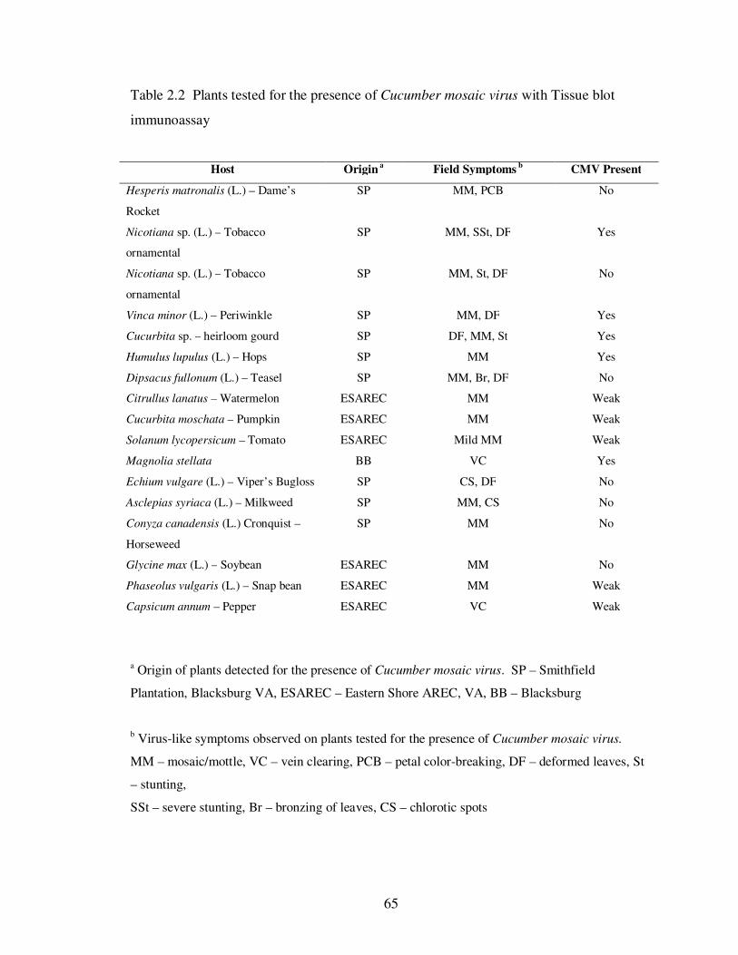
65
Table 2.2 Plants tested for the presence of Cucumber mosaic virus with Tissue blot
immunoassay
Host Origin a Field Symptoms
b CMV Present
Hesperis matronalis (L.) – Dame’s
Rocket
SP MM, PCB No
Nicotiana sp. (L.) – Tobacco
ornamental
SP MM, SSt, DF Yes
Nicotiana sp. (L.) – Tobacco
ornamental
SP MM, St, DF No
Vinca minor (L.) – Periwinkle SP MM, DF Yes
Cucurbita sp. – heirloom gourd SP DF, MM, St Yes
Humulus lupulus (L.) – Hops SP MM Yes
Dipsacus fullonum (L.) – Teasel SP MM, Br, DF No
Citrullus lanatus – Watermelon ESAREC MM Weak
Cucurbita moschata – Pumpkin ESAREC MM Weak
Solanum lycopersicum – Tomato ESAREC Mild MM Weak
Magnolia stellata BB VC Yes
Echium vulgare (L.) – Viper’s Bugloss SP CS, DF No
Asclepias syriaca (L.) – Milkweed SP MM, CS No
Conyza canadensis (L.) Cronquist –
Horseweed
SP MM No
Glycine max (L.) – Soybean ESAREC MM No
Phaseolus vulgaris (L.) – Snap bean ESAREC MM Weak
Capsicum annum – Pepper ESAREC VC Weak
a Origin of plants detected for the presence of Cucumber mosaic virus. SP – Smithfield
Plantation, Blacksburg VA, ESAREC – Eastern Shore AREC, VA, BB – Blacksburg
b Virus-like symptoms observed on plants tested for the presence of Cucumber mosaic virus.
MM – mosaic/mottle, VC – vein clearing, PCB – petal color-breaking, DF – deformed leaves, St
– stunting,
SSt – severe stunting, Br – bronzing of leaves, CS – chlorotic spots
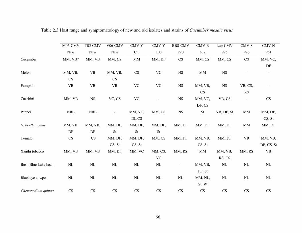
66
Table 2.3 Host range and symptomatology of new and old isolates and strains of Cucumber mosaic virus
M05-CMV
New
T05-CMV
New
V06-CMV
New
CMV-Y
CC
CMV-Y
108
BBS-CMV
220
CMV-B
837
Lup-CMV
925
CMV-S
926
CMV-N
961
Cucumber MM, VB a MM, VB MM, CS MM MM, DF CS MM, CS MM, CS CS MM, VC,
DF
Melon MM, VB,
CS
VB MM, VB,
CS
CS VC NS MM NS - -
Pumpkin VB VB VB VC VC NS MM, VB,
CS
NS VB, CS,
RS
-
Zucchini MM, VB NS VC, CS VC - NS MM, VC,
DF, CS
VB, CS - CS
Pepper NRL NRL - MM, VC,
DL,CS
MM, CS NS St VB, DF, St MM MM, DF,
CS, St
N. benthamiana MM, VB,
DF
MM, VB,
DF
MM, DF,
St
MM, DF,
St
MM, DF,
St
MM, DF MM, DF MM, DF MM MM, DF
Tomato CS CS MM, DF,
CS, St
MM, DF,
CS, St
MM, CS MM, DF MM, VB,
CS, St
MM, DF VB MM, VB,
DF, CS, St
Xanthi tobacco MM, VB MM, VB MM, DF MM, VC MM, CS,
VC
MM, RS MM MM, VB,
RS, CS
MM, RS VB
Bush Blue Lake bean NL NL NL NL NL - MM, VB,
DF, St
NL NL NL
Blackeye cowpea NL NL NL NL NL NL MM, NL,
St, W
NL NL NL
Chenopodium quinoa CS CS CS CS CS CS CS CS CS CS
67
Table 2.3 (continued)
a Symptoms observed on indicator hosts inoculated with different strains and isolates of
Cucumber mosaic virus. MM – mosaic, mottle, W – wilting, (–) – not tested, RS – ringspot
on inoculated leaf, St – stunting, DF – deformed leaves, CS – chlorotic spots, NRL – necrotic
ringspot lesions, VB – vein banding, NL – necrotic lesions, NS – no symptoms, VC – vein
clearing
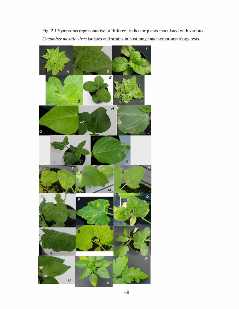
68
Fig. 2.1 Symptoms representative of different indicator plants inoculated with various
Cucumber mosaic virus isolates and strains in host range and symptomatology tests.
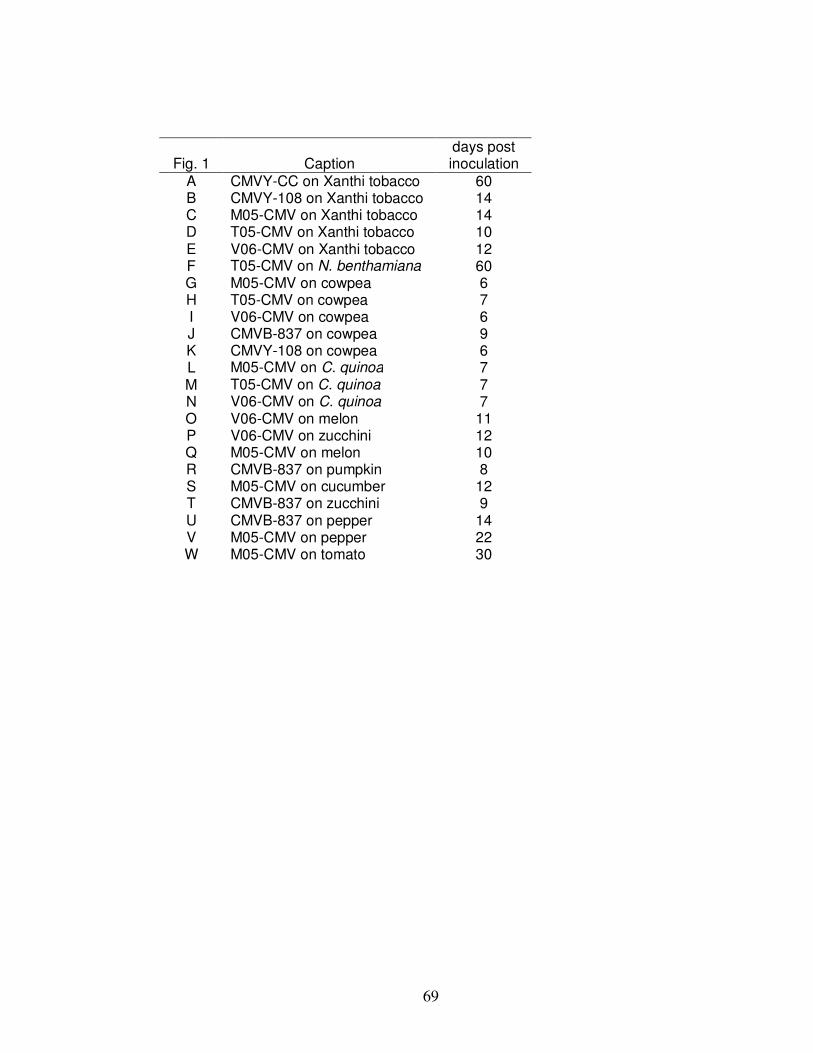
69
Fig. 1 Caption days post inoculation
A CMVY-CC on Xanthi tobacco 60 B CMVY-108 on Xanthi tobacco 14 C M05-CMV on Xanthi tobacco 14 D T05-CMV on Xanthi tobacco 10 E V06-CMV on Xanthi tobacco 12 F T05-CMV on N. benthamiana 60 G M05-CMV on cowpea 6 H T05-CMV on cowpea 7 I V06-CMV on cowpea 6 J CMVB-837 on cowpea 9 K CMVY-108 on cowpea 6 L M05-CMV on C. quinoa 7 M T05-CMV on C. quinoa 7 N V06-CMV on C. quinoa 7 O V06-CMV on melon 11 P V06-CMV on zucchini 12 Q M05-CMV on melon 10 R CMVB-837 on pumpkin 8 S M05-CMV on cucumber 12 T CMVB-837 on zucchini 9 U CMVB-837 on pepper 14 V M05-CMV on pepper 22 W M05-CMV on tomato 30
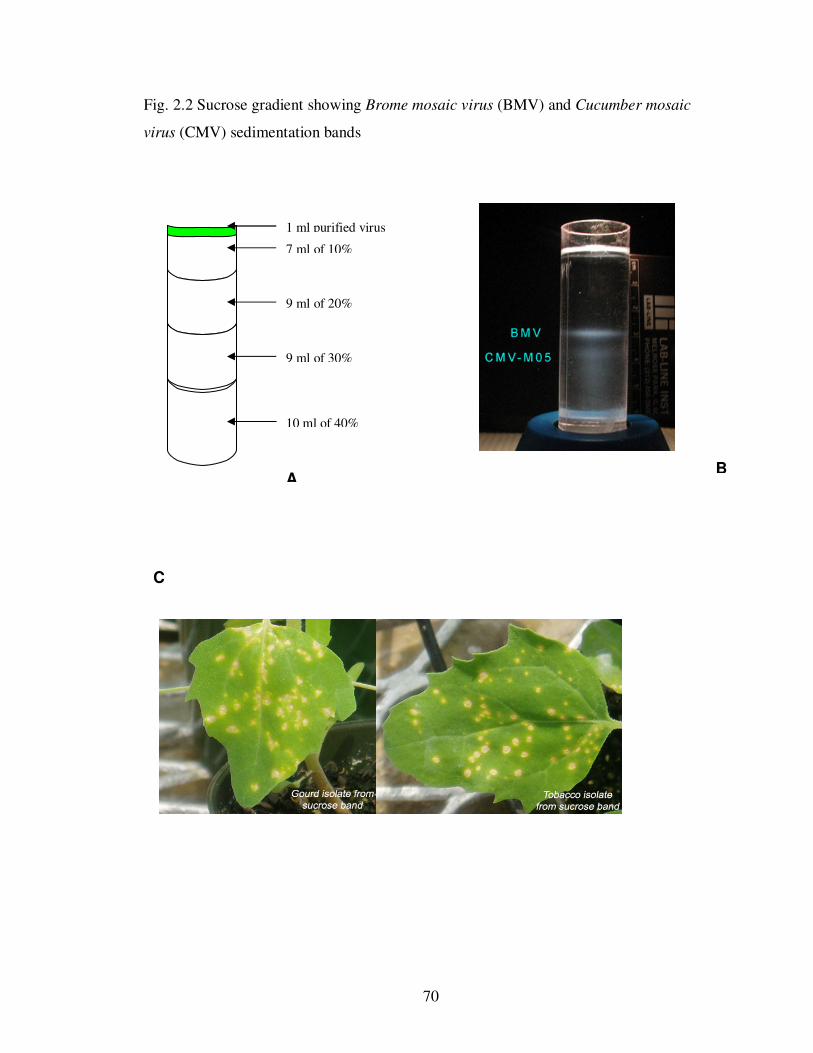
70
Fig. 2.2 Sucrose gradient showing Brome mosaic virus (BMV) and Cucumber mosaic
virus (CMV) sedimentation bands
B
10 ml of 40%
9 ml of 30%
9 ml of 20%
7 ml of 10%
A
1 ml purified virus
C
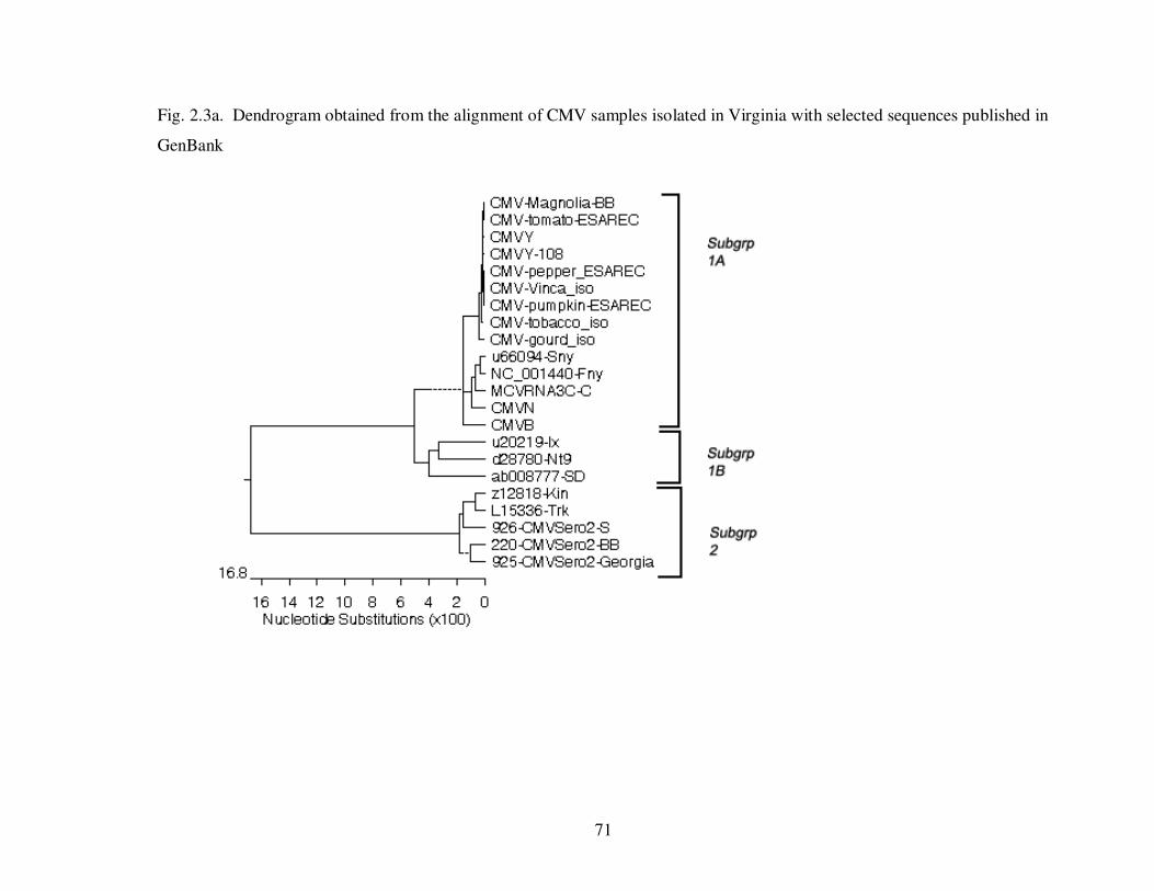
71
Fig. 2.3a. Dendrogram obtained from the alignment of CMV samples isolated in Virginia with selected sequences published in
GenBank
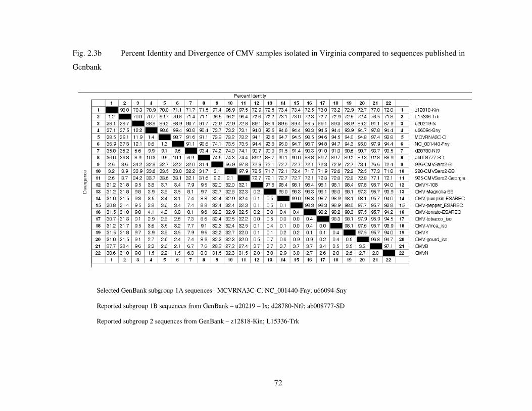
72
Fig. 2.3b Percent Identity and Divergence of CMV samples isolated in Virginia compared to sequences published in
Genbank
Selected GenBank subgroup 1A sequences– MCVRNA3C-C; NC_001440-Fny; u66094-Sny
Reported subgroup 1B sequences from GenBank – u20219 – Ix; d28780-Nt9; ab008777-SD
Reported subgroup 2 sequences from GenBank – z12818-Kin; L15336-Trk
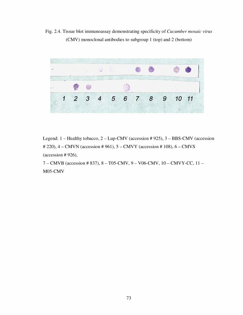
73
Fig. 2.4. Tissue blot immunoassay demonstrating specificity of Cucumber mosaic virus
(CMV) monoclonal antibodies to subgroup 1 (top) and 2 (bottom)
Legend: 1 – Healthy tobacco, 2 – Lup-CMV (accession # 925), 3 – BBS-CMV (accession
# 220), 4 – CMVN (accession # 961), 5 – CMVY (accession # 108), 6 – CMVS
(accession # 926),
7 – CMVB (accession # 837), 8 – T05-CMV, 9 – V06-CMV, 10 – CMVY-CC, 11 –
M05-CMV
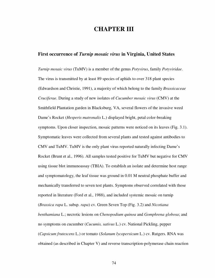
74
CHAPTER III
First occurrence of Turnip mosaic virus in Virginia, United States
Turnip mosaic virus (TuMV) is a member of the genus Potyvirus, family Potyviridae.
The virus is transmitted by at least 89 species of aphids to over 318 plant species
(Edwardson and Christie, 1991), a majority of which belong to the family Brassicaceae
Cruciferae. During a study of new isolates of Cucumber mosaic virus (CMV) at the
Smithfield Plantation garden in Blacksburg, VA, several flowers of the invasive weed
Dame’s Rocket (Hesperis matronalis L.) displayed bright, petal color-breaking
symptoms. Upon closer inspection, mosaic patterns were noticed on its leaves (Fig. 3.1).
Symptomatic leaves were collected from several plants and tested against antibodies to
CMV and TuMV. TuMV is the only plant virus reported naturally infecting Dame’s
Rocket (Brunt et al., 1996). All samples tested positive for TuMV but negative for CMV
using tissue blot immunoassay (TBIA). To establish an isolate and determine host range
and symptomatology, the leaf tissue was ground in 0.01 M neutral phosphate buffer and
mechanically transferred to seven test plants. Symptoms observed correlated with those
reported in literature (Ford et al., 1988), and included systemic mosaic on turnip
(Brassica rapa L. subsp. rapa) cv. Green Seven Top (Fig. 3.2) and Nicotiana
benthamiana L.; necrotic lesions on Chenopodium quinoa and Gomphrena globosa; and
no symptoms on cucumber (Cucumis, sativus L.) cv. National Pickling, pepper
(Capsicum frutescens L.) or tomato (Solanum lycopersicum L.) cv. Rutgers. RNA was
obtained (as described in Chapter V) and reverse transcription-polymerase chain reaction
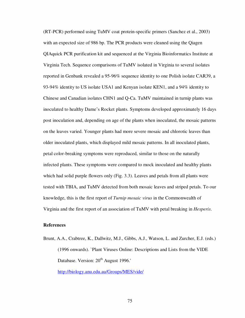
75
(RT-PCR) performed using TuMV coat protein-specific primers (Sanchez et al., 2003)
with an expected size of 986 bp. The PCR products were cleaned using the Qiagen
QIAquick PCR purification kit and sequenced at the Virginia Bioinformatics Institute at
Virginia Tech. Sequence comparisons of TuMV isolated in Virginia to several isolates
reported in Genbank revealed a 95-96% sequence identity to one Polish isolate CAR39, a
93-94% identity to US isolate USA1 and Kenyan isolate KEN1, and a 94% identity to
Chinese and Canadian isolates CHN1 and Q-Ca. TuMV maintained in turnip plants was
inoculated to healthy Dame’s Rocket plants. Symptoms developed approximately 16 days
post inoculation and, depending on age of the plants when inoculated, the mosaic patterns
on the leaves varied. Younger plants had more severe mosaic and chlorotic leaves than
older inoculated plants, which displayed mild mosaic patterns. In all inoculated plants,
petal color-breaking symptoms were reproduced, similar to those on the naturally
infected plants. These symptoms were compared to mock inoculated and healthy plants
which had solid purple flowers only (Fig. 3.3). Leaves and petals from all plants were
tested with TBIA, and TuMV detected from both mosaic leaves and striped petals. To our
knowledge, this is the first report of Turnip mosaic virus in the Commonwealth of
Virginia and the first report of an association of TuMV with petal breaking in Hesperis.
References
Brunt, A.A., Crabtree, K., Dallwitz, M.J., Gibbs, A.J., Watson, L. and Zurcher, E.J. (eds.)
(1996 onwards). `Plant Viruses Online: Descriptions and Lists from the VIDE
Database. Version: 20th August 1996.'
http://biology.anu.edu.au/Groups/MES/vide/
76
Edwardson, J. R. and Christie, R. G. 1991. The Potyvirus Group, Florida Agri. Expt.
Station Monograph 16, Vols I-IV. University of Florida, Gainesville.
Ford, R. E., Beczner, L., and Hamilton, R. I. 1988. Turnip, cucumber and ribgrass mosaic
viruses isolated from Hesperis matronalis in British Columbia. Plant Dis. 72:
101-106
Sanchez, F., Wang, X., Jenner, C. E., Walsh, J. A., and Ponz, F. 2003. Strains of Turnip
mosaic potyvirus as defined by the molecular analysis of the coat protein gene of
the virus. Virus Research 94:33-43.
77
Fig. 3.1. Mosaic (left) and petal color-breaking (right) symptoms on Dame’s Rocket
naturally infected by Turnip mosaic virus.
Fig. 3.2. Systemic mosaic symptoms on a turnip leaf inoculated with Turnip mosaic
virus.
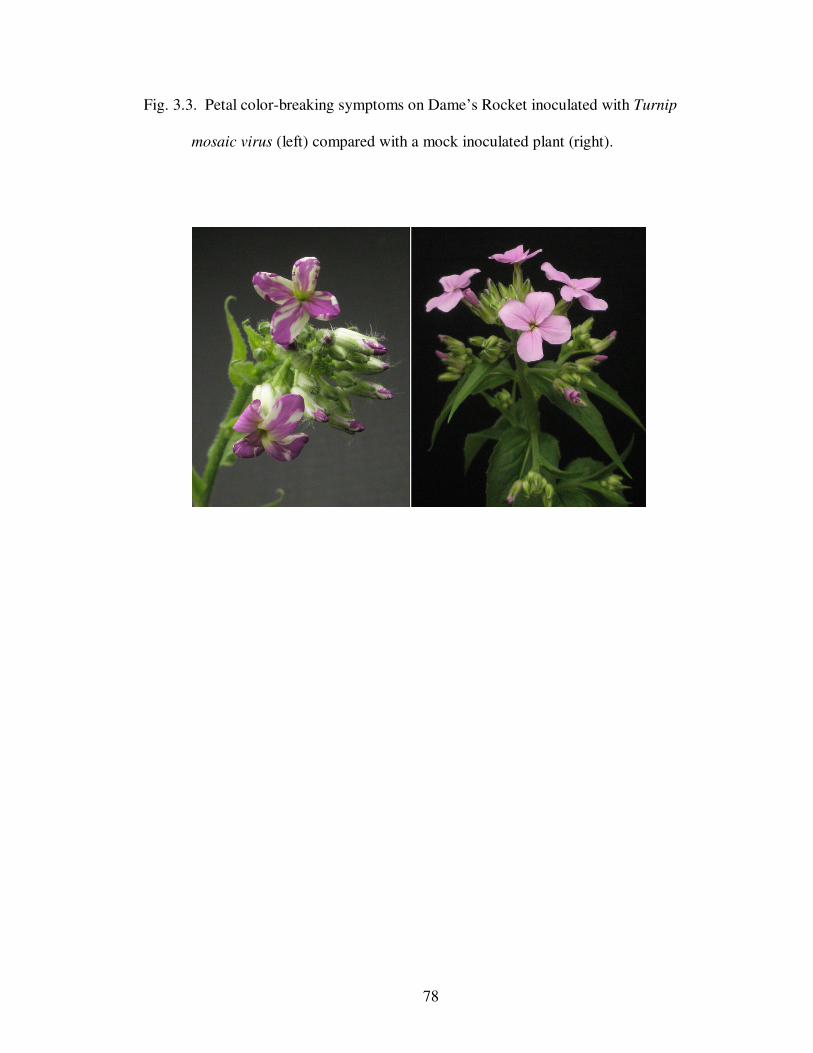
78
Fig. 3.3. Petal color-breaking symptoms on Dame’s Rocket inoculated with Turnip
mosaic virus (left) compared with a mock inoculated plant (right).
79
CHAPTER IV
Evaluation of Virus Diagnostic Capabilities of Collaborators in IPM
CRSP Host Countries
Abstract
Plant virus diagnostics plays an integral part in the development and implementation of
pest management strategies for the control of plant viruses. In this study, the diagnostic
capabilities of several plant pathology collaborators within the Integrated Pest
Management Collaborative Research Support Program (IPM CRSP) host countries and
global theme projects were evaluated by survey on their capacity to perform virus
diagnostics. Several criteria, including viral diagnostic capabilities, personnel,
infrastructure, and funding, were reported through the use of a survey. Of the 14 surveys
collected, 20 of which was disseminated, only ten reported performing virus diagnostics.
Serology and biological assays using indicator hosts were the primary diagnostic
approaches used for virus identification. Very few clinics were capable of performing
molecular assays due in part to funding, availability of specialized equipment, and
technical personnel. Clinics capable of performing virus diagnostics were either privately
owned or academically affiliated. All government affiliated clinics, and one academically
affiliated clinic, did not do molecular diagnostics of plant viruses despite some clinics
having access to PCR machines and imagers. Clients of clinics participating in this study
varied depending on the affiliation of the lab. Privately owned clinics received a majority
of their samples from seed companies and other large corporate bodies. Academically
80
affiliated clinics also dealt with companies but also had samples from researchers and
extension agents. Government affiliated clinics received a majority of their samples from
large and small farmers and extension agents. Of the samples submitted, more than 50%
of the clinics reported virus diseases on three main crops – tomatoes, peppers and
potatoes. Clinics were also asked to rate themselves in diagnostic capabilities. Many
clinics rated average to above average in the areas of infrastructure, funding, equipment,
facilities and personnel knowledge in diagnostics. Only two clinics rated themselves as
poor due to lack of equipment, trained personnel and proper, working infrastructure.
These constraints, just some of many, were shared by several clinics. The information
gathered in this survey can help diagnostic clinics in developing countries to make
international programs, such as the IPM CRSP, aware of their deficiencies and to direct
assistance where possible. This type of targeted effort could greatly increase the
diagnostic capabilities of clinics through capacity-building programs for staff, and
alternative procedures to extensive chemical controls, which can be extremely costly and
environmentally hazardous.
Introduction
Plant viruses are one of the leading causes of plant diseases in the world. They
cause a wide range of symptoms, including mosaics, mottling, stunting, and most
importantly, reduction in crop yield. The development of integrated pest management
strategies for agricultural pests has become increasingly important globally due to the
current food shortage, and the increased dangers to health and the environment with the
constant use of pesticides to control these pests. The IPM CRSP is a program funded by
the U.S. Agency for International Development (USAID) to address the health, economic
81
and environmental issues through IPM interventions. The current phase of the IPM CRSP
has specific thematic programs on plant disease diagnosis and on the assessment and
management of viruses.
The most important step in developing IPM procedures for plant viruses is the
proper identification of these viruses, and determining their distribution and alternate
hosts located adjacent to crops of interest. Plant clinics and diagnostic laboratories (both
terms will be referred to interchangeably within this chapter) play a crucial role in the
identification and detection of plant viruses. Numerous techniques are currently in use for
the detection of plant viruses, including serology with enzyme-linked immunosorbent
assay (ELISA) (Clark and Adams, 1977), tissue blot immunoassay (TBIA) (Lin et al.,
1990) and lateral flow devices, such as Immunostrips (Tsuda et. al., 1992), and nucleic
acid-based procedures, including the polymerase chain reaction (PCR) (Saiki et. al.,
1988), hybridization (Nikolaeva, 1995) and molecular arrays. In countries where the
economy is primarily dependent upon agricultural production both for local consumption
and export, the need for fully functional plant clinics and capable diagnosticians is of
paramount importance (Lawrence et. al., 2005). As part of a study on the educational
value and diagnostic capabilities of plant clinics in developing countries, Ausher et al.
(1996) conducted a survey to evaluate plant clinics within developing countries. They
reported that the capabilities of plant clinics varied depending upon several factors,
including laboratory affiliations to international companies and institutions. The affiliated
clinics had access to both moderately expensive and expensive equipment and, due to this
association, had access to specialized personnel, several of whom also worked within the
clinics. The highest educational levels of personnel working in these clinics, namely
82
doctorate and masters level, were uniformly represented within developing countries.
Personnel had access to consistent ‘in-house training’ for increased capacity building and
attendance at workshops and technical meetings on a regular basis. Despite several
constraints including funding and inadequate facilities, Ausher et al. stated that many
developed countries had shown marked improvement in diagnostic capacity and the
acquisition of trained and qualified staff compared to that reported by Black (1993) and
Black and Sweetmore (1995).
In this study the virus diagnostic capabilities of plant clinic collaborators within
the IPM-CRSP project were evaluated based on several criteria, including access to
standard and specialized equipment, personnel, virus and general diagnostics, and
funding. The evaluation was performed by the dissemination of surveys to developing
countries within the Caribbean, Latin America and Africa.
Methodology
Training
Before dissemination of the survey, a mandatory, on-line ethics course on
“Relating to Human Subjects and Ethics” was taken by all developers of the survey. The
ethics course was provided by the Virginia Tech Office of Research and Compliance
(ORC) Institutional Review Board (IRB), http://www.irb.vt.edu/ . The survey and the
protocol were submitted to the chairman of IRB for review.
Survey
Information about the diagnostic capabilities of collaborating countries within the
IPM-CRSP was obtained through a survey with an accompanying letter attached. The
83
information was subdivided into categories for easier comparisons between the
developing host countries. The categories were as follows:
i) Virus diagnostic capabilities: methods and approaches employed in the identification
and detection of plant viruses. This section also included the use of diagnostic kits for
plant viruses.
ii) Clients: researchers, home owners and other interested parties who use the diagnostic
facilities on a regular basis for virus diagnostics.
iii) Crop submission: the types of crops frequently submitted to plant clinics for plant
disease diagnosis, and the number of suspected virus-infected samples.
iv) Self evaluation of respective clinics
v) Personnel and capacity building: the number of trained and/or qualified personnel in
virus diagnostics.
vi) Funding and purchasing: the ability of the clinic to generate adequate funding for
equipment and reagent purchase, maintenance, and payment of staff.
vii) Overall constraints with performing viral and general diagnostics.
A copy of the survey is exhibited in Appendix D
Regions within developing countries were separated into 3 subgroups: The
Caribbean, Latin America, and Africa. Areas within the survey that were not answered
were considered either void or taken as a zero rating.
84
Results
Training
Upon passing the examination, a Certificate of Completion was awarded. A copy of the
approval letter, granting exemption from further IRB review, is exhibited in Appendix E.
Survey
Of the twenty surveys disseminated, fourteen were returned and included
representative clinics from each of the three regions: Latin America, the Caribbean, and
Africa. The clinics were grouped and reported accordingly: A – F were clinics within
East Africa, G – I were clinics within the Caribbean, and J – N were clinics within Latin
America, specifically Central America. The clinics were laboratories with government or
academic affiliations, or were privately owned by agricultural companies.
Virus diagnostic capabilities
Several methods can be applied for the identification and detection of plant
viruses. These methods were categorized into four main groups: physical properties of
virus particles, biological properties such as symptomatology on indicator host plants,
serological approaches, and nucleic acid-based approaches. Fig 4.1 represents the total
number of methods and/or approaches used in plant virus diagnostics by clinics within
each region.
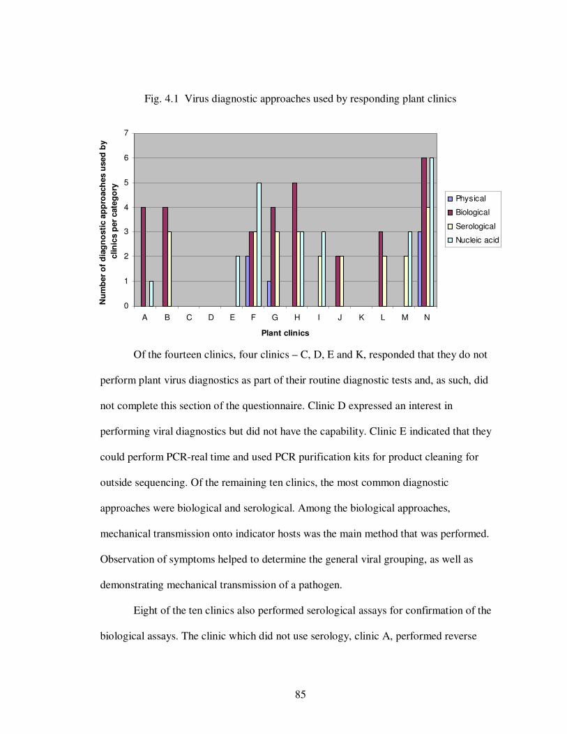
85
0
1
2
3
4
5
6
7
A B C D E F G H I J K L M N
Plant clinics
Nu
mb
er
of
dia
gn
osti
c a
pp
roach
es u
sed
by
clin
ics p
er
cate
go
ry
Physical
Biological
Serological
Nucleic acid
Fig. 4.1 Virus diagnostic approaches used by responding plant clinics
Of the fourteen clinics, four clinics – C, D, E and K, responded that they do not
perform plant virus diagnostics as part of their routine diagnostic tests and, as such, did
not complete this section of the questionnaire. Clinic D expressed an interest in
performing viral diagnostics but did not have the capability. Clinic E indicated that they
could perform PCR-real time and used PCR purification kits for product cleaning for
outside sequencing. Of the remaining ten clinics, the most common diagnostic
approaches were biological and serological. Among the biological approaches,
mechanical transmission onto indicator hosts was the main method that was performed.
Observation of symptoms helped to determine the general viral grouping, as well as
demonstrating mechanical transmission of a pathogen.
Eight of the ten clinics also performed serological assays for confirmation of the
biological assays. The clinic which did not use serology, clinic A, performed reverse
86
transcription-polymerase chain reaction (RT-PCR) as the confirmatory test. The primary
serological test performed by developing countries was the enzyme-linked
immunosorbent assay (ELISA), reagents for which were purchased as kits. Four clinics
also used Immunostrips® purchased from Agdia®, and another four clinics used tissue
blot immunoassay. Only clinic A used RT-PCR for virus diagnosis. Clinic I used only
serological and molecular assays.
The capability to perform nucleic acid-based tests was variable across the
developing countries, and was primarily confined to end point RT-PCR, gel
electrophoresis, and observation of amplicons using a transilluminator and imager. Five
of the clinics were capable of extracting nucleic acid, and those clinics (F, H, I, M and N)
had fully functional laboratories with academic affiliations or were privately owned. The
clinics that reported no nucleic acid extraction procedures were, with one exception,
government affiliated.
Clients and crop submission
Users of the plant clinic services were divided into five categories: farmers,
companies, researchers and extension officers (R/E), homeowners, and other, which
covered consultants and anyone else that did not fit the profiles of the previous
categories. The percentage of samples from each category that was received by each plant
clinic is shown in Fig. 4.2. These percentages constitute the total number of diseased
plants received by each clinic. The percentage of diseased plants diagnosed as viral-
infected ranged from as low as 2% for clinic D, which had minimal to no equipment for
virus diagnostics, to as high as 80% for clinic F, which was a fully functional diagnostic
clinic. Of the fourteen clinics, only two, E and K, did not complete the ‘Client and Crop
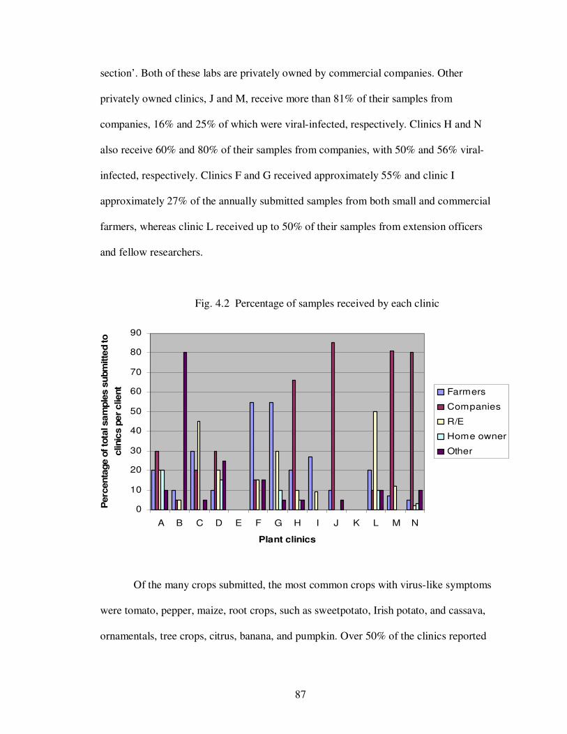
87
0
10
20
30
40
50
60
70
80
90
A B C D E F G H I J K L M N
Plant clinics
Perc
enta
ge o
f to
tal sam
ple
s s
ubm
itte
d to
clinic
s p
er
client
Farmers
Companies
R/E
Home owner
Other
section’. Both of these labs are privately owned by commercial companies. Other
privately owned clinics, J and M, receive more than 81% of their samples from
companies, 16% and 25% of which were viral-infected, respectively. Clinics H and N
also receive 60% and 80% of their samples from companies, with 50% and 56% viral-
infected, respectively. Clinics F and G received approximately 55% and clinic I
approximately 27% of the annually submitted samples from both small and commercial
farmers, whereas clinic L received up to 50% of their samples from extension officers
and fellow researchers.
Fig. 4.2 Percentage of samples received by each clinic
Of the many crops submitted, the most common crops with virus-like symptoms
were tomato, pepper, maize, root crops, such as sweetpotato, Irish potato, and cassava,
ornamentals, tree crops, citrus, banana, and pumpkin. Over 50% of the clinics reported
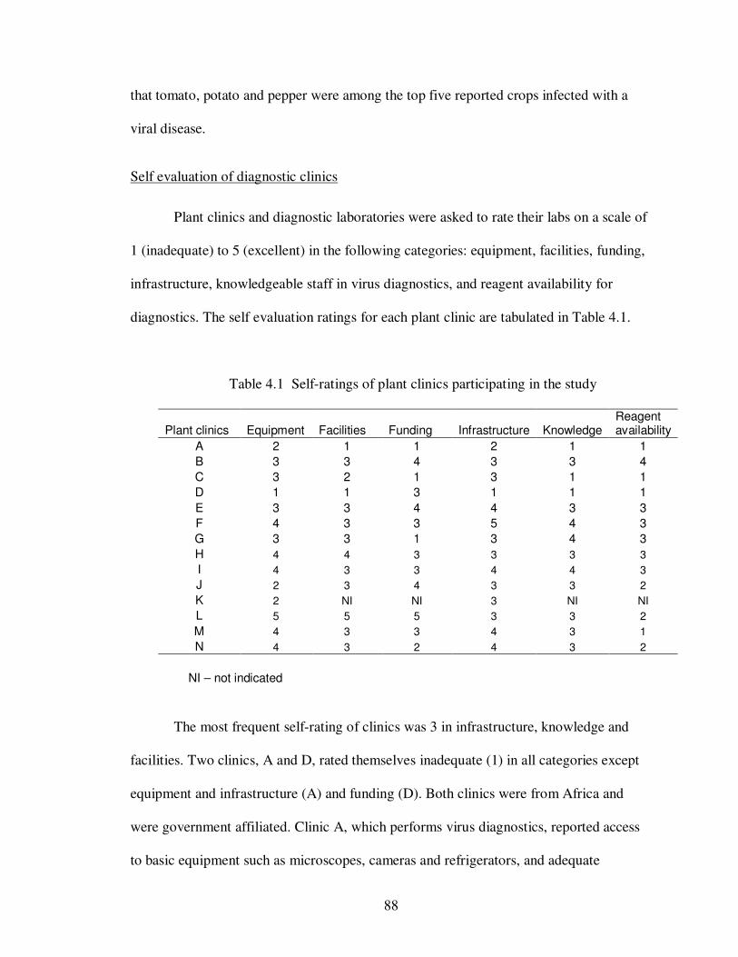
88
that tomato, potato and pepper were among the top five reported crops infected with a
viral disease.
Self evaluation of diagnostic clinics
Plant clinics and diagnostic laboratories were asked to rate their labs on a scale of
1 (inadequate) to 5 (excellent) in the following categories: equipment, facilities, funding,
infrastructure, knowledgeable staff in virus diagnostics, and reagent availability for
diagnostics. The self evaluation ratings for each plant clinic are tabulated in Table 4.1.
Table 4.1 Self-ratings of plant clinics participating in the study
Plant clinics Equipment Facilities Funding Infrastructure Knowledge Reagent availability
A 2 1 1 2 1 1
B 3 3 4 3 3 4
C 3 2 1 3 1 1
D 1 1 3 1 1 1
E 3 3 4 4 3 3
F 4 3 3 5 4 3
G 3 3 1 3 4 3
H 4 4 3 3 3 3
I 4 3 3 4 4 3
J 2 3 4 3 3 2
K 2 NI NI 3 NI NI
L 5 5 5 3 3 2
M 4 3 3 4 3 1
N 4 3 2 4 3 2
NI – not indicated
The most frequent self-rating of clinics was 3 in infrastructure, knowledge and
facilities. Two clinics, A and D, rated themselves inadequate (1) in all categories except
equipment and infrastructure (A) and funding (D). Both clinics were from Africa and
were government affiliated. Clinic A, which performs virus diagnostics, reported access
to basic equipment such as microscopes, cameras and refrigerators, and adequate
89
greenhouse and land space. Clinic D, which does not do virus diagnostics, had no
equipment and severely limited reference material. Research land space was the only
asset in large supply. The remaining clinics were adequately outfitted with basic,
moderately expensive and expensive equipment, regardless of whether they performed
virus diagnosis as part of their diagnostics protocol. Equipment included different
microscopes, pH meters, centrifuges, vortex/mixers, magnetic stirrers/hotplate, cameras
and electrophoresis units with power supplies. Many respondents had adequate research
land space, although less than 50% had fully functioning greenhouses. All clinics, with
the exceptions of A and D, reported access to reference material, including computer
access with internet capabilities.
Personnel and capacity building
Education levels and training of staff within the participating laboratories are
summarized in Table 4.2. There appeared to be very little variation in the distribution of
qualified staff among the different countries and regions. All clinics except D had at least
one member of staff with a second degree, whether it was at the Master’s or Doctorate
level. Clinic D’s highest levels were technicians, although there were a reported four
certificates among the technicians.
The total number of years of experience varied both among diagnostics labs
within the same region, and between different regions. In Africa, the total years in virus
diagnostics ranged from as high as 12 years (clinic B), to no experience (clinic C).
Region wide, clinics H, M and N had 28-35 years of experience, and clinic K had the
greatest combined experience of 60 years. Only clinic D, which expressed a desire to do
viral diagnostics, had staff with at least 1 year of experience in virus diagnostics.
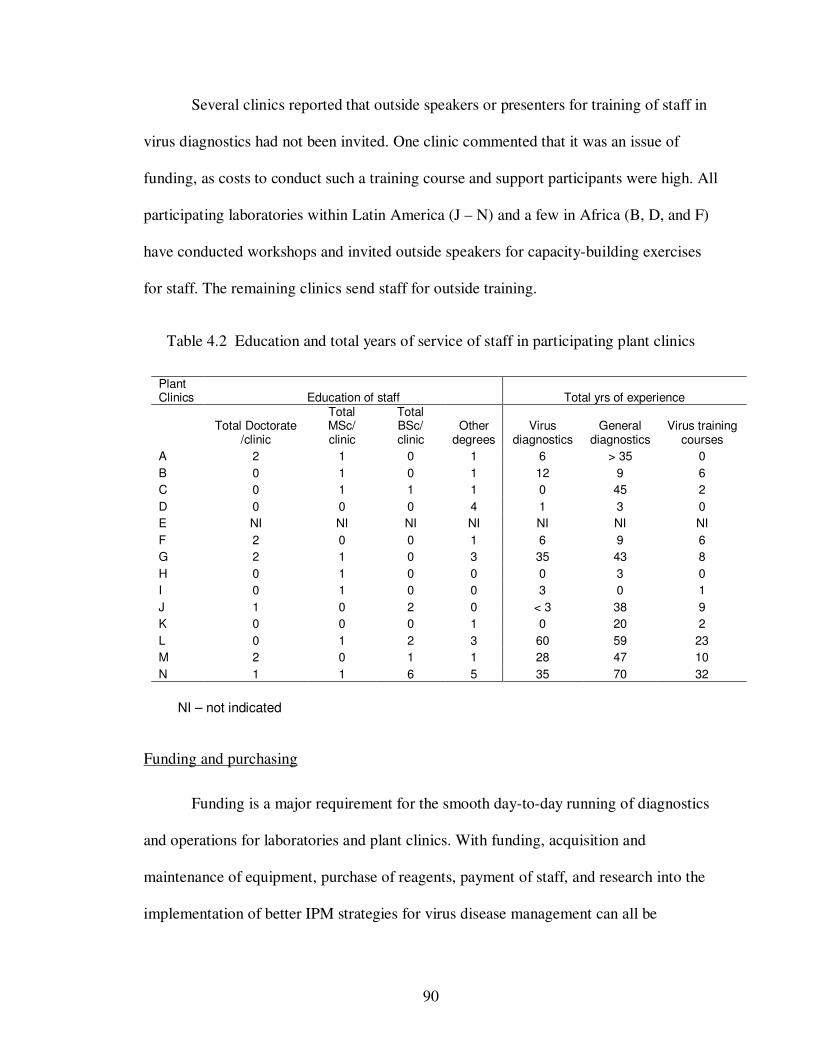
90
Several clinics reported that outside speakers or presenters for training of staff in
virus diagnostics had not been invited. One clinic commented that it was an issue of
funding, as costs to conduct such a training course and support participants were high. All
participating laboratories within Latin America (J – N) and a few in Africa (B, D, and F)
have conducted workshops and invited outside speakers for capacity-building exercises
for staff. The remaining clinics send staff for outside training.
Table 4.2 Education and total years of service of staff in participating plant clinics
Plant Clinics Education of staff Total yrs of experience
Total Doctorate
/clinic
Total MSc/ clinic
Total BSc/ clinic
Other degrees
Virus diagnostics
General diagnostics
Virus training courses
A 2 1 0 1 6 > 35 0
B 0 1 0 1 12 9 6
C 0 1 1 1 0 45 2
D 0 0 0 4 1 3 0
E NI NI NI NI NI NI NI
F 2 0 0 1 6 9 6
G 2 1 0 3 35 43 8
H 0 1 0 0 0 3 0
I 0 1 0 0 3 0 1
J 1 0 2 0 < 3 38 9
K 0 0 0 1 0 20 2
L 0 1 2 3 60 59 23
M 2 0 1 1 28 47 10
N 1 1 6 5 35 70 32
NI – not indicated
Funding and purchasing
Funding is a major requirement for the smooth day-to-day running of diagnostics
and operations for laboratories and plant clinics. With funding, acquisition and
maintenance of equipment, purchase of reagents, payment of staff, and research into the
implementation of better IPM strategies for virus disease management can all be
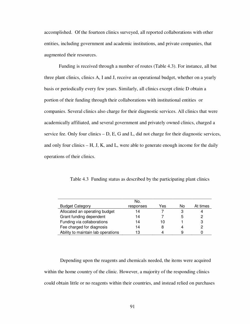
91
accomplished. Of the fourteen clinics surveyed, all reported collaborations with other
entities, including government and academic institutions, and private companies, that
augmented their resources.
Funding is received through a number of routes (Table 4.3). For instance, all but
three plant clinics, clinics A, I and J, receive an operational budget, whether on a yearly
basis or periodically every few years. Similarly, all clinics except clinic D obtain a
portion of their funding through their collaborations with institutional entities or
companies. Several clinics also charge for their diagnostic services. All clinics that were
academically affiliated, and several government and privately owned clinics, charged a
service fee. Only four clinics – D, E, G and L, did not charge for their diagnostic services,
and only four clinics – H, J, K, and L, were able to generate enough income for the daily
operations of their clinics.
Table 4.3 Funding status as described by the participating plant clinics
Budget Category No.
responses Yes No At times
Allocated an operating budget 14 7 3 4
Grant funding dependent 14 7 5 2
Funding via collaborations 14 10 1 3
Fee charged for diagnosis 14 8 4 2
Ability to maintain lab operations 13 4 9 0
Depending upon the reagents and chemicals needed, the items were acquired
within the home country of the clinic. However, a majority of the responding clinics
could obtain little or no reagents within their countries, and instead relied on purchases
92
from other countries, primarily the United States. Some clinics reported difficulties in
purchasing equipment and reagents due to high costs and to importation fees. Clinics
responding from the Latin America and Caribbean regions required intermediary
companies for the purchase of reagents and chemicals from other countries. These
intermediary companies also dealt with shipping and handling of these reagents between
companies and clinics, usually at a high cost.
Constraints
One of the major constraints reported by many plant clinics was the lack of
trained virologists and the lack of knowledge of the newer techniques for identifying and
detecting plant viruses. Other constraints reported by clinics included: time required for
importation of equipment and reagents purchased internationally, the high cost of
diagnostic kits, reagents, equipment, the requirement for importation fees and taxes
associated with international orders, and the cost of maintaining infrastructure. Other
constraints reported were the lack of available literature on new viruses and new
diagnostic procedures now being used, and the inability to attend useful diagnostic
workshops.
Discussion
Plant viruses cause some of the world’s leading plant diseases, resulting in
billions of dollars lost annually, yet viruses are one of the most difficult-to-detect plant
pathogens. Implementation of proper pest management strategies to control virus diseases
is primarily dependent upon accurate identification of plant viruses. Plant clinics play a
vital role in the detection and identification of plant viruses using a number of diagnostic
93
tools currently available. These tools can be categorized into four main groups:
identification of plant viruses using physical properties, biological characterization using
symptomatology on indicator hosts, serological detection using specific antigen-antibody
recognition, and molecular characterization using virus nucleic acids.
Ausher et al. (1996) conducted a survey among developing countries to evaluate
their diagnostic capabilities. The survey showed that, despite constraints experienced by
these clinics, which included funding, infrastructure and specialization of personnel
devoted purely to diagnostics, several clinics had made vast improvements in plant
diagnostics since the report of Black and Sweetmore (1995). Today, with an increase in
the use of molecular techniques in viral diagnostics, it was important to determine how
developing countries performed. Unlike Ausher et al. (1996), our study concentrates
primarily on the capabilities of several plant clinics within the IPM CRSP to perform
viral diagnostics, and included equipment, personnel, and other items required for proper
diagnosis. Of the twenty surveys disseminated, fourteen clinics responded, four of which
did no virus diagnosis. One of these four clinics, clinic D was unable to perform virus
diagnosis because of funding constraints. They reported that the government procurement
team for the clinic had deemed that there was no urgency for virus diagnostics and thus
had not allocated funds to increase the diagnostic capabilities of the clinic.
In our survey, the most common diagnostic approaches used were biological and
serological assays (Fig. 4.1). Several clinics used combinations of both of these assays for
the detection and identification of the plants viruses. Of the ten clinics, five clinics
reported the capability to perform molecular analysis. These five clinics were, with one
exception, either privately owned or academically affiliated. The remaining clinics, all
94
government affiliated, relied on serological techniques including ELISA, TBIA, and
Immunostrips®.
For virus detection and identification, specialized equipment is required to
perform several assays, particularly with molecular analyses. Clinics that were privately
owned or academically affiliated had well equipped laboratories, including expensive
equipment such as electron microscopes, centrifuges, and PCR machines, and adequate
facilities and infrastructure. This was in comparison to government affiliated clinics
where only three out of the six had well equipped labs. The remaining three clinics had
non-functioning greenhouses, poor infrastructure which affected the procurement of
particular equipment, and little to no equipment available for basic diagnostics.
Clinic affiliation also affected the clients that use their services. Privately owned
clinics processed samples primarily from seed companies and other corporations.
Academically affiliated labs processed samples primarily from companies, researchers
and some extension officers and government affiliated clinics worked almost exclusively
with farmers and extension officers. The number of virus infected samples varied per
year, but over 50% of the responding clinics stated that tomato, pepper and potato were
the most submitted samples containing viruses.
Clinics were asked to self-rate their diagnostic laboratories in each of five
categories (Table 4.1). The most frequent self ratings among the clinics was average (3)
throughout most of the categories. Two clinics A and D, rated themselves inadequate (1)
in almost every category, reporting several issues with the availability of equipment,
reagents, trained personnel, reference material including computers, and funding for kits
and other diagnostic tools required for virus detection. Again clinics privately owned or
95
academically affiliated rated themselves above average (4) to excellent (5) in several
categories. Clinic L, although rating itself excellent in equipment, facilities and funding,
fell short within infrastructure and knowledge. In examining the listing of available
equipment, thermocyclers and imagers necessary for amplification of DNA, were not
present. This was reflected in clinic L’s ability to perform molecular diagnostics,
resorting instead to biological and serological techniques for the identification and
detection of plant viruses (Fig. 4.1).
Several clinics reported the lack of trained personnel in both virus identification,
modern detection and molecular techniques. While there appeared to be very little
variation in the distribution of qualified staff in general diagnostics (Table 4.2), for virus
diagnostics there appeared to be a higher concentration of knowledgeable personnel
within the Latin American region. Three of the five clinics in Latin America (clinics L –
N) had at least 4 members of staff with upwards of 60 years of experience in virus
diagnostics. This was in sharp contrast to clinics within the African region where staff
was at a minimum of 2 years with at most 12 years of experience. Within the Caribbean,
clinic G had up to six staff members with a total of 35 years of experience combined.
These results can be explained by the amount of training and capacity building within
these countries. Clinics L – N host several ‘in-house’ training and capacity building
workshops on a yearly basis. Other clinics have also reported training for staff, but the
number of years of virus experience could suggest that these workshops have been more
geared towards general diagnostics. Among the clinics that do not host workshops, clinic
G has the most experience in virus diagnostics. Several members of staff were actually
trained as plant virologists.
96
From our survey, it seemed that government clinics faired the worst when it came
to molecular diagnostics. Virus diagnostics was restricted to biological and serological
techniques as many of these clinics either lacked equipment or the infrastructure to
support such equipment. It is clinics like these that work directly with the farming
community for which the IPM CRSP can make a large contribution to the economy and
the nation of that country as a whole. Assistance in capacity building through training
workshops for staff, and development of IPM strategies designed to increase the
productivity of our farmers while reducing the cost of pesticides for the treatment and/or
control of plant diseases.
Although the equipment or infrastructure is not present, these clinics can also
make a contribution to the molecular detection and diversity of viruses through the use of
simpler methods for virus identification and characterization. In the following chapters
we present an alternative use of nitrocellulose membranes, the solid matrix used in TBIA.
We focus on using this membrane to not only detect plant viruses with specific
antibodies, but to also use this membrane as a source for viral RNA for molecular studies.
Membranes would allow for the easy transport of samples to different countries for
molecular analysis without any special storage conditions and without the possibility of
introducing viruses into new locations. The ability to obtain sequence data for further
studies would contribute significantly, not just to the scientific community, but by
increasing the exposure of this laboratory through publications, attracting international
institutions for collaborations and possibly source funding for a more self-sufficient
clinic.
97
References
Ausher, R., Ben-Ze'ev, I. S., and Black, R. 1996. The role of plant clinics in plant disease
diagnosis and education in developing countries. Annu. Rev. Phytopathol. 34:51-
66.
Black, R., and Sweetmore, A. 1995. Appropriate diagnostic systems for less developed
countries. EPPO Bulletin 25:383-391.
Blackman, R. L., and Eastop, V. F. 2000. Aphids on the World's crops. John Wiley &
Sons Ltd., 2nd ed.
Clark, M. F., and Adams, A. N. 1977. Characteristics of the microplate method of
enzyme-linked immunosorbent assay for the detection of plant viruses. J Gen
Virol 34 (3):475-483.
Integrated Pest Management Collaborative Research Support Program (IPM CRSP).
[Online] Accessed 5/14/09.
http://www.oired.vt.edu/ipmcrsp/IPM_2008/draft_home.htm
Lawrence, J., Tolin, S. A., Edwards, C., Fleischer, S. J., Jackson, D. M., Clarke-Harris,
D., McDonald, S. A., Dalip, K., and Chung, P. 2005. Developing IPM packages
in the Caribbean. Pages 95-120 in: Globalizing Integrated Pest Management: A
Participatory Research Process, G. W. Norton, E. A. Heinrichs, G. C. Luther and
M. E. Irwin, eds. Blackwell Publishing, Ames, Iowa.
Lin, N. S., Hsu, Y. H., and Hsu, H. T. 1990. Immunological detection of plant viruses and
a mycoplasma-like organism by direct tissue blotting on nitrocellulose
membranes. Phytopath. 80 (9):824-828.
Lot, H., Marrou, J., Quiot, J. B., and Esvan, C. 1972. Contribution a l'etude de virus de la
mosaique du concombre (CMV). II. Methode de purification rapide du virus.
Annu. Phytopathol. 4:25-38.
Nikolaeva, O. V. 1995. Nucleic acid hybridization methods in diagnosis of plant viruses
and viriods. Pages 133-144 in: Molecular methods in plant pathology, R. P. Singh
and U. S. Singh, eds. CRC Press Inc.
98
Saiki, R. K., Gelfand, D. H., Stoffel, S., Scharf, S., Higuchi, R., Horn, G. T., Mullis, K.
B., and Erlich, H. A. 1988. Primer-directed enzymatic amplification of DNA with
a thermostable DNA polymerase. Science 239:487-491.
Thresh, J. M. 2006. Crop viruses and virus diseases: A global prospective. Pages 9-32 in:
Virus Diseases and Crop Biosecurity, J. I. Cooper, T. Kuhne and V. P. Polischuk,
eds. Springer.
Tsuda, S., Kameya-Iwaki, M., Hanada, K., and Tomaru, K. 1992. A novel detection and
identification technique for plant viruses: Rapid immunofilter paper assay (RIPA).
Plant Dis. 76:466-469.
99
CHAPTER V
Immunoassay, RT-PCR and direct sequencing of Cucumber mosaic
virus and potyvirus coat proteins from the same NitroPure
nitrocellulose membrane
Abstract
NitroPure nitrocellulose (NPN) membranes are an effective solid matrix for the
detection and identification of plant viruses by tissue blot immunosorbent assay (TBIA).
This study reports an additional use of nitrocellulose membranes as a source of viral
RNA for three potyviruses, Tobacco etch virus (TEV), Soybean mosaic virus (SMV), and
Turnip mosaic virus (TuMV), and two cucumoviruses, Cucumber mosaic virus (CMV)
and Peanut stunt virus (PSV). Discs from dried membranes blotted with symptomatic
tissue were minimally cleaned and placed directly into RT reactions with oligo-dTs or
reverse primers to initiate cDNA synthesis. An aliquot used directly in PCR resulted in
amplicons of the expected sizes for each virus only with its specific primers. The
intensity of the PCR-amplified bands from cDNA transcribed from either non-processed
and TBIA-processed nitrocellulose membranes compared favorably with those from
FTA® Plant Cards. The quantity of amplification product, estimated from band intensity
on gels, decreased over time in membranes stored for several months, but could be
increased by increasing the number of PCR cycles. The PCR products were of high
quality and could be sequenced directly. Thus, nitrocellulose membranes can be used to
100
collect and store samples that retain immunologically active virus particles, as well as
viral RNA that can be amplified by RT-PCR, for more than a year at room temperature.
Introduction
Serological techniques, including enzyme-linked immunosorbent assay (ELISA)
(Clark and Adams, 1977), lateral flow devices (Tsuda et. al., 1992), and tissue blot
immunosorbent assay (TBIA) (Lin et al., 1990) are powerful tools for the detection and
diagnosis of plant viruses. These techniques are based on an antigen-antibody binding
reactions between epitopes on virus coat proteins and the binding sites of specific anti-
virus antibodies (van Regenmortel, 1982). Although several variations of ELISA are
used for plant viruses, all are performed in microtiter plates and require extraction of
virus from fresh tissue and several hours to complete. Immunostrips® (Agdia, Inc.) are
lateral flow devices that are quick and simple to use, but are costly when testing a large
number of samples. In contrast, TBIA requires no tissue extraction, as plant leaves are
pressed or blotted directly onto a nitrocellulose membrane without mechanical disruption
of tissue. The method was first described for detection of several plant viruses by Lin et.
al. (1990), and has since become more widely used as a sensitive and reliable method for
plant virus detection (Comstock and Miller, 2004; Hsu and Lawson, 1991; Jonson et. al.,
2007; Langham et al., 2007; McDonald et al., 2004). TBIA is less costly than
immunostrips for high numbers of samples and can be completed in less time than
ELISA. A distinct advantage of TBIA is that leaves can be blotted onto membranes
directly in the field and processed at a later date (Makkouk and Comeau, 1994).
Nucleic acid-based approaches are also used extensively for detection and
identification of plant viruses, particularly since the advent of the polymerase chain
101
reaction (PCR) (Saiki et. al. 1988). Plant viruses with DNA genomes can be amplified
directly using generic or gene-specific primers for the region of amplification. Reverse
transcription (RT) of plant viral RNA genomes to a complementary DNA (cDNA)
template and amplification by cloning has been done since the early 1980’s (Owens and
Cress, 1980; Goulet, et al. 1982). Until recently, the source of viral RNA for cDNA
synthesis and RT-PCR was from purified virus particles or total RNA. Burgoyne (1996)
patented the use of FTA® Cards for the collection and storage of DNA to be used either
directly or indirectly in PCR. The cards were then modified and distributed by Whatman®
Inc. (Whatman, 2006) to produce FTA® Plant Cards. The matrix is a cotton-based
cellulose fiber membrane on a supported backing, to which infected plant tissue is
blotted. As claimed in US Patent No. 6645717 (Smith et. al. 2003), “the fibers are
conditioned with chaotrophic and other agents which lyse cells, and release and
immobilize the genetic material while inhibiting their degradation”. These plant cards,
and the methodology utilized with these cards, have proven useful for plant viruses
(Ndunguru et. al., 2005; Roy and Nassuth, 2005) and plant gene expressions (Roy and
Nassuth, 2005).
Because both FTA® Plant Cards and NitroPure nitrocellulose (NPN) membranes
are solid matrices, and TBIA is used extensively in our laboratory, it was hypothesized
that NPN could be used as a source of plant viral RNA for RT-PCR. Three potyviruses,
Tobacco etch virus (TEV), Turnip mosaic virus (TuMV), and Soybean mosaic virus
(SMV) (Genus Potyvirus, Family Potyviridae) and two cucumoviruses Cucumber
mosaic virus (CMV) and Peanut stunt virus (PSV) (Genus Cucumovirus, Family
Bromoviridae) were used to test this hypothesis. These genera are often found in mixed
102
infections in several plants including cucurbits (Pinto et. al., 2008; Wang et. al., 2002)
and ornamentals (Arneodo et. al., 2005). Both genera are economically important and
widespread. Even though all five viruses are spread by aphids, their host ranges vary
from very narrow, with SMV affecting only soybeans, to very wide, with CMV affecting
more than 1200 plant species (Palukaitis and Garcia-Arenal, 2003).
The objective of this study was to establish the versatility of NPN not only for
binding proteins of plant viruses in TBIA, but also as a source of viral RNA for RT-PCR.
The protocol required no mechanical tissue maceration, no elution of virus particles or
RNA from the membranes, and yielded good sequence information for virus
identification. Sequences obtained from PCR products enabled accurate separation of the
five viruses into the expected taxa. NPN membranes were comparable to FTA® Plant
Cards in efficiency and archival storage, and also provided a confirmation of
immunological assays.
Materials and Methods
Virus cultures and maintenance
All viruses used, with the exception of TEV, were isolated in Montgomery
County, VA. TEV was isolated in Scott County, VA, from burley tobacco and was
maintained in tobacco (Nicotiana tabacum L. cv. Burley 21) or hot pepper (Capsicum
chinense Jacq. cv. Scotch bonnet), seeds of which were obtained from Bodles Research
Station of the Ministry of Agriculture and Lands, St. Catherine, Jamaica, West Indies.
SMV, strain G1, was from soybean (Glycine max (L.) Merrill) cv. Lee and was
maintained in either cultivar Essex or Lee 68 soybean (Hunst and Tolin, 1982). TuMV
was isolated from Dame’s Rocket (Hesperis matronalis L.) and maintained in turnip
103
(Brassica rapa L. subsp. rapa) cv. Green Seven Top (Ferry-Morse Seed Co., Fulton, KY)
or Nicotiana benthamiana Domin. CMV was isolated from flowering tobacco (Nicotiana
sp.) from the Historic Smithfield Plantation and maintained on Nicotiana tabacum L. cv.
Xanthi. PSV was isolated from bean (Phaseolus vulgaris L.) cv. Bronco and maintained
on bean. All virus cultures were maintained on greenhouse-grown plants by periodic
transfer to new susceptible hosts by preparing sap in 0.01 M phosphate buffer, pH 7.0,
and rubbing onto young plants dusted with silicon carbide powder, grit 600 (Buehler®,
Lake Bluff, IL). For long term storage, leaf pieces from infected plants were stored at -
70° to -80° C or as desiccated tissue at 4° C.
Solid Matrices and Virus Deposition
Two paper matrices were tested. The nitrocellulose membrane was NitroPure, a
supported nitrocellulose membrane, 0.45µ pore size, (GE Osmonics Labstore,
(Minnetonka, MN). Pieces of desired sizes were cut and taped by the edge to index cards
for support. A template with 6 mm diameter holes, made from the membrane’s protective
paper, was taped over the NPN membrane for positioning tissue blots in an array and for
protecting non-blotted areas of the membrane. Leaf tissue from plants non-infected or
infected with each of the viruses was rolled into a tight coil. The rolled leaf was torn in
half, and the freshly torn edges were gently pressed onto the membrane surface until
uniformly green (Fig. 5.1, panel A). The second matrix was the FTA® Plant Saver Card
that uses patented Whatman® technology to collect, transport, store and purify plant DNA
(Whatman, 2006). Leaf tissue was blotted to FTA® Cards in the same manner, except that
no templates were used. After sample application, cards and membranes were allowed to
air dry a minimum of 2 hours at room temperature (26 ± 3°C) before continuing.
104
Tissue Blot Immunoassay
Virus identity and presence were confirmed by TBIA using a protocol modified
from Srinivasan and Tolin (1992) and McDonald et al. (2004). The dried membranes
were placed in 5% Triton X-100 for 10 min to remove plant debris and green color from
the membranes. Each NPN membrane was rinsed for 3 min in potassium phosphate
buffered saline (KPS) (0.02 M K2HPO4 , 0.15 M NaCl, pH 7.4) containing 0.05% Tween-
20 (Lin et al., 1990), then blocked for 20 min with 5% non-fat dry milk (Nestle
Carnation, Nestle USA Inc., Solon OH) and 0.5% bovine serum albumin (BSA) (Sigma-
Aldrich® Inc., Missouri) in KPS. The membranes were then placed for 90 min into a
combined primary antibody specific for each virus and a secondary enzyme-labeled anti-
animal antibody, at dilutions of 1:10,000 in KPS for polyclonal antibodies, and 1:15,000
in KPS for monoclonal antibodies. After antibody incubation, membranes were rinsed for
10 min in Tris buffered saline (TBS) (0.05 M Tris base, 0.15 M NaCl, pH 7.6) containing
0.05% Tween-20, followed by two additional rinses each for 5 min. Finally, membranes
were immersed in substrate, nitroblue tetrazolium (NBT) / 5-bromo-4-chloro-3-indolyl
phosphate (BCIP) (Zymed Laboratories Inc., Invitrogen Corporation, Carlsbad CA), for 5
– 10 min until a purple precipitate was visible on positive samples, then rinsed
thoroughly in de-ionized water and left to air dry. All procedures were conducted at room
temperature with constant, gentle agitation using a MS1 Minishaker (IKA® Wilmington,
NC) set at 200 rev/min. All chemicals were purchased from Fisher Scientific Inc., USA
unless otherwise stated.
Antibodies for PSV, SMV, TuMV or TEV were polyclonal rabbit antiserum.
Antisera to SMV and PSV were prepared in-house (Hunst and Tolin, 1982; Tolin and
Boatman, 1972). Antisera to TEV and TuMV were from G. V. Gooding (Raleigh, NC)
105
and T. P. Pirone (Lexington, KY), respectively. The secondary antibody was goat anti-
rabbit IgG (whole molecule) conjugated with alkaline phosphatase (GAR-AP), purchased
from Sigma Chemical Co. (St. Louis, MO). Primary and secondary monoclonal
antibodies for CMV were from Agdia® Inc. (Elkhart, IN), and were used at 1:15,000
dilution.
NPN membrane preparation for RNA
Discs of nitrocellulose were cut from plant cards or membranes, either before or
after TBIA processing, with a 3 mm Harris Micro-Punch on a cutting mat (Whatman®). A
single disc was placed in a 1.5 ml microcentrifuge tube and rinsed with 200 µl of either
FTA® purification reagent (Whatman®) or 5% Triton X-100 in deionized water. Tubes
were vortexed at 1,000 rpm for 5 sec and allowed to stand 5 min at room temperature.
The reagent or detergent was then removed, and the treatment repeated two additional
times. The disc was then rinsed twice with 200 µl TE buffer (10 mM Tris-HCl, 0.1 mM
EDTA disodium salt, pH 8), and allowed to air dry for a minimum of 1 hr at room
temperature.
The Micro-Punch was cleaned between samples by placing the core end of the
punch in 10% household bleach (0.5% sodium hypochlorite) for 60 sec with gentle
agitation. The punch was rinsed with de-ionized water for a further 60 sec and blotted
dry, both inside and out of the core. This differed from the manufacturer’s
recommendation of cleaning in 90% ethanol and punching a blank card between samples.
RT-PCR and gel electrophoresis
The RT step was performed with the Reverse Transcription System kit (Promega
Corporation, Madison WI). To a 200 µl PCR tube, the single sample disc was added to a
106
20µl reaction mix containing: 4 µl 25 mM MgCl2, 2 µL 10X Reverse Transcription
buffer, 2 µl 10 mM dNTP mix, 0.5 µl 40 U/µl RNAsin®, 0.6 µl 25 U/µl AMV reverse
transcriptase (high conc.), 1 µl 10 µM primer, and 9.9 µl nuclease-free water. The
primers in RT were the reverse primers to the CMV and PSV coat protein genomes
(Table 5.1), and oligo(dT)15 to complement the polyA tail of the potyviruses. The RT
protocol used for each virus is as follows: TEV => 42°C for 60 min, 95°C for 5 min and
4°C for 5 min; SMV and TuMV =>42°C for 30 min; PSV and CMV => 25°C for 10 min,
42°C for 15 min, 95°C for 5 min and 4°C for 5 min. The cDNA, still containing the disc,
was stored at -20°C until required. Both RT and PCR were performed in a PCR Sprint
Thermal Cycler (Thermo Electron Corporation, Bioscience Technologies, Milford MA).
The PCR step was performed with the GoTaq® DNA Polymerase kit (Promega)
and cDNA from the RT step. To a 200 µl PCR tube, 2µl of each cDNA template were
added to a 25µl reaction mix containing the following reagents: 2 µl 25 mM MgCl2, 1 µl
10 mM dNTP, 5µl 10X Colorless Flexi GoTaq® Reaction Buffer, 0.25 µl Taq (Thermus
aquaticus strain YT1) polymerase (Promega), 1 µl of 10 µM forward and reverse primers
specific to each virus (Table 1) and 12.75 µl nuclease-free water. The protocol used for
each virus is as follows: TEV => 95°C for 5 min, [95°C for 60 sec, 50°C for 60 sec, 72°C
for 2 min] for 35 cycles, and 72°C for 15 min (modified from McLaughlin, unpublished);
SMV and TuMV => 95°C for 4 min, [94°C for 20 sec, 47°C for 30 sec, 72°C for 60 sec]
for 35 cycles and 72°C for 5 min (modified from Sanchez, et al., 2003); CMV => 95°C
for 2 min, [94°C for 30 sec, 46°C for 60 sec, 72°C for 60 sec] for 35 cycles, and 72°C for
5 min (modified from Madhubala et al., 2005); and PSV => 95°C for 2.5 min, [94°C for
30 sec, 46°C for 45 sec, 72°C for 90 sec] for 30 cycles, and 72°C for 5 min (modified
107
from Naidu et al., 1995). The specificity of the primers was tested by conducting PCR
with each cDNA and heterologous primers from the same virus genus. The PCR products
were electrophoresed in 2% agarose gel in Tris-acetate-EDTA (TAE) buffer (0.04M Tris-
acetate, 0.1 mM EDTA, pH 8.0), stained with ethidium bromide, and observed on a UV
transilluminator (UVP Inc., San Gabriel CA) and AlphaInnotech Imager (San Leandro,
CA). A low range exACTgene® DNA ladder was included in all gels. At least ten
replications of RT-PCR were performed for each virus with both FTA® plant cards and
NPN processed and non-processed by TBIA.,
Sequence data
The remaining PCR products were cleaned with either the QIAquick PCR
purification kit (Qiagen, Valencia, CA) or with shrimp alkaline phosphatase/ exonuclease
1 (SAP/EXO1) (usb-Affymetrix, Cleveland, OH) and sequenced at the Virginia
Bioinformatics Institute at Virginia Tech, Blacksburg VA, or the CRC-DNA Sequencing
Facility at the University of Chicago (Chicago IL), respectively. These sequences were
aligned with reported sequences from GenBank for each of the five viruses using the
Cluster W multiple alignment program in MegAlign, Lasergene. PCR products were
sequenced at least three times for each virus except for PSV which was not sequenced.
Longevity of virus and RNA on NPN membranes:
Longevity of virus on membranes was tested by infectivity assay on plants. SMV,
CMV, and healthy soybean and tobacco plants (negative controls) were blotted onto NPN
membranes 6, 5, 4, 2, 1 day and 2 hr before inoculation preparation. Six 6 mm circular
blots were removed from the membrane and soaked in 1 ml 0.01 M sodium phosphate
buffer, pH 7.0, for 30 min with gentle agitation every 10 min. Cotton tipped applicators
108
(Puritan, Hardwood Products Company, Guilford ME) were used to inoculate SMV to 9
soybean cv. Essex plants, and CMV to 6 tobacco var. Xanthi and 9 cowpea (Vigna
unguiculata (L.) Walp. cv. Black-eye) plants for each time period. Negative and positive
(fresh sap from infected leaves) controls for each virus were transferred to 2 plants each.
Longevity of viral RNA on membranes, as detected by successful amplification of PCR
products, was examined by sampling tissue blots from infected tissue at known intervals
of storage at room temperature.
Results
Viral antigen detection on NPN membranes and FTA®
Plant Cards
Several NPN membranes were blotted with each virus and processed against
antibodies to each virus (Fig. 5.1, panels B-F). In all cases, the antibodies detected the
appropriate, homologous virus on membrane strips, as indicated by the development of a
purple precipitate. All heterologous antibody-virus combinations within the same genus,
and non-infected controls, remained negative as no purple precipitate was observed (Fig.
5.1).
FTA® plant cards were also blotted and tested for viral antigen by TBIA. Cards
blotted with TEV were cut into strips and processed using the standard procedure with
antibody to TEV. The deposition of purple precipitate demonstrated that viral antigen
was bound to the FTA® plant cards (Fig. 5.2). However, the location of the precipitate
was not restricted to the areas originally blotted with virus infected tissue, but instead
spread to non-blotted areas along the strip. The increased possibility of obtaining false
positives negates the use of FTA® Plant Cards for possible use in serology. The localized

109
and highly intense purple precipitate on NPN membranes suggests a greater capacity for
binding viral protein, presumably as intact virions.
Cleaning FTA®
Plant Cards for viral RNA recovery
Following manufacturer’s instructions, discs from Plant Cards were treated with
Whatman FTA® purification reagent to remove plant debris, chlorophyll and any other
PCR inhibitors, leaving clean nucleic acid for PCR and downstream applications. For the
three host plants tested, soybean for SMV, pepper for TEV, and turnip for TuMV, green
residues were nearly lacking in color with two rinses with the FTA® purification reagent
if blots were processed within 1 week. Amplicons of the expected size 956 bp for TEV
(Fig. 5.3A, lane 1), 904 bp for SMV (Fig. 5.3A, lane 3), and 986 for TuMV (Fig. 5.3A,
lane 5) were observed. Older blots required additional rinse treatments to remove green
color over blotted areas. Removal of all plant residue and green color, however, was not
necessary for successful amplification (data not shown).
To determine if the FTA® purification reagent was necessary for cleaning discs
from FTA® Plant Cards, it was substituted with 5% Triton X-100. This anionic detergent
is used in the initial step of TBIA to remove interfering green color from the NPN
membrane, and it was observed to remove green color from FTA® plant cards when they
were used in TBIA. Amplicons from potyvirus cDNA obtained from FTA® plant card
discs cleaned with Triton X-100 were quite similar in band intensity to those obtained
from Reagent-cleaned discs (data not shown). As with the FTA® purification reagent, the
number of Triton X-100 washings required to remove the green color depended on the
age of the blot, but had no effect on RNA recovery (data not shown).
110
Since NPN membranes are presumed to bind intact virions, discs on which each
of the RNA viruses and healthy tissue were blotted were prepared from NPN membranes.
Discs from FTA® plant cards were similarly prepared. Each disc was cleaned as above
with FTA® purification reagent then used in RT-PCR. All five viral RNAs were
successfully transcribed, synthesized, and amplified, producing the expected size
amplicons (Fig. 5.3A). The band intensities from RT-PCR products from several different
discs were quite similar with either FTA® Plant Cards or non-processed NPN membranes
as sources of RNA for TEV (Fig. 5.3A, lanes 1 and 2), SMV (Fig. 5.3A, lanes 3 and 4),
TuMV (Fig. 5.3A, lanes 5 and 6). Similar results were achieved with CMV and PSV
(data not shown).
Because Triton X-100 was a successful alternative to the FTA® purification
reagent for plant cards, this reagent was tested for the cleaning of NPN membrane discs
for RT. The Triton X-100 rinses visibly removed more green color and plant residues
from membrane discs in the same number of washings as did the reagent. Amplicon
bands from potyvirus cDNA obtained from NPN membrane discs cleaned with Triton X-
100 (Fig. 5.3B lanes 2, 4, 6) were similar in intensity to those from FTA® purification
reagent-cleaned discs (Figure 5.3B, lanes 1, 3, 5). Results were similar with CMV and
PSV for Triton X-100 cleaned NPN membranes (Fig. 5.3E, lanes 1 and 5).
To determine if the washing step with the FTA® purification reagent was
necessary, 3 mm discs were removed from both FTA® plant cards and non-processed
NPN membranes and added directly into the RT mix. Amplification within the expected
size range was observed from discs removed from the membrane but not from the plant
card, suggesting that the chemicals reportedly impregnating the cellulose fibers of the
111
plant card (Martin et. al., 2003) inhibit the transcription of cDNA, and must first be
removed before RT.
TBIA-processed NPN membranes as sources of RNA
To determine whether TBIA processing interfered with RT-PCR from NPN
membranes, the amplification was repeated using discs from TBIA-processed NPN
membranes. The expected sized amplicons of all three potyviruses were observed on the
agarose gels with little difference in band intensity (data not shown). Band intensities of
CMV and PSV amplicons were greater from non-processed NPN membranes (Fig. 3E,
lanes 1 and 3) than from TBIA-processed NPN membranes (Fig. 3E, lanes 2 and 6). A
minimum of five replications were done for each virus. In all replications, the negative
controls were discs removed from blots of virus-free plant tissue on plant cards and NPN
membranes. These negative controls produced no amplicons when subjected to RT-PCR
using each set of virus primers (Fig. 5.3A-C, lane 7; Fig. 5.3E, lane 9).
The intensity of the bands from processed membranes was slightly lower than
from unprocessed membranes for the cucumoviruses, suggesting that some virus particles
may have been removed or dissociated during TBIA and/or during the cleaning of the
discs for RT, resulting in fewer particles and less RNA template for the RT reaction. To
test whether reduction in template affected amplicon intensity, three different size discs
were removed from a NPN membrane blotted with CMV-infected leaf tissue, then
cleaned and subjected to RT-PCR. Results shown in Figure 5.4 indicate little to no
difference in the band intensities for the 3 mm, 2 mm or 1.25 mm diameter discs (Fig.
5.4, lanes 1, 2 and 3, respectively). This suggested that the amount of transcribed cDNA
does not depend solely on the amount of RNA available.
112
To demonstrate that samples blotted onto membranes processed against
heterologous antibodies could still be used as sources of RNA without interference, discs
were removed from samples testing negative against heterologous antibodies (no purple
precipitate). The discs were subjected to the same cleaning procedure, followed by RT-
PCR with homologous primers. Amplicons of the expected sizes were generated for each
virus (Fig 5.3C and Fig. 5.3E, lanes 3 and 7) and there was little difference in band
intensities when compared to blots processed using homologous antibodies in TBIA.
To determine if the rinsing steps with the FTA® purification reagent or Triton X-
100 were necessary after TBIA, several discs from processed membranes were removed
and the number of washing steps varied, from 0 to 3. The intensity of the PCR products
amplified from membranes washed only in TE buffer were low, whereas the intensity of
the amplicon bands was higher for two and three washes with the FTA® purification
reagent or Triton X-100 (data not shown). Based on these results, discs from membranes
processed by TBIA were rinsed in the Reagent or Triton X-100 not less than twice, while
discs from Plant Cards and membranes not processed by TBIA were rinsed 3 to 4 times,
depending on the age of the blot.
Specificity of primers
The sequence diversity of viruses, particularly within a genus, is often in the coat
protein genome. In many analyses, primers are often designed to amplify the coat protein.
To confirm the specificity of the five sets of primers used in the current study, cDNA
from membrane discs processed by TBIA was used. The RT step, as before, used polydT
for the three potyviruses, and specific reverse primers for CMV and PSV. Within each
genus, cDNA was paired with heterologous primers in the PCR reaction. The PCR
113
protocols corresponded to the respective primer sets used (Table 5.2). Homologous
primers paired with cDNA were the positive controls, and discs from membranes blotted
with healthy plants paired with each primer set were negative controls. Amplicons within
the expected size range were observed for all positive controls (Fig. 5.3D, lanes 1, 4 and
7 for TuMV, SMV and TEV, respectively (Fig. 5.3E, lanes 2 and 6 for CMV and PSV,
respectively). No amplification was observed in the negative controls (Fig. 5.3E, lane 9
represents the various negative controls). In all cases, the heterologous primers did not
amplify the cDNA in PCR, demonstrating the specificity of the primers within each
genus (Fig. 5.3D, lanes 2, 3, 5, 6, 8, 9; Fig. 5.3E, lanes 4, 8). Primers were not tested
across the different genera.
Infectivity of viruses on NPN membranes
Tobacco and soybean infected with CMV and SMV, respectively, and
corresponding healthy tissue were blotted to NPN membranes on 6 consecutive days
(except T=3 days), and at 2 hr pre-inoculation. Tobacco plants inoculated with 6 CMV
blots extracted in phosphate buffer showed no symptoms for any storage time period used
in the experiment. TBIA of the leaves from the tobacco plants also gave negative results
for CMV. Soybean plants inoculated with SMV blots extracted in 0.01 M phosphate
buffer displayed characteristic symptoms of SMV, including mosaic and slight stunting,
at the storage times of 2 hr, 1 and 2 days pre-inoculation. The number of plants showing
symptoms was 7/9 for time=2 hr, 6/9 for time=1 day, and 6/9 for time=2 days. No
symptoms developed on soybean plants inoculated with blots stored for 4, 5 or 6 days.
Presence or absence of SMV was confirmed by TBIA. The soybean and tobacco plants
114
inoculated with blots from healthy tissue showed no symptoms and were negative by
TBIA. None of the other viruses used in this research were tested.
Micro-punch cleaning and maintenance
The recommended cleaning procedure by Whatman® Inc. (2006) is to swirl the
core of the Punch in 70% ethanol, blot dry, and punch blank discs between samples.
Unfortunately, all blank discs removed by this method had sufficient RNA to be
amplified and produce PCR products, even if two blank discs were taken in succession.
Different methods were explored to clean the core including the use of full strength
(100%) ethanol, 10% household bleach (0.5% sodium hypochlorite) and RNase AWAY
(Molecular BioProducts Inc., San Diego, CA) with treatment times of 30 sec, 45 sec, and
60 sec, before and after a blank disc. Ethanol, even at full strength, did not degrade RNA
left on the core of the Punch, resulting in the amplification of PCR products from blank
discs. With 10% bleach and RNase AWAY no amplification from carry-over RNA was
observed, regardless of time and without punching a blank disc,. The reproducibility of
these results led to the modification of the cleaning procedure to use bleach, which is
more cost-effective than RNase AWAY.
Sequences
The sequences obtained from cleaned PCR products aligned each virus with the
two or three representatives from each species or genus reported in GenBank (Fig. 5.5).
CMV had a 94.7-95% sequence identity to U.S. CMV strain Fny, TEV had a 95.6%
identity to U.S. TEV strain TEV-GEN, SMV had a 96% identity to U.S. SMV strain G7,
and TuMV had a 93% identity to U.S. TuMV strain USA1. No attempt was made to
obtain sequences for the PSV isolate.

115
Discussion
The objective of this study was to develop a method for the rapid immuno-
detection and molecular characterization by RT-PCR of plant viruses using a single solid
matrix, NitroPure nitrocellulose membranes. The development of paper-based
technologies has greatly improved our ability to identify, detect and characterize plant
viruses and their genomes without the use of nucleic acid extraction and virus
purification methods. It has been demonstrated that Whatman FTA® Plant Cards were a
suitable matrix as sources of nucleic acids for five RNA viruses: three potyviruses, TEV,
SMV and TuMV, and two cucumoviruses, CMV and PSV. Viral cDNA was generated
from RNA assumed to be encapsidated in virus particles bound to the FTA® plant cards.
Earlier reports of FTA® plant cards focused on detection of the DNA of geminiviruses,
including Cassava mosaic virus, Maize streak virus, and Tomato yellow leaf curl virus
(Ndunguru et. al., 2005), of plant genes (Drescher and Graner, 2002; Karle et al. 2004),
and plant gene expression (Roy and Nassuth, 2005).
The use of FTA® Plant Cards with RNA viruses has not been as extensively
studied. Ndunguru et. al. (2005) used Tobacco mosaic virus, TEV, and Potato virus Y and
demonstrated the effective retrieval of viral sequences through RT-PCR from RNA
eluted from FTA® Cards. Rogers and Burgoyne (2000), with Coxsackievirus B4 (CVB-
4), added cleaned discs directly into RT reaction mixes (or one-step RT-PCR reaction
mixes) instead of concentrating RNA eluted from several discs. The direct amplification
of RNA by RT-PCR was further demonstrated with various plant viruses and plant genes
(Nischwitz et al., 2007; Roy and Nassuth, 2005) from tissue blotted onto FTA® plant
cards.
116
In this study NPN membranes were demonstrated to be an excellent source of
plant viral RNA. Tissue blot immunoassays demonstrate that virus particles from leaf
tissue bind to membranes. In addition, NPN is known to bind nucleic acids as well as
protein (GE Osmonics, Minnetonka, MN). Logic dictated that viral RNA is present and
might be accessible as a template for RT-PCR. This hypothesis was tested by removing a
3 mm disc from NPN membranes blotted with symptomatic leaves and subjected to
FTA® purification protocols, before placing the disc in RT. Transcription of cDNA was
demonstrated by PCR amplification of TEV, SMV, TuMV, CMV and PSV. All PCR
amplicons produced from NPN membranes were of the expected size range, and were of
similar band intensity compared to RNA obtained from FTA® Plant Cards. There was
little difference in the intensity of the bands from PCR products obtained from NPN
before or after processing by TBIA (Fig. 5.3). The amplification of cDNA obtained from
both processed and non-processed NPN membranes were somewhat of a surprise since
US Patent No. 6645717 (Smith et. al., 2003) claimed that the FTA® plant card’s cellulose
fibers were “conditioned with chemicals that lysed cells and released RNA, while at the
same time immobilizing and preventing degradation of the exposed RNA”. NPN lacked
these chemicals, but amplification was nevertheless observed. One possible explanation
could be the exposure to higher temperatures during reverse transcription, which could
destabilize virus particles, exposing RNA for primer recognition. Cucumovirus and
potyvirus particles are known to be temperature sensitive, losing infectivity after
treatment in sap for 10 min at 55-70° and 50-55°C respectively (Palukaitis and Garcia-
Arenal, 2003; Shukla et. al., 1994). We suggest that, at temperatures of 42°C and higher
for a minimum of 30 min, virions are partially dissociated, exposing RNA to the primers
117
and reverse transcriptase in the RT reaction mix, and initiating cDNA transcription.
Alternatively, non-encapsidated RNA may have been bound to the NPN. The sequences
obtained from cleaned PCR products were comparable regardless of which solid matrix
was used as the original source of RNA (data not shown). The viruses were easily
assigned to their respective species based on these sequences.
Triton X-100 was demonstrated to be an alternative to the FTA® purification
reagent for cleaning plant cards. A non-ionic detergent, Triton X-100 (Octylpheno-
lpoly(ethyleneglycolether)x, is commonly used to solubilize membrane proteins. It
apparently does not affect viral coat proteins because there are reports of its use in plant
virus purification (Lot et al.,1972; Han et al., 1999) and in reducing background in dot-
blot immunoassays (Abdel-Salam, 1999). Apart from removal of more green color by
Triton X-100 than by the FTA® purification reagent, there was no difference in band
intensity using cDNA from discs treated with either solution. However, the intensity of
bands decreased markedly with discs not cleaned with either reagent.
NPN membranes have the advantage of being a good matrix for immuno-
detection of several plant viruses and now, as a source of RNA for sequence analysis,
molecular characterization and further downstream applications. The similarities between
FTA® plant cards and membranes prompted the investigation into whether FTA® plant
cards could be used for immuno-detection. With TBIA, infected tissue blotted onto the
plant cards formed a purple precipitate indicative of a positive reaction, demonstrating
that the plant card binds whole virions. However, unlike NPN, the purple precipitate was
also observed in regions beyond the blot site, suggesting movement of the virus particles
on the cards. One possibility is that cellulose fibers, to which the virions had bound, were
118
dislodged during TBIA processing. Deposition of these fibers, and thus the virions, to
other regions on the cards resulted in false positives in non-blotted regions.
Since the results indicated that adequate amounts of RNA are preserved on NPN
after TBIA, the virus particle integrity, and hence encapsidated RNA, after processing
with non-matching antibodies was tested. Discs were removed from blots on NPN testing
negative by TBIA, cleaned, and subjected to RT-PCR using homologous primers.
Amplicons within the expected size ranges were obtained for each virus with little
observed difference between the intensities of the bands for the different viruses (Fig.
5.3C). The availability of virus particles on discs affecting band intensity was
investigated using three different size discs: 3 mm, 2 mm and 1.25 mm diameter. All
three sizes gave similar band intensities (Fig. 5.5) suggesting the number of virus
particles is not the sole determinant in the amount of cDNA produced during RT.
Unfortunately, virus particles cannot be measured quantitatively on membranes to
confirm these findings.
The primers used to amplify the coat protein genome were from published papers
or were designed from sequences in GenBank . To demonstrate the specificity of the
primers within each virus species, cDNA from each virus was added to PCR reaction
mixes with pairs of primers designed for different viruses. Amplification of cDNA was
only observed using primers designed on the same virus. Not only did this demonstrate
the specificity of the primers, but it also reinforced the cleaning procedure of the
MicroPunch in eliminating contamination between samples.
One of the main attributes of TBIA is the ability to store plant viruses on
membranes over long periods of time at room temperature. Once infected tissue is blotted
119
and dried, the viruses remain immunologically active for more than 1 year. Biological
activity of plant viruses on the membrane depended on the stability of the virus. CMV
was quickly inactivated, losing infectivity after 2 hr dried on the membrane, while SMV,
which is more stable in sap than CMV (Palukaitis and Garcia-Arenal, 2003), was
inactivated after 4 days on NPN. CMV and SMV, as well as TuMV, PSV, and TEV,
could still be detected by TBIA after 10 months. Demonstration of inactivation of virus
on membranes confirms the lack or risk of pathogen introduction during transport of
samples between different parts of the world. Since the virus remained immunologically
active, the integrity of its RNA after long periods of storage was also tested.
Amplification of cDNA was obtained from membranes blotted with CMV and TEV up to
15 months, either processed by TBIA or not, has been observed (Chapter VI). The PCR
products also yielded good sequences which were used for molecular studies.
NPN membranes offer a quick and simple way to store plant viruses long term for
serological and molecular diagnostics without the need for storage of leaf tissue. The
membranes can be easily transported over long distances without the risk of new
pathogen introduction in new locations, but yet remain immunologically active for
several months. Here, we have shown an additional use for NPN as a source of plant viral
RNA for direct addition to RT-PCR. The membranes can be used either before or after
TBIA processing, and in both cases give quality PCR products suitable for molecular
analysis without costly or extensive RNA extraction methods.
In the following chapter, results of testing use of NPN on a global scale for the
detection and subgroup classification of CMV within the Integrated Pest Management
Collaborative Research Support Program host countries will be demonstrated. This was
120
done by disseminating sample cards containing NitroPure nitrocellulose membranes to
collaborators. Once the global application of NPN membranes has been established, it
will allow for a greater generation of molecular data from countries lacking molecular
capabilities, as samples can be collected on membranes and shipped to diagnostic
laboratories equipped for collecting molecular data and bioinformatics.
References
Abdel-Salam, A. M. 1999. Effect of Triton X-100 inclusion in extraction buffers in
reducing non-specific backgrounds in dot-blot immuno-binding assay (DBIA) of
plant viruses. Arab J. Biotech. 2:89-96.
Arneodo, J. D., de Breuil, S., Lenardon, S. L., Conci, V. C., and Conci, L. R. 2005.
Detection of Bean yellow mosaic virus and Cucumber mosaic virus infecting
gladiolus in Argentina. Agriscientia 22:87-89.
Burgoyne, L. A. Solid medium and method for DNA storage. US Patent 5,496,562. 1996.
March 5. http://www.patentstorm.us/patents/5496562.html
Clark, M. F., and Adams, A. N. 1977. Characteristics of the microplate method of
enzyme-linked immunosorbent assay for the detection of plant viruses. J Gen
Virol 34:475-483.
Comstock, J., and Miller, J. 2004. Reliability of leaf-midrib tissue blot immunoassay to
detect Sugarcane yellow leaf virus and differences between cultivars in rate of
spread. Pages 40-41 in: International Society of Sugarcane Technologists
Pathology Workshop J. Amer. Society Sugarcane Technologists.
121
Drescher, A., and Graner, A. 2002. PCR-genotyping of barley seedlings using DNA
samples from tissue prints. Plant Breeding 121:228-231.
Han, J-H., La, Y-J, and Lee, C-H. 1999. Use of Triton X-100 and Sephacryl S-500 HR
for the purification of Cymbidium mosaic virus from orchid plants. Plant Pathol.
15: 34-37
Hsu, H. T., and Lawson, R. H. 1991. Direct tissue blotting for detection of Tomato
spotted wilt virus in Impatiens. Plant Dis. 75:292-295.
Hunst, P. L. and Tolin, S. A. 1982. Isolation and comparison of two strains of Soybean
mosaic virus. Phytopathology 72:710-713.
Jonson, G., Park, J.-C., Kim, Y.-K., Lee, M.-J., Hyun, J.-N., and Kim, J.-G. 2007. Direct
stem blot immunoassay (DSBIA): A rapid, reliable and economical detection
technique suitable for testing large number of barley materials for field
monitoring and resistance screening to Barley mild mosaic virus and Barley
yellow mosaic virus. Plant Pathol. J. 23:260-265.
Karle, R., Holder, F. M., and Smith, M. 2004. Application of FTA®-based technology for
sample collection, transport, purification and storage of PCR-ready plant DNA.
Whatman Inc.
Kormaz, S. 2002. Application of direct tissue blot immunoassay in comparison with
DAS-ELISA for detection of Turkish isolates of Citrus tristeza Closterovirus.
Turk J. Agric. For. 26:203-209.
122
Langham, M., Tolin, S., Sutula, C., Schwartz, H., Wisler, G., Karasev, A., Herschman,
D., Giesler, L., Golod, J., Ratcliffe, S. and Cardwell, K. 2007. Legume/Virus
PIPE - A new tool for disease management in legumes. Phytopathology 97:S61.
Lin, N. S., Hsu, Y. H., and Hsu, H. T. 1990. Immunological detection of plant viruses and
a mycoplasma-like organism by direct tissue blotting on nitrocellulose
membranes. Phytopathology 80: 824-828.
Madhubala, R., Bhadramurthy, V., Bhat, A. I., Hareesh, P. S., Retheesh, S. T., and Bhai,
R. S. 2005. Occurrence of Cucumber mosaic virus on vanilla (Vanilla planifolia
Andrews) in India. J. Biosci. 30:339-350.
Makkouk, K. M., and Comeau, A. 1994. Evaluation of various methods for the detection
of Barley yellow dwarf virus by the tissue-blot immunoassay and its use for virus
detection in cereals inoculated at different growth stages. Europ. J. Plant Pathol.
100:71-80.
Martín, S., Alioto, D., Milne, R. G., Guerri, J., and Moreno, P. 2002. Detection of Citrus
psorosis virus in field trees by direct tissue blot immunoassay in comparison with
ELISA, symptomatology, biological indexing and cross-protection tests. Plant
Pathol. 51:134-141.
McDonald, S. A., Tolin, S. A., and Nault, B. A. 2004. Efficacy of stylet-oil® in
suppressing spread of TEV infections in a Scotch Bonnet pepper (Capsicum
chinense Jacquin) field in Jamaica. Trop. Agric 81:54-60.
123
Naidu, R. A., Hu, C. C., Pennington, R. E., and Ghabrial, S. A. 1995. Differentiation of
Eastern and Western strains of Peanut stunt Cucumovirus based on satellite RNA
support and nucleotide sequence homology. Phytopathology 85:502-507.
Ndunguru, J., Taylor, N. J., Yadav, J., Aly, H., Legg, J. P., Aveling, T., Thompson, G.,
and Fauquet, C. M. 2005. Application of FTA technology for sampling, recovery
and molecular characterization of viral pathogens and virus-derived transgenes
from plant tissues. Virol. J. 2:45.
Nischwitz, C., Pappu, H. R., Mullis, S. W., Sparks, A. N., Langston, D. R., Csinos, A. S.,
and Gitaitis, R. D. 2007. Phylogenetic analysis of Iris yellow spot virus isolates
from onion (Allium cepa) in Georgia (USA) and Peru. J. Phytopath. 155:531-535.
Palukaitis, P., and Garcia-Arenal, F. 2003. Cucumber mosaic virus. [accessed: 4/20/06]
http://www.dpvweb.net/dpv/showadpv.php?dpvno=400
Pinto, Z. V., Rezende, J. A. M., Yuki, V. A., and Piedade, S. M. d. S. 2008. Ability of
Aphis gossypii and Myzus persicae to transmit Cucumber mosaic virus in single
and mixed infection with two potyviruses to zucchini squash. Summa
Phytopathologica 34:183-185.
Qusus, S., Cramer, C. L., Saghai Maroof, M. A., and Tolin, S. A. 1995. Progress toward
molecular analysis of soybean mosaic potyvirus (SMV) strains. Phytopathology
85:631.
Rogers, C. D. G., and Burgoyne, L. A. 2000. Reverse transcription of an RNA genome
from databasing paper (FTA®). Biotechnol. Appl. Biochem. 31:219-224.
124
Roossinck, M. 2002. Evolutionary history of Cucumber mosaic virus deduced by
phylogenetic analysis. J. Virol. 76:3382-3387.
Roy, Y., and Nassuth, A. 2005. Detection of plant genes, gene expression and viral RNA
from tissue prints on FTA cards. Plant Mol. Biol. Rep. 23:383-395.
Saiki, R. K., Gelfand, D. H., Scharf, S. J., Higuchi, R., Horn, G. T., Mullis, K. B., and
Erlich, H. A.. 1988. Primer-directed enzymatic amplification of DNA with a
thermostable DNA polymerase. Science 230:487-491
Sanchez, F., Wang, X., Jenner, C. E., Walsh, J. A., and Ponz, F. 2003. Strains of Turnip
mosaic potyvirus as defined by the molecular analysis of the coat protein gene of
the virus. Virus Res. 94:33-43.
Shukla, D. D., Thomas, J. E., McKern, N. M., Tracy, S. L., and Ward, C. W. 1988. Coat
protein of Potyviruses. Arch. Virol. 102:207-219.
Shukla, D. D., Ward, C. W., and Brunt, A. A. 1994. The Potyviridae. CAB International.
Wallingford, UK. 516 p.
Smith, M.A., Qu, D., Iyer, M., and Davis, J. C. Solid medium and process for the storage
and rapid purification of nucleic acid. US patent 6,645,717. 2003. Nov. 11.
Srinivasan, I., and Tolin, S. 1992. Detection of three viruses of clovers by Direct tissue
immunoblotting. Phytopathology 82:721
Tsuda, S., Kameya-Iwaki, M., Hanada, K., and Tomaru, K. 1992. A novel detection and
identification technique for plant viruses: Rapid immunofilter paper assay (RIPA).
Plant Dis. 76:466-469.
125
van Regenmortel, M. H. V. 1982. Serology and immunochemistry of plant viruses.
Academic Press Inc. London. 302 p.
Vincent, J. R., Ueng, P. P., Lister, R. M., and Larkins, B. A. 1990. Nucleotide sequences
of coat protein genes for the isolation of Barley yellow dwarf virus and their
relationship to other luteovirus coat protein sequences. J. Gen. Virol. 71:2791-
2799.
Wang, Y., Gaba, V., Yang, J., Palukaitis, P., and Gal-On, A. 2002. Characterization of
synergy between Cucumber mosaic virus and Potyviruses in cucurbit hosts.
Phytopathology 92:51-58
Whatman Inc. 2006. FTA nucleic acid collection, storage and purification. [accessed:
9/10/07].
http://www.whatman.com/FTANucleicAcidCollectionStorageandPurification.aspx
126
Table 5.1. Primers used for each virus in the polymerase chain reaction procedure and the
expected amplicon size.
Virus Primer Primer sequence
5’ – 3’
Product
size (bp)
Reference
TEV PfTEVCP8429
PrTEVCP9385
TCAGCACGGAACAAACTCTG
CAAGTCCCACTGTGAATAGC
956 McLaughlin,
(unpublished)
SMV PfSMVCP8475
PrSMVCP9379
AGATATCTTGAAGTGCTGGATTT
ATAAAGCGACCCGAAATGAT
904 Qusus et al.,
1995
TuMV PfTuMVCP8705
PrTuMVCP9690
CAAGCAATCTTTGAGGATTATG
TATTTCCCATAAGCGAGAATAC
986 Sanchez et al.,
2003
CMV PfCMVRNA31163
PrCMVRNA32034
ATGCTTCTCCRCGAGATT
GTAAGCTGGATGGACAAC
871 This paper
PSV PfPSVRNA31178
PrPSVRNA32037
AGRATGCTTATCCYGAAC
TTAGCCGAWAGCTGGATG
876 This paper
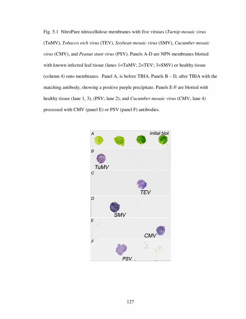
127
Fig. 5.1 NitroPure nitrocellulose membranes with five viruses (Turnip mosaic virus
(TuMV), Tobacco etch virus (TEV), Soybean mosaic virus (SMV), Cucumber mosaic
virus (CMV), and Peanut stunt virus (PSV). Panels A-D are NPN membranes blotted
with known infected leaf tissue (lanes 1=TuMV; 2=TEV; 3=SMV) or healthy tissue
(column 4) onto membranes. Panel A, is before TBIA. Panels B – D, after TBIA with the
matching antibody, showing a positive purple precipitate. Panels E-F are blotted with
healthy tissue (lane 1, 3), (PSV; lane 2); and Cucumber mosaic virus (CMV; lane 4)
processed with CMV (panel E) or PSV (panel F) antibodies.
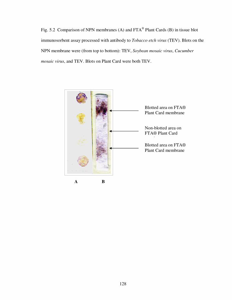
128
Fig. 5.2 Comparison of NPN membranes (A) and FTA® Plant Cards (B) in tissue blot
immunosorbent assay processed with antibody to Tobacco etch virus (TEV). Blots on the
NPN membrane were (from top to bottom): TEV, Soybean mosaic virus, Cucumber
mosaic virus, and TEV. Blots on Plant Card were both TEV.
Blotted area on FTA® Plant Card membrane
Non-blotted area on FTA® Plant Card membrane
Blotted area on FTA® Plant Card membrane
A B
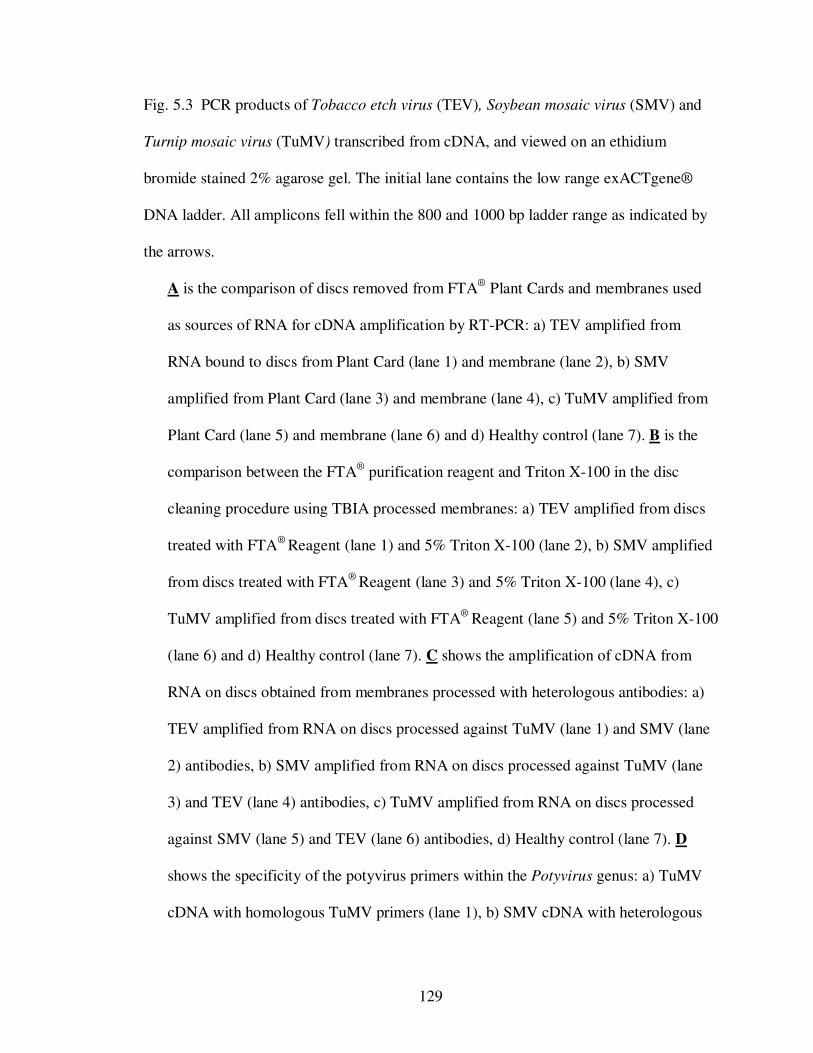
129
Fig. 5.3 PCR products of Tobacco etch virus (TEV), Soybean mosaic virus (SMV) and
Turnip mosaic virus (TuMV) transcribed from cDNA, and viewed on an ethidium
bromide stained 2% agarose gel. The initial lane contains the low range exACTgene®
DNA ladder. All amplicons fell within the 800 and 1000 bp ladder range as indicated by
the arrows.
A is the comparison of discs removed from FTA® Plant Cards and membranes used
as sources of RNA for cDNA amplification by RT-PCR: a) TEV amplified from
RNA bound to discs from Plant Card (lane 1) and membrane (lane 2), b) SMV
amplified from Plant Card (lane 3) and membrane (lane 4), c) TuMV amplified from
Plant Card (lane 5) and membrane (lane 6) and d) Healthy control (lane 7). B is the
comparison between the FTA® purification reagent and Triton X-100 in the disc
cleaning procedure using TBIA processed membranes: a) TEV amplified from discs
treated with FTA® Reagent (lane 1) and 5% Triton X-100 (lane 2), b) SMV amplified
from discs treated with FTA® Reagent (lane 3) and 5% Triton X-100 (lane 4), c)
TuMV amplified from discs treated with FTA® Reagent (lane 5) and 5% Triton X-100
(lane 6) and d) Healthy control (lane 7). C shows the amplification of cDNA from
RNA on discs obtained from membranes processed with heterologous antibodies: a)
TEV amplified from RNA on discs processed against TuMV (lane 1) and SMV (lane
2) antibodies, b) SMV amplified from RNA on discs processed against TuMV (lane
3) and TEV (lane 4) antibodies, c) TuMV amplified from RNA on discs processed
against SMV (lane 5) and TEV (lane 6) antibodies, d) Healthy control (lane 7). D
shows the specificity of the potyvirus primers within the Potyvirus genus: a) TuMV
cDNA with homologous TuMV primers (lane 1), b) SMV cDNA with heterologous
130
TuMV primers (lane 2), c) TEV cDNA with heterologous TuMV primers (lane 3), d)
SMV cDNA with homologous SMV primers (lane 4), e) TuMV cDNA with
heterologous SMV primers (lane 5), f) TEV cDNA with heterologous SMV primers
(lane 6), f) TEV cDNA with homologous TEV primers (lane 7), g) TuMV cDNA
with heterologous TEV primers (lane 8), h) SMV cDNA with heterologous TEV
primers (lane 9). E summarizes the amplification of the two cucumoviruses on
NitroPure nitrocellulose membranes: a) CMV amplified from RNA on unprocessed
membranes (lane 1), and membranes processed with CMV (lane 2) and PSV (lane 3)
antibodies, b) CMV cDNA with heterologous PSV primers (lane 4), c) PSV amplified
from RNA on unprocessed membranes (lane 5), and membranes processed with PSV
(lane 6) and CMV (lane 7) antibodies, d) PSV cDNA with heterologous CMV
primers (lane 8), e) Healthy control. The healthy control throughout these
experiments represents healthy tissue blotted to membranes and used in various RT-
PCR reactions with all conditions and primers mentioned above.
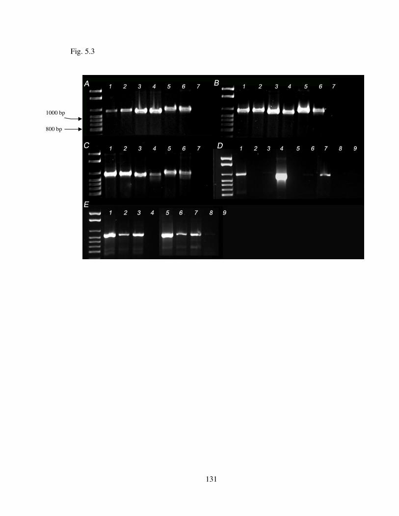
131
Fig. 5.3
800 bp
1000 bp
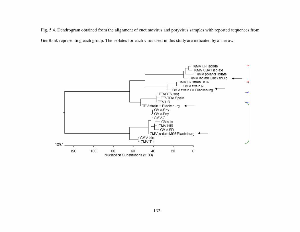
132
Fig. 5.4. Dendrogram obtained from the alignment of cucumovirus and potyvirus samples with reported sequences from
GenBank representing each group. The isolates for each virus used in this study are indicated by an arrow.
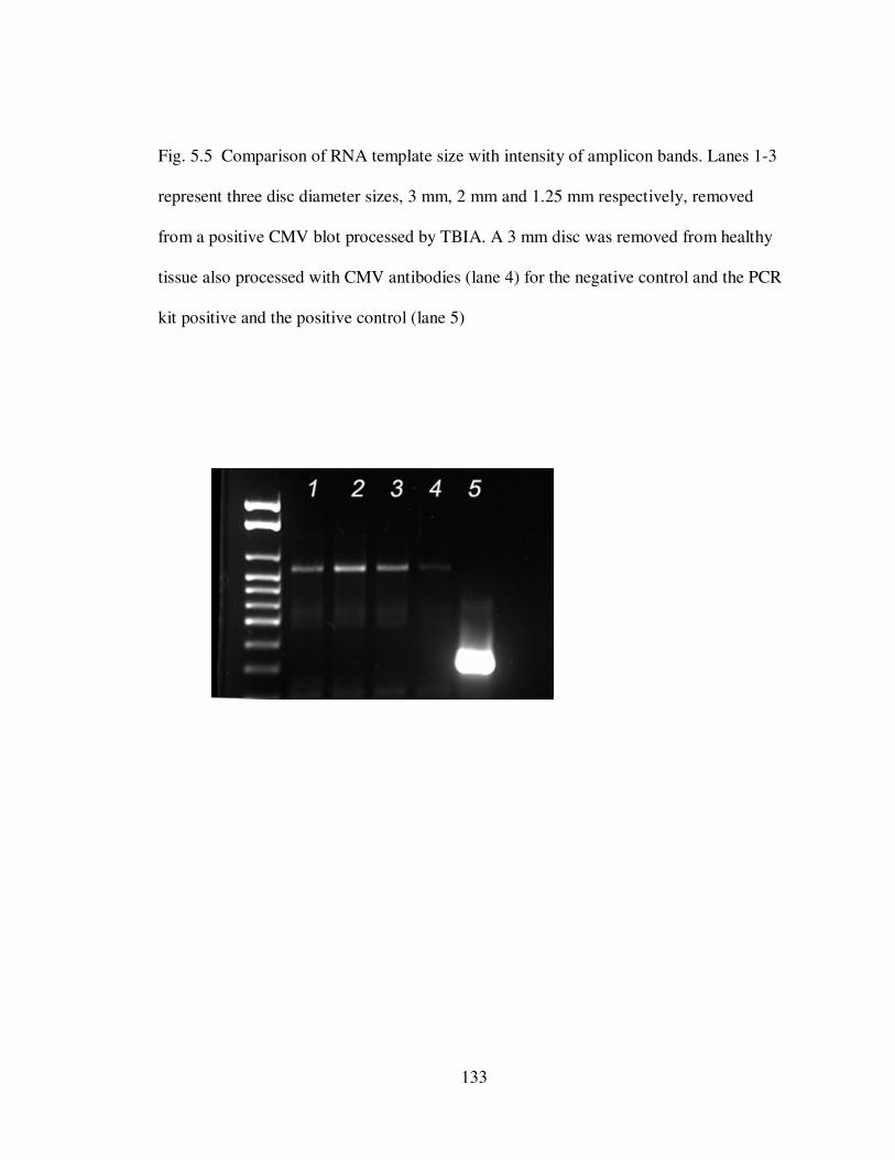
133
Fig. 5.5 Comparison of RNA template size with intensity of amplicon bands. Lanes 1-3
represent three disc diameter sizes, 3 mm, 2 mm and 1.25 mm respectively, removed
from a positive CMV blot processed by TBIA. A 3 mm disc was removed from healthy
tissue also processed with CMV antibodies (lane 4) for the negative control and the PCR
kit positive and the positive control (lane 5)
134
CHAPTER VI
Global application of NitroPure nitrocellulose membranes for virus
detection and identification
Abstract
NitroPure nitrocellulose (NPN) membranes have proven to be an effective source
of plant viral RNA. Since plant viruses are inactivated after drying, membranes offer an
easy way of transporting plant viruses over long distances without the risk of introducing
viruses to new areas. This study reports the use of NPN for the global collection,
detection, characterization and sample databasing of plant viruses.
Plant cards were disseminated to collaborators within the IPM CRSP for the
collection of plant virus samples. The cards were returned and processed using tissue blot
immunoassay (TBIA) and antibodies to Cucumber mosaic virus (CMV) and Tobacco
etch virus (TEV). Discs from dried membranes were cleaned and added directly into
reverse transcription reactions. Aliquots of the cDNA were added to PCR reactions to
obtain the expected size amplicons. The PCR products were cleaned and sequenced. The
coat protein sequences obtained placed all but two CMV positives into subgroup 1A.
Discs were also obtained from 15 month old CMV positives collected through the
Legume PIPE project processed by TBIA. PCR protocols were modified from 35 cycles
to 40 to compensate for the age of the samples. Sequences of PCR products placed all US
samples tested also within subgroup 1A. Membranes offer a quick, simple and safe way
135
of collecting samples from long distances and storing these virus samples for several
months.
Introduction
NitroPure nitrocellulose membranes (NPN) were shown in the Chapter V to
enable both the detection and molecular characterization of four plant RNA viruses using
a single membrane. In this study, the membranes were field tested by dissemination to
collaborators in the Integrated Pest Management Collaborative Research Support
Program (IPM CRSP) host countries (Table 6.1). Membranes were blotted and shipped
back to the Virology lab at Virginia Tech for processing. Samples were also taken from
previously TBIA processed cards through the 2007 Legume Pest Information Platform
for Extension and Education (PIPE) project.
Materials and Methods
Preparation and dissemination of sample cards to IPM CRSP collaborators
NitroPure nitrocellulose membranes were cut into 70 x 120 mm strips and the
edges taped to a folded 5" x 8" index card printed with sampling instructions. A template
with 6 mm diameter holes, made from the membrane protective paper, was taped over the
NPN membrane for positioning tissue blots in an array and for protecting non-blotted
areas of the membrane (Fig. 6.1 a-c). The cards were labeled numerically and distributed
to collaborators in selected IPM CRSP host countries in West Africa, Latin America and
the Caribbean (Table 6.1).
Instructions to samplers were to select symptomatic plants, select a leaf and roll it
into a tight coil, and pull to tear it in half. The torn edges were to be blotted onto five
136
spots in a row on the membrane as indicated by the arrow in Fig. 6.1. A total of nine
samples were blotted per card. Once sampling was complete, the card(s) was shipped to
the Tolin Virology lab at Virginia Tech, Blacksburg, VA, by regular mail.
Samples on BioRad nitrocellulose membranes were also received from the
Dominican Republic. This provided a comparison between two types of membranes in
this study.
Immunological assay for plant viruses from collected sample cards
Cards returned by each host country were either not processed by TBIA or were
processed for the detection of Cucumber mosaic virus (CMV) or Tobacco etch virus
(TEV). For unprocessed cards, the template was removed and strips of the membrane
were cut lengthwise. Two strips, each blotted with a representative of the nine samples,
were developed with TBIA using antibodies against CMV and TEV using the procedure
described in the previous chapters. The remaining three strips were recovered with the
template and stored at room temperature for later testing with other antibodies.
Collection of Cucumber mosaic virus samples within the United States
CMV positive samples were obtained from several states (Table 6.1) participating
in the Legume Pest Information Platform for Extension and Education (PIPE) (Langham
et al., 2007). TBIA sampling cards submitted to Dr. Sue Tolin, Virginia Tech. Using the
method described in the previous chapter, one 3 mm disc was removed from each of the
samples testing positive for CMV. All cards had been processed by TBIA at least 15
months prior to disc sample removal.
137
Membrane preparation of RNA and RT-PCR
Membrane preparation of RNA and RT-PCR was conducted as described in the
previous chapter for CMV and TEV samples. Briefly, discs, 3 mm in size, were removed
from all positive samples from the U.S. Legume PIPE cards and from representative
CMV positive samples from each of the collaborating host countries’ cards. Due to time
constraints, only selected samples were cleaned and directly sequenced. The discs were
cleaned and added directly to two-step RT-PCR with virus specific primers used as
described in the previous chapter.
To compensate for the age of the U.S. samples, the CMV PCR protocol described
in the previous chapter was modified by increasing the number of cycles from 35 to 40.
The PCR products were observed in an ethidium bromide-stained 2% agarose gel with a
UV transilluminator (UVP Inc., San Gabriel CA) and AlphaInnotech Imager (San
Leandro, CA). The remaining products were cleaned with either the QIAquick PCR
purification kit (Qiagen, Valencia, CA) or with shrimp alkaline phosphatase/ exonuclease
1 (SAP/EXO1) (usb-Affymetrix, Cleveland, OH), and sequenced at the CRC-DNA
Sequencing Facility at the University of Chicago, IL.
Analysis of sequence data
The EditSeq program of DNASTAR Lasergene software (Madison, WI) was used
to remove background signals at the starts and modify existing sequences obtained from
NCBI. The Cluster W program in MegAlign, DNASTAR (Madison, WI) was used to
align all sequences to produce a phylogenetic tree. A total of eight coat protein
sequences were used from GenBank for the comparison with the sequences from samples
received from collaborators. Three sequences from CMV strains Fny, Sny, and C, all
U.S. isolates, represented CMV subgroup 1A; three sequences, Ix, SD and Nt9, all
138
isolated from Asia, represented CMV subgroup 1B; and 2 sequences, Kin and Trk7, from
Scotland and Hungary, respectively, represented CMV subgroup 2. All sequences have
been used previously in evolutionary studies of CMV (Roossinck, 1999).
Results
Immunological assay from collaborator sample cards
A total of 362 samples were obtained from the IPM CSRP collaborating host
countries. A summary of the results is shown in Table 6.1. A majority of the samples
received were blotted from different varieties of pepper and tomato, particularly from
countries in West Africa and Latin America. Several mixed infections of CMV and TEV
were observed from peppers in Dominican Republic, as indicated by TBIA (Fig. 6.2).
Several samples were negative to both CMV and TEV, even though plants were showing
symptoms of viral disease. From the Jamaica, up to 90% of the samples were blotted
from infected cucurbits including watermelon, pumpkin and squash. Three samples
blotted in Jamaica from pumpkin displaying mosaic patterns on leaves tested positive for
Watermelon mosaic virus (WMV2) but were negative Peanut stunt virus, and for all
potyviruses using a general potyvirus monoclonal antibody.
U.S. isolates of CMV
A total of 24 positive CMV samples were selected from Legume PIPE cards from
a total of 7 locations in 5 states, namely, New York, Wisconsin, Michigan, North
Carolina and Minnesota. No other participating state had samples that were positive for
CMV. Discs were taken from a range of TBIA positive samples, from purple specks on
blotted areas to deep purple spots, which indicated a high concentration of virus particles.
139
RT-PCR and sequence data
Based on sequences obtained, all Legume PIPE samples were aligned in subgroup
1A, with percentage identities between 93 and 96%. Sequences obtained from the
Dominican Republic samples gave variable identity percentages, but most were also
aligned with subgroup 1A with 87 to 92% identities (Fig. 6.3a and b). Direct observations
of chromatograms of the Dominican Republic samples revealed multiple Ns within the
genome sequences resulting from several overlapping peaks. This was noticed especially
in samples from Ocoa (from CMV card 96) and Paya (CMV card 97), the two outliers in
the phylogenetic tree (Fig. 6.3a). These samples share only 50-70% sequence identity
with the other samples and CMV sequences from GenBank. Many plants were co-
infected with TEV. In the pepper field from which the samples were obtained 100% of
the plants showed severe symptoms, including extreme mottling and stunting.
Two types of membranes were used in this research, the NitroPure nitrocellulose
membranes commonly used in the Tolin lab, and the Bio-Rad pure nitrocellulose
membranes (Hercules, CA) used by collaborators in the Dominican Republic. With
TBIA, the Bio-Rad membranes had a higher background, making it difficult to determine
weak positives of CMV and TEV from negatives in TBIA. There was no difference
between the membranes when they were used as sources of RNA for RT-PCR as the
series of Ns in the Dominican Republic samples was not associated the type of
membrane.
Discussion
As shown in Chapters II, III and V, NitroPure nitrocellulose membranes have
made an invaluable contribution to the detection and molecular characterization of four
140
economically important plant viruses in two different virus families. The use of
membranes as sources of RNA, regardless of whether the membrane has been processed
by TBIA or not, eliminate the need for the collection and storage of fresh tissue, and the
extensive and often laborious task of RNA extraction.
Our data demonstrate that NPN can be used to store virus samples for long
periods of time without any special storage conditions, even after TBIA processing.
Previous experience with TBIA had shown that membranes remained immunologically
active for up to 1 year (data not shown). This led to the assumption that if intact virus
particles were still bound to the membranes, RNA should be available as a template for
RT-PCR. Using membranes from the 2007 Legume PIPE, cDNA was amplified from
samples that had tested positive for CMV by TBIA performed 15 months previously.
Amplification of cDNA by PCR, and sequencing of PCR products, demonstrated that
RNA was still available for molecular analysis after 15 months.
In Chapter IV, the viral diagnostic capabilities of several developing countries
within the IPM CRSP were investigated. Despite the availability of thermocyclers and
imagers, molecular diagnosis of viruses was not practiced by more than 50% of the
clinics that responded to the questionnaire. We assumed that obtaining reagents and
specialized equipment could be the cause of the lack of molecular analysis. For clinics
that cannot do molecular analysis but need to have these data for diagnostics or
publication, the development of a centralized hub lab for molecular analysis for
surrounding clinics is an option. We tested NPN for the ease of transport of plant viral
material between countries to replicate countries submitting virus samples to a
centralized clinic for molecular diagnostics. Virus samples blotted onto NPN in sample
141
cards were received from collaborating countries within the IPM CRSP. Our results show
that samples blotted to NPN can be sent between countries and used to obtain nucleic
acid sequences.
Samples from the Dominican Republic had lower percentage identity to
representative CMV sequences for each CMV subgroup. A closer look at the sequences
of these samples showed several N’s, primarily due to several overlapping peaks. Since
these samples had been obtained from an old, infested field (Fig. 6.2), the possibility of
multiple strains of CMV within one plant exists. We suggest that sampling younger
plants might give cleaner sequences, as there would be less chance of mixed infection or
mutation.
Finally, we have shown that virus samples can be collected safely and easily on
membranes and sent to other laboratories or countries for molecular testing. It was also
demonstrated that NPN can be used as a databasing matrix for storing non-infectious
plant viruses for several months at room temperature. Whether or not membranes had
been processed by TBIA, both old and newly blotted membranes were successfully used
as sources of viral RNA for molecular characterization of CMV coat protein. Further
work using a wider range of plant viruses and testing within other developing countries
would broaden the application of these methods
References
Chang, P. S. and Tolin, S. A. 2009. Immunoassay, RT-PCR and direct sequencing of
Cucumber mosaic virus and potyvirus coat proteins from the same NitroPure
nitrocellulose membrane. Chapter V in: Dissertation
142
Langham, M., Tolin, S., Sutula, C., Schwartz, H., Wisler, G., A., K., Hershman, D.,
Giesler, L., Golod, J., Ratcliffe, S., and Cardwell, K. 2007. Legume/Virus PIPE -
A new tool for disease management in legumes. Phytopathology 97:S61.
Roossinck, M. J., Zhang, L., and Hellwald, K.-H. 1999. Rearrangement in the 5' non-
translated region and phylogenetic analyses of Cucumber mosaic virus RNA 3
indicate radial evolution of the three subgroups. J. Virol. 73:6752-6758.
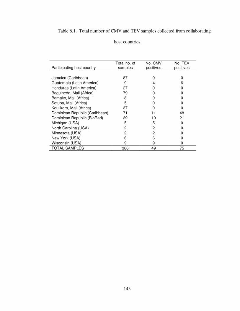
143
Table 6.1. Total number of CMV and TEV samples collected from collaborating
host countries
Participating host country Total no. of
samples No. CMV positives
No. TEV positives
Jamaica (Caribbean) 87 0 0
Guatemala (Latin America) 9 4 6
Honduras (Latin America) 27 0 0
Baguineda, Mali (Africa) 79 0 0
Bamako, Mali (Africa) 8 0 0
Sotuba, Mali (Africa) 5 0 0
Koulikoro, Mali (Africa) 37 0 0
Dominican Republic (Caribbean) 71 11 48
Dominican Republic (BioRad) 39 10 21
Michigan (USA) 5 5 0
North Carolina (USA) 2 2 0
Minnesota (USA) 2 2 0
New York (USA) 6 6 0
Wisconsin (USA) 9 9 0
TOTAL SAMPLES 386 49 75
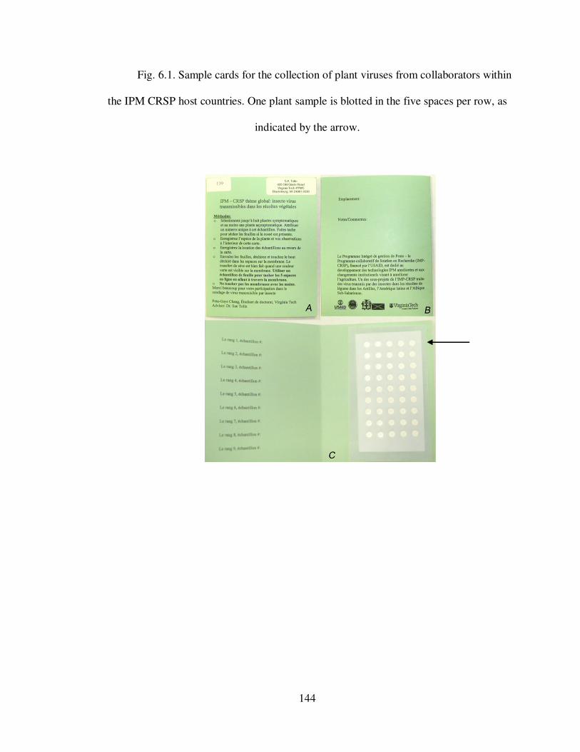
144
Fig. 6.1. Sample cards for the collection of plant viruses from collaborators within
the IPM CRSP host countries. One plant sample is blotted in the five spaces per row, as
indicated by the arrow.
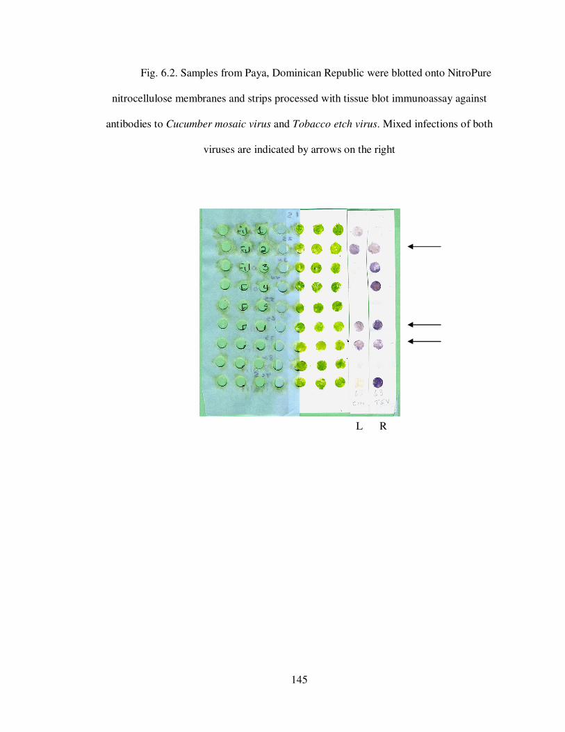
145
Fig. 6.2. Samples from Paya, Dominican Republic were blotted onto NitroPure
nitrocellulose membranes and strips processed with tissue blot immunoassay against
antibodies to Cucumber mosaic virus and Tobacco etch virus. Mixed infections of both
viruses are indicated by arrows on the right
L R
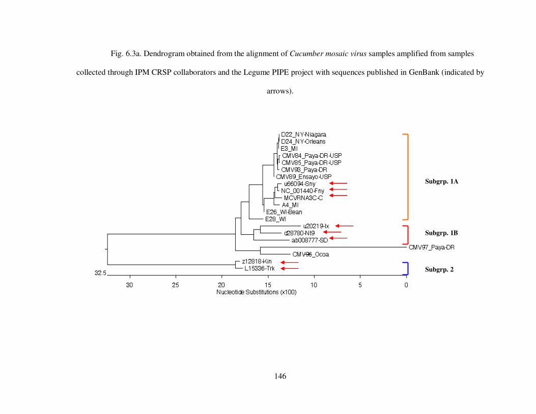
146
Fig. 6.3a. Dendrogram obtained from the alignment of Cucumber mosaic virus samples amplified from samples
collected through IPM CRSP collaborators and the Legume PIPE project with sequences published in GenBank (indicated by
arrows).
Subgrp. 2
Subgrp. 1A
Subgrp. 1B
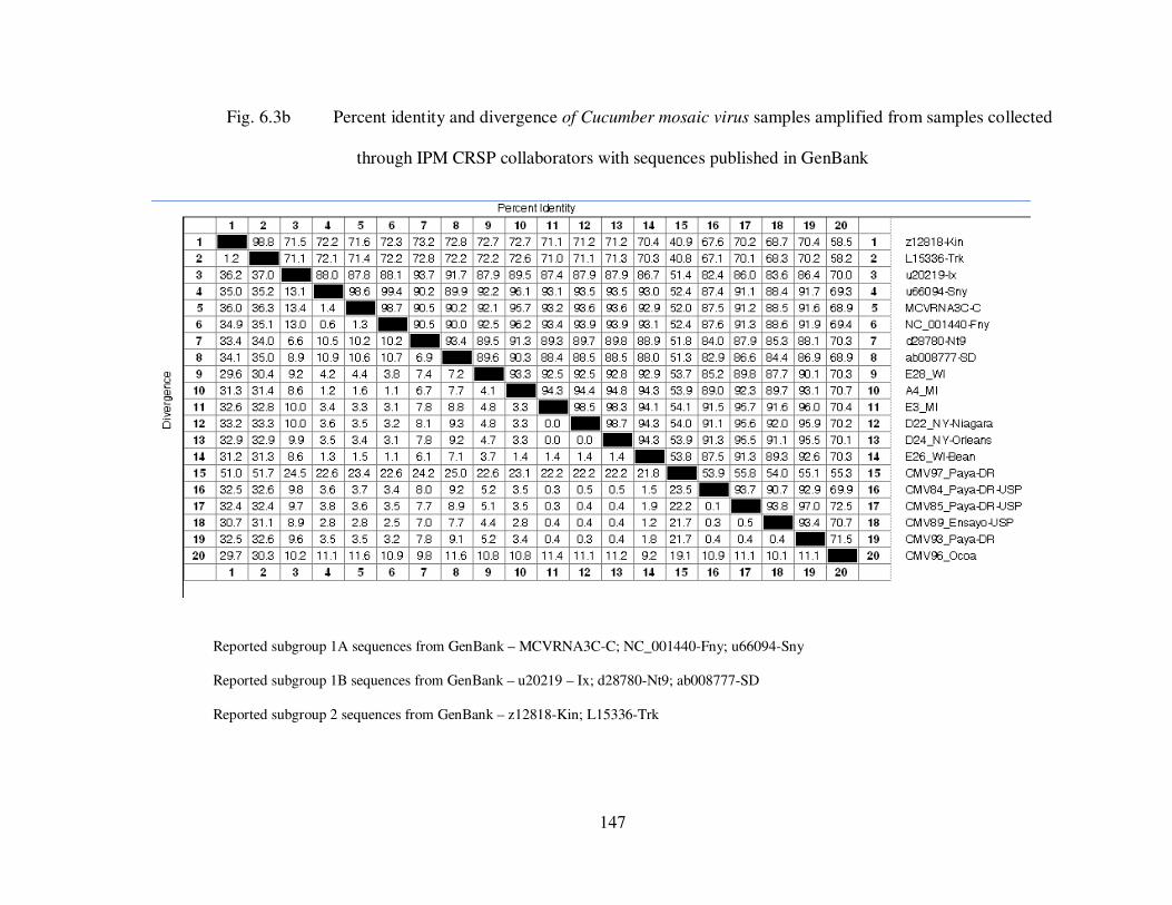
147
Fig. 6.3b Percent identity and divergence of Cucumber mosaic virus samples amplified from samples collected
through IPM CRSP collaborators with sequences published in GenBank
Reported subgroup 1A sequences from GenBank – MCVRNA3C-C; NC_001440-Fny; u66094-Sny
Reported subgroup 1B sequences from GenBank – u20219 – Ix; d28780-Nt9; ab008777-SD
Reported subgroup 2 sequences from GenBank – z12818-Kin; L15336-Trk
148
CHAPTER VII
Conclusions and Recommendations
Diagnostics plays an important role in any agricultural system. There are four
general approaches to virus diagnosis, including) biological, ii) physical, iii) serological,
or iv) molecular techniques. Three of these approaches were used in the identification and
characterization of three recent isolates of Cucumber mosaic virus (CMV; Genus
Cucumovirus; Family Bromoviridae) from the garden at the Historic Smithfield
Plantation in Montgomery County, VA. The CMV isolates were obtained from flowering
tobacco (Nicotiana sp. L.), bowl gourd (Cucurbita sp. L.) and periwinkle (Vinca minor
L.). Symptoms included mosaic and leaf deformation of varying severities. Positive
reactions were obtained in tissue blot immunoassay (TBIA) using CMV polyclonal
antibody from the Tolin lab (code HCV-7409), and monoclonal antibodies donated by
Agdia Inc. The isolates were compared to CMV isolates collected in the 1970s and stored
as dry tissue for over 25 years. Biological characterization using indicator hosts induced
symptoms similar to those described for CMV (Palukaitis and Garcia-Arenal, 2003).
CMV RNA was first obtained from purified CMV particles through SDS release. The
cDNA was amplified with CMV coat protein specific primers, and the PCR products
cleaned and sequenced. All CMV sequences were aligned using the Clustal W program in
the Lasergene software (Madison, WI), which produces a phylogenic tree (Fig. 2.3a). The
sequences of the new CMV isolates were 94.5% identical to representative sequences for
CMV subgroup 1A from GenBank. Three of the old isolates were classified as subgroup
2, which was not previously reported to occur in Virginia.
149
Another virus, Turnip mosaic virus (TuMV; genus Potyvirus, family Potyviridae),
was isolated from Dame’s Rocket (Hesperis matronalis L.) located along the periphery of
the Smithfield Plantation gardens. The plant displayed symptoms of mosaic on leaves and
petal color-breaking in flowers. TuMV was detected using TBIA with antibodies donated
from T. P. Pirone (Lexington, KY). Symptoms on indicator plants and sequence data
confirmed the identity of TuMV. Petal color-break symptoms were reproduced upon
inoculation of young Dame’s Rocket plants. This is the first reported case of TuMV in
Virginia.
Because diagnostics is especially important in developing countries where the
export of agricultural produce contributes heavily to the economy (Lawrence et. al.,
2005), the capabilities of clinics in developing countries to perform viral and general
diagnostics were investigated. Ausher (1996) previously reported that the diagnostic
capabilities in developing countries were often limited by funding and the availability of
trained diagnosticians. We conducted a survey to look more closely at the current viral
diagnostic capabilities of several clinics in developing countries, collaborators of the IPM
CRSP. Clinics privately owned or affiliated with academic institutions had better
equipped laboratories than clinics that were affiliated with the government. Clients that
used clinic services varied, depending on the affiliations to the different institutions.
Privately owned clinics obtained a majority of their samples from companies and
corporations, academically affiliated clinics obtained samples from corporations and
extension agents, and clinics affiliated with the government received samples primarily
from farmers and extension agents. It was interesting to note the contrasting diagnostic
approaches employed by these various clinics. Unlike clinics privately owned or
150
affiliated to academic institutions that used molecular diagnostics, government affiliated
labs employed biological and serological approaches for virus diagnosis. The
unavailability of specialized equipment and personnel, reagents and kits has hampered
the use of molecular techniques in virus diagnosis.
For laboratories that require molecular data for quarantine and phytosanitary
purposes, characterization studies and virus identification but are unable to perform such
tasks, samples can be collected and shipped to diagnostic laboratories. However, because
of state, nation, and International Plant Protection Organization (IPPO) regulations,
special permits have to be obtained for the importation of any pathogen into a locality or
political division where the pathogen has been either controlled or is not present.
Presented with this issue, our laboratory turned to NitroPure nitrocellulose (NPN)
membranes as a solid matrix on which pathogens are non-infectious and can be
transported without permit. These membranes, used in TBIA, bind whole virus particles
to its surface. Our hypothesis was that if whole particles are present on membranes, then
RNA will also be present and might be available for molecular studies. We proved this
hypothesis using five viruses in two genera, CMV and Peanut stunt virus (PSV) in the
genus Cucumovirus, and Soybean mosaic virus, Tobacco etch virus, and Turnip mosaic
virus in the genus Potyvirus. Amplicons of the expected sizes for each virus were
produced from viruses blotted to NPN membranes. CMV had a 94.7-95% sequence
identity to U.S. CMV strain Fny, TEV had a 95.6% identity to U.S. TEV strain TEV-
GEN, SMV had a 96% identity to U.S. SMV strain G7, and TuMV had a 93% identity to
U.S. TuMV strain USA1.
151
A limited trial of the international application of this procedure was conducted in
several countries in West Africa, Latin America and the Caribbean. Special sampling
cards were designed, and distributed to collaborators with instructions for blotting with
plant tissue displaying virus-like symptoms. Non-processed membranes received from
collaborators in these countries were developed by TBIA using monoclonal antibodies to
CMV and a polyclonal antibody to TEV (#601). No CMV or TEV positive samples were
obtained from West Africa or Jamaica. Samples positive to CMV and TEV were obtained
from the Dominican Republic, but the possible presence of two or more strains of CMV
within the samples, shown as multiple peaks in the chromatograph, interfered with a good
sequence reading. The samples were, however, approximately 70-80% identical to CMV
subgroup 1A sequences, compared to 60% to subgroup 1B and 30% to subgroup 2.
Diagnostics is important for the management of plant viruses. However, without
the right tools, equipment and knowledge, proper management practices cannot be
accomplished. Our work has compared both the classical approaches to plant virus
diagnostics and a new, simple method for both detecting plant viruses and obtaining
sequence data from one solid matrix, nitrocellulose membranes. To further explore
membrane applications, we propose studies into the use of membranes as sources of
nucleic acid for other plant RNA and DNA viruses. It is possible also to explore the use
of membranes for other pathogens, including bacteria, as bacteria has been detected using
TBIA protocols (Lin et al., 1990)
I would also like to use this method to survey the current virus diversity in
Jamaica and, if possible, in surrounding islands as well. Nitrocellulose membranes would
allow for the collection of several viral samples from various locations. With the
152
knowledge of the types of viruses found within the region, islands would be better able to
develop management strategies that would benefit farmers, both large, and small for
increased productivity within agriculture.
References
Ausher, R., Ben-Ze'ev, I. S., and Black, R. 1996. The role of plant clinics in plant disease
diagnosis and education in developing countries. Annu. Rev. Phytopathol. 34:51-
66.
Lawrence, J., Tolin, S. A., Edwards, C., Fleischer, S. J., Jackson, D. M., Clarke-Harris,
D., McDonald, S. A., Dalip, K., and Chung, P. 2005. Developing IPM packages
in the Caribbean. in: Globalizing Integrated Pest Management: A Participatory
Research Process, G. W. Norton, E. A. Heinrichs, G. C. Luther and M. E. Irwin,
eds. Blackwell Publishing, Ames, Iowa.
Lin, N. S., Hsu, Y. H., and Hsu, H. T. 1990. Immunological detection of plant viruses and
a mycoplasma-like organism by direct tissue blotting on nitrocellulose
membranes. Phytopath. 80 (9):824-828.
Palukaitis, P., and Garcia-Arenal, F. 2003. Cucumber mosaic virus. [accessed: 4/20/06]
http://www.dpvweb.net/dpv/showadpv.php?dpvno=400

153
APPENDICES
154
Appendix A
Virus purification according to Lot et. al. (1972)
Six virus isolates: M05-CMV, T05-CMV, CMVY-CC, V06-CMV, CMVY-108
and CMVB were each inoculated to six 8-week old Xanthi tobacco plants. Each plant had
four to six fully expanded leaves. Eight days after inoculation, the leaves from all plants
were harvested for a combined weight of 20 – 30 gm. The leaves were cooled at 4°C for
2 hr and then homogenized in a Waring blender with 0.5M citrate buffer, pH 6.5 (sodium
citrate – citric acid), containing 0.1% thioglycollic acid at a rate of 2 ml/gm of tissue.
The tissue was macerated for 10 sec at low speed. Cold choloroform, at a rate of 2 ml/gm
of tissue, was added to the crude sap and homogenized at high speeds for 30 sec. The
homogenate was quickly moved to a chilled beaker, mixed, and centrifuged at 7500 rpm
for 10 min. The aqueous layer was decanted into a cooled beaker. To the total volume of
the decanted layer, PEG 6000 (10% w:v) was added and magnetically stirred for 15 min
at 4°C. The suspension was allowed to stand in an icebath at 4°C for 40 min, and then
centrifuged at 10,000 rpm for 20 min. The supernatant was discarded and the pellet re-
suspended in 0.05 M citrate buffer, pH 7, containing 2% (v:v) Triton X-100, at a rate of
0.5 ml/gm of original weight of leaves. To ensure maximum re-suspension, the pellets
were magnetically stirred at 4°C overnight. The suspension was centrifuged at 7500 rpm
for 20 min. The supernatant was centrifuged using a Type 65 rotor (Beckman L8-80
Ultracentrifuge (Fullerton, CA. USA), for 2 hr at 40,000 rpm. The supernatant was
discarded and the pellets re-suspended in distilled water, with gentle agitation, overnight.
After low speed centrifugation of 13,600 g (Microcentrifuge Model 235B, Fisher

155
Scientific) for 2 min, the supernatant was removed and made up to 0.5 M in citrate buffer.
The purified virus was stored at 4°C.
156
Appendix B
Virus purification according to Lane et. al. (2003)
Three CMV isolates: M05-CMV, T05-CMV and V06-CMV, were each
inoculated to two 8-week old Xanthi tobacco plants. Eight days after inoculation, a
combined weight of 1 gm of infected leaf tissue was harvested from both plants and
stored at 4°C for 2 hr. The leaves were homogenized in a small Waring blender at high
speed with 22 ml cold sodium citrate buffer, pH 6.5, 150 µl 0.5 M sodium
diethyldithiocarbamate (DIECA) (Acros Organics, Geel Belgium) and 125 µl 0.2 M
Iodoacetamide (Sigma-Aldrich, St. Louis, MO USA). The mixture was expressed through
a damp cheesecloth into ultracentrifuge tubes and centrifuged at 40,000 rpm for 10 min
using a Type 65 rotor. The supernatant was decanted into new ultracentrifuge tubes.
Eight drops of 5% Triton X-100 were added, and the supernatant mixed before
centrifugation at 40,000 rpm for 40 min. The resulting supernatant was discarded and the
pellets rinsed with de-ionized water. The pellets were left to air dry for 15 min, re-
suspended in 50 µl 0.05 M sodium phosphate, and stored at 4°C.
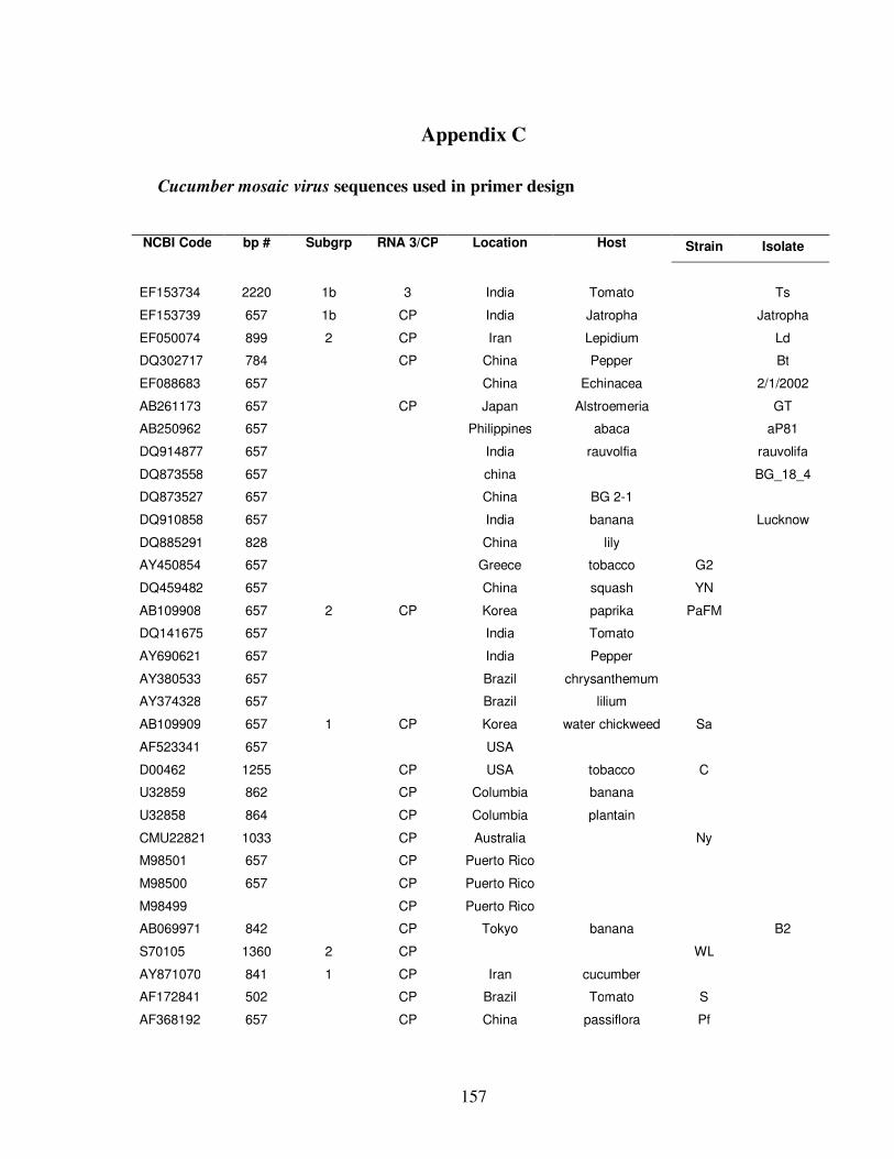
157
Appendix C
Cucumber mosaic virus sequences used in primer design
NCBI Code bp # Subgrp RNA 3/CP Location Host Strain Isolate
EF153734 2220 1b 3 India Tomato Ts
EF153739 657 1b CP India Jatropha Jatropha
EF050074 899 2 CP Iran Lepidium Ld
DQ302717 784 CP China Pepper Bt
EF088683 657 China Echinacea 2/1/2002
AB261173 657 CP Japan Alstroemeria GT
AB250962 657 Philippines abaca aP81
DQ914877 657 India rauvolfia rauvolifa
DQ873558 657 china BG_18_4
DQ873527 657 China BG 2-1
DQ910858 657 India banana Lucknow
DQ885291 828 China lily
AY450854 657 Greece tobacco G2
DQ459482 657 China squash YN
AB109908 657 2 CP Korea paprika PaFM
DQ141675 657 India Tomato
AY690621 657 India Pepper
AY380533 657 Brazil chrysanthemum
AY374328 657 Brazil lilium
AB109909 657 1 CP Korea water chickweed Sa
AF523341 657 USA
D00462 1255 CP USA tobacco C
U32859 862 CP Columbia banana
U32858 864 CP Columbia plantain
CMU22821 1033 CP Australia Ny
M98501 657 CP Puerto Rico
M98500 657 CP Puerto Rico
M98499 CP Puerto Rico
AB069971 842 CP Tokyo banana B2
S70105 1360 2 CP WL
AY871070 841 1 CP Iran cucumber
AF172841 502 CP Brazil Tomato S
AF368192 657 CP China passiflora Pf
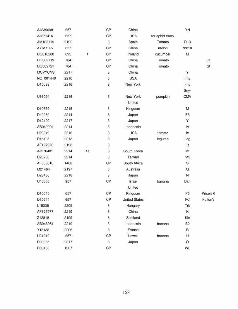
158
AJ239098 657 CP China YN
AJ271416 657 CP USA for aphid trans.
AM183119 2192 3 Spain Tomato Ri-8
AY611027 657 CP China melon 99/10
DQ018288 895 1 CP Poland cucumber M
DQ302719 784 CP China Tomato Gf
DQ302721 784 CP China Tomato Sf
MCVYCNS 2217 3 China Y
NC_001440 2216 3 USA Fny
D10538 2216 3 New York Fny
U66094 2216 3 New York pumpkin
Sny-
CMV
D10539 2215 3
United
Kingdom M
D42080 2214 3 Japan E5
D12499 2217 3 Japan Y
AB042294 2214 3 Indonesia IA
U20219 2216 3 USA tomato Ix
D16405 2213 3 Japan legume Leg
AF127976 2198 3 Ls
AJ276481 2214 1a 3 South Korea Mf
D28780 2214 3 Taiwan Nt9
AF063610 1468 CP South Africa S
M21464 2197 3 Australia Q
D28486 2218 3 Japan N
U43888 957 CP Israel banana Ban
D10545 657 CP
United
Kingdom P6 Price's 6
D10544 657 CP United States FC Fulton's
L15336 2209 3 Hungary Trk
AF127977 2219 3 China K
Z12818 2199 3 Scotland Kin
AB046951 2219 3 Indonesia banana B2
Y18138 2206 3 France R
U31219 657 CP Hawaii banana Hi
D00385 2217 3 Japan O
D00463 1267 CP WL
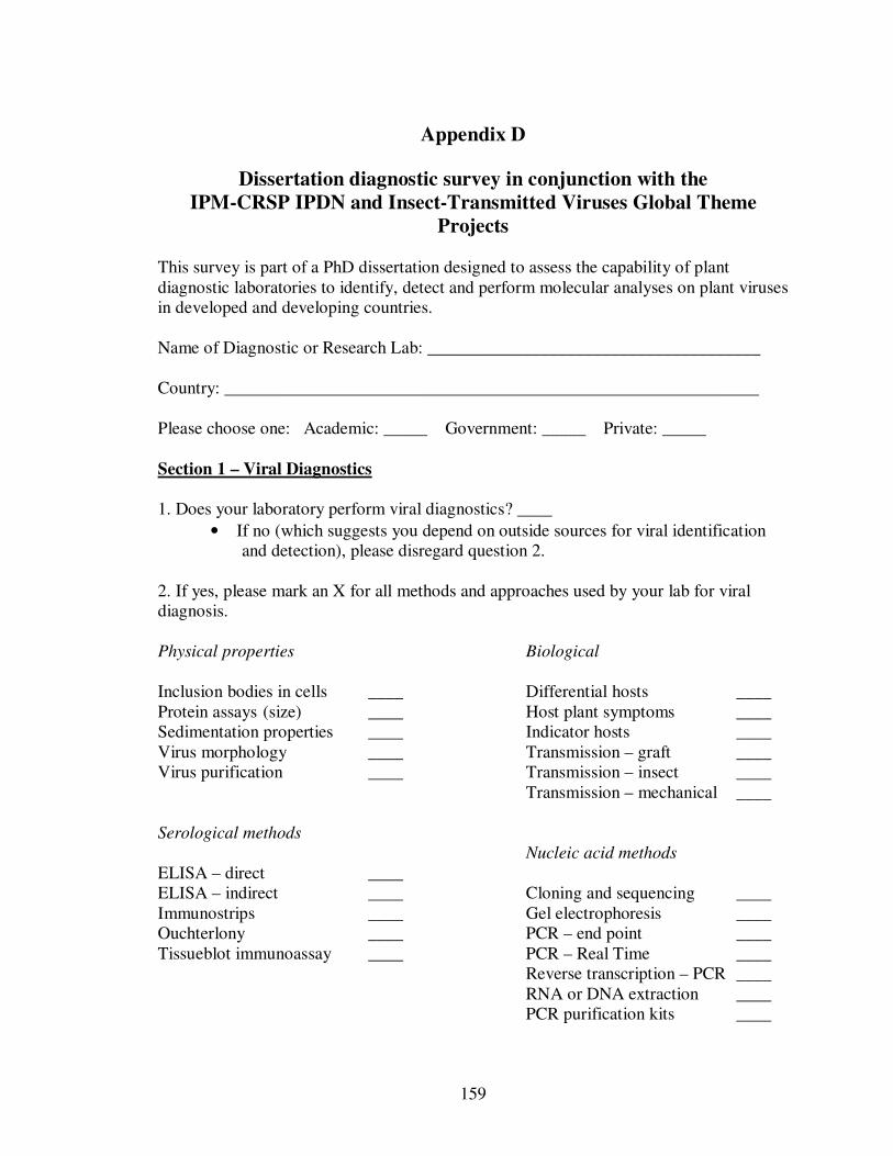
159
Appendix D
Dissertation diagnostic survey in conjunction with the
IPM-CRSP IPDN and Insect-Transmitted Viruses Global Theme
Projects This survey is part of a PhD dissertation designed to assess the capability of plant diagnostic laboratories to identify, detect and perform molecular analyses on plant viruses in developed and developing countries. Name of Diagnostic or Research Lab: ______________________________________ Country: _____________________________________________________________ Please choose one: Academic: _____ Government: _____ Private: _____ Section 1 – Viral Diagnostics
1. Does your laboratory perform viral diagnostics? ____
• If no (which suggests you depend on outside sources for viral identification and detection), please disregard question 2.
2. If yes, please mark an X for all methods and approaches used by your lab for viral diagnosis. Physical properties Biological Inclusion bodies in cells ____ Differential hosts ____ Protein assays (size) ____ Host plant symptoms ____ Sedimentation properties ____ Indicator hosts ____ Virus morphology ____ Transmission – graft ____ Virus purification ____ Transmission – insect ____ Transmission – mechanical ____ Serological methods Nucleic acid methods ELISA – direct ____ ELISA – indirect ____ Cloning and sequencing ____ Immunostrips ____ Gel electrophoresis ____ Ouchterlony ____ PCR – end point ____ Tissueblot immunoassay ____ PCR – Real Time ____ Reverse transcription – PCR ____ RNA or DNA extraction ____ PCR purification kits ____
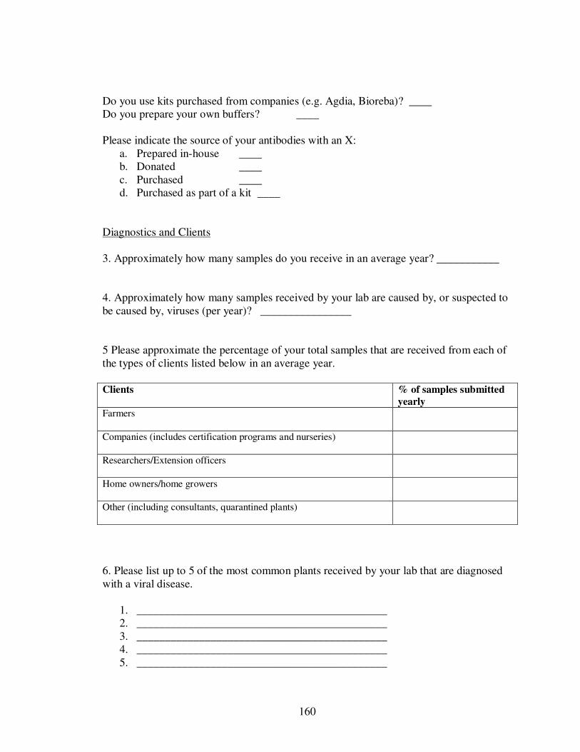
160
Do you use kits purchased from companies (e.g. Agdia, Bioreba)? ____ Do you prepare your own buffers? ____ Please indicate the source of your antibodies with an X:
a. Prepared in-house ____ b. Donated ____ c. Purchased ____ d. Purchased as part of a kit ____
Diagnostics and Clients 3. Approximately how many samples do you receive in an average year? ___________ 4. Approximately how many samples received by your lab are caused by, or suspected to be caused by, viruses (per year)? ________________ 5 Please approximate the percentage of your total samples that are received from each of the types of clients listed below in an average year. Clients % of samples submitted
yearly Farmers
Companies (includes certification programs and nurseries)
Researchers/Extension officers
Home owners/home growers
Other (including consultants, quarantined plants)
6. Please list up to 5 of the most common plants received by your lab that are diagnosed with a viral disease.
1. ____________________________________________ 2. ____________________________________________ 3. ____________________________________________ 4. ____________________________________________ 5. ____________________________________________
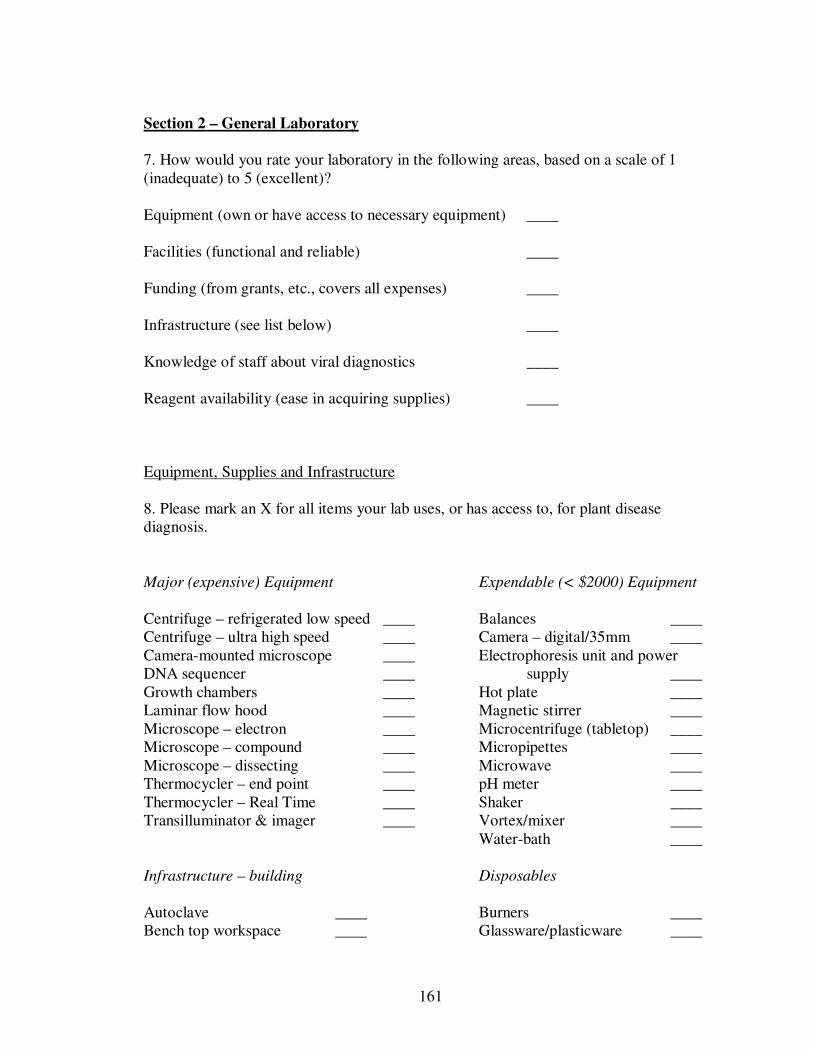
161
Section 2 – General Laboratory
7. How would you rate your laboratory in the following areas, based on a scale of 1 (inadequate) to 5 (excellent)? Equipment (own or have access to necessary equipment) ____ Facilities (functional and reliable) ____ Funding (from grants, etc., covers all expenses) ____ Infrastructure (see list below) ____ Knowledge of staff about viral diagnostics ____ Reagent availability (ease in acquiring supplies) ____ Equipment, Supplies and Infrastructure 8. Please mark an X for all items your lab uses, or has access to, for plant disease diagnosis. Major (expensive) Equipment Expendable (< $2000) Equipment Centrifuge – refrigerated low speed ____ Balances ____ Centrifuge – ultra high speed ____ Camera – digital/35mm ____ Camera-mounted microscope ____ Electrophoresis unit and power DNA sequencer ____ supply ____ Growth chambers ____ Hot plate ____ Laminar flow hood ____ Magnetic stirrer ____ Microscope – electron ____ Microcentrifuge (tabletop) ____ Microscope – compound ____ Micropipettes ____ Microscope – dissecting ____ Microwave ____ Thermocycler – end point ____ pH meter ____ Thermocycler – Real Time ____ Shaker ____ Transilluminator & imager ____ Vortex/mixer ____ Water-bath ____ Infrastructure – building Disposables Autoclave ____ Burners ____ Bench top workspace ____ Glassware/plasticware ____
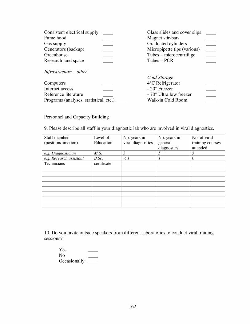
162
Consistent electrical supply ____ Glass slides and cover slips ____ Fume hood ____ Magnet stir-bars ____ Gas supply ____ Graduated cylinders ____ Generators (backup) ____ Micropipette tips (various) ____ Greenhouse ____ Tubes – microcentrifuge ____ Research land space ____ Tubes – PCR ____ Infrastructure – other Cold Storage Computers ____ 4°C Refrigerator ____ Internet access ____ - 20° Freezer ____ Reference literature ____ - 70° Ultra low freezer ____ Programs (analyses, statistical, etc.) ____ Walk-in Cold Room ____ Personnel and Capacity Building 9. Please describe all staff in your diagnostic lab who are involved in viral diagnostics.
10. Do you invite outside speakers from different laboratories to conduct viral training sessions? Yes ____ No ____ Occasionally ____
Staff member (position/function)
Level of Education
No. years in viral diagnostics
No. years in general diagnostics
No. of viral training courses attended
e.g. Diagnostician M.S. 3 5 5
e.g. Research assistant B.Sc. < 1 1 0
Technicians certificate
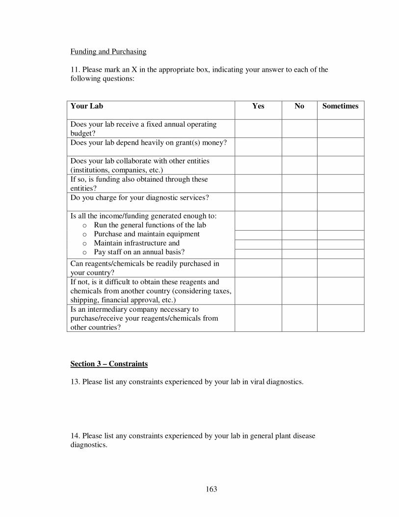
163
Funding and Purchasing 11. Please mark an X in the appropriate box, indicating your answer to each of the following questions:
Your Lab Yes No Sometimes
Does your lab receive a fixed annual operating budget?
Does your lab depend heavily on grant(s) money?
Does your lab collaborate with other entities (institutions, companies, etc.)
If so, is funding also obtained through these entities?
Do you charge for your diagnostic services?
Is all the income/funding generated enough to: o Run the general functions of the lab o Purchase and maintain equipment o Maintain infrastructure and o Pay staff on an annual basis?
Can reagents/chemicals be readily purchased in your country?
If not, is it difficult to obtain these reagents and chemicals from another country (considering taxes, shipping, financial approval, etc.)
Is an intermediary company necessary to purchase/receive your reagents/chemicals from other countries?
Section 3 – Constraints 13. Please list any constraints experienced by your lab in viral diagnostics. 14. Please list any constraints experienced by your lab in general plant disease diagnostics.
164
15. For IPM-CRSP demographic purposes are you a. Male ____ b. Female ____ Thank you very much for your participation. Peta-Gaye Chang PhD student Dr. Sue Tolin (advisor) Professor/Virologist Virginia Tech
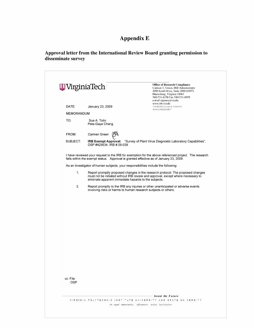
165
Appendix E
Approval letter from the International Review Board granting permission to
disseminate survey


![RESEARCHARTICLE ResistancetoSriLankanCassavaMosaic … · 2017. 7. 4. · virus [15],Cucumbermosaic virus,Zucchiniyellow mosaic virusand Watermelon mosaic virus [16–19],Beangolden](https://img.pdfslide.us/doc/110x75/6127a5c32d450a74e22164b0/researcharticle-resistancetosrilankancassavamosaic-2017-7-4-virus-15cucumbermosaic.jpg)