Embed Size (px)
Citation preview

F OCU S A R T I C L E
Tuning the size, shape and structure of RNA nanoparticlesfor favorable cancer targeting and immunostimulation
Sijin Guo1,2,3,4 | Congcong Xu1,2,3,4 | Hongran Yin1,2,3,4 | Jordan Hill5 | Fengmei Pi5 |Peixuan Guo1,2,3,4
1Center for RNA Nanobiotechnology andNanomedicine, The Ohio State University,Columbus, Ohio2Division of Pharmaceutics andPharmacology, College of Pharmacy, TheOhio State University, Columbus, Ohio3Dorothy M. Davis Heart and LungResearch Institute, The Ohio StateUniversity, Columbus, Ohio4James Comprehensive Cancer Center,College of Medicine, The Ohio StateUniversity, Columbus, Ohio5ExonanoRNA LLC, Columbus, Ohio
CorrespondencePeixuan Guo, Sylvan G. Frank EndowedChair in Pharmaceutics and Drug Delivery,The Ohio State University, 2 BiomedicalResearch Tower (BRT), 460 W 12th Ave.,Columbus, OH 43210.Email: [email protected]
Funding informationNational Cancer Institute, Grant/AwardNumbers: R01EB019036, U01CA207946;CM Chen Foundation
AbstractThe past decade has shown exponential growth in the field of RNA nanotechnology.
The rapid advances of using RNA nanoparticles for biomedical applications, espe-
cially targeted cancer therapy, suggest its potential as a new generation of drug. After
the first milestone of small molecule drugs and the second milestone of antibody
drugs, it was predicted that RNA drugs, either RNA itself or chemicals/ligands that
target RNA, will be the third milestone in drug development. Thus, a comprehensive
assessment of the current therapeutic RNA nanoparticles is urgently needed to meet
the drug evaluation criteria. Specifically, the pharmacological and immunological
profiles of RNA nanoparticles need to be systematically studied to provide insights
in rational design of RNA-based therapeutics. By virtue of its programmability and
biocompatibility, RNA molecules can be designed to construct sophisticated
nanoparticles with versatile functions/applications and highly tunable physicochemi-
cal properties. This intrinsic characteristic allows the systemic study of the effects of
various properties of RNA nanoparticles on their in vivo behaviors such as cancer
targeting and immune responses. This review will focus on the recent progress of
RNA nanoparticles in cancer targeting, and summarize the effects of common physi-
cochemical properties such as size and shape on the RNA nanoparticles' bio-
distribution and immunostimulation profiles.
This article is categorized under:
Biology-Inspired Nanomaterials > Nucleic Acid-Based Structures
Diagnostic Tools > in vivo Nanodiagnostics and Imaging
Therapeutic Approaches and Drug Discovery > Nanomedicine for Oncologic
Disease
KEYWORD S
biodistribution, immune response, immunomodulation, immunostimulation, nanobiotechnology, RNA
nanoparticle, RNA nanostructure, RNA nanotechnology
1 | INTRODUCTION
Nowadays, nanotechnology-based platforms such as lipid-based particles (Puri et al., 2009), nucleic acids nanoparticles(Andersen et al., 2009; Guo, Tschammer, Mohammed, & Guo, 2005), viral nanoparticles (Singh et al., 2007), and synthetic
Received: 23 January 2019 Revised: 13 July 2019 Accepted: 18 July 2019
DOI: 10.1002/wnan.1582
WIREs Nanomed Nanobiotechnol. 2019;e1582. wires.wiley.com/nanomed © 2019 Wiley Periodicals, Inc. 1 of 18https://doi.org/10.1002/wnan.1582

inorganic and polymeric particles (Astruc, 2012) are finding ever-increasing applications in various fields, includingnanomedicine. Nevertheless, one challenge in clinical development of nanomedicine is the lack of sufficient evidence regardingtheir safety, immunological, and pharmacological profiles. Inadvertent recognition of nanomaterials as invaders by the immunesystems frequently results in varying levels of immunostimulation or immunosuppression, which consequently leads to toxicityand reduced therapeutic efficacy. Particularly, the common challenges in the immunotoxicology assessment of nanomaterialshave been summarized by the NCI Nanotechnology Characterization Laboratory, highlighting four key areas (Chemistry, Effi-cacy, Pharmacology and Toxicology, and Hematology and Immunology) requiring thorough characterization to efficiently trans-late nanoparticle-formulated drugs toward the clinic (Dobrovolskaia, 2015). Typically, their physicochemical characterizationneeds to be well-assessed, and they are expected to display favorable pharmacokinetics (PK), pharmacodynamics (PD), andstrong safety profiles while retaining potent drug efficacy against the targeted disease. Some strategies, for example, poly (ethyl-ene glycol) (PEG) coating on cationic lipid-based nanocarriers (Suk, Xu, Kim, Hanes, & Ensign, 2016), have been proposed toengineer nanoparticles that minimize unwanted immunotoxicity while improving their in vivo performance. However, additionalside effects induced by PEG-specific antibodies were also reported (Zhang, Sun, Liu, & Jiang, 2016). Refining nanoparticle sizeis another approach proposed to diminish immune response because phagocytic cells in the immune systems tend to pick uplarger nanoparticles. Unfortunately, some nanoparticles were formulated with unpredictable or heterogeneous size distributions,making consistent assembly particularly difficult (Desai, 2012). While immune responses are vital, the favorable in vivo bio-distribution of a nanoplatform to achieve specific cancer targeting is another key factor for efficacious drug delivery. With activetargeting ligands, nanoparticles exhibit distinct advantages over traditional small molecule therapeutics for overcoming PK limi-tations by virtue of their prolonged circulation time and extended accumulation in the tumors (Singh & Lillard Jr., 2009). Uponsystemic administration, nanoparticles are exposed to the physiological environment rich with proteins, cells and tissues. Sizeand shape have both been reported to play important roles on the PK profile and biodistribution of nanoparticles (Hoshyar, Gray,Han, & Bao, 2016; Toy, Peiris, Ghaghada, & Karathanasis, 2014). Nanoparticles smaller than 5 nm undergo significant filtrationby the kidneys and are mainly excreted in urine. Larger particles, ranging from 20 to 100 nm, are easily engulfed by macro-phages or sequestered in healthy tissues, thus causing nonspecific and undesirable accumulation in healthy organs (Gustafson,Holt-Casper, Grainger, & Ghandehari, 2015). Nanoparticles with distinct shapes also exhibit different hemorheological dynamicsand cellular uptake. For instance, Discher et al reported prolonged circulation time of filamentous polymer micelles compared tospherical counterparts (Geng et al., 2007). Additionally, the surface characteristics of nanoparticles such as hydrophobicity andsurface charge can greatly influence protein adsorption and cellular membrane interactions that are important to the in vivo per-formance of nanoparticles (Albanese, Tang, & Chan, 2012). Adsorption to specific opsonin proteins such as the complement pro-teins, IgG and laminin will facilitate the recognition and uptake by immune cells, such as macrophages. As a result of thisnanoparticle uptake, macrophages secrete cytokines, possibly leading to immunostimulatory responses. Thus, notwithstandingthe achievement of nanotechnology has shown in drug delivery, the translation of synthetic or biological nanoparticles into clini-cal trials will mandate extensive investigations in accordance with strict criteria.
As a naturally-occurring biopolymer, RNA is unique compared to other nanomaterials. RNA molecules possess diversesequences, secondary structures, tertiary and quaternary interactions by nature (Hendrix, Brenner, & Holbrook, 2005). Unlikeother biomacromolecules (e.g., DNA and protein), RNA is structurally more flexible and functionally more versatile (Guo,2010; Jasinski, Haque, Binzel, & Guo, 2017). Taking advantage of both canonical Watson-Crick (A-U, G-C) and non-canonical (e.g., G-U) base pairing, a substantial number of natural RNA motifs and self-folding RNAs have been discovered.These include, but are not limited to: kink-turns (Huang & Lilley, 2013; Huang & Lilley, 2016), kissing hairpins (Bindewald,Hayes, Yingling, Kasprzak, & Shapiro, 2008), paranemic motifs (Afonin, Cieply, & Leontis, 2008), pseudoknot (Bindewald,Afonin, Jaeger, & Shapiro, 2011), three-way (Shu, Shu, Haque, Abdelmawla, & Guo, 2011; Zhang et al., 2013) and multi-helix junctions (Laing, Jung, Iqbal, & Schlick, 2009), bulges and loops (Zacharias & Hagerman, 1995). As a result of thehighly influential research in previous traditional RNA biology, the field of RNA nanotechnology has experienced an expo-nential growth in the past decade (Jasinski et al., 2017; Li et al., 2015). Myriads of sophisticated RNA architectures withhighly-ordered structures and multivalent functionalities were assembled using RNA as building blocks, such as RNA poly-gons (Boerneke, Dibrov, & Hermann, 2016; Bui et al., 2017; Dibrov, McLean, Parsons, & Hermann, 2011; Huang & Lilley,2016; Jasinski, Khisamutdinov, Lyubchenko, & Guo, 2014; Khisamutdinov et al., 2014; Ohno et al., 2011; Severcan, Geary,Verzemnieks, Chworos, & Jaeger, 2009), RNA polyhedrons (Afonin et al., 2010; Hao et al., 2014; Khisamutdinov et al.,2016; Li et al., 2016; Severcan et al., 2010; Xu et al., 2019; Yu, Liu, Jiang, Wang, & Mao, 2015), RNA rings (Geary,Chworos, Verzemnieks, Voss, & Jaeger, 2017; Grabow et al., 2011; Shu et al., 2013), RNA dendrimers (Sharma et al., 2015),jigsaw puzzles (Chworos et al., 2004), and RNA filaments (Nasalean, Baudrey, Leontis, & Jaeger, 2006) (Figure 1).
2 of 18 GUO ET AL.
Recently, the three-way junction (3WJ) motif of packaging RNA (pRNA) originated from phi29 DNA packaging motorhas been exploited as a scaffold for fabrication of multi-functional and thermodynamically stable RNA nanoparticles (Shuet al., 2011). Different components including therapeutic (miRNA, siRNA, chemical drugs), targeting (RNA aptamers orchemical ligands), and imaging (fluorophores or fluorogenic aptamers) modules were incorporated to the 3WJ motif withoutaffecting the authentic folding and original functions of modules (Shu, Khisamutdinov, Zhang, & Guo, 2013). Tremendousefforts have been made to improve the enzymatic stability of RNA by chemical modifications such as 20-fluorine (2’-F) onpyrimidines, making them dramatically resistant to RNase degradation (Corey, 2007). These RNA nanoparticles are highlyprogrammable, namely their physicochemical properties can be easily engineered to favor in vivo therapeutic delivery (Guoet al., 2017; Jasinski et al., 2014; Jasinski, Li, & Guo, 2018; Jasinski, Yin, Li, & Guo, 2018; Khisamutdinov, Li, et al., 2014;Pi et al., 2018; Shu et al., 2018). Upon systemic injection in tumor-bearing mice, the RNA nanoparticles displayed favoredaccumulation at the tumor site, with little to no accumulation in vital organs (Binzel et al., 2016; Cui et al., 2015; Lee et al.,2015; Pi et al., 2018; Rychahou et al., 2015; Shu et al., 2015; Shu et al., 2018; Shu, Haque, et al., 2013). The negativelycharged backbone of RNA disallows nonspecific interaction with negatively charged cell membranes. The pRNA-basednanoparticles showed favorable pharmacological profiles and induced negligible interferon or cytokine secretion in vivo(Abdelmawla et al., 2011; Guo et al., 2017; Khisamutdinov, Li, et al., 2014; Shu et al., 2018). Thus, the significant progressof therapeutic RNA nanoparticles has revealed its potential to pioneer a new generation of drug for the future market.It waspredicted that RNA drugs, either RNA itself or chemicals that target RNA, will become the third milestone in drug develop-ment, following the first generation of small molecule drugs and the second generation of antibody drugs (Shu et al., 2014).
In order to develop therapeutic RNA nanoparticles into a new drug, they must meet all the FDA's drug evaluation criteriaand overcome the obstacles limiting their clinical advancement. To date, many important barriers (e.g., chemical stability andthermodynamic stability) in the field of RNA nanotechnology have been successfully overcome (Corey, 2007;Khisamutdinov, Jasinski, & Guo, 2014; Shu et al., 2011). The progress in these aspects has been discussed in previous reviews(Jasinski et al., 2017; Li et al., 2015; Xu et al., 2018). Recently, however, more studies have been reported on understandingthe broad mechanisms that define the pharmacological and immunological profile of RNA nanoparticles. A variety of theirphysicochemical properties were recognized to be associated with their in vivo behaviors, including size, shape,stoichiometry/module density, surface chemistry, composition, hydrophobicity, and elasticity (Abdelmawla et al., 2011;Afonin et al., 2014; Guo et al., 2017; Halman et al., 2017; Hong et al., 2018; Jasinski, Li, & Guo, 2018; Jasinski, Yin, et al.,2018; Johnson et al., 2017; Ke et al., 2018; Khisamutdinov, Li, et al., 2014; Lee, Urban, Xu, Sullenger, & Lee, 2016; Pi et al.,2018; Shi et al., 2018; Shu et al., 2018). Here, this brief review aims to integrate the recent progress seen in the study of RNAnanoparticle on biodistribution and immunostimulation, while focusing on the defining aspects of their design andphysiochemical properties precisely tuned to treat different cancers.
2 | DEFINITION OF RNA NANOTECHNOLOGY
RNA nanotechnology is a relatively new field distinctive from traditional RNA biology. The concept of RNA nanotechnologyis defined as the study on the design, construction, and application of RNA nanoparticles at the nanometer scale that are pri-marily composed of RNA via bottom-up self-assembly (Guo, 2010). The major frame of RNA nanoparticles can include scaf-folds, targeting ligands, regulatory moieties, and therapeutic modules. The field focuses on the inter-RNA interactions andquaternary interactions of small RNA motifs, which is different from classical RNA research focusing on intra-RNA interac-tions and 2D/3D structure–function relationships. Nevertheless, the broad knowledge gained from classical RNA biologyresearch has laid a solid foundation for RNA nanotechnology. Since the introduction of the concept describing RNAnanoarchitectures in 1996 and 1998 by RNA tectonics and reengineered pRNA molecules respectively (Guo, Zhang, Chen,Trottier, & Garver, 1998; Jaeger, Westhof, & Leontis, 2001), RNA nanotechnology has grown rapidly as a platform withextensive applicational studies in nanomedicine over the past decade (Jasinski et al., 2017; Li et al., 2015).
3 | ADVANTAGES OF RNA NANOTECHNOLOGY FOR CANCER TARGETINGAND IMMUNOMODULATION
3.1 | RNA nanoparticles are distinct from traditional therapeutic RNAs
Traditional therapeutic RNAs, such as siRNA (Elbashir et al., 2001), miRNA (Bartel, 2004), anti-miRNA (Elmen et al.,2008), mRNA (Sahin, Kariko, & Tureci, 2014), viral immunostimulatory RNA (isRNA) (Bourquin et al., 2007), and
GUO ET AL. 3 of 18

FIGURE 1 Modular design and construction of RNA nanostructures. (a) pRNA hexamer from phi29 DNA packaging motor (Reprinted withpermission from Shu, Haque, et al. (2013). Copyright 2013 RNA society); (b) RNA square with tectoRNA motif (Reprinted with permission from Chworoset al. (2004). Copyright 2004 The American Association for the Advancement of Science); (c) RNA triangle with IIa motif from SVV IRES (Reprinted withpermission from Boerneke et al. (2016)). Copyright 2016 John Wiley & Sons, Inc.) & RNA square with IIa-1 motif from HCV (Reprinted with permissionfrom Dibrov et al. (2011). Copyright 2011 National Academy of Sciences; (d) RNA triangle and square with k-turn motif (Reprinted with permission fromHuang and Lilley (2016). Copyright 2016 The Royal Society of Chemistry); (e) RNA triangle with tetra-U motif (Reprinted with permission from Bui et al.(2017). Copyright 2017 Elsevier Inc).; (f) RNA-protein triangle with k-turn motif (Reprinted with permission from Ohno et al. (2011). Copyright 2011Nature Publishing Group); (g) RNA tectosquare with RA, 3WJ, and tRNA motifs (Reprinted with permission from Severcan et al. (2009). Copyright 2009American Chemical Society); (h) RNA nanoring based on RNAI/II inverse kissing complex (Reprinted with permission from Grabow et al. (2011).Copyright 2011 American Chemical Society); (i) RNA cube designed in silico (Reprinted with permission from Afonin et al. (2010). Copyright 2010Macmillan Publishers Limited); (j) pRNA-3WJ motif-based RNA tetrahedron (Reprinted with permission from Li et al. (2016). Copyright 2016 JohnWiley & Sons, Inc.), pyramid (Reprinted with permission from Xu C et al. (2018). Copyright 2018 Springer Nature), & nanoprism (Reprinted withpermission from Khisamutdinov et al. (2016)). Copyright 2016 John Wiley & Sons, Inc.); (k) RNA polyhedron made of tRNA subunit (Reprinted withpermission from Severcan et al. (2010). Copyright 2010 Macmillan Publishers Limited); (l) triangular and tetragonal RNA prism from re-engineered pRNA(Reprinted with permission from Hao et al. (2014). Copyright 2014 Macmillan Publishers Limited); (m) homo-octameric RNA prism with T-junction RNAtile (Reprinted with permission from Yu et al. (2015). Copyright 2015 Macmillan Publishers Limited); (n) RNA dendrimers from pRNA-3WJ motif(Reprinted with permission from Sharma et al. (2015). Copyright 2015 Elsevier Inc.); (o) RNA nanoheart from a syntax of RNA modules (Reprinted withpermission from Geary et al. (2017)). Copyright 2017 American Chemical Society
4 of 18 GUO ET AL.
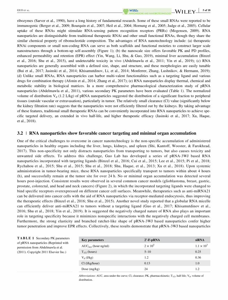
ribozymes (Sarver et al., 1990), have a long history of fundamental research. Some of these small RNAs were reported to beimmunogenic (Berger et al., 2009; Bourquin et al., 2007; Heil et al., 2004; Hornung et al., 2005; Judge et al., 2005). Cellularuptake of these RNAs might stimulate RNA-sensing pattern recognition receptors (PRRs) (Mogensen, 2009). RNAnanoparticles are distinguishable from traditional therapeutic RNAi and other small functional RNAs, though they share thesimilar chemical property of ribonucleotide composition. The advantages of RNA nanotechnology include: (a) therapeuticRNAi components or small non-coding RNA can serve as both scaffolds and functional moieties to construct larger scalenanostructures through a bottom-up self-assembly (Figure 1); (b) the nanoscale size offers favorable PK and PD profiles,enhanced permeability and retention (EPR) effect (Yin, Wang, Li, Shu, & Guo, 2019), minimal liver accumulation (Binzelet al., 2016; Shu et al., 2015), and undetectable toxicity in vivo (Abdelmawla et al., 2011; Yin et al., 2019); (c) RNAnanoparticles are generally assembled with a defined size, shape, and structure, and these morphologies are easily tunable(Bui et al., 2017; Jasinski et al., 2014; Khisamutdinov, Li, et al., 2014; Monferrer, Zhang, Lushnikov, & Hermann, 2019);(d) Unlike small RNAs, RNA nanoparticles can harbor multi-valent functionalities such as a targeting ligand and variousdrugs for combination therapy (Afonin et al., 2014; Zhang et al., 2017); (e) RNA nanoparticles display thermal, chemical andmetabolic stability in biological matrices. In a more comprehensive pharmacological characterization study of pRNAnanoparticles (Abdelmawla et al., 2011), various secondary PK parameters have been evaluated (Table 1). The normalizedvolume of distribution Vd (1.2 L/kg) of pRNA nanoparticles suggested the distribution of a significant fraction to peripheraltissues (outside vascular or extravasation), particularly in tumor. The relatively small clearance (Cl) value (significantly belowthe kidney filtration rate) suggests that the nanoparticles were not efficiently filtered out by the kidneys. By taking advantageof these features, traditional small therapeutic RNAs can be conveniently incorporated into RNA nanoparticles to achieve spe-cific targeted delivery, an extended in vivo half-life, and higher therapeutic efficacy (Jasinski et al., 2017; Xu, Haque,et al., 2018).
3.2 | RNA nanoparticles show favorable cancer targeting and minimal organ accumulation
One of the critical challenges to overcome in cancer nanotechnology is the non-specific accumulation of administerednanoparticles in healthy organs including the liver, lungs, kidneys, and spleen (Shi, Kantoff, Wooster, & Farokhzad,2017). This non-specificity not only distracts nanoparticles from transporting to tumors, but also causes toxicity andunwanted side effects. To address this challenge, Guo Lab has developed a series of pRNA-3WJ based RNAnanoparticles incorporated with targeting ligands (Binzel et al., 2016; Cui et al., 2015; Lee et al., 2015; Pi et al., 2018;Rychahou et al., 2015; Shu et al., 2015; Shu et al., 2018; Shu, Haque, et al., 2013; Xu et al., 2018). Upon systemicadministration in tumor-bearing mice, these RNA nanoparticles specifically transport to tumors within about 4 hours(h), and successfully remain at the tumor site for over 24 h. No or minimal organ accumulation was detected severalhours post-injection. Consistent results were observed in several common cancer models (glioblastoma, breast, gastric,prostate, colorectal, and head and neck cancers) (Figure 2), in which the incorporated targeting ligands were changed tobind specific receptors overexpressed on different cancer cell surfaces. Meanwhile, therapeutics such as anti-miRNA21can be delivered into cancer cells with the aid of RNA nanoparticles via receptor-mediated endocytosis, thus improvingthe therapeutic effects (Binzel et al., 2016; Shu et al., 2015). Another novel study reported that a globular RNA micellecan efficiently deliver anti-miRNA21 to tumors without a targeting ligand (Guo et al., 2017; Khisamutdinov et al.,2016; Shu et al., 2018; Yin et al., 2019). It is suggested the negatively charged nature of RNA also plays an importantrole in targeting specificity because it minimizes nonspecific interactions with the negatively charged cell membranes.Furthermore, the strong elasticity and branched ratchet-like shape of pRNA-3WJ based nanoparticles confer highertumor penetration and improve EPR effects. Collectively, these results demonstrate that pRNA-3WJ based nanoparticles
TABLE 1 Secondary PK parametersof pRNA nanoparticles (Reprinted withpermission from Abdelmawla et al.(2011). Copyright 2011 Elsevier Inc.)
Key parameters 2’-F-pRNA siRNA
AUClast (hour�ng/ml) 2 × 10⁵ 1.1 × 10³
T1/2 (hours) 5–10 0.25
Vd (l/kg) 1.2 0.36
Cl (l/kg/hour) 0.13 1.0
Dose (mg/kg) 24 1.2
Abbreviations: AUC, area under the curve; Cl, clearance; PK, pharmacokinetic; T1/2, half-life; Vd, volume ofdistribution.
GUO ET AL. 5 of 18

can be conveniently engineered with active targeting ligands to achieve specific cancer targeting with low accumulationin healthy organs. This favorable biodistribution is an important indication of RNA nanoparticles' pharmacologicalprofiles.
FIGURE 2 Specific cancer targeting in vivo of RNA nanoparticles to (a) brain cancer (Reprinted with permission from Lee et al., 2015).Copyright 2015 Impact Journals; (b) breast cancer (Reprinted with permission from Shu et al. (2015). Copyright 2015 American Chemical Society);(c) gastric cancer (Reprinted with permission from Cui et al. (2015). Copyright 2015 Macmillan Publishers Limited); (d) prostate cancer (Reprintedwith permission from Binzel et al. (2016). Copyright 2016 Elsevier Inc.); (e) colorectal cancer (Reprinted with permission from Rychahou et al.(2015); Xu, Pang, et al. (2018). Copyright 2015 American Chemical Society & Elsevier B.V).; (f) Head & Neck cancer (Reprinted with permissionfrom Shu, Haque, et al. (2013). Copyright 2013 RNA society); (g) specific cancer targeting of RNA/EVs (Reprinted with permission from Pi et al.(2018). Copyright 2018 Springer Nature Publishing), and (h) RNA micelles (Reprinted with permission from Shu et al. (2018)). Copyright 2018Elsevier Inc)
6 of 18 GUO ET AL.

3.3 | RNA nanoparticles intrinsically display immunologically inert property and non-toxicity
Due to the lack of a universal nomenclature to categorize traditional therapeutic RNAs and RNA nanoparticles, the literatureson RNA immunogenicity have been controversial. Though the immunogenicity of traditional small RNAs has been widelyinvestigated, only a limited number of studies focused on the immunogenicity of RNA nanoparticles. Recent studies revealedthat pRNA-based RNA nanoparticles intrinsically display immunologically inert properties (Abdelmawla et al., 2011; Guoet al., 2017; Khisamutdinov, Li, et al., 2014; Shu et al., 2018). Specifically, no or negligible cytokine induction includingTNF-α (Tumor Necrosis Factor-α), IL-6 (Interleukin-6) and IFN-α (Interferon-α) has been observed following treatment withpRNA nanoparticles in vitro and in vivo. TNF-α is a cytokine involved in systemic inflammation and the acute phase reaction.IL-6 is an interleukin secreted to stimulate immune responses during infection. IFN-α, which belongs to type I interferon, isalso a cytokine involved in pro-inflammatory reactions released in response to the presence of viral pathogens. Besides, nostimulation of TLRs (Toll-like receptors) pathway, and no damage to normal tissue and organs was detected in multiple celltypes and mice (Cui et al., 2015; Zhang et al., 2017). Similarly, RNA polygons (RNA triangle, square, and pentagon) con-structed from the thermodynamically stable pRNA-3WJ were studied (Guo et al., 2017; Khisamutdinov, Li, et al., 2014).These RNA polygons have been considered nonimmunogenic and nontoxic because undetectable or negligible cytokineinduction and cytotoxicity were observed in vitro and in vivo, compared to positive controls. Consistent results were found inthree dimensional pRNA-based nanoparticles, including the RNA tetrahedron, RNA nanoprism, and RNA micelles (Guoet al., 2017; Khisamutdinov et al., 2016; Shu et al., 2018; Yin et al., 2019). Additionally, RNA aptamers, a family of RNA oli-gonucleotides commonly incorporated into RNA nanoparticles as targeted ligands to enhance binding specificity, have beenreported to go unrecognized by the host immune system in various animal studies (Song, Lee, & Ban, 2012). These findingsdemonstrate that RNA nanoparticles equipped with targeting ligands can serve as safe delivery vectors in therapeutic interven-tions. Studies by Afonin Lab using a different system have also shown no immune response detection upon treatment withRNA nanoparticles in human peripheral blood mononuclear cells (PBMCs) from healthy donors (Hong et al., 2018). Interest-ingly, only complexation with a delivery carrier such as lipofectamine 2000 induced immunorecognition by PBMCs.
4 | PHYSICOCHEMICAL PROPERTIES OF RNA NANOPARTICLES AFFECTIN VIVO BIODISTRIBUTION AND IMMUNE RESPONSE
Nanotechnology offers a substantial number of benefits over traditional routes for drug delivery, but unfavorable immuneresponses and liver accumulation have also been reported (Buzea, Pacheco, & Robbie, 2007; Zolnik, Gonzalez-Fernandez,Sadrieh, & Dobrovolskaia, 2010). It has been suggested that the adverse effects were elicited by numerous physicochemicalcharacteristics, including size, shape, surface chemistry, or hydrophobicity (Dobrovolskaia, 2015; Dobrovolskaia, Shurin, &Shvedova, 2016). Engineering these properties with precision and homogeneity is a common strategy to improve the in vivoperformance of nanomaterials. One of the advantages of RNA nanotechnology is its high programmability. In other words,their physicochemical properties are easily tunable (Figure 3), and the production process is highly consistent. Therefore, theeffects of these properties can simply be studied as a result of reproducible nanoparticle assembly. The following subsectionswill focus on the main physicochemical properties of RNA nanoparticles and the corresponding effects on their immuno-stimulation and biodistribution.
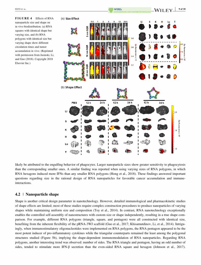
4.1 | Nanoparticle size
Nanoparticle size represents one of the most critical considerations in the design and construction of RNA nanoparticles. Itwas found that size significantly dictates nanoparticle performance at the nano-bio interface, including vascular transportation,plasma protein binding, and cellular membrane interaction (Albanese et al., 2012; Hoshyar et al., 2016). Large particles(>100 nm) tend to be trapped in the liver and spleen as a result of the stronger recognition by the mononuclear phagocytic sys-tem (MPS) in these organs (Gustafson et al., 2015). Particles with a small diameter (<10 nm) are more likely to have a fasterrenal clearance, thus leading to a shorter half-life in vivo (Longmire, Choyke, & Kobayashi, 2008). In a systemic in vivo bio-distribution study, the effect of RNA nanoparticle size on their circulation time and accumulation in healthy organs and tumorshas been evaluated (Jasinski, Li, & Guo, 2018). Specifically, RNA squares of three different sizes (5 nm, 10 nm, 20 nm) wereintravenously administered into tumor-bearing mice. Internal organ imaging at 12 and 24 h time points showed the rapid elim-ination of 5 nm RNA squares from vital organs with significant accumulation in the tumor after 12 h (Figure 4a). It wassuggested that renal excretion is the primary excretion route for the nanoparticles (Piao, Wang, Binzel, & Guo, 2018). For the
GUO ET AL. 7 of 18

10 and 20 nm RNA squares, stronger interaction with macrophages and slower metabolism in the liver was observed, whichis possibly caused by the different protein binding profiles of large nanoparticles compared to that of small ones. The correla-tion of size with the biodistribution profile should be considered from the perspective of its effects on renal clearance and mac-rophage uptake. Particularly, smaller RNA nanoparticles exhibited less uptake by macrophages of the MPS due to less serumprotein binding. However, they are more rapidly excreted by kidney filtration, while the opposite trend is seen with the largerRNA nanoparticles. Thus, the final in vivo fate of RNA nanoparticles will take a balance between these two size-dependentelimination pathways (i.e., macrophages and urinary excretion).
The effect of varied sizes will manifest itself in the interaction between particles and immune system as well. Small sizeswill benefit from not being recognized by the bulky opsonins in complement cascade, a key component of the immune system,due to the inadequate accommodation on particle surfaces (Ventola, 2012). RNA nanoparticles have been deliberately con-structed in a size range from 10 to 40 nm, making them advantageous for drug delivery. Recently, the effect of size on theirimmunostimulation has been studied (Guo et al., 2017). RNA nanoparticles with identical square shape but varying size wereused as the model (Figure 5a). After extending the single-stranded sequence at the vertexes, RNA squares were endowed withimmunostimulatory activity in a size-dependent fashion. Small RNA squares (7.10 nm) elevated the immunomodulation tosome extent, while stronger responses were observed with medium (12.31 nm) and large (21.15 nm) RNA squares. This can
FIGURE 3 Construction ofRNA nanostructures with tunableproperties. (a) RNA squares withsmall, medium, and large size bytuning the length of the connectinghelix (Reprinted with permissionfrom Jasinski et al. (2014).Copyright 2014 AmericanChemical Society). (b) RNAtriangle, square and pentagon bytuning the interior pRNA-3WJangle (Reprinted with permissionfrom Khisamutdinov, Li, et al.(2014). Copyright 2014 AmericanChemical Society). (c) 3D RNAcube, planar RNA nanoring, andlinear RNA fiber by differentconnectivity (Reprinted withpermission from Hong et al.(2018). Copyright 2018 AmericanChemical Society)
8 of 18 GUO ET AL.
likely be attributed to the engulfing behavior of phagocytes. Larger nanoparticle sizes show greater sensitivity to phagocytosisthan the corresponding smaller ones. A similar finding was reported when using varying sizes of RNA polygons, in whichRNA hexagons induced more IFNs than any smaller RNA polygons (Hong et al., 2018). These findings answered importantquestions regarding size in the rational design of RNA nanoparticles for favorable cancer accumulation and immune-interactions.
4.2 | Nanoparticle shape
Shape is another critical design parameter in nanotechnology. However, detailed immunological and pharmacokinetic studiesof shape effects are limited; most of these studies require complex construction procedures to produce nanoparticles of varyingshapes while maintaining uniform size and composition (Toy et al., 2014). In contrast, RNA nanotechnology exceptionallyenables the controlled self-assembly of nanostructures with custom size or shape independently, resulting in a true shape com-parison. For example, different RNA polygons (triangle, square, and pentagon) were all constructed with identical size,benefiting from the inherent flexibility of the pRNA-3WJ scaffold (Guo et al., 2017; Khisamutdinov, Li, et al., 2014). Intrigu-ingly, when immunostimulatory oligonucleotides were implemented on RNA polygons, the RNA pentagon appeared to be themost potent inducer of pro-inflammatory cytokines while the triangular counterparts remained the least among the polygonalstructures studied (Figure 5b), implying the shape-dependent immunomodulation of RNA nanoparticles. Regarding RNApolygons, another interesting trend was observed: number of sides. The RNA triangle and pentagon, having an odd number ofsides, tended to stimulate more IFN-β secretion than the even-sided RNA square and hexagon (Johnson et al., 2017).
FIGURE 4 Effects of RNAnanoparticle size and shape onin vivo biodistribution. (a) RNAsquares with identical shape butvarying size, and (b) RNApolygons with identical size butvarying shape show differentcirculation times and tumoraccumulation in vivo. (Reprintedwith permission from Jasinski, Li,and Guo (2018). Copyright 2018Elsevier Inc.)
GUO ET AL. 9 of 18

Additionally, a significant difference was found in immune responses between RNA nanoparticles with varying dimensionalstructures. A 3D RNA tetrahedron carrying immunostimulatory oligonucleotides exhibited stronger immunostimulatory activ-ity than the planar triangular counterpart, when size and payload stoichiometry were controlled to be equivalent (Guo et al.,2017). Likewise, globular RNA cubes induced stronger immunostimulation compared to planar hexameric RNA rings, whichwere more immunostimulatory than RNA fibers (Figure 5e) (Hong et al., 2018). These findings suggest a trend of increasingimmunostimulatory properties of RNA nanoparticles from linear, to planar, and to 3D structure. One interpretation may be thatthe increased surface area intrinsic to 3D structures provides a spacious surface for complement opsonins assembly and depo-sition, while a greater proportion of opsonins released into the surrounding medium with linear and planar RNA structures.
The shape of nanoparticles has been shown to dictate the interactions that occur with cell membranes and circulating serumproteins. For instance, studies have suggested that oblate-shaped nanoparticles with discoidal geometries are more likely to
FIGURE 5 Effects of RNA nanoparticles' physicochemical properties on immunostimulation. RNA nanoparticles with (a) varying size,(b) varying shape, (c) different stoichiometry, (d) different sequence, and (e) different dimension induced cytokines and interferons secretion tovarious levels. (figures A-D and E reprinted with permission from Guo et al. (2017). Copyright 2018 Elsevier Inc). and (Reprinted with permissionfrom Hong et al. (2018). Copyright 2018 American Chemical Society, respectively)
10 of 18 GUO ET AL.

migrate toward blood vessel walls and establish greater interactions with endothelial cells of blood vessels in comparison tospherical nanoparticles (Muller, Fedosov, & Gompper, 2014). The biodistribution profiles of RNA polygons of different shapebut uniform size were compared in tumor-bearing mouse models after systemic administration (Jasinski, Li, & Guo, 2018).Different retention in organs were observed at 12 h time point as nanosquares showed high fluorescent signal intensity whiletriangle nanoparticles showed none and the pentagon very little. In the spleen, pentagon nanoparticles exhibited the highestfluorescence. A similar biodistribution in organs was found among the particles after 24 h (Figure 4b). Therefore, the proteincorona formation on the RNA nanoparticles may drastically change in response to nanoparticle shapes, which will furtherimpact their elimination pathways. Additionally, the cellular interactions with nanoparticles as well as internalization are alsoclosely related to the size and shape. Cell receptors that mediate the endocytosis are of various sizes and shapes, so it will pro-vide beneficial information on rational design of nanoparticles that possess favorable binding to the receptors, thus enhancingnanoparticle recognition and cellular uptake. Considering the controllable size, shape and other physicochemical properties,RNA nanoparticles could potentially be designed with enhanced tumor cell uptake and retention.
4.3 | Sequence signature and modular stoichiometry
As a biocompatible nanomaterial, RNA nanoparticles are immunologically inert. In contrast, some special RNA sequenceshave been reported to trigger immune responses, named isRNAs, due to the specific recognition by toll-like receptors (TLRs)or cytosolic sensors (PKR, RIG-1, and MDA-5) in immune cells (Berger et al., 2009; Bourquin et al., 2007; Heil et al., 2004;Hornung et al., 2005; Judge et al., 2005). Incorporation of these isRNA sequences can turn immunologically inert RNAnanoparticles to immunologically active, or even enhance the immune response associated with an incorporated module. In astudy from Guo Lab, a specific RNA SEQ was extended to the vertexes of RNA squares and dramatically engendered the pro-duction of pro-inflammatory cytokines in vitro and in vivo (Figure 5d) (Guo et al., 2017). The immune responses were indirect proportion to the stoichiometry of single-stranded RNA extensions. RNA squares with increasing copies of payloadinduced stronger cytokine levels (Figure 5c). Conversely, mutation or complementary blockage of the extension sequenceresulted in reduced immune responses, while scrambling the extension sequence led to complete abrogation of immuneresponse. This study affords a new sight in the design and construction of RNA nanoparticles—they can be constructed toserve as safe therapeutic nanocarriers with non-immunogenicity, or deliberately trigger a strong immune response forimmunotherapy.
4.4 | Surface chemistry
Surface characteristics have been shown to be a significant parameter in the pharmacokinetics and pharmacodynamics ofmany nanomaterials, as well as influencing their immune system interactions (Albanese et al., 2012). Some cationicnanoparticles, such as polyethyleneimines, can easily interact with cell membranes, causing nonspecific cytotoxicity and giv-ing rise to complement system activation (Merkel et al., 2011). In contrast, RNA nanoparticles have consistently exhibited theadvantage of causing no or undetectable cytotoxicity in many studies due to their polyanionic nature (Shu et al., 2014).Surface-projected composition is another factor that greatly defines the in vivo fate of nanoparticles (Merkel et al., 2011). Asa naturally aqueous-soluble biopolymer, RNA nanoparticles are distinct from many synthetic nanomaterials in that they do notrequire surface modifications, such as polyethylene glycol (PEG) grafting (Suk et al., 2016), to increase aqueous-solubilityand consequently limit their immune response and increase in vivo circulation. This advantage provides RNA nanoparticlesthe flexibility to incorporate various functional modules, including RNAi therapeutics, chemical drugs, fluorophores, andtargeting ligands, to achieve multi-functionality. Particularly, some of these surface compositions, especially drugs,fluorophores, or other hydrophobic compounds, might be important factors in determining how RNA nanoparticles communi-cate with cells or proteins in vivo. Nanoparticles decorated with more hydrophobic reagents will result in greater plasma pro-tein binding, and therefore greater accumulation in the liver or other organs. Jasinski et al. reported that different chemicalsincorporated to RNA nanoparticles altered the RNA hydrophobicity to varying degrees (Figure 6a) (Jasinski, Yin, et al.,2018). The changes in vital organ accumulation as a function of hydrophobicity variation was investigated. Weaker organaccumulation was detected for RNA nanoparticles (3WJ-Fluor) containing hydrophobic fluorophores (Cyanine5.5,Sulfonated-Cyanine 5.5, and AlexaFluor700) than these fluorophores alone (Figure 6a-c), clearly indicating the capacity ofRNA nanoparticles to solubilize hydrophobic compounds. In another study, paclitaxel (PTX), an antitumor chemo-drug withpoor aqueous-solubility, was conjugated to micellar RNA nanoparticle (Figure 6b) (Shu et al., 2018). Consequently, the RNAmicelle/PTX complex showed significantly enhanced water-solubility and efficient cancer targeting in vivo. Additionally,
GUO ET AL. 11 of 18
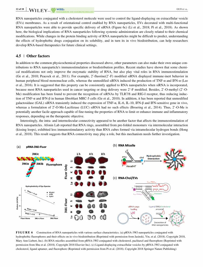
RNA nanoparticles conjugated with a cholesterol molecule were used to control the ligand-displaying on extracellular vesicle(EVs) membranes. As a result of orientational control enabled by RNA nanoparticles, EVs decorated with multi-functionalRNA nanoparticles were able to provide specific delivery of siRNA (Figure 6c) (Li et al., 2018; Pi et al., 2018). As shownhere, the biological implications of RNA nanoparticles following systemic administration are closely related to their chemicalmodifications. While changes in the protein binding activity of RNA nanoparticles might be difficult to predict, understandingthe effects of hydrophobic drugs conjugation on its solubility, and in turn its in vivo biodistribution, can help researchersdevelop RNA-based therapeutics for future clinical settings.
4.5 | Other factors
In addition to the common physicochemical properties discussed above, other parameters can also make their own unique con-tributions to RNA nanoparticle's immunostimulation or biodistribution profiles. Recent studies have shown that some chemi-cal modifications not only improve the enzymatic stability of RNA, but also play vital roles in RNA immunostimulation(Ge et al., 2010; Peacock et al., 2011). For example, 20-fluorine(2’-F) modified siRNA displayed immune-inert behavior inhuman peripheral blood mononuclear cells, whereas the unmodified siRNA induced the production of TNF-α and IFN-α (Leeet al., 2016). It is suggested that this property can be consistently applied to RNA nanoparticles when siRNA is incorporated,because most RNA nanoparticles used in cancer targeting or drug delivery were 2’-F modified. Besides, 2’-O-methyl (2’-O-Me) modification has been found to prevent the recognition of siRNAs by TLR7/8 and RIG-I receptor, thus reducing induc-tion of TNF-α and IFN-β in human fibroblast MRC-5 cells (Ge et al., 2010). In addition, it has been reported that unmodifiedgalactosidase (GAL) siRNA transiently induced the expression of TNF-α, IL-6, IL-10, IFN-β and IFN-sensitive gene in vivo,whereas a formulation of 20-O-Me-Luciferase (LUC) siRNA had no such effects (Broering et al., 2014). Thus, 2’-O-Me ispotentially another facile approach capable of fine-tuning the properties of RNA to limit or enhance immune and inflammatoryresponses, depending on the therapeutic objective.
Interestingly, the intra- and intermolecular connectivity appeared to be another factor that affects the immunostimulation ofRNA nanoparticles. Afonin Lab reported that RNA rings, assembled from pre-folded monomers via intermolecular interaction(kissing loops), exhibited less immunostimulatory activity than RNA cubes formed via intramolecular hydrogen bonds (Honget al., 2018). This result suggests that RNA connectivity may play a role, but this mechanism needs further investigation.
FIGURE 6 Construction of RNA nanoparticles with various surface characteristics. (a) pRNA-3WJ nanoparticles conjugated withhydrophobic fluorophores and their effects on in vivo biodistribution (Reprinted with permission from Jasinski, Yin, et al. (2018). Copyright 2018,Mary Ann Liebert, Inc). (b) RNA micelles assembled from pRNA-3WJ conjugated with cholesterol, paclitaxel and fluorophore (Reprinted withpermission from Shu et al. (2018). Copyright 2018 Elsevier Inc). (c) Ligand-displaying extracellular vesicles by pRNA-3WJ conjugated withcholesterol, ligand aptamer, and fluorophore (Reprinted with permission from Pi et al. (2018). Copyright 2018 Springer Nature Publishing)
12 of 18 GUO ET AL.

Additionally, it has been reported that nanoparticle elasticity influences vascular transport, biodistribution, cellular internal-ization, and immunostimulation (Anselmo et al., 2015). RNA as a biopolymer has shown rubber-like elastic property (Chiuet al., 2014; Jacobson, McIntosh, Stevens, Rubinstein, & Saleh, 2017), allowing RNA nanoparticles to “squeeze” through vas-culatures of the tumor microenvironment by blood pressure without altering its thermodynamic stability. Meanwhile, manyRNA nanoparticles were constructed to be ratchet-shaped after incorporating multiple modules (Haque et al., 2012; Shu et al.,2011), preventing them from returning to blood circulation. Therefore, the effects of these properties favor the transport ofRNA nanoparticles toward tumors and enhance the EPR effect.
5 | PERSPECTIVES
5.1 | RNA nanotechnology for potential immunotherapy
One of the most important recent breakthroughs in cancer research is cancer immunotherapy (Couzin-Frankel, 2013; McNutt,2013). Extensive studies revealed important information regarding the complicated cancer-immune system relationship, andmany researchers are now looking to summon the self-defense system in the hosts to kill cancer. The most common immuno-therapies include chimeric antigen receptors (CAR) T-cell therapy (June, O'Connor, Kawalekar, Ghassemi, & Milone, 2018),immune checkpoint blockade (Pardoll, 2012), monoclonal antibodies (mAb) (Weiner, Dhodapkar, & Ferrone, 2009), and can-cer vaccines and adjuvants (Temizoz, Kuroda, & Ishii, 2016), just to name a few. Particularly, nucleic acid aptamers haveemerged as a new type of therapeutic for immunotherapy (Pastor et al., 2018). Aptamers are short nucleic acid oligomers(12-80 nt) selected from SELEX (systematic evolution of ligands by exponential enrichment) and are capable of binding tar-gets specifically and tightly. Various kinds of co-stimulatory molecules belonging to the B7/CD28 family have been selectedto trigger cell mediated immune responses. For instance, CTLA-4 RNA aptamer developed by Santilli-Marotto et al. can bindCTLA-4 with high affinity, inhibit CTLA-4 function, and enhance tumor immunity in mice (Santulli-Marotto, Nair, Rusconi,Sullenger, & Gilboa, 2003). Furthermore, aptamers targeting the TNF/TNFR family which are involved in the later phase ofT-cells activation have been developed, including OX40 and 4-1BB aptamers (McNamara et al., 2008). In order to take advan-tage of the RNA nanoparticle platforms, nucleic acid aptamers can be incorporated into the RNA scaffold to achieve strongerimmunotherapeutic effects. Meanwhile, the multivalent property allows the additional incorporation of RNAi therapeutics orchemotherapeutic drugs to realize combination therapy.
Although immunotherapy has shown success in various cancers, some clinical challenges remain, such as the safety andefficacy concerns derived from the systemic dosing of immunomodulatory agents (Whiteside, Demaria, Rodriguez-Ruiz,Zarour, & Melero, 2016). RNA nanotechnology, as a safe and efficient drug delivery platform, can potentially enhance theefficacy as well as reduce the side effects of such immunotherapies by improving the delivery, retention, and release of immu-nomodulatory agents in targeted cell populations and organs. RNA, as a biomacromolecule, can be successfully recognized bythe immune system as a self-entity, and thus RNA nanoparticles intrinsically display immunologically inert property. Asdescribed above, though naturally inert, RNA nanoparticles can be manually designed using their tunable and programmableproperties to exhibit no, low, or high immunostimulation, thus allowing them to be employed as safe therapeutic carriers with-out triggering an immune response, or as potential immunomodulators for cancer immunotherapy.
5.2 | Understanding the interactions of RNA nanoparticles at the nano-bio interface
Upon introduction into biological environment, nanoparticles interacting with proteins, membranes, cells and organs establisha series of nanoparticle/biological interactions at the interfaces which govern the in vivo fate of nanoparticles (Cheng, Jiang,Wang, Chen, & Liu, 2013; Nel et al., 2009). As little is known about the interactions of RNA nanoparticles with biologicalcomponents, a better understanding at the nano-bio interface will be essential to the rational design of RNA nanoparticlescapable of targeted delivery (Xu, Haque, et al., 2018). At the molecular level, protein corona formation around nanoparticlesdrastically influences their in vivo behavior, resulting in different elimination pathways (Kim, Faix, & Schnitzer, 2017; Tenzeret al., 2013). Moreover, the component in the protein corona, such as the complement proteins, can potentially lead to alteredimmune responses (Chen et al., 2017). Previous studies on serum protein binding of RNA polygon nanoparticles showed thatthe size and shape play critical roles in protein binding (Jasinski, Li, & Guo, 2018). However, the composition of proteincorona formation on RNA nanoparticles has not been comprehensively determined. At the cellular lever, it appears the fre-quency of RNA nanoparticle uptake into macrophages is closely related to their morphology and size, as these factors impactthe engulfing process (Guo et al., 2017). The internalization of RNA nanoparticles with targeting ligands into cancer cells is
GUO ET AL. 13 of 18

proposed to be receptor-mediated pathway (Shu et al., 2014). However, the impacts of physicochemical properties on theintracellular trafficking of RNA nanoparticles are still not completely understood and await more investigation.
6 | CONCLUSION
RNA nanotechnology is growing exponentially, though its inception lags behind other nano-delivery systems. As shownwithin this review, RNA nanoparticles display many advantages in biomedicine. As drug carriers, RNA nanoparticles haverepeatedly shown immunologically inert behavior, while can be concomitantly manipulated to exhibit controlled immuno-stimulation. RNA nanoparticles possess a variety of advantageous physicochemical properties over other nanomaterials,including the capacity to be precisely programmed. As a result, RNA nanoparticles can be rationally designed, optimized, andconstructed for specialized in vivo applications. As a biocompatible nanomaterial, RNA nanoparticles show favorable tumortargeting proficiency, as evidenced in various pre-clinical cancer models. The extensive research conducted in order to under-stand the safety, immunological, and pharmacological profiles of RNA nanoparticles have positively paved a path toward clin-ical trials. Evidently, RNA nanotechnology is bespeaking a bright future in cancer therapy.
ACKNOWLEDGMENTS
The research in P.G.'s lab was supported by NIH grants R01EB019036 and U01CA207946. The authors would like to thankLora E. McBride for her constructive comments and revisions on the article. P.G.'s Sylvan G. Frank Endowed Chair positionin Pharmaceutics and Drug Delivery is funded by the CM Chen Foundation.
CONFLICT OF INTEREST
P.G. is the consultant of Oxford Nanopore Technologies and Nanobio Delivery Pharmaceutical Co. Ltd, as well as thecofounder of Shenzhen P&Z Bio-medical Co. Ltd and its subsidiary US P&Z Biological Technology LLC, as well asExonanoRNA, LLC and its subsidiary ExonanoRNA (Foshan) Biomedicine Co., Ltd.
RELATED WIREs ARTICLES
Role of particle size, shape, and stiffness in design of intravascular drug delivery systems: insights from computations,experiments, and nature
Nanoscale imaging in DNA nanotechnologyCurrent advances in Phi29 pRNA biology and its application in drug deliveryRNA versatility, flexibility, and thermostability for practice in RNA nanotechnology and biomedical applications
ORCID
Peixuan Guo https://orcid.org/0000-0001-5706-2833
REFERENCES
Abdelmawla, S., Guo, S., Zhang, L., Pulukuri, S. M., Patankar, P., Conley, P., … Li, Q. X. (2011). Pharmacological characterization of chemicallysynthesized monomeric phi29 pRNA nanoparticles for systemic delivery. Molecular Therapy, 19, 1312–1322.
Afonin, K. A., Bindewald, E., Yaghoubian, A. J., Voss, N., Jacovetty, E., Shapiro, B. A., & Jaeger, L. (2010). In vitro assembly of cubic RNA-basedscaffolds designed in silico. Nature Nanotechnology, 5, 676–682.
Afonin, K. A., Cieply, D. J., & Leontis, N. B. (2008). Specific RNA self-assembly with minimal paranemic motifs. Journal of the American Chemi-cal Society, 130, 93–102.
Afonin, K. A., Viard, M., Kagiampakis, I., Case, C. L., Dobrovolskaia, M. A., Hofmann, J., et al. (2014). Triggering of RNA interference withRNA-RNA, RNA-DNA, and DNA-RNA nanoparticles. ACS Nano, 9, 251–259.
Afonin, K. A., Viard, M., Koyfman, A. Y., Martins, A. N., Kasprzak, W. K., Panigaj, M., et al. (2014). Multifunctional RNA nanoparticles. NanoLetters, 14, 5662–5671.
Albanese, A., Tang, P. S., & Chan, W. C. (2012). The effect of nanoparticle size, shape, and surface chemistry on biological systems. Annual Reviewof Biomedical Engineering, 14, 1–16.
14 of 18 GUO ET AL.
Andersen, E. S., Dong, M., Nielsen, M. M., Jahn, K., Subramani, R., Mamdouh, W., … Kjems, J. (2009). Self-assembly of a nanoscale DNA boxwith a controllable lid. Nature, 459, 73–76.
Anselmo, A. C., Zhang, M., Kumar, S., Vogus, D. R., Menegatti, S., Helgeson, M. E., & Mitragotri, S. (2015). Elasticity of nanoparticles influencestheir blood circulation, phagocytosis, endocytosis, and targeting. ACS Nano, 9, 3169–3177.
Astruc, D. (2012). Electron-transfer processes in dendrimers and their implication in biology, catalysis, sensing and nanotechnology. Nature Chemis-try, 4, 255–267.
Bartel, D. P. (2004). MicroRNAs: Genomics, biogenesis, mechanism, and function. Cell, 116, 281–297.Berger, M., Ablasser, A., Kim, S., Bekeredjian-Ding, I., Giese, T., Endres, S., … Hartmann, G. (2009). TLR8-driven IL-12-dependent reciprocal
and synergistic activation of NK cells and monocytes by immunostimulatory RNA. Journal of Immunotherapy, 32, 262–271.Bindewald, E., Afonin, K., Jaeger, L., & Shapiro, B. A. (2011). Multistrand RNA secondary structure prediction and nanostructure design including
pseudoknots. ACS Nano, 5, 9542–9551.Bindewald, E., Hayes, R., Yingling, Y. G., Kasprzak, W., & Shapiro, B. A. (2008). RNAJunction: A database of RNA junctions and kissing loops
for three-dimensional structural analysis and nanodesign. Nucleic Acids Research, 36, D392–D397.Binzel, D., Shu, Y., Li, H., Sun, M., Zhang, Q., Shu, D., et al. (2016). Specific delivery of MiRNA for high efficient inhibition of prostate cancer by
RNA nanotechnology. Molecular Therapy, 24, 1267–1277.Boerneke, M. A., Dibrov, S. M., & Hermann, T. (2016). Crystal-structure-guided Design of Self-Assembling RNA Nanotriangles. Angewandte
Chemie (International Ed. in English), 55, 4097–4100.Bourquin, C., Schmidt, L., Hornung, V., Wurzenberger, C., Anz, D., Sandholzer, N., … Endres, S. (2007). Immunostimulatory RNA oligonucleo-
tides trigger an antigen-specific cytotoxic T-cell and IgG2a response. Blood, 109, 2953–2960.Broering, R., Real, C. I., John, M. J., Jahn-Hofmann, K., Ickenstein, L. M., Kleinehr, K., … Schlaak, J. F. (2014). Chemical modifications on
siRNAs avoid toll-like-receptor-mediated activation of the hepatic immune system in vivo and in vitro. International Immunology, 26, 35–46.Bui, M. N., Brittany, J. M., Viard, M., Satterwhite, E., Martins, A. N., Li, Z., et al. (2017). Versatile RNA tetra-U helix linking motif as a toolkit for
nucleic acid nanotechnology. Nanomedicine, 13, 1137–1146.Buzea, C., Pacheco, I. I., & Robbie, K. (2007). Nanomaterials and nanoparticles: Sources and toxicity. Biointerphases, 2, MR17–MR71.Chen, F., Wang, G., Griffin, J. I., Brenneman, B., Banda, N. K., Holers, V. M., … Simberg, D. (2017). Complement proteins bind to nanoparticle
protein corona and undergo dynamic exchange in vivo. Nature Nanotechnology, 12, 387–393.Cheng, L. C., Jiang, X., Wang, J., Chen, C., & Liu, R. S. (2013). Nano-bio effects: Interaction of nanomaterials with cells. Nanoscale, 5,
3547–3569.Chiu, H. C., Koh, K., Evich, M., Lesiak, A., Germann, M. W., Bongiorno, A., et al. (2014). RNA intrusions change DNA elastic properties and
structure. Nanoscale, 6, 10009–10017.Chworos, A., Severcan, I., Koyfman, A. Y., Weinkam, P., Oroudjev, E., Hansma, H. G., & Jaeger, L. (2004). Building programmable jigsaw puzzles
with RNA. Science, 306, 2068–2072.Corey, D. R. (2007). Chemical modification: The key to clinical application of RNA interference? Journal of Clinical Investigation, 117,
3615–3622.Couzin-Frankel, J. (2013). Breakthrough of the year 2013.Cancer immunotherapy. Science, 342, 1432–1433.Cui, D., Zhang, C., Liu, B., Shu, Y., Du, T., Shu, D., et al. (2015). Regression of gastric cancer by systemic injection of RNA nanoparticles carrying
both ligand and siRNA. Scientific Reports, 5, 10726.Desai, N. (2012). Challenges in development of nanoparticle-based therapeutics. The AAPS JournalAAPS, 14, 282–295.Dibrov, S. M., McLean, J., Parsons, J., & Hermann, T. (2011). Self-assembling RNA square. Proceedings of the National Academy of Sciences of
the United States of America, 108, 6405–6408.Dobrovolskaia, M. A. (2015). Pre-clinical immunotoxicity studies of nanotechnology-formulated drugs: Challenges, considerations and strategy.
Journal of Controlled ReleaseJ, 220, 571–583.Dobrovolskaia, M. A., Shurin, M., & Shvedova, A. A. (2016). Current understanding of interactions between nanoparticles and the immune system.
Toxicology and Applied Pharmacology, 299, 78–89.Elbashir, S. M., Harborth, J., Lendeckel, W., Yalcin, A., Weber, K., & Tuschl, T. (2001). Duplexes of 21-nucleotide RNAs mediate RNA interfer-
ence in cultured mammalian cells. Nature, 411, 494–498.Elmen, J., Lindow, M., Schutz, S., Lawrence, M., Petri, A., Obad, S., et al. (2008). LNA-mediated microRNA silencing in non-human primates.
Nature, 452, 896–899.Ge, Q., Dallas, A., Ilves, H., Shorenstein, J., Behlke, M. A., & Johnston, B. H. (2010). Effects of chemical modification on the potency, serum stabil-
ity, and immunostimulatory properties of short shRNAs. RNA, 16, 118–130.Geary, C., Chworos, A., Verzemnieks, E., Voss, N. R., & Jaeger, L. (2017). Composing RNA nanostructures from a syntax of RNA structural mod-
ules. Nano Letters, 17, 7095–7101.Geng, Y., Dalhaimer, P., Cai, S., Tsai, R., Tewari, M., Minko, T., & Discher, D. E. (2007). Shape effects of filaments versus spherical particles in
flow and drug delivery. Nature Nanotechnology, 2, 249–255.Grabow, W. W., Zakrevsky, P., Afonin, K. A., Chworos, A., Shapiro, B. A., & Jaeger, L. (2011). Self-assembling RNA Nanorings based on RNAI/-
II inverse kissing complexes. Nano Letters, 11, 878–887.Guo, P. (2010). The emerging field of RNA nanotechnology. Nature Nanotechnology, 5, 833–842.Guo, P., Zhang, C., Chen, C., Trottier, M., & Garver, K. (1998). Inter-RNA interaction of phage phi29 pRNA to form a hexameric complex for viral
DNA transportation. Molecular Cell, 2, 149–155.
GUO ET AL. 15 of 18

Guo, S., Li, H., Ma, M., Fu, J., Dong, Y., & Guo, P. (2017). Size, shape, and sequence-dependent immunogenicity of RNA nanoparticles. MolecularTherapy--Nucleic Acids, 9, 399–408.
Guo, S., Tschammer, N., Mohammed, S., & Guo, P. (2005). Specific delivery of therapeutic RNAs to cancer cells via the dimerization mechanismof phi29 motor pRNA. Human Gene Therapy, 16, 1097–1109.
Gustafson, H. H., Holt-Casper, D., Grainger, D. W., & Ghandehari, H. (2015). Nanoparticle uptake: The phagocyte problem. Nano Today, 10,487–510.
Halman, J. R., Satterwhite, E., Roark, B., Chandler, M., Viard, M., Ivanina, A., … Afonin, K. A. (2017). Functionally-interdependent shape-switching nanoparticles with controllable properties. Nucleic Acids Research, 45, 2210–2220.
Hao, C., Li, X., Tian, C., Jiang, W., Wang, G., & Mao, C. (2014). Construction of RNA nanocages by re-engineering the packaging RNA of Phi29bacteriophage. Nature Communications, 5, 3890.
Haque, F., Shu, D., Shu, Y., Shlyakhtenko, L., Rychahou, P., Evers, M., et al. (2012). Ultrastable synergistic tetravalent RNA nanoparticles fortargeting to cancers. Nano Today, 7, 245–257.
Heil, F., Hemmi, H., Hochrein, H., Ampenberger, F., Kirschning, C., Akira, S., et al. (2004). Species-specific recognition of single-stranded RNAvia toll-like receptor 7 and 8. Science, 303, 1526–1529.
Hendrix, D. K., Brenner, S. E., & Holbrook, S. R. (2005). RNA structural motifs: Building blocks of a modular biomolecule. Quarterly Reviews ofBiophysics, 38, 221–243.
Hong, E., Halman, J. R., Shah, A. B., Khisamutdinov, E. F., Dobrovolskaia, M. A., & Afonin, K. A. (2018). Structure and composition defineImmunorecognition of nucleic acid nanoparticles. Nano Letters, 18, 4309–4321.
Hornung, V., Guenthner-Biller, M., Bourquin, C., Ablasser, A., Schlee, M., Uematsu, S., … Hartmann, G. (2005). Sequence-specific potent induc-tion of IFN-[alpha] by short interfering RNA in plasmacytoid dendritic cells through TLR7. Nature Medicine, 11, 263–270.
Hoshyar, N., Gray, S., Han, H., & Bao, G. (2016). The effect of nanoparticle size on in vivo pharmacokinetics and cellular interaction.Nanomedicine (London, England), 11, 673–692.
Huang, L., & Lilley, D. M. (2013). The molecular recognition of kink-turn structure by the L7Ae class of proteins. RNA, 19, 1703–1710.Huang, L., & Lilley, D. M. (2016). A quasi-cyclic RNA nano-scale molecular object constructed using kink turns. Nanoscale, 8, 15189–15195.Jacobson, D. R., McIntosh, D. B., Stevens, M. J., Rubinstein, M., & Saleh, O. A. (2017). Single-stranded nucleic acid elasticity arises from internal
electrostatic tension. Proceedings of the National Academy of Sciences of the United States of America, 114, 5095–5100.Jaeger, L., Westhof, E., & Leontis, N. B. (2001). TectoRNA: Modular assembly units for the construction of RNA nano-objects. Nucleic Acids
Research, 29, 455–463.Jasinski, D., Haque, F., Binzel, D. W., & Guo, P. (2017). Advancement of the emerging field of RNA nanotechnology. ACS Nano, 11, 1142–1164.Jasinski, D., Khisamutdinov, E. F., Lyubchenko, Y. L., & Guo, P. (2014). Physicochemically tunable poly-functionalized RNA square architecture
with fluorogenic and ribozymatic properties. ACS Nano, 8, 7620–7629.Jasinski, D. L., Li, H., & Guo, P. (2018). The effect of size and shape of RNA nanoparticles on biodistribution. Molecular Therapy, 26, 784–792.Jasinski, D. L., Yin, H., Li, Z., & Guo, P. (2018). Hydrophobic effect from conjugated chemicals or drugs on in vivo biodistribution of RNA
nanoparticles. Human Gene Therapy, 29, 77–86.Johnson, M. B., Halman, J. R., Satterwhite, E., Zakharov, A. V., Bui, M. N., Benkato, K., et al. (2017). Programmable nucleic acid based polygons
with controlled neuroimmunomodulatory properties for predictive QSAR modeling. Small, 13, 1701255.Judge, A. D., Sood, V., Shaw, J. R., Fang, D., McClintock, K., & MacLachlan, I. (2005). Sequence-dependent stimulation of the mammalian innate
immune response by synthetic siRNA. Nature Biotechnology, 23, 457–462.June, C. H., O'Connor, R. S., Kawalekar, O. U., Ghassemi, S., & Milone, M. C. (2018). CAR T cell immunotherapy for human cancer. Science,
359, 1361–1365.Ke, W., Hong, E., Saito, R. F., Rangel, M. C., Wang, J., Viard, M., et al. (2018). RNA-DNA fibers and polygons with controlled immunorecognition
activate RNAi, FRET and transcriptional regulation of NF-kappaB in human cells. Nucleic Acids Research, 47, 1350–1361.Khisamutdinov, E., Li, H., Jasinski, D., Chen, J., Fu, J., & Guo, P. (2014). Enhancing immunomodulation on innate immunity by shape transition
among RNA triangle, square, and pentagon nanovehicles. Nucleic Acids Research, 42, 9996–10004.Khisamutdinov, E. F., Jasinski, D. L., & Guo, P. (2014). RNA as a boiling-resistant anionic polymer material to build robust structures with defined
shape and stoichiometry. ACS Nano, 8, 4771–4781.Khisamutdinov, E. F., Jasinski, D. L., Li, H., Zhang, K., Chiu, W., & Guo, P. (2016). Fabrication of RNA 3D nanoprism for loading and protection
of small RNAs and model drugs. Advanced Materials, 28, 100079–100087.Kim, S. M., Faix, P. H., & Schnitzer, J. E. (2017). Overcoming key biological barriers to cancer drug delivery and efficacy. Journal of Controlled
ReleaseJ, 267, 15–30.Laing, C., Jung, S., Iqbal, A., & Schlick, T. (2009). Tertiary motifs revealed in analyses of higher-order RNA junctions. Journal of Molecular Biol-
ogy, 393, 67–82.Lee, T. J., Haque, F., Shu, D., Yoo, J. Y., Li, H., Yokel, R. A., et al. (2015). RNA nanoparticles as a vector for targeted siRNA delivery into glio-
blastoma mouse model. Oncotarget, 6, 14766–14776.Lee, Y., Urban, J. H., Xu, L., Sullenger, B. A., & Lee, J. (2016). 2'Fluoro modification differentially modulates the ability of RNAs to activate pat-
tern recognition receptors. Nucleic Acid Therapeutics, 26, 173–182.Li, H., Lee, T., Dziubla, T., Pi, F., Guo, S., Xu, J., … Guo, P. (2015). RNA as a stable polymer to build controllable and defined nanostructures for
material and biomedical applications. Nano Today, 10, 631–655.
16 of 18 GUO ET AL.

Li, H., Zhang, K., Pi, F., Guo, S., Shlyakhtenko, L., Chiu, W., … Guo, P. (2016). Controllable self-assembly of RNA tetrahedrons with preciseshape and size for cancer targeting. Advanced Materials, 28, 7501–7507.
Li, Z., Wang, H., Yin, H., Bennett, C., Zhang, H. G., & Guo, P. (2018). Arrowtail RNA for ligand display on ginger exosome-like nanovesicles tosystemic deliver siRNA for cancer suppression. Scientific Reports, 8, 14644.
Longmire, M., Choyke, P. L., & Kobayashi, H. (2008). Clearance properties of nano-sized particles and molecules as imaging agents: Considerationsand caveats. Nanomedicine (London, England), 3, 703–717.
McNamara, J. O., Kolonias, D., Pastor, F., Mittler, R. S., Chen, L., Giangrande, P. H., et al. (2008). Multivalent 4-1BB binding aptamers costimulateCD8+ T cells and inhibit tumor growth in mice. Journal of Clinical Investigation, 118, 376–386.
McNutt, M. (2013). Cancer immunotherapy. Science, 342, 1417.Merkel, O. M., Urbanics, R., Bedocs, P., Rozsnyay, Z., Rosivall, L., Toth, M., et al. (2011). In vitro and in vivo complement activation and related
anaphylactic effects associated with polyethylenimine and polyethylenimine-graft-poly(ethylene glycol) block copolymers. Biomaterials, 32,4936–4942.
Mogensen, T. H. (2009). Pathogen recognition and inflammatory signaling in innate immune defenses. Clinical Microbiology Reviews, 22,240–273.
Monferrer, A., Zhang, D., Lushnikov, A. J., & Hermann, T. (2019). Versatile kit of robust nanoshapes self-assembling from RNA and DNA mod-ules. Nature Communications, 10, 608.
Muller, K., Fedosov, D. A., & Gompper, G. (2014). Margination of micro- and nano-particles in blood flow and its effect on drug delivery. ScientificReports, 4, 4871.
Nasalean, L., Baudrey, S., Leontis, N. B., & Jaeger, L. (2006). Controlling RNA self-assembly to form filaments. Nucleic Acids Research, 34,1381–1392.
Nel, A. E., Madler, L., Velegol, D., Xia, T., Hoek, E. M., Somasundaran, P., et al. (2009). Understanding biophysicochemical interactions at thenano-bio interface. Nature Materials, 8, 543–557.
Ohno, H., Kobayashi, T., Kabata, R., Endo, K., Iwasa, T., Yoshimura, S. H., … Saito, H. (2011). Synthetic RNA-protein complex shaped like anequilateral triangle. Nature Nanotechnology, 6, 116–120.
Pardoll, D. M. (2012). The blockade of immune checkpoints in cancer immunotherapy. Nature Reviews Cancer, 12, 252–264.Pastor, F., Berraondo, P., Etxeberria, I., Frederick, J., Sahin, U., Gilboa, E., & Melero, I. (2018). An RNA toolbox for cancer immunotherapy.
Nature Reviews Drug Discovery, 17, 751–767.Peacock, H., Fucini, R. V., Jayalath, P., Ibarra-Soza, J. M., Haringsma, H. J., Flanagan, W. M., et al. (2011). Nucleobase and ribose modifications
control immunostimulation by a microRNA-122-mimetic RNA. Journal of the American Chemical Society, 133, 9200–9203.Pi, F., Binzel, D. W., Lee, T. J., Li, Z., Sun, M., Rychahou, P., … Guo, P. (2018). Nanoparticle orientation to control RNA loading and ligand dis-
play on extracellular vesicles for cancer regression. Nature Nanotechnology, 13, 82–89.Piao, X., Wang, H., Binzel, D. W., & Guo, P. (2018). Assessment and comparison of thermal stability of phosphorothioate-DNA, DNA, RNA, 2'-F
RNA, and LNA in the context of Phi29 pRNA 3WJ. RNA, 24, 67–76.Puri, A., Loomis, K., Smith, B., Lee, J. H., Yavlovich, A., Heldman, E., & Blumenthal, R. (2009). Lipid-based nanoparticles as pharmaceutical drug
carriers: From concepts to clinic. Critical Reviews in Therapeutic Drug Carrier Systems, 26, 523–580.Rychahou, P., Haque, F., Shu, Y., Zaytseva, Y., Weiss, H. L., Lee, E. Y., … Evers, B. M. (2015). Delivery of RNA nanoparticles into colorectal
cancer metastases following systemic administration. ACS Nano, 9, 1108–1116.Sahin, U., Kariko, K., & Tureci, O. (2014). mRNA-based therapeutics—Developing a new class of drugs. Nature Reviews Drug Discovery, 13,
759–780.Santulli-Marotto, S., Nair, S. K., Rusconi, C., Sullenger, B., & Gilboa, E. (2003). Multivalent RNA aptamers that inhibit CTLA-4 and enhance
tumor immunity. Cancer Research, 63, 7483–7489.Sarver, N. A., Cantin, E. M., Chang, P. S., Zaia, J. A., Ladne, P. A., Stephens, D. A., et al. (1990). Ribozymes as potential anti-HIV-1 therapeutic
agents. Science, 24, 1222–1225.Severcan, I., Geary, C., Chworos, A., Voss, N., Jacovetty, E., & Jaeger, L. (2010). A polyhedron made of tRNAs. Nature Chemistry, 2, 772–779.Severcan, I., Geary, C., Verzemnieks, E., Chworos, A., & Jaeger, L. (2009). Square-shaped RNA particles from different RNA folds. Nano Letters,
9, 1270–1277.Sharma, A., Haque, F., Pi, F., Shlyakhtenko, L., Evers, B. M., & Guo, P. (2015). Controllable self-assembly of RNA dendrimers. Nanomedicine:
Nanotechnology, Biology and Medicine, 12, 835–844.Shi, J., Kantoff, P. W., Wooster, R., & Farokhzad, O. C. (2017). Cancer nanomedicine: Progress, challenges and opportunities. Nature Reviews Can-
cer, 17, 20–37.Shi, Z., Li, S. K., Charoenputtakun, P., Liu, C. Y., Jasinski, D., & Guo, P. (2018). RNA nanoparticle distribution and clearance in the eye after sub-
conjunctival injection with and without thermosensitive hydrogels. Journal of Controlled ReleaseJ, 270, 14–22.Shu, D., Khisamutdinov, E., Zhang, L., & Guo, P. (2013). Programmable folding of fusion RNA complex driven by the 3WJ motif of phi29 motor
pRNA. Nucleic Acids Research, 42, e10.Shu, D., Li, H., Shu, Y., Xiong, G., Carson, W. E., Haque, F., et al. (2015). Systemic delivery of anti-miRNA for suppression of triple negative
breast cancer utilizing RNA nanotechnology. ACS Nano, 9, 9731–9740.Shu, D., Shu, Y., Haque, F., Abdelmawla, S., & Guo, P. (2011). Thermodynamically stable RNA three-way junctions for constructing multifuntional
nanoparticles for delivery of therapeutics. Nature Nanotechnology, 6, 658–667.
GUO ET AL. 17 of 18

Shu, Y., Haque, F., Shu, D., Li, W., Zhu, Z., Kotb, M., et al. (2013). Fabrication of 14 different RNA nanoparticles for specific tumor targeting with-out accumulation in normal organs. RNA, 19, 766–777.
Shu, Y., Pi, F., Sharma, A., Rajabi, M., Haque, F., Shu, D., et al. (2014). Stable RNA nanoparticles as potential new generation drugs for cancertherapy. Advanced Drug Delivery Reviews, 66C, 74–89.
Shu, Y., Yin, H., Rajabi, M., Li, H., Vieweger, M., Guo, S., … Guo, P. (2018). RNA-based micelles: A novel platform for paclitaxel loading anddelivery. Journal of Controlled ReleaseJ, 276, 17–29.
Singh, P., Prasuhn, D., Yeh, R. M., Destito, G., Rae, C. S., Osborn, K., … Manchester, M. (2007). Bio-distribution, toxicity and pathology of cow-pea mosaic virus nanoparticles in vivo. Journal of Controlled Release, 120, 41–50.
Singh, R., & Lillard, J. W., Jr. (2009). Nanoparticle-based targeted drug delivery. Experimental and Molecular Pathology, 86, 215–223.Song, K. M., Lee, S., & Ban, C. (2012). Aptamers and their biological applications. Sensors (Basel), 12, 612–631.Suk, J. S., Xu, Q., Kim, N., Hanes, J., & Ensign, L. M. (2016). PEGylation as a strategy for improving nanoparticle-based drug and gene delivery.
Advanced Drug Delivery Reviews, 99, 28–51.Temizoz, B., Kuroda, E., & Ishii, K. J. (2016). Vaccine adjuvants as potential cancer immunotherapeutics. International Immunology, 28, 329–338.Tenzer, S., Docter, D., Kuharev, J., Musyanovych, A., Fetz, V., Hecht, R., … Stauber, R. H. (2013). Rapid formation of plasma protein corona criti-
cally affects nanoparticle pathophysiology. Nature Nanotechnology, 8, 772–781.Toy, R., Peiris, P. M., Ghaghada, K. B., & Karathanasis, E. (2014). Shaping cancer nanomedicine: The effect of particle shape on the in vivo journey
of nanoparticles. Nanomedicine (London, England), 9, 121–134.Ventola, C. L. (2012). The nanomedicine revolution: Part 1: Emerging concepts. PT, 37, 512–525.Weiner, L. M., Dhodapkar, M. V., & Ferrone, S. (2009). Monoclonal antibodies for cancer immunotherapy. Lancet, 373, 1033–1040.Whiteside, T. L., Demaria, S., Rodriguez-Ruiz, M. E., Zarour, H. M., & Melero, I. (2016). Emerging opportunities and challenges in cancer immu-
notherapy. Clinical Cancer Research, 22, 1845–1855.Xu, C., Haque, F., Jasinski, D. L., Binzel, D. W., Shu, D., & Guo, P. (2018). Favorable biodistribution, specific targeting and conditional endosomal
escape of RNA nanoparticles in cancer therapy. Cancer Letters, 414, 57–70.Xu, C., Li, H., Zhang, K., Binzel, D. W., Yin, H., Chiu, W., & Guo, P. (2019). Photo-controlled release of paclitaxel and model drugs from RNA
pyramids. Nano Research, 12, 41–48.Xu, Y., Pang, L., Wang, H., Xu, C., Shah, H., Guo, P., et al. (2018). Specific delivery of delta-5-desaturase siRNA via RNA nanoparticles sup-
plemented with dihomo-gamma-linolenic acid for colon cancer suppression. Redox Biology, 21, 101085.Yin, H., Wang, H., Li, Z., Shu, D., & Guo, P. (2019). RNA micelles for systemic delivery of anti-miRNA for cancer targeting and inhibition without
ligand. ACS Nano, 13, 706–717.Yin, H., Xiong, G., Guo, S., Xu, C., Xu, R., Guo, P., & Shu, D. (2019). Delivery of anti-miRNA for triple-negative breast cancer therapy using
RNA nanoparticles targeting stem cell marker CD133. Molecular Therapy, 27, 1252–1261.Yu, J. W., Liu, Z. Y., Jiang, W., Wang, G. S., & Mao, C. D. (2015). De novo design of an RNA tile that self-assembles into a homo-octameric
nanoprism. Nature Communications, 6, 5724–5729.Zacharias, M., & Hagerman, P. J. (1995). Bulge-induced bends in RNA: Quantification by transient electric birefringence. Journal of Molecular
Biology, 247, 486–500.Zhang, H., Endrizzi, J. A., Shu, Y., Haque, F., Sauter, C., Shlyakhtenko, L. S., … Chi, Y. I. (2013). Crystal structure of 3WJ Core revealing divalent
ion-promoted thermostability and assembly of the Phi29 hexameric motor pRNA. RNA, 19, 1226–1237.Zhang, P., Sun, F., Liu, S., & Jiang, S. (2016). Anti-PEG antibodies in the clinic: Current issues and beyond PEGylation. Journal of Controlled Rel-
easeJ, 244, 184–193.Zhang, Y., Leonard, M., Shu, Y., Yang, Y., Shu, D., Guo, P., & Zhang, X. (2017). Overcoming Tamoxifen resistance of human breast cancer by
targeted gene silencing using multifunctional pRNA nanoparticles. ACS Nano, 11, 335–346.Zolnik, B. S., Gonzalez-Fernandez, A., Sadrieh, N., & Dobrovolskaia, M. A. (2010). Nanoparticles and the immune system. Endocrinology, 151,
458–465.
How to cite this article: Guo S, Xu C, Yin H, Hill J, Pi F, Guo P. Tuning the size, shape and structure of RNAnanoparticles for favorable cancer targeting and immunostimulation. WIREs Nanomed Nanobiotechnol. 2019;e1582.https://doi.org/10.1002/wnan.1582
18 of 18 GUO ET AL.


![Functionalization of silica nanoparticles for nucleic acid ... · nucleic acids, such as plasmid DNA (pDNA), small interfering RNA (siRNA), and antisense oligonucleotide (ASO) [22]](https://img.pdfslide.us/doc/110x75/5f2af20a89da2955404162da/functionalization-of-silica-nanoparticles-for-nucleic-acid-nucleic-acids-such.jpg)