Embed Size (px)
Citation preview

Page 1/22
Prognostic value of Onodera’s Nutritional Index forintermediate and high risk gastrointestinal stromaltumors treated with or without Tyrosine KinaseInhibitorsFeng Wang
Research Institute of General SurgeryTingting Tao
Research Institute of General SurgeryHeng Yu
Research Institute of General SurgeryYingying Xu
Research Institute of General SurgeryZhi Yang
Research Institute of General SurgeryXuefeng Xia
Research Institute of General SurgeryMeng Wang ( [email protected] )
Research Institute of General SurgeryLiang Zong ( [email protected] )
Changzhi People's Hospital https://orcid.org/0000-0003-4139-4571Wenxian Guan ( [email protected] )
Research Institute of General Surgery
Research
Keywords: Gastrointestinal stromal tumor, Neutrophil-to-lymphocyte ratio, platelet-to-lymphocyte ratio,Onodera’s prognostic nutritional index, propensity score matching, prognostic marker
Posted Date: March 19th, 2021
DOI: https://doi.org/10.21203/rs.3.rs-127802/v2
License: This work is licensed under a Creative Commons Attribution 4.0 International License. Read Full License

Page 2/22
Version of Record: A version of this preprint was published at World Journal of Surgical Oncology onAugust 3rd, 2021. See the published version at https://doi.org/10.1186/s12957-021-02345-9.

Page 3/22
Abstract
BackgroundImmunoin�ammatory and nutritional markers such as peripheral blood neutrophil-to-lymphocyte ratio(NLR), platelet-to-lymphocyte ratio (PLR) and onodera’s prognostic nutritional index (OPNI) have gainedconsiderable attention and revealed preliminaryly as prognostic markers in gastrointestinal stromal tumor(GIST).
MethodsIn this study, we �rstly investigated the prognostic value of OPNI in GIST treated with or without TKIsbased on the propensity score matching (PSM) method. All of the patients had received surgical resectionfor primary GIST, the data from 2010 to 2018 were initially and retrospectively identi�ed from ourgastrointestinal center. Recurrence-free survival (RFS) was calculated by the Kaplan-Meier method andcompared by the log-rank test.
ResultsThese patients who were treated with TKIs and those who did not were divided into two groups, and weused propensity score matching method to make them have more uni�ed baseline data. Multivariate Coxproportional hazard regression models were applied to identify associations with outcome variables. Atotal of 563 GISTs were initially chosen and 280 of them were included for analysis under an inclusioncriteria. After PSM, there were 200 patients included. Multivariate analyses identi�ed OPNI was anindependent prognostic marker, and was associated with primary site, tumor size, mitotic index, tumorrupture, necrosis, and modi�ed NIH risk classi�cation. Low OPNI (< 42.6; HR 0.315; P 0.001) wereassociated with worse RFS.
ConclusionsPreoperative OPNI is a novel and useful prognostic marker for GISTs both treated with or without TKIs.
IntroductionGastrointestinal stromal tumor (GIST) is the most common mesenchymal tissue neoplasms of digestivesystem. It often occurs in stomach and small intestine, accidentally found in abdominal and pelvic,omentum, colorectal, esophagus, pancreas, etc [1]. According to literatures, the incidence of GIST is about0.001%~0.0015%[2], which only accounts for a small part of gastrointestinal tumors. And the elderlysuffer more. GIST is now considered to originate in the interstitial cell of Cajal, and the most commoncause is mutations in receptor tyrosine kinases, especially among adults with proto-oncogene c-kit and

Page 4/22
platelet-derived growth factor receptor A (PDGFRA) [3]. Treatment methods for GIST are relatively limitedbecause it is not sensitive to radiotherapy and chemotherapy, so surgical resection is the �rst choice, andis the only potentially curative therapy. Tyrosine kinase inhibitors (TKIs) are used as routine clinical drugsfor GIST patients of medium to high risk due to their signi�cant effects [4]. Despite the availability of TKIssuch as imatinib mesylate (IM), which greatly promoted the disease-free survival (DFS), the relapse ofGIST is common, even the tumors are R0 resected. So GIST is not easy to manage, let alone the prevalentside effects (fatigue, diarrhea, nausea, periorbital edema, muscle spasm, rash, etc) and resistance to IM.There are also approximately 15% of GIST patients are innately resistant or intolerant to �rst-line imatinibtreatment[5–7].Therefore, accurate risk classi�cation schemes are becoming increasingly signi�cant forscreening out patients who are most possibly to bene�t from systematic IM therapy. Nowadays, fourwidely accepted factors which can re�ect the prognosis of GIST patients are tumor location, size, mitoticindex and tumor rupture as suggested by National Institutes of Health (NIH) consensus criteria[8], ArmedForces Institute of Pathology (AFIP) criteria [9], and modi�ed NIH consensus criteria [10]. As time goes by,more and more independent prognostic factors are proposed, such as antigen identi�ed by monoclonalantibody Ki-67 index and surgery options[11, 12],
In addition, tumor-associated in�ammatory cells, which consist tumor microenvironment, promote theproliferation, invasion and metastasis of tumor cells. Thus enhance the development and progression oftumor [13]. As many studies shown, GIST is also affected by immunoin�ammatory factors such asperipheral blood neutrophil-to-lymphocyte ratio (NLR), as well as platelet-to-lymphocyte ratio (PLR)[14],which are readily measurable, reproducible and inexpensive systemic in�ammatory marker. The that highlevel of NLR or PLR were reported to associate with poor prognosis of various solid tumors. However,investigations on the prognostic value of NLR and PLR for GISTs are lacking and the results remaincontroversial[15–17].Onodera’s prognostic nutritional index (OPNI) was initially used to evalulate theimmune-nutritional state of patients who are given gastrointestinal surgery [18]. Several studies haveshown that the OPNI is a crucial prognostic factor in some speci�c human cancers such as gastriccancer [19], pancreatic cancer [20], colorectal cancer [21] and esophageal cancer [22]. Recently, an articleabout OPNI and GIST illustrated that OPNI plays a crucial role in prediction for GISTs that were nottreated with medicine[23]. However, whether OPNI is a prognostic marker for GIST treated with TKIs hasnot been expounded, and the predictability difference between GIST treated with or without TKIs remainsunknown. In this study, we investigate this �rstly.
MethodsPatients
We retrospectively retrieved the 563 cases of GIST ranging from the lowest to high risk according to themodi�ed NIH risk classi�cation, in Nanjing Drum Tower Hospital from January 2010 to December 2018.Among them, 349 cases were not treated with TKIs, and the other 214 cases received TKIs therapy. In thisstudy, we selected patients classi�ed as the intermediate and high risk, and divided them into two groups:TKIs-using group and TKIs-unused group. We intended to investigate whether OPNI can be a prognostic

Page 5/22
marker to these two groups. The inclusion criteria was set as follows: (1)Classi�ed as intermediate andhigh risk according to modi�ed NIH risk classi�cation; (2) primary localized GISTs with R0 resection; (3)no other synchronous primary tumors; (4) complete medical records; (5) patients whose follow-up wasdone. Eventually, 280 GISTs were enrolled in this investigation. Among them, 102 patients received notherapies of imatinib, while 178 patients were treated by imatinib after operation. This study wasapproved by the Ethics Committee of Nanjing Drum Tower Hospital. And written informed consent wasacquired from all the patients in this program.
Preoperative peripheral blood routine tests and OPNI evaluation
All the results of preoperative peripheral blood routine and blood biochemistry were obtained within 5days before surgery. The NLR value was calculated as neutrophil count (109/L) divided by thelymphocyte count (109/L). The value of platelet-to-lymphocyte ratio (PLR) was calculated same as NLR.The OPNI was calculated as serum albumin (g/L) + 5×total lymphocyte count (109/L).
Clinicopathological features
All GISTs were initially diagnosed as gastrointestinal mesenchymal tumors by pathological ways basedon a combination of histopathological evaluation and immunohistochemistry for CD117 or DiscoveredOn GIST 1 (DOG1). They are further con�rmed by CD34, desmin, SMA, S-100 expression. DNA mutationanalysis of PDGFRA gene exons 12 and 18 or c-kit gene exons 9, 11, 13 and 17 were also made partly todetermine the application of TKIs. In this study, clinical data and histopathological parameters are allcollected from medical records. Clinical data includes age, gender, initial complaint, primary tumor site,tumor size, surgery options, tumor rupture (preoperative or intraoperative), whether the TKIs were usedand hospitalization time. Tumor size was accurately measured by pathologists after surgery.Histopathological factors include predominant cell type (spindle, epithelioid, or mixed), mitotic index (per50 randomly selected high power �elds [HPFs]), tumor necrosis and Ki-67 index. Risk strati�cation ofeach case was determined by modi�ed NIH consensus criteria covering tumor size, mitotic index, tumorsite, and rupture.
Follow-up
The patients after surgery were followed up through routine peripheral blood tests, abdominalultrasonography, endoscopy and computed tomography (CT) every 6 months in the �rst 5 years, and thenannually after 5 years to evaluate tumor recurrence or distant metastasis. Follow-up information wasobtained by outpatient or hospitalized records, or direct contact with patients or their family. Relapse-freesurvival (RFS) is more suitable to evaluate patients’ survival than overall survival (OS). RFS wascalculated from the date of surgery to the date of GIST relapse, metastasize or to the last follow-up date.Median follow-up time was estimated by Kaplan-Meier method.
Statistical analysis

Page 6/22
All statistical analyses were calculated by using IBM SPSS Statistics, version 22.0 (IBM, New York, USA)and R 3.6.3. The ranked and unordered categorical variables were respectively assessed by Mann–Whitney U and Chi-square test. The correlation of continuous variables was calculated by Pearsoncorrelation coe�cient, while discrete variables by Spearman’s correlation coe�cient. Cox’s regressionmodel was used to perform multivariate survival analyses. The log-rank test and Kaplan–Meier methodwere utilized to calculate univariate survival. The PLR, NLR, OPNI cut-off value was determined by R 3.6.3which was performed based on the recurrence state at 9-year follow-up. A P-value <0.05 was indicated tobe statistical signi�cant, and con�dence intervals (CI) were calculated at the 95 % level.
In this study, we applied 1:1 propensity score matching to adjust patients for gender, age, primary tumorsite, tumor size, mitotic index and risk strati�cation in order to reduce the effect of potential confoundingfactors and selection bias, such as patients’ baseline clinicopathologic factors or unequal patientsdistribution between the TKIs-used and TKIs-unused groups. A 0.05-width caliper of the standarddeviation of the logit was set to match the two groups.
ResultsThe median age of 280 patients was 60 years old (range 26 to 83 years old), with 114 patients (40.7%)aged > 60 years. Among them, there were 143 men and 137 women. Primary manifestations of GISTswere as follows: abdominal discomfort or pain (n = 65), GI bleeding (n = 56), obstruction (n = 17), tumorperforation or rupture (n = 24), medical examination reported (n = 104), and other symptoms (n = 14). Theprimary tumor sites were mainly stomach (n = 182), secondly small intestine (n = 84), and colorectum orintraperitoneally with unknown origin in the next place (n = 14). The tumor size varied from 1.0 to 30.0 cm(median, 7.5 cm). Histologically, the spindle cell type was most common (n = 162), followed by epithelioidcell type (n = 12) and mixed type (n = 6). The mitotic index, necrosis, and more detailedclinicopathological variables of our patients before and after PSM are summarized in Table 1.

Page 7/22
Table 1Clinicopathological features of 280 patients with primary GIST that classi�ed as medium and high risk.Characteristics Before matching (n = 280) After matching (n = 200)
TKIs-usedgroup(n = 178)
TKIs-unusedgroup(n = 102)
Pvalue
TKIs-usedgroup(n = 100)
TKIs-unusedgroup(n = 100)
Pvalue
Gender 0.446 0.257
Male(%) 100(56.2) 43(42.2) 52(52.00) 43(43.00)
Female(%) 78(43.8) 59(57.8) 48(48.00) 57(57.00)
Age (years) 0.064
≤ 60 years(%) 116(65.2) 50(49.0) 52(52.00) 50(50.00)
> 60 years(%) 62(34.8) 52(51.0) 48(48.00) 50(50.00)
Clinicalmanifestation
0.469 0.362
Abdominaldiscomfort orpain(%)
41(23.03) 24(23.53) 36(36.00) 39(39.0)
Gastrointestinalbleeding(%)
36(20.22) 20(19.61) 17(17.00) 19(19.00)
Obstruction(%) 11(6.18) 6(5.88) 2(2.00) 2(2.00)
Perforation orrupture(%)
14(7.87) 10(9.80) 8(8.00) 9(9.00)
Medicalexaminationreported(%)
68(38.20) 36(35.29) 32(32.00) 27(27.00)
Others(%) 8(4.49) 6(5.88) 5(5.00) 4(4.00)
Preoperativelaboratoryvariables
Hemoglobin(g/L, χ̅ ± s)
113.3±22.3 108.5±26.4 0.206 116.3±25.6 108.3±26.5
White blood cell(109 /L, χ̅ ± s)
6.4±2.5 6.7±4.2 0.662 6.4±2.5 6.7±4.2
Neutrophil count(109 /L, χ̅ ± s)
4.4±3.2 4.5±4.0 0.809 4.2±2.3 4.6±4.0

Page 8/22
Characteristics Before matching (n = 280) After matching (n = 200)
TKIs-usedgroup(n = 178)
TKIs-unusedgroup(n = 102)
Pvalue
TKIs-usedgroup(n = 100)
TKIs-unusedgroup(n = 100)
Pvalue
Lymphocytecount (109 /L, χ̅± s)
1.5±0.6 1.6±1.4 0.397 1.6±0.6 1.6±1.4
Platelet count(109 /L, χ̅ ± s)
230.4±87.7 237.8±103.3 0.616 226.6±78.4 237.4±104.4
Albumin (g/L, χ̅± s)
38.9±4.3 38.9±4.8 0.992 39.0±4.0 38.9±4.9
NLR (χ̅ ± s) 4.0±5.5 3.9±5.1 0.939 3.5±3.8 3.9±5.1
PLR (χ̅ ± s) 182.7±136.1 194.2±143.3 0.339 170.3±121.6 193.9±143.9
OPNI (χ̅ ± s) 46.4±5.7 46.4±8.6 0.015 47.2±5.7 46.4±8.6
Primary tumorsite
< 0.001
0.137
Stomach(%) 114(64.04) 68(66.67) 75(75.00) 66(66.00)
Smallintestine(%)
54(30.34) 30(29.41) 20(20.00) 30(30.00)
Colorectum(%) 3(1.69) 2(1.96) 2(2.00) 2(2.00)
Intraperitoneallywith unknownorigin(%)
7(3.93) 2(1.96) 3(3.00) 2(2.00)
Tumor size (cm,χ̅ ± s)
7.75±3.64 7.17±4.45 0.051 6.67±2.75 7.16±4.49 0.469
≤ 5.0(%) 32(17.98) 27(26.47) 23(23.00) 26(26.00)
5.1–10.0(%) 112(62.92) 62(60.78) 72(72.00) 61(61.00)
> 10.0(%) 34(19.10) 13(12.75) 5(5.00) 13(13.00)
Predominant celltype
0.685 0.795
Spindle(%) 170(19.50) 92(90.30) 95(95.00) 90(90.00)
Epithelioid(%) 5(2.81) 7(6.86) 4(4.00) 7(7.00)
Mixed (%) 3(1.69) 3(2.94) 1(1.00) 3(3.00)
Mitotic index(per 50 HPFs)
0.875 0.250

Page 9/22
Characteristics Before matching (n = 280) After matching (n = 200)
TKIs-usedgroup(n = 178)
TKIs-unusedgroup(n = 102)
Pvalue
TKIs-usedgroup(n = 100)
TKIs-unusedgroup(n = 100)
Pvalue
≤ 5(%) 85(47.75) 47(46.08) 53(53.00) 45(45.00)
6–10(%) 35(19.66) 26(25.49) 21(21.00) 26(26.00)
> 10(%) 58(32.58) 29(28.43) 26(26.00) 29(29.00)
Necrosis 0.014 0.002
Yes (%) 62(34.83) 32(31.37) 33(33.00) 37(37.00)
No (%) 116(65.17) 70(68.63) 67(67.00) 63(63.00)
Tumor rupture < 0.001
< 0.001
Yes (%) 23(12.92) 14(13.73) 6(6.00) 7(7.00)
No (%) 155(87.08) 88(86.27) 94(94.00) 93(93.00)
Riskclassi�cation
0.096 0.024
Intermediate(%)risk
59(33.15) 44(43.14) 58(58.00) 42(42.00)
High risk(%) 119(66.85) 58(56.86) 42(42.00) 58(58.00)
CD117 0.279 0.031
(–) (%) 3(1.69) 5(4.90) 2(2.00) 5(5.00)
(+)(%) 37(20.79) 27(26.47) 23(23.00) 25(25.00)
(++)(%) 35(19.66) 10(9.80) 17(17.00) 11(11.00)
(+++)(%) 103(57.87) 60(58.82) 58(58.00) 59(59.00)
CD34 0.257 0.531
(–) (%) 20(11.24) 12(11.76) 10(10.00) 12(12.00)
(+)(%) 33(18.54) 36(35.29) 15(15.00) 33(33.00)
(++)(%) 22(12.36) 8(7.84) 12(12.00) 8(8.00)
(+++)(%) 103(57.87) 46(45.10) 63(63.00) 46(46.00)
Ki-67 index (%, χ̅± s)
7.87±7.66 7.31±7.59 0.019 7.01±6.62 7.35±7.69 0.766
≤ 5(%) 110(61.80) 64(62.75) 67(67.00) 63(63.00)

Page 10/22
Characteristics Before matching (n = 280) After matching (n = 200)
TKIs-usedgroup(n = 178)
TKIs-unusedgroup(n = 102)
Pvalue
TKIs-usedgroup(n = 100)
TKIs-unusedgroup(n = 100)
Pvalue
6–10(%) 35(19.66) 25(24.51) 16(16.00) 24(24.00)
> 10(%) 33(18.54) 13(12.75) 17(17.00) 13(13.00)
Follow-up time(months, χ̅ ± s)
44.47±25.07 55.01±29.39 0.003 48.57±24.61 53.98±28.75 < 0.001
Follow-up status < 0.001
< 0.001
No Relapse (%) 146(82.02) 72(70.59) 85(85.00) 70(70.00)
Relapse (%) 32(17.98) 30(29.41) 15(15.00) 30(30.00)
According to the recent study, OPNI is a prognostic marker to GIST[23]. We used the continuous variableNLR, PLR and OPNI of 200 patients after PSM, and their RFS and outcome as the state variable. TheHosmer-Lemeshow test and the value of c-statistic (0.71) showed fairly excellent calibration (p = 0.08)and discrimination, respectively, between the 2 groups. The ASD values after matching ranged from 0 to8%. The cut-off point of OPNI is 42.6 (P < 0.001), NLR is 5.1(P < 0.001) and PLR is 98.6(P = 0.008). Exp(coef), univariate P-value and Hazard ratio of NLR, PLR and OPNI were summarized in Table 2, Fig. 1.
Table 2analysis for NLR, PLR, OPNI.
NLR PLR OPNI
Exp(coef) 1.038 1.002 0.933
Univariate P-value 0.129 0.084 0.002
Best cut-off point 5.1 98.6 42.6
Hazard ratio 3.000 1.795 0.315
Log rank P < 0.001 0.008 < 0.001
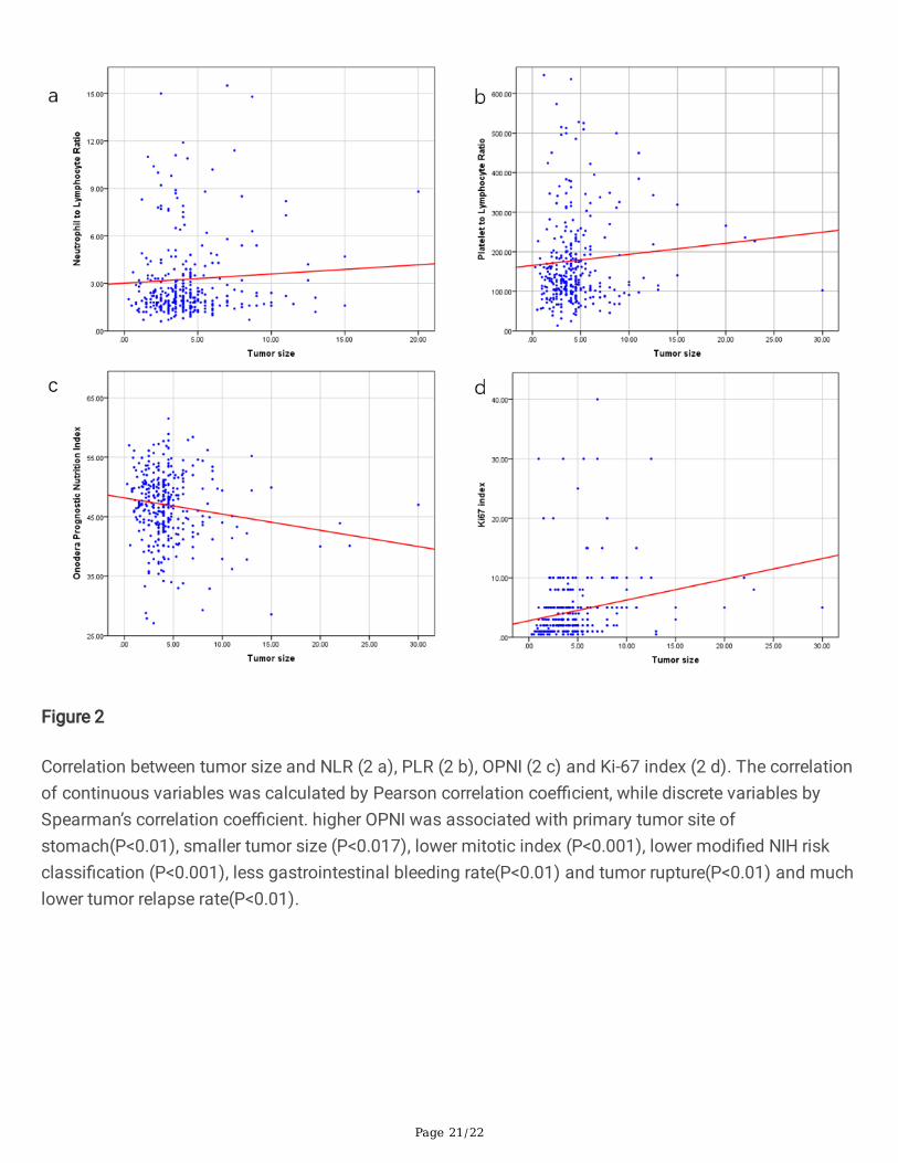
As we saw in Spearman Correlation analysis, higher OPNI was associated with primary tumor site ofstomach(P < 0.01), smaller tumor size (P < 0.017), lower mitotic index (P < 0.001), lower modi�ed NIH riskclassi�cation (P < 0.001), less gastrointestinal bleeding rate(P < 0.01) and tumor rupture(P < 0.01) andmuch lower tumor relapse rate(P < 0.01). A strong correlation was observed between NLR and tumorsite(P = 0.01), GI bleeding(P < 0.01), tumor rupture(P = 0.03) and relapse(P = 0.02). And PLR was also

Page 11/22
connected with tumor site(P < 0.01), GI bleeding(P = 0.04) and relapse(P = 0.03). (Table 3, Fig. 2 andTable 4)
Table 3Correlation analysis of tumor size and mitotic index with NLR,
PLR, OPNI and Ki-67 index.
Tumor size Mitotic index
Spearman r P-value Spearman r P-value
NLR 0.06 < 0.01 0.11 0.07
PLR 0.28 < 0.01 0.15 0.01
OPNI -0.27 < 0.01 -0.14 0.02
Ki-67 index 0.35 0.02 0.44 < 0.01
Table 4
Correlation analysis of OPNI, NLR and PLR with tumor site, gastrointestinal bleeding (GIbleeding), tumor rupture, relapse, CD34 and CD117 .
Factors OPNI NLR PLR
Spearman r P-value Spearman r P-value Spearman r P-value
Site -0.26 < 0.01 0.18 0.01 0.19 < 0.01
GI bleeding -0.26 < 0.01 0.20 < 0.01 0.15 0.04
Tumor rupture -0.13 0.06 0.15 0.03 0.12 0.09
Relapse -0.23 < 0.01 0.16 0.02 0.15 0.03
CD34 -0.07 0.26 0.02 0.74 0.07 0.27
CD117 -0.06 0.36 -0.01 0.81 0.05 0.38
Patients were followed for a median of 48 months (range: 8months– 103months). This was calculatedby Kaplan-Meier method, we concluded that our estimated median follow-up time was 47.98 months(P < 0.001). 62 patients experienced tumor relapse during the follow-up period. There were 30 of them had notbeen treated by IM, and 32 of them had received IM therapy with a duration ranged from 1 months to 5years. Until December 2018, 13 patients without medication had died, with a mortality rate of 12.7%(12/102), while the mortality rate in the drug treatment group was 3.3% (6/178). Metastasis to the lymphnodes was not spotted.
Our univariate survival analysis showed that tumor size (Log-rank P = 0.002), mitotic index (Log-rank P < 0.001), modi�ed NIH risk strati�cation (Log-rank P < 0.001), Ki-67 index (Log-rank P = 0.053), age (Log-

Page 12/22
rank P = 0.005) and OPNI (Log-rank P = 0.002) were all signi�cant prognostic parameters for RFS. Resultsof univariate survival analysis are in Table 5. Some sorted factors were analyzed in the Cox proportionalhazards model in enter strategies. The results of the Cox regression analysis are listed in Table 5. Highmitotic index (P = 0.001), age more than 60(P = 0.020), larger tumor size(P = 0.007), high NLR (P = 0.033),and low OPNI (P = 0.007) were statistically signi�cant independent negative prognostic indicators forRFS.

Page 13/22
Table 5Univariate and multivariate analysis of the prognostic factors for recurrence-free survival of
patients after PS matching.Characteristics Univariate analysis Multivariate analysis
HR(95%CI) P value HR(95%CI) P value
Age/year
≤ 60 1 0.005 1 0.020
>60 0.230(0.091-0583) 2.015(1.117–3.635)
Gender
Male 1 0.728
Female 0.859(0.364–2.024)
GI bleeding
Yes 1 0.054
No 0.655(0.270–1.585)
Primary site
Gastric 1 0.001 1 0.864
Non-gastric 0.513(0.189–1.389) 1.066(0.560–2.029)
Tumor size
≤ 5.0 cm 1 0.002 1 0.007
> 5.0 cm 1.943(0.795–2.921) 2.367(1.067–3.759)
Predominant cell type
Spindle 1 0.419
Epithelioid 0.765(0.263–3.376)
Mixed 0.735(0.363–3.289)
Mitotic index
≤ 5 per 50 HPFs 1 < 0.001 1 0.001
6–10 per 50 HPFs 0.288(0.068–1.216) 1.787(1.254–2.545)
> 10 per 50 HPFs 0422(0.120–1.490) 2.395(1.786–4.502)
Tumor rupture
No 1 0.097

Page 14/22
Characteristics Univariate analysis Multivariate analysis
HR(95%CI) P value HR(95%CI) P value
Yes 0.477(0.092–2.488)
NIH risk classi�cation
Intermediate risk 1 < 0.001 1 0.013
High risk 0.456(0.138–1.508) 2.514(1.218–5.191)
Ki-67 index
≤ 5 1 0.053
6–10 1.630(0.429–6.189)
> 10 2.696(0.617–11.787)
NLR
< 5.1 1 0.084
≥ 5.1 1.123(0.301–4.188)
PLR
< 98.6 1 0.129
≥ 98.6 1.695(0.429-6.700)
OPNI
< 42.6 1 0.002 1 0.007
≥42.6 3.320(1.146–9.621) 0.433(0.236–0.794)
We divided patients with or without medical treatment into several groups according to relatively low orhigh OPNI, NLR and PLR. And we observed that patients who used TKIs had a better relapse free survival.In addition, patients with higher OPNI showed preferable RFS than those with lower OPNI (P < 0.0001).Furthermore, higher NLR (P < 0.0001) and PLR (P = 0.0003) were factors leading to poor prognosis(Fig. 3).
DiscussionIn this study, according to recent investigations of Sun JY’s team that showed OPNI was an independentpredictive factor of RFS in GIST patients with no TKIs treatment [23]. We initially base our survival cut-offanalysis on the 200 GIST patients with median or high risk after PSM and get the best cut-off points ofNLR, PLR and, OPNI. Then we examined the univariate and multivariate survival analysis of our patients

Page 15/22
after PSM study. It was our aim to investigate the prognostic value of OPNI in intermediate and high riskgastrointestinal stromal tumors treated with or without TKIs. Eventually, analysis proved that OPNI wasan independent prognostic marker for both two groups.
A more precise risk classi�cation criterion that can be applied to determine the postoperative prognosis ofpatients with GIST is eagerly required. Of which the items should be simply and economically detectedand calculated by clinicopathological data,
Nowadays, the most widely used criterion to estimate the risk of relapse after surgery in GIST are the AFIPcriteria, and modi�ed NIH consensus criteria. Studies have proved that their prognostic accuracy issimilar, by and large [24]. Moreover, Memorial Sloan-Kettering Cancer Center sarcoma team developed anomogram that could estimate the probability of RFS at 2 and 5-year after surgery for primary GIST andwas more precise than NIH criteria to a certain extent [25]. Joensuu H further demonstrated the KIT andPDGFRA mutations may have widely varying risks for recurrence, and those with KIT exon 11 duplicationmutation or deletion of one codon have favorable RFS with surgery alone [26].
OPNI is a nutrition index which is �rstly raised by Onodera and his colleagues. The previous studiesshowed that patients with high OPNI shared a signi�cantly better prognosis than those who had a lowervalue of OPNI [22]. And similar results regarding Crohn’s disease and stage III colorectal cancer have beenalso reported [27, 28]. In our study, the border value of the OPNI was determined to be 42.6 for TKIs-usedgroup and TKIs-unused group according to survival cut-off analysis by R 3.6.3. A detailed analysisdemonstrated that higher OPNI was associated with primary tumor site of stomach, smaller tumor size,lower mitotic index, lower modi�ed NIH risk classi�cation, better gastrointestinal bleeding rate and tumorrupture and much lower tumor relapse rate. In univariate and multivariate survival analysis, OPNI wasindependent prognostic indicators too. Lower OPNI may result from low hypoproteinemia and/orlymphopenia, which can be explained by several potential phenomena: (1) nutritional supplementation ofbranched-chain amino acids can improve hypoproteinemia and reduce tumor recurrence in patient ; ( 2)lymphocytes play an important role in the host immune response, eliminating tumor formation andprogression.
In our univariate and multivariate analysis of the prognostic factors for recurrence-free survival ofpatients after PS matching, we found that high mitotic index, age more than 60-year-old, larger tumorsize, high NLR, and low OPNI were statistically signi�cant independent negative prognostic indicators forRFS. Moreover, when we had split patients into groups according to the best cut-off value of NLR, PLRand OPNI, we found that relatively lower NLR, lower PLR and higher OPNI were signi�cantly linked tobetter RFS.
There does exist limitations of this study. Firstly, it is a single-center retrospective study, therefore, amulticenter study is eagerly required to enlarge the sample to minimize the de�ciency during the analysis.Secondly, the best cut-off value in this study is determined by survival cut-off analysis. However, it is stillunclear what cut-off value is the best optimal cut-off value for clinical diagnosis of GIST due to thelimited amount of patients. Thirdly, mutations were not well considered because there were just part of

Page 16/22
patients had gene test. In general, exploring the exact best cut-off value and studying its intrinsicmolecular mechanism will be the future research direction.
In conclusion, we found connection among immuno-in�ammatory factors (NLR and PLR), nutritionalfactors (OPNI), clinico-pathological characteristics and the RFS of intermediate and high risk GIST treatedwith or without TKIs. OPNI is an independent indicator for RFS in GIST treated with or withoutTKIs.Furthermore, OPNI also might be a ponderable factor for predicting tumor biological behavior fromperipheral blood.
AbbreviationsNLRneutrophil-to-lymphocyte ratioPLRplatelet-to-lymphocyte ratioOPNIonodera’s prognostic nutritional indexGISTgastrointestinal stromal tumorsPSMpropensity score matchingRFSrecurrence-free survivalTKIsTyrosine kinase inhibitorsIMimatinib mesylateNCCNNational Comprehensive Cancer NetworkNIHNational Institutes of HealthAFIPArmed Forces Institute of PathologyDFSdisease-free survivalOSoverall survivalPDGFRAplatelet-derived growth factor receptor AHPFshigh power �elds

Page 17/22
CTcomputed tomographyROCreceiver operating characteristicCIcon�dence intervals
DeclarationsEthics approval and consent to participate
This study has been approved by the Ethics Committees of Nanjing Drum Tower Hospital.
Consent for publication
No
Availability of data and materials
Access to the data and the calculation method can be obtained from the authors by email([email protected]).
Competing of Interests
The authors declare that they have no competing of interests.
Funding
This work was supported by Jiangsu Provincial Key Research and Development Program (socialdevelopment project) , China (No. BE2016603)
Authors’ contributions
FW, TT and LZ contributed to the study design, drafted the manuscript. HY and ZY worked on studydesign and data analysis. XX and YX were involved in data collection and extraction. MW, LZ and WGrevised the manuscript. All authors have read and approved the �nal manuscript.
Acknowledgments
The authors gratefully acknowledge all of the investigators for their contributions to the trial.
References1. Keung EZ, Raut CP. :Management of Gastrointestinal Stromal Tumors. Surg Clin North Am.
2017;97(2):437–52.

Page 18/22
2. Nishida T, Goto O, Raut CP, Yahagi N. Diagnostic and treatment strategy for small gastrointestinalstromal tumors. Cancer. 2016;122(20):3110–8.
3. Heinrich MC, Corless CL, Duensing A, McGreevey L, Chen CJ, Joseph N, Singer S, Gri�th DJ, Haley A,Town A, et al: PDGFRA activating mutations in gastrointestinal stromal tumors. Science299(5607):708–710,2003.
4. Seifert AM, Zeng S, Zhang JQ, Kim TS, Cohen NA, Beckman MJ, Medina BD, Maltbaek JH, Loo JK,Crawley MH, et al:PD-1/PD-L1 Blockade Enhances T-cell Activity and Antitumor E�cacy of Imatinibin Gastrointestinal Stromal Tumors. Clinical Cancer Research 23(2):454–465,2016.
5. Sodergren SC, White A, E�cace F, ,Sprangers M, Fitzsimmons D, Bottomley A. JohnsonCD:Systematic review of the side effects associated with tyrosine kinase inhibitors used in thetreatment of gastrointestinal stromal tumors on behalf of the EORTC Quality of Life Group. CriticalReviews in Oncology Hematology 91(1):35–46,2014.
�. Holdsworth CH, Badawi RD, Manola JB, Kijewski MF, Israel DA, Demetri GD. Van den Abbeele AD:CTand PET: early prognostic indicators of response to imatinib mesylate in patients withgastrointestinal stromal tumor. Am J Roentgenol 189(6):W324–330,2007.
7. Cassier PA, Blay JY. Imatinib mesylate for the treatment of gastrointestinal stromal tumor. Expert RevAnticancer Ther. 2010;10(5):623–34.
�. Fletcher CD, Berman JJ, Corless C, Gorstein F, Lasota J, Longley BJ, Miettinen M, O’Leary TJ, RemottiH, Rubin BP, et al: Diagnosis of gastrointestinal stromal tumors: a consensus approach. Hum Pathol33(5):459–465,2002.
9. Miettinen M, Lasota J. Gastrointestinal stromal tumors: pathology and prognosis at different sites.Semin Diagn Pathol 23(2):70–83,2006.
10. Joensuu H. Risk strati�cation of patients diagnosed with gastrointestinal stromal tumor. Hum Pathol39(10):1411–1419,2008.
11. Zhou Y, Hu W, Chen P, Abe M, Shi L, Tan SY, Li Y, Zong L. Ki67 is a biological marker of malignant riskof gastrointestinal stromal tumors. Medicine. 2017;96(34):e7911.
12. Zhi X, Jiang B, Yu J, Røe OD, Qin J, Ni Q, Sun L, Xu M, Zhu J. Ma L:Prognostic role of microscopicallypositive margins for primary gastrointestinal stromal tumors: a systematic review and meta-analysis.Scienti�c Reports 6:21541,2016.
13. Mantovani A,Allavena P,Sica A,Balkwill F. Cancer-related in�ammation. Nature 454(7203):436–444,2008.
14. Feng F, Tian Y, Liu S, Zheng G, Liu Z, Xu G, Guo M, Lian X, Fan D, Zhang H. Combination of PLR, MLR,MWR, and Tumor Size Could Signi�cantly Increase the Prognostic Value for Gastrointestinal StromalTumors. Medicine. 2016;95(14):e3248.
15. Atila K, Arslan NC, Derici S, Canda AE, Sagol O, Oztop I. Bora S:Neutrophil-to-lymphocyte ratio: couldit be used in the clinic as prognostic marker for gastrointestinal stromal tumor?Hepatogastroenterology 61(134):1649–1653,2014.

Page 19/22
1�. Racz JM, Cleghorn MC, Jimenez MC, Atenafu EG, Jackson TD, Okrainec A, Venkat Raghavan L.Quereshy FA:Predictive ability of blood neutrophil-to-lymphocyte and platelet-to-lymphocyte ratios ingastrointestinal stromal tumors. Ann Surg Oncol 22(7):2343–2350,2015.
17. Goh BK, Chok AY, Allen JC Jr, Quek R, Teo MC, Chow PK, Chung AY, Ong HS. Wong WK:Bloodneutrophil-to-lymphocyte and platelet-to-lymphocyte ratios are independent prognostic factors forsurgically resected gastrointestinal stromal tumors. Surgery 159(4):1146–1156,2016.
1�. Onodera T, Goseki N, Kosaki G. Prognostic nutritional index in gastrointestinal surgery ofmalnourished cancer patients. Nihon Geka Gakkai Zasshi 85(9):1001–1005,1984.
19. Migita K, Takayama T, Saeki K, Matsumoto S, Wakatsuki K, Enomoto K, Tanaka T, Ito M, KurumataniN, Nakajima Y. The prognostic nutritional index predicts long-term outcomes of gastric cancerpatients independent of tumor stage. Annals of Surgical Oncology 20(8):2647–2654,2013.
20. Hubbard TJE, Lawson-Mclean A, Fearon KC. Nutritional predictors of postoperative outcome inpancreatic cancer. British Journal of Surgery 98(2):268–274,2011.
21. Nozoe T, Kohno M, Iguchi T, Mori E, Maeda T, Matsukuma A, Ezaki T. The prognostic nutritional indexcan be a prognostic indicator in colorectal carcinoma. Surgery Today 42(6):532–535,2011.
22. Nozoe T, Kimura Y, Ishida M, Saeki H, Korenaga D, Sugimachi K:Correlation of pre-operativenutritional condition with post-operative complications in surgical treatment for oesophagealcarcinoma. European Journal of Surgical Oncology 28(4):396–400,2002.
23. Sun J, Mei Y, Zhu Q, Shou C, Tjhoi WEH, Yang W, Yu H, Zhang Q, Liu X, Yu J. Relationship ofprognostic nutritional index with prognosis of gastrointestinal stromal tumors. J Cancer.2019;10(12):2679–86.
24. Joensuu H, Vehtari A, Riihimäki J, Nishida T, Steigen SE, Brabec P, Plank L, Nilsson B, Cirilli C, BraconiC,et al: Risk of recurrence of gastrointestinal stromal tumour after surgery: an analysis of pooledpopulation-based cohorts. Lancet Oncol 13(3):265–274,2012.
25. Gold JS, Gönen M, Gutiérrez A, Broto JM, García-del-Muro X, Smyrk TC, Maki RG, Singer S, BrennanMF, Antonescu CR,et al: Development and validation of a prognostic nomogram for recurrence-freesurvival after complete surgical resection of localised primary gastrointestinal stromal tumour: aretrospective analysis. Lancet Oncol 10(11):1045–1052,2009.
2�. Joensuu H, Rutkowski P, Nishida T, Steigen SE, Brabec P, Plank L, Nilsson B, Braconi C, Bordoni A,Magnusson MK,et al: KIT and PDGFRA Mutations and the Risk of GI Stromal Tumor Recurrence.Journal of Clinical Oncology 33(6):634–642,2015.
27. Ihara K, Yamaguchi S, Shida Y, Fujita J, Matsudera S, Kikuchi M, Muroi H, Nakajima M, Sasaki K,Tsuchioka T, et al: Nutritional status predicts adjuvant chemotherapy outcomes for stage IIIcolorectal cancer. J Anus Rectum Colon 3(2):78–83,2019.
2�. Kang WM, Zhu CZ, Yang XX, Yu JC, Ma ZQ, Ye X, Li K, Liu D. Application of the Onodera prognosticnutrition index and neutrophil-to-lymphocyte ratio in risk evaluation of postoperative complicationsin Crohn's disease. Sci Rep. 2017;7(1):8481.

Page 20/22
Figures
Figure 1
ROC analysis of NLR (1 a), PLR (1 b) and OPNI (1 c) in TKIs-unused patients. The PLR, NLR, OPNI cut-offvalue was determined by R 3.6.3 which was performed based on the recurrence state at 9-year follow-up.And the cut-off point of OPNI is 42.1 (P<0.001), NLR is 5.1(P<0.001) and PLR is 98.6(P=0.008).
Page 21/22
Figure 2
Correlation between tumor size and NLR (2 a), PLR (2 b), OPNI (2 c) and Ki-67 index (2 d). The correlationof continuous variables was calculated by Pearson correlation coe�cient, while discrete variables bySpearman’s correlation coe�cient. higher OPNI was associated with primary tumor site ofstomach(P<0.01), smaller tumor size (P<0.017), lower mitotic index (P<0.001), lower modi�ed NIH riskclassi�cation (P<0.001), less gastrointestinal bleeding rate(P<0.01) and tumor rupture(P<0.01) and muchlower tumor relapse rate(P<0.01).

Page 22/22
Figure 3
Recurrence-free survival analysis of 200 patients after PSM. Kaplan-Meier curve analysis demonstrated aworse relapse-free survival for patients presenting with (3 a) higher NLR, (3 b) higher PLR, and (3 c) lowerOPNI. Patients treated with TKIs had better prognosis in our study and low NLR, low PLR and high OPNIalso indicated better prognosis.