Embed Size (px)
Citation preview
Tumor Seminar Conducted by
MALCO_LM B. DOCKERTY, M.D.
T ..HE; rhirreemh aonuaf " uno.c seminar of the San Anconio Sociecy of PathologistS at llrookc Army
Hospi[al, l'on Sam Housron, Texas1 on November 3, 1956, was opeo.ed py Dr. louis J. Manhoff, Jr., president of the sociery, who <lcknowledged rhc senerous supporr given by the Sourh Central Regional Commii ree, College of American Pathologists and the Tex:as Dh,islOn of t he Amerkan Cancer Society.
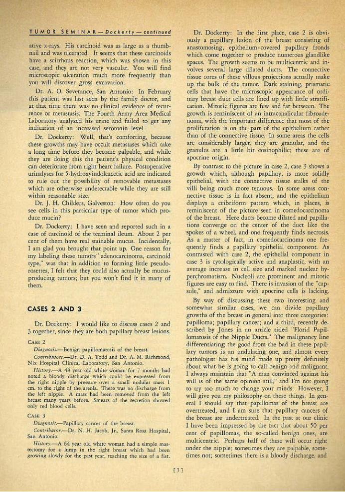
Dr. Malcolm B. Dockerty, mem 4
bcr of the Moyo Cfin ic s.toff, conducted thi$ seminar for the Son Antonio Society of Pothole~ gists a t Fo rt Scm HOuSton on November 3. 1 956.
The commanding general of Brooke Army Mediall Cemer, Majoc Geqeral William E. Shambora, MC, and the cOm<l'laodant of the Army Medical Service School and moderaror of the seminar, Brigad ier Genel'al Elber~ DeCou(SC)', MCt were introduced in curn. GeneraiUeCoursey inrroduced Dr. Mulcolm B. Dockeny, 1\fayo roundation, Rochester~ Minn., conch,;ICtOr of che seminar.
CASE_ 1
Oiacgnosi$.-Urcinoid of duodeo\lm. CouJribJJJ()r.- Dr. A . 0 . Sc:vcrancc:, Baptist Memorial
Ho~pit.ll , .Sao. Antonio. IiiiJOry.-A 55 year old \Yoman 6 _yers p;tSC meno!X'luse
hnd jlad urerine bleedins, of 2 months' dutl'ltion. She also gave a histOC)' of belching, indigestion, nausea, vomiting. and ao occ-.,15ioo;tl dull otc-be io the epigastrilUn of se\'eral } 1ears' duration. Dilatation and curt'ttilgc: _revc:tled adenoc:ucinomn of endQm,errium, (Qt wblc,h c.be }Xltie.nt received rndium and 6 weeks .later hjld a panhyscCrcctOmy. At rl:ie time of suq~cry an ;tpparcndr supt::rfi~ial bn.rd tunlor, 2 em. in di;'lroete-r, WJ..s Jound in the wntl of chc duodenum near rh~ pylorus. After rcmov:1l of the nlJllOt ilnd report of bi·opsy a subtot:d gastre<:tonly was petFor:mcd; togechcr wicb foJther removal of porri()ns of the dllodeown.
Dr. Dockcrty: There are rn•o roentgenograms oo the first case, and Col. Henry C. Harrell, MC, radiologisr ar Brooke A rmy Hospiral, has k indly consenred
•
to dis<.t-uss chem, I might add thar in this at~, ns with rhe other Cl\Ses having toemgenogr~ms, Colonel Harrell h:lS had no opponuniry t() srudy them, an-d he is further handicapped in having ooJy a «production co inrerprer.
E:olonel Harrell : You notice tl1erc is a sort of a ''3" shaplid sign which giv.es rhe impressioo. rliar there is n little rumefaciion extending knucklelike inro the ouodeoum. The cap is lar.ge, 'o ir suggeSts obstrucrion and tll.Olefacrion in the ·wall As fat as n differential diagnosis goes, from the x-ray One would consider an enlargement of Brunner·s glands, which Can do rhis, bur rhar is nsuaJiy all around; carcinoicj of COlltse is~ _possibili t)', as well as some orher rumor of the small bowel wall. The mucosa cannot be made our over die mmefacrion tOO welL I think perhaps rhe Jn\ICOS:t may be destroyed.
Dr. Dockercy: I am glad rhe colonel went over rbese rO\'Otgen,ograms, because the older roxtbooks told us that carcinoid rumor-1 an1 g-iving you an jnkling now us c.o 9;-·hnt my djngoosis is-that ca.~cinoid tumor wa.s an ·~rmpcomatic plnytbin$ of the pathologists which for the most pau. \Vas cronf.ned to rhe tip of dJ.e appen~lix. Jvhn)' of these extr-0-appendiceal case,s, however, are symf,comadc.
Uno;!cr low power we see rhac the neoplasm is submucos:ll and cbac the Ol\1COS1'l is imncc- o"•ec it. TI1ese
growrhs are supposed to come from the Kulcschitzky cells at the base of the cryp t< of Lieberkiihn, ona it is remarkable that perhaps onl)• 1 case in 10 o£ a carclnojd rumor wil1 exhibjc mucos:tl ulceration ro rhe extent rha< the patiem will bleed, os patients ,will bleed from ga.mic and duodennl ulcers, myomas. and so on. ClinlcaJJy, rbey do obstruCt, however.
I rhjnk we can _ru)e our immediarely the possibil.iry Chat this g rowrh js a metastasiS frorn rhe p~~iem1s en4ometr~~\J c:l.r.cinoma. 1lte Iauer was _nor partiCularly invasive in the Se<.'tioos· I ex~mined. This also ~vould be :1 most unusual . .site for a solitary m1!J;:JS(asls: Such g(owths, when tbey spread, will go ro nodes or spread by peritoneal sedimemadon. 'Fhe scirrhous re· acdon in this growd1 would be unusual for an endometrial cru:cinom.'l~ ;)nd rhe composicion Of prismatic cells containing pink cyropl:t.smic granules is unusual for met:tSt:atic cumor from che urerus. The
- rlnodenal tumor is r. carcinoid tO anyone who has seen tnore than one OC CWO examples, and WC <,i.o OOt
need silver and "gold " st-ains ro prove Qur point.
! I l
I would l ike co say i l word abOut auci.noids in geoerol, aod duodenal carcinoids in panicular. As far as 1 am concerned, ca.rdnoid rumors are dll can· cers>. regardless of location or size. Giveo time-, io Ill)'
opinjon, evet.y cardnoid romQr even:mall.y wiJ[ mcrascasizc. Or.hcrs in the Jitemture h~ve also come to chj:; conclusion> which is nr variance, of course., -with chc notion caught in the oMe r rextbooks, chnr c-arcinoids were carcinoma-like, microscopicall;1, but chat t.hey d id nor behave clinicolly like. c-.mce.t> at aU. In our

TUMOR SEMINA R- .Ooc Jt erty-contitJueJ
experience 13 of OUI' last 16 curci.t>oids of the small imesline wer~ associarcd with 1ner:1Srasis, 2nd in the literature me figure of incidence for me spread of exm-appendiccal carcinoids is about 35 per cenr. The ileal ones have " greater tendenc)' tO metastasize tlwl do the rta:U or the go.suic or the duodenal ones, and a.\ I say, the figure there will run perhaps more rhan 50 per cenc in generaL Now tbe appendiceal growths ll!e smalL If you go over the age incidence for people with oppendiceal cnrc.inoid rumors. you will find that the patients arc young (between 20 und 30). lr oppears dw these lirrle growrbs, which are somtcimcs IOCtted proximally io the appendix, but more fr«Juendy ar rhe tip, perhaps will produce mild symptoms of obstruction, and they nrc removed, b6couse of those symptoms, early. 1u d1e ileum, however~ where 1hey have more of a chance ro grow be· fore rhey produce any symptoms, rhey fll(!l11SrnSizt, nod che average age for pacicms wicb ile:'l c:uciooids is around 50 tO 60. or pc:t·haps 1 should say 40 t() 60. So the fuetor of size is ' '"'Y impomutr. and the as~ ft~cror closely pnrall~ls ir. In the good old d•ys 1'rocrologists were p,rone co fuJgurntc the submucosal nodules in rh~ rectum widmut ch~ benefir of a biopsy. but nobody ever got inro trouble wirh them. Nowa· d•ys proctologists >re excising them for microscopic cx:unination. nnd we are finding in our dinic dt:lt rectal carcinoids ncrually ore mor<: £requenr chan appendiceal cnrcinoids.
Jt! is jmpoctanc to tcmember that che lump rhar you feeljo the patient witb a c1.1rdnoid mmor is the mrraslllSis and nor me primary. This is chnncretistic. I will not accept os carcinoid any primary lesion in tho bowel chat is over I inch in dinnierer, As regards rh~ metaStatic nodules, howe"tr, 1 ha\'e seen 2.:,000 Gm. of liver metastaseS from a 1 Gm. ileal OU'Cinoid, which jn rncmscasizing Also i.ovolved tbe root of the mesenrery, con>p!crely blocked off the mesenteric "Vessels, and killed me pnrienr by infarcting nloog one segment of bowel
W hen you obmin such a large bulk of carcinoid rumor from 2 mewraric lesion, you :.tre likely to have che as.socinced carcinoid syndrome recendy described ..b)' BiOrck. Thorson~ nnd ochers. Titis syndrome consisrs of an iml'~etable diarrhea which ac dmcs sus· gests that the patirot has a g:utrooolic fistula. Po· cients h:tve palpitation, flushing, md cy"-nosis~ with patchy reddish discQI<lred are:J.S over rhcir bodies. Eventually signs of right be:" r failure ensue from subendocardial fibrosis rhar may kill d1e pntieor. There 3re peculior signs on cle<:troc><diographic smclies. Pullnonary hyperrension nnd aschmn are nlso common in the syndrome. _If you anal}rze ahe urjnt for seroronin, which is rhe product of the normal "c11rcinoid" cell (or Kultschiczky cell), you will find rhac its end pr<1ducr, 5·hydroxyind()leaceric acid, is
I 2 I
prescnr in large ll!llOtllltS. Accordingly, ir now becomes possible ro diognose aucinoids preopcrncively.
Since it is possible, chcre(ore, for " carcinoid rumor wich mecas~si.s. ro kill the patient b)' ruining the righr side of his heart, ir becomes the duty of the pathologist ro make the diagnosis by fresh frozen sections where.! poss ible, so chac che surseon m:t)"' deal with it aud its metastasis appropri3<cly. In od1er words, it is re<:OO>meodcd that a partial hepnte<:romy be done for a. large mernsmrlc carcinoid t\lmor in rhc Liver, or th~1c orher sirnl1ar metastatic masses of carcinoid ntmor be excis«< as completely as possible.
The IOCtrion of carcinoids in rhe duodenum, :J.S in this parcirub r nodule, is tll!e. As h r as J could garhor, there '"" only 9 duodenal carci.noids in the lirtr2rure. This compares with an incidenct of about 25 corcinoids of rhe st011111ch, hundreds from the ileum, and pcrhnps 50 from the re<Jnun. Seven of the 9 duoden~l examples have mec.tstasizcd. Only 1 duodenal carcinoid in the literanrr<: has produced symptoms, and this C:l$t is prob•bly anoth~r such example.
S•l>miud Oiotl • ose-s.-<an:inoKi. 41: a.Jrooateinoma. 2; islet CtJI tumor, t; islet cell autinoma ( in ecropic pan· creas?), 1.
Dr. Dockcrry: \"\fe notice rhar most persons sub· miuing diagn05<S thought this was a cnrcinoid. As for !ldenocarcinoma., in the lirerncure 1 have called these "11denocarciooma, a'rcinoid type/' to emphasize that carcinoids were conccrs, bur hove called mem grade I ro iudicncc char rhey we«! low grade, well differentiated~ and slowl)' growing cancers. Isler ceU rumor is an imetesting diagnosis. Islet cell rumors micCOSCQpiCllU)' look much like OU'Cinoids, bur I think rhoc parbologisrs sometimes have ro be philooophers, and they have m be biased a lirtle bit by the clinical hisrory. lf this patient had a blood sugar of I 0 mg., I probably would have cnlled this an islet ceU 1\omor. 111e t-wo are \'Cry similar, and in dte liternrure islet cell rumors fn."<)~enrly hnve been confused wirh car· cinoid rumors. 1 might s•y thnt I per cent of isler cdl tumors occur in ectopic locari()Os.
Or. Leo Lowbeer, Tulsa, Okla: We had n 32 year old male par;ent who had a microcytic hypochromic anemia and occulr blood in the srooL The rocncgcnogrnms of me gnmoiorestinal tract were entirely nega· rive. Ute sutgeon did an exploratory laparotomy, and at firsc found norhing; but sinct there was so obviously loss of blood from the intestinal traer, he wcnr carefully through all small intestinal loops until he got n tiny nodule, which he excised, and wbidt proved to be a small (less rl>an ) mm.) Cllrciuoid wh icb was ulcerated, aod the only symptom of which was bleeding.
Dr. Dockeny: Our experience has been rhe srunc. 1 memiooed che figure of 10 per ceoL We had 1 patient who presen<ed with the problem of severe t:x.SA I'IJ)uinaring gastroincesrinaJ hcmorrhnge~ wirh neg-
TUMOR SEMINAR DocRer'ty continued
at<ive x-rays. H i$ carcinoid w.as· as large as a chLUnbn.ll.'il~and was uJcera.ted. lr seems that these cardnoids ..have .a. scirrhous reaction. whkh was shown ip [h-is. ""'!•. "and: rhey are not very vascular. You will find rh!c.t:o~opic ulceration much more. frequently [haq )'O\l wi~ discover gross excavation.
Dr. A. 0. Severance, San .A.ncooio: In February this parienc was lase seen b)' the family doctor, nlld ·ar rhnt time there was no dinital evidence o f Ielur· renee or motascasis. 1l1e Fourth Army /\rea Medrcal Laborarory· analyzed his urine and failed to gee any indicnrio,n of an increased serQronin Jevel.
Dr. DockertF Well, rbat's '<omforriog, because these growths may have occult metastases which i.ake a long tinae before chey become palpable, illld while they :u:e doing this the pncient's ph.ysicnl condition con deteriorate from right bean failure. l'osropcracive· urinalyses for 5·hydro~yindQle<Jcecic acid are indicated to rule out the possibility of removable metastases which are otherwise underctrable while they are still wilhin reasonable size.
Dr. J. H. Childers, GalvestOn : How often do you .see <:elfs jn this- parrkula.c: type of rumor v,.rhkh produce 01\lcin?
Dr. Dockercy: I have seen and reported such in a case of carcinoid of the terminal ileum. Abouc 2 per eeoc of them have real srainable mucus. Incidentally, I al)l glad you brough t tl}at poim up. 0oe reason for my labeling rhese mJm5(s· '.' adeoocar:cinoroa, C$rcinoid cn~e;' was cba[ in :"tcl~it-Lon ro fo.tllling Hctlc pseudo. roserres, I felr chat they couJd aJso acmally be mutus· producing rumors; b1" you won't find it in many of fh~m.
CASES 2 AND 3
Or. Dockeny: 1 would like to diSti.J.SS cases 2 and 3 cogcthcr,. since they are bocb papillary breast lesions.
CAsll 2 Di.t.gmJJis.-BeoigJ\ papillomarosis o( the breast. ConJribuJ()rJ.- D r. D. A. Todd and Dt. A. M. Ric;hmond,
Nix Hospital CJiniC\'11 L'lboratory, San Antonio. fi}IJ()r-y.-A 48 ye.ar old white: -wQ(l'l3n for 7 mQnths had
norcd -a blpodf JjsdlJtSe whi<h could be expre:Med· fr.oLn the!' right o i_pple by pressu~ over 11 small nodull!t mass 1 em: to rh~ right <>f the ar~b. T bere ,,.,..as 110 diseha,rg:e from the left nippk·. A mass lutd been. rtm<wed from rhc: left brea!it m:an}• y~:us- l>c(oce. Smears of the secre<ion show~d only rt-;:1 blood eells.
CAso3 Dittg-tJOJis.-PapiUary <:unc<>r o£ rhe breast:
C¢tNr1'bi(IOI.-Dr.. N. H. Jacob, JJ., S<lot'~ RO$<t Hospital, San Anronio.
lliuur,y.- A 64 l't'OU: o ld white woman had :l simple- mM· rectoroy for a lump In rhe rigbt brea.sr wbich bad been g(O'wins ;low.l}' _for th~ p;'I.St }'ear, r<::'3ching rhe size of 01 fist.
[ 31
Dr. Docke.n:y: [n the Jim place, case 2 is obvi· ously • papilla'}' lesion of tbe bteast consisting of anastomosing; e_pi rbeliuru ·covered papillary fronds which come together ro produce numerous g!andHke spaces. Tlic growth seems to be multicentric and in· volves sever~ large d ilated duces. The conneCtive rissue cores o~ chese villous projeccions ncrually m!tke up rhe bulk of the mmor. D-ark scaitting, ptismatic c.el-ls th~t haVe the microscopic appearance of ordi~ nary breaSr duct celJ;; are lined up With' licde strMffi. cation. J\.fiti>r ic figures are few Md far between. The growrh is reminiscenr o{ nn inrrncanalicular fibroade· noma, '\~t i~h the important d iffe retlce thac m0$t of ~h~ proliferation ..is on the parr of the epithelium ratbct chan of rhe c()nneccive· tissue. In some areas t he cellci are COO!;i·dera.bly hu:ge,t , they ate gr.anu lftr, :md the granules are a little bit eosinophilic; these are of apocrine ori~in.
By conuasc ro the pict.ure in. case 2, c.~se 3 sl1ows" growth whicl!, although papillary, is more solidly epiehelial, v.r~rh rhe connect-ive tissne scalks of [he villi being 1nucb more tenuous. In some ar.c~s connC'<:tive tissue is in fact absem~ and t he epithelium displays -a cribriform. _patte rn which. it) places, is reminiscent of ebe picture seen .io come<Locarcinoma 9 f rhe breasc Here duces become dilated and papilla· Uohs ·col)verge on ~he center of cbe ducr like d)e
-spoke.s of a wheel, and one J requendy finds necrosis. .As a mane[ of f~cc. in corpedQCarcinoma _one fee· quem,ly finds • papillary epithelial component. As comr.tsted w;th a tse 2,. rbe epithelial component in case 3 is q:ocologically active and anaplastiq. wirh an ~\1erage increase in ceU size !ind m!lrked nuclear hyper~hroma[iSm. Nucleoli arc p rominent arld mitOtic f igures -a.re easy to find. There is. invasion of the '1cap· sule," and admixn11e with upcxdne cel1s is lacking.
By w:~y of discus$1ng ehese two imeresring and somewhat simi)(U' cnses, \Ve OllO divide papillary growehs of rhe brcasr in general imo ehree caregories': papilloma~ pap ilhtry cancer ; and a thi.rd, recendy described by Jones in au arricle titled "fl.oricl Papil· lomarosis of the N ipple Duets." The malignancy line differentiating the good from the bad in these· papil· l~1ry elunors js an undubtlrtg one~ and alnlost every pathologist has his mind made up p retty de(inirely a~out what h;: is goin-g to caJ r benign •od malignant. 1 ahv.ays mainmJn thar "A map conviocecl againsc- hi~ will is of t he ~ame opinion srill,'" and l'm not going to -cry roo much ro change your minds. Ho<vevet, I will give you my philosophy on rhtse chings. In ge.n· era! I should "'Y rbar papillomas o f the breasr nre o vemeared. and I arn sure that papillary cancers of the brea.sc are undertre:ned. lu the. p<tSr at our clinic 1 bave been i:m_pressed by the fact that about 50 per cent of papillomas~ ehe sQ·called benlgn onesi nre mttlticentric. Perhaps half of chese will occur tighc under tl1e. nipple~ some~ imes rhey 9re p:.'llpable, some· times not; sometimes there is a bloody discharge, aod

T U M 0 k SEM I N AR-Do d: or t y- co ntinued
somerimt!S there isn'c. 1 ha,·e nor been impressed by <he <hesis <hat the rtmaining muh ic<ncric p:tpillnry growdls in this 50 per C<:ot are in fttct confined ro one segment of the breast. Accordingly, I was not roo unhappy over che rcndcocy of our senior S\~rgcons to perform simple mastectomies for papillomas. Some of the younger generntioo are currently injeaing radiopaque dye. They are slitting nipple ducts, and they arc cutting "pie'' segments mi.r of rbc brcasc, while rhey main min rhar they, coo, a1·e gercing J 00 per cf'nr cures. Thus rhe creaunent ha.s swung. cenainly ac our insti· rution. coward a_ much more coosetvncive approach to papilloma of the brenst.
On the other band l have always advocated radic11l mastectomy for what I caU boneSt·ro-soodness papillary cancer. In 3 instances I 1,., .• seen nodes in· volved, and io 3 additional cases I have seen rite p:t· dents die ns a result of ex-tensive l<:>cnl recurrence and chesc wnll invasion following simple mascecroJny. One such recurrence rook 6 years ro mnkc irself manifest.
What arc some of the gross diffcrC1lces berwt<11 papilloma and papillary cancer? In papillomns there usually is no lump, but if there .is one it is a riny nub· bin righr under d1e nipple. About 50 per cent of <hese nrc located in rhe ampulb. l'bere may be a bloody discharge. Papillary cancer frequendy forms " lump or occuxs in the wall of a cyst. In a recently studied series of 48 cystic cancers of the breasr :It our iosrirucion no less d1an 33 -were papillary in nalure.
Microscopically rhc presence of rather fibrous, thick, heavy stalks with luyering of perha~ two ccUs at most over these !tn.lks; good vasculnrity of the stalks; the presence of apocrine glands; lack of in· vasioo; 1nd multicemric locarion Wider the nipple, all favor rht diagnosis of a beni&n p-•pillary rumor of the breasr. In this cancer I think the mosr impOtC<Ulr evidence fot malignnncy is cytologic. If I find parho· logic mirOtic figures in one or chese proliferAtive growr.hs:, it is cancer, in my esrimatioa In addition, rbe~ells will show the usual signs of Mnplasi:l : hyperchromnrism, many mirotic fihJUrcs, nnd frequencl}' an over-all comedo appenrnncc.
My diagnosis in case 2 is p:tpilloma, or papillomatosis, nod in case 3. gmde n papillory adcnOCllrcinoma of tbe breMt
Folknli-U/1, Case 2.-0r. IL M.. Richmond, Nix HotJ>ital: The puie-nc is living and ~:ell. ahe.r 6 ran.
Fellou .. Up, C111e 3.-0r. N. ff. J:acob. Santa Rosa Hos-~ic-.U: This paciem w:as oper;a.ccd upon in l948 and duri08 this pa.st ye:u (1956) there was :\ recurrence of ~ ,,<:ldt~ lc in the a.xill:l which sho,vcJ the. s!lme his1ologic t'icturc ns the ori,gioal Jaion.
Dr. Dockeny: I am cerrainly interesred <0 hear obout rhi• cc•ulr in case 3. That is on 8 year d~lnyed recurrence; without a doubr, though, rhe c~tnccr was
[4]
there in 1948. (t simply cmphasi>es rhar popillary cancers of the brt'ilSt are slowly growing. hue can merastaSize just I ike aD)' other breasc c:mCt[.
SubmitteJ o;tJIIfOJtJ, Ctu-11 2.-lorraduaa.l papiUomJ. 23; intntduetal pnpilloma with cystic dlsea.se, 12; adenoma, 1; sclerosing adcnosi,J, L; inuotduc;ml c:arcinoma, 1: inttaducml Ptl)illat)' ~denocardooma, 6.
Or. Dockerry: In <he analyses of case 2 we ba\'e essentially 35 for papillom:a, benign, and I woo'r quat· rei too much with rhe term adcnomo. As tO scleros· illS odcnosis, )'OIL do somerirnes find pnpillary projecrions in chis lesion, but for rhe mosc parr in sclerosing adctiOSis you have solid inlilrraring cancer-like areAS in the breasr wi<h • lor of fibrous rissue, which is sometimes hyali11ized acound these well diffctemiated ducrs. It can be mixed " I' with papilloma, bur l don't think that <hot is the diagnosis here. Seven persoos vored for cancer, but I would noc go <bar f .. r. As I said in rhc beginning, rhe borderline of diasnosis between benignnnC)' ·nnd malignancy in p-•pilLuy growths is an unduJadng one, und some pathologists, ro be safe, call rhem all cancers.
SNbmisred Di•X'~Ol#l1 GIJ, J.-P~pilla.ty ad«1oarcinoma. 32; cardnoma, L: ln.filttatirv; adcnocaz:clnomll, 1: ovarisn setoull cyn:-:tdcnoCicdooma, I~ C)'StOSoltc;omn J,byllodd, l; angiomn, Kapo$1 rypc, 1; papliJoma, LSi intr:aductal papiJIal')' adeoomll, lo !.d.enoma, sweat sb-nd rype, I.
Dr. Dockerty: In case 3 we h•.e 4 voting for cancer and 1 for mecastatic ovarian cancer. Of course: if you foMd n growtb like rhis in <he ovnry, you per· haps would nor suspect that it was primary in rhe breast; you \\'Ould call it on ov:uiao cysr~deoOOlrcinom:t, p:tpillnry type, and you would look for psammoma bodies. 1 have seen a grnde 1 papillary cancer of the ovary metastasize co the axiUa, but I have net,er see-n such a cancer roecasrasize 10 the bre-asc. As a matter of fact, rhe only meta:~lllti<: anctr.; 10 <he breast l have in my series ote a cast of Hodgkin's disense and n C11Se of cancer of tbe prostate. In any Iorge series you will find that the brensr is frequendy the seat of primocy cancer, bur very, very infrequenrly the sear of mewtases, and I doubt <hat an ovarian carcinoma of '"'h a low grade of ronlignoncy could metastasize co chc brensc.
C)•sros;,ooma phyllodes, or phyUoides, ro my mind is a malisnant fibroadenoma, and <here is a uemendous increase in the coonective tissue~ rather lha.n the epithelium. Case 3 sho•ved on epithelial prolifera· tion, whereas cystosnrcom:t phyllodcs is n connective rissue prolifernrion. One per cenr of fibroadenomas arc malignant, and pe<haps 5 or 6 per cent of the giant fibroodcnomos nre molignaoc I rhink <hac we should do awoy with the sarcoma parr of cystosar· coma phyllodes. Clinically, of course, they ace like sarcomas; they ulcenre the skin, and the pathologist has 8f<O' difficulty r:oovincing <he clinician du.t the growth is not n "bad cancer," but a surpruingly small percentage of them acmally nrc cytologically molig-
T U MO R S EMfNA R. Do c- ke1ty-continu~tl
omt. Whet~ they metastaSize it is almost 100 per cent by me blood stream, and they mctllS!asi:ze usually .s strnight sarcomas. I have ~n onl)' 2 cases of carcinoma in fibroadenomas.
The lesion in case 3 was vascular, hue I ch ink that ios epithelial nanore is fair!)' definite, so d><u 1 would nor consider angioma, Kaposi cype. The rt S[ of the diagnoses were those of benign lesions, bur I believe tloRI this ltsion is definitely a c-•ncer.
Or. ]. ]. Andujar, Fort W orth: I nOfe mar you graded this as grade 11. Are diCie two grades, grade I, in which you cannoc be sure it is m>lignant, ..,d gr.de II, aU cbe o<hers, or do you coli them grades 1, II , ill, and IV?
Dr. Dockercy: I do oot call all papillom:!S grade 1 cancer, although I used co a number of years ago. I also recognize grades I, II, Ill, and IV in the papillary lesions.
Dr. Andujar: You disagree, then, with the origin1Ll work of Or. Broders in which he called chem grade I carcinom!LS?
Dr. Dockeny: I do, to some exteno. 1 have ~n gr.de I papillary cancer of the bteast met:JStaSize. I have never seen a growth like the lesion in case 2 memstasizc.
General DeCoursey: Many years ago, ac me first one of chese seminars I c'•cr atrended, the philosophy was expressed that ~rhen you looked :lt n bre':lst twllOt
and weren't sur:e whether ic was benign or malignant, it should be collod btnign. Have pachologim changed sine<, or bas cb>e philosophy changed since?
Dr. Dockeny: Well, let's put ic this way: lf you make the mistake of calling benign a borderline losion-:t grade I papiUacy cancer of the breast-ond the surgron has done an adequate excision around i1, tbe chances are perhaps 95 to 5 tb"' d>ere will be no further. rrouble.
Dr. T. R. Sunbury, Temple: I called case 2 a grndc l papillary alleoocacciaoma. Abouc the tione these slides came in we had 1 case in which a radical mnstecoomy was done for a nonpapill•ry adeoocarcioorna. and we found a metnscasis in one lymph node which was papil!ll<y and did aQ( look any more rna· lignanc th•n cose 2. On going bock <hrough che breasr we found a small papillary adtDOCO«'inolllll, whid1 I believe moSt people here "·ould have called an inrrnducml papilloma.
Dr. Dockercy: You ·u get suq>rises every once in n while; but you found that papilbry cancer after you had found the papillary formation in the axilla, and ic wns noc in che lesion rhar you first Saw in the brc:ost. That brings up the question t;>f how ofcen popilloma is associated with somcching more serious else'(J.lhtrt. r run sure th3t chC'te is a somewhac in,· creased inciden~ of c::mc<r in bre:uts that are the seat of papillomas, bur I frankly do not believe <hat
[)]
if a p.1pillom~ is Jefc long enough it is going to become cancer. 1 will admit tint you s« me cwo tO· ge<her once in a while.
CASE 4
Oiagmuh.-Ucnisn sjot.ot cel.l tumor ()( 1icapula. Colilrib11ror.- Dr. S. \Y/. Boh1s, Au.srio Swce Hospi(:tl,
Austi n. HiJliJ?·-A 26 year old white man compla.iot<l of a
lump in chc leEr sc:tpul;tt tcgjon and rain in his ltft 11rm 6 weeks aJ1er re-ceiving an injury. Thcrr wts t linn. non· ttodcr, ftmon sittd m:us on tbe medial bocder of rhe leh sapula. hdf \\Yay betwttn the: spine: and ;an.glt:. Roentgen ray revealed a.n irregulu. boor. c.tOJivc: process wirh che appe.1nncc of a nulig.runt lesion at this sitt. No Other abnorrnalitit$ we.re noted in lhe s-houlder. Jnd sl::cletal iurvey was n<'g:uive.
Dr. Docke ny: 1 believe chece nre two roentgeno· grams.
Colonel H:~rrell: This lesion involving the scapula is well described in your protocol l t has coo smooth :.n edge to be a malignant lesion. There usually is more infiltration and irregular desrnoction of bone in a malignancy, though the part down ot the tip does show some irregulaciry.
Geoeral D tCow:sej•: Is rhere an x-ray diagnosis? Colonel H!trrell : I'll accept the opinion in the
pr()OOC()I, thac <his is a malignaol{ lesion, alchougb so>ne of rhc feacures of it ure not typical.
Dr. Oocke,rcy: There are also cc1<uin microscopic features thac are not too typical. One is srrttcl<, at first, with rhe cremendous numbtrs o f giant cells; they conmin from 10 to 80 nuclei, and they are pretty obviO<osly osceocl>scs. They probably arise from fusion of the intervening suomal cells. "•hich are me basic etlls in this growrb. The stroma is composed of spindle ceUs and is very, very cellulnr. Mitotic figures are present. We ha"e nor, bowc\'er. in out experience routld the micodc index, or count, among these srrornal cells in biopsy rnactriul to be belpfui in uny WO)' in pre<llcting the ouu:ome of a ses.
In some arens we see ~lnoc.ber fencure. namely, the presence of rather young osrcoid tissue. I would like to emphasize chat irr none of chis osteoid tissue, in none of the giant cells, and in none of me spindle celt.. arc any miroses found which are pathologic. 1n no ploce are there giant cells of the Joe Louis or Rocky Gro~i!tnO rype, big owl-eyed cells which are obviously malignant. The gl3nr cells ore •ll osreocla$!$. Now <he presence of this osteoid rissue io a giant cell ttlll10r is not coo disturbing, especially in cases in which rhere- has been n history of jojury. Fractures occur through these bones, the bony meson· chyme is pres-em, and d1e bone heals. We are perhaps <hankful rh1t there are rremendO<os numbc;rs of the giant cells, be01usc it helps us a little in pronouncing this tull)or benign ramer <han tnalignant. You may ~ mony osreocl<~Sts in cenain areas of osu:ogenic

TUMOR SEMIN.AR Oockertr 'ontjnuei:l
sarcomas chat have undergone bemotrbage and necrosis., hue when one fiods diem in numbers· StiCh as in chis case
1 one begins to think. of sometbing orher than
30 osteogenic sarcoma Their presence in great num. hers also wiU help us tO rale ouc certain co·ndltions Jjke, osteitis fibrosa cystica, fibtO\lS dysplasia, benign chonclroblustoma, and ucbw; growths that contain giant Ct>Us. l'.{}' diagnosis in this particular. case. ·is benign gianr cell tumor of th~ scapula.
By ' Y."Y of discuS$iOn I mighr say i har De. D, C babli~:'my ilssoda[c, has recently gone Q\ler 101 ~a~·es of giann :'e)l tilmor in which he had all cbe·ol,d slides and all d1e old tissue p(eserved in fotm.Un. The dioidll behavior Of c.hesc. rumors \V:ts nor me!lsured b}' me mitotic :ted vi t)1 1L'\ shown in specimens from curettemenrs. ·ren of che cases were mal~gnanc, but in only 2 did malignancy appear as a primacy change .in che tissue odgioally examined. Thc-.se 2 tLU\'IOtS exh·jbiced· fibrosnrcorna in :'1 neoplasm which ¢lsc· wb.ere h•d all rhe feamrcs of giant cell cwnor, The other 8 of chose growths ( 7 fibrosarcomas and l osleogenic sarcoma) were from patienrs who had had an inadefl'J:lte curenage from ·f ro 20 yen.rs previ· ously, and then- bad l:i_ceJ> ueaced with iccadiarion rbcr'\I)J'. It i~ Qr, D•luin's and my feeling rhar roent· gen·my therapy ro a gianr tell rumor exposes che patient co chc danger of developing al,l irradiation irtdu<;ed sarcoma.
I 9-dr;nir thnc there are sCvcml c.ases in che lite.ramre in which a neop i11Sm resembUng tbis microscopitally {many gfant cclls and a tichl)'" vascular stroma-l' with no pathologic mitotic figures) bas merastasized to ch·e. lungs. I saw one ar che Armed Forct·s Institute of Pathology; I saw aoorher ~:e{erred case; and I reaa of a case io rhc Brittsb ]o"ro"t of l?<~tholoft)' a num· ber of years ago. Tbose c-.~rses are ver)r, ·very mre, and deser.ving of rcporcs in rhe liceramre as real "odd balls." No le>s tbau 10 per cent of Dr. Dahlin's orig· ina] gcoup designated as ·giaor cell nunors proved on mote adeguace sampling to be osteogenic sarcomn1
whi_cb bdngs up che impOrtilnt pol m t•hat rbe srudenc can do the ampumdon, buc the. ptofessor should do che biopsy and obt~in adequate m;ucrial which should be sampled adequately by the pathologist co rule O<lt
the presence of ll sarcoma wich giant ceU-Iik~. areas. Sixcy per cent of our series of IOl giant cell tumors
were in the epiphyses of rhe lowe.r extremic.ies, and none ·were SC$pldar in ()rjgin. or prim"ry in the vene. b~al bodies, All of oW: vertebral and skull rumors o,Jgit>ali)' Iep orred as giant ecU nln)Ots rurned out on analysis tO be ex~mples of aneurysm~! booe cysts. These c-an be neac6:1 and Cl1recl in almosr al1 caSt'S b}' curetcf:lge. Such treatment is inadeqoa.te for giam cell rumor oi rhc ocdinary tYI"' in a long bone.
StllmJitJ(ltl Diag~J(JJes.-G-ianc cef:l tumor., 27: g.i~Of <ell rumor ln ane-uq•smnric bone cyst with fibroUs dysplasia! 1;
os[eof'ibroilil'l with J1e·mor rhagc1 L; QSteh:is fibrosa q•n ica. 1 ; maJignanr giant ceU ruJllOr, 9: osteogenic s:t.r<Om:f, .5~ giant ccU osceosa.r(oma. 1.
Gr. Dockett) ' : The diagnosis of giant cell mmor in aneu~ysmadc bone cyst ~tith fjbrous dys.P1asia js
perhaps sparked up by the areas of bone and the are'S of fibrosis wbich were present. 1 beljeve in an aneurysmal bone cyst rhe diagnosis .is fairly easy co csrnblish. ln the first place, grossly tbere is a solicl, meaty mass it> the bone which sbells our readily; $Ions wich this :ue rreroeod9us, vascular spaces, so that you m"y ha"e difficulry getting good se<:dons through solid meil$ of mmoc. Giant cells of course are- preSenc in che:.sol.id ·aren:s, but <?.mer zones h:t\'e f.-" of them In this particuL" case che giant cells were scaccereO pr~tt}' evenl}' chroughour. As regards osreitis fibrosa cysticaf rou c:tn pick out from such a case zones in whjch chere arc rnany gi11m cells. It would be difficulr, howe•er, co duplicate the slide placed in our semlnar grot~.
i do noc believe chat tbis is a malignant giant· ceU mmor, and I also do oor auemtn to gr~!de such curnors.
( 6 )
1J1e diagnosjs of osteogenic sarcoma was prob~bly based on the presence of osteoic.l, which I interpreted a~ being bQne thar· founed as the result of frnccure thtougn rhe m.ruor. 'The scapula i.'i ·a chin bone, suO· jccc to a lot of unuma, and it will break ea5ily, As a mnHer of fact, ir ·is difficulc m esrabHsh in the scapula rhe fllcc rhftt ·:~ given mmbr i.s ceAnmil in origin) be· cause che bone in cercain areas is ooh' a millimeter Of cwo chick. ]}y giaor cell osteosarcoma I suppose is l'ne:mc an os.reoge-nic sarcoroa wlrh nwnerous giant cells. and I pointed out cbe r<'llSOO fot my rejecting that particular diagnosis. The giant cells are jusc roo numer0\1$, and rtiere arc no y~thoJogic mitotic figures in the moma. My diagnosis is ccllu l~r, but benign, giaor cell rumor of cbe scapula.
Dr .. S. W. Bohls, Austin : r be patient is still living and wdl ufte~ ooe year.
Dr. teo I.owbeer, Tulsa, Okl:>. : Could you ·give us )'Our id~s abo:ttt che. namre of rhc giant ceUs in g innc cell mmors~ aod also ::tbom the relation bcrwc..-en rhe gianc cell mmors as we know them 'Mlc! those mmors which b~we been Cllled wicb var.ious naOleS1 soch as chondroblascorna and chondtor;nyxofibroma?
Dr. Docker:ry: I am noc :;ute 1 can answer yO\lf
quesrion. I personallj• regard the osreoclasr-like <ells in giant cell rumor of booe :rod j n fibrous dysj>las.ia as fused spindle ce~s. I think char their origin is bone mesend 1yme, 1 feel that mcseochymru cells arc phu:ipoteotinl :1nd 1hac., rhe same cells which io this case went ou •ncl laid down Osteoid also produced giaor cells. \V/c see them producing co<tilage, par· cicu}ady in os reiris fibrosa C)'stica. Chondroblastoma of Codman is ~ very jmeresring rumot. Some '3teas in it are fairly rypictl of giant cell mmor; other zones recall rhe pictwe of chondro,.r<O<ua, You liave to go
. back co rour ·philosophy again and realize that per·
T l:J M 0 R S EMINAR D ock.CI t .y- <:0.!1tif' u·ed
baps both o{ rhose .cells crune frqm the S:lme mesenchyme and tllac when you find che cwo il1association, the neoP.iasm is benign. [n ·our -series we had .sevend Codman rumors that wtrc cillcd malignant, and ma· jor surgef)' was done for them; ye r in recenr years we have been just locally excising chem.
CASE 5
Di4$110Jis.- Grade IV dlondrosarcqma Qf scaj)ub. C((t'ftribmor..._- Dr. L J. Manboff, Jr., Robert B . . Green
Hospital, San i)..ntonio. HiJroty.-A 64 year o ld woman was ndmitted to the hos
pital with a huge rumor o f the shoUlder o.f 2 yC3.rs• dur:uion. At one poinr it extended thr<HlSh the skin, producing a 4 by 6 e:rn. area of superficial ulcc·ration. Roenrgenograms .showed destruction of almost tlle ·eotire scapula, and erosion o£ rhe uppet end o£ the humerus, la tef31 p~:mion of tbe clavide. and two adjacent ribs. ·ne lunss were clear:, and an inrcaveoous p;•etosram \vas normal. At or.Crndon the tUmor \vas essentially a ~insJe, large, well circum.sctibcd m-ass. The c;entml p<.mioa W;l$ necrotic, cyscic, and hemor· rl)ttgk, buc the P.ecipheral :portion ~-as firm. gra~·-white, and ..somewhat tr2nslucent: wjth {In ill defined Jobulsr p;rctern..
D r. Dockei'Cy: I beHove chere is one roenrgenogrnl)) in this case.
Colonel Harrell: Acmally all time you can see on this slide is ehe en ormous sofc cis.~ue wmor. There are some scrands of cal<.:ifkation chac appear to be in rbe ttunor, rhough the bone decaH. Is noc well defined. T be humerus merges w ith and disappears in the cumor. l r i$ hard co see on chis lancem slide .rhe I it tle detilils which we like co see; but l assun1e chat rhi.s meanS eh:lC rhe head of the bumerus is cnticel}' destroyed. It speaks in the p tococol abouc some ribs l:>~iJlg ll~suoyed; i '"n noc make chat out, either, buc I assume cbat i har would be just by pressure. Tite soft ri:isue caldfitation would suggest an origin ia synovia or some structure abour t he joinr, or petiosceum. Occasion;.dJy, of course, c:util~tglnous rumors will progress co rhC poim o f calcificacion. Osceochondromas ot chondJ:osnrcomas \Viii lay down calcium our in rhelr peripheries.
Dr. Dockercy: This, incidencally, is a real Texas rumor, the size of a Texa-s grapefruir. \Yfe .are cold char it is .scapular in origin, and I gladly accept chat biasing information heciuse I ch ink chat in many o f these growths the p:uhologist needs all the help he can gee. He muse pay accendon to the age of the padem, rhe locadoo of r-he nunor, and so on. For inStttnce, 1t basal-squmnous <:ell cancer from the side of cbe face aces like a basal cell cancer. It rony eac your head off evencually if yoo do noc do sornccbing_ abouc it, bur it is. not going to merosmsjze. The same sm· p~aronce sho\vn in 'ln ?tno.r..ectal cancec has ~erloL1S
impo<L So che clinical dam ilre impotrant. T hey always should be "available to rhe pathologist, a long
with a generous biopS)' specirnen so dtat he mity sample differtnc areas. As we look " this neoplasm we nore rhac: ic is exrremeJy cellulae; .there is some hemorrh118e jn ir, a,nd areaS of nCX:rosis: all of which are help ful .in arriving n.r he lea.st one smtjon along die way. T ltis gwwtli is malignant.
BMically rlte cells are more or less round, buc unlike thOSe o f reticulum ceU sarcoma many seem, ro be surrounded b)' narrow d~ar zones. The margins of rhese boundari(!S seem ro be ouclined by reticulin. G ianr ccU~ ... b joucJeated forms. and numerous mitoses a ttest tO a l.tigb oegree of anaplasia. A few of r5·e cells have va~uolaced cycoplasro. He(e add chcre is a hie of scain:tble ground substance 'vhkh to me ceseoJbles chondr.omucin rather chan osseomucin. In a desperate efforr co arrive ~-tt a diagnosis in ch·is rumor 1 wenr ro che literature and a-lso reviewed OllJ: sedes of 30 tO 40 malig nanc nu11ors of the scapula. 1 found tl)at chondn.>:sarcoma accoumed for well <.>vee half of the primary malignanc cumors. Thac was helpful, bec:tuse th is mighr be a chondrosru-comtt. Y<·t, as 1 wen1 over our series of choodtooarcomas I d id noc find any rhar were quite as malJgnam as chis 6ne. I-t is r.u c co get a degree of anaplasia so marked chat one can n(lt even de-cide oo che basic cell Ackerman and O 'N eil, in a rec~nt arcide, however, do show an ex·
_ ample ·of a gwde IV chondrosarcoma wich a compamblt microscopic picnue. Finally, l got out our s lides on Codman's r.unJor. because I \vamed to see what re<d juicy-looking <:artii:.ge cells appeared like when cht·y were packed closely rogerher. Codrn•n's rumor is- :1 b enign rumor; bo.t it does sbow youog cartilage .cell:;. I was struck by d1e resen:i\>.lance <0 the ronnded cells wicb che clear zooes ~bout chcm, and finally dedded that here, perhaps, if we were dealihg wirh a primary· 'bone rnmo~~ ie wns I! high grade chbndrQsarcpma of th~ ~"llp~J~ .
[ 7)
From the mi.croscopic picture we can ensil}' rule Out ocher primary bone neoplasms S\IC.h {'IS 1:ttydoma, where the p lasma cells ·are prett)' e:>sily identifk-d. We can also rule our by r~e same roken Ewiog·s mmor, wi·th its t·iny sph\d1c shaped Ewing·rumorcyces. (~like to c.IJ them this because 1 -do noc ki19w where chey com·e f.rom.) ·we cannot qu ice IUle ouc :u.J. ostcO· genic sarconia. An oseeogeo! sarcoma could show th is degree of malignancy, be VQU would expect to lind a little osteoid in ir. Actually, I do nor rhink ir would make much difference ro rhe padem whether this tumor is an osceogenk sarcoma, a rhabdomyosar<:oma, or a chor'ld.rosarcoma. l r is a grade LV sar~ coma- and che pwgnosis should be poor indeed. Qf the .soft tissue rumors, syoovjal cell sarCOillal rbab· domyosarconl:t, nnd liposarcoma are .most likely co conh.1se the issue:. Jn ~ynovial ce~rt ~rcomas )'Oll usually have clefcs, and you will Jind c-&kific"tion. T he cells are spindle sllllped, ,and che presence of !liant cells is um\Su!lJ. RJutbdomyosarcoma is somNi.mes difficulc ro d iagnooe, bur chank.~ co i\rcbur Pnrdy
TUM 0 k S C: MIN A k Oockerty - continuttcl
Stout and others we have come to regard fibrosarcomo-like lesions with giant cells ., being basic.lly rhnbdomyosorcoma or lipOS<trcoma. 1 ~!ways like to give the patient the benefit of any doubt, Md if 1 cum10t decide whether a ctunor is Uposnrcoma or rhabdomyosarcoma, l will pur my first choice as liposarcoma because somt of those respond in a mirnculous wA)' to roenrgcn-ray ue:umcot.
Dr. Manhoff: The chest plotc ot the time of surge<y was .perfectly cl<:or, but 4 weeks after surgery the chest was completely loadc<l with huge met11smric nodules :md tlle paricnr died 3 ·weeks h\Cer. t\ c nuropsy there '''ere abund~tnt 1necnstase.s alrnosr completely replacing the ltJnwo. Tbere was one sinsle nodule in the ]jvor, one small merosraric nodule in the ileum, and abundlUlt recurrence at the site of rhe original mmoc.
Or. Dockercy: Titoc perbops is not surprising in view of the very on11p1asric narncc. of chat lesion.
s,,JJ,millt'tt DMguOft'J.- Snr<:t;>mll, 37 (retiQI(Um tell, 7: chondmsarcomil, 7; liposarcom:., 6; undif-fert nrintcd, ~; Ewing's. 4; S)·noviaJ, 2~ 2-.n,ltiosa.roorna, "2; OSlent.nrC'Oma, 2~ rhabdomy0$1_rcoma, I; fibrosarcoma, 1) ; metasca1ic o.rcinoma. 4; m.isc:db.oeow mali,;nant rumors. 4.
Dr. Dockeny: I tended to role our reticulum cell sarcoma because of rhe clear zones around the cells nnd <he presence of giant cells, which obviously wore nclr Reed cells. A few folks I see did agree with me on che diagnosis of chondros<lrcoma. lc could be " liposarcoma, bur I have seen onl)· 1 liposarcoma in a bone. Of course a tumor of this size with involvemen< of booe could have been primarily a soft tissue rumor. Undifferm<iored sarcom. I think is an excellent diagnosis. Ewing sarcoma 1 would disagree with because of che cell siu . The cells in the rypic;:~l l!wing's tUmor are nbout the size of lymphocytes; they are abom as d osell' packed, and have about ns much cytoplasm around rhem as one sees in n lym· phosarcoma. Synovial cell sarcoma J ICjea becouse of lad< of spaces and tht presence of giant ceUs. The l~ion did behave like a synovial ccll sarcol!lll, bowe,•er, in mewtasizing lO the lungs. Jr coold be m ;tngiosarcoma, buc 1 wus ooc os impressed p<:rbnps with the vascuJ:tricy a.s wete che rwo people who made <his diagnosis. It cerwioly could be osteogenic sorcorno, but r do not think tbe evidence in the slide tl>ot J had was good lor on our-and-our diagnosis. Tite presence of big StrnpUke cells- indeed suggests o diagnosis of rhabdomyosarcoma. 1 rhink tbat a "fibrosarcoma" with such giont cells more likely would be a liposarcoma~ however. M('tn5t-1!tic cnrcinomt' is a _pQssibHiry ( ho.cl not thought of; however, r belie-ve that tf it is n cancer~ it js one which is sc:> closely imiroting • sorcoma as not to affeet the treatment.
[8 j
CASE 6
Oidgnorii.-Primuy chondrosarcom~a (g:ratle I-ll) o( bone (humerus).
ComrilniiOt.-Capt. R. C. $dl2f-fe.r, USAF Hospital, L<tck-1-and Air Porte" Duse, ~n Antonio.
Hiuory.-A 3:5 ye.1.t old man first complained of stiff· ness and p;1in lo llis right. hip a year before. An operation, of the hlp "'•' pc:tformcd, and the syno\•lal membnnc: was a.ppuentl)' rtmo\•cd, a1thott.sh it appe.ared normal Stiffnest and p.1in e:oouou«< and anocher opendoo. 'fo'&S performed 9 months b•cr, at \\'hich time irtcJU_lat, srar Lr.as:mtno Dlf1.5\lring up tO 2 em. were- remcwt<l from the joint. Slides -are from thi!J operation.
Colonel Harrell: I am afr:tid I will noc be able co conuiburc much froro this roentgcnogrrul). There is marked soft tissue swelling and soft tissue rumefacrion around me risht hip. There St-cros to be some booe destruction of the ilium juSt abo,•e the acerabulum, but the soft tissue rumor g«s clear c:J<n,·n inro the thigh and the hip. lr is on enormous rumor. The app<lmnce certainly is highly suggestive of malignanq in this case, tbe pcin1ary silc of whicb I would have co gue-ss at cmireJy. The Hium on che medial side appears normal.
Dr. Dorkerty: \\1ould che contributor core to inrtrpr« the gross pho<el,T.lphs in this case?
Captain Scb:tffe!: There """ a 4 em. rumor projecting thrO\tgh the obroouor for111tl<n- The rumor h•d olso grown through the acer.tbulum and fonned • 2 em. mass on the inner aspen of the wiog of the ilium.
When hemi.sccced the rumor wns seen to extend outward from the capsular oren for a dist:tnce of from 4 co 8 em. N o remaining joinr capsule or synovial membran• could be found. The joioc sp1ce was linc<l by rumor except for the central porrion of the !ltticular surfoce. There was partial destruction of the joint cortilnge of che acembulum and of the heod of the femur.
Dr. Dockcrcy: While our of line a litclc, I think I would like tO give you "' chis tirnc my diagnosis, which is chondrosarcoma of the pelvic bones. Whether ir scarred io the ilium or the femur l cannot tell, and 1 do net think ic would m•ke too grear a difference. In our series of cases 60 per eeoc of chondrosarcomas orosc from the pelvic girdle, and if che upper ends of borh femurs, che proxumu ends of the hwneri. nnd chest gi[d1e ~re added, the fjgure cenches more rhnn 90 per rent It is well to remember th:ll 90 per ccnc of these rumors come from ;1rea.s close ro the crunk. I would make the diagnosis of choodrosarc01llll on the original materi•l, and certainly on rbe rtcurrence-.
Microscopically, "-e see an obviO<osly cMcilaginous mmor. Th~ b~ckground m-atrix i.s chondromucin, in anybody's language. Wl e norice ch:tt for a chonllromn, the lesion 15 just too cellulnc; the nuclei are plump wirh prominent nucleoli. There is variations in size of the cells, with some being multinucleoted; ond,
T UM 0 R S £MI N A It Do c ll ~r tr - cont inued
more imporram, the multinudeation is easy to find. ln my opinion multinudearioo in a choodromaroos rumor is a worrisome feorure. Y oo canuo< be guided by mitOtic aCtivity because these chondromatous ru· mon do not have many mitotic figures, except •he one that we just diagnosed, in case 5. The islands of neoplastic appearing bone I rcgasd as being me••· plastic, and more or less incidental.
Dr. Dahlin recenlly has gone over more than 200 d>ondros•tcomns of bone seen at the Mayo Clin ic over a period of 50 years. These chondrosarcomas comprise some 10 per cem of our grou p of p rimary maJigmmt neoplasms of bone~ being rwice as common as Ilwing's mmor nnd hnlf ns common as osteogenic sarcoma. It hl\$ taken pathologiStS about 50 years tO recognize that the credemials for malignancy in cat· tilaginous mmors are not che S3Jlle tb3t they ~ for other neoplasms, ~nd as a rcsulr these rumors wert undcrdiagnosed. Twenty·five years ago the surgeons decided thai rhe pnthologists did nO< know one ou· tibgiooos rumor from anO<hcr and began doing big operations for all of them, regardless of what me pnrhologist ~porred. However, mt surgeons erred in one important respcet. In response 10 me pathologisu' n-*qLU.:ost. for more nnd more biopsy material they would incise 1 hese growths and "spill" them. The recurrence rnre bc."Camc almos[ JOO per cenr. ( h wns dose co chat in our series when such tre;ttment was clll'rietl OUI'. ) Originally these rumors were called cellular chondromas, and liquefying chondromns. I remember I liquefying chondroma in the femur of a womao which 1 clingaosed. 1 was amazed 2 yea.rs lalcr co find thn< she had genemted several chondromas in rbe uterus, and one in the urinary bladder. 1 began 10 pull in my horns nnd review me literarure on chon· dmma1oos rumors.
Jo review, nnd usmg the criteria of LichtenStein IUld Jafft, I believe tha< these rumors are <j'tOlogiOI!· Jy gro"·ths which tan recur, wiU recur in a high per· cemage, and mcrastaSize in 10 to 20 per cent of the cas.es. Ool)' 2 per cent of malignant chondromatous tumors in ou r series involved the hands and dte feet, which is 3 point ro remember. One. observes n1nny cases of multiple chondrom3tosis of merararsals and n1ecacnrj.xdsJ but mrdy obtains> let us say, malignancy in a c:m ilnsinous cumor on rhe back of the hand. Dr. Dell remarked mnny )'e:\rS ago that rbe further nWa)' frOm the heart, rhe lower me incidence Of n:Ja• Ugnon~y. Unlike Lichrensrein, in our series we did not find any recurrenr chondcos.arcomas which were centrally locnred in bone and which ar the first operation we~ benign. 11>ey were one and all pri· marily malignant. Ackerman has had me same ex. pcciencc. l'en per cenr of our series meroscasizcd, and of course a 1remendoos number JCCUrred. Our policy on these growrhs now is to gee the professor
tO do rhe biopsy. W e make • fro>eo section of the rumor and render an immediate diagnosis if possible, bur if we c:mnot we "'ill hold it over for 3 day. In ony '"-"'• the surgeon seols up his incision cnrefully. During me "big" dissection he never •gain ollows himself to opproach the rumor. becouse of the ten· dency for rhese rumon to seed.
My diagnosis is chondrosarcoma, arising from somewhere in the pelvic girdle.
Dr. N. E. Pond, USAF Hospi1al, l.acklnnd Air Force B3sc: Ac d)e time of auropS)', nc che sire of the hemipelvectOmy there was a large pseudobur511 cornplerdy filled wirh 11 cnn ilnginous groMh which cxrcnded up into me soft tissue of the pelvis with cwo nodules on che inner nspecr of the pelvis, nnd then extended by direct continuity up the epiduml sp"'e of rhe spin"! cord and imo the brnin, ttgain b)' direct conrinuir:y. I opened up rbe whole spine. You could follow the rumor right up imo me head. There w:u • Iorge epidural rumor in rhe skull, and in the skull it also in filrrared through the dura so that there wns large subdural occipital. extension of this rumor. In odd ilion it· bad eroded through the skull to form • large mass benearh me scalp in the occipital region. TI1ere was iovasion of cvCf}r single verrtbrn except the ctrvical. -which I djd not ex-amine. Ther~ were
... nonronrinnous mernstases to three ribs on 1he Id e, one rib on th e right. and me right tempornl lobe of the brnin, ond borh lungs were alinosc completel)' rcplncecl by c11mor.
f?l
Dr. Dockcny; l arn glad that you put this C!ISC on, because ic is not often rhat we find ooe of rhese lower grnde chondrosarcQmas behaving in such an unquestionably malignant fashion, and over a fairly shon period of time.
Or. Pond: One of rhe consult1lnts who saw the original lesion and who is a bone and joint pnthologist made rhe sttuemeot that io many c:a.ses in which the surgeon nO<es choodromacosis of the syoo••ia, if it is confined. to the synovia icsclf one can mo~ or less overlook some of the disrurbing histologic fe•· tures, because he has _rarely seen a chondrosarcoma arising from chondromaros-is.
Or. Dockerty: T know of only 2 cases of rhon· dromntosis of .synovial lining from which sn:r:comn d<vcloped, and I am not sure that one of 1hcm is tOO c1e41f' cur.
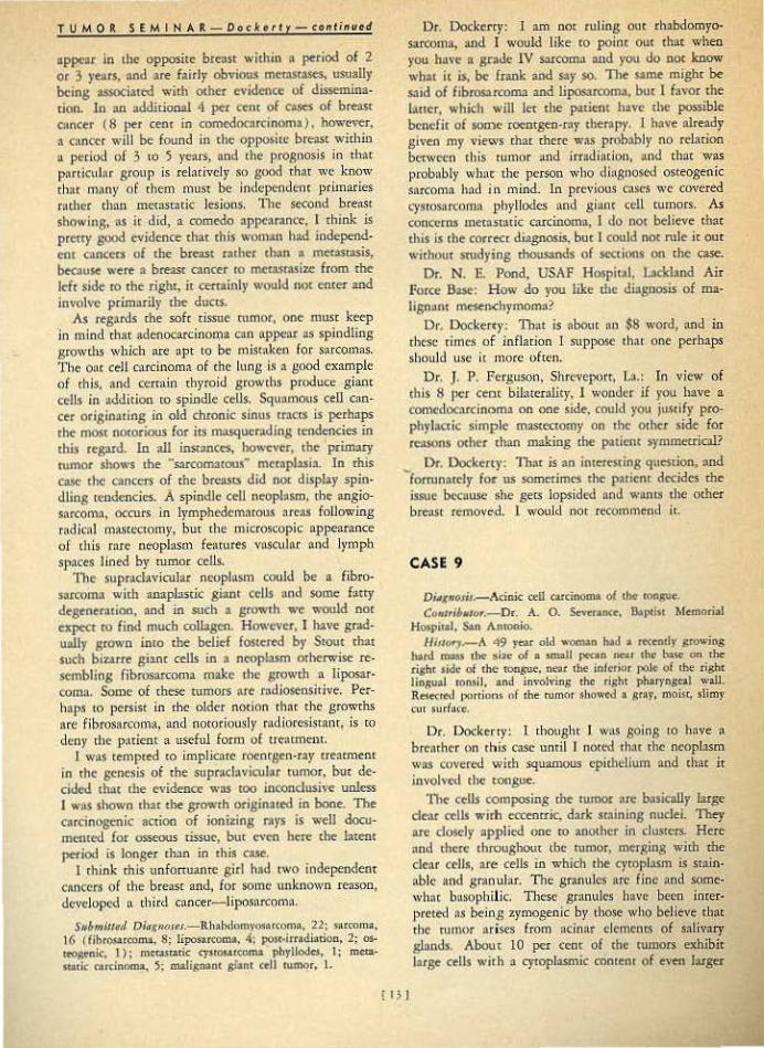
Snbmirud Did,?IJQ.fes.-cbondrosarcorna, -10: chondroma. pc>tc:nti;~lly malignant, l; e»tcocbondtomatOSLs1 2; benif:n ehondroblastoma, I; c.bo:l<lroma, L
Or. Dockerty: The majority favor chondrosJtcoma. The pathologi.sr who made the diagnosis of chondroma, potemially mruignaor. was probably worried by the ctllul>rity. The OSteochondrorearosis vorcrs 2~ pretty well OUtnumbered. I would argue with the diagnosis of benign chondroblasroma because tbcrc were no osteodasdc giaot cells, number one; and number rwo,
TUMOR SEMINA R-Dodarty - continuod
the cnnilaginous cells v:uiro a grear deal in site •nd shape and thty were nor closely p3ckro rogether. I wonder if this ll>t is chordoma, or chondroma. I am surprised someone did nor include chordoma, because we find chordomas in unusual locations, ;md they are prone m r<'CUr. TI••Y arc prone to spill un.less one goes widely around them. The rreatmem of chordom~ts tu cht Mnyo Clink consists of a colbbor:uion of P"'hologisrs, roentgenologists, neurosurgeoo.s, orthopedic surgeons, general surgeons, bowel surgeons. and s>•nt-<:ologic surgevos. Tbey all mill around; one mkes cu·e of the nerves, dtc orher the cecrurnl the ocher rhc blood vessels, and the other rhe bt.dder. We are gcrring good rc>'Uits from ·chis combined, [crrific, aiJ.out ::mack on chordomas. The pachologis.r:, ho't\•t>Vtr, has dHficuhy getting inco the opercujng room in lhtsc cases.
Gener.tl DeCoursey: Do you want ro give us • grade?
Dr. Dockerty: lr is a good grade L I will grade it low because I waor to impress on the group that these borderline chondrosarcomas can roeustasize by tho blood srreorn and kill rapidly. They ore dangerous lesions.
Or. Schoffer: I notice tim cwo people thought cbis w.ts ostcochondromarosis. I vo~onder if }'Ou would comment ns tO whether you saw :my evidence here tl~<~t you rhought could h:•ve puc this originally in rhnt c>regory, It wos supposed to hnve looked like ~n osreochondrom:ttosis when it was originaUy Op· erated on, and the surgevn thought tbey had gotten it aU our.
Dr. Oockeny: I am glad 1hat you were oot con· fused by that biasing information. I c;m see what me surgeons were tllking abouL lr bad 10 do wirb che bumpy appearance on the surface, wbicb is par for the courte in chondromarous rumors; ;er you note rh:ar in their report the)• said that the syno,•ial lining itself appeorro normal. They apparently were grnsping for srraws in trying ro bdp the p><ienL As was mention«!, there are only 2 c~ in che litem· nrre of cartilaginous malignancy which were sul"'r· imposed on om'Ocbotldromatosis. I suppose, on the mhec hand) rhnt when n curnot becomes as exre-nsive ns this one, it would be diffietdt to decide on the poim of origin.
CASE 7
DUt, ntJJis.-'Paroneal C»lcogeni£ sucom:3. of che humerus. Co-ntribmt>r.-Brooke Army Hospital, f-ott Sam Houtron. flisJory.-A 17 yeu old white boy oocico:l limituio.o o(
motion in his leh um 6 months pr:ior tO surgery. -:1nd in· crene in .site of hit ld1 shoulder a.nd pain 2 months prior co surgery. Roencgcnogzams v.·e.re suggeni\·e o! osteo~ic ssrcoma. Th~ firn biopsy spedmco ~.u reported ~ bon~.
librosi!0 chronic inflamnntioo, a.od new bone lorJ\llltiOn. Slidts arc from later material
Dr. Dockeny: In view of che original pllhologic di:>gnosis of "bone, fibrosis, chronic inflamm•tioo, •nd new bone for:roation," me fourth assistlnt •ppar· endy did rhe biopsy in chis case :>nd rhe professor the for<quarter runpuration which \\'IU performed about 1 month later. We h.n•e some coenrsenogroms of the lesion.
Colonel Hurrell: There iS marked overgrowth of bone which is sclerotic and dense. The cortex ;ppears to be destroyed on both. sides, ond nr least half rhe bone involved, cl1e shoulder joinr itself being spur«!. 11u:rc is n greac protrusion of overgrowing osseous ris~uc out into the axilla. l l1is :1ppearnnce ccrrninly suggests the sclerosing •YP" of osrc6gcnic snrconlu. There is some spirularion.t although jc is not the sun~
burst cay spiculation that oftentimes :lccomp~nies these rumors. C<-minly the diagnO<is here is highly suggesri,·e of a malignant primary bone rumor.
The rocnrgeoo,gram of the longirudinall)• secrionro ampuratro specimen sbows pracrically the same rhing that rhc original roenrgenograrn does. I might odd rhar laminographic StUdies of these lesions before chey are ampucared give you 2 simillr stetiou:al •PI"'~ ranee.
Dr. Doc.kerry: The gross specimen shows involve•nenr of the p'Jrosieal zone, an enc:~sing and cnshcath· ing, very extensive lesiOn of che upper half of the shaft of the humems.
Mictosoopic"IIY ir can be seen that the bony proJifermion in this rumor is invading d1e scrinced mus· de of the upper arm. The outstanding fcocurc is the invasion of muscle and the production of new bone in the form of rather narrow, rrabeculnr be:tms which nre fajrJ)' immarure, or ''tumor bone." They are a SOrt of cross berween me bone that you might find in rbc he:tling of a fnaure and cbar of the "rumor bone" rhar is ...,. in more m:llignanr osteogenic s:arcomas.
[ 10 J
1be rrabecube ote long and narrow with rorber cenuO\&S nnastomoseS1 one with another. L'ilfge irreg· ular compartments intervene, and these nre fiHcd with clongt~tcd spindle cells in p<lisade. TI•e spindle cell elcJ11COl is n ceUular one. There is linle inflnman:uory infilr rntc. The spindle cells seem ro run parnllcl tO chc surface of [he neare-st bon~· [rabc:etllac.
In sorne areas only chc spindle cell elemcnr is ob· S<'rved, nnd che piccure is -reminiscent of fibrous dysplasia except thor gisnt cells arc few in number and the fibrous tissue is distinctly immarurc. Zoot$ of myxomatous change and of carrihginous meraplosi> are much more commonly seen man in fibrous d)'Splasia.
Mitoric figures are seen in some of thc fibrobi<Urs, which is unusual in fibrous dysplasia; there is pleomorphism, and some prominent nucleoli.
It is impor<anr ro obtain a really generous biopsy

TUM 0 It SE MINA It Oock~rt r co ntlnucJ
s.ropk from one of these rumors. You wam to get the bone ot the growing edge; you wont to get enough bone to show the arrangemtnt of the fibroblasts, and how juicy :llld active they are.
We must admit d>at if we are going to atll this a fibrosa=m•, it ctminly would bt "" accllub.r one. In this CO$(; the history, the location, the typical roentgenographic appetrance, the combination of the bony b011ms that nre son of borderline between neo· p l(lstic bone :md healing bone, the appearance of the fibroblasu, nnd their relation to the bOO)' rtabeculnc th'u nre fonned musr be considen..-d in order ro a[civc M the correct dingnosis.
By way of discussion I 'an say tb;n
Vice: j, a mon.~cer of $0 ftiS}lduJ mieo, As tO be hated needs bu! ro be seen; Yec seen coo oft, f:tmili~r with her £a<e, We lir.sc endure, then piry_, then emhrare.
-Alexander Pope.
It is juSt nbour like this in a p;uhologisr's attitude toward this rumor. To make a diagnosis of sarcom• on such :Ill acellular mixrure of booy and fibrous cle· mems is l'lt first :tbhorrent; but after the pad1ologist has stcn a couple of c•ses, and perhaps gotten stung by one of them, nod has talked to his colle>J,'llcS, he then begi1u to emermin du: diagnosis of malignancy on the bnsis of rh:u m ixmre~ '3nd f.ioall ~', ai1er he goes over a personal series, he is williog to embrace the concept and he bt-comcs bullheaded and biased abouc ir. even as I am now. We have seen 20 of these tu· mors in young people. Four of our 20 tumors exhjbited oo morc.- ccllu.larhy chan this case, but eventu4
ally mecumsiud. The original diagnosis in some of che older castS in our series .;vas osteochondcomat05is wirb inflamm.'lory change--fibrous, inflammatory tissue~ jusr 25 ir W3S here; bur we have learned now ro listen tO the rocmgeoologist and ro look at the roemgen·tuy places ourselves and correlate the whole pictu~.
.My diagnOoSis, bued on the appearance ht·re in comparison whh 20 of our own1 is '3 parosreal 0$1CO·
genic s:trcomn: :1 mmor rh:u wiU recur; n ruroor thnt will mcmswsi:tc in n high percentage of cases~ a tumor that hns lO Pc teemed radkally nt rhe SN'll't,
afcct hnvi n~ been recognil'.ed as such, and nor j;J'll f·
tinily excised. Such an att;,ck gives chc patient the b(.'St ebnncc for n cure.
SubmiJJtJ Ort~gfWit't.-Fibcous dysplasia. 10; myoslfi$ ossificans, 6; OSttOChondrom:t, 5; neorofibcorna, l ; os:tiCying fibroma. 1: osccom:a, I: bone ttngio~ 1; osreochondritL$. rcpa.nuivc .scagc, I; nrconu, 15 (osfeQSenic, l2; fibros.ar· comit, 2~ paroscta.l, 1); OS«<ehondroma with malignant change, 3.
Dr. Dockerry: Bone and cartihge are '"par"' for fibrous dyspbsi•; bur tbe bone is noc laid down in these little renuous beams, and cl>e fibroblasts ace
[ I I ]
nor os mun~cous; they do not tun pamllel co the trabeculae. Myositis ossificans can re•lly cauS<: dif· licuhy, bec:tuse in ic you observe £ibrous tissue rh" is even much more ceUulru: than in this CliSC: you find bone tb$t looks like rumor bon<; but again, the roen~nographic appearance is hdpful becouse myosiris ossific:tns begins in the muscl~ nor in the bone. and the rumor as seen grossly and by roentgen t>y wiJJ rouch the bone tangeotia.lJy, bur j, 'viii nor CllC1.1St
it. TiliS is on important difference. lncidcn"lly, myt)Siris t)SSificans is mucb mor~ fr<'quenrly called rnoJjgnnm [ban is chjs c.ondidon~ so we have tht· pnritdox chnt du: benign condition, myo..~ i tis ossificnns, is often callc<i malignant, :>od On rite other hand rhis IY.lrosrcal osrcogcnic sarcoma is interpreted ~1s u be· nign uunor bc<ause of the strange cc.:llu lar mixcure, lnck of cclluhrity, inflammatory component, nod so forth. Tile p resence Of S<:anty :IJnOulltS Of cartilage dOtS nOt justify a diagnosis of osteochondroma.
Somebody thougb< this wos a neurofibroma. I have never seen a neurofibroma prinlary in bone. I have tried <o find ooe among a gro<~p of painful OoSreoid ostromas, but withou< success. If you found such a cellular composition ( without bone and C3rtil•ge) in an eighth nen·e rumor. you mighr djagnose it neurofibroma; buc you certainly should worry nbouc che presence of mirodc figures. Ossifying fibromn is not n tumor £hoc encases the uppc:r c;·nds of lnng bones in young p<.>ople. Fa.voriog angioma W!lS the fncc thnt chis w:ts quire \'ascuJili; but in my ~xpcrlcncc :tngi~
om!lS of b011e arc mosdy in che vcrtcbrol column or rhey :m; nssodaced wid, coogen icnl onom:,Jics ~1nd clong:uion of !1. JUnb, and the vascuJnc comp(_lntnt is tnuch more pronounced th:lu it wns in chis case.
This looks much roo nunefact.i\•e co be :sn osrrochondritis, reparodvc stage. \Ve h•vc 1 ~ diagnoses of • low grnde neoplasm that is producing bune :llld a little bit of Clrrilnge. As for O<Steochoodroma with maligo!lm change, 1 do not think there was enougl> evidtJlCC of cartilage in tbi.s ~; •nd thto as T mcnrioned before, these mher critcri:t. nnmely. the chnmcter of <he bone beams nod their relation ro 1he fibroblasts. are rhe feamres which su·e charac· tcriscic of paroste;tl tnmors of chis type.
Tn c:tscs soch as this the pathologist should fer chc roentgenologist make rhe diagnosis. He should go out nnd have a nice d3y of golf, and chen go bnck ro chc l:!bornrorr ~bree or four days Iacer and dis..*:ecr out the ampucaced specimen, inSiend of deferring for a week.a biopsy diagnosis such as ""fibrosis, new bone, and chronic in£hunmation." Well, I am ouc on :a limb now. Parosteal osteogenic sarcoma, low gr>d<, an unusual newcomer in the field of rumor pathology.
Lt. Col. James L Hansen, Brooke Army Hospital: TIIC only follow-up we have is thac after the forequarter ampuudon the paciem did gtt oloog well. We do nor h>ve a late follow-up on him.
Dr. Dockerry: As I mentioned, 20 per cenr of our
T UMOR SE M I NAR Dock~t tt r conth tue il
series of 20 mcCIS<asized, and the figUIC in the literarurc is •bout 10 to 20 per cenc They have ro be ttcared radicnlly ., the sran, as this one was, ro expect good resulrs.
Gcncrnl DcCoursc)': Jo.h )' I ask if you would be influenced if this young mao hnd come in giving :t bisrory of h:•ving been prcviousJy hurc in . sny, a foor~ ball game and had developed o swell ing in • muscle?
Dr. Dockcrry: Well, again, I would ler rhe roem· gcnologin help me.
General DeCoursey: Or would you sec rhis some picture microscopically?
Dr. Dockerry: TI1e point is that I do nor believe you would see rhe same gross aod microscopic piccure. The bone in a myoskis ossificans is more like tumor bone, nnd ~the rrabccuJ:te are irregular, wid1 Chinese and J<'i'•nese ch:uactets; the fibroblasts do not line up as in this mmor; lt is Jnfinit:cl}' more cellular; and rhere is rbe tell-role hemorrhage. Also, myositis ossi£icans, when ir involves bone, involves booe by • globul.,. mass touching rhc booe tnngenriall)'.
Colonel Horrell: This leswn, the calcified hematoma, ns I like ro refe.r to it more than as myositis ossificons, touches bone but dnes not desrroy booe cortex. The acmal cortex of the bone mny merge into ir. bm it is not demoyed. I might odd that fibrous dysplusia involves the correx of rho bone, but it :tlso expnnds the cortex. It does nor grow on the outside of ic, and it does not tlesuoy ir. Your x·my consultnnr can steer you clc:-..r of fibrous d)'Splasia and cnlcified hematomas prett)' well.
Dr. Dockerry: I scroogly recommend thor you get on friC'Odly terms with your x·ray colleague. He will even tc:llCh yon something about iuterpreuuinn of his plates. W hen you see the clinici.'UlS setting up and looking ut roentgenograms and dingnosh1g duodenal ulcers, perhnps you should be a little bit •shamed that )'0\1 cnnnoc at least do chat 1nuch.
Or. J. H. Childers, Galvesron: At leasr in some of the sections from four sets which we h:~d ao opportuniry m review, there wC're little nreas of osseous tissue ond fibrous rissue wbich w<re covered by aortilaginous cops with uniform hr~line c.util3ge oells. Where did these carrilage cells come from?
Dr. Dockcrry: I believe that these fibroblms are osteogon ic, nnd they are chondrogenic. too. T he rumQr is trying to diHecentiate into bone, and it hns a cellulor fibrous t01l1]'0nenr which can nlso lay down cartilage c~lls over the end of the bone that they hove produced. That perhaps is wb)' the suggestion wos mode thM this was n malignancy in an osteochon
drom•.
CASE 8
DittiiWJiJ.-Bilat~u.l metacbtooow. ma.mm.a.rr c:a.rcinorna and :wociarcd mew;(:1Jizing liposarcoma..
ContribMIO,..-Brookc Arlll)' Hospital, r-ort Sam Howton. lJiJtory.-A 27 )'ear old ~:oman h.ad a ld't rAdical JnU•
te<:tomy for t'lrcinocna 10 December, 1950. ru which rime one axi.LJ.ar)' node: contained rumor and she wns siven pon~ o pcrarive ifrMIJ:ulon. In O<wbet. 1953, she had a right cadi<.::tl r(lnSCt.'CCOillY (l)r carcinoma, wh ic:h wall inlt't i)Jt led i\.\
a s<:0011d pritn:.\r y tumor. In April, 19,4, o tumor in che lch supr:adavic:ul:lr region was noted. 'rh is 1uca WAS ~~~ajn t~ted with roencgeB rar. f urdter irrad iation co the nr('tl. was gi.ven in December, 1954, and in June, l9),, Ac che clme oi ~'b if"' December, 195), thc.rc w.u a l~ar~:e (ungacing and uJccnt~ lesion invol"in8 the le£1 sho-alder and e.xteodio.s 1hrou.gh the chcsr "';!)I into rhc hanJ.
Dr. Dockerry: ln this case tbe big question is ·whether we arc de-:rling wich one, two, or three ma· lignant tumors. n.e left breast lesion w•s n grnde UJ scirrhous :tdcnocnrcinorna with involvement of one axillary lymph node. 1 would have liked to see more sections of rhis if rhey had been a~ailoble, becnusc it would not have surprised me tO find some comcdQ areas. In our e.<perience, while 4 per cent of ordioory brtnSt cancers are noosimulroneously bilareral, rbat figure is nt lca5t double wbeo the type of growth in the first breast is a comedo, or duct,, a.ncer.
The right breast growth '1\'hich appeared almosr 3 years later was, in my opinion, a grnde Ill comedo adeoocarcine>mn wi[h CX(ension C01 or rarht r, muJdcent ric involvcmen:£ o f, m2ny re-cmino.l acini. Stewan calls lhc:m lobulnr c;~rciuom3S jn siru because of the rhoughr cxp1·essed by some people that the real acini in the br<:nst :tre something that pop up onl)' during pregnancy; but even in male btensts one nO<es riny buds nt th~ ends of ducrs, and in certain cancers, n001blr in comedOCllrciooma involving luge duas, the process, being multicencric, goes our and invoh·es the terminal buds. 1 could not be <rrr.tin •bour breaching of rhe duetnl basement membrone in chis lesion and accordingly ft,d that rhc evidence is strong for the thesis thu the rn·o breast growths :ue inde]Jendent.
[ 12]
111c highly onoplaStic spindle cell oeopbsm wh ich fung(tted through che soft tissues of the left shoulder, and ttrrtlinally •necnstllsized ro rhe lungs. is dif£crcnr from the two cancers which preceded it. The anaplasia culminlUCS in_ the form.:uion of hugr, sometimes vacuolac~ uninudared and multinucle:ued gi3nr cells. No areas of cpitbeliogeoesis are app•renr. Fat stains are weakl)• positive.
One per cent of our breast c.ancecs 2re simulr110e· ously bilnteml, n11<ll think thar • good ""'"Y of those are mcta5ta.~cs from one side or the mhcr, for two reasons. }\{O$t of chem are medial lesion,s, which we know tend tO spre-Jd to the other side; t~nd only 10 per cenr of rhc porientS live 5 years, which prognosis is about four rimes as bad 1IS for ordinary single crutce-r in one brlt3.St. Five pet cent of brtast cancers
TUMOR SEMINAR Oo c k er tr -continverJ
appcor in the opposite bteost within n period of 2 01 3 years, and ore fairly obvious mci:ISt~, usually being associated wirb ochet evidence of dissemina<ia<L In an addi<ional 4 per cent of c:..ses of breast canctr ( 8 per cent in comedocarcinoma.). howtver, a cone<r will be found in the O)>posite breast within a period nf 3 ro 'i years, and the l>rOgnosis in tb.r partic\ll:tr group is relatively so good rhnt we know that many of them must be independent prima.ries rather dum memnadc le-sions. ·n,c second breast showing, n.s ir did. a comedo appcnrnnce, I think .is preuy good evidence rhat this woman had independem cuncers of the breast ratbr:t dun a met;lS:nts-i.s, btoause were a breast cancer to mcwr:uize from rbe left side ro the righr, it ceminly would nor emer and involve prim~rily the duCtS.
As reg1rds [he sofc ris.-suc rumor. on<: musr keep in mind that adenocarcinoma can oppea.r as spindling growths which :1re ~pt to be misc~lkten for ~lrC01.nas. The one cell carcinoma of the lung is a good example of chis, and certain thyroid growths produce giant cells in uddition co spindle cells. Squomous cell cancer origin:tting in old chronic sinus rncts is perhaps the m<l5r notorious for its masquerading tendencies in this regard. In all insrances, however, the prirooty tumor shows the ·•sarcomatous"' mempbsia. In chis case <he cancers of the breasu did nOt display spindling tendencies. A spindle cell ocopl.sm, the angiosnrcom:t. occurs in lymphedc:m:'ltous nrcas folJo~ring wdkol mascectom>'~ bur [he miccoscopic appearance of rhis rnre neoplasm (Cllcures vasculnr and lymph spaces l ined by mmoc cells.
The suproclovicular neoplasm could be • fibrosorcomn •vith anaplastic giont ceUs and some fatty degenencion, and io sucb a. growth we would _oot expect ro find much collagen. However, I have gradually grown into 1he belief f<»<ered by Stout chat such bizarre ghnr cells in a nroplasm Otherwise re~ sembling librosarcomn make the growth a liposarcoma. Some of cht-se nunors ;~.re rndiosensitive. Per· haps 1n persist in chc older notion rhnc rhc growchs ar.c fibros:1rcoma, ~od ooco.riou~ly rnd iorcsisr:-anc, is [0
den)' the patient a useful form of ue~trmenr. I wCLs tempced to implicate roencgen·tQy ue:umeor
in the genesis or tbe supraclavicular tumor, but de· cided thnc <he cYiden<:e was roo inconclusive unless I was shown char the growth origina~ed in bone The Cltrdnogenic ac<ioo of ioni•ing ta)'S is wcll documented foe osseous cissuc, but even here rbe latent ~riod is longer than in this case.
l think this uoforruante girl had 1wo independent cancers of [he breast and, for some unknown reason, developed n third cancer- liposorcomo.
S1~6rniltt(t Diagll(JftV.-RhJabllom}'Ollllrcome~, 22~ $arcom-a. t6 ( fibr()S.3rcoma. S; liposarcom~~;, 4: pOSC·irrlldi!ttion. 2; ~ ttOgenic, t ) i mce3St:!tic qsroJarcoma phyllodc:s, 1 ; mccastlltic arcinoma, 5; malignant g:f,nt «II rumor. l.
Dr. Dockercy: 1 am not ruling our rhabdomyo$3tCom., :~nd I would like <o point out chat when you ha vc n grade IV satcoma and you do not know wbat i< is, be frank aod say so. n.~ same might be said of fibros:ucoma and lip0$3tCO<Ila, but I favor the Iauer, which will let the patient hove the possible benefit of some roentgen-toy thernpy. I have olready gi"e.n my views that cbere was probably no rebcioo becweto this tumor ~nd irracliadon, .and d1ac wasprobnbly what rbe person who diugnosed osteogenic sarcomn hnd in mind. In previous cases we covered q•stosucomn phyllodes nod g inn< cell tumors. As concerns mecasr.atic carcinoma, l do nor believe chat this i.s che correcc diagnosis~ but I could nOt rule ic our without smdying thousaods or sections 011 rhe case.
Or. N. E. Pood, USAF H<»pi••l, L1ckland AU Force Bose: How do you like the diagn<»is of malignanc me.stll<.hytnoma?
Dr. Dockerty: Tb:<t is about an $8 word, :md in rhcsc times of infla<ion I suppose thor one perhaps should usc it more often.
Dr . .J. P. Ferguson, Shrevepott. L.1.: In view of this 8 per cent bibtemlity, 1 wonder if you have a comedocarcinoma on one side, could you juscify prophylactic simple mastectomy on rhe orher side fat r...ons other <han making the patient symmetrical?
_ Dr. Dockerty: Tb.Jc is an intelcsring qnes<ion, ond forrunardy for us sometimes the p;>tionr docides the issue becallSe she gei< lopsided ond wan<> the other b.rensr rtn•oved. I would not recommend ir.
CASE 9
! U I
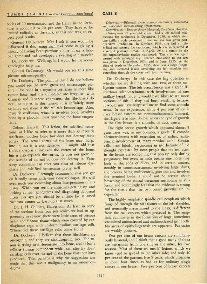
DhtznoJis.-Acinic ceil ('j,.(ci.noms of the rongue. C0111ribMitw.-Dr. A. 0. ScvcrllfKC, Baptist MMtorial
Hospital. ~n Atuoo.H>. H i11Q? . .-A 49 rear old •"<llVn bJd a rturulr ,S:CO'fo"ing:
bard mass the si2e of -a small JXCI-n ncar the 1we on tbC' right stele of the toogue, neat: the inferior p0Je of the ri,ght lin~tu:tl U)nsiJ, :and in\"ofving the righr phBryngeal wall. Rcsc:cre;;) I)C')rl.iollS or c:he tUmor showed a ~tra~· , moisr, slimy cut 11urfnee.
Dr. Dockerty: t rhoughr I wns soing 10 hnve a bre:lthtr on ellis case unril l noted rhoc rhc neoplasm was covered with squamous epithelium ~nd that it involved the tongue.
The cdls composing the rwnor :ue basictilly bitge dear cells wirh ecreouic, dark smining nuclei. They ore doscly applied one 10 ana<her in clus1ers. Here :md there throughout the IUU"lar, m<rging wi<h the cle:tr cells, are cells in which rhe cycopl:'lsm is stain~ able and gran uhr. The granules are fine :<nd some· wh:~t busophi!ic. Th<'"' g ranules h• ve been imerpreted as being zymogenic by t'hose who believe elm d1e tumor arises from acinnr clemt.•ncs of salivary gland!. About 10 per cenr of the <umors exhibit large cells wirh ll cytoplasmic a>n<en< of eveo l:uger

T U M 0 R S E M I N A R - 0 o c Jc e rt r- continue tl
grnnules, ~nd occasionol rumo<> contain cells of all three types.
Tbe Slroma is scancy, and one <foc-.s nor eocounrer in i£ the artilaginous m3rcriaJ of a mixed lUIDOr nor the hf3line ground subsconce of a cylindroma. The cells are not ~1Upposcd to be mudcarminophiJjc as in mucoepidermoid rumors, bur I have seen one e'<cep· doo m (h is ru le. T hese tumors do occosionnlly have mitOses.
My di>gnosis in this cnse is acinic cell carcinoma of the tongue, 11 rel:uive newcomer in the field of oncoloh'l'· 'These tumors were originally dtsccibed in 1924 b)' Masson; called pawtbyro id resr tumors in 1929 by Frnnnsen; termed s•livary glandular adeno· mas by God,.•in and Colvin in 1948; and finally str· cled once and for. all as a rumor sui ~ncri5 in 1953 by Foote and Fm>ell, who reponed a series of 21 c:ues. T hree o f rheir 2 1 paticms died o r the disease, 2 wicb clistn.nt mecasr:ascs, and the recurrence race was very high. Not enough of these rumors have beeo reported in rhe lireraiUrC ., yer ro decide upon criteria for malignancy, but some no and do metnsca· size. I W01tJd nor like ro say whether this one is cap11ble Qf rnec:l..'itaSizing or: notj it is 110t pnrdcularly ceiJuJar, however, and mito1ic figures arc r:tre.
To my knowledge no such r11mor Ius been reported as being primary in rhe tongue, but this neither ~llatms nor surprises me. Most investigators have nor yet goncn around m reporring rheir p:trotk1 exam· pies, simply because reco~n icioo of rhe tumor us :'1
distinct t}'J:te has been so recent. Ir is my firm con· \~icrion tbar in due cimt" acinar cclJ cancers will be reponed as urisiog fmm rhc submaxill•ry and l•ch· rymol glands; the fJoor and roof o f the mouth; and rhe phiU)'nX, nasopharynx, lrftchea, nnd esoph!lgus. Possibly rite dermatologist also will discover th91 sorne of their poorly undersrood IUtnOrS of sw~r glimds belong co this selfume category.
Subn;ined DiiiK1JO.ts.t.-Adnic cell carcin<ml", II : adena-. C"J.rdnoma, 8i muCoepidermoid c:sucino•na, 5; rnew.natic:. c;tr· cinom:t fmnt kidney. 4; mucin l>rQducing curdooma, 1; adcnom3 of sali\'at)• sJand, 8; mixed n.1mor ol &alh·ary gl•nd. 3; lymphosarronu, I; tbcmode<tom.., I; lympbo· epithelioma.. I ; chordoma, I; craniophlryn,.~:.ioma, 1.
Dr. Dockcrcy: Quire a number ~gree with my diagnosis and chnt includes, l suppose, (lt le:lSt some of those who did not specify rhe rype of atrdnoma. This rumor did noc exhibit che epidermoid fcanues of a mucoepidermoid a.rdnoma~ but these lesions coo conmin mucus. Up to yesrerday 1 knew of l acinic cell rumor char had Jn\.U,'\IS in ir, and ycsrcrda}' l was shown another that concn.i.oed p ltnr)' of mucus. As • result, "'c are &'<>ing to ha''" uoublc dilferentiaring some of rhese rumors, but I do nor suppose that ir would make • gre<~t dell of difference if yo<~ lllilde che mistBke of calling an :tcin ic cell rumor a rot•co-
I I~ I
cpidccmoid Clrcinom:>, becnuse they have about the same degree of cliniCil malib'111111C)'.
The diogn()Sis o f mcmmric carcinoma from the kidney is prob<1bl)' based on the dear nppeat"~nce of the ceUs; and us a marrcr of fnct, as far as the granu· lar cells are concerned, ar I~ 2 per cent of hypernephromas ore purdy gn~nular, and nn IU!diciooal 10 per cent will show some gm1ulnr cell!. lr would be unusual, however, tO hnve a soHd h)•pernepbcoma meta..c;tasize co. che tongue. You mighr consider a mucinous cardnoma, and hc:tc we have such a diagnosis. The oudej get pushed off ro one side in the cle-M cell!, giving them a signet ring nppeat11nce; howcvei, stains for mucus on this fY.!r:ticulnr rumor were negative. 111ere -a.re some salivary gland adenoro=ts in tht li re.raru.rc, buc this looks too unlike s:ali"ary gland for me co .ccept such a diagnosis. The oncocyromas nrc granulor cell rumors, perfeccly benign, nnd chty sometime.s contain tiny ducts. Alrhough rhcr~ were some salivar)' ducts in thi$ pa.rtiru· lor neoplasm of rhe tongue, they were normal included duces, and Wf« nor llte product of the rumor. The •bsence of a cartilaginous-like Stiomo and ductli.~e suucrures preuy weU rules out a mixed rumor. I believe the <:clJs in this uunor arc coo large nnd coo dear for • lymphosarcoma. As for chemode<:toma, such rumors :uc J_nuch more vascuJar [han [h is one; the granular cells, which are impon:uu in making the diogoosis, are much larger aod they wry ucmen· dous.ly in si1.e: nnd tbe rumor has ;~. djscinctive lobular :uchicecrurc wherever you see ir. I do nor think chis could be • lymphoepirhcliontn: this parrkuhtr cumor had roo many dear cells for me to enrtrcain that p.uticular nOt ion. I 1IJ1l glad co see the diagnosis of chordoma. Chordomas have cleat cells •nd granular cells; bur whereots this rumc)r g rew in tremendous sbeer.s, rbc du.l~domn. cends co run out iJl Htde cords and islands. Ooe is likely co mistake a chordoma for a mucinous C)ncer on the one band. or Q choodrosar· coma on rhc other. The rongue would be •n unU$Ual ploce for a craniopharyngioma. You should be nble ro idoncify squarnous ep ithelium, b:15al epithelium, uud srella1c !l'eliculum; and l'hose feat'lu·es arc oor present her(\
Dr. A. 0. Severance, &n Anronio: \'1/e did muci· cnrmine stltin.s on chis b«-ause of chose de-.tr cells, .md if' you look long and hnrd, an occ:tsionnl cell 5mins wirh u:~ucicann ine, ond n rare g landlikt space wliJ have some pink mi'\ccrial in it. Seconc.ll)', d1is pacitnr .received roenrgen~ray d1erapy, Md now. seen rhis faU ( 19%), some 16 months after excision, "'aS
still well nnd wjchour evid~nce of recurrence or mecnscasis.
D r. Leo T..owbeer, T uls•, Okla.: Did these cells com·ain lipids-?
Dt. Dockerty: Nor co my knowJedge. Lt. Col. ]. H. Hanse11, Drooke Army Hospirol: \Ve
also saw a reccmc case of chc: snme rype of rumor-

TUMGR SEMINAR D o·c ke rt { -continued
acinic cell carcinoma-which was in -a minor sali~·n.f}~
gl'nd of (he hypopharynx, and out fat scains on thar were oegative.
General DeCoursey: Except for chis case how many people hav~ seen aCinic ceJJ nJnots outside of the parodd? O nly one!
Dr. j. J3. Hutcheson, Dal)as: T his or her wa~ one Of D r. CharJes Martin's cases in ])~lias, ~nd the acinic cell lesion arose in one of rho accessory salivary gland deposits in tbe postecior pharynx.
General DeCoursey: May I ask, does· this prio,g up an)' discussion on the ph ilosophy rhat a pathologist looking rbrough the microscope -hisrologically cannor tell wl>ether a rumor is benign or maligMnt? All be can: do is identify rhe picrure, and rb.en by know· i.ng rhe oarural hisroqr of this p icture, say whac is gQiog to happen.
Dr. Docketty: T hat is righr. V iJtliow, looking through his rnicrosCQpe nr)d seeing his fi rsr diondrosarcoma, without knowin_g the clinical hisroty could nor sa)1 "Nh:u that mmor would do. J3m pathologists leato from e:~perience. A g iven microscopic picture usuall.)' wiU produce a certain omcome1 and I think thot wirhjn 10 years we perhaps will bo able to look through rhe mic-r-oscope aod decide wh ich of the,se acinic cell cumors are going to mernstttsizc and whicb ones are not.
CASE 10 fVC I ._
DiagnpJis ........ Angioljpoleiom}'Oin<l.S (muldple) o£ kidoey. Co-ntrib;iJor.-Dr. Dockert~·.
1 Hissory.-A 29 ye:u: old womao had a mass in ("be rig:bt side ror . 6 monrhs, wirh' fa irly rapid increase in size and
1some tcndcrn-e.ss £or 2 months. PyeJosraphy <lisdosed ~ mass involving the upper" pole:- of c.ht: right kidn'ey.
Dr. Dockeny: Gross fearures of Jnteresr in rhis kidney in_cluded the occurrence of no less th!ll\ 6 )•eiJowish brown, encapsulated m;lsScSJ ch¢ largesc meas.tariog: 10 em. in d iamerer and che remainder varying from 2 em. tO 5 rom: Tile J>otieot did not have ruberous sclerosis.
The micwscopic high lights feature a mix.ture of embryonic smooch muscle cells, far cells, aoa rhc presence of nurneroos and ' hick walled blood vessels. These vcssCis constit~,Jte rhe (nosr: pecoliar feature of rhis)e:sion, as chey hav<: che caliber and will ch ick· oess of MrCrie,st ~~ec they are different jn chat special s ta.ins revl-al absence of elastica. Around c-he1n and disp~ed in a scmiconce.ncric fashion_ are aggcegac~ of smooch muscle cells which appear tO he young. Tbe third compooent1 namel)r> the adipose connective cissuc. likewise is peculiar in thac it cxhib irs a range of cytOlogy ''arying all rhe way from embryon ic lipocytes to [,illy mnmre fat cells, The cellularity of the entire mixrure is such that malignanC)• is strong ly
[ 15 ]
suspe~ted, and wicbout a Qoubc man}r of these ((1Ses
·have been hidden away under suc:h d iagnoses as Lipo· s~coma :;nd PliJ<~ rumor. l.oi.itotic figures, p~radoxically enough, are hard ro fl.nd. Periodic acidSchiff stains show occasional pink staiujng grouules io the spindle tell demenrs. T he growth is an angiolipoleiomyoma.
Jacobeus in 1905 fir1;t noted the association of sizeable xen:ll "capsulomas" and tuberous sclerosis. His capsuJomas were remarkable in char chey conrained thick walled vessels. Gordon in 193~ first llored chat SOlne nunors of this type could attain quire a large si.zc. and cequ.ire- surgieal -artendon. To my knowledge there •1e about S similar rumors in rhe lite,qtture. SCvtr:1l lHtvc been called mat.ignanr. alchough none haVe merasrosized. The tull1QJ in Berg's patient was io.retesting in rwo resp.ocrs: (1) it had inv11ded 8 tribunuies of the renal vein aod ( 2 ) it was associated ~rich meriucasis· from a bl'onchogenic carcino~a. 1 have seen 2 of 'rhcse rumors in out Jaboracory ilnd ha-.'e revie.wed t ~dditionn-1 case in con· mlmtion. They are about rhe only large ben ign growths seen• in che kidney. The;• have a 1nicroscop ic appeamnc~ .~dentkal to thM seen in the tin;' cap· sulornas Qf chc necropsy parhologisr- and cbe 'ince.rmediare sized lesions chat hav.e interested rhe ocuro· pnthologjsrs because. o( an association wirh ruberous S<;lerosis.
Are cbey vascular in origin.? Are: chey ham,1rt0mas? Are they neuroectodermal and basically vascular neurilemom~s? 111c answers to .these qbesdo.ns. are not known.
Sf.lbm.'iiJtd Diag1Jo.ios.-Ao~iomyolipocoa, 13: lipoma, 2; h~mangioeodoihelioma. 3; hc:niangiopcricyroma, 3~ par-a.· gangliollla, 2; l ijmsarc¢m~. 7i (ibroHposarromil., L: ~~~.~cular leiomyos:ucom<t; 1; angiosarcoma, J; malignant angiomyoliPoma. 2; meseil.<'hyn:totna, 6; Wilms' rumor. ;2: cacdncma of suprarenal, l.
Dr. Dockerry : Angioroyolipoma i~ perfectly acceptable. My nrgurnent •gainst lipoma is rim rhis is coo ccUular.. Watch out for celluJaricy1 however, in rcrrop<:rironeaJ lipomatOus cumors and in lipon:)a,S that occur around rhe pelvic girdle,•nd the chest and upper arm. 11te di•goosis of, bemangioendodtelioma was wldoubte<Uy broughr abour by <\>e observation that ch is js a very, very vascular (wnor. Agajosr sudl a din/it>osis is the fact rbar these blood vessel walls arc so uccncndously rbickened. The11, of course, you have to exp,lain away the fat, and in addjrion d1e pericytes. ,Hemangiopericytomas_ do nor have a (at comp6nenc,. wh~reas d1ese nunors are tm::aysulnted. and rhe fat is , an ltitegral J?art of rbe neoplasm; it is nor jtiSt something chac has been inv:ided. The vasculariry wou id suggesc a ~r~8fiuglioma nnd some of the peric}'tes were Iitrger than othcrs
1 but you do
nor get chick walled vessels devofd of elnsr.ic.1 in paragangliomAS. Uposnrcom• has been the commonest error in .diagnosis in our series> and fibroUposarcoma
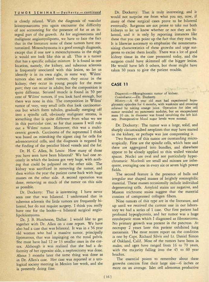
TUMOR SEMINA R Doclcerty continued
is closely rclau.'d. With the diagnosis of vascular leiomyosorcoma you aguo encounter the difficulty of nOt accounting for the prescooe of far as an inregral p:lrt of the growth. As for angiosarcoma and malign•nt angiomyolipoma, we have ro f..:e the f•ct that in dte lireramre oOfle of tbe$e wmors have memsrasized. Mesenchymoma is a sood enough diagnosis, excepr rh•r if one saw a rncsenchymoma in the thigh lt would nor look like this at niL This is n tumor ch:u has a specific cellulru mix:mre. Jr is found ln one loc.,rion, nnmc:ly, the kidney, and tuberous sclerosis is frequel''ltl)' nssoci:mx! with ·this lesion, so Icc us idenrify it in i1$ own rigbtJ in some w:\)'· Wilms' tumors nlso nre "'ixed rumors; they occur jn rhe kidney; they occur in )•oung people for me most parr; cht;• Clln occur in adults; bur the composition is quite different. Striated muscle is found in 50 per cent of Wilms' rumors. if you look bllrd enough, bm the~ wu none in d1ls. The composition in Wilms' tumor of very, very small cells thar Jook carcinomorous bur which form tubules, gradually merging off into a spindle cell, obviously malignant suoma1 is somerhing rhat is quite differcn1 from what we ~ in this particular cnse. so lor thac reason 1 will rult: ouc a \Y/ ilms' tumor. ~{orco,•er, this \\•a.s a multicencric growch. Carcinoma of the suprarenal 1 think was based on misrakiog rhe signer ring fat cells for adrenocorrical cells. and the diag.oosis is negated by rhe finding of the pecu liar blood vessels and the f<lt.
Dr. H. C. Allen, Sr. l.ouis: How mnny of those you have seen hove been bilareral? We bod one re· cently in which the lesions go1 very huge, wi1h noth· ing rim could be palpated on tbe other side. TI1e kidney was sacrificed io removing the rumor, and then wirhin rhe year the padenr came back with huge masses on the or her side. A secood operation was dooe, removing as mucb of <he rumor oo this side as possible.
Or. Dockerty: 11llt is interesting. l have never seen one <hat was bilaccrnl. I underStand that in ruberous scl<roois the little tumors ace frequently bi· lateral, but do no< require smgery. 1 think you really have one for the books--a bilatenll surgical angio· lipolciomyomo.
Dr. J. ll. ~huchcson, Dallas: I would like ro get togerhC!'r wilh Dr. Allen on a ca.'\e Itpo.cr.. We h:~ve also h•d n case tht•t was bilateral. I< was in a 56 year old womnn who had n ma.~sive romoc, principally lipomnrous, rhot was impinging on rhc renal peh•is. She rnusr ha"e bad 12 or 13 smaller ones in the cor· tex. Altl1ough it was realized thor she had a deformity of her opposite side, a oephrecromy was done. About 3 months later the same thing was done as in Dr. Allen's case. Her case was reported at a urological sociecy meeting in Mexico lase week, and she is presently doing fine.
Or. Dockerty: That is truly inr<rcsdng, and it would not surprise me. from wh:\t you say, now, if maoy of these surgical cases prove tO be bilarernl eventually. Surgeons are nor prone co take OUt both kidneys to Jer us Jmow wherher or not d>ey are bilarcml, oncl it is only by reponing insrances like these lha< you can point up the facr thac they may be. It is likewise necess:uy co emphasize the nonmet:UI1l· sizing cha.tac[eristics of these growcbs !lnd \ttge surgeons to excise them locally. There was a lot of good kidney tissue in the case presented today, and the surgeon could have skimmed off the bigger lesion. Ht would httv-e left 6 others, buc chose might h:tve raken 30 )•ears to give die patient trouble.
CASE 11
Di•gn.oJ;s.-Hypoglycem.it rumor of kidn~)'· ContNb,ror.-.Oc. Dockerry. Hi.1toq.-A 48 yeu old m-an had t-xperitnctd hypo.
gl,.a:mk episode$ for 4 months, • •ith wnlc.nt$S a.nd S\\'f!a~io& tdicvcd by takins oran~ juice. Operation fOf l:sltt cell rumor reo.·calcd. no poaocrcacic nodules. but •n encapsulated mass 10 an. in diamcu·r WM (Q~Und invo'lvintt the lcJr kidney. Pouopetath-e blood sugu levels 1l'Cre: nocmal.
Dr. Dockercy: The rumor wos ~ l>rge, solid, and sharply circumsccibed neoplAsm that may h>vc scatted in rht kidney, or perhaps was jusc coanprcssinS it.
[ 16 J
Two feacures of chis rumor ~· re outstanding microscopk1111)'. First tlte the spindle cells, which here Ulld there are aggccgated inco bundles, and e.lsewhere "l'P<'"" ro be closelj• applied ro th in w3llcd vascular spaces. Nucld are oval aod nor p:micularly hyper· chromntic. NucleoJi are small :~,od mhosts :tee infrequc-nr, averaging one in every six or seven high power fields.
n,. second fearurc is •he presence of balls and irregular SrM shaped masses of brightly eosinophilic m11crial. 1besc masses occasionally include individual dtgener:uing cells. Amyloid szains lltC ncgacive, and Masson trichrome smins suggest char the material consists of compressed collagen fibers.
Nine tumors of chis type are in the licenttu.re, nnd up \IDtiJ we received the current one in our laboratory we had a. series of l case. Our first patient had pro round hypoglycemia, and her rumor was a huge rctrobepntic mass which I diagnosed os fibrosarcon'la, No primary growth was present in rhc pancreas. At nt'<'ropsy 2 years later this p:ttiem exhibited lung mct-:tstasis. The mosr recem reporc on the cond ition is one by Capr. Richard Silvis nnd lc. Daniel Simon of Oakland, C..t.if. Most of the rumors ha\'C b""n io males, •ncl ages have ranged from 16 to 70 years, with the ma joriry falling into che -!) to 60 year brncket.
11>e t'SSClltinl points to remember about rb<SC growlhs concern first their l11tge sizc>-6 inches 0<
more on an average. .Isler cell adtnom>S produaive

Fi$1, 1. Co~e 1. Carcinoid o l· duodenum. X 60. l'l9 . 2. Case I. F11J. 3. Case I?· Ccucinoir! in dcrmald !:):St of avery with Biord:.~Tnor&on .!:yndromc. Both ore <Of':",Posed of u-niform ori&motk .cells wl1h Dn"k cytootosmk Qt01'ules. x 760, Fig. 4. Coso 2. Benign po;:~ll lomotosts of ba:-ost. The DC:lll lory !>talks hove OO•~I"'C>t livc lil:'oe «>res, a nd the eptthefla 1 c!emenh e re unit~rm with little &trotifkotion. Fig. S . CQr.c 3~ Paoillcry cordnomo of the b1oost. As com-
po re-d 10 Co~to 2., the growth iS; more solfd l v ~i11V.d1o1 and f lie conn~ivc hi>~>UCI ;tolks moro tcmoO\ls. x 170, Fig. 6 . Co&c 4.. Boni~.n glon~ cell tt•mor: of ICCDtdo GiClhf" coils ore num~rous or-d contain loro~ numbtlrs. <If (lOCtei, x 1?0. tig,' 7. Coso 5, Grode IV chondrosarcoma of--scopuiO. The teMOfL is eellu tca wilh a r'Coll ot ne-crwis. x 60 Fig. 8. Cc::>e 5. Mora or lc:.~ rounded cclfs w•th mart)! mito!ic:s. nnd o w rroondlno d ear ~ooe, '< 760,

Fig. l l . Co~;c: 7. F•br<lblo:oh .s.howi:ng morlc:t ale p /eomorphlim a nd l'nii0$1$ (ccnlot'). x 475. Fig, U. C0$6 8. Bllot~rcll mccochronous mommory c.orciM"ffO Cfld CW)e!oted m.etc-~l osl.z:lnQ lioosotoomo. Thh. f!Qure &h OW$ the onoplost ic /jposon:cma wtth g icnf end bi::crtc cells. X 105. Fig. 14. Cm.e 8 , Grade IV l~inhoul c.ard nomo ol lett breast, )( 105. Fig. 1 s. Cose 8. Gtode h i comcdocorc•notna of rloht br"OJ' op· pc renlly !I ti ll 11'1 situ. x I ,0,
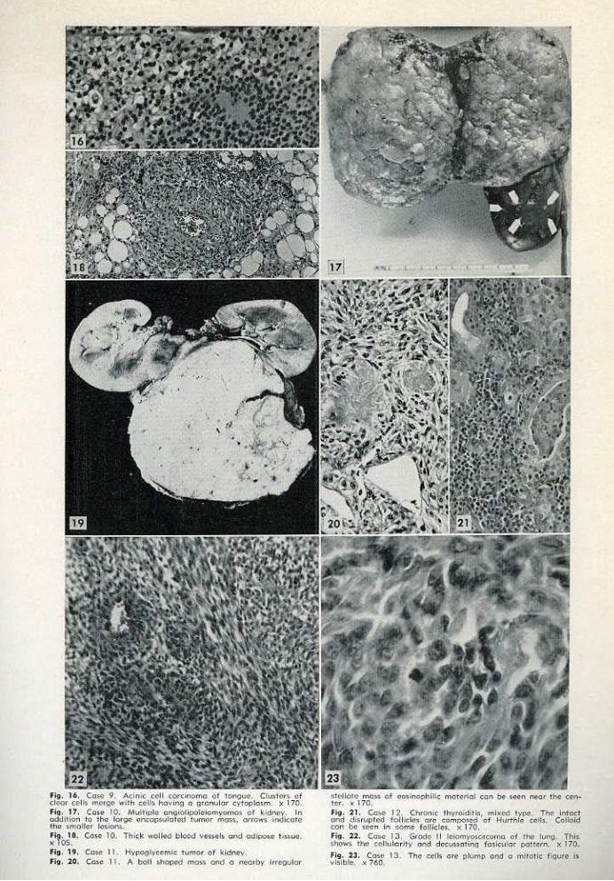
F1g. 16. Coso 9, Aclnk C<'ll cordnomo of tonou~. C!uslcrs of clear cells meoroe wlth cells h:.'lving c gH·:nulor cvtoplca m . )c 170, Fig. 17, Co~t> JO. Multip le on9iOiipolciomyomO$ of kidney, In addition to lh<; Iorge on::opwtotcd tvmor mas~, orrows lndlcota t ho $mOiler lor;iom. Fig, 18 . Co~o 10 . Thlck walled bleed\'~'!.~ !~ orld odlpo~c th.~uc, X lOS. Fl9. 19 . e'o~ I I. Hypoglyc-~mk t u mQr o f kidnoy, Fl; . 20. Ccae I I . A boll l:ihnpod moss ond o nearby lfreQulor
stellate mc:-n of codnot~hiti<: motorial cofl be seen neot tho& cenH~r. x 170, Fig. 2 1. Cow 12. Chr01l ie thy~okhtis, mi~ed type. The infoc:f ond d fsruplcd toll1dC5 ore cOmpO.'IC'd o f Hurthl,::: coetk: Co!T.oid con 00 !.e-en in ::omct fo llicle:s. x 170. Fig. 12. Ccsc 1 :.J, Grodo II lelom~o{coroo of rho Juno~ Thls shows the eelluler-itv Qnd dcC\Inotm9 fosicular poncrn. x ~ 70.
Fig. 23. Cos~ 13. The C¢115 ore Plumo o.nd a mhotic. rigvro. Js visib lt-, x 760
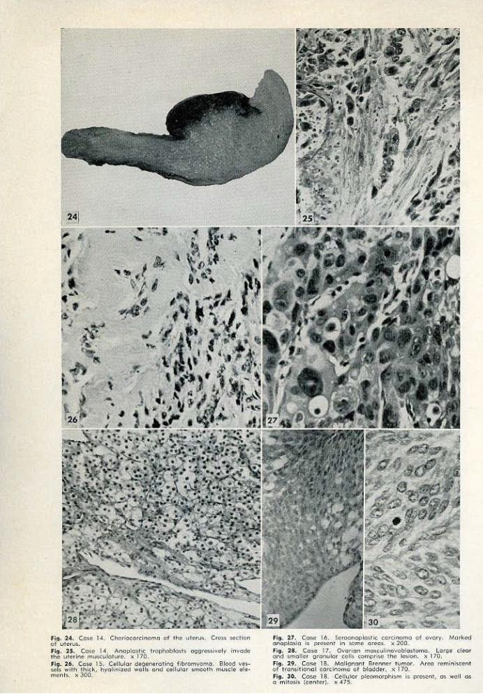
Fig, 24. Co:;.o. 14, Chorio~ordnom.a o f the. uleruo;. Cro,.s ~ection of u1er1.1~. fl9. 2-5. Case 1-'1. Afloplost lc Trophoblast$ a90rc~>sivcly in-.ot;lc the uterine musculoTure. x 170. Fi&J. 26. Case 15. Cellular degenerat ing fibtomyomo. Blood \14!$· ~I:> with' t hick, hy atm i:zcd wall:; and ca)lulo r smooth· musd e e!e-· ments. x 300.
Flp. 27. Cose l 6. ~foonoplosrlc co.rcinomo of ovary, Morkird onopla:~~o i.$ pre:~ent in some cteo,. x 200. Fig. 28. Co:se 17. Ovarian mo!.cu linov6bhl:~lomo. LOtQe tleof ond WlOflcr gronulor eel£$. oomori~>C Jhc losf9n, ) f 170. Fig. 29. co~c 1$, Molignont Brenner t U I'I"oOf , r\rco reminl$-CCnt of uomltlonol cordnomo of bladder. x 170. Fig, 30. Cosc 18 Celfulor Qleol'norphlsm Is pr("...ent, os wall os o mJtosis {center;. x .475.

TUMOR S. EMI NAR Dockerty-c·ontinued
of hypetiilso lin·ism .are measured in millune1ers rather [han inches. Functioning lslec ce]L .cancers ~r.c larger rban rheir benign .coumerparrs, bur still frurly smalL Se<:ood, they are all exrrapanCr<!ad c. SeV'erol have been located in che chest, l of our rumots w11s- ln che liver, and rhe remainder have occurred as rccro· periton.e;d growths, ofren arrachcd co one of the kid· neys. T hird, nobody bas excracred insulin from the &rowcl1s to date. Fourth, the rumors ate all fibroge-nic aod var)' in cellulariq'. Some arc histologic~ll~' ma· Lignanc and the)' can JO.etasmsize. In ihe case reponed by Silvis and Simon the g rowtb il)vaded the spinal cw<l. Pimtllj•, nc;>body has ,any dear 'Conception ~s co wliy ~these twnors ioduce hypogl)•cemia. Th~ obser· vacion o f Evans c:hac eluting cbe symparherk nerve supply ro the li,•ers' of cars produces low blood sugar levels may or may noc be significam. These nunors are large enough tO press on a lor of Otf,'!lns~ bur on the ot·her hnnd so do uterine fibroids ·Md o ther tU· mors which Me nor associared with th is peculiar clinical S}!ndcome.
Snbmill.etl Di4ghoJeJ.-5i\t<Omas, 11 ~ islet cell w mor, 5; m.-.l,isn;tm isle~ cell rumor, 3; carcinoma, 1; spindle ceH caidl.ioma, l ; adrt-na.J <ortical <:trdnoma, t: hemangiopericy:rQmn, S: pheOchromocytoma, 3 ; mi::;ceUaneous benit n tu· mors. 6; mis<ellaneous m;~(iso~nr rumors, 3.
Dr. Dockerty: Sarcoma is • s:1fe d i:.gnosis for [hese mmorsj they ace as low grade fjbrogcniC' sar· coma.s, ~nd abour one4 rhird Of che cases in the litera· cur~ have roet.Jstasized. 'Moljgo:1nc islet eel) iumors chnt I hal•e seen have nor I<!sembled a fibrbgenic lesion at all. In our expet·ience 80 per cenc of islet cell rumors are benign and ·small, mettsured in milli· me[e rs rarher than cencime·rers, and exhibit no micork l ccivity; 10 per cenc of th em a re intermediate in s ize, measuring 3. or ·4 em., and exhibi t enough m itOtic activicy for me [0 call chem grade 1 islet cell C~1ncers.
I have noc seen an }' of these mecasw.si:ce. Anocher 10 per cem o f islet ce ll rumors are obviously ffi;tlignanc and often mecascasize to rhe liver by che blood srreru1); but d1ey are all epitbe}i;;d nunors~ whereas rhis is a spindle cell g rowth. I do not see enough d ear cells in this case to suggest adrenal conical carcinoma. TillS lesion js not vascular enoughJ h as EOO much fibrosis, 'wd is coo lacking ln · arrangement tlround blood vessels co be a hemangiopericycoma. Pb('O· chromoc~·cc;Hnas can be p.tetty fibrogenic, buc in my experieuce you will practically alw•ys find marked vascular icy aod grear big granular cclls like chose -you see in .!;ranulac ce)l nJ)'Obll11!tOmas; you will also some• rimes find some pigment; and che tumor is 9bviously mut:b more anaplastic rhan this. I do not know what is iodude<l in rbe miscellaneous grqups, but you can ~U tbis, if you wane to, a miscellaneous b en ign fibrogenic tumor, possibly malignant, indUcing, for some unknown reason, clin ical severe b)rpoglyccm.ia.
9enc<al Dt:Coursey; I wonder if D r. Goldzleher h.S qad any <noughts on rhls tak or any experience whh .any simHnf· cases. Here wits -a rumor of rhe kid· ney char cnu51'd hypoglycemia, but oo cl1emiql exarnlo:lt:joo did not conrain in$ulin.
Dr. J. \Y!, GoJdzieber, SM A.otOoJo: I r-•ke it chis rumor did n oc destroy enough of the liver co interfe re w ith carbohyd ra·rc merabo1ism.
'Dr. Dockcrcy: No, chJs was a neoplasm that looked like a low g(ade fibrosarcoma. \Ve have bad the onlr. <:ase t hat v;·as retrobepa.tic :'IOd rhat mighr have in terfered wich 1fhe fun<:cion of rhc liver in any way.
Dr. Goldzieher: I cerminly bavt! nothing ro add, buc I would say that the iMbility to demon.s"ace the presence of insulin or a hypoglycemic substance would not be conchtsi-ve in my mind, because ·e\•en with all the work that b(IS been donee on crying to demonstrate insu lin in the blood, this is still an un· accep.rable proceduJ'e ro mosr endocrinologists. There could weU bave been insulin in rhe cuinor, and it jusc was noc gotten out.
Dr. Dock'ert.y: J suspeq, also, thnt the pnrhologists got these insulin determinations as 'afrcrthoughrs, afrer the rumor had been in formaldehyde for se:veral days. It happened in our case.
CASE 12
( 17)
Di(lgncti.~ .-01 rouic thr rojditis, mixed rype. Com rib1110r.-Dr. A. 0. Severance! Baptist Memorial
I-:{O$pic.a1, ·San ,\nconlo. flislory.- A 64 ye-at o1d \\·mr.an wu.s adn:Utted to th~ hos
pital because o{ '3 harli sw~Hins io her throat of 3 co ·1 nlomhf dur-ation, and an associated choking sc.nsar.jon of l m~mrh's duration. 'the palpa~le mass involVed both s~de~ of. the th;'rpid gJilnd. At surgtry a walnut·sized masS was f<mod ·at>ovc rhe thyroid ,gl3.nli in the tesion of the thyt<l· g:.IO$.SiU duc.t. Doth lobe$ of tbe thrroid and the isthmus were cock h1ud a.nd were temoved. 'fi1e hlsw1ogic pictu(e of the thyroid and the mass ubcwe \\•ere si.milnr. rrhe J>.'l· dent is still :tlivc wirh a ma.~s.- in her neck l ~ years after surgtry•.
Dr. Dot:kc.n:y: The microscopic alceracions in chis case concern both che Cl)itheliutn -a1rd rhe scrqma. The former~ here ~nd tbcrt, appears still cb be in rhe business of producing cOlloid, buc even in these zones: there is enhugement and gronularicy of the cells which have the hallmarks of .Hiirdlle cells. TI1e acini a re sepamted by 1)•mp hocyric dusters; acinar. boundaries are disrupted; and giant cells, probably consist· ing of fused hi stiocyces, arc~ foun d enmeshed in the coi!Qi<l sobsrance.
Else\yhere the epithelial dement.s aro mu'ch d istoned and rhey are disposed singly, in sm:l\1 alveo)~r cluste.rst M lcl in. thf fqnn · of shore conci.m10t~ files.. In spite of [bis aisorganization, however. mitocic fig· ures are absenc .and nudeol.i remaio sn1all.
The srromal chnnge comprises lymphocyric infil· tration on chc one hand and fibrosis and byaijnizarion
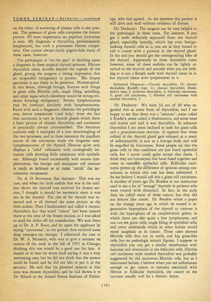
TUM 0 R Sf MI NAR - ·Oo c lce r.t y - aon tinu ed
on the orher. A scm eting of plasma reUs is also presom. The presence .of giant cells c9mp!etes rhe bizarre picnue. Of mOSc importance, no papillary form8t'ion is· seen. ~iy diagposis is thyroiditis, predominantly lympbo~:yric, but with a p(ominenr EibtQUs compo-' nenr. One cmnot always nicdr pigeon-bole m.aoy of these cases, however. ·
T he pathologist is "on rhe spoc" in deciding upon a diagnosis in these arypical cl1yroid pictUies. Fibrous thyroiditis often invades berond the capsule of the gland, gi¥ing the sucgeon a srroog in1pression thnt ad inoperable mali~;nancy is p rl'Senc His biopsy specimen is not likely ro be generous. Microscopical· ly th is lesion, alrhough benign, fearures such things as giant cells, Hiitthle oells, single filing, spindling, and other signs which elsewbere would be suong evl. deoce favoring maJignanq•. Strwna Ly.rnphomatosa may be confused ~im!Jady wirh lymphosarcoma. Faced wi<h such a diagn&cic dilemma the· pacl•o!ogisr moy derive consola:cioo (and help) from rhr face tba< c:ucinonla is taj;e in thyroid s!ands which show a. b~jc picture . of chronic thyioidids, ~n<.l c•1rcino.mil is pt:'lctically ahvays· cjrcumscrib·ed. The Jheranue contains only 4· exa.tpples of a true iocermingliog of rhe cwo pr9cesses, and in <hese inscances the papillary nature of the .cardnoma made tbe diagnosis easy. Lymphosarcoma of the tltyroid, likewise. ·quire rare, displars a ''solid'' cellularity with cywlogkolly immature celis showing Lirde admlxru.re with epitheli· um. Alcl1ough found occasionally with srruma lympho.macosa, the benign and malignant cell mi>.'ture is rardy so intimate as 10 mal<.e "occuk' the sarcomatous component.
D r. A. 0 . Severance, San Anronio: 11lis was mv case, and when the litd~ nodule that wa:s in the -mid· line above che chyroid was re<reivc..-d for frozen seccion, I rl1ought ii- shoUld be mccascatic since i t was not in rhe thyroid. The rest of the thyroid ·was re. mo•;red and it aU .showed ·the same picrure as dte little nodUle. Then 1 backtracked and c;,Ued i t chronic rhyroidicis, bm thnr word ''cancer'' had been uttered there ac the cime. of the frozen settion, so I was asked to send the s)ides off for consulrorion. \'>l'e sene chem up to Dr. A. P. Stout, and he upset che applecnrc by SQ)'iag "car:cinoma"; $t) che patient chen received some deep roentgen-ray cherapy. I submicted che case m Dr. W . A. 'Meissner, who was tO g ive a seminar on rumors. of the nee~ in che fali of 1955 in Chicago, thinking thi$ one would be a goqd one for him. A month or so later he wrore back saying it was a vert irtterescing case, btu he did noc chink that the ~nswer could be found and he did noc like co pur ic in the seminar. He said char bis personal opinion ar that time was chronic' dly~oiditi.s, aad he had shown ic co Dr. K linck at the M:med Forces Institute of Pathol-
( 18)
ogy, ~rho bad-agreed. At the moment this paciem is scill aJive and well ~~idlour evidenc:::e of disease.
Dt. Dockerty: U te $Urgeoo. can be very helpful to the pathologist in rbl'So cases. For instance, if you get a node delinirely separated lrom the thrroid gland, especially laterally, which bas even benign· looking thyroid cells in i~ you are in dmy bound to call it cance; w ith a primary in the thyroid gl•nd. In the end you should get the cortespo0diog lobe of cl1e thyroid. Apparently in the'se thyroiditis c.ses, hc)~rev~r, ~.me of these nodules can be lightly 3t· cached to the iliyroid and acrually whl! you nre looking at is not ·a lymph noi:lc with thyroid tissue in it; but: thyroid tiSSt!.le 'lvith lymphocyces in ir.
Su/Hn.ito'.etl Dia~'IJOJru.-Chronic thyroidiris, 18; dHooic thrroiditis, Riedefs Wl*· 11; chronic th)·roitlitis. H!LShi~ moto .. s type-, 5; subaunc thyroiditis, 4; follicular carcinoma, 3-; gi~oc celJ (';ltcinom.1, l : Hi.ixthle cell q.t~iooma with ma'rkcd ffiyroidiris.1 I.
Dr. Dockercy: We ~have 34 out of 40 who .re· garded rltis as some form of rhycoi<litis, and I am happy to see t11at there was a ''mix.rurC:'~ some called it Rfeders, some atlled it Hashimoto's, and some w~re real hon~st and said it was thyroidi tis. In subacute thyroiditis I am. more inclined ro l90k for giant cells and a granulomatous reaCdo~l. It ·appears · th~c when colloid of che thyroid gland gets released as a res11lt of inf1ammati00, it aces as a foreign body and will be engulfed by hisriocytes. Some people say that l'he giant cells in that condition are just fused epithelial cells, but 1 neve.r could qu.ire accept that thesis. 1 think they ore h.isriocyces that have fused together nnd come ro roscmble epithelial cells. Follicular cardnom~ poim~ up ~he di.ffere1.1t;e of ol?inign "1!\Qng t;Qn· sult.~nts ro wbooo th"is C.ftSe has beea submicced.. I do nor bdieve 1 would call this a giant ~ell carcinoma. A n,tn:tber of years ago Dr. D_roders nc our insdrutio·n used to sec. a Jot of "strange" thyroids in parient~ who were ueared ·t~rhh thioucaciJ. Jn face, in rhc earJy d:tys we called some of these cancer; buc they did noc behave like cancer. Dr. Broders wrote a paper on the Change yefl.cS n.go in which he named ir re· genern~\ve hyperplasia of che thy.roid ro COI\trnsr it wi<h .the hyperp las,ia of ~n exophthalmic goicer, in which there >arc alSo quite a few lymphocytes, artd one can see siaoc cells, single fi lins., of epichelinl cells,. and orbec credentials which in ocher lesions would mean anaplasja. as in cancer. These cases showed Hi.irtble- cells tlhat ran in cords, and big giant-like cells, bur no pathologic Jnicocic figures. 1 suppose in chyroidicis )'OU can get a similar interference with funcrion 3.Ud :Uber{ltions in the foHkular celfs. HUrtble cell carcinoma with marked thyroiditis was probably sugge$ett>d by en~ numerous HlircWe cells, .hue as I mendoned befo.~;e> I believe .... •hen )'OU flre unforcunare enough ro gee cancer or sarcoma aSsociated with fibrous or follicular chyroiditis, rhe cance.r or the sarcoma. usually will be a discincr lesion.

TUM Q R: SEMINA k Doc.k·ertr cor1t.in ued
CASE 13 ., v' <\~
pi.a:g-t:Oiis.-Gradc l[ lciQm)'OSarcoma of lung, Co-nrri-bJIJor.-~t.ooke Army Hosp,ical, Fort Sam Houston. 1-lissor'):.- A 23 year ()]d whit<: man fl.ad a JllStory of
right lower dlesr nnd upper abdominal ~in for appr<>xi· mare)y ; yc.'llrs. A mass in rhe lower lobe of the r i;s.hr lung was seen on routine" roentgenogram 4 month~ prior to surgery.
Colonel· Harrell: This ma..s is faidy ro\lfld. Tbe n1argin.s of ir a re rather shatp, rtod ir does nor seem co be' invading, Acmally, from chis fi~n, 1 do ooc rbink one Cilll cell wbecbet it is in che middle 6r lower lobe. There· is nQ rib in\'olvement to indicare metascaSis, nor ~tie ' rhere a11y or her lcmions Or nodes that I can see in che che.<r. Tbe best probabilities at 23 years of age are thac (his is inflaJU.IXHttory inscead of a m~UignanC)', unless ir is a rnetascaric lesion. I llke co srudy chose ltsions by laminogr~phj•, for if they ~ue., maligoam tl}ey ofcemimts show umbilic~ .. tions in their pexiP.hedes where cbe blood vcisels which supply rhe lesion emer. I have picked up a few that W<1}'1 whiCh m,ade me feel !:).tber coufidenr dur the lesion was metast:\rict or ar least maJignaar, bec:wse benign rumors usually do not demoo.strace this umbilication. 1\ooche~ thing that laminogmpby shQws, which sometimes is 00( shown on the plain film, is ·whecher or not rbesc Jesions- coocain calcium, and l think this also is a good differential point. As far as 1 can wlll:iy lookiug at the reproducrion, there is no calcium 'in this lesion.
Dr. Dockert)' : This growth is in the midcUe of tbe lobe. lc is fair.ly solid) ~mel does n ot llave rhe caseous nppea.rance that ·we ex,P.CCt in tuberculomas. On cue '""face we set tMt nlchough ir displaces a bronchus i~ does noc 3cruall}' ulcerate the mucosa. The wash-ings in this case perhaps would be neg~.ti\'e. With a c.ircurnscribed growth in rhe lung that looks like a neqplasm an d \vith the ~g¢ and oU we would suspect th{lt this mi-ght be merasmtic.
Bnsic~lly rb:is very ceUular neoplasm is comprised ot plump spiodle ceUs With elong~ced oval nuclei and pink smining cymplasm ·which sometitnes h itS a granul~r -uppe;~~oce. There is a definite tendency for cell grmtping inco fasides ~nd bundles which dOC\~SS!LtC one w it h another. Here ana there arc a few bandS of brightly .srn.ined hy::tline m:uerial.
Vascularity is ~rooouoced, hut there :tppetli' co be no vaSCllloinvasive rendenci<tS even rhougb .-.some of the vessels ate very thin walled. Malignancy is indi· caced by fairly m•rkc:d m·itotic occi'vicy and by che ptesence of inf~trccion which s~ems co be on the basis of the tumor outgrowing its blood supply. Peripherall}r there are fibrosis ::1nd hyalini~~don which m1ty be rel:tted to ancient infarction. J\.fa.sson 's ukhrorne stain Sives a red -appearance con.siscenc wich young fib rous cissue or mu$de.
[ 1·9)
To recapirubue, chen, one is impressed by che faq tha.: ·~ve J1ave ltere a spindle cell cumor with r:.lther granular, eosinophilic cytoplasm; n definite tendency to form whorls, or bundk'S, or fasicJes, thac inte:rlock one with anorh er; fnirry pronOunced va.scul:ui.cy; and M cleft formation. }.{y diagnosis, right or wrong, in chi~ case is leiomyosartoma, grndc 11, of che lung, po.<sibl)• coming from d1e musttilaruro of a bronchus.
The lw1g is a derivative of rhe primitive gasrro· intestinal tr:tcc-"' w hich frcqu~ntly harbots myomacous tumors, benign aod malignant. In the esophagus abouc 20 per cent will si19W malrgnant change, and tbis will reacb 30 to 35 pet C£ntiCI the stomach and ~[;out 50 per cent in the small incestine. Tbey will not go direcdy to lyrnpb nodes, bur having breached the peritoneum, they will implant. When rbey do reach the lymph oOdes, t11ey usuaUy do so via the blood scream from mems~u-ie-_pulmooacy Je.sioos. Ac-cording to \Varson and Anlyan, there are 10 r.efe£ences in the 1 i.cerarure oo .indiVidual case reports of leiOmyosarcoma (at lease collc:d chac) presenr in rhe lung. Inceresdngly enough, rhc 1"\tienrs wece all )'Omlg persons. In other repqrced art icles tl,tere ~re 10 references to benign MO.Piasros of sroOQth .ri1uscle io che hmg; so apparently in the lung, as ia tb~ gascroinresdnal cr3Ct, there is a great tendency for ll'!):'Omacous rumors to be malignanr, a change wbic.h we expecr in only nbouc .5 pee cenr of the ordinaryuterine fibroids. According w Evans, who I think did the best w'ork on these ksions, the safest criterion foe. diagnosing. malignancy in a srnoorb .muscle tumor js the presence of more rh1n J mi~ock figure in 10 ordinary h?gh, dry fields. One lnttSt be very c:ltreful abour cnllin·g benign any smooth muscle rumor in t he uterus or gasttoiotescioaJ traer which ·exhibits miro~jc
figorcs tltar nre not roo hard to find. One would suspect in this case char che rumor was- mer.astatic-. The commot~e.sr example of myos..rcom• in the lung is mcrasrasis ·from malignant uterine fibroidSj but chis was a ma~e rocher rhafl a female, and we do not bave any evidence in this case frdm ochet Stlldics tbac there was> in face, a _primary rumor :mywhere in the body ocher chan in che lung. It has been Strongly suggested that rhis lesion might be a mew.scatic synovioma; however, in a rev,ie~ of £he lh era· mre, 1 ha\•e no~ found one synovioma presenc1ng lung met~stas;s in the absence of :r. rec<Jgni~d pri· IDa!)' growth. The lack of nlveolar clefts and of cal· cifitation1 lltl.cl rhe telati ~·el}' lnfge · si1~ Qf rhe t;umor cells furdrer incline me away from this diagnosis.
.M;dignonr mesothelioma was suggested by one of my colleagues_ In our own group of 24 fibrous meso· theliomas, 6 were iatrapulmonary, but in the region of an inrcrlobar fissure. 11tis lesion involved the basilar segment of the right Jowet lobe, T~mQrs of thls type show Clefts, they are more fibrogenic ilian m)•Ogenk, and they freqtrenily exhil>it a papillary pattem. I compared che sections on dlis case wich

TUMOR SEMIN A R Oodctrtr con tlnuefl
sections of ouc malig nanc mesorheliom:u of the p leura, and the resemblance was not so ciking.
I came to the conclusion thar this was leiomyo· sarcoma, primary in rhe lung. t ''ery rare lesion. 11>ese low sr•de Jeiomyosorcomns in the gut, uterus, and elsewbete ate not tOO prone ro mensrnsize; and yet one must follow the pmtienrs for 10 rears or more.
S11bmiue:J DU.(Nose::r.-I.dom)·ouroorna, 7: mnochelioma, 13 (benign 9. maligNnt 4): fobrosam>ma, '' bronchogenic c.ucinom1, ); O$tCOJtoic ur<oau. 3; melaoorna, 1; rnttaSCatic sucoma. 1; nturobbstoma. t : Mman,;topc:ricycoma, 5; bmnchi!ll ad~noma. 2.
D r. Dockerty: We luve disaossed leiOm)'OS:Ircoma and mesothelioma, and I think that these are the two main choices. I believe thmt • fibrosarcom• of this low order of mllignancy would have plenty of col· lagen formation so that it would nor be confused with myogenic cytoplasm. A b rond 1ogen ic carcino m3 imitating sarcoma would be more ana.pln.st ic1 witb more mitotic figures, and would not form ffts icles or buodles. An osteogenic snrcomo would be rare, but [ suppose possible; however, [ d id not recogni:te any osteoid or bone. W e should think o f a n>:ti(gnttnt melanomo, ns they may 1pindle our nnd they can be amelanm ic. Mecnscnric snrcom:t i.s certainly n poss i· bility. A malignant rumor from the gur, however, U$ttaUy would have tO fi lccr chl'ough t·hc liver, nnd from cbcre spread tO the lung. l)y dmt time the d iagnosh; would h:wc Peen m11.de- becnuse the pacienc probnbly would hnve nn intestinal obsrruction. Ncu· roblastnma is probably suggested by the peculiar palisading of the cells; but rh.r is n characteristic o f smooth muscle cumors of the gastrointestinal t ract. As concerns hemangiopericyroma, I don'r belleve even Dr. Scou t would d1ampion the diagnosis in chis case. Bronchial adenoma I tl>ink is ruled out ~ausc tl>OS<: rumors are p«rty much recognizablo as epithelial, and th is was more of n spindlo coli groMh.
Fol/ow-Up.-Approximaedy 11 montht fotlowins the origi02l .su.rgery a second thotacotomy "..., ~formtd, -.·ith the mnoval of a 1.) em. subpnaral Jes;on (rom the upper lobe of rhe right June: sho•·iq. a piaurc: similar tO that o( the etisinjl tumor. ~nd incerpreccd ac c.be time u beins an impbnc. Rot-nc,seoo~;raphjc loUow•up at' 18 monchs re,·~1~ muhipl~ asymptomatic !c;;om: throushout che entire right lung fidd.
CASE 14
DkJ.~nOJ;J.-choriocardnnmll ol the uteru.s. Conrribut()r.-Or. L J. Mtnholl, Jr., Robete 8. Green
HospiCll, S:1.n Anto nio, HiJtQrJ•.-A 3 I yt;~r old whitt w()man pauc:d u large
hydatid ifo rm mo le. Two mo nth$ later • vu.gin:ll nodule w~u $ubjected co biopSy, rollowinJ.!: which n pnnhystc.r«tom)' was per(crmed. The ut~rus showe<:l ll bul~; lng 4 by 2 by 1.) em. friable ,mass, and lhere wos a 5 em. corpus Ju«:um q•u of
[ 20 l
the left OVA!)'· 7wo months alter h ysttrtctotny anotlter vagJnaJ noduJe wa,s excised. Tbe vagioa1 nodules and uterine mass aU sho wed the !3nte bist01ogic picture. The p3ticnt wu Jiving and well 2 ye:us after hysterecromy.
Dr. Dockcn y: Grossly me p ictute of the original lesion is what anyone would call a hydatidiform mole. ..Grapes" ate present in great abundance. 11>e microscopic sections exhibit well formed ,.iJii with focal hyperplasia of me mantling rrophoblas~S, both syncytial and Laogba.tls. Like Hertig, 1 cbink that these more aCtive moles are the ones most likely ro proceed inro cboriocarcinolll3. In any given specimen, however, reganlless o f location, I lean .strongly away from a diagnosis of cboriooic m:tlignancy when even one identifiable villus is prescnL Novak, Herrig, and ochers conrur in this opinion.
The uterus showed a red hemorrhagic nodulo, bur it is perlups significant that this nodule does not extend entirely through the uterine w'illl.
The vaginal nodules and the hysterectom y speci· men show marked anaplasia o f uophobla.srs, invnsion o f veins, nec.rosls of muscle, and much hemorrhage. Tbese features, however, do not suffice for Lt diag· nosis of choriocarcinoma unless villi are -abs-enc, rtnd many .secd ons muse he taken co prove their absence. l t has been In)' expetjeoce witb ch;lt o f •n nny others that villi can become deponed tO the vogina nnd even to the lung. To my knowledge there ore only 2 cnses in d1C liter:m.rre jo which denth de:•ling pulmonary metastases coorrunc.-d viJli. Novak norcd only I insrance in h is 74 cases o f true cl)oriocardnoma.
No vil1j were presen r in [he urerioe and ' 'aginal nodules, but tbis does not certify the diagnosis of choriocarcinoma The sampling muse be generous. and the fioding of ooe villus should negate a malig· nam diagnosis. Tlus musr be our philosoph)• if we are t·o get a proper perspective on creatment and prognosis. Currently we are studying 3 cases with tbt"se borderlioe lesions kno"''" as mernsrnsi.zing chorioadcnoma desuueos, associated w ith long shadows. Each of the p rimary growths exhibited occasional villi in the bad company o f very malignant appearing cells. These lung shadows ore disoppearing under roentgen-ray therapy ud it is tho experience of somo rlur dx.'f dis•ppear without any rreaunenr. We persuaded one of onr younger surgeons tO obrain o biopoy sample of one of these nodules, a nodule whicb the radiologists bad called memstaoic choriO· carcinoma, and villous suuc."tUres were identified.
Th= opinions expressed oo the subject aro b>sed on a personal srudy of 40 moles ond 13 casts of choriocucinoma p lus the wad ing rhrough of volumes of llteratuxe ( tberc arc more than 1,000 :trtides on rumors o f the trophobhsr) . Boiled down <O rhe nonsaponifiable reSidue this materi-al bas given 0"1¢ t he following philosophy;
1. True cho r iocacdnoma is a rare disease. l c is occasion:l iJ}' Ctrmble. The vase majort~y of pat iems
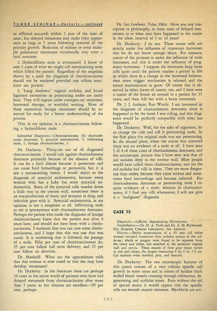
T U M OR. SEMit!! A R- Do<korty-cont·iftuod
so a£fliaed succumb ~'ithin 1 year of che time of on.s<;c, bm delayed merns.oses mny make tbeir appear· ance as long as 5 y.ears foi·iowing ranov3l of cbe prima!)' lJ!OWtb. Resection of solltar:y or even multi· ple pulmonary mcrasrost.'.s occt~sioo:illy m·ay ~wen a faral ourcome.
2. HydatidifOml mole is overaeated. I know of ·only 2 cases of wha.r we mighc caJJ metastasizing mole which killed rbe padenc. R~garclless Of the •n~plasia shown by a mole che diagnosis of choriocarcinoma should not be rendered provjded any villous suuc· rures arc presegc.
3. "Lung· shadows," vaginal nodules, and, broad ligamem extensions in penecraring nJoles are rarely, fnenJ. They ~~·iJ l rtgress uode.r roentgeJt·.rny ueaU)"'ent, bormooal cherap)', or watChful waiting. More of these mysterious benign mecastases should be remov'-d for stud)• lor a better understand lng of rhe process.
T his, in my opinion, is a choriocarcinoma following a hyd»ridiform mole.
Stt/J.mituil DiJgno.reJ.- Choriocarcinot'l1a, 36: choriO~<lenQma destrucns-, S; S}' IH'.yti~l eodoroclrid~. 3: iofiJ tr:.ldng mole. I : benign choriocarduomn, I.
Dr. Dockerty ; Thirty-six O"m of 46 diagnosed chociocartinoma; 1 ·would rule agai.osr chorio!tdenotna dcstmens primar.il)' because of d1e absenc:e of villi. lr can be ~ fat-:d disease because ir peneuares and can <;ause farn1 hemorrhage or pedcoaids) but ic is not ~ 01CW$t~~(zing tumor. I would ob jeer tO the d iagnosis of syucycinl end(uneuicisJ because every woman who has a babj• has some syncytial eodomecritis. Some of the syncytial cells wander down a litde way to the uterine v;;-aiJ; sometimes there is an ovet·J?roduction of rbcm, and invasion is <~eep~ and infection goes with it. Syn<;ytial endmnecrid~) iO. my opinion} is not a neoplasm ar a iJ. Infi1traring mole co me is synonymous with c.:horioadenoma destcueus. Pethaps the person who made the diagnosis oCbenign chor.ioc:ucinoma knew that the patient W9S alive 4 yeats -later
1 and should not bavc been wicb a. chorio
carcinoma. 1 maintain chat you can cure some chorio-rnrdnomas. and I hope cht~t this was ooe that ,.._,as cured. l! is interesting chac ic followed the passage of a mole. Fifty per cenr of choriocarcinomas do; 25 l'er eeoc follow full term delivery; "nd 25 per cent' foiJow an l}borrion.
Dr. !yfanhoff; \Whac are the appr.oximace odds rhat this woman is no·w cured or rhac she may Iacer develop mccasmses?
D..r. Dockercy: In the _literature chere are pe.rhaps to cases in the enl ire world of pacienwwho have had delayed rilerastasis hom chorioc:\icinoma after more rhan 3 years, so her chances are excellenc-99 -~er cenc, pe.dtttps:.
[ 2 1 J
Dr. Leo LO\\•beer, TursaJ 0kln: Have )'Ou any con· ception or philosophy, in chest cases of delayed metasta.ses, as ro whac rna;' have happened to che curn6r io the silent iorerval of 3 or 1.0 years?
Dr. Docket!}': 1 d<;> not. These tumor cells are scricztly \lOder the rnfJueoce of jmporrnnc hocmooes that we do nor know enough aboUt. For insrance, cancer of the prQSrare is tmder che influence of ml\le horroox..tesJ and ·this is under rhe influence of pregnancy horm9ne,, l suppqse the hormones !(eep these celJ,s guier umil che pntienr readies a period in li(e in. which th<:re jS ;l ch~nge in the hormonal .balance; rhen some criggcr mechanism is released and the rumor· reco.mJveoces to gi'O\V. Of course this is observed in other; forms of cancer, coo~ ;tnd I have seen a cancer of rhe breast s ic around in a ¥ottiem {or .35 ;'ears, and then kill her ~rich a brain mecasms.i.s. D~. J. J. And., jar, Forr Worth: I am interested in
rhe diagnosis of fhorioodcnoma destrueos1 since lt happened to be the horse [ wos riding, and chis diagnosis would be perfectly compacible wirh what bas happened.
Dr. Doc kerry: Well, for che sake of argument, let us change the Ude- and call it penetc-ariog mole. ln chc f.irst place che original mole was not peoetrnring. In the -second placeJ when the orerus was ramoved there \Vas no e''idence of a mole ar ajJ. I bave seen 3 tr 4 of these cases of chorioad'enoma desrntects, ood the microscOpic picnue r.hat I haye seen was tba.t of red nodules deep in tbe uterioe wall. Mosr people ~·ouJd have c~tlled them ch.oriocarcinOmai and yet r-he re~ nodu"les bad villi in them. They are moce dangerous th~u mole;, because they cause serious and some· rimos facal hemorrhage and become infecced. For chorioadenoma descruens or penetrating mole 1 rc~ quire evidence of a mole; whereas in chodocardn_oma,_ if I find. any villi whatsoever, 1 wlll nOt giv"e it a "roalignnn(· diagnosis.
CASE 15
O.i4&nosis.-Cellular <legef)etaliog fibwmyoma. COtl~ribt~lon:-Dr. D. A. To.dd and. Dr. A. lvL Richmond,
Nix Hospjl:J.l CLinicotJ Laborat<~ry, S~o Amonio. Riitory.-Pclvic exurninacibn of !l ~ year Old ,.;.bjte
woma n revealed numerous f irm nodular masses in· the culde-sac, which at llurgcrr ~ere found to be sep<irlltC- from lhe ()vary and rubes. but -attached co the po$tcrior wginal wall and cervix. The~e masses <1f fiun grii.)' tiss.ue varied in, $i2e :tfJd J b:tpc. the largt.ost: !DC'.tSurinJ; 8 f;)· 6 by 2.5 . em. Cue surf~<:es wc·rc- mocded, gra}•. an<l m\Koid.
Dr. Dockerry; The rwo microscopic features of this ~moe consist of , a very ceiJulac spindle ceU lJ!Owth in some are:•s and in others of hyaline thick walled blood ''essels coursing through edemacm1s, de· gcJleracing, and ·aceljul~r zones. Even wirbout rhe ~.id of special scains ic would app<-ar chat tho spindle ceUs a re smomh muscle ~l~mems. &lyofibrils are· evi-
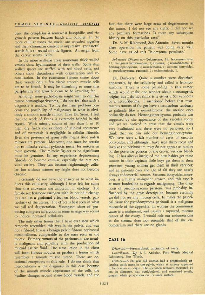
TUM 0 R SEMINAR- Oocl:trty- continu.ed
dcnr, rhe Cj•roplasm is somewhat biiSOphilic, and the growch pattern fearures bands and bundles. In the more cellular zones the nuclei ore crowded togerher nnd rheir chromatin ·Content js impressive; yec careful se"rch fails to reveal mitotic (iguces. An origin from the cervix seems likoly.
In rhe more acellular areas numerous thick walled ves.1els show hyalinU..tion of rheir wall$. Some thin wallt-d sp.1ces are sruffed with red blood cells, and others show thrombosis wid> org•ni%3rioo and re· Olnaliution. In the edematous fibrous tissue •bout rhcsc vessels only a few viable smooth muscle cells are ro be found. Ir may be disturbing ro some that pcriphcrnlly rhc g rowth sc<:ms ro be invading far.
AJrhough some parbologiscs rnighr \vish 10 c:tH rbls cumor hcmn.ngiopericycoma. l do noc feel thnt: sucb a diagnosis is rcnahle~ To me che mnin problem con· ccrns rhe possibWcy o f malignnn<j in whlt is obviously a smooth muscle rumor. Like Or. Srour, I feel rh>r rhe work of Evans is extremely helpful in this regard. Wirh mirocic c:uunrs of less rhan 1 in 10 high, dry fields the evidence of diniCill recurrence and of meramsis is negligible in cellular fibroids. Even rhe presence of g1anr cells U"'t:llls I inle unless mil-oses nre presen~;.. J\.{oreover, one must be certain oot w mistnke cena~n p)1knotic nuclei for mitoses ln chcse growrhs. The mitotic fisurfs ro be sigoificancmusc b~ genuine. In my experience degener;.cing fibroids do become cellular, especially the r:tre cerYiCII variety. They cnn become !righreningly 'ellu· lar, bur wirhour miroses my frighr docs nor become chronic.
l cerc;~.inly do nor h:we the :mswtr as tO wluc in· duces this cellul~riry, oltbougb 1 have fdr for some dm<: thlll :moxe1nia was lmpurrnur in etiology. The fcJtmlc: sex hormone esuogcu with ics periodic change in liter has a profound effect on blood ''essels, par· ri<ulal'ly or the urerus. The effecc is best seen in what we C>ll red degeneration. V>SOSpasm ShOrt of producing c:omp1etc infarccion in some smmgc "'ay seems ro induce increased ccllularicy.
The only orher lesion rlut I ba,•e ~r seen which rc.morely rtSembled tl1is was in the pelvis, and was not a fibroid. It was • benign pelvic fibrous pedroneal nli!'SOthf:Jiomn, comparable to the ones seen in the thorox. Primnty rumors of che perironcum are usunJ. ly tnnlignanr and papill:u:y with che pruducrion of mucoid nscicic fluid. Tbe same lesion in rhe chest will form fibrous nudules or produce • lesion which rC$tmb1ts a. smooth muscle tumor. 1l1ere ~re occuional .xceprions ro this rule. I do nor think tbt mCSOihelioma is the diagnosis in this c:ue, because of the smooth muscle appearance of rhe cells, the hyaline changes around these blood ves,.,ls, and the
[22 1
&.ct rhar there were large ~~teas of degener.uion in the rumor. I did not see any cleftS: I did nor see any papillary form.Uons. Is rhere any subsequenr history on this particular case?'
Or. A.M. R ichmond, San Amonio: Seven monrbs {lftcr op,•rario.n the paricnt was doing: very well. Some have called rhis "leiomyoma pec11l inre."
$11bmin1d DltrgnouJ.-1.eiomyorrta, 18; lt'iomyosar<Oma, 17: nualign:am Schwannoma, :l; fibroma. l; nc:urQ.fibroms, 1; heman.s:ioperkrt-oma, l ; nc:u.rilcmoma, I ; s:ttrontt bncrynide$, I: pseudomrxoma periconii, 1; eodomtu-iosis, 1.
Or. Oockorry: Qtrire a number were disrurbed, apparently, by thc cellularity and Clllled ir leiomyo· sarcomn. 11lerc is some p:11is:tdins in rhis mmor,
which would mak~ one wonder ~bout " l'ltnrosenic orisin; but 1 do nor think ic Is elrhCl' a Schw~l'lnoma or a ncun)fibroma. 1 mentioned before that myomntou5 rumors of che gur have ~l tremendous tendency co p:Uisade like a. neurofibroma; the uterine ones ordinarily do nor. Hemangiopericyroma probably was suggested by the appewwce of the ' '<UCIJinr zones, and yet we noticed in areas that the vessels were very hyalinized and there were no ~icyres, so I think rhar we can role out hemaogiopericyroma. \Vle hnve seen a fair pumber of cases o f sarcoma borryoides, ana although I have seen rhtm recur and involve the pcrironeltm, they do not rtppear as mmors on chc posterior portion of the cervix without b i<'Cd· ing. It hn.s always iorrigued me how babies ger these rumors in their vaginas; linlc boys gee: them in rheir pt'Qst1res: young women get them in their cervices: und in potieots over the age of 60 rhey are nearl)' always endomtuial rumors. Sarcoma borryoides, moreover, is a highly malignam growth, :tnd chis case is ar mosr borderline as regards malign~ncy. The diog· nosis of pseudomyxoma perironii wn.s p1·obably in· fluencl!<l by r(\e g ross description, bccnuse certainly we did not see an;' mucolls cells. In mnle$ chc prim:i~
pal cnuse for pseudom;--xomn pel'icon ii i$ n malignant mucocele of rhe appendix. In women rhe commonest e>use is • malignant, and usually • ruprured, mucous cancer of rhe ovarv. 1 would ruk: our endometriosis as the S<roma d~ nor resemble thll of the en· domerrium and there are no s)ands.
CASE 16
Diflg'IIOJis.-Se-ro~-lnapJastic <:arcinom.;1 of ovary. Contribt~s;,t-,-Pr. j. J. Andujar, Focc \'V'arth ~cdic:ll
Labor-JtOfy, fort Worth. HiJtQry.-A 60 rear old woman had a prosressivdy m ·
la.rsiog (J1dc nuss in che pelvis. wbkh at surAt'tJ appea.rtd ro be on. rUn in origin. 'The .specimen removal measurnl J 3 em. in di.lmtttr .... "lS ooolobul11ed, and comAlned sessi!t, ,grayish \\•bitt projections o n its inner .s:utfact.
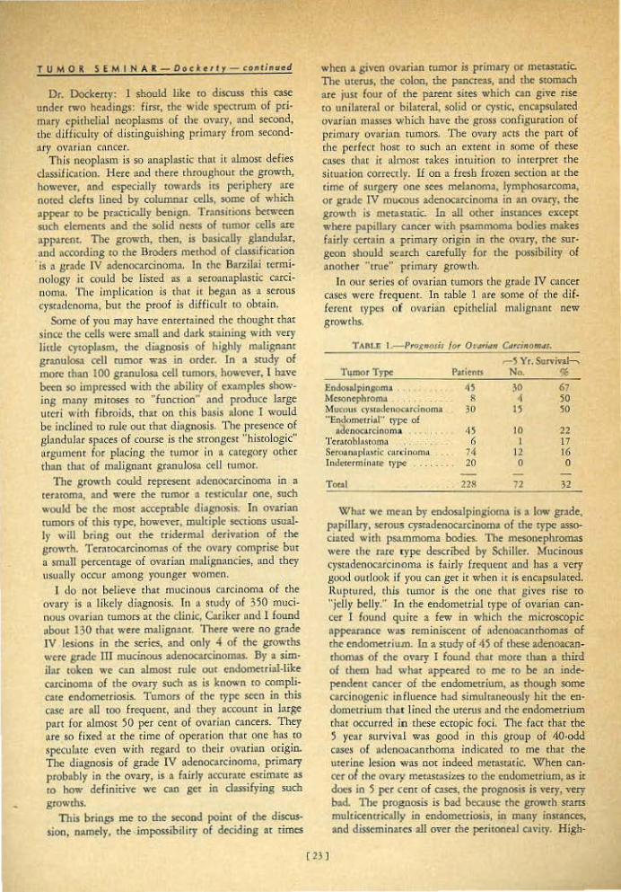
T U MO R SE MIN AR Doci:etcr continued
Or. Docl<eny: I should like 10 discuss this case under rn•o headings: firsr, •he wide spectrum of prim•ry epi<helinl neoplasms of rhe ovary, ond se<:ond, the difficult)' of disringuishing primary from secondary Q\'nri!ln cancer.
This neoplasm is so anaplas<ic that it ulmoS< defies dassifiC:ICiOu. Here aod there thrmaghout the growth, however, and c:sp<.-<:ially row>rds itS periphery are noted clefts lined by col= cells, some of which oppe<tr co be prncticolly benign. Transitions betw<m such elemenrs and tbe solid nests of mmor cells are apparent. The growm, chen, is basiaUy glandular, nod according to tbe Broders me< hod of clnssification
·is a S"dc IV adenocarcinomo. In the Barzilai te<mi· nology it could be lisced as a seroannplascic cnrd nomu. 'l'bc:: in'ljlliCMioo is thot it beg:ul ns !l serous cyscadcnomn, buc <he proof is difficult 10 obtain.
Some of you may have encenained the thought that since the cells were small and dark staining witb very little cytoplosm, the diagnosis of highly moligrunt gr:mulosa ceU rumor was in order. In a srudy of more than 100 granulosa cell rumors, ho"""'er, I have lx:<:o so impressed with the ability of exrunples showio8 m:tny mitoses ro ''function·' :\nd produce Jacge u<eri with fibroids, that oo this basis alone T would be inclined ro mle our rhar diagno$is. T he presence of gJnndu lnr S():tces of course is the strongcsr "hiscologlc" nrgumcnr for placing the rumor in n category ocher than that of malignant g<amdosa cell cumor.
The growth could represent adenocarcinoma in • reuroma., a.nd were the rumor a resricular one, such would be the most occeptable dingnosis. In ovarian rumors of this type, however, multiple sccrions usual· Jy wiU bring out the tridermal deriv:nion of dle growth. Tcr.uoca.rcinomas of the O\'ary comprise bur a small pcrce-ncage of ovarian Jnalig,n:tncies, and they usu:\lly occur among younger w01ncn.
. 1 do not belie~e rhnt mucinous cn.rcinomn of che ovory is n l ikely ding~>osis. In a study of '150 mucinous ovnrhtn rumors ac dtc clinic, Cariker nnd 1 found obout 130 thot were lllillignant. There were no grade IV lesions in the series, and ooly 4 of the yowtbs were grade 111 mucinous adenOClltcinomas. By a similar token we can almost rule out endometrial-like nrcinoma of the ov~rJ such as is known tO compli· cnte endometriosis. Tumors of the type seen in this cnso ore all too frequem, and they account in l:uge p;~rt for almost 50 per cent of ovnrinn cancers. Tbey a.:e so fixed nr the time of opemtion tl"" one has to speculate even wirh regard to their ovarian origin. TI1e diagnosis of grade IV ndeoocnrcinomo, primary probably in rhe ovary, is a fair ly accurate cscimare as to how definitive we cao get in dlllSifying such IJI'Owths.
This brings me ro tbe second point of the discussion, namely. rhe impossibility of deciding ar rimes
when a given ovarillo rumor is primary or meta.Staric. The u<erus, the colon, the pancreas, and the swmach are jusr four of the parenr sites whid\ C.'ln gi·\'e rise to unilareml or bilateral. soUd or cystic, oncnpsulared ovarian masses which have the gross configuration of prinlt•ry ovad,tn nwtors. Tbe OVM)' nct·s the part of rhe pcrfccr hose ro such an extent in some of chese cases chnc ic a.lmos; t:tkes intuition tO inccrpret the situ;ttion correctly. If on a fresh frozen .section a t the time of surgery one sees melanoma, 1)1mphos..ucoma, or grude IV mucous adenocarcinoma in an ovary, the growth is mcmscuic. In all <Kher insutnces except where J»pillory c:~ncer with psammoma bodies makes fairly cenain a primary origin in the ovary, the surgeon should seJ!rch carefully for the possibility of another ··uue" prirnaty growth.
In our series ('If Ovarian ctunOrs che grnde IV cancer cases were frequent. ln table 1 nre sorne of the different types of ovarian epithelial malignant new growchs.
1'Anl.r 1.-P..ot~UJJi.s /fff" Ot.JNIIII Curinom4S.
,-) Yr. Survivah Tumor Type: Puitntf No. %
EndosaJpin,gom1 . 45 ~0 67 Meso nephroma 8 ~ 50 Mucous cysrttden()Cardnoma 30 1) 50 "EndomeHIIll" t)'JX of
aJcno<:ardnom;t 45 10 22 'l'eratoblas«Jmn 6 I 17 Seroannt>lasrk t'l:lrcinom~ 74 12 16 lndct('rrniOilte cype 20 () 0
To .. I 228 72 '!2
\'<'hot we mean by end05alpingioma is a low grade, papillary, srrous cys.,denocnrcinoma of the type associa<ed with ps:tmmoma bodies. The mesonephromns were the rare <ype descr ibed by Schiller. Mucinous cystadcnocnrcinornn is fairly frequent and has a very good ou tlook if you <-,.n gec it when it is encopsu l~red . Ruptured, chis rumor is the one thAt gives rise ro "jelly belly." In che endometrial type of ovarian cancer I found quicc a few in which tht microscopic appearJncc was reminiscent of adenoacanchomas of me endometriu.ro. In a srudy of 45 of these :Wenoacaothoous of me ovary I found that mote <han a third of d""'' had what appeared ro me ro be an independent c:~nccr of tbe endometrium, as though some carcinogenic influence h!!.d simultaneously hit d~e en· domcrrium that lined the uterus and the endometrium that occuned in these eccopic foci. Tile fact that rbe 5 )'t·ar survival was goOd i~ this grou p of 40-odd cases of adenoacanthoma indicated to me that the uttrine lesion was noc indeed mec;.\Srotk. When can· ccr of tht ovj,ry metastasizes m the cndomecrlum, as ic does in 5 per cenr of =. the prognosis is very, very bad. 1llc prognosis is bad beauS< the IJI")Wth S<ll!tS
multicenrrietUy in endometriosis, in rrusny inst~nces,
and disseminates aU over rhe peritone11 covity. High-
( 23 J
TUMOR SEMINAR Dockertr co nt1nuccJ
ly malignant teratomas were diagnosed by making mulrjple sections and finding different types of tissue -again, a prognosis was bad; bu r rhe series is. very small. U1e seroanaplasdc carcinomas, usoally grade liJ or IV, have a bad prognosis. The type '\'35 d ifficult to das·sif)• jn 20, and lo this smaU series there ~ve-re no S Y""' survivals. The over-all sutvtval, including J>•· ciems- ueared by surge.cy nnd irradiatiOn, is 32 per. cenr.
Sti.bmiJI&/. Didgntm!.l. - Adenocarcinoma of ovar~·, 16; gmnuJosa ceU tumor.t 4~ ulutigni\nt granulosa ("ell tUmor, 6; teca(ocardoom\1, 2; C:lrcioOm:~o of fallopian n1bc, 6; Brenner rumor> J; mi~cei.laneous mulig nant lumots, 6.
Dr. Docl<ercy: We have alre•dy d iscussed grnoulosa cell mmors aod reracomas. C3.rcinoma of chc fallopian ruBe is a possibility. C~rcinoma of clie fallopihn rube derives fro•n tpithelium d>at may occur ecto.piClllly in rhe ovaryf and ovarian_ and rubaJ tardno!oas m ay look much a I ,ike, .even to rhe. (ormation of .psammoma bodies. We bavc nor actually ruled out tbe posslbiliry of rhis growth being primacy in che cube; but I will say dlis. th:tt it is much rnore common co have ovat:Jan cance r involving We ru_b c and pwducing a pkcure of maiignanc hyd rosaJpjox rl tao che reverse. In a series of D)Oce chao 4,000 ovarian and n•!;.al malignancies we have seen onlr about 24 in which the p.d.U)JlTY was present in the rube. T hese cancers, before they become large, mero;;rasize ro rhe o\•ary; b~t the cuba! component is qearly .always considerably faiger chan t he ovarian secondary. As for B renner rumocs, 1 bave not seen oc reod from the literature about ex:ample!) rhac were very anaplastic.
D r. L. R. Hershberger, ~an Angelo: We recendy have had a COISt of endomet,rja} c-ardnoola chat :usurger.f aiso sho"•cd ovarian card oo.ma. .Is char a: blood borne meta~t~~is, or is ic ·an implanrorion -via cbe cube?
Dr. Dockerty: 1 once chought that cancer of che o~ary ordinarily spread co the endomecdwn by lymphatic or direcr exc.eosion, bm I believe more and more now cbnt j, is by cransir tbcoug h the fallopinn rube. There ace ..recenr studies in che journal Cancer to show rhac if one washes out t he f~llopian rubes in ~ases of \ltedne cancer (where it is twict as difficulr for cbc cancer cells to spread from the endom,_etrlum tQ che oVary as c-he reverse) , O.~le find$ PQSitiYC' smeilr-s in_ mor.e t han 50 per cent:
CASE 17 ' IJ! t..\. l v
Viag.JuJsis.-Ovariao ma~linovoblasroma. Co1itri6p1or.-Dc. l... ], .i\1a,uhoff •. J~;, , Robert B. Green
Hospjt~J. San .AnroniQ. lJhtofi,-A 40 }'tar ol.d womao was a(lmhted to the hos·
\") it<'LJ beca.use of' ;,tbdorn in~l pain. She had increased gr<>wth o f hair of male distribution wh ich ne<essitated sh:cvjog a l·
[ 24 ]
most da il}\ and sh~ had had no menses for 8 years. She had a h!ood pres5\l(e of 160/ 100, a <lecp m-asculine voice, a hea\'Y muscular t;·ody, an enlarge{! clitoris. and a milss ln rhe Jelt sjd_e of the peh•i-s. Urinar~· 17 ·k('t0stero ids were 55 mg. in 2.4 hours and ah~r :.dmini~wuion of ACJ;'Hincteased to 100 mg. Conko.id)i :a.nd l asting blQOd sugar wt'rc normal. Glucose cole!ancc curve spowcd a ,t.)(O'Ik 01 248 m_g. per J 00 cc. in ~ houc and l!'etutn to normal in 2 hour:s. At Sut:gery il. 520 Gm., lobuhu~d. ye11owish tumor was found replacing rhe Jcit O\·ary.
Dr. Dockerryc I beli~ve ·Dr. Man hoff h<S some gross pictures in this case.
Dr. Manhoft: These Kodachromes ru:e chrou·gb the courtesy of D r . J. W. Goldz-ieher.
The clitoris .is considerablj• hyperuopb.ied and there is marked hirsutism. l believe d1e _patient was shaving every day. The tumor grossly was lobulate<l •nd th" cut surface showed a !'atiegated appear:<nce. Grossly the ova.ry was ooc jdentifiablc~ although in some of the sections there is ovarian cisslle jde-mifi<\ble in the edge of che nunor.
Dr. Docktrty: ~n chis· caSe we are pccsenced with a large, solid, hemor.rh~glc, yellow tumor of the ovary. Microscopic:dly one is struck by rhe composidon of L"ge dear and smaller gron1~at cells which have an epirheJial npp~rance and :m arrangement in ah•eo1ur dus~ers. There is an incimnte tf]ation becween the rumor cells and the lumens of the ownerous and very chjn w:dled b1ood -.:•t>sscls. tfhis picmre is reminis~enc
of hypcraephi:oma on the one hand, and cer~io tu" mors of cl)e ducrless gl!lnds on cbe other.
The cytoplasm of che cells concaio.s lipoid material. The nudei are roun~ some\...:hat h yperchromatic) and poss•ssed of sharply defined nuclear membranes. Nucleoli are l~qW and mitotic figures arc noc diffi· colt to find, especially in cells which appear more anaplastic rl)<\tl their neighbors.
W ithour any knowledge of the clinical histOry 1 believe that we cao rule our such thiogs a~ ltypernephroid Carcin oma. of the ovat)', whlch to my mind is an ordinary solid cySt1,denocarcinoma with h)•dcopic cells. The$e gto~·chs are much more accive th;ln rhc ru.mor under considemtioo
1 and t).ley Jack che beauti
ful alveolar ''""nsement of cells d isplayed bj• this case. Metastatic hypernephroma can be excluded for sim ilar reasons. \Vith three remaining choices, namel y, Jureinized grli.Oulosa cell cumor, Leydig cell neo· plasm, and adrenal -like carcinoma of the ovary1 one rurns to the h iscory for nssiscance. Since generous ~runpliog of rhe CllmOr foils. co d isclose typical areas of granulosa tell rumor we are not sucprised chat chis parieor is fnasculiJl'ized. Remaining co be an· sweted j:; rhc question of tesriculac \'e.rsus adr~naJ bistogl:nesis.. ,
The literature contains 20 c-~scs of large yellow ov~rian rumors assodared wich Cushing'S syndrome, and I personally have studied and reponed 1 sucb case. Ie}•dig cell mmors give puie mascuJioi zacion. They do not induce osteoporosis, plethoric obesity,

T U MOR SE M I N AR Ooc lc ert r co11titt uetl
hypertension, suiac, diabetes, 2nd so forth. Unfocmn~tely in this case che dioicnl picmre is strongly flavored with masculinization. I believe d1a t the hypertension and che l1emoglobin tl'lldiug of F .3 Gm. point strong ly tO •n effect o f hor~nones other thnn rcsrosrcronc. I wonder if the poricnr's abdotnin;l.i pains did not include backache, and if rocmgcnogroms of the long bone< ru1d ol the spine would nor have shown osteoporosis? Positive evidence in this regard would be decisive, since cestoscerooe, tbe product or imcrstiti!l] cells. pms protein imo the bones, producing sclerosis. whereas che adrenal hormones produce osteoporosis. The high blood sugar peak with chc prompt return ro normnl signifies that no dioberogcnic hormone was being produced. The ketosteroid levels are veey high for an ~tdrcnal nll)lOr, and the oorJll:.d v:•Jues for corticos1eroids somel\•hac disturbing.
We can $11)' then rhat in this case the clinical and loborarory piCtUre is mixed, bur is predominrulrly rhac of ''iriliz.ation. ~7e muse dKidc th~ issue oo tbe b:ISis of pathology alooe. My vote is for adrenal rest carcinom• for the following reosons:
1. TI~e gross rumor is large nnd yellow. Most of rhe viritizing testicuJar rest cumors that I bave seen or read 11bouc have been small hiJur noduh.-'S, ofte-n bilatcrol.
2. Microscopio;,dly, I think rhar the cells ore lnrger rhnn Lcyd ig cells. Tbe1r alvt'OI.r arraogemcnc is identi~d with thac seen in adrenal rumors wi~h Cush· iog's syndrome.
3. I believe that Teilum and others hove gone roo far in ascribing a rescicuJar origin tO ov:a.rian g.ro"•rhs of rhis rypc and then blinding rhems<:lves to th<: faa th~tr che rumors produce hormones which are native only to the :~drenal.
Suhm/tr11l Diagfl(ue.r.-Masculinovoblt~t<)ma (adrenal rest tumor), 36: ndreoal cor1ical c~'\tdnoma ol <wttry, 3; tmheno"' fllnsromn, 3; llysgr:rmino nut, I.
Dr. Docket!')': l r seems that mascul inovoblasroma is the choice ol the majoriry, ood chis should include rhose '"ho cilled ir an adrenal mrtical arcinoma. I hO\•e almdy attempted co rule out on arthenoblastoma. and 1 could not buy chis for n dysgeuoinOflla, which in my terminology is a growth which micro~ scopically is a dead-ringer for seminoma of che cestis. Dysgorminomns consist o f vety anaphtSric cells which one ,·:tnnor ascribe roan epithelial ur co a connecdve t issue origin; they are large polygonal cells arranged in n mosnic pattern without glands or papillary forn·uH.ion, and interspersed be.rc :1nd there ~virh nests of lymphocytes. We did oot see any lymphocyces in rl>is rumor, IUld the cells were dear instead of dark Moreover, dysgerminoma is a so-called "neutralizing" rumor, although there are 2 or 3 ex.unples in ch<: literorure of ma.sculinizing dysgerminom:IS. I think,
[25]
however, that if these cases were analyzed funhcr, adrenal ondl or cesriculn.r tissue would ~ found in· tcrminglcd with rhe other elements.
Dr. Monboff: This was D r. Gold%iehe<'s patient, and as endocrioolog isr 21,1d director o f rhc hor mone [CSl":l~Ch l:1bOtiHOI)' ac che South·o:vest foundation for EducAtion ond Research, he has done cxren.sive hormone studies. I particularly osked hiln the q uestion whether his studies could help solve the problem as ro whtther these rumor cells are acrually true adrenal cells, as has been long argued in the literarure, or wbcrh<:r tbcy arc just hormone producing cells th:u: resemble adrenal cells. I think h<: olso h:u some iotere<ting current follow-up data.
Dr. ). W. Goldz.icher, San Antonio: We should h:tvc been able to make. the correcr diagnosis in chis case on the b~ is of the urinary horrnonc scudies. 'lltc control lwei of ketosteroid cxcrerion wnged abour 50 mg. a day, and because we are doing some research on hirsutism of .;1arious kinds, we mn a number of addid onal rescs, one of which consisted of the injeccino of 60 units of ACTH gel. To our grt-.tr surprise the urinary 17-ke«>Sreroids rose co 100 following this injeaioo, which is quire • response. Thtrf wns :aJso no con-isone suppression, v .. hich meant rhac adrenal hyperplasia was nor responsible for rhis syndrome. I chink, with hindsighc, rhese tests indicated chat chere were p:~rtly ;;u.uonomous adrenal·resc cells w hich were sri II <:npablo of re<ponding co ACTH with nn incrcosed p roduction of the 17·kcrostcroids. r have nor combed the literature, but I do nor know of any other sinoilar rumor rhac h:u been tcsred in this w~y. Dltring we pos~operacivc Collow·~p the hormone Jevcls went down co :a normal range; a gre3t deal of tbe facial hirsutism dL<.1ppeared; and the patient shaved once a week insteod of once a d11y. Uucly, however, her hormone levels have shown n progressh•e ri.s.e, so I think ,here is either a recurrence or :1 mctasu~s.is. One vecy interesting rh iog wbkh is nm )'et in che liceracurc is l'ltoc l'hc grou.p :u 1he \."Vorcestet Foundation in :l\ft~ssachuserts, as wd l as rhc Mossachusens General Hospital group, h!lve been incubating adrenal and tesriculllC rumor tissue with radio.,cti"e-ot<bon·l•~led sreroids, and to th<:ir gmr surprise they h3\'C found thar in!t'miri<l.l cell rumors of the testis were •ble to produce chemicnl changes, such ao hydroxylation at position 11 of the sceroid molecule, which arc known 10 be characceristic o r che adrenal. Tc mny be that there will be cases in which the !Ustolog!c appearance is ty]>iCltl o f !ld1·cnnl Ol' interstitial cell. aod the chcmisrrv wHJ be quire the rtverse; so ::tt -rhe moment Lhcre fs a greac deal of interest in wb:u these rissues possess ln rc:rms or hydroxylating enzymes.
Dr. Oockerry: This parricular gro•\'th is \'OI)' unusual, and "'hen I reported our C:ISC with Kepler a feo.· years •go dlere were abour 18 in the liru.>rure_ I do nor know how m•ny have been reported since
TUMOR SEM I NAR. - Docked y- contillued
rhe advenr of che steroids, bur I rhi"k Dr. Goldzicher's studies are probably the first of. <heir kind. I ce.rrainly hope rhat the chemi.srs and che hormonologists will -suaighcen us our on some .of t hese tul'nors, because imerscirjal cells of the testis can look a good deal lij<e cells of che adrenal correx. .Maybe Ii iscop :ttM iogy is not d>e ailsWe< in sOJ)'le of these Cases.
!Dr. I.eo J.owbeer, 'Tulsa, Okla.: How m3.0y of chese rumors beba••e nmlig rumdy?
DJ:. Dockeny: You cannot answer tbat from che licen rure, b&~cise ;t surgeon usually .re'porrs d1ese C.1Ses, frequencly before the phtiem sees our of the bosp it1d, and there is ng follow-up report'. All that 1 can say is chac o ur ''series!' ,9f 1 c~e was noc USS:Q·
d ated wirh nie wst:cits, but rnic.coscopicilUy ic was l)'lit·
Ug(Janc, and l . would noc be surprised if chefe were a recu rrence.
Lr. Col. J. H. H•nsen, Brooke Am>y Hospind: In Dr. Iverson's ser ies reported in 1947, 2 our of 17 cases had metascacic lesions.
D i:. Docke(t}': T hat ls. interesting, but I am sure char we do not bave adequate follow-up data on rbe majoricy of c~es, for the rc~son cl1ar they arc r.eJ)Orted tOO eaclr .
CASE 18
n;a~m.lSit.-M.aljgnanr Brenoer cumor. Comribu:o.,,- Dr. Dockertr. fli.stOr:}'.-A 59 ~car old white. woman_ comph\ined of ab
dominaJ et\lnigemem .of 2 weeks· dur-1rion. Sbe:foon )'e'lrs e;)rl ie r a 4 cro.. nodule fouJld (ln pelvic «!"xanli nntio n hn.d been re$<1.rded as an as}'mptomatic fibro irl. At openuion n list sizedt p:~rd}' c.ysric nod pt~rdy solid left ovarian cumo1 was re-mcwed along -wlth Urc{U.s, mbcs, 3nd the rem~irUng o vary.
Dr. Docken )•: This tleoplasm had a relatively small solid componen t, attached to wh ich was a bilocular cystic mass. A r rhe junccion of rhe solid :"tnd cystic areas the lining bad .a shaggy appearance suggesting a p apillacy cystadenoma or cystadenocarcinoma. E'•ecy once 'in a while ·a fibtOma will pe seen in the wall of a cystadenoma, il.Od chac is whac \Vas expecred in chis c--ase.
The solid portions of this comor were very firm a na fibrous. Nficroscopically rhey, concained rhe usual islnods of pale epithelial cells 'wlrh grooved nuclei whicb we have :~1 come co J ecognjze as being ryplcal of .Brenner rnm.or. Ocher cjssue se<:d ons «~.ken from rbe papi ll~ry ~t.e~s which oompri.s~d rbe soft marsh)• bach-..1acers of chc large 'cenct;al cyst. bowever. showed a p iccure reminiscent of papiiJary rraoSi!iOnal cdl catei.nOnla of the urin;uy bladder. 'The srrorna, so dense in che solid porcion$ of rhe rumor;, ,-..•as here reduced co delicate frills which form ce.no6us suppom for many layers of epithelial cells. TI1e latter, while rerain iog rheir pale c::olor, polygona l outlines, ~u'd
[ 26:1
groOved riudei~ ace Oisrincdy moce ana·pJnstic chao their "kissing cousins." Micotk f igULes cart be found to the tune of one in every three or four fields. T here is cellular pleomorphism. My diagnosis i's malign!U)t Brenner. cl.inlor.
Wirh Jenienr judgmenc we perhaps can accep£ 6 c:.tst:..s of malignan t Brerlner tumor ~mtuig t he ht•nilreds ol Brenner nuuors repQrred in rhe littrarure. Tho 2 j>.ltientsseporred by Von N wner.; died of"d>eir malignancy, but wirhour rhe penefit ·ol necropsy. Rawson's patienr exb.ibited e.xrensive local spread wirh iuvolverncnr of the right kidney and ureter. ~lflt face of 3 ocher p aciems: is unkriown. Ou r. caseis a recent one in which the malignancy wa.o; com~ plercly removed, and dam on Colvin 's case ( unpublished ) are n.or ava il able as to ourcorue.
I c would a -ppe!tr, rhen1 that rhe epithelium of t he lowly Brenner mmor can assume aggressive ·growth characrerisdcs and produce metascasis~ Sofc succulenc areas or pap illar)' featu res cha,rncceri:Ze the aggressh·e variantS. Tbe change can affect solid Brenner tumors ::1s well its che f}'pe associa ted w ith mucinous cysrs.
Submiued DiagnQU:>!.-Dermoid cysr o f ovary, 5; m alig· nant Brenner rumor, 3; Brenner cumoc, ll~ te n'i toca rdooma-. 4i fuuctionio,g feminizjng tumors, }; function ing masculin izing tumor, I ; adenoaCln(bOi:oat 4~ qstadeoo~rdoom;t, 2; uo.differ<'ettiace<! carcinoma, 5; rron:;irional ceU carcinoma, I ; W ahhard ncsr""'q•st, 1.
Dr. Dockerry: Appacenrly rhose who diagnosod de,nnoid cyst were impressed by rhe squamous epiche~ liaJ t har.accerisrics of rhis rumor . .It has bc:en observed rhat in BrenrJer nunors chc cp il-hclium is squamouslike, bur it lacks epitlwlj~l bri<!g~s and kennohyalin~ granules~ res . .embJing cransidonal epithelium more than squamOI,lS epithelium. A sm~ minority appears to agree-wittt my diagnosis; aitd sever~! were not impressed wich, qr diCI noc commit chemselves as· co, maJignanczy. Teracod.ccjn.om·a was- l'!O dot1Pr d i:1gnos.ed because of the mixture of benign ~nd malignant elemems. As a. matt er of face-, it has been suggested that the Brehoe.J: cumor (s pa1·r of a reraroma, inas· much as one-fourth ro one-third of the cases are found in rho. walls of mucinous cysts of the ovary. As fo r a ftmccioning feminizing rumor, we muse recall char Brenner rumo r in che pasc has been confused, and srill is confused, wirh grao\tlosa cell tumor of the ov,ary. The malisoruu pord.on of chis n1roor was cellular llke " grnnulosa cell tumor, but the benign port ion WM ' Yl>k :tl of a Brenner tumor. I would find dlfficulq~ in reading funccion ing nuslulinizing rumor inco d1is lesion, aod I saw nothing resembling imersritial cells. for adenoae:tmhoma l like ro see the g)ondular epir~elium and rhe squamous epirh~lium sid~ by side. As a maetec of face, Brenner tumors and Oldenoac~.uubomas resemble each ocher in this re· speer. Jn a B renner rumor rhe squ.amous epirhelhuu surroun<Js the glaodula:r epithelium; in the adenoacanthom" ic i:S the reverse; >'ou get che squamous cells
TUM 0 R SEMINAR.- Do~ker:tr COfltinued
in tl\e cemer and the gland$ on the oursi~e. There are some interesting cases wrongly- _reponed io the licer:uure as Brenner nunors, which ttre typidil adenoa·canthomas of che ov-ary. As for cystadenOcarcinom~. che lesion. was C)~stic, and there -were gl:10dS Ln ic. I [biok_. bo~·ever, che atnce.r is· n.lote like a uansitional cell carcinoma than m adcnocarciaoma. As for the r~st of che di :~gnoses, 1 like uansirional ccU caccinOilJ:a best because the malig;nanc cells cerminly look like the J>apiilary .growths that c ue sees in the b lndder.
CASE 19
Dit~gnOJM.-C:u:d.noid tumor io de-rmoid cysr ~'irh BiOrck· Thorson syndrome.
C(m:r-ibutor:- Dt. Doc.l<trcy. HisJo!'y.-A '0. year old woman had a h istory of pf:'CuHar
hot ftashe:s for 2 years. Thirtet"n rears before, anificia.l mell,C)o p.'l.uSe bad been iodltce<.l wirh .t::adium. T he )lm Hashes lasted 3hout 5 minutes following bowel movements and Were ac4
comptut ied b}' J;alpit:~otioo. D uring the an:\cks her ski·o, parcirularl)• over rhc du>rnx anti fnc<:'. -would b'eromc fiery red. Eor 7 months she- bad brad di<1rrbe~. wjtb 15 o r moce bowel movementS daily. BJood prc::;surc was 235/95. Rectal ex· ttmimuion revettJed a sofid, lO em. mass in the cul·de·sac. An nu ac_k w3s p rcci(, i tnted b)' pressure o n th!! mas.s.
D r. Dockcrc)': \Vc beg;'~O rhis se1ninar. wlth a Cll'd11Qicl. and ic js p·crhaps approp riate tO end it with -another. A dhtgno.~is of carcinoid syndrome was made in this QtSC) bur bec:n,ase of the s ize of the mass ic was believed rbat ic was a metastasis from a cardnoid elsewhere. A laparotomy was p erformed with removal of the Jeft ovMy. which was the seac ol a well encapsulated, somewhat lobulated 6 b)• 8 em. curoor which on cut secrion .showed a slight g'stk component, but SS t6 90 per ce1tt was solid. lobulated, and yellow. It was palpation of this rumor which induce9 the attlleks of cyanosis, along w ith che diarrhea.
Microscopically this solid ~eJl<:>w portion of the tumor did nm differ m~rerially from case I , being composed of clusters of uniform prismatic cells with granular cycopL1sm. \Vlch speci_~l silver stains argen4
tnffin granules can be denionstrnted io the cympb sm. T he solid yellow _portion <;>f rhe rumor showed rhe Same piccure chroughout; elsewhe.te it showed che typical fenrures of a dermoid cyst.
For rhe fiisr half century of rhe knowo bistory of card ooids, or "Lirde carcinomas," they were regarded as mere p laythings for the pathologist. Next certain obstructive features wete. recognized dinkally, an d Miller a-nd Hermann in the early lortiC$ dC$cribed a rocnrgen-r~y sisn consisting of acute buckling or -an · guladon of the ilcwn associAted wirh the presence of ':1. sm!!.ll rumor occurring at the apex of the "kink" Carcinoid w:ts the only rumor which could produce such a phenomenon, b1.1t :Mille,c and_ Hermzann ~·ere
( 27]
t be onl}• invesdgators who were able co use che sign co adva·mage, and in 1 case ooJy, ac char. An occasionally .associated diarrhea was attribnted co rhe effects of obsuuccion. 1?1uiems wich coocomirno~ skin changes were sent to th~ deJ;ln~tologisc or ro the allergist, who alwnys came up with d iagoQSCS that con fused rbe j:ssue.
J.lin,;Jiy in I9?4 'niorson, Biorck, ·-ajorkmau, .and W alde.osrrom tied the loose ends into a syndrome with which you :dl ore fami liar -aod whith appears ro be more and. more common as.-we review our oldet sexics Q( cases of au:cinoid mmors, especially those associated w ich JllCt:JStasis.
The hormone p roduced is ap),'arently serotonin, d aboration of which by the Kultschir<ky cells promot'e:>, among ocher -cbings, normal intcsr.iaal peri$ral · $iS.. ItS end product of excrecioo in rhe urine is 5· h ydroxyindoleaecdc acid, or 5-HIAA. It was prig· jnally thought char che liver. in some tt1.1t10er r.l~rox.i· fied the-excessi:ve iUllOOOCS of SC(OtOoin p rodUced ·by carcinoid rumors so d1nr ead y cases of che a trcinoid syndrome wer-e :ill associated wjtb bulky Jiver secondaiies. The.se were supposed 1:0 have imerfered with deroxi!icat (on. However, Schciflcy o.n~ 1 wirh od1ers have reported inst;'lnces of che carcinoid syndwme producing death from r.ighr-sidcd heard ailure. in which no ll~ler meroscasis was present. T he present care is a beautiful one tO iU\JSt.nue the poinr chat all O~le requires for producdon of rbe c:ucinold syodrome is a large mruss of carcinoid rumor t issue, locl ted anywhere in t he J:~Qdy. Noa'lmetaSrasiziog carciooids do nor elicit tb e syndtomc because rhey are roo smaJI. One should al ways hesitate to desigruM any tomor _a.~ · a carcinoid if the size. of the primary qr.owtb ex~ ceeds 1 ineb. Th~re nre few exceptions co this 01le, and t his case <s one of them.
T he present ca$e, so far .as I am awru::e, is the fift h instance of carcinoid nunor ·in a dermoid cyst of the ovary. Rupert A. W illis reporred rbe first 2 io,smnces of the combination in the early 1940's. J believe, however) cbl'lt thjs is rhe only example of tbe- carcino id .synd.r:oroc assodaced with catdooid overgr.owth in an O\l'nrian dermoid.
•StJin,n-iued D1<"tguosn.-CI}n:inojd tumor, 2~; pheoChromo· cytoma, 7; chemodectoma. 3; gcanulosa cell r;umor, ovary. 3-; ad.ren:tl. rumor o f .ov:Lr}'~ J.
Dr. Dockercy: Cm:ciooid tttmot appears to E>.e the cho ice of the majority. Pbeocbromoq'toroa and d)emodec[oma. are abour che' s'am e·, and the ones who mode these d.iagnoses were prob;tbly thrmm off by rhe grnnulari~ of rhcsc cells. Cens in a pheochtn· mocyroma are much larger and more pleomorphic rh an they arc in a carcinoid. Possibly both of cl1ese tumorS are relaced, i:hougb, co sympathetic nerve tissue._ Gmuulosa cell cumor of the ovacy i~ someiimes difficulr to distinguish from a.; C.1rcinoid. fossip)y speciaJ stnins woUld help, acid of cour.se a ce.st of chc ur.ine for 5-bydroxyindol~cetic add wo.uld se«le the

TU MGR ·$EM 1 N A R - Doc.kert Y - cq nt.lnuetl
issue. As for aarenal rumor of cbe ovary, I chink per· Jiap:s tbar die cells ' are tQO dack and granolar fo• chac pacticulltr diagnO.sis._
Dr. F. P. B<trnsrein, El Paso: Would you_ know whether this rumor wa$ associated _in the teracoma with ··a mucous membrane resembling 'either imesrio~l
or respirorory epithelium? I v.•onder about rhc histo,. genesis and wherher some of these-cardnoids in der· moid cysts ·are not much more similar tO bt:oochial adenornas chan they are ro carcinoids.
Dr. Dockercy: In I of· our first 2 case.< we found rhe gastroinrcsdna1 epithelium fcom which <he oardnoid arose~ ln the od1er case we did nm, but in this patticoht r case tbe rumor wM so ve.ry~ very large that it was diffindc ro ascribe rhc origin co an}' one point. It certainly was a carcinoid chemically and bistolog· icnlly. 1 would lik.c ro mention that T run "9' cecmin. char cardnoids of the bronchus are carcinoid tutnOIS
ac all, nod ~ 1~6pe someone will prove .ic by examining chc mmc for 5.-hydroxyindole>eetic .cid.
REFERENCES
1. Dcsg. }. W.: An~iolipOrnyOb.ICOmll of Kido-cy (M:align11nr Hun3nOJ~t<>us All$ioli_J:()m)'Om.1) lo Cue witb Solimy Mttasr~is .£tom D:ooebo~tnk Cardnom:s. Canccs- 8: 75.9-16; (Jllly-Au,g.) 19:5}.
2. Blacitt.'tlJ. w. ) .. iltld Doc.kertr. M. B.: Ar.seoWfin Car<:i· noMA ( Gardaciid Twt~.os) Aiisi..,4 in Oru-ian-~srooi.:r Crst, Am. J, ObH. & Gyocc. }1:515·51'1 ( l\prlJ} 1946.
-3. :Suck.i~, -:R. Et : Utcinoid T\\moc of Di.!od;qum l.omplic:u~d. hr Crclic Vom~ring1 A.?-.f.4, M<h. S'lt~. 69~84-6$7 <No,·. ) 19}4.
4, Oug~tt. Q. T .; P.j~DDnf!ld , J. R.; 11-n~ Sch~irl t, .H. \'<'.: Local· h«l Fibrous ).!e;,oc-hclioma of J>icuu, J. Thondc- Su.r~;. 24 :213·2~0 (SOpL) 19~2.
5. Ctlle, G., Jr., and Pi~hcr, E. Jt.: Simulttoeou~ Ckt.uu i-nte ol Th.fJOiditls • nrl P:1pil111ry C:m::,lnom:•: Rq.on ol 2 C:•set. Caoctr 6:$7· 62 (}11.n. ) 19.H. 1
6, O:l.hlif1, D. C .. nnd Hc-nder~on. E. D.: <2hoodrOS3.1'(():n;l, Su.tgi.· cal :uu! ~thologia.l Problem: .R~tev.· <lf 212 CUd, J . .BOne & Joint Surt. ~-8-A:S, 1025·1038. eMslm ( Oct.) 19>6. .
7. D~hJj.Q, D . C.; GbotmJey, R. K.~ and. Pll&h. D. G-.: Giaot Cell :rumor of 11oM: Dmcttoti:ll Dittbosis. Proc. Sti.fi M¢et. Ma1-o OJtt. <3HZ) :H-t·2 O j n, 25) 195G.
8. Dockeut'. • M .. .B.,:. -Ck~ti~u NeoJ::l:u.nu; COUec-ri-ve lt~·icow ol Re«-nt. Litctllt\ltc-. I'otttna<. Abs<. S\lt*' 8J: 179-204, 1?4}, in Su.rg., Gya«. ~ Obst. ( SC';lC.) 19H.
9. Dockettr, Jot. D .. aod ScheiBer. C. H.: Mtta5r:.l.$i%in,g ~rcinoid; ltepon of lfp,wua.l Cu e whh. EpisocUc C:::-yll.ft()Sif, Am. ), .!;lin , Pluh. 25;770·71·1 ouJr) 195~.
I 0 . Eben~· R. E.; Pukiu.lut, G. F.; ·Melendy, 0. /1..; Md Q'shl:>ul~, .M, 1'.; PritJ»ry Tamor.s ol Duod.eoum, -S\Itg .. Gro«. fi:. Obn. 9J:13j-ll9 <Aus. ) l9Sl. .
l l. Foote, F. W., Ji., and Fra.~.cl ). "F.. r .. : Tumon of Major Sallvuy GlnndJ, C.:t.rtccc- 6:1065-llH (Nov~) 195>.
1'"2. H~;~-n{cr, J. S .• Jr .• n~:~d 0(('\ce<ny, }.(. 8. : Ghotiocllrcinom.a, O~t. & CylWc. ) :}98:(;12 ( April) 195:},
13. J ones,_ D~ B.: Florid Pnpjilomar0$i5 oi Nipple Cum, Cancec 8:31$~319 (~JII.rch •April} 19}}·, •
14. Kcn}'Ou. R., a.ed Ackeptu n , L. V.: Matit:OII.Ill L)·m~omil. o! Thl•toid AppuCn!-b Arislne; in Suum~ L}·mphbmucl5~ Canoe-.r 8~964· 969 (Sc;x.·Oet.) 19$5.
.l5. K~plct, E. J,: Doc:kCift, 1\f. B,: and PtiNdey, }. T.: Adt~tl· Like Tomor ll1i0c:ilurd witb Cushing~a 'Syndtom~ (_So-C:tlJW. Ma&C1.l· JjnQ\'Obln$c~mu. ' L\ltroma,. Hrpcr111'pt1roma. ·Adt<'tl:al Conii':tt C:vd· .nom~ 9( 0\'IIIY1· Am. J, Obst. & G1•oec. 4i :4",·62 (Jan. ) l9•H.
16. ltnwsnn,. A. ) ,, ~od Hclroat,, M. R.: Malisoam Drenoc:t Tu· mor; kepon c1f C.~e. Am. J, Obst. (.( Grn« . 69:429·434 (Feb,') 19H .
17. Silvts. ~- S., and Sirooo. D. S.: Mnhd J:<lrJX>i>'f:rt"~ i2. :\,SJO> <j attd with NJ?nSXLnne.uic Tu.m,ors, Ne"'' -r:flol:.nd ), Mcd. 254·: 1·4·11 (Jon. 5) 19)6.
J'8; Skillern. P. G .. )r.: ~kCormacl: , L J.: H<--wlrn, ) . S.: n.nd Crilc, v· .. Jr.: f:h'Pt1'iDSUIJiif.Sm Due tO bltr·<:ell J.\imo: J Stmubrinr;$uoOroa; Repcm of 2 CIISCS of -L:uge Tumon CQrnpas~l u! kou'!d ud Spindle CeiJ11· AswdaJcd wjd • Hrpogh-<m.lt~., .Diul:«e 3:1~3·14:0 (M"irc:h•April ) 1?54.
19. St:tpl~·. L, A.; Dockcnr. M. D.: and Ha.rt!oe:«>n. S. \'Q,: Con:edocnrcinonu cf Utt:1St, 511C8 .. G)' IIC'C. & OMt. 100: 70Ft 15 (Jlul~) 19)5 .
20. ThOliOn, .J_.: Bilirc.k. G.;· 6;6d<mlltl, G.; and \\?aldensu{im, ] .: .\btign11nt Cardnoi1l ol Stntl}l Jnt(>Jtin!' ... ·irh Mceuta$et co ti ~·cr. Vul· ''lll'~r Dite-Jhc of Rie.hc Sidt of Hearl:. { Pulmoo:•tT S~non~ and 1'ri· cu;J)i4 At:sursitli(ion \'C'idiobt Sepc;tl Ocl«t'l). Pet iplttnl V!L10rnl)t()J S)TI)JY.O(l)S, 0Jon-:i•ocoosttkdon, and Ut•usutll q'rpe Of C)·;mt!it~ . Am. Hc:~a ]. ·17:795-817 (JuM>) 195·t
2.1. Twtt-t!~a!e, D. N.; U:•>:~,·e, c;. J.; .M<:Oon:dd. ) , R.: •od Qdp. 0 . S.: An!;ioliP.Oid om:f(lm:.. of Kidney; Rer<>ff ol Case wich OOOc:M· cions on Hiscogcnesis, C!u~cer $: 764·770 (July.AU.S.) l95"},
22. \X'acsoo, W, :L., :and AnJ,·an, A. J.: PJjm:.ry l.ei<.lOOYQJ;&r<QO)II oC UlO$. Cancer 7 :25(),2}8 ( March) l9}•1.
• Dr. T.>ockeccy, M:1yo Clinic~ .Roc.hes(c-r. Mi nn.
COPYRIGHT. 19:18 Tlb::. \S Ml!DICAI. AS~1.\TlON
, /t.OSJ'IN, in'X.-\S
[ 2~)

"" /
m:LR'l!EEN'M ANNUAL 'l'lMOR SEMINAR
BROOKE Am« MEDICAL CENTER
AND
SAN ANTONIO SOCIET:( - -OF PM:'HOLOOISTS
Conducted by
DOCTOR MALCOlM B, DOCKER'.lY
Mayo Clinic
Rochester, Minnesota
BROOKE Am« HOSPITAL
Fort Ssm Houston, Texas
NOVEMBER 31 1956 .
v """

The meeting vill be held in the Auditorium, Building 1026, at Brooke Army Hospital on November 3, 1956, from 9:00a.m. to 12:00 noon and fran 1:30 to 5:30p.m.
Behind the Main Hospital
The cases vill be presented by means of lantern slides. The registrants are advised not to bring microscopes, since there vill be no occasion for their use.
Please fill in your diagnoses on the form on the last page, detach, and mail to the Chief of Pathology Service, Brooke IuTey Hospital, Fort Sam Houston, Texas, by October 27, so that diagnoses may be summarized for presentation at the Seminar. These need not be signed.
* * *
This Tumor Seminar is supported in part by grants !ran the AMERICAN CANCER SOClETY, TEXAS DIVISION, and the SOO'm ClElmiAL REGIONAL CCMMITTEE of the COI.IEGE OF AMERICAN PA!l'BOLOGISTS .
' Microscopic slides vere prepared by the laboratories o:r the participants and contributors.

CASE 1
Contributed by Baptist ~1emorial HosPital, San Antonio, Texas.
A ~~-year-old female six years past menopause had had uterine bleeding of two months ' duration. She also gave a history of belching, indigestion, nausea, vomiting, and an occasional dull ache in the epigastrium of several years• duration. D & C revealed adenocarcinoma of endoaetrium, for which the patient received radium and six weeks lnter had a panhysterectomy. At the time of surgery an apparently superficial hard tumor, 2 om. in diameter, was found in the wall of the duodenum near the wlorus. After removal of the tumor and report of biop• 111 a subtotal gastr ectomy was performed, together with tw1ber removal of portions or the duodenum.
~di~oeist ----------------------------------
Dr. Dockerty1s diagnosist --------------
C~ntt -------------------------------------
CASE 2
Contributed by N:lx Hospital Clinical Laboratory, 511.!1 Antonio. (41-li-~0 and 39-N-375')
A hS-year- old white female had for seven months noted bloody discharge which could be expressed from the right nipple by pressure over a small nodular mass 1 em. to the right of the areola. There was no discharge from the left nipple. A mass had been removed from the left breast many years before. Papanicolaou smears of the secretion showed only red blood cells.
111 diagnosiat ----------------------------------
Dr. Dockerty ' s di.agnosia t -------------
Caamentt
CASE 3
Contributed by Santa Rosa Hospital, San Antonio, Texas.
A 61!-year- old white female had a siJnple mastecto1111 tor a lump in the right breast which had been growing slowly f or the past year until it was the size of a fist.
~~~osis: -----------------------------------
Dr. Dockerty ' s diagnosis:-------------
Coommt: -------------------------------------
CASE 4
Contributed by Austin State Hospital, Austin, Texas.
A 26-year-old white male complained of a lump in the left scapular region and pain in his left arm six weeks ~r receiving an injury, There was a firm, nontender, lemon- sized mass on the medial border of the left scapula, halfway between tho spine and angle. X-ray revealed an irX'egular, bony, eX'oaive process with the appearance of a malignant lesion, at this site. No other abnormalities were noted in the shoulder, and skeletal survey was negative,
Mydi~osis t ----------------------------------
Dr, Dockerty• e diagnosis:--------------
Comment: ------ -------------------
CASE 5
Contr ibuted by Robert B. Green Hospital, San Antoni o, Tel(f.ll.
A 64-year-old female was admitted with a huge tumor of the shoulder of two years ' duration. At one point it extended t hro\1811 the skin in a 4- x-6-cm. area of superficial ulceration. X-ra;ys showed destruction of all!loat the entire scapula, and erosion of the upper end of the humerus, later al portion of the clavicle, and two adjacent ribs . The lungs were clear and intravenous pyelo· gram wee normal. At operation the tumor was essentially a single, large, well- circumscribed mass, The central portion was necrotic, cystic, and hemorrhagic, but the peripheral portion was finn, grey- white, and somewhat translucent , with an ill....Jefined lobular pattern.
l!y diagnosi8t -------------------
Dr. Dockert;r1s diagnosis: -------------
Comment: ---------- -----------
CASE 6
Contributed by U. S. A. F. Hospital, Lackland Air Force Base, Son Antonio, Texas
A JJ-year- old male first complained of stiffness and pain in hiB right hip a year ago. An operation of the hip was perfo:med, and the synovial membrane was apparently removed. Stitfuess and pain continued and another operation was perfonned nine months later, at which time i rregular, grey tragmeJlts measuring up to 2 em, were removed from the joint. Slides are from this operation.
tly diagnosis: ------------------
Dr. Dockerty•s diagnosis:-------------
Comment: ------------~---------
CASE 7
Contributed by Brooke Army Hospital, Fort Sam Houston, Texas,
A 17-year-old white male noticed limitation of motion in his left ann six months prior to surgery, and increase in size of left shoulder and pain two months prior to surgery, x- rays were suggestive of osteogenic sarcoma. The first biopsy was r eported as bone, fibrosis, chronic innammation and new bone formation . Slides are fi'om later material.
tly diagnosis: -----------------
Dr. Dockerty's dia8)'losis: ---------------
Comment1 ----------------------------------
CASE 8
Contributed by Brooke Army Hospital, Fort Sam llouston, Texas,
A 27-year-old female had a left radical mastec~ for carcinoma in December 1950, at which time one axillary node contained tumor and she was given postoperative irradiation, In October 1953 she had a right radical mo.stectomy for carcinoma, which was interpreted as a second primary tumor, In AprU 195h a tumor in the l eft supraclavicular r egion was noted. This area was again treated with x-ray, Further irradiation to the area was given in December 195h and in June 1955. At time of death in December 19!)5 there was a large fungating and ulcerated lesion involving the left shoulder and extending through the chest wall into the lung.
v~~agnosis : ----------------------------------
Dr. Dockerty's ~agnosia: --------------------
Commentt ------------~------------

CASE 9
Contributed by Baptist Memorial Hospital, San Antonio, Texas.
A 49-year- old female had a recently growing hard mass the size of a small pecan uear the base on the r ight side of the tongue, near the inferior pole of the rigllt lingual tonsil, and involving the right pharyngeal wall . Resected portions of the tumor showed a gra,y, moist, sl:!Jn;y surface,
My diagnosis r --------- --- --- ---
Dr. Dockerty1s diagnosis: - --- --- ------
~entr ---------------- -------
CASE 10
Contributed by Dr, Dockerty.
A 29-year-old female had a mass in the right s i de for six months, with fairly rapid increase in size and some tenderness for two months. Pyelography disclosed a mass involving the upper pole of the r iflht kidney, At nephrectomy several similar twnor masses were found, the largest of which measured 12 x 10 x 10 em.
H;y diagnosis:
llr. Dockerty•s diagnosisr ----- ------- --
Commentr --------- -----------------
CASE 11
Contributed by Dr. Dockerty.
A 46-,-ear-old male had e~erienced hypoglemic episodes tor four months, with weakness and sweating relieved by taking orange juice. Operation for islet cell t\DIIor revealed no pancreatic nodules, but en encapsulated m88S 10 em. in diameter was found involving the lei't. ld.dney. Postoperative blood- sugar levels were nonnal.
My di~osis:--------------------------------
Dr. Dockert;r 'e di~osis : -----------------------
~entr __________________________________ __
CASE 12
contributed by Baptist Memorial Hospital, San Antonio, Texas.
A 64-,-ear- old female was admitted to the hospital because of a hard swelling in her throat of three t o .tour months 1 duration, and an associated choking sensation of one month 1 s duration. 'lbe palpable Jaase involved both sides of the teyroid gland. At surgerya walnut-oized mass was found above the thyroid gland in the region of the thyroglossal duct. Both lobes ol the thyroid and the isthmus were rock-hard, and were removed. 'lbe histologic picture of the thyroid and the mass above were similar. The patient is still alive with a mass in her neck one-and- one-half years after surgery.
My diagnosis 1
Dr. Dockert;r•s diagnosis: ----------------------
c~entt -------------------------------------

CASE 13
Contributed by Brooke Arm:; Hospital, Fort Sam Houston, Texas.
A 23-year- old white male had a history of right lower chest and upper abdominal pain for approx:in\ately three ;rears. A mass in the right lower lobe was seen on r outine x-r~ four months prior to surgery,
M:r diagnosis: --- - ------------ --
Dr, Dockerty's diagnosis:--------- --- -
Commentt --------------------------------------
CASE 14
Contributed by Robert B. Green Hospital, San Antonio, Texas.
A )1-;rear- old wtrl.te female passed a large hydatidifom mole. Two months later a vaginal nodule was biopsied, following which a ponhyaterectom;y was performed. The uterus showed a bulging L x 2 x 1 . 5 em. triable mase and there was a 5- cm. corpus luteum cyst of the left ovacy. Two months after hysterectom;y another vaginal nodule was excised. The vaginal nodules and uterine mass all showed the same histologic picture. The patient was living and well two ;rears after h;rsterectany.
My diagnosis : ____ ______________ __
Dr. Dockerty1e diagnosis: - -------------------------
Comment: ---------- ------ ------

CASE 15
Contributed by Nix Hospital Clinical Laboratory, San Antonio, Texaa,
Pelvic examination of a 45-year-old white female revealed numerous f:l.rlll nodular masses in the cul- de- sac, which at surgery were found to be separate fran the ovary and tubes, but attached to the posterior vaginal wall and cervix, lhese masses of firm grey tissue varied in siZe and ehape, the largest measuring 8 x 6 x 2.5 em. cut surfaces were mottled, grey, and mucoid.
My diagnosis: ---,----------------
Dr. Dockerty•s diagnosis:-------------
Comment: ---------- -----------
CASE 16
Contributed by Fourth /lrriry Area Hedical Laboratory, Fort Sam Houston, Texas.
A 60- year-old female had a progr~ssively enlarging cystic mass in the pelvis, which at surgery appeared to be ovarian in oriein. The specimen r emoved measured 13 em. in diameter, was non-lobulated, and contained sessile, greyish - white projections lining its :inner surface.
~dia~os~~-------------------
Dr. Dockerty•s diagnosis: _ ____________ _
Commmt: _________________________________ __

CASE 17
Contributed b;y Robert B, Green Hospital, San Antonio, Texas,
A Lo-year-old female was ad!1!itted because of abdominal pain, She had increased growth of hair of male distribution which necessitated shaving almost daily, and she had had no menses for eigllt years, She had blood pressure of 160/1001 a deep masculine voice, a heavy muscular body, an enlarged clitoris, and a 111ass in the left side of the pelvis, Urinary 17-ketosteroide were 55 mgm. in 24 hours and after ACTH increased to 100 mgm, Corticoid& and fasting blood sugar were normal , Glucose tolerance curve showed a peak of 2118 mgm, per cent in one hour and return to normal in two hours. At surgery a 52Q-gram, lobulated, yellowish tumor was found replacing the• left ovary.
~ d~osis: ---------------------------------
Dr. Dockerty 1s diagnosis r -------- --- ---
Oommentt ------------------------------ --
CASE 18
Contributed by Dr. Dockerty,
A 59..,-ear-old white female complained o! abdominal enlargement of two weeks 1 duration. Sixteen years earlier a 4-CII1. nodule round on pelvic examination had been regarded as an asymptomatic fibroid. At operation a fistsized, partly cystic and partly solid left ovarian tumor was removed along with uterus, tubes, and the remaining ovary,
Hy diagnosis: ------------------------
Dr. Dockerty•s diagnosis:--------------
Commentr -----------------------

CASE 19
Contributed ~ Dr. Dockert,r.
A S'O.;year-old female had a history of peculiar hot nashea for two years. Thirteen years ago artificial menopause had been induced with radi~m~ . The hot naehes lasted about five minutes following bowel movements and were accompanied by palpitation. During the attacks her eldn, particularly over the thorax and face, would become fiery red. For seven months she had bed diarrhea, with fifteen or more bowel movements daily. Blood pressure was 235'/95'. Rectal ex.amination revealed a solid, 10- cm. mass in the cul- de- sac. An attack was precipitated ~ pressure on the mass.
Hy diagnosis: - - - --------------- -
Dr. Dockerty's diagnosis : ---- ---------
Comment: ---- - - --- - -------------
,
IIAG!l OSES:
CASE 1:
CASE 2:
CASE .3:
CASE u: CASE 5:
CASE 6 :
CASE 7:
CASE 8:
CASE 9:
CASE 10:
THIRTEENTH ANNUAL TtlHOR SllilNAR SAN ANTC!HO SOCIETY OF PATHOLOGISTS
BROOKE ARMY HOSPITAL Fort Sam Houston, Texas
November .3, 1956
CASE 11: -----------------
CASE 12: ----------------------------------
CASE1.3: -----------------------------
CASE14: --------------------------------CASE15: ______________________ ____ ___
CASE 16: ----------------------------------
CASE17: ---------------------------
CASE18: - -------------------------------
CASE19a ---------------------------~-----
Please fill in your diagnoses on this form, detach, and rnail to the Chief of P~tthology Service, Brooke Amy Hospital, Fort Sam Houston, Texas, by October 27, so that diagnoses may be slllllllarized for presentation at the Serninar. These need not be signed.