Embed Size (px)
DESCRIPTION
medical
Citation preview
UVEAL TUMOURS
Classification
• Uveal tumors can be classified according to their location, etiopathology, histopathology, histogenesis, genotype, and various other ontological methods.
• Etiopathogenic classification;- This system categorizes uveal tumors as congenital, traumatic, inflammatory, neoplastic, degenerative and idiopathic. This classification is far from perfect.
• The anatomical listing is only approximate
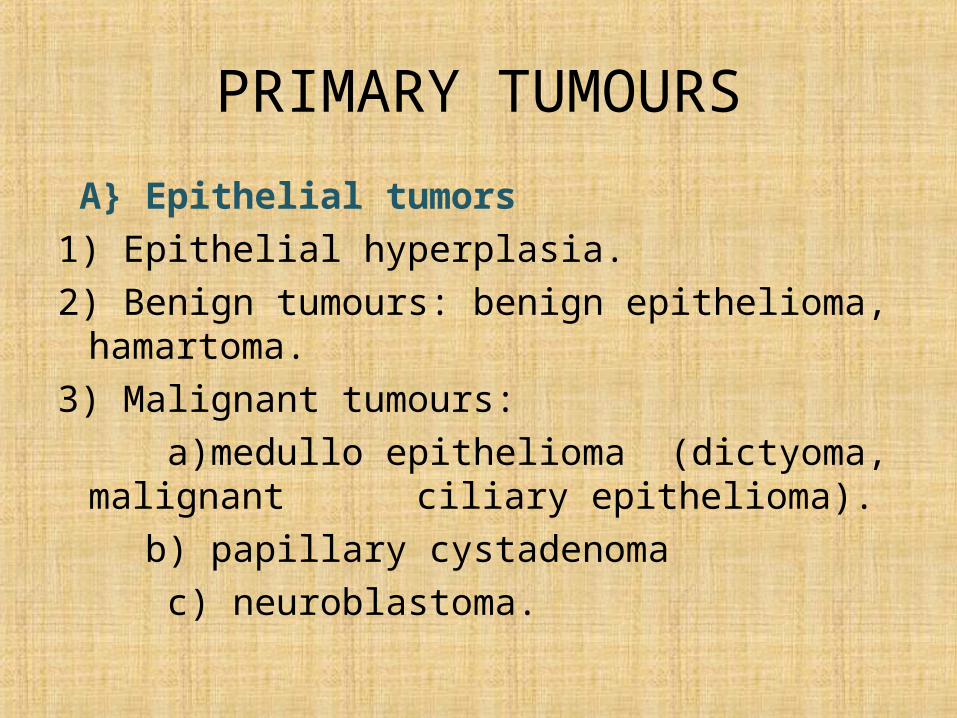
PRIMARY TUMOURS
A} Epithelial tumors 1) Epithelial hyperplasia.2) Benign tumours: benign epithelioma, hamartoma.3) Malignant tumours: a)medullo epithelioma (dictyoma, malignant
ciliary epithelioma). b) papillary cystadenoma c) neuroblastoma.
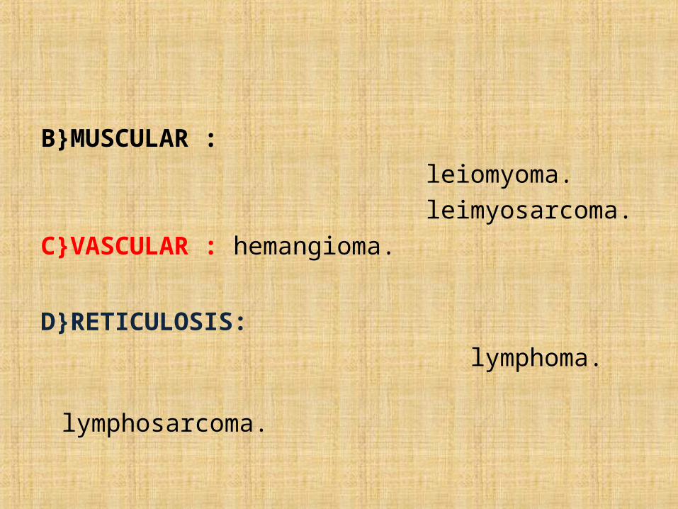
B}MUSCULAR : leiomyoma. leimyosarcoma.C}VASCULAR : hemangioma.
D}RETICULOSIS: lymphoma. lymphosarcoma.
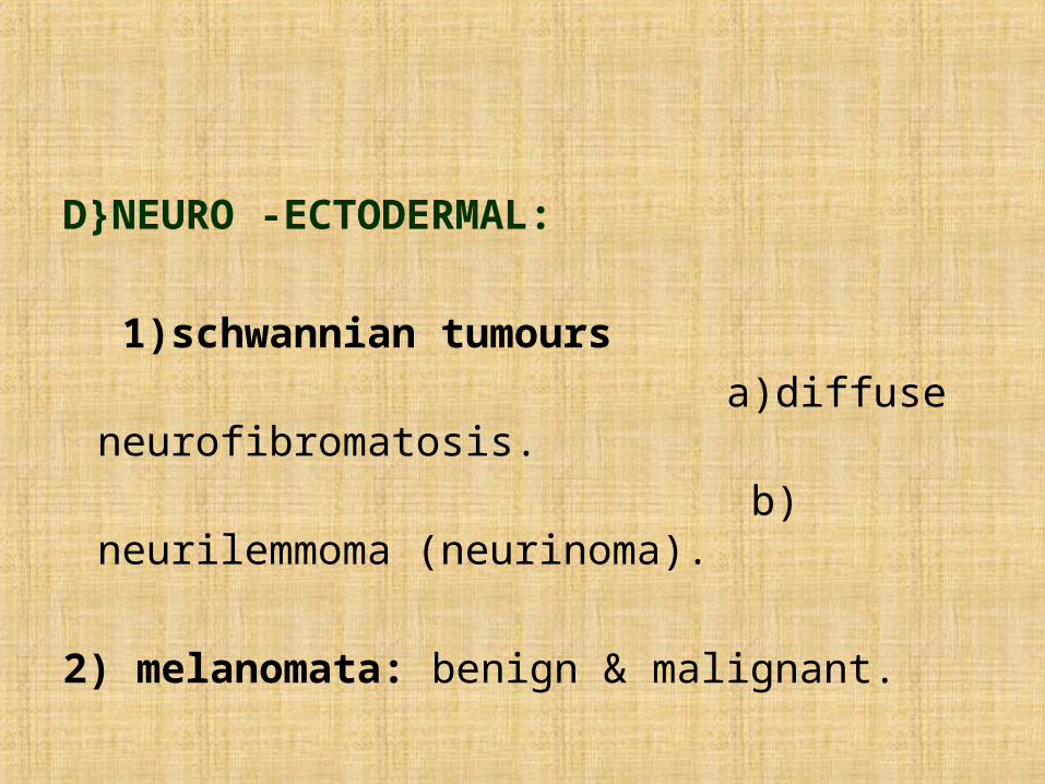
D}NEURO -ECTODERMAL:
1)schwannian tumours a)diffuse neurofibromatosis. b) neurilemmoma (neurinoma).
2) melanomata: benign & malignant.
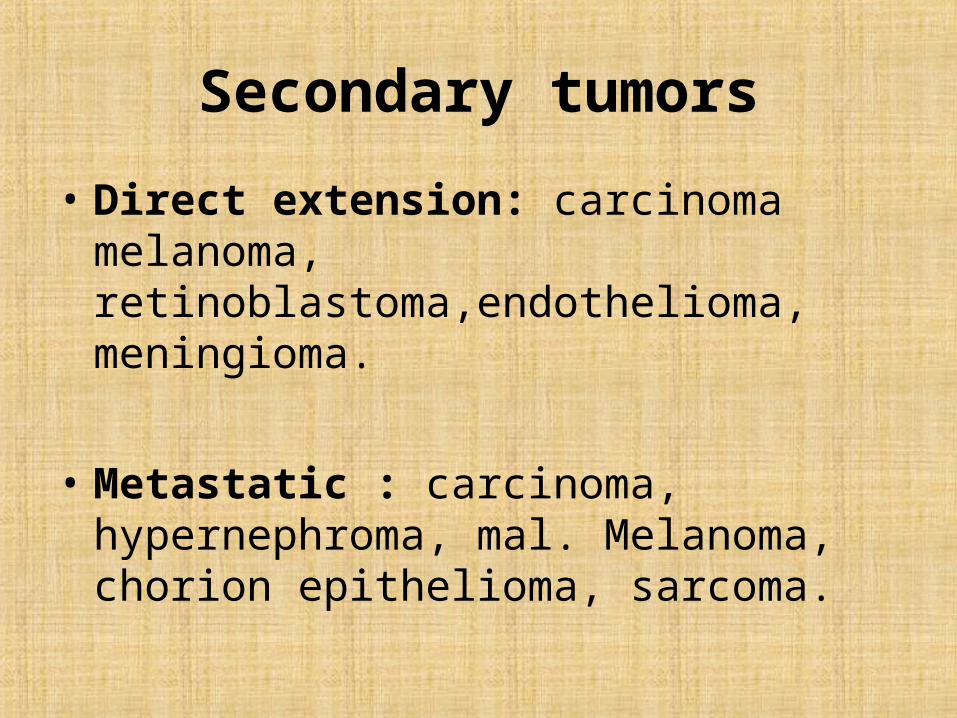
Secondary tumors
• Direct extension: carcinoma melanoma, retinoblastoma,endothelioma, meningioma.
• Metastatic : carcinoma, hypernephroma, mal. Melanoma, chorion epithelioma, sarcoma.
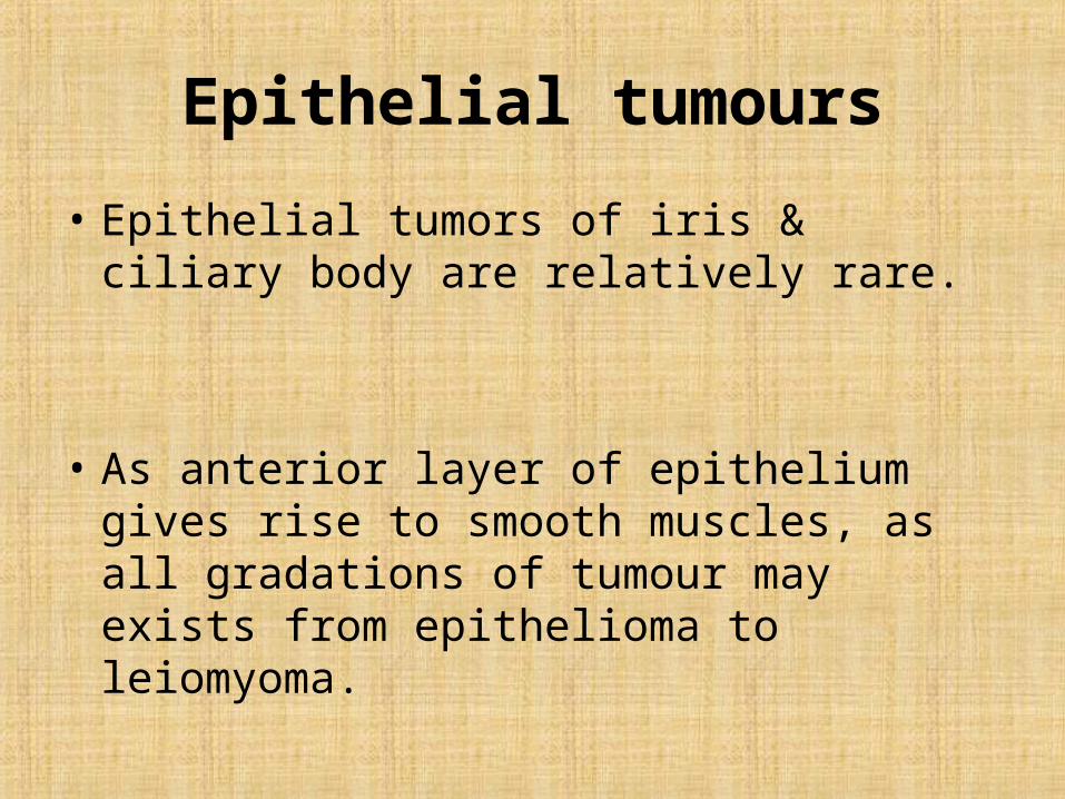
Epithelial tumours
• Epithelial tumors of iris & ciliary body are relatively rare.
• As anterior layer of epithelium gives rise to smooth muscles, as all gradations of tumour may exists from epithelioma to leiomyoma.
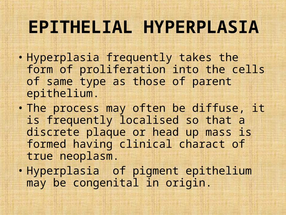
EPITHELIAL HYPERPLASIA
• Hyperplasia frequently takes the form of proliferation into the cells of same type as those of parent epithelium.
• The process may often be diffuse, it is frequently localised so that a discrete plaque or head up mass is formed having clinical charact of true neoplasm.
• Hyperplasia of pigment epithelium may be congenital in origin.
EPITHELIAL HYPERPLASIA
• It usually appears as discrete, flat & deeply pigmented lesion showing no tendency to grow, mainly seen on pupillary margin.
• EPITHELIAL hyperplasia of IRIS: simple hyperplasia of pigmented layer of posterior surface is particularly after prolonged iridocyclitis, trauma (including surgery), glaucoma or degenerated eyes.
EPITHELIAL HYPERPLASIA
• In primary glaucoma: The epithelial cells may migrate through the stroma of iris to apper as velvety black spot on anterior surface of iris, mainly at collarette.
• EPITHELIAL hyperplasia of ciliary body: Fuch emphasized difference between simple reactive hyperplasia & epithelioma. Both this conditions occurs in blind degenrative eyes.
EPITHELIAL HYPERPLASIA
• Hyperlasia is direct continuation of ciliary epithelium forming tumour like masses projecting inwards but showing no distinct pattern of cells.
• In case of old RD the ciliary epithelium may show hyperplasia near ora forming plaque may develope into metaplasic changes.
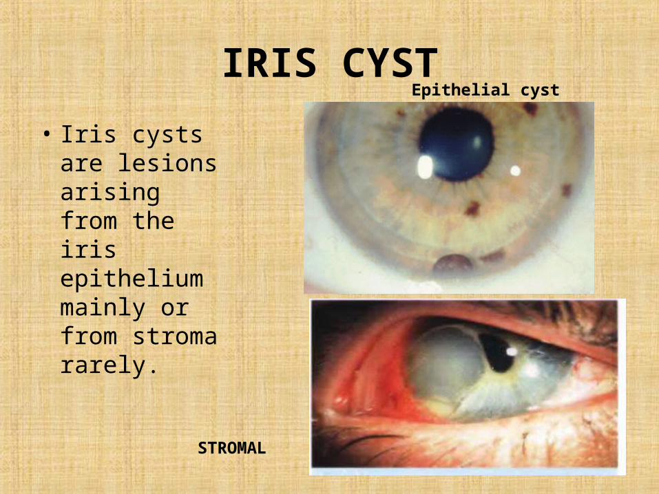
IRIS CYST
• Iris cysts are lesions arising from the iris epithelium mainly or from stroma rarely.
Epithelial cyst
STROMAL
IRIS CYST
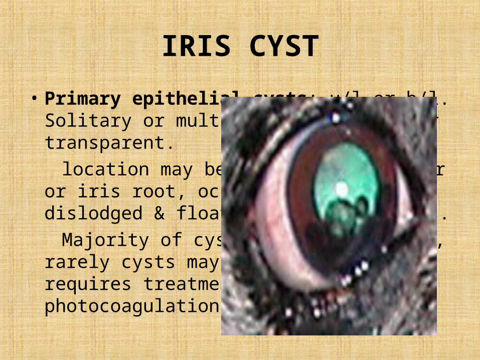
• Primary epithelial cysts: u/l or b/l. Solitary or multiple may be brown or transparent.
location may be at pupillary border or iris root, occasionally gets dislodged & floats in AC or vitreous.
Majority of cysts are asymptomatic, rarely cysts may obstruct vision & requires treatment with argon laser photocoagulation.
IRIS CYST
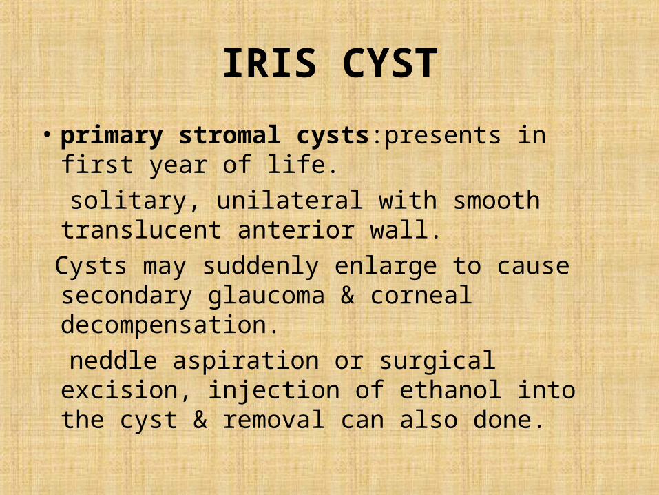
• primary stromal cysts:presents in first year of life.
solitary, unilateral with smooth translucent anterior wall.
Cysts may suddenly enlarge to cause secondary glaucoma & corneal decompensation.
neddle aspiration or surgical excision, injection of ethanol into the cyst & removal can also done.
Secondary cysts
• Implantation: m/c type originate after deposition of surface epithelial cells from conjunctiva or cornea on the iris after trauma.
• Pearls: white, solid lesion with opaque walls located on stroma, not connected to wound.
• Serous: translucent cysts filled with fluid, connected to wound, frequently enlarge leading to corneal edema, antr uveitis & glaucoma.
Secondary cysts
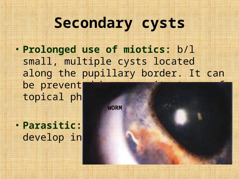
• Prolonged use of miotics: b/l small, multiple cysts located along the pupillary border. It can be prevented by concomitant use of topical phenylephrine 2.5%.
• Parasitic: very rarely cysts may develop in parasitic infection.
WORM
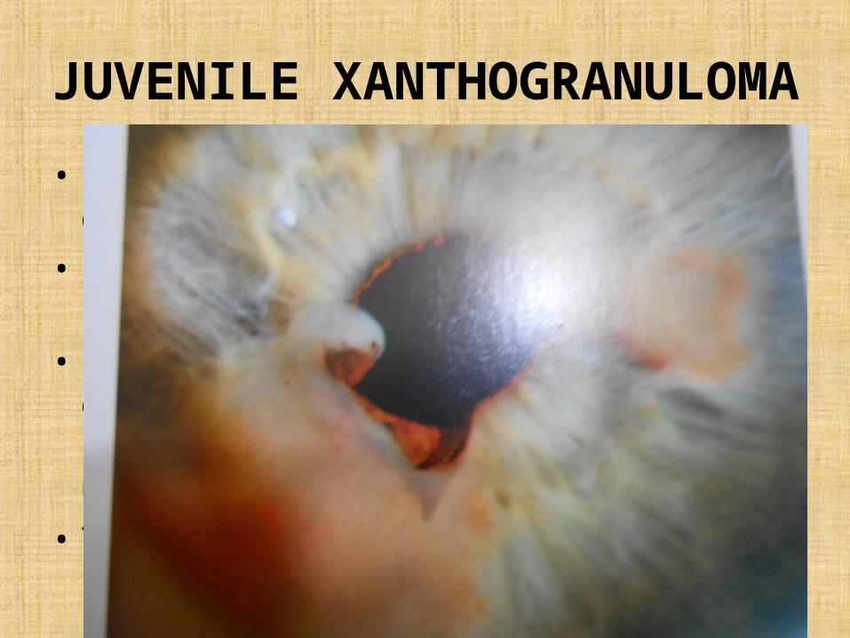
JUVENILE XANTHOGRANULOMA
• Rare idiopathic granulomatous d/s of childhood.
• Involves skin,muscle,stomach, salivary glands.• Iris involvement cause localised or diffuse
yellow lesion with spontaneous hyphema, anterior uveitis or glaucoma
• TOC: topical steroids.
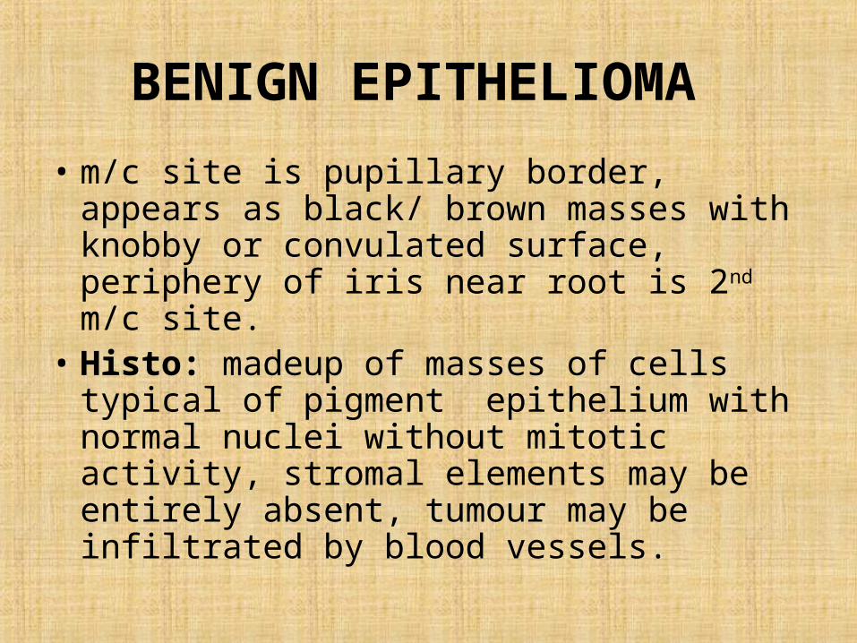
BENIGN EPITHELIOMA
• m/c site is pupillary border, appears as black/ brown masses with knobby or convulated surface, periphery of iris near root is 2nd m/c site.
• Histo: madeup of masses of cells typical of pigment epithelium with normal nuclei without mitotic activity, stromal elements may be entirely absent, tumour may be infiltrated by blood vessels.
BENIGN EPITHELIOMA
• Difference between epithelioma & melanoma is dense blackness of former & its usual sharp differentiation from surrounding.
• Management: observation over sometime, preferably by photography. If doubt iridectomy is done,many eyes involved have been excised owing to the fear of malignancy.
EPITHELIOMA OF CILIARY BODY
• First description of benign epithelioma was given by Fuch in 1883 in the eye of 70 year female pt having absolute glaucoma known as Fuch’s epithelioma.
• Occurs in wide age group 10 to 90 years of age, no sex prediction.
• Prolonged irritations such as inflammation, trauma or other intraocular tumour predisposes their formation.
EPITHELIOMA OF CILIARY BODY
• Tumour is usually small around 1mm but occasionally reaches to 5mm in diameter.
• Gonioscopy in dilated pupils shows brown pigmented mass oval or round in shape lying on the ridge of ciliary process or in intervening valley.
• Histo: high degree of differentiation, seperated from stroma, may lose its pigment or become atrophied.
EPITHELIOMA OF CILIARY BODY
• In transitional cases which is between typical simple growth & malignant medulloepithelioma have been reported, this tumour has cellular growth.
MEDULLO EPITHELIOMA
• Fuch classically divided medullo epithelioma to two groups
A)Resesmbling embryonic retina DICTYOMATA.B)Ciliary epithelium (malignant epithelioma of
ciliary body).
DICTYOMA
• A.k.a Embryonal medullo-epithelioma.• It is a tumour arising from non pigmented layer
of ciliary epithelium having structure of embryonic retina.
• Never b/l, no multicentric in origin & no hereditary tendency, young children, present as u/l buphthalmos/glaucoma/ cataract.
• Histo: made up of bands of cells arranged in one/ several rows forming intricate convolutions.
DICTYOMA
• Dictyoma should be suspected in all cases of u/l buphthalmos, glaucoma in child , staphyloma, u/l catarctous or dislocated lens or nay cyst formation in the Ac
• Management: only treatment of choice is enucleation.
MALIGNANT CILIARY EPITHELIOMA
• Unlike dictyoma this type is formed by mature cells.
• Involves eyes which have severe inflammation.
• Small dark non translucent nodule bulging from behind & eventually appearing over pupillary margin.
• Course of tumour is slow & benign eye becomes atrophic & degenerated.
MALIGNANT CILIARY EPITHELIOMA
Histo:• a single layer of cell, tube formation may
occur giving the tumour in section a lace like effect.
• Rosette like structures, primitive nerve fibres formations resembles primitive optic vesicle.
MUSCULAR TUMOUR
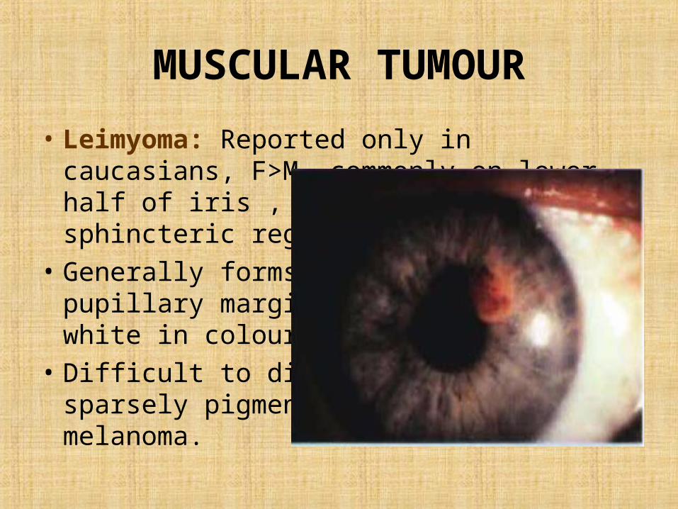
• Leimyoma: Reported only in caucasians, F>M, commonly on lower half of iris , temporally & on sphincteric region.
• Generally forms a sessile mass on pupillary margin, pink/ greyish white in colour.
• Difficult to diagnose from a sparsely pigmented malignant melanoma.
VASCULAR TUMOUR
• Haemangiomas are more common in choroid as compare to iris & ciliary body.
• Iris: vascular tumour is localised on the iris surface, which may give rise to periodic bleeding.
• Choroidal haemangioma is divided into circumscribed. diffuse.
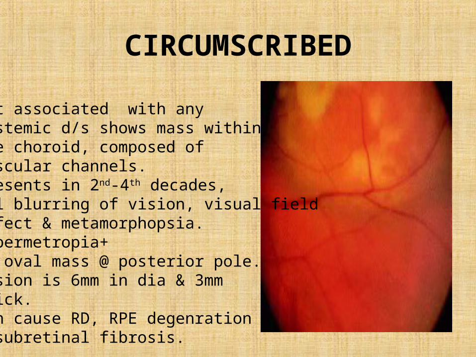
CIRCUMSCRIBED
Not associated with any systemic d/s shows mass withinthe choroid, composed ofvascular channels.Presents in 2nd-4th decades,u/l blurring of vision, visual fielddefect & metamorphopsia.Hypermetropia+An oval mass @ posterior pole.Lesion is 6mm in dia & 3mm thick.Can cause RD, RPE degenration& subretinal fibrosis.
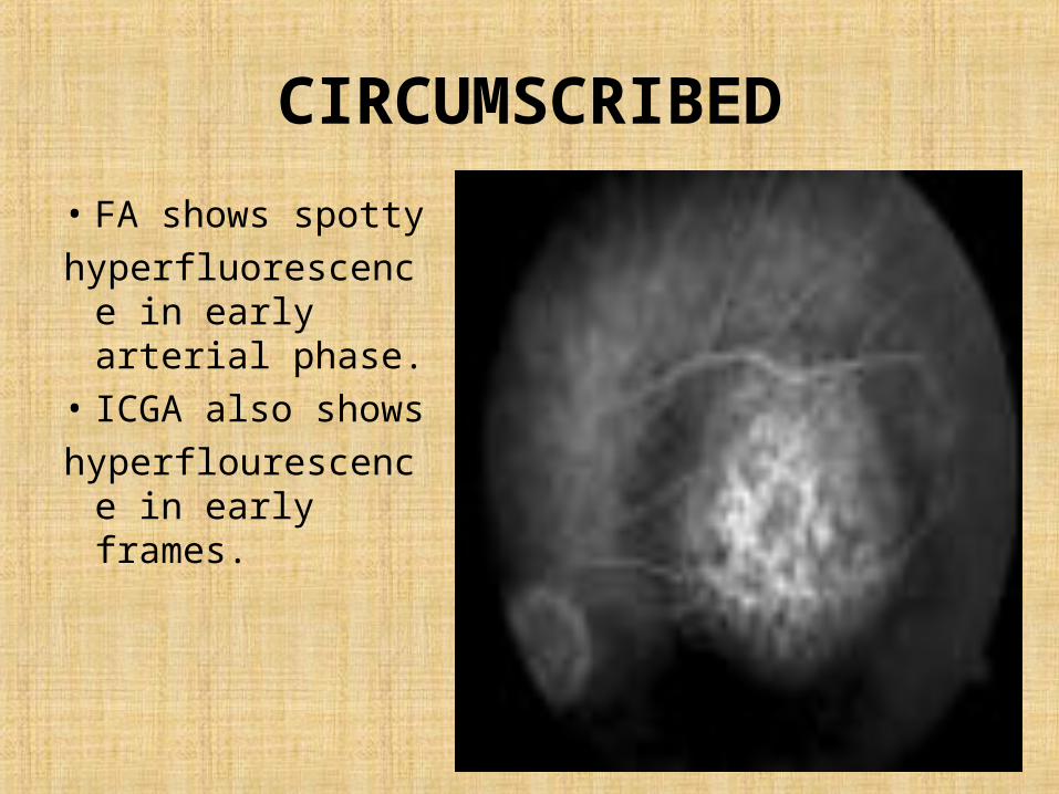
CIRCUMSCRIBED
• FA shows spotty hyperfluorescence
in early arterial phase.
• ICGA also shows hyperflourescence
in early frames.
CIRCUMSCRIBED
• US shows acoustically solid lesionwith sharp anterior surface.
• Treatment: photodynamic therapy- may be repeated after few months.
• TTT:if lesions are not involving macula.• Radiotherapy.• Intravitreal anti-VEGF therapy.
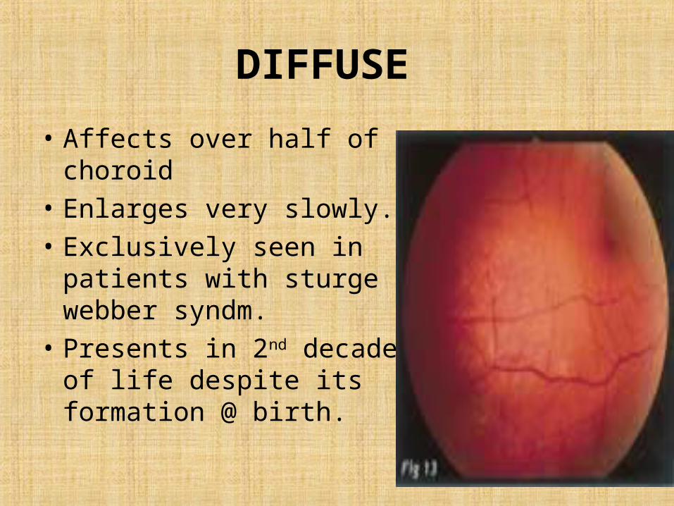
DIFFUSE
• Affects over half of choroid• Enlarges very slowly.• Exclusively seen in patients
with sturge webber syndm.• Presents in 2nd decade of
life despite its formation @ birth.
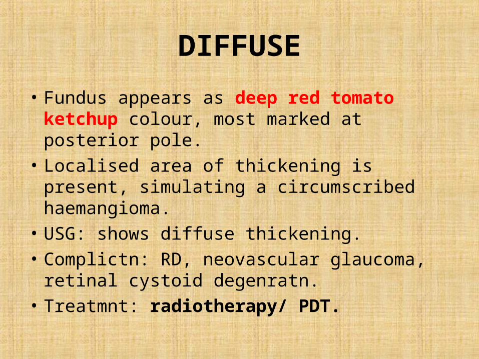
DIFFUSE
• Fundus appears as deep red tomato ketchup colour, most marked at posterior pole.
• Localised area of thickening is present, simulating a circumscribed haemangioma.
• USG: shows diffuse thickening.• Complictn: RD, neovascular glaucoma, retinal
cystoid degenratn.• Treatmnt: radiotherapy/ PDT.
CHOROIDAL OSTEOMA
• Very rare benign,slow growing ossifying tumour.
• b/L in 25% cases, F>M.• Mature cancellous bone which causes
overlying RPE atrophy.• Presents in 2nd- 3rd decades , yellowish-white
flat, minimally elevated lesion near disc or posterior pole present.
CHOROIDAL OSTEOMA
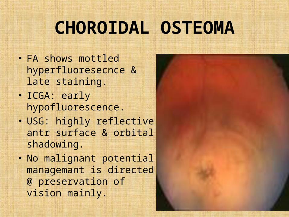
• FA shows mottled hyperfluoresecnce & late staining.
• ICGA: early hypofluorescence.
• USG: highly reflective antr surface & orbital shadowing.
• No malignant potential, managemant is directed @ preservation of vision mainly.
NEUROFIBROMA
• Congenital condition representing a diffuse proliferation schwann cells of the peripheral nerves.
• HISTO: increase in thickness of choroid & ciliary body, pear shaped deformity of the pupil resembling colobomata may occur.
• Rx: Excision------ enucleation.
NEURILEMMOMA
• Benign encapsulated tumour of nerve sheath.
• Tumour composed of schwann cells arranged in interlacing cords with palisading of the nuclei.
METASTATIC TUMOUR
• Choroid is m/c site accounting for about 90% followed by iris & ciliary body.
• Most frequent primary site is breast & bronchus.
• A fast growing creamy white lesion with indistinct margins most frequently at posterior pole may be present.
• Deposits are multifocal, may cause RD.
METASTATIC TUMOUR
• Management 1, Observation. 2, Radiotherapy. 3, TTT (transpupillary thermotharapy). 4, Systemic therapy for 10 tumour
RETICULOSIS
• Any part or whole of uveal tract may be involved & ocular lesion may be B/L.
• In anterior segment: severe iridocyclitis, nodular mass in AC.
• In posterior segment: pan uveitis, RD, vision fails rapidly.
RETICULOSIS
HISTO: lymphocytic cells arranged either diffusly, in reticular cells large round oval nuclei & small cytoplasmic process resembling the cells of germinal centre of lymph follicles are present lying within the network of reticulum.
MANAGEMENT: palliative, in some cases enucleation is required.
RETICULOSIS
• MULTIPLE MYELOMA: abnormal plasma cells in bone marrow.
deposition of plasma cells in the tissue particularly in retina / optic nerve & bony orbit.
UVEAL NEVUS
• The uveal nevus is a benign tumour that arises from melanocytic cells derived from the neural crest.
• m/c in whites, aprox 20% develop at least one choroidal nevus >50yrs of age, M=F.
• 1 in 4000-5000 undergoes malignant changes.• Asymptomatic may cause visual loss in
macular choroidal nevi.
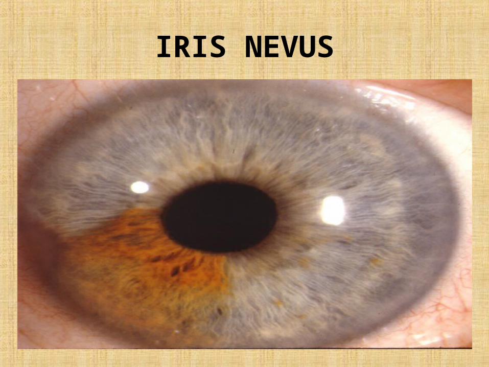
IRIS NEVUS
IRIS NEVUS
• IRIS NEVUS :< 3mm in diameter, < 0.5mm thick,pupillary peaking, focal ectropion iridis or both, abnormal iris vasculature.
• Choroidal nevus:small grey to brown choroidal tumour, < 5mm in diameter, < 1mm in thickness, causes blurred/distorted vision serous subretinal fluid, cystic degenration of retina involving macula or choroidal neovascularization.
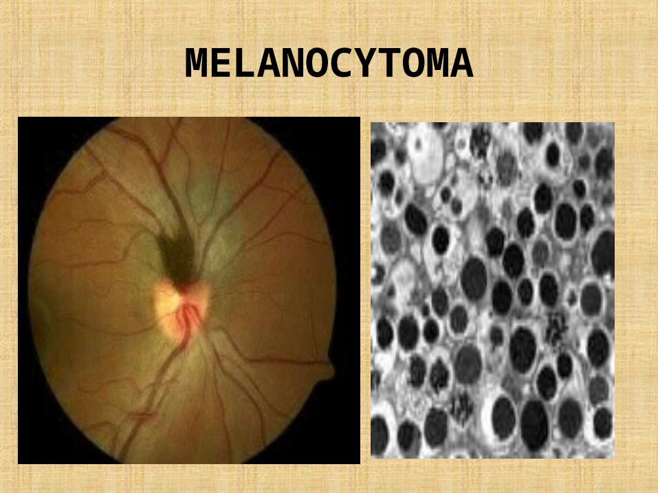
MELANOCYTOMA• Special type of nevus “melanocytoma”of optic
disc.• Composed entirely of maximally pigmented
polyhedral nevus cells (magnocellular nevus cells).
• Lesion appears striated because of insinuation of pigment cells between axons in the nerve fibrelayer which may inturn causes visual field defect.
• Magnocellular nevus can occur in choroid, ciliary body & iris.
MELANOCYTOMA
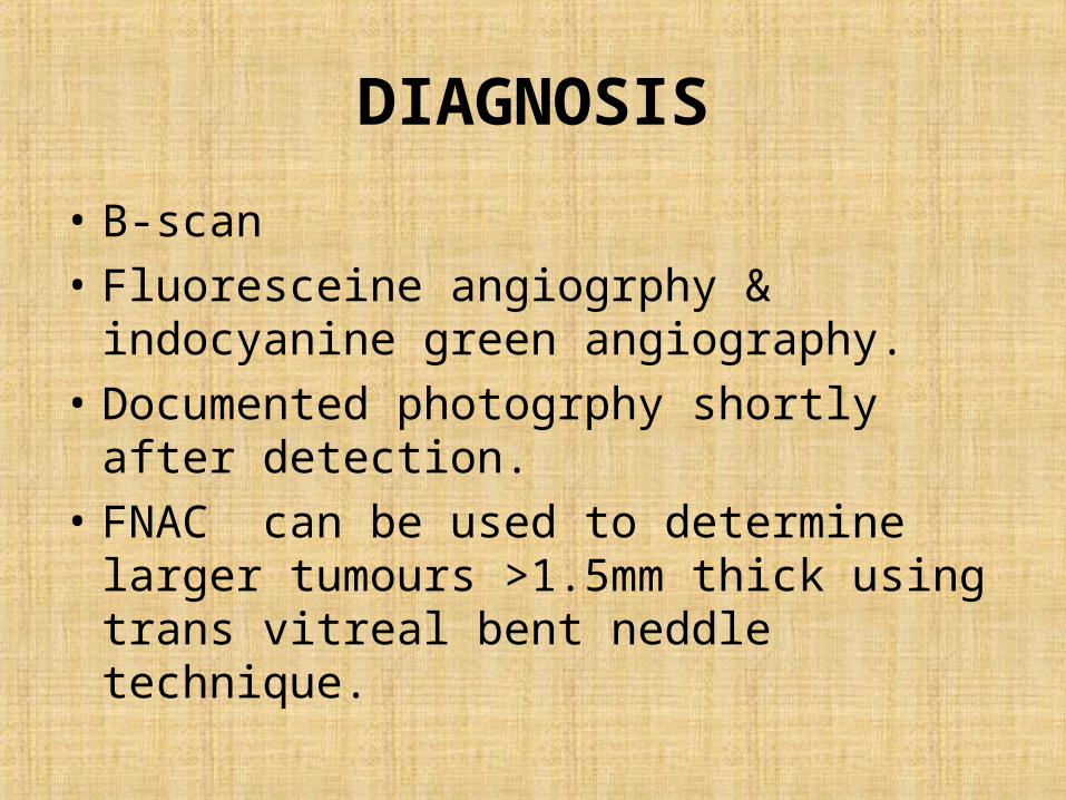
DIAGNOSIS
• B-scan• Fluoresceine angiogrphy & indocyanine green
angiography.• Documented photogrphy shortly after
detection.• FNAC can be used to determine larger
tumours >1.5mm thick using trans vitreal bent neddle technique.
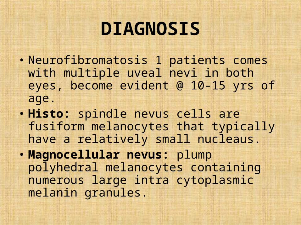
DIAGNOSIS
• Neurofibromatosis 1 patients comes with multiple uveal nevi in both eyes, become evident @ 10-15 yrs of age.
• Histo: spindle nevus cells are fusiform melanocytes that typically have a relatively small nucleaus.
• Magnocellular nevus: plump polyhedral melanocytes containing numerous large intra cytoplasmic melanin granules.
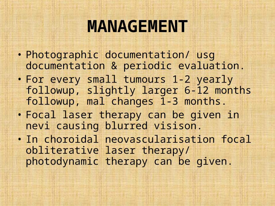
MANAGEMENT
• Photographic documentation/ usg documentation & periodic evaluation.
• For every small tumours 1-2 yearly followup, slightly larger 6-12 months followup, mal changes 1-3 months.
• Focal laser therapy can be given in nevi causing blurred visison.
• In choroidal neovascularisation focal obliterative laser therapy/ photodynamic therapy can be given.
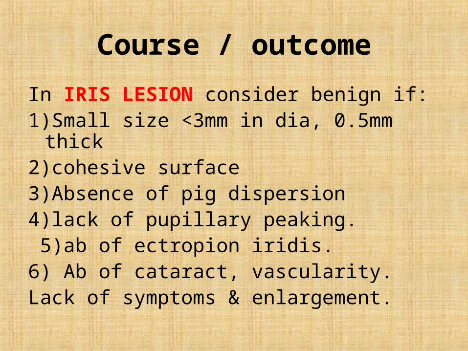
Course / outcome
In IRIS LESION consider benign if:1)Small size <3mm in dia, 0.5mm thick2)cohesive surface3)Absence of pig dispersion4)lack of pupillary peaking. 5)ab of ectropion iridis.6) Ab of cataract, vascularity.Lack of symptoms & enlargement.
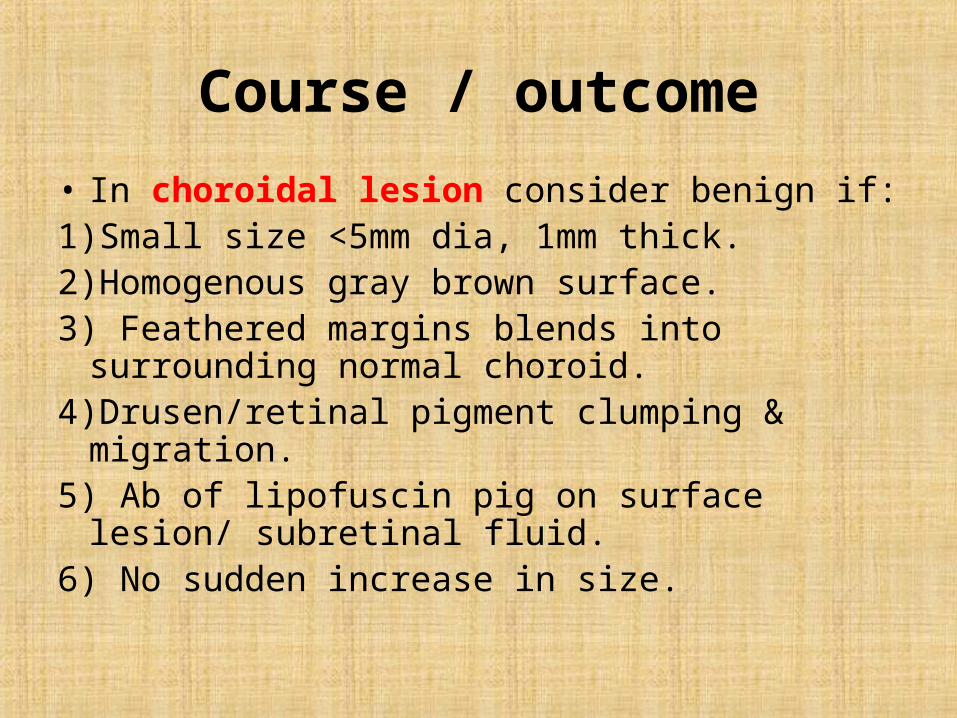
Course / outcome
• In choroidal lesion consider benign if:1)Small size <5mm dia, 1mm thick.2)Homogenous gray brown surface.3) Feathered margins blends into surrounding
normal choroid.4)Drusen/retinal pigment clumping & migration.5) Ab of lipofuscin pig on surface lesion/ subretinal
fluid.6) No sudden increase in size.
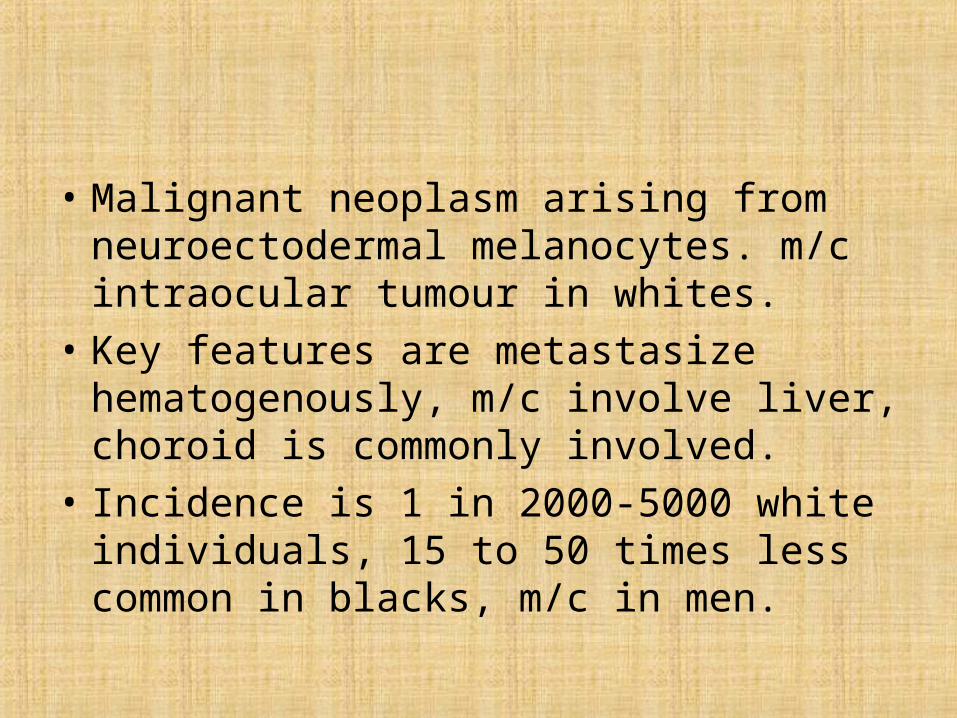
MALIGNANT MELANOMA
• Malignant neoplasm arising from
neuroectodermal melanocytes. m/c intraocular tumour in whites.
• Key features are metastasize hematogenously, m/c involve liver, choroid is commonly involved.
• Incidence is 1 in 2000-5000 white individuals, 15 to 50 times less common in blacks, m/c in men.
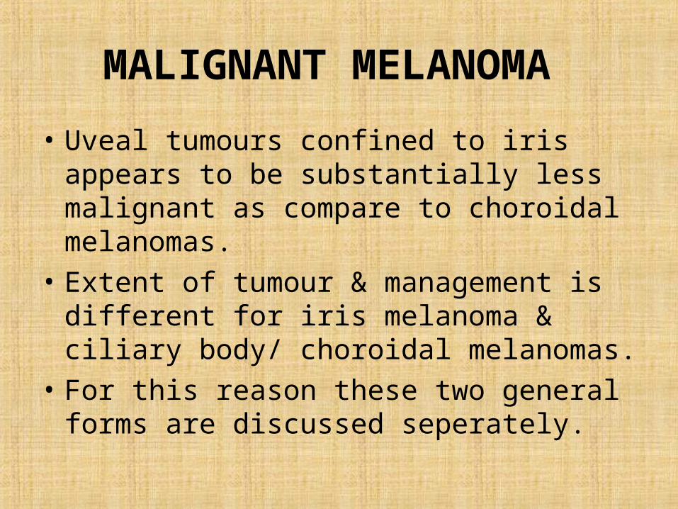
MALIGNANT MELANOMA
• Uveal tumours confined to iris appears to be substantially less malignant as compare to choroidal melanomas.
• Extent of tumour & management is different for iris melanoma & ciliary body/ choroidal melanomas.
• For this reason these two general forms are discussed seperately.
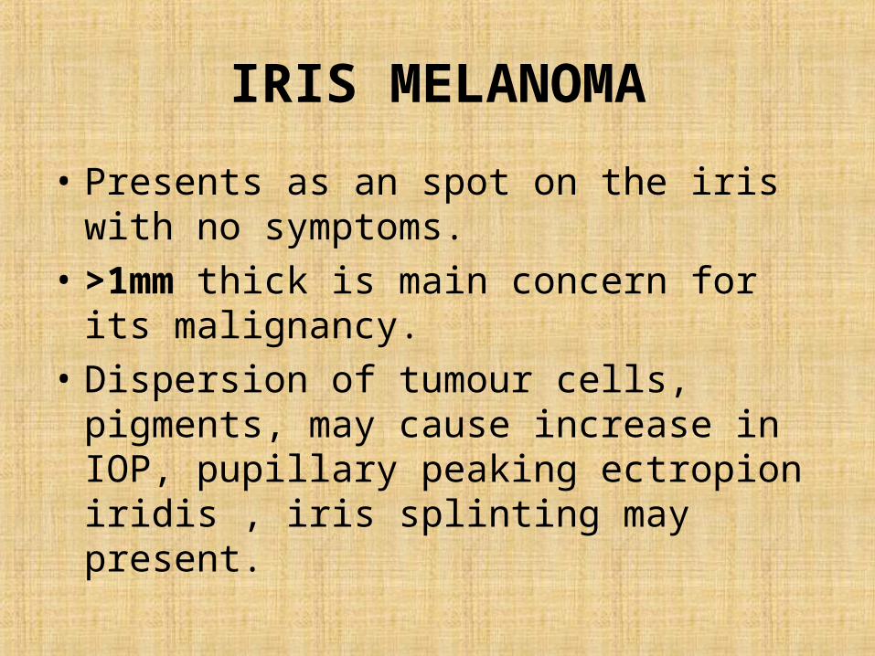
IRIS MELANOMA
• Presents as an spot on the iris with no symptoms.
• >1mm thick is main concern for its malignancy.
• Dispersion of tumour cells, pigments, may cause increase in IOP, pupillary peaking ectropion iridis , iris splinting may present.
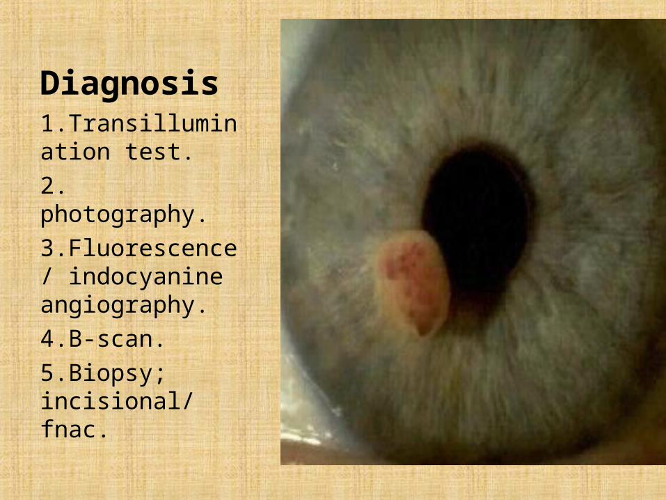
Diagnosis1.Transillumination test.2. photography.3.Fluorescence/ indocyanine angiography.4.B-scan.5.Biopsy; incisional/ fnac.
IRIS MELANOMA
• Histo: composed of atypical melanocytic cells, larger nuclear to cytoplasmic ratio.
• Tumour cells have a fusiform shape & mild atypical spindle melanoma cells.
• Those that have more spherical & more pronounced anaplasia are called as epithelioid melanoma cells.
MANAGEMENT
• Small melanomas should be observed without intervention.
• Excision: iridectomy/ irideocyclectomy is done.• Plaque radiotherapy/ photon beam irridiation
performed in small number of cases but appears to be effective in short term followup.
• Enucleation: for large iris melanomas, extensive seeding, transcleral spread, blind painful eye.
course
• Iris tumour grows relatively slowly.• Replaces porportion of iris & ciliary body.• Can cause secondary glaucoma.• Post excision patient must be followed for
every 6 months for first 2-3 years & yearly there after.
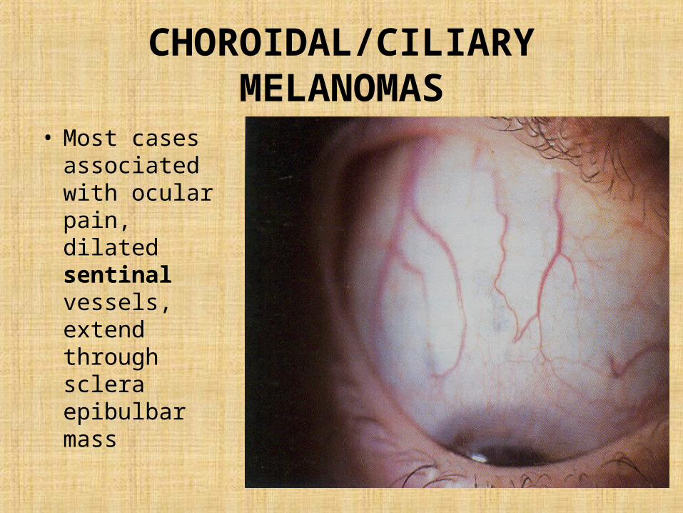
CHOROIDAL/CILIARY MELANOMAS
• Most cases associated with ocular pain, dilated sentinal vessels, extend through sclera epibulbar mass
CHOROIDAL/CILIARY MELANOMAS
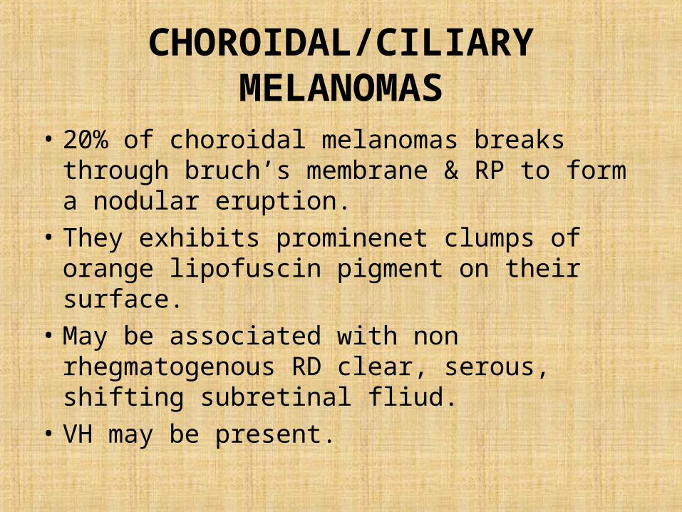
• 20% of choroidal melanomas breaks through bruch’s membrane & RP to form a nodular eruption.
• They exhibits prominenet clumps of orange lipofuscin pigment on their surface.
• May be associated with non rhegmatogenous RD clear, serous, shifting subretinal fliud.
• VH may be present.
CHOROIDAL/CILIARY MELANOMAS
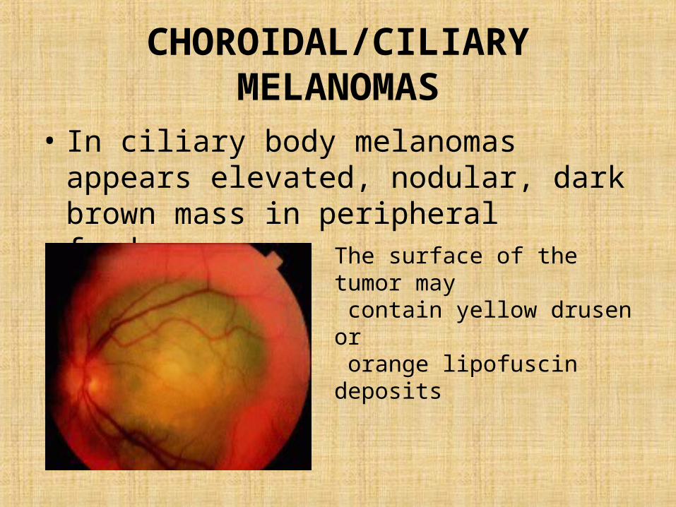
• In ciliary body melanomas appears elevated, nodular, dark brown mass in peripheral fundus.
The surface of the tumor may contain yellow drusen or orange lipofuscin deposits
• Clinical features • asymptomatic• incidentally during ophthalmoscopy. • In general, the more anterior their origin, the longer the delay of any
symptoms. • Blurred visual acuity • growth of the melanoma into the subfoveal retina• cystoid macular edema disruption of choroidal circulation ischemia
degeneration of retinal photoreceptors The retina overlying the tumor separates into cystoid spaces and larger schisis cavities.
• Retinal detachment - Exudation of fluid into the subretinal space retinal detachment enlarge the field loss can lead to total retinal detachment
• vitreous hemorrhage - Erosion of the melanoma into blood vessels in adjacent tissues, or areas of necrosis within the tumor
• cataract – involvement of lens • Paracentral scotoma • if the tumor affects the perifoveal retina.• visual field loss Painless and progressive as peripheral melanoma grows• Floaters when areas of necrosis within the tumor or adjacent structures
produce vitreous hemorrhage or hyphema.• Severe ocular pain Occasionally when they impinge into posterior
ciliary nerves. • can also cause pain secondary to high intraocular pressure from acute
angle-closure glaucoma.

• History of weight loss, marked fatigue, cough, or change in bowel or bladder habits, should prompt consideration of primary non-ocular malignancy with choroidal metastasis
DIAGNOSIS
• The diagnostic error rate in the COMS was only 0.48%.
• Misdiagnoses occur in less than 4% of enucleated eyes at tertiary referral centers.
• The diagnostic error rate in the COMS, however, was measured under ideal conditions; all tumors were well visualized and photographed. Patient were excluded from the COMS if the ocular media was opaque. In real-world situations, the misdiagnosis rate for posterior melanoma may be greater than that reported by the COMS
• A-scan ultrasound • tumors more than 2-3 mm thick. • characteristically shows an initial prominent spike, followed by low-to-
medium internal reflectivity. Vascular pulsations can be seen as fine oscillations of the internal spiking pattern within the tumor. Standardized ultrasonography has a diagnostic accuracy of more than 95%.
• B-scan ultrasound to help establish the diagnosis, to evaluate possible extraocular extension, and to estimate tumor size for periodic observation and plan therapeutic intervention
• Low-to-medium reflectivity• Excavation of underlying uveal tissue• Internal vascularity• An acoustic quiet zone at the base of the tumor called acoustic hollowing
• Fluorescein angiography do not show pathognomonic signs of choroidal melanoma but can help point to its diagnosis.
• Small choroidal melanomas may show fluorescein angiographic changes similar to some choroidal nevi, as follows: ranging from normal angiography to hypofluorescence secondary to blockage of background fluorescence.
• Larger melanomas may show a patchy pattern of early hypofluorescence and hyperfluorescence followed by late intense staining.
• intrinsic vascularization, visible throughout the angiogram. • The angiographic sign called "double circulation" pattern refers to
simultaneous fluorescence of retinal and choroidal circulation within the tumor. When it occurs, it is fairly distinctive of choroidal melanomas
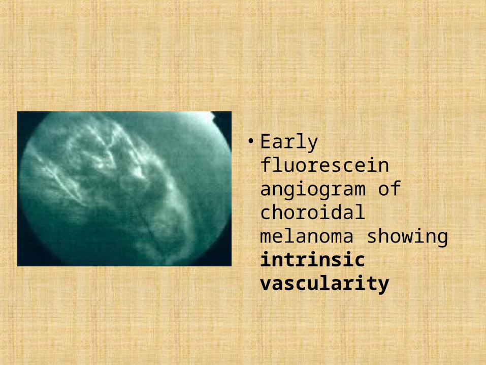
• Early fluorescein angiogram of choroidal melanoma showing intrinsic vascularity
• CT scan orbit
• is more expensive than ultrasound and• currently is not as sensitive. • It is useful to see extraocular extension and may help
differentiate between choroidal or retinal detachment and a solid tumor.
• Choroidal melanoma shows enhancement with contrast, where as exudation does not.
• CT scan also is very sensitive detecting calcium, a feature of some tumors that are different than uveal melanoma (characteristically choroidal osteoma).
• Fine needle and incisional biopsy
• usually are not required • to distinguish amelanotic melanomas from metastatic tumors,
and if results from other ancillary tests are equivocal.• accuracy of more than 95% in tumors larger than 3 mm.• Incisional biopsy is more invasive and may have more associated
complications, but it has less false-negative and false-positive results.
• Risk of spread of cancerous cells with needle biopsy is small for choroidal melanoma (unlike retinoblastoma). Follow biopsy by prompt treatment to avoid extrascleral extension.
• Histologic evaluation - after enucleation can confirm the diagnosis and evaluate prognosis.
• Three distinct cell types are recognized: • Spindle A - elongated nuclei and uncommonly have mitotic figures• Spindle B - prominent nucleolus• found more commonly, have an elongated profile but are slightly
larger than spindle A cells• Epithelioid – most aggressive behavior and carries a poorer
prognosis for the patient's long-term survival• highly anaplastic, poorly cohesive, and have considerable • frequent mitotic figures.
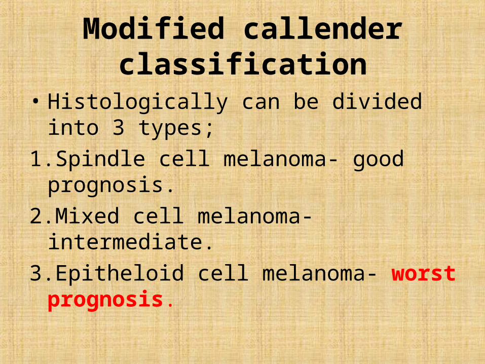
Modified callender classification
• Histologically can be divided into 3 types;1.Spindle cell melanoma- good prognosis.2.Mixed cell melanoma- intermediate.3.Epitheloid cell melanoma- worst prognosis.
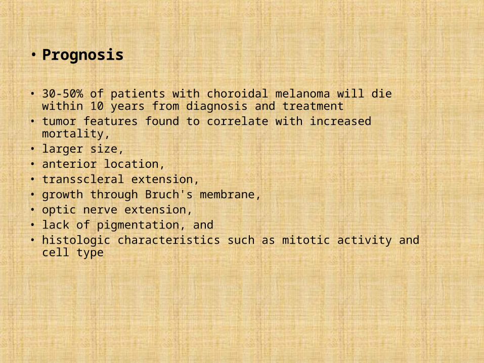
• Prognosis
• 30-50% of patients with choroidal melanoma will die within 10 years from diagnosis and treatment
• tumor features found to correlate with increased mortality, • larger size, • anterior location, • transscleral extension, • growth through Bruch's membrane, • optic nerve extension, • lack of pigmentation, and • histologic characteristics such as mitotic activity and cell type
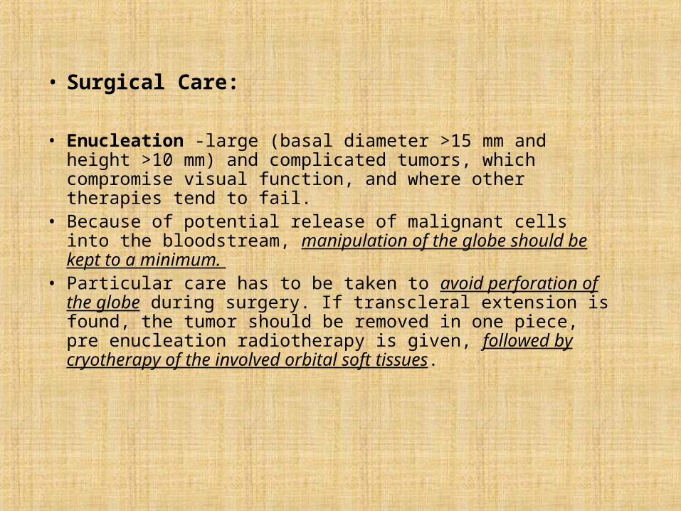
• Surgical Care:
• Enucleation -large (basal diameter >15 mm and height >10 mm) and complicated tumors, which compromise visual function, and where other therapies tend to fail.
• Because of potential release of malignant cells into the bloodstream, manipulation of the globe should be kept to a minimum.
• Particular care has to be taken to avoid perforation of the globe during surgery. If transcleral extension is found, the tumor should be removed in one piece, pre enucleation radiotherapy is given, followed by cryotherapy of the involved orbital soft tissues.
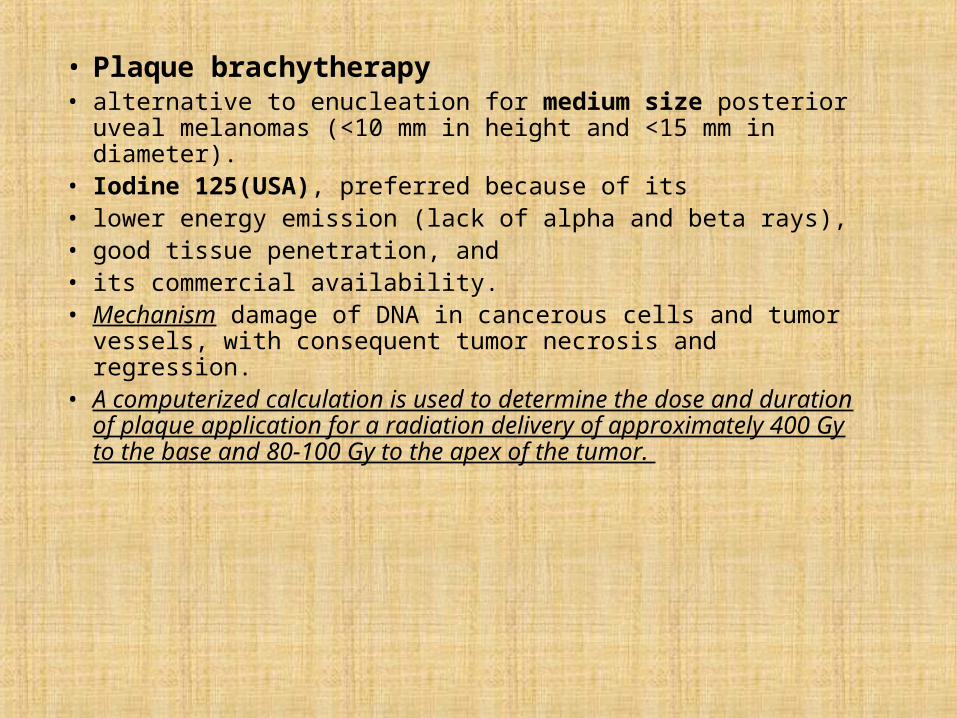
• Plaque brachytherapy • alternative to enucleation for medium size posterior uveal
melanomas (<10 mm in height and <15 mm in diameter). • Iodine 125(USA), preferred because of its • lower energy emission (lack of alpha and beta rays), • good tissue penetration, and • its commercial availability. • Mechanism damage of DNA in cancerous cells and tumor vessels,
with consequent tumor necrosis and regression. • A computerized calculation is used to determine the dose and
duration of plaque application for a radiation delivery of approximately 400 Gy to the base and 80-100 Gy to the apex of the tumor.
• A margin of 2 mm over the largest tumor basal dimension is adequate. • Postoperative imaging confirmation of correct plaque localization is
required. Radioactive plaques are left in place for 3-7 days. • The goal of successful treatment is to achieve arrest of tumor growth
or regression in size. • Local recurrence, usually requiring enucleation, occurs at a rate of
about 12-16%. • complications
– cataract, – rubeosis, – scleral necrosis, – keratopathy, – radiation retinopathy, and – optic neuropathy but at a reduced rate compared with external beam irradiation
• External beam irradiation using charged particles, either protons or helium to treat medium size choroidal melanomas (<10 mm in height and <15 mm in diameter), although it has been used for larger tumor It has similar indications and success rates to plaque brachytherapy.
• radiopaque tantalum rings usually are sutured to the sclera to serve as reference markers for alignment of the radiation beam. A collimated beam delivers about 70 Gy, divided usually in 5 sessions.
• Vital ocular structures are avoided through careful positioning of the head and eye. Irradiation causes damage of DNA in cancerous cells and tumor vessels, with consequent tumor necrosis and regression..
• survival rate comparable to those treated by enucleation.
• Small choroidal melanomas, when they are located away from the fovea and are less than 3 mm in thickness
• Laser photocoagulation - to seal off the blood supply to the tumour and destroy it
• Transpupillary thermotherapy• Photodynamic Treatment
– Animal study– melanin precursors become extremely phototoxic in melanoma
tumor cells when activated with certain light sources – No additional phototoxic agents are administered – differs from conventional photodynamic therapy in that it uses
pulsed, longer wavelength light
The Collaborative Ocular Melanoma Study
• initiated in 1986 • long-term, multicenter, randomized
controlled trials • conducted in 43 clinical centers located in
major population areas of the United States and Canada
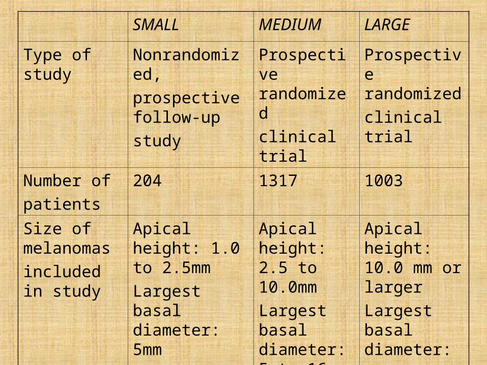
SMALL MEDIUM LARGE
Type of study Nonrandomized,
prospective follow-up
study
Prospective randomized
clinical trial
Prospective randomized
clinical trial
Number of
patients
204 1317 1003
Size of melanomas
included in study
Apical height: 1.0 to 2.5mm
Largest basal diameter: 5mm
Apical height: 2.5 to 10.0mm
Largest basal diameter: 5 to 16 mm
Apical height: 10.0 mm or larger
Largest basal diameter:
16 mm or larger
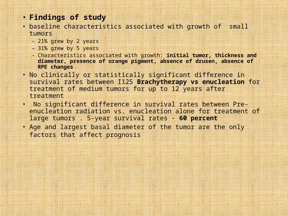
• Findings of study • baseline characteristics associated with growth of small tumors
– 21% grew by 2 years – 31% grew by 5 years – Characteristics associated with growth: initial tumor, thickness and diameter,
presence of orange pigment, absence of drusen, absence of RPE changes
• No clinically or statistically significant difference in survival rates between I125 Brachytherapy vs enucleation for treatment of medium tumors for up to 12 years after treatment
• No significant difference in survival rates between Pre-enucleation radiation vs. enucleation alone for treatment of large tumors . 5-year survival rates - 60 percent
• Age and largest basal diameter of the tumor are the only factors that affect prognosis

THANKYOUTHANKYOU