Embed Size (px)
Citation preview
Proc. Nati. Acad. Sci. USAVol. 92, pp. 9328-9332, September 1995Neurobiology
Tumor necrosis factors a and ,f protect neurons against amyloid,8-peptide toxicity: Evidence for involvement of a ucB-bindingfactor and attenuation of peroxide and Ca2+ accumulation
(Alzheimer disease/cell death/confocal laser scanning microscopy/fura-2 imaging/reactive oxygen species)
STEVEN W. BARGER*t, DOROTHEE HORSTER*, KATSUTOSHI FURUKAWA*, YADONG GOODMAN*,JOSEF KRIEGLSTEINt, AND MARK P. MATTSON*§¶*Sanders-Brown Research Center on Aging and §Department of Anatomy and Neurobiology, University of Kentucky, Lexington, KY 40536-0230; and *Instituteof Pharmacology and Toxicology, University of Marburg, Marburg, Germany
Communicated by Dominick P. Purpura, Albert Einstein College of Medicine, Bronx, NY, June 22, 1995 (received for review March 27, 1995)
ABSTRACT In Alzheimer disease (AD) the amyloid,B-peptide (AO8) accumulates in plaques in the brain. Af3 canbe neurotoxic by a mechanism involving induction of reactiveoxygen species (ROS) and elevation of intracellular freecalcium levels ([Ca2+]1). In light of evidence for an inflam-matory response in the brain in AD and reports of increasedlevels oftumor necrosis factor (TNF) inAD brain we tested thehypothesis that TNFs affect neuronal vulnerability to Ap.A.3-(25-35) and A,3-(1-40) induced neuronal degeneration ina concentration- and time-dependent manner. Pretreatmentof cultures for 24 hr with TNF-13 or TNF-a resulted insignificant attenuation of A.8-induced neuronal degeneration.Accumulation of peroxides induced in neurons by Ap wassignificantly attenuated in TNF-pretreated cultures, andTNFs protected neurons against iron toxicity, suggesting thatTNFs induce antioxidant pathways. The [Ca2+]1 response toglutamate (quantified by fura-2 imaging) was markedly po-tentiated in neurons exposed to Af3, and this action of A,6 wassuppressed in cultures pretreated with TNFs. Electrophoreticmobility-shift assays demonstrated an induction of a acB-binding activity in hippocampal cells exposed to TNFs. Ex-posure of cultures to IcB (MAD3) antisense oligonucleotides,a manipulation designed to induce NF-cB, mimicked theprotection by TNFs. These data suggest that TNFs protecthippocampal neurons against A,l toxicity by suppressingaccumulation of ROS and Ca2+ and that icB-dependent tran-scription is sufficient to mediate these effects. A modulatoryrole for TNF in the neurodegenerative process in AD isproposed.
Several lines of study have led to the hypothesis that aninflammatory reaction is involved in the pathogenesis ofAlzheimer disease (AD) (1). Recently, amyloid 3-peptide(A,B) was found to cooperate with interferon ry in the activationof cultured microglia, resulting in elevation of tumor necrosisfactor a (TNF-a) release (2). Reactive glia are found inproximity to amyloid plaques; TNF-a is elevated in these glia(3), as are other cytokines such as interleukin 1 (IL-1) andS100f3 (4). It has been proposed that these factors are involvedin maturation of the plaque or propagation of the pathology tosurrounding tissue. For instance, IL-1 elevates expression ofthe ,B-amyloid precursor protein in endothelial cells (5) andinduces the amyloid-promoting factor antichymotrypsin inastrocytes (6), and S100,B may stimulate the aberrant neuriticgrowth in plaques (7, 8).We recently found that TNF-a and a related cytokine,
TNF-,B, protect primary neurons from damage induced byglucose deprivation and glutamate (9) and protect primary
astrocytes from acidosis (10). TNF-a and TNF-,B (also knownas lymphotoxin a) bind the same set of receptors, which aremembers of a family of related cell-surface molecules (11).TNF-a also facilitates axon elongation through injured nerve(12), suggesting that elevation of TNF may promote regener-ation of injured fiber tracts. Insults such as ischemia (13) andtraumatic brain injury (14, 15) elevate TNF-a levels in brain,suggesting that TNFs play a role in the brain's defense againstneuronal damage and that the elevation of TNF-a in AD mayreflect a compensatory response. While serum levels ofTNF-ahave been reported to be elevated in AD (16), other studieshave found the levels to be unchanged or even decreased (17,18). Such discrepancies argue that any role TNF plays in ADis not essential for progression of the disease, suggesting thatits elevation may be incidental-perhaps as a reaction to theincipient damage.
Reactive oxygen species (ROS) have been implicated inmany aspects of aging and in neurodegenerative diseases,including AD (19, 20). The neurotoxicity of AP3 appears toinvolve ROS; AP3 generates free radicals in solution (21) andinduces peroxide accumulation in cultured neuroblastomacells (22) and primary hippocampal neurons (23), and AP3toxicity can be blocked by antioxidants (21-23). Predictably,AP3 also induces NF-KB (22), a transcription factor that can beactivated posttranslationally by oxidative stress (24). A proto-typical member of the Rel family of KB-binding transcriptionfactors (25), NF-KB is also one of the most ubiquitouslyconserved components of TNF signaling. In addition, thegenes for TNF-a and TNF-3 are themselves activated byNF-KB. Therefore, it is possible that tissues damaged by anoxidative attack from AP3 respond with an increase in the levelsof cytokines that offer protection against the insult. We testedthis model by assaying the effects of TNFs on the toxicity ofAP.
MATERIALS AND METHODS
Hippocampal Cell Culture and Analysis of Neuronal Sur-vival. Dissociated cell cultures of hippocampus were estab-lished from 18-day-old rat embryos according to proceduresdetailed pr-eviously (26). Similar procedures were used formouse (C57BL/6) cultures. All experiments were done in 6- to10-day-old cultures. Human recombinant TNF-a and TNF-,3were purchased from PeproTech (Rocky Hill, NJ). Two dif-ferent synthetic AP3 peptides of human sequence (Bachem)
Abbreviations: AP3, amyloid 3-peptide; AD, Alzheimer disease; DCF,2,7-dichlorofluorescein; EMSA, electrophoretic mobility-shift assay;ROS, reactive oxygen species; TNF, tumor necrosis factor; [Ca2+]i,intracellular [Ca2+]; AO, antisense oligonucleotide.tPresent address: Department of Medicine, University ofArkansas forthe Medical Sciences, Little Rock, AR 72205.tTo whom reprint requests should be addressed.
9328
The publication costs of this article were defrayed in part by page chargepayment. This article must therefore be hereby marked "advertisement" inaccordance with 18 U.S.C. §1734 solely to indicate this fact.
Proc. Natl. Acad. Sci. USA 92 (1995) 9329
were used: A13-(1-40) (lot ZK600) and A,B-(25-35) (lotZL650). The toxicity profiles of these peptides were charac-terized in our previous studies (23, 27). Control peptides withreverse amino acid sequences [i.e., A13-(35-25) and AP3-(40-1)]were generous gifts from Athena Neurosciences, Inc. Neuronalsurvival was quantified as described (26, 28).Measurement of Cellular Peroxides and Intracellular Free
Calcium Levels ([Ca2+];). Relative levels of cellular peroxideswere quantified by confocal laser microscope image analysis ofcultured cells loaded with 2,7-dichlorofluorescein diacetate asdetailed previously (23, 27). Procedures for quantifying [Ca2+]sin individual neurons by radiometric imaging of the Ca2indicator dye fura-2 are detailed in our past studies (26, 28, 29).Values represent the average [Ca21], in the neuronal cell body.
Electrophoretic Mobility-Shift Assay (EMSA) of cB-Binding Activity. Nuclear extracts were prepared by themethod of Ostrowski et al. (30). This procedure yielded 15-30jig of nuclear protein from -8 x 10⁶ cells. EMSA wasperformed with a commercial kit (GIBCO) according to themanufacturer's instructions. Five micrograms of nuclear ex-tract was incubated with a 32P-labeled DNA sequence con-taining a tandem repeat of an NF-KB binding site (boldface):
5'-GATCCAAGGGGACTTTCCATGGATCCAAGGGGACTTTCCATG-3'3'-GTTCCCCTGAAAGGTACCTAGGTTCCCCTGAAAGGTACCTAG-5'
The DNA-protein complexes were resolved by nondenaturinglow ionic strength TBE (90 mM Tris/64.6 mM boric acid/2.5mM EDTA, pH 8.3)/PAGE. The gel was dried and exposedfor autoradiography.IKB Antisense Oligonucleotides (AOs). Experiments with
MAD3 AOs were performed in cultures established from micedue to the availability of sequence data. Oligonucleotides weresynthesized by Oligos Etc. (Guilford, CT) or IDT (Coralville,IA). AO against mouse MAD3 was 5'-TGGCTGAAACAT-GGC-3'; its mismatch control was 5'-TAGTTGGAACAC-GGC-3'. Oligonucleotide sequences were compared to allvertebrate GenBank sequences and no significant matcheswere found other than mouse MAD3.
RESULTSTNFs Protect Hippocampal Neurons Against A,B Toxicity.
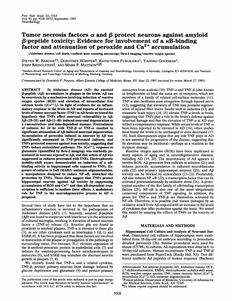
Exposure of hippocampal cell cultures to A13-(25-35) resultedin a concentration- and time-dependent reduction in neuronalsurvival (Fig. 1A). Af3-(1-40). also caused a progressive reduc-tion in neuronal survival, while control peptides did notsignificantly reduce neuronal survival during a 3-day exposure
> .0*5 E
-(U-(U.0
2 .E
100
90
80-
70
60
50
40
30,
20-
ACon,3d "
I
-.-.-._._ TI
-*-- 24hr---a-- 48hr-~-**-*- 72hr
0 10 20AD1 Concentration (,NM)
100-
80
60-
40-
20
50
period (Fig. 1). Based on our previous data with excitotoxicand metabolic insults (9), we tested whether TNFs wouldprotect hippocampal neurons against Aj3 toxicity. Cultureswere pretreated for 24 hr with TNF-ca or TNF-13 (100 ng/ml)and then exposed to Af3-(25-35) or A,B-(1-40) [control cul-tures received A,B-(35-25)]. Neuronal degeneration induced by50 ,uM A13-(25-35) or 20 AM A,B-(1-40) was significantlyreduced in cultures pretreated with either TNF-a or TNF-f3(Fig. 1C). TNF-a and TNF-,3 alone had no significant effect onbasal level of neuronal survival during a 72-hr treatment (datanot shown).TNFs Suppress A.3-Induced Accumulation of Cellular Per-
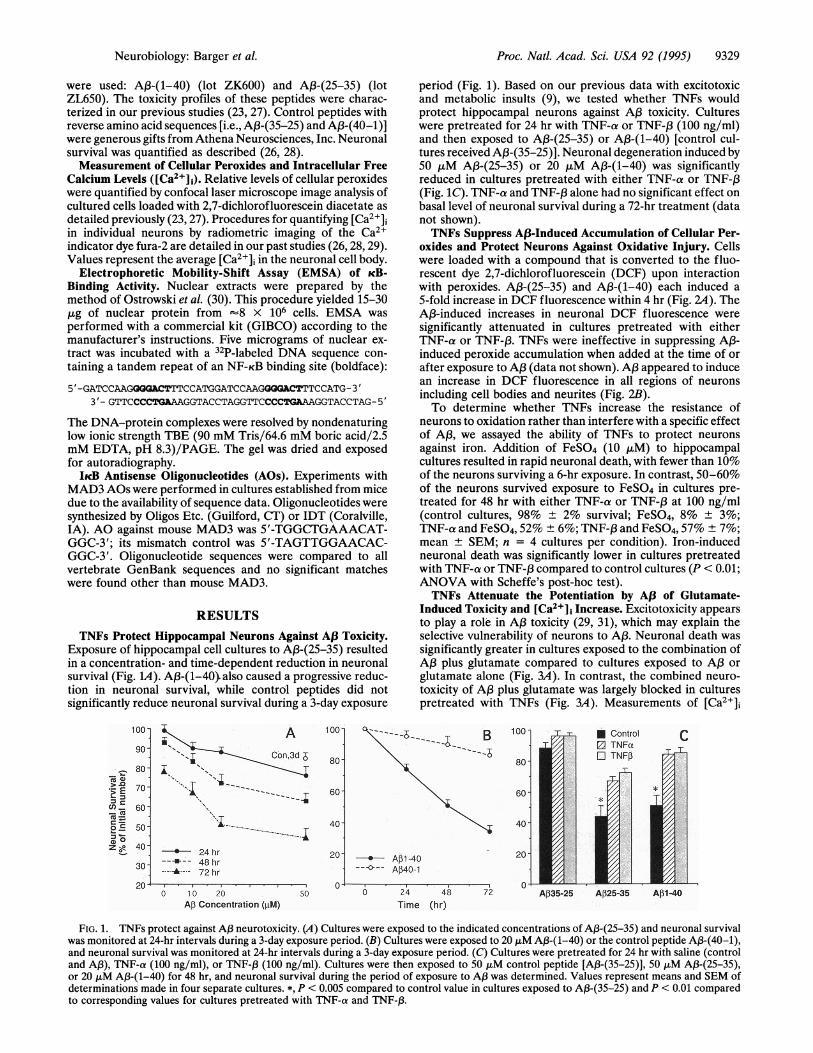
oxides and Protect Neurons Against Oxidative Injury. Cellswere loaded with a compound that is converted to the fluo-rescent dye 2,7-dichlorofluorescein (DCF) upon interactionwith peroxides. A,B-(25-35) and A,B-(1-40) each induced a5-fold increase in DCF fluorescence within 4 hr (Fig. 24). TheAP3-induced increases in neuronal DCF fluorescence weresignificantly attenuated in cultures pretreated with eitherTNF-a or TNF-13. TNFs were ineffective in suppressing A,B-induced peroxide accumulation when added at the time of orafter exposure to AP3 (data not shown). AP3 appeared to inducean increase in DCF fluorescence in all regions of neuronsincluding cell bodies and neurites (Fig. 2B).To determine whether TNFs increase the resistance of
neurons to oxidation rather than interfere with a specific effectof AP3, we assayed the ability of TNFs to protect neuronsagainst iron. Addition of FeSO4 (10 ,uM) to hippocampalcultures resulted in rapid neuronal death, with fewer than 10%of the neurons surviving a 6-hr exposure. In contrast, 50-60%of the neurons survived exposure to FeSO4 in cultures pre-treated for 48 hr with either TNF-a or TNF-3 at 100 ng/ml(control cultures, 98% ± 2% survival; FeSO4, 8% ± 3%;TNF-a and FeSO4, 52% ± 6%; TNF-P and FeSO4, 57% ± 7%;mean ± SEM; n = 4 cultures per condition). Iron-inducedneuronal death was significantly lower in cultures pretreatedwith TNF-a or TNF-,3 compared to control cultures (P < 0.01;ANOVA with Scheffe's post-hoc test).TNFs Attenuate the Potentiation by A13 of Glutamate-
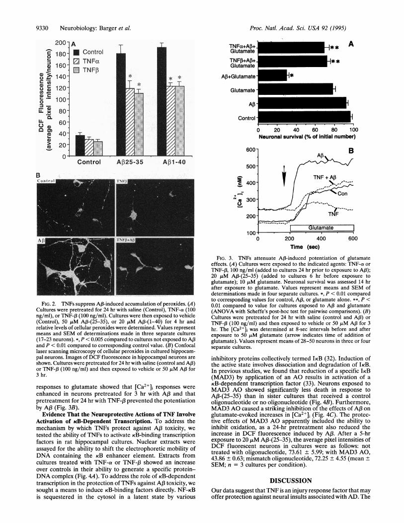
Induced Toxicity and [Ca2+1] Increase. Excitotoxicity appearsto play a role in AP3 toxicity (29, 31), which may explain theselective vulnerability of neurons to A,B. Neuronal death wassignificantly greater in cultures exposed to the combination ofAP3 plus glutamate compared to cultures exposed to AP3 orglutamate alone (Fig. 3A). In contrast, the combined neuro-toxicity of A,B plus glutamate was largely blocked in culturespretreated with TNFs (Fig. 3A). Measurements of [Ca>]
B 1001-T
* Apl -40---<>- AP40-1
6 24 48Time (hr)
72 Aj35-25 AP25-35 Afl-40
FIG. 1. TNFs protect against A,3 neurotoxicity. (A) Cultures were exposed to the indicated concentrations of A,3-(25-35) and neuronal survivalwas monitored at 24-hr intervals during a 3-day exposure period. (B) Cultures were exposed to 20 ,M A13-(1-40) or the control peptide A,B-(40-1),and neuronal survival was monitored at 24-hr intervals during a 3-day exposure period. (C) Cultures were pretreated for 24 hr with saline (controland A,B), TNF-a (100 ng/ml), or TNF-f (100 ng/ml). Cultures were then exposed to 50 ,uM control peptide [A,B-(35-25)], 50 ,iM A,3-(25-35),or 20 ,uM Aj3-(1-40) for 48 hr, and neuronal survival during the period of exposure to A,B was determined. Values represent means and SEM ofdeterminations made in four separate cultures. *, P < 0.005 compared to control value in cultures exposed to A3-(35-25) and P < 0.01 comparedto corresponding values for cultures pretreated with TNF-a and TNF-B3.
Neurobiology: Barger et al.
9330 Neurobiology: Barger et al.
A13
TNFa+Ap+ E I **Glutamate
TNF,+Am+ iuGlutamate_
+Glutamate
Glutamate
Control
A
AP i
Iiiuiui0 20 40 60 80 100Neuronal survival (% of initial number)
600
Control AP25-35 A,l1-40
0-
2.
c
+M,
0 200 400
FIG. 2. TNFs suppress A,B-induced accumulation of peroxides. (A)Cultures were pretreated for 24 hr with saline (Control), TNF-a (100ng/ml), or TNF-1 (100 ng/ml). Cultures were then exposed to vehicle(Control), 50 ,lM A13-(25-35), or 20 ,uM A1-(1-40) for 4 hr andrelative levels of cellular peroxides were determined. Values representmeans and SEM of determinations made in three separate cultures(17-23 neurons). *, P < 0.005 compared to cultures not exposed to APand P < 0.01 compared to corresponding control value. (B) Confocallaser scanning microscopy of cellular peroxides in cultured hippocam-pal neurons. Images of DCF fluorescence in hippocampal neurons are
shown. Cultures were pretreated for 24 hr with saline (control and A,B)or TNF-f3 (100 ng/ml) and then exposed to vehicle or 50 ,tM A,B for3 hr.
responses to glutamate showed that [Ca2+]i responses were
enhanced in neurons pretreated for 3 hr with AP3 and thatpretreatment for 24 hr with TNF-,B prevented the potentiationby A,B (Fig. 3B).
Evidence That the Neuroprotective Actions of TNF InvolveActivation of cB-Dependent Transcription. To address themechanism by which TNFs protect against AP3 toxicity, we
tested the ability of TNFs to activate KB-binding transcriptionfactors in rat hippocampal cultures. Nuclear extracts were
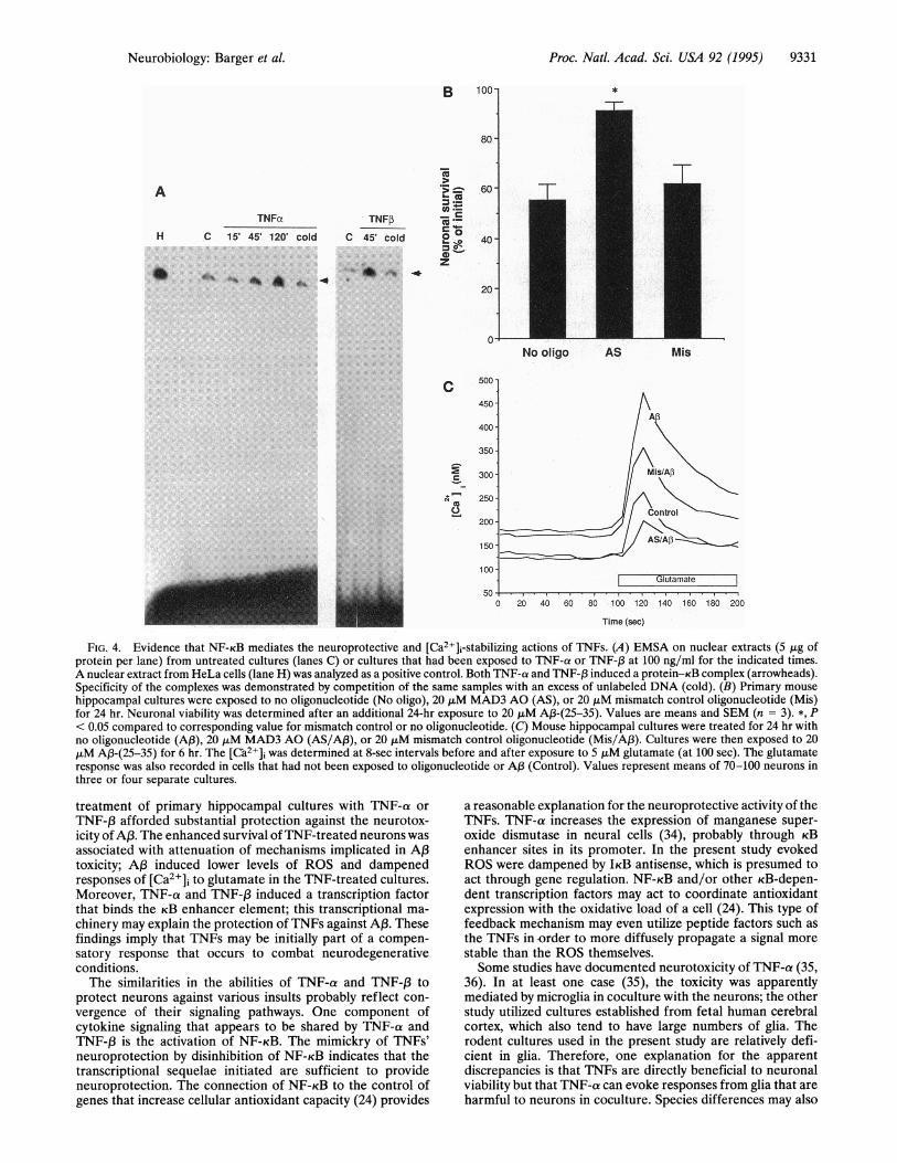
assayed for the ability to shift the electrophoretic mobility ofDNA containing the KB enhancer element. Extracts fromcultures treated with TNF-a or TNF-,B showed an increaseover controls in their ability to generate a specific protein-DNA complex (Fig. 4A). To address the role of KB-dependenttranscription in the protection of TNFs against AP3 toxicity, wesought a means to induce KB-binding factors directly. NF-KBis sequestered in the cytosol in a latent state by various
600Time (sec)
FIG. 3. TNFs attenuate AP3-induced potentiation of glutamateeffects. (A) Cultures were exposed to the indicated agents: TNF-a or
TNF-f3, 100 ng/ml (added to cultures 24 hr prior to exposure to A,);20 ,uM A,B-(25-35) (added to cultures 6 hr before exposure toglutamate); 10 ,uM glutamate. Neuronal survival was assessed 14 hrafter exposure to glutamate. Values represent means and SEM ofdeterminations made in four separate cultures. *, P < 0.01 comparedto corresponding values for control, A,B, or glutamate alone. **, P <0.01 compared to value for cultures exposed to AP3 and glutamate(ANOVA with Scheffe's post-hoc test for pairwise comparisons). (B)Cultures were pretreated for 24 hr with saline (control and AP3) or
TNF-,B (100 ng/ml) and then exposed to vehicle or 50 ,uM A,B for 3hr. The [Ca2+]i was determined at 8-sec intervals before and afterexposure to 50 JtLM glutamate (arrow indicates time of addition ofglutamate). Values represent means of 28-50 neurons in three or fourseparate cultures.
inhibitory proteins collectively termed IKB (32). Induction ofthe active state involves dissociation and degradation of IKB.In previous studies, we found that reduction of a specific IKB(MAD3) by application of an AO results in activation of aKB-dependent transcription factor (33). Neurons exposed toMAD3 AO showed significantly less death in response toAP3-(25-35) than in sister cultures that received a controloligonucleotide or no oligonucleotide (Fig. 4B). Furthermore,MAD3 AO caused a striking inhibition of the effects of A,B onglutamate-evoked increases in [Ca2+], (Fig. 4C). The protec-tive effects of MAD3 AO apparently included the ability toinhibit oxidation, as a 24-hr pretreatment also reduced theincrease in DCF fluorescence induced by AfB. After a 5-hrexposure to 20 p,M A,B-(25-35), the average pixel intensities ofDCF fluorescent neurons in cultures were as follows: nottreated with oligonucleotide, 73.61 + 5.99; with MAD3 AO,43.86 + 0.63; mismatch oligonucleotide, 72.25 + 4.55 (meanSEM; n = 3 cultures per condition).
DISCUSSIONOur data suggest that TNF is an injury response factor that mayoffer protection against neural insults associated with AD. The
0a-
. U)C
e4-.- ._
o0_L x
LL~IL
.)0a-0
4)
B
r-
Proc. Natl. Acad. Sci. USA 92 (1995)
Proc. Natl. Acad. Sci. USA 92 (1995) 9331
B 100-
80 -
A
TNFuxH C 15' 45' 120' cold
S
I-Z
TNFP X-
C 45' cold 0
zt ~ z,4kA f -4
60
40 -
20-
0-
500
450
400
350-
r_ 300-
A,_ 250 -
200 -
150
100I '.Ulutamate
0 20 40 60 80 100 120 140 160 180 200
Time (sec)
FIG. 4. Evidence that NF-KB mediates the neuroprotective and [Ca2+]i-stabilizing actions of TNFs. (A) EMSA on nuclear extracts (5 ,tg ofprotein per lane) from untreated cultures (lanes C) or cultures that had been exposed to TNF-a or TNF-,B at 100 ng/ml for the indicated times.A nuclear extract from HeLa cells (lane H) was analyzed as a positive control. Both TNF-a and TNF-f, induced a protein-KB complex (arrowheads).Specificity of the complexes was demonstrated by competition of the same samples with an excess of unlabeled DNA (cold). (B) Primary mouse
hippocampal cultures were exposed to no oligonucleotide (No oligo), 20 ,uM MAD3 AO (AS), or 20 ,iM mismatch control oligonucleotide (Mis)for 24 hr. Neuronal viability was determined after an additional 24-hr exposure to 20 ,uM A,B-(25-35). Values are means and SEM (n = 3). *, P< 0.05 compared to corresponding value for mismatch control or no oligonucleotide. (C) Mouse hippocampal cultures were treated for 24 hr withno oligonucleotide (A,B), 20 ,uM MAD3 AO (AS/Af3), or 20 ,tLM mismatch control oligonucleotide (Mis/A,B). Cultures were then exposed to 20,tM A3-(25-35) for 6 hr. The [C'a2+], was determined at 8-sec intervals before and after exposure to 5 ,tM glutamate (at 100 sec). The glutamateresponse was also recorded in cells that had not been exposed to oligonucleotide or A,B (Control). Values represent means of 70-100 neurons inthree or four separate cultures.
treatment of primary hippocampal cultures with TNF-a or
TNF-,B afforded substantial protection against the neurotox-icity of A,B. The enhanced survival of TNF-treated neurons was
associated with attenuation of mechanisms implicated in AP3toxicity; A,B induced lower levels of ROS and dampenedresponses of [Ca2+]j to glutamate in the TNF-treated cultures.Moreover, TNF-a and TNF-,3 induced a transcription factorthat binds the KB enhancer element; this transcriptional ma-
chinery may explain the protection of TNFs against A,B. Thesefindings imply that TNFs may be initially part of a compen-satory response that occurs to combat neurodegenerativeconditions.The similarities in the abilities of TNF-a and TNF-,3 to
protect neurons against various insults probably reflect con-
vergence of their signaling pathways. One component ofcytokine signaling that appears to be shared by TNF-a andTNF-,B is the activation of NF-KB. The mimickry of TNFs'neuroprotection by disinhibition of NF-KB indicates that thetranscriptional sequelae initiated are sufficient to provideneuroprotection. The connection of NF-KB to the control ofgenes that increase cellular antioxidant capacity (24) provides
a reasonable explanation for the neuroprotective activity of theTNFs. TNF-a increases the expression of manganese super-oxide dismutase in neural cells (34), probably through KBenhancer sites in its promoter. In the present study evokedROS were dampened by IKB antisense, which is presumed toact through gene regulation. NF-KB and/or other KB-depen-dent transcription factors may act to coordinate antioxidantexpression with the oxidative load of a cell (24). This type offeedback mechanism may even utilize peptide factors such asthe TNFs in-order to more diffusely propagate a signal more
stable than the ROS themselves.Some studies have documented neurotoxicity of TNF-a (35,
36). In at least one case (35), the toxicity was apparentlymediated by microglia in coculture with the neurons; the otherstudy utilized cultures established from fetal human cerebralcortex, which also tend to have large numbers of glia. Therodent cultures used in the present study are relatively defi-cient in glia. Therefore, one explanation for the apparentdiscrepancies is that TNFs are directly beneficial to neuronalviability but that TNF-a can evoke responses from glia that areharmful to neurons in coculture. Species differences may also
I
T1T
No oligo AS Mis
Neurobiology: Barger et al.
co
Proc. Natl. Acad. Sci. USA 92 (1995)
contribute to discrepancies between studies. Gelbard et al. (36)treated human cultures with human TNF-a, whereas we haveused human TNF-a in rodent cultures. Human TNF-a does notactivate the rodent p75 TNF receptor (37); therefore, theneuroprotective effects ofTNF-a may be mediated specificallyby signaling events arising from the p55 receptor, whichinclude production of ceramide and activation of NF-KB (38).Other glial-derived growth factors and related signals have
been shown to lessen the toxicities of A,B and glutamate (seeref. 39 for review). Basic fibroblast growth factor affordscomplete protection against AP3 toxicity in the same type ofcells studied here (28). Transforming growth factor 13 (TGF-,B)has also been reported to lessen excitotoxicity (40) and APtoxicity (41). Galindo et al. (41) also reported an increase in theexpression of P-amyloid precursor protein (P3APP) in neuronaland glial cultures treated with TGF-,3. This may indicate anindirect action for TGF-,3, as secreted forms of I3APP (sAPP)can also protect against A,B toxicity (23). We recently foundthat sAPP also induces KB-dependent transcription (42), fur-ther linking this transcription system with neuroprotection.Several growth factors are induced in glia by AP3 itself andaccumulate in neuritic plaques (43). Accumulation in AD ofother glial-derived neuroprotective proteins such as proteasenexin I and al-antichymotrypsin may similarly reflect anoverreaction of the glia-neuron axis as it attempts to managethe neurodegeneration. This and other systemic considerationslimit the potential therapeutic usefulness of TNFs; however,the data presented here suggest that pharmacological mim-ickry of the effects ofTNFs in neurons-particularly inductionof KB-dependent transcription-may offer clues for alleviatingA,B-induced neurodegeneration.
We thank J. Begley and S. Bose for technical assistance and R. E.Rydel for critical comments on this work. This work was supported byfunds from the National Institutes of Health (S.W.B. and M.P.M.), theFrench Foundation (S.W.B.), and the Alzheimer's Association (M.P.M.;Evelyn Stone Fund and Zenith Award).
1. Aisen, P. S. & Davis, K. L. (1994) Am. J. Psychiatry 151, 1105-1113.
2. Meda, L., Cassatella, M. A., Szendrei, G. I., Otvos, L., Jr., Baron,P., Villalba, M., Ferrari, D. & Rossi, F. (1995) Nature (London)374, 647-650.
3. Dickson, D. W., Lee, S. C., Mattiace, L. A., Yen, S. H. & Bros-nan, C. (1993) Glia 7, 75-83.
4. Griffin, W. S. T., Stanley, L. C., Ling, C., White, L., MacLeod, V.,Perrot, L. J., White, C. L. I. & Araoz, C. (1989) Proc. Natl. Acad.Sci. USA 86, 7611-7615.
5. Goldgaber, D., Harris, H. W., Hla, T., Maciag, T., Donnelly,R. J., Jacobsen, J. S., Vitek, M. P. & Gajdusek, D. C. (1989) Proc.Natl. Acad. Sci. USA 86, 7606-7610.
6. Das, S. & Potter, H. (1995) Neuron 14, 447-456.7. Marshak, D. R., Pesce, S. A., Stanley, L. C. & Griffin, W. S. T.
(1992) Neurobiol. Aging 13, 1-7.8. Sheng, J. G., Mrak, R. E. & Griffin, W. S. T. (1994) J. Neurosci.
Res. 39, 398-404.9. Cheng, B., Christakos, S. & Mattson, M. P. (1994) Neuron 12,
139-153.10. Mattson, M. P., Cheng, B., Baldwin, S. A., Smith-Swintosky,
V. L., Keller, J., Geddes, J. W., Scheff, S. W. & Christakos, S.(1995) J. Neurosci. Res. 42, in press.
11. Smith, C. A., Farrah, T. & Goodwin, R. G. (1994) Cell 76,959-962.
12. Schwartz, M., Solomon, A., Lavie, V., Ben-Bassat, S., Belkin, M.& Cohen, A. (1991) Brain Res. 545, 334-338.
13. Liu, T., Clark, R. K., McDonnell, P. C., Young, P. R., White,R. F., Barone, F. C. & Feuerstein, G. Z. (1994) Stroke 25, 1481-1488.
14. Shohami, E., Novikov, M., Bass, R., Yamin, A. & Gallily, R.(1994) J. Cereb. Blood Flow Metab. 14, 615-619.
15. Tchelingerian, J. L., Quinonero, J., Booss, J. & Jacque, C. (1993)Neuron 10, 213-224.
16. Fillit, H., Ding, W. H., Buee, L., Kalman, J., Alstiel, L., Lawlor,B. & Wolf-Klein, G. (1991) Neurosci. Lett. 129, 318-320.
17. Cacabelos, R., Alvarez, X. A., Franco-Maside, A., Fernandez-Novoa, L. & Caamano, J. (1994) Methods Find. Exp. Clin.Pharmacol. 16, 25-35.
18. Huberman, M., Shalit, F., Roth-Deri, I., Gutman, B., Brodie, C.,Kott, E. & Sredni, B. (1994) J. Neuroimmunol. 52, 147-152.
19. Coyle, J. T. & Puttfarcken, P. (1993) Science 262, 689-694.20. Smith, C. D., Carney, J. M., Tatsumo, T., Stadtman, E. R., Floyd,
R. A. & Markesbery, W. R. (1992) Ann. N.Y Acad. Sci. 663,110-119.
21. Hensley, K., Carney, J. M., Mattson, M. P., Aksenova, M., Harris,M., Wu, J. F., Floyd, R. A. & Butterfield, D. A. (1994) Proc. Natl.Acad. Sci. USA 91, 3270-3274.
22. Behl, C., Davis, J. B., Lesley, R. & Schubert, D. (1994) Cell 77,817-827.
23. Goodman, Y. & Mattson, M. P. (1994) Exp. Neurol. 128, 1-12.24. Schreck, R., Albermann, K. & Baeuerle, P. A. (1992) Free
Radicals Res. Commun. 17, 221-237.25. Kabrun, N. & Enrietto, P. J. (1994) Semin. Cancer Biol. 5,
103-112.26. Mattson, M. P., Barger, S. W., Begley, J. & Mark, R. J. (1995)
Methods Cell Biol. 46, 187-216.27. Goodman, Y., Steiner, M. R., Steiner, S. M. & Mattson, M. P.
(1994) Brain Res. 654, 171-176.28. Mattson, M. P., Tomaselli, K. J. & Rydel, R. E. (1993) Brain Res.
621, 35-49.29. Mattson, M. P., Cheng, B., Davis, D., Bryant, K., Lieberburg, I.
& Rydel, R. E. (1992) J. Neurosci. 12, 379-389.30. Ostrowski, J., Sims, J. E., Sibley, C. H., Valentine, M. A., Dower,
S. K., Meier, K. E. & Bomsztyk, K. (1991) J. Biol. Chem. 266,12722-12733.
31. Koh, J.-Y., Yang, L. L. & Cotman, C. W. (1990) Brain Res. 533,315-320.
32. Gilmore, T. D. & Morin, P. J. (1993) Trends Genet. 9, 427-433.33. Barger, S. W. & Mattson, M. P. (1994) Soc. Neurosci. Abstr. 20,
687.34. Mokuno, K., Ohtani, K., Suzumura, A., Kiyosawa, K., Hirose, Y.,
Kawai, K. & Kato, K. (1994) J. Neurochem. 63, 612-616.35. Chao, C. C., Molitor, T. W. & Hu, S. (1993) J. Immunol. 151,
1473-1481.36. Gelbard, H. A., Dzenko, K. A., DiLoreto, D., del Cerro, C., del
Cerro, M. & Epstein, L. G. (1993) Dev. Neurosci. 15, 417-422.37. Lewis, M., Tartaglia, L. A., Lee, A., Bennett, G. L., Rice, G. C.,
Wong, G. H. W., Chen, E. Y. & Goeddel, D. V. (1991) Proc. Natl.Acad. Sci. USA 88, 2830-2834.
38. Kolesnick, R. & Golde, D. W. (1994) Cell 77, 325-328.39. Mattson, M. P., Cheng, B. & Smith-Swintosky, V. L. (1993)
Semin. Neurosci. 5, 295-307.40. Prehn, J. H., Backhauss, C. & Krieglstein, J. (1993) J. Cereb.
Blood Flow Metab. 13, 521-525.41. Galindo, M. F., Prehn, J. H. M., Bindokas, V. P. & Miller, R. J.
(1994) Soc. Neurosci. Abstr. 20, 1248.42. Barger, S. W. & Mattson, M. P. (1995) Ann. N.Y Acad. Sci., in
press. -43. Araujo, D. M. & Cotman, C. W. (1992) Brain Res. 569, 141-145.
9332 Neurobiology: Barger et al.