Embed Size (px)
Citation preview

Tubulointerstitial Disease
Mark D. Baldwin D.O. FACOI
ACOI Board Review Course 2018

Disclosures
• None, just working for The Man

Features of Tubulointerstitial
Disease
1. Proteinuria- usually less than 1 Gm/da
2. Anemia-due to low level of Erythropoetin
3. Acidosis-RTA’s are common
4. Hypertension-common
5. Urinalysis-WBC’s and WBC casts seen
6. Electrolyte Abnormalities-Na and K
Many of the above features are seen at relatively
mild elevations of Serum Creatinine

Features of Glomerular Disease
1. Proteinuria->3 Gm/da
2. Anemia-uncommon until late
3. Acidosis-uncommon until late
4. Hypertension-may occur at any time
5. Urinalysis-may see Oval Fat Bodies
6. Electrolytes-May see low Na
Unlike Tubulointerstitial Disease, many of these
feature do not occur until late in the course of
the underlying disease.

Types of Tubulointerstitial Disease
1. Acute Interstitial Nephritis
2. Chronic Interstitial Nephritis
3. Acute Tubular Necrosis
4. Renal Tubular Acidosis
5. Multiple Myeloma

Chronic Interstitial Nephritis (CIN)
• A chronic condition involving fibrosis of the
interstitium and tubular destruction.
• Macroscopically normal kidneys
• The final common pathway of most
chronic renal diseases

Causes of Chronic Interstitial Nephritis
• Mechanical: ureteral reflux, obstruction, stones, infection,
neurogenic bladder, medullary cystic disease, Alport’s
• Drugs: NSAIDs, Lithium, PPI’s, cyclosporine, tacrolimus, indinavir,
cisplatin
• Heavy Metals: Hg, Pb, Cd, arsenic, gold, uranium, too much Ozzy
• Metabolic: hyperuricemia, hypercalcemia, hypokalemia,
hyperoxaluria, cytinosis
• Radiation
• Immune mediated: ANCA, SLA, Sjorgren's, sarcoid
• Vascular: atherosclerotic renal disease
• Heme/Onc: myeloma, amyloid, lymphoma, sickle cell, PNH
• Late glomerular disease
• Aristolochic acid: Balkan nephropathy, Chinese herb nephropathy

Aristolochic acid
• Acute exposure: Chinese herb
nephropathy-AKI, rapid decline in renal
function
• Chronic exposure: Balkan endemic
nephropathy-CIN/CKD slow decline over
years from chronic exposure
Debelle Kidney Int 2008;74: 158-169

Aristolochic acid Nephrotoxicity
Debelle Kidney Int 2008;74: 158-169

Clinical Features of CIN
• Usually Asymptomatic
• STERILE PYURIA-The Hallmark of CIN
• Anemia
• Acidosis-Renal Tubular Acidosis
• Hypo or hyperkalemia
• Minimal Proteinuria
• Hypertension

Chronic Interstitial Nephritis

Chronic Interstitial Nephritis

Treatment of CIN
• Do NOT give antibiotics for pyuria unless
there is bacteria present-this is a chronic
inflammatory condition, NOT an infection
• BP control-The MOST important treatment
• ACE-I or ARBs-The drugs of choice
• Anemia control
• Acidosis control
• Phosphorus control

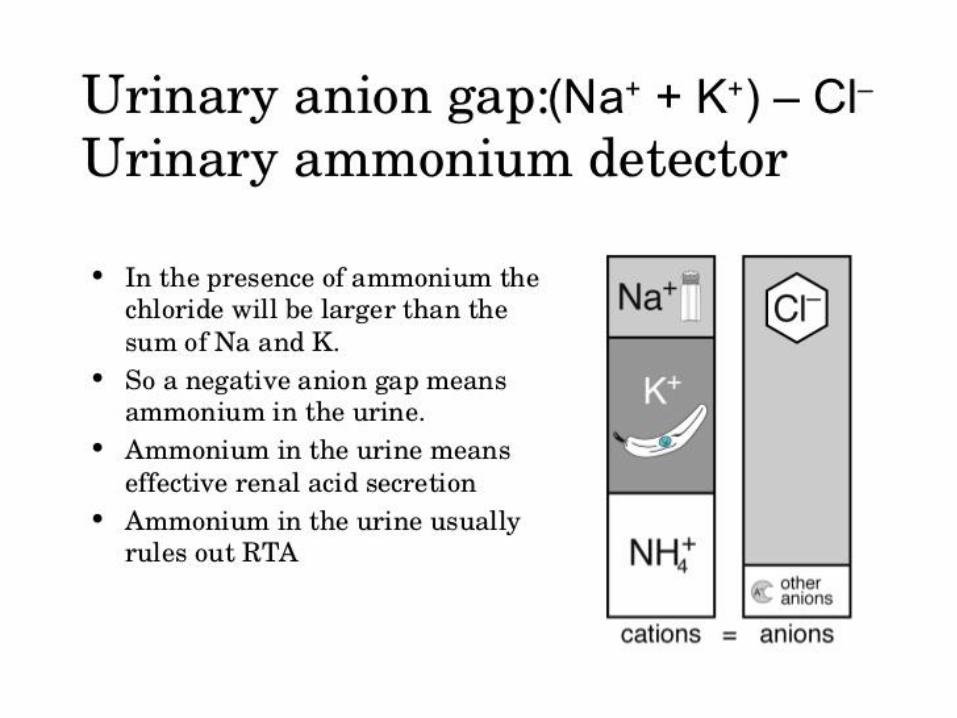
Renal Tubular Acidosis (RTA)
• Distal Type I RTA -associated with Chronic Urinary Tract Obstruction, Bicarb<15*NAG, hypokalemia, urine pH>5.5
• Proximal Type II RTA -associated with Fanconi’s Syndrome Bicarb 15-21*NAG, hypokalemia, urine pH>5.5
• Distal Type IV RTA- Most common RTA, Seen w/ DM and CKD, NAG, Hyperkalemia urine pH<5.5
• All RTA have +UAG* Point of differentiation, NAG non anion gap acidosis


Myeloma and the Kidney
• 10% of all hematological malignancies
• Plasma cell clone of Immunoglobulins usually IgG
• Renal, Cardiac and Liver are the most common organs
involved
• Renal impairment-acute or chronic- is commonly seen
~50% of cases with severe involvement in 15-20% of
cases
• Proteinuria-globulin or albuminuria is seen in >80% of
cases
• Myeloma can involve the vascular, glomerular or
tubular/interstitial segments of the kidney
• Frequent cause of mortality and morbidity
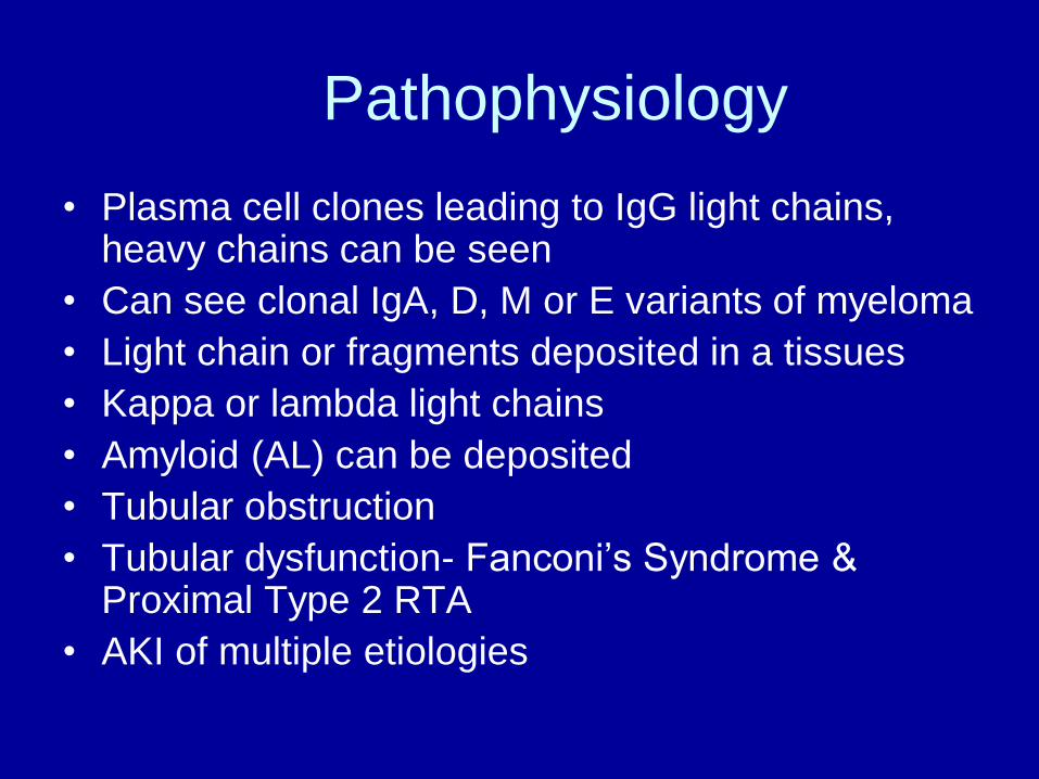
Pathophysiology
• Plasma cell clones leading to IgG light chains, heavy chains can be seen
• Can see clonal IgA, D, M or E variants of myeloma
• Light chain or fragments deposited in a tissues
• Kappa or lambda light chains
• Amyloid (AL) can be deposited
• Tubular obstruction
• Tubular dysfunction- Fanconi’s Syndrome & Proximal Type 2 RTA
• AKI of multiple etiologies

Renal Effects
• Glomerular: Amyloid light chain (AL) or heavy chain (AH)
amyloidosis, Light Chain Deposition Disease (LCDD) or
Heavy Chain Deposition Disease (HCDD) plasma cell
infiltration
• Tubular: Cast nephropathy “Myeloma kidney”, tubular
dysfunction, hypercalcemia, hyperuricemia, contrast
induced AKI
• Interstitial: Plasma cell infiltration, pyelonephritis

Amyloid MM v LCDD
• Amyloid myeloma (AL):
• lambda>kappa, +congo red, +fibrils on EM
• Light chain deposition disease(LCDD):
kappa>lambda, -congo red, -fibrils on EM

Cast Nephropathy
• Most common cause of renal failure in myeloma
• Globulin light chains are filtered at the glomerulus
• Can exceed 10-20 grams/day and are toxic to the tubular
cells, negative dipstick d/t globulins not albumin
• Light chain are partially reabsorbed damaging to
proximal tubular cells and delivered distally, combing
with Tamm-Horsfell protein produced in the thick
ascending limb occluding the tubule
• Obstructing casts lead to inflammation, fibrosis and
tubular rupture

Presentation
• >50 years old
• Males>females
• African Americans >other groups
• Long history of back pain or “arthritic” pain
• Pathological fractures
• Fatigue
• Anemia
• Infection
• Renal failure
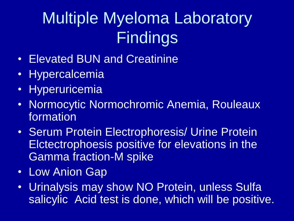
Multiple Myeloma Laboratory
Findings
• Elevated BUN and Creatinine
• Hypercalcemia
• Hyperuricemia
• Normocytic Normochromic Anemia, Rouleaux formation
• Serum Protein Electrophoresis/ Urine Protein Elctectrophoesis positive for elevations in the Gamma fraction-M spike
• Low Anion Gap
• Urinalysis may show NO Protein, unless Sulfa salicylic Acid test is done, which will be positive.
Serology
• Serum protein electrophoresis/urine
protein electrophoresis
• Free light chain assay
• Immunofixation: quantifies IgA, IgD, IgE,
IgM, IgG
• Cytogenic analysis: karyotyping
• Flow cytometry

Congo Red “Apple green
birefringence”
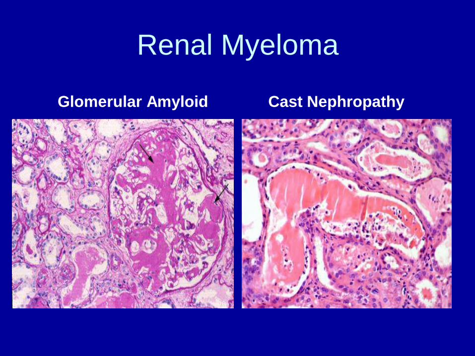
Renal Myeloma
Glomerular Amyloid Cast Nephropathy
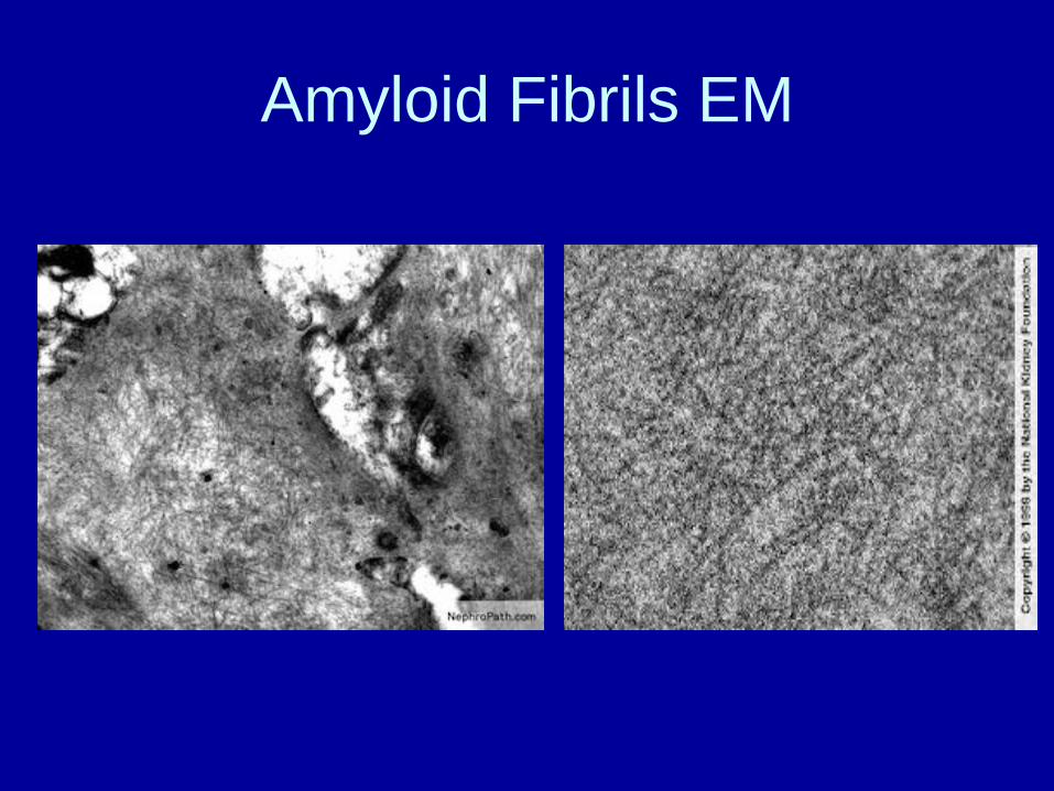
Amyloid Fibrils EM
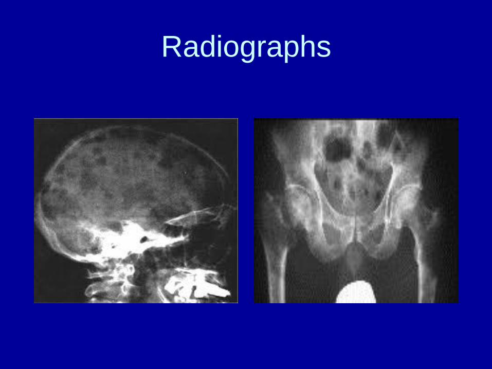
Radiographs

Treatment of Myeloma Renal
Disease• Assure hydration status with alkalization of the urine
but avoid fluid overload
• Allopurinol
• Bortezomib-dexamethasone-cyclophosphamide or
• Bortezomib-thalidomide-dexamethasone can
decrease light chain production and may improve
cast nephropathy
• Dialysis if needed (poor outcomes)
• ?role of plasmapheresis

Malignancy Related
Hypercalcemic Renal Failure• Prostate
• Renal Cell
• Breast
• Lymphoma/Leukemia
• Lung
• Myeloma
• Thyroid
• Other malignancies

Mechanism of Hypercalcemia
1. Humoral Hypercalcemia of Malignancy (HHM) 80%:
-PTH related protein (PTHrP)
-1,25 dihydroxy Vit D (also seen w/ sarcoid and T.B.)
-PTH-like substance (very rare)
2. Osteolytic Metastasis: 20%
-Bone mets stimulate osteolysis via Osteoclast Activating
Factor, RANKL, Il, VEGF, TNF, TGF and PTHrP
3. Prostaglandin mediated

Presentation
• Decreased cognition, fatigue
• Anorexia, N,V, constipation
• Abdominal and bone pain
• Pancreatitis
• Short QT, ST changes pseudo MI pattern
• HTN
• AKI, Nephrogenic DI
N AM J Med Sci 2011;7(11): 483-493.

Treatment of Hypercalcemia of
Malignancy• Normal Saline: Restores BP, decreases Ca
reabsorption
• Calcitonin
• Bisphosphonates (Zolendronic acid most potent)
• Denosumab: Ab to RANKL, decreases OAF,
best in cases refractory to Bisphosphonates
• Loop diuretics: enhance Ca excretion, don’t give
thiazides will increase Ca reabsorption

Tubulointerstitial Diseases-
Conclusions
• Often overlooked as a cause of Chronic Renal Disease
• Look for Drug causes or Sepsis as a cause of Acute Renal Failure (i.e. AIN or ATN)
• Tubulointerstitial Diseases frequently have electrolyte abnormalities, acid-base disorders, and anemia as a common feature.


![Research Article Relationship between Hyperuricemia and ...downloads.hindawi.com/journals/bmri/2015/127596.pdf · hyperuricemia and dyslipidemia [ , ]. When establishing the diagnosis](https://img.pdfslide.us/doc/110x75/5f1058267e708231d448a554/research-article-relationship-between-hyperuricemia-and-hyperuricemia-and-dyslipidemia.jpg)