Embed Size (px)
Citation preview

TRENDS in Cell Biology Vol.12 No.10 October 2002
http://tcb.trends.com 0962-8924/02/$ – see front matter © 2002 Elsevier Science Ltd. All rights reserved. PII: S0962-8924(02)02364-4
479Review
Matthew Buechner
Dept of MolecularBiosciences, 1200Sunnyside Ave., 8035Haworth Hall, Universityof Kansas, Lawrence,KS 66045-7534, USA.e-mail: [email protected]
Most efforts to understand tubulogenesis have focusedon the complicated tubes of vertebrates, such as bloodvessels, nephrons, lung sacs, mammary glands andsecretory vessels. These structures are constructedfrom large numbers of epithelial cells enclosed by abasement membrane, and the tubule lumen oftenexhibits large variations in diameter and specializationalong the length of the tubule. To understand themolecular basis of tubulogenesis and regulation oftubule diameter, however, it is useful to considersimilar structures in simpler creatures. In fact, longtubules can be formed from single cells. Such asingle-celled epithelium greatly simplifies considerationof the elements that regulate lumen formation anddiameter. In a cross-section of a typical thinmulticellular lumen such as a kidney nephron, lungalveolus, mammary gland or capillary (Fig. 1a), theapical (lumenal) surface is connected to the surroundingbasal surface via the lateral surfaces of each of themultiple epithelial cells. But, in a single-celled tubule,there is no lateral surface present in a typicalcross-section (Fig. 1b). The apical and basal surfacesmeet only at the end of the tubule and are thereforecompletely separated from each other along the lengthof a single-celled tube.
Single-celled tubules have been studied in theDrosophila trachea (see the forthcoming article byC. Samakovlis in this series) and in the geneticallytractable roundworm Caenorhabditis elegans, thesubject of this review. The nematode excretory canalsregulate the osmolarity of this organism [1,2]. Theexcretory cell is shaped like the letter ‘H’and formstwo canals, one on each side of the worm (Fig. 2). Eachcanal is a hollow tube that runs the entire length ofthe nematode beneath the hypodermis on the animal’slateral side. The lumen diameter is much narrower in
the anterior canals than in the posterior canals.The lumen tapers towards the tips of the canals andwidens as the animal grows. The canals are flexibleand bend with the animal as it slithers along. Justbeneath the pharynx, there is a tubular bridge thatconnects the left and right canals to each other and toa duct cell and pore cell through which excess water iseliminated. The single nucleus is located in thecytoplasm bridging the two canals. In addition toregulating lumen formation and the initial growth ofthe canals during embryogenesis, this busy cell mustcontinuously regulate lumen diameter both spatiallyand temporally throughout the nematode’s larval andadult life. Various mutations have been found thataffect the identity of the excretory cell, the length ofits tubules, the location of the tubules within theorganism, and the diameter of the tubule lumen.Here, I discuss the development of the excretory canals,the identity and function of some of the large set ofgene products that affect canal structure, and bringthese components together towards understandingthis appealing example of tubule morphogenesis.
Development of the excretory canals
The excretory cell is the largest cell in C. elegans, andits birth provides an easily identified landmark midwaythrough embryonic development [3]. The cell is locatedon the ventral side near the developing pharynx.Several transcription factors affect the developmentof the canals, of which the best characterized is thePOU/homeobox CEH-6 [4]. Within an hour of its birth,the excretory cell extends two processes dorsolaterally
The formation and regulation of tubule shape and size is fundamental to the
development and function of many tissues and organs in metazoan organisms.
The excretory canals of the nematode Caenorhabditis elegans are a fascinating
example of cell morphogenesis, as the tiny worm manages to create a
complicated set of tubular epithelia within a single cell. In addition to the
inherent attraction of studying this cytoengineering feat, the excretory cell
provides a simple genetically tractable model for studying tubule formation
and regulation of tubule diameter. Mutations in the exc genes alter the
diameter of the lumenal surface of these tubules. Cloning of these genes
reveals a set of proteins that both control tubule diameter and regulate the
comparative growth of the apical and basal tubular surfaces.
Published online: 3 September 2002
Tubes and the single C. elegansexcretory cell
Matthew Buechner
Tube morphogenesis series
TRENDS in Cell Biology
Basal lamina
Lumen
Apicalsurface
Basal surfaceLateralsurface
Multicellular tubule Unicellular tubule(a) (b)
Fig. 1. Comparison of a multicellular tubule and a unicellular tubule.(a) Multicellular tubules contain lateral surfaces throughout the tubuleto connect the component cells. (b) The apical and basal surfaces of aunicellular tubule need meet only at one end of the tubule.

over the ventral muscle quadrants to the lateral surface(Fig. 3a,b). At the same time, vacuoles within the cellbody form and appear to coalesce to form the apical(lumenal) surface (D.H. Hall and M. Buechner,unpublished). A thick material then forms in the lumenduring embryogenesis, visible via electron microscopy(http://www.wormatlas.org/handbook/excretory.htm)(Fig. 3c). By the time of hatching, this lumenal material
has disappeared, while a different electron-densematerial forms in the cytoplasm to surround thelumenal membrane (Fig. 3d). This changeover inelectron-dense material may be related to the cellbeginning to function as an osmoregulatory organ inlate embryos; water movement within the lumen isinferred from the observation of pulsations in the ductcell beginning at this time [2]. A similar electron-densematerial is seen in vertebrates in the terminal websurrounding the lumen of the digestive tract, where itserves to anchor the microvilli [5]. The nematode canallumen is surrounded by myriad small channels, calledcanaliculi, that greatly increase the apical surfacearea [1] (Fig. 4). Arrays of microtubules that extendlongitudinally surround the canaliculi. Mitochondriaand Golgi bodies are dispersed throughout the canals,so translational control over tubule components canoccur throughout their length.
Once both the apical and basal surfaces have formed,they extend together, even though the two surfaceshave no lateral membrane connecting them throughoutthe canals. The only membrane connections betweenthe lumenal and outer surface are within the cell body,at the point where the excretory cell lumen connectsto the duct cell. The canal tips grow dorsalward overthe muscle quadrants until they reach the lateralhypodermis. At this point, the tips cross the hypodermalbasement membrane, bifurcate and begin growinganteriorward and posteriorward along the lateralsurface of the hypodermis (Fig. 3d). For this growth, thecanals insinuate themselves between the hypodermal(skin) cells and the basement membrane secreted bythe hypodermis; it is not clear whether the canalssecrete basement membrane in addition. By the timethe worm hatches, the posterior canals have reachedapproximately half the length of the organism (Fig. 3e).They continue to extend during the first larval stage,over the next 12 hours. The organism itself growsrapidly (from ~130 to over 300 µm long) during thisperiod, while the canals increase their overall lengthby a factor of five during this time. Upon finishingextension, the canals reach from the anterior tip ofthe organism, past the anus, to near the tip of the tail.As the animal continues to grow through three morelarval stages to adulthood and its full length of 1 mm,the canals grow passively along with the animal, butdo not extend any farther relative to the hypodermis.The canals are connected to the hypodermis viaextensive gap junctions along the entire canal length,as well as through interactions with the hypodermalbasement membrane. Therefore, once canal extensionis complete, growth of the canals occurs evenlythroughout their entire length, to match animal growth.
The length of the canals is affected by later eventsas well as by early development. As the adult ages andsenesces, the connections between the canal basalsurface and the hypodermis weaken. In older adults,a canal tip sometimes tears free of the hypodermis, andthat canal shortens instantly. The affected canal tipswells in diameter to become a wide ball of cytoplasm
TRENDS in Cell Biology Vol.12 No.10 October 2002
http://tcb.trends.com
480 Review
TRENDS in Cell Biology
VentralAnterior
Excretory canalsPosterior
Pore Connection to duct
Excretory cell body
Dorsal
Nucleus
Pharynx
Fig. 2. Structure of the excretory canals in a late larval stage worm. The position of the excretory cellbody is shown beneath a grey shadow representing the pharynx. Diameters of the lumena are slightlyexaggerated for clarity. The apical/lumenal (red) and basal (blue) surfaces of the excretory cell meetonly at the connection to the duct cell – the tips of the canals are enclosed, and the lumen is thereforecompletely surrounded by apical membrane.
TRENDS in Cell Biology
(e)
(f)
(g)
(c)(b) (d)(a)
Fig. 3. Development of the excretory cell. (a) Individual excretory cell is surrounded by presumptivebasal surface (shown in blue). (b) Apical surface (in red) develops as vesicles coalesce, then presumablyjoins to the basal surface at the junction to the excretory duct cell. (c) The tips of the excretory cellmigrate dorsalward to the lateral surface; the apical surface expands within the cell. A thickelectron-dense material in the lumen determines the lumen diameter. (d) Upon reaching the lateralsurface, the canals branch, and extend anteriorward and posteriorward. Lumenal material disappears,and is replaced by thick terminal web material (dark gray) in the cytoplasm surrounding the apicalsurface. (e) In adult animals, the posterior canal tips reach the back of the worm. Note that the tips areclosed, unlike the ends of nephridia of rotifers and flatworms. In nematodes, excess coelomic liquidmust pass through the canal cytoplasm to be pumped into the lumen. (f) The canals often ‘snap back’from their full length during senescence to create a ball of cytoplasm filled with apical lumen at the tip.(g) In exc mutants, the terminal web material breaks down or is improperly constructed, allowing thelumen to swell into large fluid-filled cysts.

as the canal shrinks in length (to as short as 1/2 to 3/4of its normal adult length) (Fig. 3f) (E.M. Hedgecockand M. Buechner, unpublished). The lumenal surfaceretains its normal length and diameter, however, andappears as a folded-up tubule within the ball ofcytoplasm. That the collapse of the basal cytoskeletonappears to have no effect on the apical cytoskeletonsuggests that these two surfaces are essentiallyindependent of each other.
Canal outgrowth uses neural guidance cues
Canal extension is mediated through interactions of thebasal surface with the cell environment and utilizesmany of the same cues and mechanisms as do neuralguidance and outgrowth [6,7]. Mutations in basementmembrane proteins and their receptors prevent thecanals from extending to their full length. For example,a mild hypomorphic allele of the β1-integrin genepat-3 [8] exhibits diverse shortenings of several neuraltracts, and the excretory canals extend only to mid-bodyin adult animals. Inspection of canal extension revealsthat the canals grow ~30% slower during the first larvalstage than do wild-type canals, and so never reach theends of the animal. Interestingly, in later larval stages,the canals do continue to grow with the rest of theanimal, but do not extend further along the hypodermis.Canal extension at the tip during embryonic and earlylarval growth therefore appears to be a process separatefrom passive canal extension in response to growth ofthe underlying hypodermis.
Other mutations affect canal and neuron outgrowthsimilarly. These include mutations in genes encodingvarious basement membrane components: epi-1 (thelaminin α chain), lam-1 (laminin β chain), unc-52
(perlecan) [9–11]; and the α-integrin chains thatdimerize with PAT-3: pat-2 and ina-1 [8,12]. Mutationshaving similar effects also occur in genes encodingcytoplasmic proteins, including mig-15 (Nck-interactingkinase), unc-53 (a novel actin-binding protein), unc-73(a rho-type GDP–GTP exchange factor), mig-10 (Grb7),unc-34 (Enabled), and the unc-104 and unc-116 kinesingenes [13–19].
Just as axons must be guided during neuraloutgrowth, the canal tips must be guided during theirextension. Dorsalward growth both of the canals andof axonal commissures is mediated by the UNC-6netrin and its receptor, UNC-5 [20–22]. Mutants in thelin-17 gene (encoding a Frizzled receptor [23]), whichaffects the polarity of asymmetrical cell divisions,including tail hypodermis, actually have canals thatare longer than normal; this observation suggests thattail hypodermal cells act as guideposts to determinethe limits of canal extension. Mutations in several othergenes (e.g. unc-34 and bli-6) affect the very earliestcanal guidance decision: whether the excretory cellshould extend to the left or the right upon its birth.In some of these animals, two full-length canals areseen extending along the same side of the animal!Finally, canals in dpy-17 mutants travel posteriorwardnormally until hatching, but then occasionally turn180° and extend forward to the head (‘canals on thebrain’). dpy-17 mutants show drastic alteration inhypodermal shape during the first larval stage [24],and bli-6 also affects hypodermal structure [25], so theencoded proteins presumably mediate interactionsbetween the canal tips and the surface over whichthey travel. The similar effect of all of these mutationson both canal and neural outgrowth suggests thatoutgrowth and guidance of the excretory canal tips ismolecularly similar to axonal migration and thatcanal guidance is mediated through molecules on itsbasal surface.
Lumenal mutants – the exc files
None of the mutations discussed above shows defectsin the ability of the canals to form a tubule or toregulate the diameter of the lumen. A distinct seriesof mutations has been discovered that affects thesetubular qualities, and these mutations affectpredominantly the canal’s apical membrane [1].In these mutants the apical surface loses the ability tomaintain a narrow tubular structure, and swells intoa series of fluid-filled cysts (Fig. 3g, Fig. 5). Thisphenotype, named Exc, has been the focus of studiesto determine the mechanism by which cells measurethe diameter of their tubular lumen and how theymodify the apical cytoskeleton to adjust that diameter.To date, mutations causing a penetrant Exc phenotypehave been found in twelve genes.
Mutations in two genes that cause the Excphenotype, let-4 and let-653, result in extremely largecanal lumena, swollen so large as to be visible bylow-power dissecting microscope. let-4 has not yet beencloned, but let-653 encodes a secreted mucin [26].
TRENDS in Cell Biology Vol.12 No.10 October 2002
http://tcb.trends.com
481Review
TRENDS in Cell Biology
lu ap
tw
b
mt
ca
bl
mi
lu ap
tw
b
mt
mi
Fig. 4. Transmission electron micrograph cross-section of a posterior excretory canal. (lu) lumen;(ap) apical membrane; (tw) the electron-dense terminal web surrounding the lumenal membrane;(b) basal membrane; (bl) basal lamina, the basal extracellular matrix; (mt) microtubule;(mi) mitochondrion; (ca) canaliculi; (arrowheads) although resembling vesicles, these are actuallyother canaliculi; serial sections reveal that all of these are connected to the lumen. Bar, 0.5 µm.
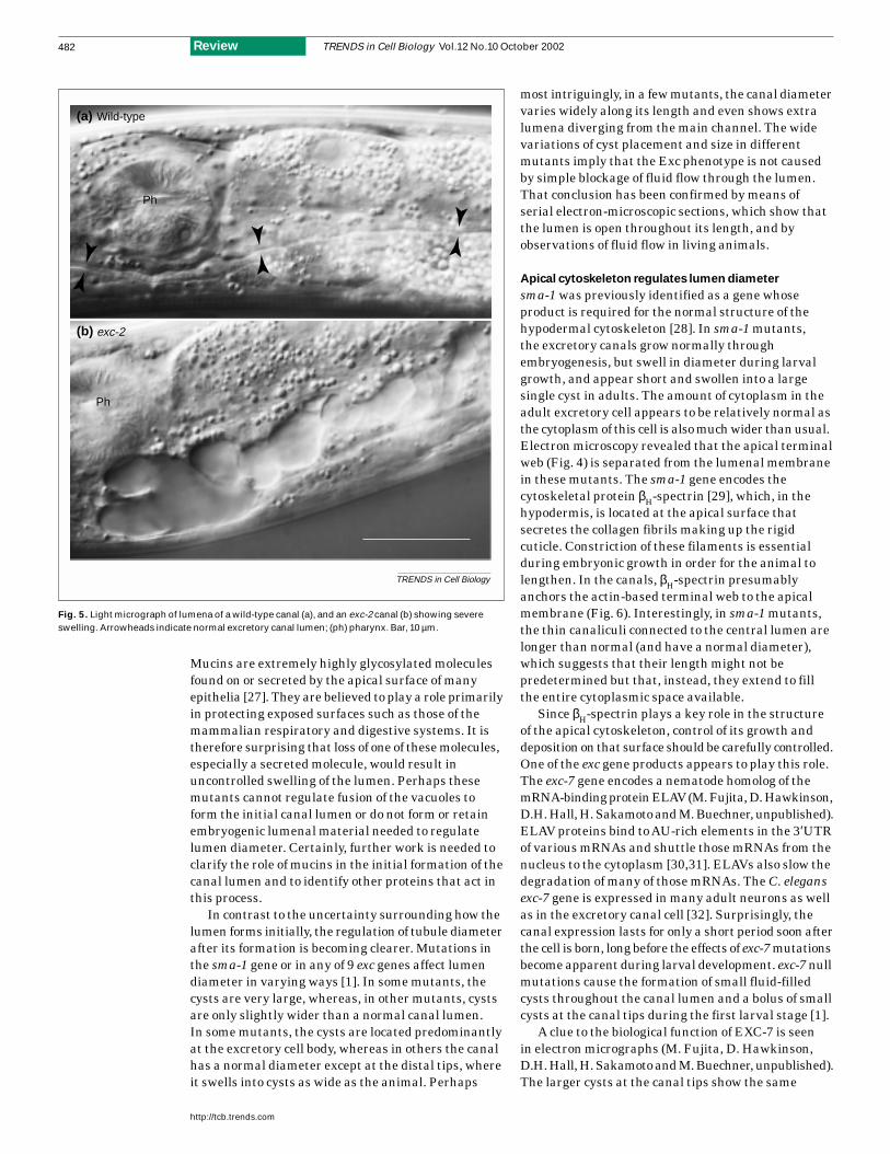
Mucins are extremely highly glycosylated moleculesfound on or secreted by the apical surface of manyepithelia [27]. They are believed to play a role primarilyin protecting exposed surfaces such as those of themammalian respiratory and digestive systems. It istherefore surprising that loss of one of these molecules,especially a secreted molecule, would result inuncontrolled swelling of the lumen. Perhaps thesemutants cannot regulate fusion of the vacuoles toform the initial canal lumen or do not form or retainembryogenic lumenal material needed to regulatelumen diameter. Certainly, further work is needed toclarify the role of mucins in the initial formation of thecanal lumen and to identify other proteins that act inthis process.
In contrast to the uncertainty surrounding how thelumen forms initially, the regulation of tubule diameterafter its formation is becoming clearer. Mutations inthe sma-1 gene or in any of 9 exc genes affect lumendiameter in varying ways [1]. In some mutants, thecysts are very large, whereas, in other mutants, cystsare only slightly wider than a normal canal lumen.In some mutants, the cysts are located predominantlyat the excretory cell body, whereas in others the canalhas a normal diameter except at the distal tips, whereit swells into cysts as wide as the animal. Perhaps
most intriguingly, in a few mutants, the canal diametervaries widely along its length and even shows extralumena diverging from the main channel. The widevariations of cyst placement and size in differentmutants imply that the Exc phenotype is not causedby simple blockage of fluid flow through the lumen.That conclusion has been confirmed by means ofserial electron-microscopic sections, which show thatthe lumen is open throughout its length, and byobservations of fluid flow in living animals.
Apical cytoskeleton regulates lumen diameter
sma-1 was previously identified as a gene whoseproduct is required for the normal structure of thehypodermal cytoskeleton [28]. In sma-1 mutants,the excretory canals grow normally throughembryogenesis, but swell in diameter during larvalgrowth, and appear short and swollen into a largesingle cyst in adults. The amount of cytoplasm in theadult excretory cell appears to be relatively normal asthe cytoplasm of this cell is also much wider than usual.Electron microscopy revealed that the apical terminalweb (Fig. 4) is separated from the lumenal membranein these mutants. The sma-1 gene encodes thecytoskeletal protein βH-spectrin [29], which, in thehypodermis, is located at the apical surface thatsecretes the collagen fibrils making up the rigidcuticle. Constriction of these filaments is essentialduring embryonic growth in order for the animal tolengthen. In the canals, βH-spectrin presumablyanchors the actin-based terminal web to the apicalmembrane (Fig. 6). Interestingly, in sma-1 mutants,the thin canaliculi connected to the central lumen arelonger than normal (and have a normal diameter),which suggests that their length might not bepredetermined but that, instead, they extend to fillthe entire cytoplasmic space available.
Since βH-spectrin plays a key role in the structureof the apical cytoskeleton, control of its growth anddeposition on that surface should be carefully controlled.One of the exc gene products appears to play this role.The exc-7 gene encodes a nematode homolog of themRNA-binding protein ELAV (M. Fujita, D.Hawkinson,D.H. Hall, H. Sakamoto and M. Buechner, unpublished).ELAV proteins bind to AU-rich elements in the 3′UTRof various mRNAs and shuttle those mRNAs from thenucleus to the cytoplasm [30,31]. ELAVs also slow thedegradation of many of those mRNAs. The C. elegansexc-7 gene is expressed in many adult neurons as wellas in the excretory canal cell [32]. Surprisingly, thecanal expression lasts for only a short period soon afterthe cell is born, long before the effects of exc-7 mutationsbecome apparent during larval development. exc-7 nullmutations cause the formation of small fluid-filledcysts throughout the canal lumen and a bolus of smallcysts at the canal tips during the first larval stage [1].
A clue to the biological function of EXC-7 is seen in electron micrographs (M. Fujita, D. Hawkinson,D.H. Hall, H. Sakamoto and M. Buechner, unpublished).The larger cysts at the canal tips show the same
TRENDS in Cell Biology Vol.12 No.10 October 2002
http://tcb.trends.com
482 Review
TRENDS in Cell Biology
exc-2
(a)
(b)
Wild-type
Ph
Ph
Fig. 5. Light micrograph of lumena of a wild-type canal (a), and an exc-2 canal (b) showing severeswelling. Arrowheads indicate normal excretory canal lumen; (ph) pharynx. Bar, 10 µm.

separation of apical membrane from terminal web asoccurs in sma-1 mutants. In addition, exc-7 mutantsshow synergistic effects with the sma-1 mutations: indoubly mutant exc-7; sma-1 animals, the canals aremuch more drastically affected than in either one ofthese mutants alone, to the point that the lumen swellsinto one or two extremely large cysts at the excretorycell body, similar to the canals seen in let-4 and let-653mutants. EXC-7 binds to the 3′UTR of sma-1 mRNA.Based on these observations, we have proposed thatEXC-7 regulates translation of sma-1 mRNA toensure that there is enough βH-spectrin during thespectacular increase in canal growth that occursduring the first larval stage (Fig. 6). exc-7 also showssimilar synergistic effects with the exc-3 gene (not yetcloned) [1], which suggests that exc-3 might encode aprotein that also interacts with βH-spectrin.
‘Form ever follows function’
The physiology of the excretory canal might also playa direct role in the regulation of tubule diameterbecause the lumen narrows towards the canal tips,where there should be less fluid flowing. One potentialcandidate for this measurement role is the EXC-4protein, which has recently been shown to encode ananion channel (O. Hobert, pers. commun.). This channelmight be linked directly to the apical cytoskeleton andmight allow the passage of counter-ions to balance the protons secreted by the many highly expressedvacuolar ATPases such as VHA-1 that coat thecanalicular surfaces [33] (Fig. 6). The increased saltsecretion to the lumen would pull excess water out ofthe animal into the canal lumen to be secreted. Whenexc-4 is mutated, the canals swell into a few extremelylarge vacuoles near the cell body. The same phenotypeis seen in mutants of the exc-2 gene (not cloned yet),which might suggest that these two gene productsfunction together.
Balancing it all
Finally, coordination of the growth of the apical andbasal sides of the canals is essential. EXC-5 balancesthe growth of cytoskeleton on these two surfaces ofthe canals [34]. In exc-5 mutants, the apical surfaceswells into extremely large cysts (again, visible bylow-power dissecting microscopy as refractile ‘bubbles’in the center of the animal). These cysts, however, arelocated at the tips of the canals rather than at theexcretory cell body, so the bridge of the ‘H cell’ andlumen near that point retain normal diameter. Thebasal surface of the canals in exc-5 mutants appearsnormal, and the canals extend normally until apicalswelling prevents further growth. In animals wherethe exc-5 gene is overexpressed, the opposite phenotypeis seen: the canal lumen has a normal diameter andlength, but the basal surface fails to extend, which is the same canal phenotype seen in basementmembrane mutants. The result is an extremely largecell body, filled with folded up, normal-diameter lumen.exc-5 encodes a GDP–GTP exchange factor homologousto the human FGD1 (Faciogenital dysplasia,Aarskog–Scott syndrome) gene product [34,35], whichactivates the small rho GTPase CDC-42 [36]. CDC-42is known to regulate cytoskeletal polarity in the one-cellembryo of C. elegans [37,38], as well as polarity inorganisms ranging from yeast to human [39,40].exc-5::gfp is expressed at highest levels along the entirecanal apical surface. The expression pattern, togetherwith the under- and overexpression phenotypes,suggests that EXC-5 and CDC-42 regulate the ratio ofapical to basal growth. High EXC-5 activity activatesCDC-42 and stimulates apical construction whileinhibiting basal construction, and low EXC-5 activityhas the opposite effect (Fig. 6). Interestingly, inmammals, CDC42 stimulation of JNK activity isimplicated as a downstream intermediary of signalingfrom the polycystins, cation channels that regulatenephron diameter [41]. Nephrons missing polycystinfunction develop large fluid-filled cysts similar inappearance to the nematode EXC mutations (see theforthcoming article by G. Germino in this series). It is tempting to speculate whether EXC-4 or otheryet-to-be discovered apical ion channels similarlyregulate EXC-5 function. The identification of othermembers of this signal-transduction pathway will beinstrumental in understanding both how EXC-5 effectsapical construction and how it measures the amountof apical and basal surface already synthesized. Ofnote, exc-1 mutants exhibit the same type of cystformation, and this gene is therefore another potentialparticipant in this pathway.
Concluding remarks
An intriguing picture of organ formation is emergingfrom the study of this appealing model tubule (Fig. 6).Physiology, cell polarity, rate of protein synthesis andcell and animal growth are all balanced to allow thesimultaneous formation and extension of a functionaltubule containing a complex lumenal surface.
TRENDS in Cell Biology Vol.12 No.10 October 2002
http://tcb.trends.com
483Review
TRENDS in Cell Biology
Nucleus
Apical surface
EXC-7ELAV
sma-1mRNA
3′UTR
Terminal web
Basalsurface
Basal lamina/extracellular matrix
βH-SpectrinEXC-5GEF
EXC-1?
CDC-42P
VHA-1H Pump
EXC-4Cl Channel
EXC-2?
Fig. 6. Model of excretory cell development. Apical-specific mRNAs forgenes such as sma-1 (encoding βH-spectrin) are regulated via the EXC-7protein (homolog of the mRNA-binding protein ELAV) and guided tothe apical surface by the putative EXC-1–EXC-5–CDC-42 pathway, whichalso inhibits development of the basal cytoskeleton. The chloridechannel EXC-4 and its putative partners EXC-2 and the VHA vacuolarproton pumps affect lumenal diameter through regulation of ion flux from the cytoplasm to the lumen of the canals. Abbreviations: GEF, GDP–GTP exchange factor; 3′UTR, 3′ untranslated region.
Acknowledgements
I thank Judith Austin andHiroshi Sakamoto forhelpful discussions,Oliver Hobert fordisclosing unpublishedresults, Victoria Corbinand Erik Lundquist forvery helpful comments onthe manuscript, Dave Hallfor longtime collaborationon canal ultrastructure,and especiallyEd Hedgecock for pastguidance and fororiginally suggesting theuse of the excretorycanals as a model forneural guidance.Our work is supported by NIDDK 55526, byNIDDK 57301, the KansasInterdisciplinary Centerfor Polycystic KidneyDisease and by thePolycystic Kidney DiseaseFoundation.

Basal extension requires the expression of receptorsto basement membrane components, while lumenalsynthesis depends on synthesis of apical cytoskeletalcomponents. The two processes appear to be yoked via an EXC-5–CDC-42 signal-transduction cascade toprevent either process from proceeding without theother. It is intriguing to consider that the samemechanisms regulating tubule diameter have
conceivably been preserved from the earliestmetazoans to vertebrates. The cloning of other known exc genes will provide more pieces to thispuzzle, and the use of genetics to identify moredetailed links between all these processes will provide a better foundation for understanding themorphogenesis of this complicated cell in this simple worm.
TRENDS in Cell Biology Vol.12 No.10 October 2002
http://tcb.trends.com
484 Review
References
1 Buechner, M. et al. (1999) Cystic canal mutants inCaenorhabditis elegans are defective in the apicalmembrane domain of the renal (excretory) cell.Dev. Biol. 214, 227–241
2 Nelson, F.K. and Riddle, D.L. (1984) Functionalstudy of the Caenorhabditis eleganssecretory-excretory system using lasermicrosurgery. J. Exp. Zool. 231, 45–56
3 Sulston, J.E. et al. (1983) The embryonic celllineage of the nematode Caenorhabditis elegans.Dev. Biol. 100, 64–119
4 Bürglin, T.R. and Ruvkun, G. (2001) Regulation ofectodermal and excretory function by theC. elegans POU homeobox gene ceh-6.Development 128, 779–790
5 Drubin, D.G. and Nelson, W.J. (1996) Origins ofcell polarity. Cell 84, 335–344
6 Hedgecock, E.M. et al. (1987) Genetics of cell andaxon migrations in Caenorhabditis elegans.Development 100, 365–382
7 Antebi, A. et al. (1997) Cell and Growth ConeMigrations. In C. elegans II (Riddle, D.L. et al., eds),pp. 583–609, Cold Spring Harbor Laboratory Press
8 Gettner, S.N. et al. (1995) Characterization ofβpat-3 heterodimers, a family of essential integrinreceptors in C. elegans. J. Cell Biol. 129, 1127–1141
9 Zhu, X. et al. (1999) Identification of epi-1 locus asa laminin alpha chain gene in the nematodeCaenorhabditis elegans and characterization ofepi-1 mutant alleles. DNA Seq. 10, 207–217
10 Hutter, H. et al. (2000) Conservation and noveltyin the evolution of cell adhesion and extracellularmatrix genes. Science 287, 989–994
11 Rogalski, T.M. et al. (2001) UNC-52/perlecanisoform diversity and function in Caenorhabditiselegans. Biochem. Soc. Trans. 29, 171–176
12 Baum, P.D. and Garriga, G. (1997) Neuronalmigrations and axon fasciculation are disruptedin ina-1 integrin mutants. Neuron 19, 51–62
13 Poinat, P. et al. (2002) A conserved interactionbetween β1 integrin/PAT-3 and Nck-interactingkinase/MIG-15 that mediates commissural axonnavigation in C. elegans. Curr. Biol. 12, 622–631
14 Stringham, E. et al. (2002) unc-53 controlslongitudinal migration in C. elegans. Development129, 3367–3379
15 Steven, R. et al. (1998) UNC-73 activates the RacGTPase and is required for cell and growth conemigrations in C. elegans. Cell 92, 785–795
16 Manser, J. et al. (1997) C. elegans cell migrationgene mig-10 shares similarities with a family ofSH2 domain proteins and acts cellnonautonomously in excretory canaldevelopment. Dev. Biol. 184, 150–164
17 Forrester, W.C. and Garriga, G. (1997) Genesnecessary for C. elegans cell and growth conemigrations. Development 124, 1831–1843
18 Otsuka, A.J. et al. (1991) The C. elegans unc-104gene encodes a putative kinesin heavy chain-likeprotein. Neuron 6, 113–122
19 Patel, N. et al. (1993) Cloning by insertionalmutagenesis of a cDNA encoding Caenorhabditiselegans kinesin heavy chain. Proc. Natl. Acad. Sci.U. S. A. 90, 9181–9185
20 Hedgecock, E.M. et al. (1990) The unc-5, unc-6,and unc-40 genes guide circumferentialmigrations of pioneer axons and mesodermal cellson the epidermis in C. elegans. Neuron 4, 61–85
21 Merz, D.C. et al. (2001) Multiple signalingmechanisms of the UNC-6/netrin receptorsUNC-5 and UNC-40/DCC in vivo. Genetics 158,1071–1080
22 Wang, Q. and Wadsworth, W.G. (2002) TheC domain of netrin UNC-6 silencescalcium/calmodulin-dependent protein kinase-and diacylglycerol-dependent axon branching inCaenorhabditis elegans. J. Neurosci. 22,2274–2282
23 Sawa, H. et al. (1996) The Caenorhabditis elegansgene lin-17, which is required for certainasymmetric cell divisions, encodes a putativeseven-transmembrane protein similar to theDrosophila frizzled protein. Genes Dev. 10,2189–2197
24 Brenner, S. (1974) The genetics of Caenorhabditiselegans. Genetics 77, 71–94
25 Park, E.C. and Horvitz, H.R. (1986) Mutationswith dominant effects on the behavior andmorphology of the nematode Caenorhabditiselegans. Genetics 113, 821–852
26 Jones, S.J. and Baillie, D.L. (1995)Characterization of the let-653 gene inCaenorhabditis elegans. Mol. Gen. Genet. 248,719–726
27 Dekker, J. et al. (2002) The MUC family: anobituary. Trends Biochem. Sci. 27, 126–131
28 Hosono, R. et al. (1982) Mutants of C. elegans withDumpy and Rounded head phenotype. J. Exp.Zool. 224, 135–144
29 McKeown, C. et al. (1998) sma-1 encodes aβH-spectrin homolog required for Caenorhabditiselegans morphogenesis. Development 125,2087–2098
30 Levine, T.D. et al. (1993) Hel-N1: an autoimmuneRNA-binding protein with specificity for3′ uridylate-rich untranslated regions of growthfactor mRNAs. Mol. Cell. Biol. 13, 3494–3504
31 Brennan, C.M. and Steitz, J.A. (2001) HuR andmRNA stability. Cell. Mol. Life Sci. 58, 266–277
32 Fujita, M. et al. (1999) Neuronal expression of aCaenorhabditis elegans elav-like gene and theeffect of its ectopic expression. Biochem. Biophys.Res. Commun. 260, 646–652
33 Oka, T. et al. (2001) Four subunit α isoforms ofCaenorhabditis elegans vacuolar H+-ATPase.Cell-specific expression during development.J. Biol. Chem. 276, 33079–33085
34 Suzuki, N. et al. (2001) A putative GDP-GTPexchange factor is required for development of theexcretory cell in Caenorhabditis elegans. EMBORep. 2, 530–535
35 Gao, J. et al. (2001) The Caenorhabditis eleganshomolog of FGD1, the human Cdc42 GEF generesponsible for faciogenital dysplasia, is criticalfor excretory cell morphogenesis. Hum. Mol.Genet. 10, 3049–3062
36 Zheng, Y. et al. (1996) The faciogenital dysplasiagene product FGD1 functions as aCdc42Hs-specific guanine-nucleotide exchangefactor. J. Biol. Chem. 271, 33169–33172
37 Gotta, M. et al. (2001) CDC-42 controls early cellpolarity and spindle orientation in C. elegans.Curr. Biol. 11, 482–488
38 Kay, A.J. and Hunter, C.P. (2001) CDC-42regulates PAR protein localization and function tocontrol cellular and embryonic polarity inC. elegans. Curr. Biol. 11, 474–481
39 Johnson, D.I. (1999) Cdc42: An essential Rho-typeGTPase controlling eukaryotic cell polarity.Microbiol. Mol. Biol. Rev. 63, 54–105
40 Pruyne, D. and Bretscher, A. (2000) Polarizationof cell growth in yeast. I. Establishment andmaintenance of polarity states. J. Cell Sci. 113,365–375
41 Arnould, T. et al. (1998) The polycystic kidneydisease 1 gene product mediates protein kinase Calpha-dependent and c-Jun N-terminalkinase-dependent activation of the transcriptionfactor AP-1. J. Biol. Chem. 273, 6013–6018
Do you have an alternative view or a particular opinion to air?
We welcome correspondence on views expressed in Trends in Cell Biology Opinion, Review and Research News articles.
Send your letter to the Editor at: