Embed Size (px)
Citation preview
PneumoniaRoya-Pabon and Perez-Velez Pneumonia (2016) 8:23 DOI 10.1186/s41479-016-0023-9
REVIEW Open Access
Tuberculosis exposure, infection anddisease in children: a systematic diagnosticapproach
Claudia L. Roya-Pabon1,2 and Carlos M. Perez-Velez2,3,4,5*Abstract
The accurate diagnosis of tuberculosis (TB) in children remains challenging. A myriad of common childhooddiseases can present with similar symptoms and signs, and differentiating between exposure and infection, as wellas infection and disease can be problematic. The paucibacillary nature of childhood TB complicates bacteriologicalconfirmation and specimen collection is difficult. In most instances intrathoracic TB remains a clinical diagnosis. TBinfection and disease represent a dynamic continuum from TB exposure with/without infection, to subclinical/incipient disease, to non-severe and severe disease. The clinical spectrum of intrathoracic TB in children is broad,and the classification of clinical, radiological, endoscopic, and laboratory findings into recognized clinical syndromesallows a more refined diagnostic approach in order to minimize both under- and over-diagnosis. Bacteriologicalconfirmation can be improved significantly by collecting multiple, high-quality specimens from the mostappropriate source. Mycobacterial testing should include traditional smear microscopy and culture, as well asnucleic acid amplification testing. A systematic approach to the child with recent exposure to TB, or with clinicaland radiological findings compatible with this diagnosis, should allow pragmatic classification as TB exposure,infection, or disease to facilitate timely and appropriate management. It is important to also assess risk factors for TBdisease progression and to undertake follow-up evaluations to monitor treatment response and ongoing evidencesupporting a TB, or alternative, diagnosis.
Keywords: Latent tuberculosis, Algorithm, Diagnostic techniques and procedures, Specimen handling, Risk factors
BackgroundDiagnosing tuberculosis (TB) in children is challenging(Table 1) [1] and often it is only considered after the childhas failed various therapeutic trials for other disorders.Even with intensive specimen collection and optimal mo-lecular and culture-based diagnostics, most children withnon-severe pulmonary TB are not confirmed bacterio-logically, despite having an exposure history, immune-based confirmation of infection and clinical featuresconsistent with this diagnosis [2]. Nonetheless, withcurrently available tools, it is possible to make an accurateclinical diagnosis of intrathoracic TB in most diseasedchildren. This review presents a systematic approach todiagnosing intrathoracic TB in children.
* Correspondence: [email protected] Tuberculosis Valle-Colorado (GTVC), Medellin, Antioquia, Colombia3Tuberculosis Clinic, Pima County Health Department, Tucson, AZ, USAFull list of author information is available at the end of the article
© The Author(s). 2016 Open Access This articInternational License (http://creativecommonsreproduction in any medium, provided you gthe Creative Commons license, and indicate if(http://creativecommons.org/publicdomain/ze
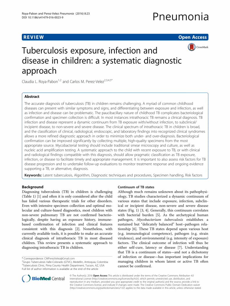
Continuum of TB statesAlthough much remains unknown about its pathophysi-ology, TB studies characterized a dynamic continuum ofvarious states that include exposure, infection, subclin-ical or incipient disease, non-severe and severe diseasestates (Fig. 1) [3, 4]. Generally, this continuum correlateswith bacterial burden [5]. As the archetypical humanpathogen, Mycobacterium tuberculosis establishes asustained but “delicately balanced” host–pathogen rela-tionship [6]. These TB states depend upon various host(e.g. immunological competence), pathogen (e.g. strainvirulence), and environmental (e.g. intensity of exposure)factors. The clinical outcome of infection will thus beeither self-cure, latency or disease [7]. Understandingthat TB is a continuum of states—and not a dichotomyof infection or disease—has important implications formanaging children in whom latent or active TB oftencannot be confirmed.
le is distributed under the terms of the Creative Commons Attribution 4.0.org/licenses/by/4.0/), which permits unrestricted use, distribution, andive appropriate credit to the original author(s) and the source, provide a link tochanges were made. The Creative Commons Public Domain Dedication waiverro/1.0/) applies to the data made available in this article, unless otherwise stated.
Table 1 Challenges in diagnosing TB exposure, infection and disease in children
Disease state Main challenges Current status & limitations Recent advances & future prospects
Infection Differentiating betweenTB exposure (withoutinfection), and TBinfection
Current immune-based tests (TST and IGRAs)may not convert to positive until 2–10 weeksafter acquiring M. tb infection
Mycobacteria-specific cytokine biomarkers –alone or in combination (i.e., biosignatures) –may distinguish between TB exposure(without infection), and TB infection [90]
Differentiating betweeninfection and subclinicaldisease
Chest radiography is the first-line imagingmodality, but may not reveal abnormalitiesconsistent with TB disease in all cases –especially those in early states of thecontinuum of TB
Chest CT, MRI, and PET [91] scan mayreveal findings consistent with TB diseasebefore symptoms develop
Disease Detection of TB diseaseand of drug resistance
Currently available immune-based tests (TSTand IGRAs) do not differentiate betweeninfection and disease
Mycobacteria-specific cytokine biomarkers – aloneor in combination – may distinguish between TBinfection and TB disease [90]
Currently available tests (e.g. NAATs; culture)for bacteriological confirmation have limitedsensitivity for detecting M. tb in young childrenwith paucibacillary disease–especially in earlystates of the continuum of TB
- Xpert MTB/RIF Ultra (Cepheid): next generation,ultrasensitive NAAT for detection of both M. tb &rifamycin resistance; in vitro study demonstratedsensitivity comparable to culture [92, 93].- GeneXpert Omni (Cepheid): single-cartridgebattery-operated platform that is portable/mobile;study pending [93]- Xpert XDR NAAT (Cepheid): study anticipatedin 2018 [93]
Specimen collection for bacteriologicalconfirmation currently consists of serial samplingof three gastric aspirates/lavages or induced sputaand requires trained personnel and facilities withairborne infection control
Strategies consisting of “intensive” collectionof combinations of various specimens(e.g., nasopharyngeal aspirates; string tests;stool; fine needle aspirate of diseasedlymph node) that have similar or superiorbacteriological yield, require less training,and may be carried out as an outpatientover 1–2 days
Monitoring responseto treatment
Mycobacterial culture is only useful in thosechildren who had positive cultures at time ofdiagnosis (minority of cases).
Cytokine biomarkers and biosignatures(possibly including IFN-γ, TNF-α, IL-2, IL-6,IL-10 and/or IL-12) [94, 95]
18 F-FDG PET/CT is sensitive for the detectionof TB disease (in different states of the continuum)and for monitoring response to treatment [91]
CT computed tomography, IGRA interferon-gamma release assay, MRI magnetic resonance imaging, M. tb: Mycobacterium tuberculosis, NAAT nucleic acidamplification test, PCR polymerase chain reaction, PET positron emission tomography, TB tuberculosis, TST tuberculin skin test, XDR extensively drug-resistant
Roya-Pabon and Perez-Velez Pneumonia (2016) 8:23 Page 2 of 18
Clinical spectrum of diseaseOnce infected with M. tuberculosis, young children(aged <5 years) are at greater risk than adults of pro-gressing to disease, including its most severe forms.This depends on the child’s susceptibility, which ishighest during the first years of life, probably from im-munological immaturity. Without Bacille Calmette-Guerin (BCG) vaccination, approximately 30% ofinfected infants (<1 year old) will progress to intratho-racic TB, and 10–20% will develop disseminated dis-ease. In children aged 1–2 years, the risk of progressingto intrathoracic TB is 10–20 and 2–5% for disseminateddisease. These risks decline slowly until around 10 yearsof age when adult-type disease starts to emerge [8, 9].Thus, early diagnosis is important, especially in infantsand young children who are at greatest risk of rapiddisease development [8] and clinicians should considerthe full clinical spectrum of intrathoracic syndromes[10].
Clinical classification of tuberculosisClassifying intrathoracic TB by immunopathogenesis(Table 2) assists understanding how each possible “state”in the continuum is managed [11]. For example, a childwith a history of TB exposure can have the features ofsubclinical disease [12] outlined in Table 2, which insome hierarchical diagnostic classification systems corre-sponds to “possible” intrathoracic TB. A typical exampleis that of a young child with isolated uncomplicated hilarlymphadenopathy [13]. Such a child may not meet suffi-cient criteria to be clinically diagnosed with “probable”intrathoracic TB given their lack of symptoms and phys-ical signs, [14] and consequently may not receive treat-ment for tuberculosis disease or infection. Whether thisintermediate state will progress to clinically manifestdisease or be contained as latent infection is dependenton the child’s level of immunocompetence. In those withrisk factors for progression to TB disease, treatment isrecommended. Children with disease can be further
Fig. 1 Continuum of TB states and correlations with bacterial load and with radiological and clinical manifestations. CFU: colony-forming units;LED: light-emitting diode; LOD: limit of detection; mL: milliliter; NAAT: nucleic acid amplification test; RT-PCR: real-time polymerase chain reaction.Adapted from C.M. Perez-Velez. Diagnosis of Intrathoracic Tuberculosis in Children. In: Handbook of Child and Adolescent Tuberculosis (p. 149),J.R. Starke and P.R. Donald (Eds.), 2016, New York, NY: Oxford University Press. Copyright by Oxford University Press [15]. Adapted with permission
Table 2 Clinical classification of intrathoracic TB based on immunopathogenesis
Clinicalclassification
Immunopathogenesis TST/IGRA Imaging Clinicalmanifestations
Myco-bacterialdetection
TB exposure Self-cure (infection eliminated byinnate immune response; noT-cell activation)
Negative Normal None Negative
Latent TB infection Quiescent infection (non-replicatingbacteria persisting with very lowmetabolic activity; infectionwell-contained)
Positive - Calcified non-enlargedregional lymph nodes
- Calcified lung nodules- Pleural thickening
None Negative
Subclinical TB Incipient disease (replicating bacteriathat are metabolically active;infection contained)
Usuallypositive
- Uncomplicated hilar/mediastinal lymphadenopathy
- Non-calcified lung nodules- Uncomplicated pleuraleffusion
None Usually negative(may be transientlypositive)
Non-severe TB Mild-to-moderate disease (replicatingbacteria that are metabolically active;infection only partially contained)
Usuallypositive
- Uncomplicated hilar/mediastinal lymphadenopathy
- Non-calcified lung nodules- Uncomplicated pleuraleffusion
Mild-to-moderate Positive cultures(10–30% of cases)
Severe TB Severe disease (replicating bacteriathat are metabolically active; infectionnot contained)
Usuallypositive
See spectrum ofdisease (Fig. 2)
Severe Positive cultures(30–70% of cases)
Adapted from C.M. Perez-Velez. Diagnosis of Intrathoracic Tuberculosis in Children. In: Handbook of Child and Adolescent Tuberculosis (p. 149), J.R. Starke and P.R.Donald (Eds.), 2016, New York, NY: Oxford University Press. Copyright by Oxford University Press [15]. Adapted with permissionIGRA Interferon-gamma release assay, PCR polymerase chain reaction, TB tuberculosis, TST tuberculin skin test
Roya-Pabon and Perez-Velez Pneumonia (2016) 8:23 Page 3 of 18
Roya-Pabon and Perez-Velez Pneumonia (2016) 8:23 Page 4 of 18
classified as severe or non-severe, depending on whetheror not infection is contained and on the presence andextent of complications.
Systematic diagnostic approachAs it is impossible to achieve bacteriological confirm-ation in many childhood TB cases, systematically identi-fying findings suggestive of TB can allow for its clinicaldiagnosis. Excluding other differential diagnoses andobserving a positive therapeutic response increases theprobability of TB being the correct diagnosis. Thefollowing systematic approach to diagnosing TB inchildren consists of (i) identifying findings suggesting TBdisease; (ii) identifying findings supportive of TB as theetiology; (iii) screening for risk factors for progression todisease; and (iv) follow-up evaluations to further supportor exclude TB as the etiology (Table 3) [15].
Table 3 Systematic approach to the diagnosis of intrathoracicTB in children
Step 1: Identify findings suggestive of TB disease
• Clinical evaluation: history & physical exam
• Radiological imaging: chest radiography; computed tomography;ultrasonography
• Laboratory studies: composite measures (cell count and chemistry)of body fluids (e.g., pleural fluid)
• Endoscopic studies: bronchoscopy
Step 2: Identifying findings supportive of TB as the etiology
• TB exposure history
• Immune-based tests: TST; IGRA
• Biochemical markers: ADA in body fluids (e.g., pleural fluid; pericardialfluid)
• Mycobacterial detection: smear microscopy; NAAT; culture; antigentest (in HIV-infected adolescents, lateral flow lipoarabinomannan inurine with CD4 < 100)
• Histopathological & cytopathological studies
• Excluding other differential diagnoses
Step 3: Screen for risk factors for progression to TB disease
• Age groups (e.g. immunological immaturity of infancy)
• Immunocompromising conditions (e.g., HIV infection)
• Immunosuppressive medications (e.g., TNF-α) antagonists
• Contained TB infection-disease (e.g., noncalcified fibronodular lesions,especially apical, on chest imaging
• Environment (e.g., continued exposure)
Step 4: Follow-up evaluation to support or exclude TB as the etiology
Adapted from C.M. Perez-Velez. Diagnosis of Intrathoracic Tuberculosis inChildren. In: Handbook of Child and Adolescent Tuberculosis (p. 149), J.R.Starke and P.R. Donald (Eds.), 2016, New York, NY: Oxford University Press.Copyright by Oxford University Press [15]. Adapted with permissionADA adenosine deaminase, CD4 cluster of differentiation 4, HIV humanimmunodeficiency virus, IGRA interferon gamma release assay, NAAT nucleicacid amplification test, TB tuberculosis, TNF-α tumor necrosis factor alpha, TSTtuberculin skin test
STEP 1: Identify findings suggestive of TB diseaseEach intrathoracic clinical syndrome of TB disease hasits own constellation of clinical, radiological, laboratory,and endoscopic (if indicated) findings, although manyare shared by more than one clinical syndrome. Further-more, miliary lung disease may also involve potentiallyany organ system (Additional file 1: Textbox 1). Mostclinical manifestations of intrathoracic TB result fromthe overall balance of beneficial and harmful immuneresponses to M. tuberculosis and a severe inflammatoryreaction can be triggered by a relatively low burden oforganisms. There are no clinical features pathognomonicof intrathoracic TB, but combinations of symptoms andphysical signs with certain temporal patterns can helpdifferentiate it from other etiologies that might mimicthis disorder.
Clinical evaluationPulmonary TB is frequently associated with intrathoraciclymphadenopathy, and sometimes with pleural or peri-cardial disease, and therefore “intrathoracic TB” is thepreferred term in children. Localized symptoms andphysical signs depend on which intrathoracic organs areinvolved, while non-localized symptoms and signs areindependent of the organ-specific clinical syndrome.Symptoms and physical signs that are well-defined havehigher specificity. However, in children who are im-munocompromised (e.g. less than three years of age withimmunological immaturity), HIV-infected, or severelymalnourished, these symptoms and signs have lowersensitivity and specificity [16].Systemic symptoms and signs may appear early or late
in the disease course [17]. Daily fever is characteristically>38.0 ° C, intermittent or persistent throughout the day,and usually lasts >1 week. Night sweats are uncommon,subjective and nonspecific, and are significant only whenthey drench the child’s clothes and bedding. Chills andrigors are rare, except in disseminated disease. Anorexiaand associated wasting or failure to thrive during thepast 3–6 months, or having lost >10% of body weightover any interval of time, are sensitive—albeit nonspeci-fic—signs in most TB clinical syndromes in young chil-dren [16]. The immunocompromised state from severeunder-nutrition can increase the risk for a paradoxicalreaction when they receive TB treatment and nutritionalrehabilitation [18]. Fatigue, asthenia, and malaise maymanifest in young children as listlessness (e.g. decreasedplayfulness) and in infants as apathy (e.g. less interactivewith caregivers) and should be persistent and not attrib-utable to other causes.Peripheral lymphadenopathy from TB typically con-
sists of a unilateral, enlarged, non-painful, rubberylymph node, sometimes becoming fluctuant, with or
Roya-Pabon and Perez-Velez Pneumonia (2016) 8:23 Page 5 of 18
without spontaneous drainage forming a sinus tract [19].Respiratory symptoms and signs depend on the site, anddegree of involvement (e.g. of airway obstruction). Thecough is usually unremitting for >2 weeks and may be“dry” or “wet”. When the airway is compressed by anenlarged lymph node, there may be persistent cough,wheezing or stridor that does not improve with inhaledbronchodilators (Additional file 1: Table S1). Characteriz-ing the temporal pattern (including the onset, progressionand duration) of symptoms helps clinicians to identifycases with likely intrathoracic TB.
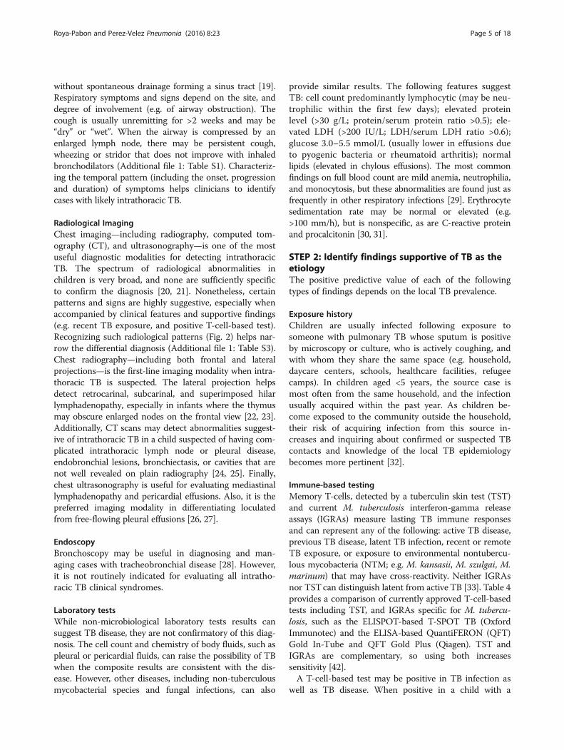
Radiological ImagingChest imaging—including radiography, computed tom-ography (CT), and ultrasonography—is one of the mostuseful diagnostic modalities for detecting intrathoracicTB. The spectrum of radiological abnormalities inchildren is very broad, and none are sufficiently specificto confirm the diagnosis [20, 21]. Nonetheless, certainpatterns and signs are highly suggestive, especially whenaccompanied by clinical features and supportive findings(e.g. recent TB exposure, and positive T-cell-based test).Recognizing such radiological patterns (Fig. 2) helps nar-row the differential diagnosis (Additional file 1: Table S3).Chest radiography—including both frontal and lateralprojections—is the first-line imaging modality when intra-thoracic TB is suspected. The lateral projection helpsdetect retrocarinal, subcarinal, and superimposed hilarlymphadenopathy, especially in infants where the thymusmay obscure enlarged nodes on the frontal view [22, 23].Additionally, CT scans may detect abnormalities suggest-ive of intrathoracic TB in a child suspected of having com-plicated intrathoracic lymph node or pleural disease,endobronchial lesions, bronchiectasis, or cavities that arenot well revealed on plain radiography [24, 25]. Finally,chest ultrasonography is useful for evaluating mediastinallymphadenopathy and pericardial effusions. Also, it is thepreferred imaging modality in differentiating loculatedfrom free-flowing pleural effusions [26, 27].
EndoscopyBronchoscopy may be useful in diagnosing and man-aging cases with tracheobronchial disease [28]. However,it is not routinely indicated for evaluating all intratho-racic TB clinical syndromes.
Laboratory testsWhile non-microbiological laboratory tests results cansuggest TB disease, they are not confirmatory of this diag-nosis. The cell count and chemistry of body fluids, such aspleural or pericardial fluids, can raise the possibility of TBwhen the composite results are consistent with the dis-ease. However, other diseases, including non-tuberculousmycobacterial species and fungal infections, can also
provide similar results. The following features suggestTB: cell count predominantly lymphocytic (may be neu-trophilic within the first few days); elevated proteinlevel (>30 g/L; protein/serum protein ratio >0.5); ele-vated LDH (>200 IU/L; LDH/serum LDH ratio >0.6);glucose 3.0–5.5 mmol/L (usually lower in effusions dueto pyogenic bacteria or rheumatoid arthritis); normallipids (elevated in chylous effusions). The most commonfindings on full blood count are mild anemia, neutrophilia,and monocytosis, but these abnormalities are found just asfrequently in other respiratory infections [29]. Erythrocytesedimentation rate may be normal or elevated (e.g.>100 mm/h), but is nonspecific, as are C-reactive proteinand procalcitonin [30, 31].
STEP 2: Identify findings supportive of TB as theetiologyThe positive predictive value of each of the followingtypes of findings depends on the local TB prevalence.
Exposure historyChildren are usually infected following exposure tosomeone with pulmonary TB whose sputum is positiveby microscopy or culture, who is actively coughing, andwith whom they share the same space (e.g. household,daycare centers, schools, healthcare facilities, refugeecamps). In children aged <5 years, the source case ismost often from the same household, and the infectionusually acquired within the past year. As children be-come exposed to the community outside the household,their risk of acquiring infection from this source in-creases and inquiring about confirmed or suspected TBcontacts and knowledge of the local TB epidemiologybecomes more pertinent [32].
Immune-based testingMemory T-cells, detected by a tuberculin skin test (TST)and current M. tuberculosis interferon-gamma releaseassays (IGRAs) measure lasting TB immune responsesand can represent any of the following: active TB disease,previous TB disease, latent TB infection, recent or remoteTB exposure, or exposure to environmental nontubercu-lous mycobacteria (NTM; e.g. M. kansasii, M. szulgai, M.marinum) that may have cross-reactivity. Neither IGRAsnor TSTcan distinguish latent from active TB [33]. Table 4provides a comparison of currently approved T-cell-basedtests including TST, and IGRAs specific for M. tubercu-losis, such as the ELISPOT-based T-SPOT TB (OxfordImmunotec) and the ELISA-based QuantiFERON (QFT)Gold In-Tube and QFT Gold Plus (Qiagen). TST andIGRAs are complementary, so using both increasessensitivity [42].A T-cell-based test may be positive in TB infection as
well as TB disease. When positive in a child with a
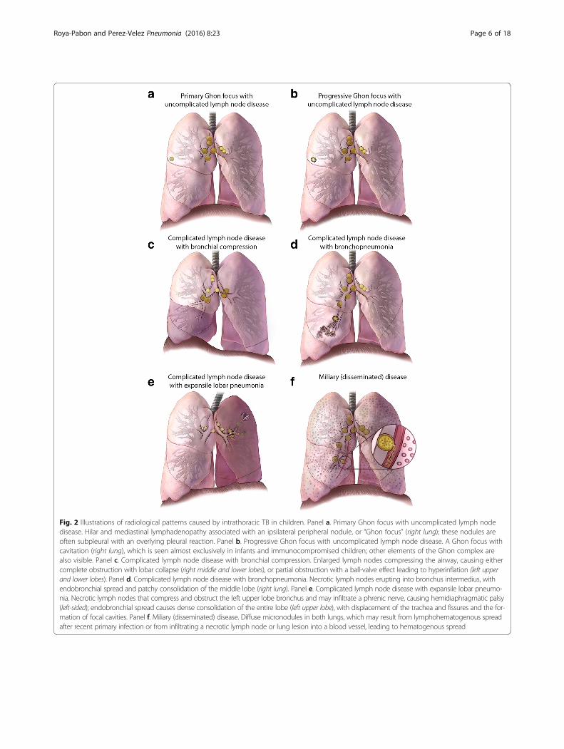
Fig. 2 Illustrations of radiological patterns caused by intrathoracic TB in children. Panel a. Primary Ghon focus with uncomplicated lymph nodedisease. Hilar and mediastinal lymphadenopathy associated with an ipsilateral peripheral nodule, or “Ghon focus” (right lung); these nodules areoften subpleural with an overlying pleural reaction. Panel b. Progressive Ghon focus with uncomplicated lymph node disease. A Ghon focus withcavitation (right lung), which is seen almost exclusively in infants and immunocompromised children; other elements of the Ghon complex arealso visible. Panel c. Complicated lymph node disease with bronchial compression. Enlarged lymph nodes compressing the airway, causing eithercomplete obstruction with lobar collapse (right middle and lower lobes), or partial obstruction with a ball-valve effect leading to hyperinflation (left upperand lower lobes). Panel d. Complicated lymph node disease with bronchopneumonia. Necrotic lymph nodes erupting into bronchus intermedius, withendobronchial spread and patchy consolidation of the middle lobe (right lung). Panel e. Complicated lymph node disease with expansile lobar pneumo-nia. Necrotic lymph nodes that compress and obstruct the left upper lobe bronchus and may infiltrate a phrenic nerve, causing hemidiaphragmatic palsy(left-sided); endobronchial spread causes dense consolidation of the entire lobe (left upper lobe), with displacement of the trachea and fissures and the for-mation of focal cavities. Panel f. Miliary (disseminated) disease. Diffuse micronodules in both lungs, which may result from lymphohematogenous spreadafter recent primary infection or from infiltrating a necrotic lymph node or lung lesion into a blood vessel, leading to hematogenous spread
Roya-Pabon and Perez-Velez Pneumonia (2016) 8:23 Page 6 of 18
Fig. 2 (continued) Panel g. Multiple focal pulmonary nodules. Multiple focal pulmonary nodules involving the right middle lobe withenlargement of regional lymph nodes (right lung). Panel h. Cavitary (“adult-type”) pulmonary disease. Cavity formation in both upper lobes, withendobronchial spread to the right middle lobe. Nodules or cavities in apical lung segments are typical of adult-type disease and are pathologicallydistinct from the other cavities shown. Panel i. Bronchitis and endobronchial granulomas. Inflammation of the mucosa of main stem bronchuswith purulent secretions (left lung), and a necrotic lymph node that has eroded into the right middle lobe bronchus leading to endobronchialspread and subsequent development of endobronchial granulomas extending proximally to the bronchus intermedius and main stem bronchus,and distally to the lower lobe bronchus (right lung). These findings are best visualized by bronchoscopy. Panel j. Bronchiectasis and tree-in-bud-pattern.Bronchiectasis that extensively involves the upper lobe (right lung), and shows tree-in-bud pattern observable on CT scans – reflecting dilated centrilobularbronchioles with mucoid impaction – involving the upper lobe (left lung). Panel k. Pleural effusion. A pleural effusion that is usuallyindicative of recent primary infection, with a hypersensitivity response to tuberculoprotein leaking from a subpleural Ghon focus (oftennot visible) into the pleural cavity; in rare cases this effusion may also result from a chylothorax. Panel l. Pericardial effusion. A pericardialeffusion that occurs when tuberculoprotein leaks from a necrotic subcarinal lymph node (shown in “close-up” window) into the pericardialspace; it may also occur after hematogenous spread. Conceptualization and original sketches by C.L. Roya-Pabon, MD; finished artwork byMesa Schumacher, MA (used with permission). Adapted from C.M. Perez-Velez. Diagnosis of Intrathoracic Tuberculosis in Children. In: Handbook ofChild and Adolescent Tuberculosis (p. 154–155), J.R. Starke and P.R. Donald (Eds.), 2016, New York, NY: Oxford University Press. Copyright by OxfordUniversity Press [15]. Adapted with permission
Roya-Pabon and Perez-Velez Pneumonia (2016) 8:23 Page 7 of 18
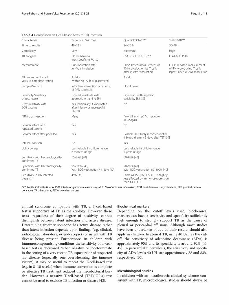
Table 4 Comparison of T cell-based tests for TB infection
Characteristic Tuberculin Skin Test QuantiFERON-TB®* T-SPOT-TB®**
Time to results 48–72 h 24–36 h 36–48 h
Complexity Low Moderate High
TB antigens PPD-tuberculin(not specific to M. tb.)
ESAT-6; CFP-10; TB-7.7 ESAT-6; CFP-10
Measurement Skin induration afterin vivo stimulation
ELISA-based measurement ofIFN-γ production by T-cellsafter in vitro stimulation
ELISPOT-based measurementof IFN-γ-producing T-cells(spots) after in vitro stimulation
Minimum number ofvisits to complete testing
2 visits(within 48–72 h of placement)
1 visit
Sample/Method Intradermal injection of 5 unitsof PPD-tuberculin
Blood draw
Reliability/Variabilityof test results
Limited variability withappropriate training [34]
Significant within-personvariability [35, 36]
Cross-reactivity withBCG vaccine
Yes (particularly if vaccinatedafter infancy or repeatedly)[37, 38]
No
NTM cross reaction Many Few (M. kansasii, M. marinum,M. szulgaii)
Booster effect withrepeated testing
Yes No
Booster effect after prior TST Yes Possible (but likely inconsequentialif blood drawn < 3 days after TST [39]
Internal controls No Yes
Utility by age Less reliable in children under6-months of age
Less reliable in children under5-years of age
Sensitivity with bacteriologically-confirmed TB
75–85% [40] 80–85% [40]
Specificity with bacteriologically-confirmed TB
95–100% [40]With BCG vaccination 49–65% [40]
90–95% [40]With BCG vaccination 89–100% [40]
Sensitivity in HIV-infectedpatients
45% [36] Same as TST [36]; T-SPOT.TB slightlyless affected by immunosuppressionthan QFT [41]
BCG bacille Calmette-Guérin, IGRA interferon-gamma release assay, M. tb Mycobacterium tuberculosis, NTM nontuberculous mycobacteria, PPD purified proteinderivative, TB tuberculosis, TST tuberculin skin test
Roya-Pabon and Perez-Velez Pneumonia (2016) 8:23 Page 8 of 18
clinical syndrome compatible with TB, a T-cell-basedtest is supportive of TB as the etiology. However, thesetests—regardless of their degree of positivity—cannotdistinguish between latent infection and active disease.Determining whether someone has active disease ratherthan latent infection depends upon findings (e.g. clinical,radiological, laboratory, or endoscopic) consistent with TBdisease being present. Furthermore, in children withimmunocompromising conditions the sensitivity of T-cell-based tests is decreased. When negative or indeterminatein the setting of a very recent TB exposure or of suspectedTB disease (especially one overwhelming the immunesystem), it may be useful to repeat the T-cell-based test(e.g. in 8–10 weeks) when immune conversion is completeor effective TB treatment reduced the mycobacterial bur-den. However, a negative T-cell-based (TST/IGRA) testcannot be used to exclude TB infection or disease [43].
Biochemical markersDepending on the cutoff levels used, biochemicalmarkers can have a sensitivity and specificity sufficientlyhigh enough to strongly support TB as the cause ofpleural or pericardial effusions. Although most studieshave been undertaken in adults, their results should alsoapply in children. In pleural TB, using 40 U/L as the cut-off, the sensitivity of adenosine deaminase (ADA) isapproximately 90% and its specificity is around 92% [44,45]. In pericardial tuberculosis, the sensitivity and specifi-city of ADA levels 40 U/L are approximately 88 and 83%,respectively [30].
Microbiological studiesIn children with an intrathoracic clinical syndrome con-sistent with TB, microbiological studies should always be
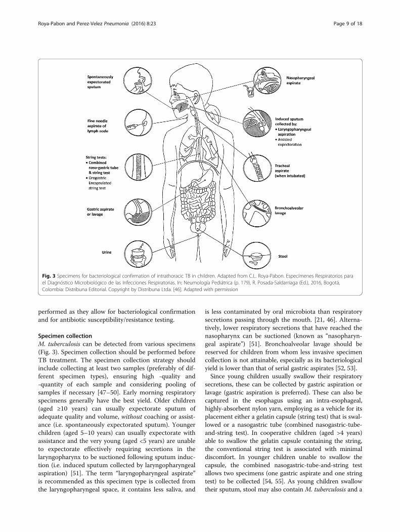
Fig. 3 Specimens for bacteriological confirmation of intrathoracic TB in children. Adapted from C.L. Roya-Pabon. Especímenes Respiratorios parael Diagnóstico Microbiológico de las Infecciones Respiratorias. In: Neumología Pediátrica (p. 179), R. Posada-Saldarriaga (Ed.), 2016, Bogotá,Colombia: Distribuna Editorial. Copyright by Distribuna Ltda. [46]. Adapted with permission
Roya-Pabon and Perez-Velez Pneumonia (2016) 8:23 Page 9 of 18
performed as they allow for bacteriological confirmationand for antibiotic susceptibility/resistance testing.
Specimen collectionM. tuberculosis can be detected from various specimens(Fig. 3). Specimen collection should be performed beforeTB treatment. The specimen collection strategy shouldinclude collecting at least two samples (preferably of dif-ferent specimen types), ensuring high -quality and-quantity of each sample and considering pooling ofsamples if necessary [47–50]. Early morning respiratoryspecimens generally have the best yield. Older children(aged ≥10 years) can usually expectorate sputum ofadequate quality and volume, without coaching or assist-ance (i.e. spontaneously expectorated sputum). Youngerchildren (aged 5–10 years) can usually expectorate withassistance and the very young (aged <5 years) are unableto expectorate effectively requiring secretions in thelaryngopharynx to be suctioned following sputum induc-tion (i.e. induced sputum collected by laryngopharyngealaspiration) [51]. The term “laryngopharyngeal aspirate”is recommended as this specimen type is collected fromthe laryngopharyngeal space, it contains less saliva, and
is less contaminated by oral microbiota than respiratorysecretions passing through the mouth. [21, 46]. Alterna-tively, lower respiratory secretions that have reached thenasopharynx can be suctioned (known as “nasopharyn-geal aspirate”) [51]. Bronchoalveolar lavage should bereserved for children from whom less invasive specimencollection is not attainable, especially as its bacteriologicalyield is lower than that of serial gastric aspirates [52, 53].Since young children usually swallow their respiratory
secretions, these can be collected by gastric aspiration orlavage (gastric aspiration is preferred). These can also becaptured in the esophagus using an intra-esophageal,highly-absorbent nylon yarn, employing as a vehicle for itsplacement either a gelatin capsule (string test) that is swal-lowed or a nasogastric tube (combined nasogastric-tube-and-string test). In cooperative children (aged >4 years)able to swallow the gelatin capsule containing the string,the conventional string test is associated with minimaldiscomfort. In younger children unable to swallow thecapsule, the combined nasogastric-tube-and-string testallows two specimens (one gastric aspirate and one stringtest) to be collected [54, 55]. As young children swallowtheir sputum, stool may also contain M. tuberculosis and a
Roya-Pabon and Perez-Velez Pneumonia (2016) 8:23 Page 10 of 18
nucleic acid amplification test (NAAT), such as XpertMTB/RIF (Cepheid, United States of America), on stoolcan bacteriologically confirm approximately 45% of clinic-ally diagnosed cases of pulmonary TB [56, 57].In children with enlarged peripheral lymph nodes
(usually cervical), a fine needle aspiration biopsy is thespecimen of choice, and should be submitted for: (i)mycobacterial testing, i.e. NAAT (Xpert MTB/RIF has asensitivity of ~83% using culture as reference) andculture; and (ii) pathological studies (cytopathology ofaspirate; histopathology of biopsied tissue) [58, 59].Serosal fluids (e.g. pleural and pericardial) should becollected and submitted for biochemical markers, myco-bacterial testing, and cytopathological studies. The diag-nostic yield of serosal fluids increases as more types oftests performed. Serosal tissue generally has a higher cul-ture yield and so biopsy (e.g. of the pleura or pericardium)may be justified, especially when drug-resistant TB is sus-pected (allowing susceptibility testing to be undertaken).
Mycobacterial detectionAcid-fast staining and smear microscopyAcid-fast staining and smear microscopy is a rapid andrelatively inexpensive test for detecting acid-fast bacilli(AFB). Unfortunately, the sensitivity of smear micros-copy varies greatly based on AFB load. For reliabledetection, a sample must contain AFB of at least 1000–10,000 colony-forming units (CFU)/mL [60]. This rela-tively high detection limit, together with the paucibacil-lary nature of TB disease in children, contributes to thevery low sensitivity of smear microscopy.Acid-fast stains are also not specific for M. tuberculosis
complex as they cannot differentiate between mycobacter-ial species. Nonetheless, in a child with a high pre-testprobability of having pulmonary TB, a positive result has ahigh predictive value, and studies using culture as areference standard report a very high specificity (~95%)[61–63]. Microscopy’s low sensitivity and inability to differ-entiate between AFB species (especially relevant for gastricaspirate specimens), means it should not be used as a solemycobacterial test for detecting M. tuberculosis.
Nucleic acid amplification tests or antigen detectionNAATs are rapid tests that include real-time polymerasechain reaction (RT-PCR) and line probe assays (LPAs)(Additional file 1: Table S2). Recently developed NAATscan also simultaneously detect genes conferring drug resist-ance, allowing prompt and more appropriate treatment ofcases with drug-resistant disease. The fully automatedXpert MTB/RIF test has high sensitivity (pooled estimate95–96%) in sputum smear-positive samples using cultureas a reference standard, but only moderate sensitivity(pooled estimate 55–62%) in smear-negative samples [64].In 2013, the World Health Organization recommended
using Xpert MTB/RIF in samples from children, especiallythose suspected of multidrug-resistant TB or HIV co-infection [64]. Certain LPAs detect M. tuberculosis with/without drug resistance mutations, as well as commonNTM, such as M. avium, M. intracellulare, and M.kansasii. GenoType MTBDRplus® (Hain Lifescience,Holland) or Genoscholar NTM+MDRTB® (Nipro Europe,Germany) are especially useful for simultaneously detectingisoniazid- and rifampin-resistance in microscopy-positivesamples or culture isolates [65–67]. Regarding antigen de-tection tests the urine lateral flow lipoarabinomannan (LF-LAM) assay may be useful in adolescents with advancedHIV disease and CD4 counts <100 cells/L [68–70]; how-ever, in young children it has poor diagnostic accuracy [71].
Mycobacterial cultureMycobacterial cultures have the highest sensitivity andspecificity for bacteriological confirmation of intratho-racic TB in children. The limits of detection of liquidand solid media are approximately 10–100 CFU/mL and50–150 CFU/mL, respectively (versus 100–150 CFU/mLfor RT-PCR or 1000–10,000 CFU/mL for fluorescentLED microscopy) [72]. In most prospective studies ofchildren with a clinical diagnosis of probable pulmonaryTB, cultures of respiratory specimens are positive in 10–20% of cases. Studies reporting higher rates (i.e. >30%)of culture confirmation are often retrospective andinclude only children who are hospitalized (probablyhave more severe disease and better specimen collectionstrategies) or diagnosed following passive case finding[73]. For definitive species identification followinggrowth in mycobacterial culture, the following methodsmay be utilized: (i) phenotypic analysis; (ii) antigen tests;(iii) molecular tests such as nucleic acid hybridizationprobes, matrix-assisted laser desorption/ionization time-of-flight mass spectrometry, and DNA sequencing.
HistopathologyHistopathological studies should be considered in intratho-racic clinical syndromes compatible with either TB diseaseor malignancy, especially when bacteriological tests fail toconfirm an infectious etiology. Potentially useful tissues tobiopsy include lymph nodes, pleura, pericardium and lung.Findings suggestive of TB are numerous granulomas invarious developmental stages, some with central caseousnecrosis [74]. However, granulomatous inflammation is notsufficiently specific to diagnose TB and differential diagno-ses include bacterial (e.g. NTM, nocardiosis), fungal (e.g.histoplasmosis, coccidioidomycosis), helminthic (e.g. schis-tosomiasis) and, protozoal (e.g. toxoplasmosis) infections,autoimmune diseases (e.g. granulomatosis with polyangii-tis), idiopathic etiologies (e.g. sarcoidosis), and foreignbodies.
Roya-Pabon and Perez-Velez Pneumonia (2016) 8:23 Page 11 of 18
Excluding alternative diagnosesIn infants and children, the clinical diagnosis of intratho-racic TB is not always certain, as other disorders canpresent with similar clinical, radiological, and laboratoryabnormalities, or may be present concomitantly. Chroniccough, failure to thrive and prolonged fever for example,have multiple etiologies (Additional file 1: Table S1). Itmay be possible to exclude some differential diagnosesby using sensitive diagnostic tests or if the child fails adiagnostic-therapeutic trial (i.e., no sustained improve-ment with appropriate empiric therapy) [75]. Examplesof the latter include antibiotics for possible pneumonia,antimalarial agents for fever from presumed malaria,and nutritional support for failure to thrive fromsuspected under-nutrition. Excluding alternative diagno-ses provides further support for a clinical diagnosis ofTB disease.
STEP 3: Screening for risk factors for progressionto TB diseaseIdentifying risk factors for progression from TB infectionto disease (Additional file 1: Textbox 2) is importantwhen intrathoracic TB (both pulmonary and extrapul-monary) is suspected. If these are present, this shouldhasten the diagnostic evaluation; expedite TB treatment(beginning immediately after collecting specimens formicrobiological studies) if there are sufficient findingsfor a presumptive TB diagnosis; and guide preventivetherapy in children with TB exposure and infection.
STEP 4: Follow-up evaluation to further supportor exclude TB as the etiologyIn very young or immunocompromised children, intra-thoracic TB can present acutely; however, in otherwiseimmunocompetent children, it usually presents as a sub-acute or chronic illness. In the early stages, there may beinsufficient findings to make a presumptive diagnosis,and, even if culture confirmation is attained, this cantake weeks. It is therefore critical to perform follow-upevaluations to reassess the patient, whether or not treat-ment has been initiated, by continuing to reassess steps1 and 2. On follow-up evaluations, failure to thrive maybecome more apparent, respiratory symptoms emerge,chest radiography may reveal new or increasing abnor-malities, immune-based tests (TST/IGRA) may becomepositive, and M. tuberculosis is detected in respiratoryspecimens. As most (>90%) children develop diseasewithin the first 12-months of their primary infection,periodic reassessment during the first year of their infec-tion being diagnosed is important.
Structured diagnostic approachesThe lack of a sensitive diagnostic test for intrathoracicTB means that many diagnostic approaches have been
developed. Some are numerical (scoring systems), somehierarchical (case definitions for classification), andothers binary (presence or absence of disease). Few havebeen validated against a gold standard [76]. Althoughsome perform well in advanced disease where clinicaland radiological findings are florid, they perform lesswell in patients with early or mild disease, in young chil-dren, and in immunocompromised patients, all of whomare challenging to diagnose [77]. Commonly usedapproaches have poor agreement with one another andyield highly variable case frequency results from differ-ences in purpose (screening versus diagnosis; patientcare versus research versus epidemiological surveillance);healthcare setting (community versus hospital); diseaseseverity (mild versus severe); and prevalence of tubercu-losis and/or HIV infection (low versus high) [13].
Clinical case definitions and management algorithmsClinical case definitions of TB exposure, infection, andpresumptive and confirmed intrathoracic TB in childreninvolve findings suggestive of TB disease (clinical, radio-logical); findings supportive of TB as the etiology(exposure, immune-based testing, mycobacterial testing,therapeutic response to TB treatment); and risk factorsfor progression to disease (Table 5). Figure 4 shows analgorithm providing recommendations for diagnosingand managing children with recent exposure to TB(active case finding), or with clinical and/or radiologicalfindings suggestive of TB disease (passive case finding).
TB exposure [ICD-10: Z20.1]TB exposure is defined as recent close contact with anadult or adolescent with infectious pulmonary TB (pre-sumptive or bacteriologically confirmed), but withoutevidence of infection, and lacking clinical or radiologicalfindings suggestive of disease. Not all contacts becomeinfected with TB, but most who do will demonstrate apositive T-cell-based test result within 2–10 weeks [43].Therefore, in the initial evaluation of a child in a contactinvestigation, it is not always possible to determinewhether a TB exposure has resulted in infection demon-strable by a T-cell-based test. Consequently, until ahighly accurate test is developed for detecting an acuteTB infection soon after it occurs, it is important torecognize “TB exposure” as a diagnosis, especially inchild contacts with risk factors for progression to diseasewho will benefit from post-exposure prophylaxis. To be-come infected with M. tuberculosis, a susceptible childmust inhale droplet nuclei (1–5 microns in diameter)from someone with infectious TB disease who is cough-ing. This usually involves close (i.e. shared air space inan enclosed environment) contact with an infectiouscase. Indeed, the longer the duration of exposure andcloser the proximity to the case, the higher the risk for
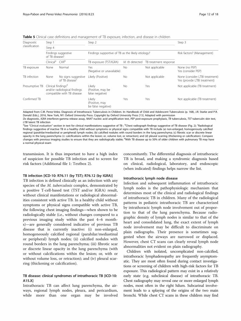
Table 5 Clinical case definitions and management of TB exposure, infection, and disease in children
Diagnosticclassification
Step 1 Step 2 Step 3
Step 4
Findings suggestiveof TB disease?
Findings supportive of TB as the likely etiology? Risk factors? (Management)
Clinicala CXRb TB exposure (TST/IGRA) M. tb detected TB treatment response
TB exposure None Normal Yes(Negative or unavailable)
No Not applicable None (no PEP)Yes (consider PEP)
TB infection None No signs suggestiveof TB diseasec
Likely (Positive) No Not applicable None (consider LTBI treatment)Yes (provide LTBI treatment)
Presumptive TB Clinical findingsd
and/or radiological findingscompatible with TB disease
Likely(Positive, may befalse negative)
No Yes Not applicable (TB treatment)
Confirmed TB Likely(Positive, maybe false negative)
Yes Yes Not applicable (TB treatment)
Adapted from C.M. Perez-Velez. Diagnosis of Intrathoracic Tuberculosis in Children. In: Handbook of Child and Adolescent Tuberculosis (p. 168), J.R. Starke and P.R.Donald (Eds.), 2016, New York, NY: Oxford University Press. Copyright by Oxford University Press [15]. Adapted with permissionDx diagnostic, IGRA interferon-gamma release assay, NAAT nucleic acid amplification test, PEP post-exposure prophylaxis, TB tuberculosis, TST tuberculin skin test,LTBI latent TB infectionaSee “Clinical evaluation” section in text for clinical manifestations suggestive of TB ; bChest radiograph findings suggestive of TB disease (Fig. 2); cRadiologicalfindings suggestive of inactive TB in a healthy child without symptoms or physical signs compatible with TB include (a) non-enlarged, homogenously calcifiedregional (parahilar/mediastinal or peripheral) lymph nodes; (b) calcified nodules with round borders in the lung parenchyma; (c) fibrotic scar or discrete linearopacity in the lung parenchyma (±: calcifications within the lesion; or, volume lost, or retraction); and (d) pleural scarring (thickening or calcification). Comparechanges with previous imaging studies to ensure that they are radiologically stable; dWith TB disease up to 50% of older children with pulmonary TB may havea normal physical exam
Roya-Pabon and Perez-Velez Pneumonia (2016) 8:23 Page 12 of 18
transmission. It is thus important to have a high indexof suspicion for possible TB infection and to screen forrisk factors (Additional file 1: Textbox 2).
TB infection [ICD-10: R76.11 (by TST); R76.12 (by IGRA)]TB infection is defined clinically as an infection with anyspecies of the M. tuberculosis complex, demonstrated bya positive T-cell-based test (TST and/or IGRA) result,without clinical manifestations or radiological abnormal-ities consistent with active TB. In a healthy child withoutsymptoms or physical signs compatible with active TB,the following chest imaging findings—when shown to beradiologically stable (i.e., without changes compared to aprevious imaging study within the past 4–6 month-s)—are generally considered indicative of previous TBdisease that is currently inactive: (i) non-enlarged,homogeneously calcified regional (parahilar/mediastinalor peripheral) lymph nodes; (ii) calcified nodules withround borders in the lung parenchyma; (iii) fibrotic scaror discrete linear opacity in the lung parenchyma (withor without calcifications within the lesion; or, with orwithout volume loss, or retraction); and (iv) pleural scar-ring (thickening or calcification).
TB disease: clinical syndromes of intrathoracic TB [ICD-10:A15.X]Intrathoracic TB can affect lung parenchyma, the air-ways, regional lymph nodes, pleura, and pericardium,while more than one organ may be involved
concomitantly. The differential diagnosis of intrathoracicTB is broad, and making a syndromic diagnosis basedon clinical, radiological, laboratory, and endoscopic(when indicated) findings helps narrow the list.
Intrathoracic lymph node diseaseInfection and subsequent inflammation of intrathoraciclymph nodes is the pathophysiologic mechanism thatdetermines most of the clinical and radiological findingsof intrathoracic TB in children. Many of the radiologicalpatterns in pediatric intrathoracic TB are characterizedby intrathoracic lymph node involvement out of propor-tion to that of the lung parenchyma. Because radio-graphic density of lymph nodes is similar to that of theheart and consolidated lung, the exact extent of lymphnode involvement may be difficult to discriminate onplain radiographs. Their presence is sometimes sug-gested when the airways are narrowed or displaced.However, chest CT scans can clearly reveal lymph nodeabnormalities not evident on plain radiography.Children with isolated, uncomplicated non-calcified,
intrathoracic lymphadenopathy are frequently asymptom-atic. They are most often found during contact investiga-tions or screening of children with high-risk factors for TBexposure. This radiological pattern may exist in a relativelyearly state (e.g. subclinical disease) of intrathoracic TB.Chest radiography may reveal one or more enlarged lymphnodes, most often in the right hilum. Subcarinal involve-ment leads to a splaying of the origins of the two mainbronchi. While chest CT scans in these children may find
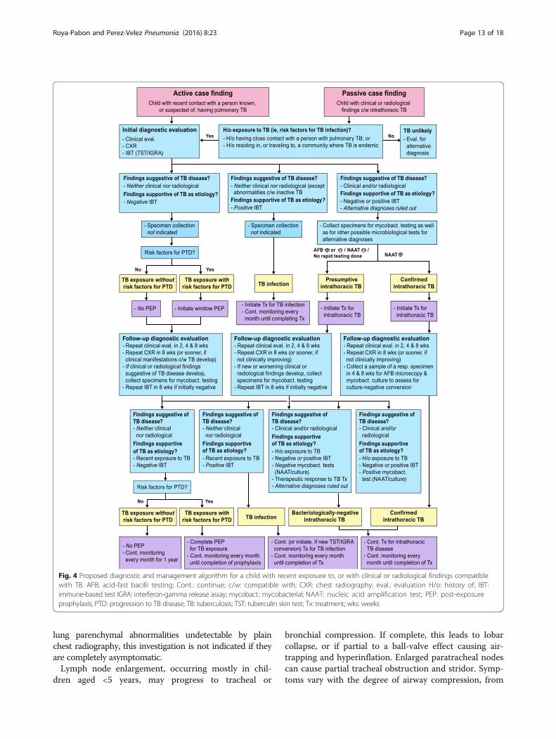
Fig. 4 Proposed diagnostic and management algorithm for a child with recent exposure to, or with clinical or radiological findings compatiblewith TB. AFB: acid-fast bacilli testing; Cont.: continue; c/w: compatible with; CXR: chest radiography; eval.: evaluation H/o: history of; IBT:immune-based test IGRA: interferon-gamma release assay; mycobact.: mycobacterial; NAAT: nucleic acid amplification test; PEP: post-exposureprophylaxis; PTD: progression to TB disease; TB: tuberculosis; TST: tuberculin skin test; Tx: treatment; wks: weeks
Roya-Pabon and Perez-Velez Pneumonia (2016) 8:23 Page 13 of 18
lung parenchymal abnormalities undetectable by plainchest radiography, this investigation is not indicated if theyare completely asymptomatic.Lymph node enlargement, occurring mostly in chil-
dren aged <5 years, may progress to tracheal or
bronchial compression. If complete, this leads to lobarcollapse, or if partial to a ball-valve effect causing air-trapping and hyperinflation. Enlarged paratracheal nodescan cause partial tracheal obstruction and stridor. Symp-toms vary with the degree of airway compression, from

Roya-Pabon and Perez-Velez Pneumonia (2016) 8:23 Page 14 of 18
asymptomatic to persistent cough, wheeze or stridor;dyspnea and respiratory distress from extensive atelec-tasis; or hyperinflation created by pressure from the en-larged lymph nodes on adjacent structures. Chestradiography (especially high-kilovoltage radiography)and CT scans may demonstrate severe narrowing of abronchus leading to either collapse or hyperinflation,most commonly of the right upper or middle lobes, orthe left upper lobe.
Tracheobronchial diseaseEndotracheal and endobronchial disease most often resultsfrom bronchogenic spread of TB after a diseased lymphnode erodes into the airway, most commonly the left orright main bronchus and bronchus intermedius [24, 28].Disease may be diffuse or localized with visible granulationtissue [78]. Damaged bronchi may dilate (bronchiectasis) ordevelop bronchostenosis [79]. Tracheobronchial disease canhave an acute, insidious, or delayed onset, with symptomsor physical signs of airway obstruction that depend on thelocation and severity, including persistent cough, rhonchi,wheeze, stridor, and/or dyspnea. Chest radiography is notsensitive in detecting tracheobronchial disease, unless it issevere or has an associated fibronodular appearance in thelung parenchyma. Bronchiolar disease is revealed on CTscans and may appear as a tree-in-bud pattern or as centri-lobular nodules consisting of dilated bronchioles that arethick-walled and filled with mucus. Bronchiectasis is alsomore easily noted on CT scans, which may show bronchialdilatation and wall-thickening. Bronchoscopy may demon-strate abnormalities suggestive of tracheobronchial disease,including hyperemia, edema, ulcers, masses, stenosis,granulation tissue or caseous lesions [80, 81].
Parenchymal diseaseIf inhaled M. tuberculosis bacilli are not destroyedimmediately by the innate immune response, a small paren-chymal focus of infection (primary/Ghon focus) may de-velop and drain via local lymphatic vessels to regionallymph nodes. Most nodular TB lung disease in children re-solves spontaneously and is identified only by radiographicscreening during contact investigations. Multiple, focal pul-monary nodules may be seen on chest imaging in the earlystages of a TB bronchopneumonia. A child with a solitarypulmonary nodule, with or without associated lymphaden-opathy, is most often asymptomatic. Chest radiographymay reveal isolated lung opacity with enlarged ipsilateralthoracic lymph nodes, known as a primary/Ghon complex.When lymph node lesions are calcified, it is a Ranke com-plex. Chest CT scans are more sensitive at detecting smallill-defined airspace nodules that tend to coalesce in someparts, but are different from the discrete, sharply definedmicronodules seen in miliary disease.
When the primary infection is poorly contained, myco-bacteria replicate and the initial lesion may enlarge (lobarpneumonia). Hilar lymph nodes may also enlarge andsometimes compress or infiltrate contiguous bronchi, mostcommonly the right or left main bronchus, or bronchusintermedius [24]. If a necrotic hilar lymph nodeerupts into a bronchus, endobronchial spread leads topatchy or multifocal consolidation of the respectivelobe (bronchopneumonia). When enlarged hilar lymphnodes are also compressing the bronchus, the endo-bronchial spread may cause distal expansion anddense consolidation of the entire lobe (expansilepneumonia) displacing the trachea, bowing the fis-sures and forming focal cavities. Cavities are uncom-mon in children, occurring predominantly in infantswith extensive, uncontained disease or in adolescentswith “adult-type” disease. Chest radiography and CTscans may reveal an oval-shaped lucency that is eitherisolated or within a consolidation or nodule, with wallsthat may be either thin or thick. In older childrenand adolescents there may be multiple cavities,located typically in the apical segments of the upperor lower lobes [9].
Pleural diseaseTB pleural effusions typically occur 3–6 months after aprimary infection and are usually unilateral, mostly result-ing from a delayed-type hypersensitivity reaction to M. tu-berculosis antigens that leaked into the pleural space froma subpleural primary focus. Pleural thickening is a com-mon component of the primary complex, but it rarelyleads to a significant effusion. Large effusions are seenmore often in older children (age >5 years) and adoles-cents. The child most often presents with pleuritic chestpain (58%), cough (80%) and fever (67%) [82]. Chestradiography will reveal a homogeneously opacified fluidlevel, with pulmonary parenchymal abnormalities (usuallyconsolidation) and intrathoracic lymphadenopathy oftenbecoming visible post-drainage [21]. Chest ultrasonographyis useful in determining the nature and quantity of the effu-sion and detecting early loculations and septations. ChestCT scans are useful in cases with complicated pleural effu-sion, detecting associated parenchymal lesions and intra-thoracic lymphadenopathy, and differentiating betweenpleural thickening and a chronic loculated effusion or em-pyema. TB pleural fluids are most often exudative withlymphocytic pleocytosis. Because of its protein-rich nature,care must be taken to not remove too much pleural fluid ina severely malnourished child because this can acutelyworsen the child's oncotic pressure. TB empyema hasalso been described [83], where pleural fluid is purulent[84]. Chylothorax is a rare type of pleural effusionresulting from disruption or obstruction of the thoracic
Roya-Pabon and Perez-Velez Pneumonia (2016) 8:23 Page 15 of 18
duct (or its tributaries), leading to lymphatic fluid(chyle) leakage into the pleural space. The pleuralfluid typically has a milky white appearance, and ispredominantly lymphocytic with elevated levels of tri-glycerides (>1.2 mmol/L) [85].
Pericardial diseaseTB is one of the most common causes of pericardial effu-sion in children in TB-endemic countries, and approxi-mately 1–4% of children with TB develop pericarditis [86].It has three main presentations: pericardial effusion (themost common), constrictive pericarditis, and a com-bination known as effusive-constrictive disease. Itmost frequently occurs after an infected contiguoussubcarinal lymph node infiltrates the pericardium. Itcan also arise from lymphohematogenous dissemin-ation of M. tuberculosis. HIV infection predisposes apatient to such disseminated disease, and is associatedwith greater severity of pericardial TB [87]. Childrenwith TB pericarditis usually present with symptomsand signs of heart failure, including persistent cough(70%), dyspnea (77%), chest pain (30%), hepatomegaly(77%), elevated jugular venous pressure (7%), softheart sounds, and a pericardial friction rub (18%), inaddition to fever (52%), night sweats, failure to thrive(36%), fatigue, and malaise [88]. Chest radiographytypically reveals cardiomegaly with a globular heartsilhouette (91%). Echocardiography is the most sensi-tive study to confirm a pericardial effusion, and mayreveal associated mediastinal lymphadenopathy orother complications.
Disseminated/miliary diseaseMiliary lung disease results from a TB lesion infiltratinginto a blood vessel, leading to hematogenous dissemin-ation [89]. The temporal pattern of miliary disease isusually acute, but it can also present with a delayed on-set. Pulmonary involvement and respiratory symptomsoccur relatively late in the disease. Given the multisys-tem involvement, presenting symptoms may includecough (72%), dyspnea, diarrhea and vomiting (33%), ir-ritability, headache, convulsions, hepatomegaly (82%),splenomegaly (54%), lymphadenopathy (46%), fever(39%), chills, loss of appetite and failure to thrive (40%),fatigue, generalized weakness, decreased activity, andmalaise. The main complication is TB meningitis [89].Chest radiography may reveal innumerable roundedmicronodules (≤3 mm in diameter) scattered diffuselythroughout both lungs, but in the initial stages of dis-seminated disease the radiological abnormalities maynot be apparent (9%) [79, 89]. Often these nodules arebest seen on the lateral projection of the chest radio-graph in the retrocardiac area.
ConclusionsUsing currently available tools, a systematic diagnosticapproach to the child with recent exposure to, or withclinical or radiological findings compatible with, TB canallow the clinician to classify most patients into one ofthe major diagnostic categories of TB exposure, infec-tion, or disease. In cases of TB exposure and infection,identifying risk factors for progression to disease helpshasten diagnostic evaluation and initiating appropriateprophylaxis or treatment when indicated.
Additional file
Additional file 1: Table S1. Differential diagnosis of chronic cough inchildren. Table S2. Nucleic acid amplification tests for detecting Mycobacteriumtuberculosis complex and genes encoding targets of mutations conferring drugresistance. Table S3. Differential diagnosis of clinical-radiological syndromesassociated with intrathoracic TB in children. Textbox 1. Spectrum of possibleorgan involvement in TB disease. Textbox 2. Risk factors for TB infection inchildren. (PDF 617 kb)
AbbreviationsADA: Adenosine deaminase; AFB: Acid-fast bacilli; BCG: Bacille Calmette-Guerin;CFU: Colony-forming unit; CT: Computed tomography; DNA: Deoxyribonucleicacid; HIV: Human immunodeficiency virus; ICD-10: International statisticalclassification of diseases and Related health problems, 10th revision;IGRA: Interferon-gamma release assay; LDH: Lactate dehydrogenase;LED: Light-emitting diode; LF-LAM: Lateral flow lipoarabinomannan;LPA: Line probe assay; M.: Mycobacterium; MDR: Multi-drug-resistant;NAAT: Nucleic acid amplification test; NTM: Nontuberculous mycobacteria;PPD: Purified protein derivative; RT-PCR: Real-time polymerase chain reaction;TB: Tuberculosis; TST: Tuberculin skin test
AcknowledgementsSonia L. Villegas, MD, MPH, Charite - Medical University Berlin, Germany forher contributions to previous versions of the diagnostic and managementalgorithm for tuberculosis in children.
FundingThere were no funding sources for this review.
Availability of data and materialsNot applicable.
Authors’ contributionsBoth authors defined the scope of the review, searched the literature,assessed the evidence base, synthesized included studies, analyzed thefindings, designed and drafted the manuscript and tables and figures,critically revised the manuscript, and approved the final version of themanuscript.
Authors’ informationWe decline this option, as we have nothing substantive to add to aid thereader’s interpretation of the article, or to understand the standpoint of theauthors].
Consent for publicationNot applicable (some figures and tables were adapted from figures andtables previously published by the authors [CMPV & CLRP] with permissionfrom the publishers).
Competing interestsThe authors declare that they have no competing interests.
Ethics approval and consent to participateNot applicable.
Roya-Pabon and Perez-Velez Pneumonia (2016) 8:23 Page 16 of 18
Author details1Division of Pediatric Pulmonology, Department of Pediatrics, Faculty ofMedicine, University of Antioquia, Medellin, Antioquia, Colombia. 2GrupoTuberculosis Valle-Colorado (GTVC), Medellin, Antioquia, Colombia.3Tuberculosis Clinic, Pima County Health Department, Tucson, AZ, USA.4Division of Infectious Diseases, College of Medicine, University of Arizona,Tucson, AZ, USA. 5College of Medicine, University of Arizona, 1501 NorthCampbell Avenue, P.O. Box 24503985724 Tucson, AZ, USA.
Received: 20 October 2016 Accepted: 3 November 2016
References1. Cruz AT, Starke JR. Clinical manifestations of tuberculosis in children.
Paediatr Respir Rev. 2007;8:107–17. PMID:17574154. http://dx.doi.org/10.1016/j.prrv.2007.04.008.
2. Marais BJ, Graham SM. Childhood tuberculosis: a roadmap towards zerodeaths. J Paediatr Child Health. 2016;52:258–61. PMID:24923706, http://dx.doi.org/10.1111/jpc.12647.
3. Young D, Stark J, Kirschner D. Systems biology of persistent infection:tuberculosis as a case study. Nat Rev Microbiol. 2008;6:520–8. PMID:18536727, http://dx.doi.org/10.1038/nrmicro1919.
4. Barry 3rd CE, Boshoff HI, Dartois V, Dick T, Ehrt S, Flynn J, et al. Thespectrum of latent tuberculosis: rethinking the biology and interventionstrategies. Nat Rev Microbiol. 2009;7:845–55. PMID:19855401.
5. Young DB, Gideon HP, Wilkinson RJ. Eliminating latent tuberculosis.Trends Microbiol. 2009;17:183–8. PMID:19375916. http://dx.doi.org/10.1016/j.tim.2009.02.005.
6. Deffur A, Mulder NJ, Wilkinson RJ. Co-infection with Mycobacteriumtuberculosis and human immunodeficiency virus: an overview andmotivation for systems approaches. Pathog Dis. 2013;69(2):101–13.
7. Salgame P, Geadas C, Collins L, Jones-López E, Ellner JJ. Latent tuberculosisinfection–revisiting and revising concepts. Tuberculosis (Edinb). 2015;95:373–84. PMID:26038289. http://dx.doi.org/10.1016/j.tube.2015.04.003.
8. Marais BJ, Gie RP, Schaaf HS, Hesseling AC, Enarson DA, Beyers N. Thespectrum of disease in children treated for tuberculosis in a highly endemicarea. Int J Tuberc Lung Dis. 2006;10:732–8. PMID:16848333.
9. Marais BJ, Gie RP, Hesseling AH, Beyers N. Adult-type pulmonarytuberculosis in children 10–14 years of age. Pediatr Infect Dis J. 2005;24:743–4. PMID:16094237. http://dx.doi.org/10.1097/01.inf.0000173305.04212.09.
10. Wiseman CA, Gie RP, Starke JR, Schaaf HS, Donald PR, Cotton MF, et al. Aproposed comprehensive classification of tuberculosis disease severity inchildren. Pediatr Infect Dis J. 2012;31:347–52. PMID:22315002. http://dx.doi.org/10.1097/INF.0b013e318243e27b.
11. O’Garra A, Redford PS, McNab FW, Bloom CI, Wilkinson RJ, Berry MP. Theimmune response in tuberculosis. Annu Rev Immunol. 2013;31:475–527.PMID:23516984. http://dx.doi.org/10.1146/annurev-immunol-032712-095939.
12. Achkar JM, Jenny-Avital ER. Incipient and subclinical tuberculosis:defining early disease states in the context of host immune response. JInfect Dis. 2011;204 Suppl 4:S1179–86. PMID:21996700. http://dx.doi.org/10.1093/infdis/jir451.
13. Hatherill M, Hanslo M, Hawkridge T, Little F, Workman L, Mahomed H,et al. Structured approaches for the screening and diagnosis ofchildhood tuberculosis in a high prevalence region of South Africa. BullWorld Health Organ. 2010;88:312–20. PMID:20431796. http://dx.doi.org/10.2471/BLT.09.062893.
14. Graham SM, Ahmed T, Amanullah F, Browning R, Cardenas V, Casenghi M,et al. Evaluation of tuberculosis diagnostics in children: 1. Proposed clinicalcase definitions for classification of intrathoracic tuberculosis disease.Consensus from an expert panel. J Infect Dis. 2012;205 Suppl 2:S199–208.PMID:22448023. http://dx.doi.org/10.1093/infdis/jis008.
15. Perez-Velez C. Diagnosis of intrathoracic tuberculosis in children. In: StarkeJR, Donald PR, editors. Handbook of child and adolescent tuberculosis. NewYork: Oxford; 2016. p. 147–76.
16. Marais BJ, Gie RP, Hesseling AC, Schaaf HS, Lombard C, Enarson DA, et al. Arefined symptom-based approach to diagnose pulmonary tuberculosis inchildren. Pediatrics. 2006;118:e1350–9. PMID:17079536. http://dx.doi.org/10.1542/peds.2006-0519.
17. Hossain MM, Norazmi MN. Pattern recognition receptors and cytokines inMycobacterium tuberculosis infection–the double-edged sword? BioMedRes Int. 2013;2013:179174.
18. Boulware DR, Callens S, Pahwa S. Pediatric HIV immune reconstitutioninflammatory syndrome. Curr Opin HIV AIDS. 2008;3:461–7. PMID:19373006.http://dx.doi.org/10.1097/COH.0b013e3282fe9693.
19. Mohapatra PR, Janmeja AK. Tuberculous lymphadenitis. J Assoc PhysiciansIndia. 2009;57:585–90. PMID:20209720.
20. Marais BJ, Gie RP, Schaaf HS, Starke JR, Hesseling AC, Donald PR, et al. Aproposed radiological classification of childhood intra-thoracic tuberculosis.Pediatr Radiol. 2004;34:886–94. PMID:15300340. http://dx.doi.org/10.1007/s00247-004-1238-0.
21. Perez-Velez CM, Marais BJ. Tuberculosis in children. N Engl J Med. 2012;367:348–61. PMID:22830465. http://dx.doi.org/10.1056/NEJMra1008049.
22. Smuts NA, Beyers N, Gie RP, Schaaf HS, Talent JM, Nel E, et al. Value of thelateral chest radiograph in tuberculosis in children. Pediatr Radiol.1994;24:478–80. PMID:7885777, http://dx.doi.org/10.1007/BF02015003.
23. Andronikou S, Van der Merwe DJ, Goussard P, Gie RP, Tomazos N.Usefulness of lateral radiographs for detecting tuberculouslymphadenopathy in children–confirmation using sagittal CT reconstructionwith multiplanar cross-referencing. South Afr J Radiol. 2012;16:87–92.http://dx.doi.org/10.4102/sajr.v16i3.288.
24. Andronikou S, Joseph E, Lucas S, Brachmeyer S, Du Toit G, Zar H, et al. CTscanning for the detection of tuberculous mediastinal and hilarlymphadenopathy in children. Pediatr Radiol. 2004;34:232–6. PMID:14710313.http://dx.doi.org/10.1007/s00247-003-1117-0.
25. Kim WS, Moon WK, Kim IO, Lee HJ, Im JG, Yeon KM, et al. Pulmonarytuberculosis in children: evaluation with CT. AJR Am J Roentgenol. 1997;168:1005–9. PMID:9124105. http://dx.doi.org/10.2214/ajr.168.4.9124105.
26. Daltro P, Nunez-Santos E, Laya B. Pediatric tuberculosis. In: Garcia-Peña P,Guillerman R, editors. Pediatric chest imaging. 3rd ed. Berlin Heidelberg:Springer; 2014. p. 285–303.
27. WHO. Guidance for national tuberculosis programmes on the managementof tuberculosis in children. Geneva: World Health Organization; 2014.
28. Goussard P, Gie R. The role of bronchoscopy in the diagnosis andmanagement of pediatric pulmonary tuberculosis. Expert Rev Respir Med.2014;8:101–9. PMID:24378192. http://dx.doi.org/10.1586/17476348.2013.863712.
29. Wessels G, Schaaf HS, Beyers N, Gie RP, Nel E, Donald PR. Haematologicalabnormalities in children with tuberculosis. J Trop Pediatr. 1999;45:307–10.PMID:10584477. http://dx.doi.org/10.1093/tropej/45.5.307.
30. Rasmussen TA, Søgaard OS, Camara C, Andersen PL, Wejse C. Serumprocalcitonin in pulmonary tuberculosis [i.]. Int J Tuberc Lung Dis.2011;15:251–6. PMID:21219690.
31. Herlina M, Nataprawira HM, Garna H. Association of serum C-reactiveprotein and leptin levels with wasting in childhood tuberculosis. SingaporeMed J. 2011;52:446–50. PMID:21731999.
32. Schaaf HS, Michaelis IA, Richardson M, Booysen CN, Gie RP, Warren R, et al.Adult-to-child transmission of tuberculosis: household or communitycontact? Int J Tuberc Lung Dis. 2003;7:426–31. PMID:12757042.
33. Mack U, Migliori GB, Sester M, Rieder HL, Ehlers S, Goletti D, et al. C. Lange;TBNET. LTBI: latent tuberculosis infection or lasting immune responses to M.tuberculosis? A TBNET consensus statement. Eur Respir J. 2009;33:956–73.PMID:19407047. http://dx.doi.org/10.1183/09031936.00120908.
34. Perez-Stable EJ, Slutkin G. A demonstration of lack of variability among sixtuberculin skin test readers. Am J Public Health. 1985;75:1341–3. PMID:4051078. http://dx.doi.org/10.2105/AJPH.75.11.1341.
35. van Zyl-Smit RN, Zwerling A, Dheda K, Pai M. Within-subject variability ofinterferon-g assay results for tuberculosis and boosting effect of tuberculinskin testing: a systematic review. PLoS ONE. 2009;4:e8517. PMID:20041113.http://dx.doi.org/10.1371/journal.pone.0008517.
36. Trajman A, Steffen RE, Menzies D. Interferon-gamma release assays versustuberculin skin testing for the diagnosis of latent tuberculosis infection: anoverview of the evidence. Pulmonary medicine. 2013;2013:601737.
37. Farhat M, Greenaway C, Pai M, Menzies D. False-positive tuberculin skintests: what is the absolute effect of BCG and non-tuberculousmycobacteria? Int J Tuberc Lung Dis. 2006;10:1192–204. PMID:17131776.
38. Pai M, Zwerling A, Menzies D. Systematic review: T-cell-based assays forthe diagnosis of latent tuberculosis infection: an update. Ann InternMed. 2008;149:177–84. PMID:18593687. http://dx.doi.org/10.7326/0003-4819-149-3-200808050-00241.
39. Dheda K, van Zyl-Smit RN, Sechi LA, Badri M, Meldau R, Meldau S, et al.Utility of quantitative T-cell responses versus unstimulated interferon-gamma for the diagnosis of pleural tuberculosis. Eur Respir J. 2009;34:1118–26. PMID:19386693. http://dx.doi.org/10.1183/09031936.00005309.
Roya-Pabon and Perez-Velez Pneumonia (2016) 8:23 Page 17 of 18
40. Chiang SS, Swanson DS, Starke JR. New diagnostics for childhoodtuberculosis. Infect Dis Clin North Am. 2015;29:477–502. PMID:26188605.http://dx.doi.org/10.1016/j.idc.2015.05.011.
41. Metcalfe JZ, Everett CK, Steingart KR, Cattamanchi A, Huang L, Hopewell PC,et al. Interferon-γ release assays for active pulmonary tuberculosis diagnosisin adults in low- and middle-income countries: systematic review andmeta-analysis. J Infect Dis. 2011;204 Suppl 4:S1120–9. PMID:21996694,http://dx.doi.org/10.1093/infdis/jir410.
42. Adetifa IM, Ota MO, Jeffries DJ, Hammond A, Lugos MD, Donkor S, et al.Commercial interferon gamma release assays compared to the tuberculinskin test for diagnosis of latent Mycobacterium tuberculosis infection inchildhood contacts in the Gambia. Pediatr Infect Dis J. 2010;29:439–43.PMID:20068506, http://dx.doi.org/10.1097/INF.0b013e3181cb45da.
43. Lalvani A, Millington KA. T cell-based diagnosis of childhood tuberculosisinfection. Curr Opin Infect Dis. 2007;20:264–71. PMID:17471036. http://dx.doi.org/10.1097/QCO.0b013e32813e3fd8.
44. Merino JM, Carpintero I, Alvarez T, Rodrigo J, Sánchez J, Coello JM.Tuberculous pleural effusion in children. Chest. 1999;115:26–30. PMID:9925059. http://dx.doi.org/10.1378/chest.115.1.26.
45. Goto M, Noguchi Y, Koyama H, Hira K, Shimbo T, Fukui T. Diagnostic valueof adenosine deaminase in tuberculous pleural effusion: a meta-analysis.Ann Clin Biochem. 2003;40:374–81. PMID:12880538. http://dx.doi.org/10.1258/000456303766477011</jrn>.
46. Roya-Pabon C. Especímenes respiratorios para el diagnóstico microbiológicode las infecciones respiratorias. In: Posada-Saldarriaga R, editor. Neumologíapediátrica. Bogotá: Distribuna; 2016. p. 171–92.
47. Warren JR, Bhattacharya M, De Almeida KN, Trakas K, Peterson LR. Aminimum 5.0 ml of sputum improves the sensitivity of acid-fast smear forMycobacterium tuberculosis. Am J Respir Crit Care Med. 2000;161:1559–62.PMID:10806154. http://dx.doi.org/10.1164/ajrccm.161.5.9908063.
48. Mpagama SG, Mtabho C, Mwaigwisya S, Mleoh LJ, Boer IM, Heysell SK, et al.Comparison of overnight pooled and standard sputum collection methodfor patients with suspected pulmonary tuberculosis in northern Tanzania.Tuberc Res Treat. 2012;2012:128057.
49. Singh S, Singh A, Prajapati S, Kabra SK, Lodha R, Mukherjee A, et al. DelhiPediatric TB study group Xpert MTB/RIF assay can be used on archivedgastric aspirate and induced sputum samples for sensitive diagnosis ofpaediatric tuberculosis. BMC Microbiol. 2015;15:191. PMID:26420261.http://dx.doi.org/10.1186/s12866-015-0528-z.
50. Cruz AT, Merchant O, Zafar A, Starke JR. Tuberculosis exposure, infection anddisease among children with medical comorbidities. Pediatr Infect Dis J. 2014;33:885–8. PMID:24642517. http://dx.doi.org/10.1097/INF.0000000000000343.
51. Zar HJ, Workman L, Isaacs W, Munro J, Black F, Eley B, et al. Rapid moleculardiagnosis of pulmonary tuberculosis in children using nasopharyngealspecimens. Clin Infect Dis. 2012;55:1088–95. PMID:22752518. http://dx.doi.org/10.1093/cid/cis598.
52. Abadco DL, Steiner P. Gastric lavage is better than bronchoalveolar lavagefor isolation of Mycobacterium tuberculosis in childhood pulmonarytuberculosis. Pediatr Infect Dis J. 1992;11:735–8. PMID:1448314. http://dx.doi.org/10.1097/00006454-199209000-00013.
53. Somu N, Swaminathan S, Paramasivan CN, Vijayasekaran D,Chandrabhooshanam A, Vijayan VK, et al. Value of bronchoalveolar lavageand gastric lavage in the diagnosis of pulmonary tuberculosis in children.Tuber Lung Dis. 1995;76:295–9. PMID:7579310. http://dx.doi.org/10.1016/S0962-8479(05)80027–9.
54. Chow F, Espiritu N, Gilman RH, Gutierrez R, Lopez S, Escombe AR, et al. Lacuerda dulce–a tolerability and acceptability study of a novel approach tospecimen collection for diagnosis of paediatric pulmonary tuberculosis. BMCInfect Dis. 2006;6:67. PMID:16595008. http://dx.doi.org/10.1186/1471-2334-6-67.
55. Perez-Velez C, Wilches-Luna E, Hernández-Sarmiento J, Casanova-ReynoldsA, Hernandez N, Moreno-Ortega S, editors. Preliminary results of acomparative yield study of induced sputum, string test, and gastric aspiratefor the microbiological diagnosis of pulmonary tuberculosis in children.New York: Amer Thoracic Soc; 2010. AMERICAN JOURNAL OF RESPIRATORYAND CRITICAL CARE MEDICINE.
56. Walters E, Gie RP, Hesseling AC, Friedrich SO, Diacon AH, Gie RP. Rapiddiagnosis of pediatric intrathoracic tuberculosis from stool samples usingthe Xpert MTB/RIF assay: a pilot study. Pediatr Infect Dis J. 2012;31:1316.PMID:23188101. http://dx.doi.org/10.1097/INF.0b013e318266c21c.
57. Nicol MP, Spiers K, Workman L, Isaacs W, Munro J, Black F, et al. Xpert MTB/RIF testing of stool samples for the diagnosis of pulmonary tuberculosis in
children. Clin Infect Dis. 2013;57:e18–21. PMID:23580738. http://dx.doi.org/10.1093/cid/cit230.
58. Wright CA, Warren RM, Marais BJ. Fine needle aspiration biopsy: anundervalued diagnostic modality in paediatric mycobacterial disease. Int JTuberc Lung Dis. 2009;13:1467–75. PMID:19919763.
59. Denkinger CM, Schumacher SG, Boehme CC, Dendukuri N, Pai M, SteingartKR. Xpert MTB/RIF assay for the diagnosis of extrapulmonary tuberculosis: asystematic review and meta-analysis. Eur Respir J. 2014;44:435–46. PMID:24696113. http://dx.doi.org/10.1183/09031936.00007814.
60. J Clin Tuberc Other Mycobact Dis. 2016;4:33–43. http://dx.doi.org/10.1016/j.jctube.2016.05.005.
61. Gómez PD, Torronteras SR, Caro MP, López BA, Macías MP, Andrés MA, et al.An Esp Pediatr. 2000;53:405–11. PMID:11141361. http://dx.doi.org/10.1016/S1695-4033(00)78620–6.
62. Zar HJ, Hanslo D, Apolles P, Swingler G, Hussey G. Induced sputum versusgastric lavage for microbiological confirmation of pulmonary tuberculosis ininfants and young children: a prospective study. Lancet. 2005;365:130–4.PMID:15639294. http://dx.doi.org/10.1016/S0140-6736(05)17702–2.
63. Laven GT. Diagnosis of tuberculosis in children using fluorescencemicroscopic examination of gastric washings. Am Rev Respir Dis.1977;115:743–9. PMID:857714.
64. WHO. Automated real-time nucleic acid amplification technology for rapid andsimultaneous detection of tuberculosis and rifampicin resistance: Xpert MTB/RIF assay for the diagnosis of pulmonary and extrapulmonary TB in adults andchildren: policy update. Geneva: World Health Organization; 2013.
65. WHO. Molecular line probe assays for rapid screening of patients at risk ofmultidrug-resistant tuberculosis (MDR-TB). Geneva: World HealthOrganization; 2008.
66. WHO. The use of molecular line probe assay for the detection of resistanceto second-line anti-tuberculosis drugs: expert group meeting report,Geneva, February 2013. Geneva: World Health Organization; 2013.
67. Sanchini A, Fiebig L, Drobniewski F, Haas W, Richter E, Katalinic-Jankovic V, et al.European reference laboratory network for TB members. Laboratory diagnosis ofpaediatric tuberculosis in the European Union/European Economic Area:analysis of routine laboratory data, 2007 to 2011. Euro Surveill. 2014;19. PMID:24679723. http://dx.doi.org/10.2807/1560-7917.ES2014.19.11.20744.
68. Lawn SD, Kerkhoff AD, Vogt M, Wood R. High diagnostic yield oftuberculosis from screening urine samples from HIV-infected patients withadvanced immunodeficiency using the Xpert MTB/RIF assay. J AcquirImmune Defic Syndr. 2012;60:289–94. PMID:22531759. http://dx.doi.org/10.1097/QAI.0b013e318258c6af.
69. Lawn SD, Dheda K, Kerkhoff AD, Peter JG, Dorman S, Boehme CC, et al.Determine TB-LAM lateral flow urine antigen assay for HIV-associatedtuberculosis: recommendations on the design and reporting of clinicalstudies. BMC Infect Dis. 2013;13:407. PMID:24004840. http://dx.doi.org/10.1186/1471-2334-13-407.
70. Mutetwa R, Boehme C, Dimairo M, Bandason T, Munyati SS, Mangwanya D,et al. Diagnostic accuracy of commercial urinary lipoarabinomannandetection in African tuberculosis suspects and patients. Int J Tuberc LungDis. 2009;13:1253–9. PMID:19793430.
71. Nicol MP, Allen V, Workman L, Isaacs W, Munro J, Pienaar S, et al. Urinelipoarabinomannan testing for diagnosis of pulmonary tuberculosis inchildren: a prospective study. Lancet Glob Health. 2014;2:e278–84. PMID:24818083. http://dx.doi.org/10.1016/S2214-109X(14)70195–0.
72. Society AT. Diagnostic standards and classification of tuberculosis in adultsand children. Am J Respir Crit Care Med. 2000;161:1376–95. PMID:10764337.http://dx.doi.org/10.1164/ajrccm.161.4.16141.
73. Marais BJ, Hesseling AC, Gie RP, Schaaf HS, Enarson DA, Beyers N. Thebacteriologic yield in children with intrathoracic tuberculosis. Clin Infect Dis.2006;42:e69–71. PMID:16575719. http://dx.doi.org/10.1086/502652.
74. Demers A, Whitelaw A, Eisenach K. Microbiology and pathology oftuberculosis. In: Starke J, Donald P, editors. Handbook of child andadolescent tuberculosis. New York: Oxford; 2016. p. 13–29.
75. Hamilton G. Emergency medicine: an approach to clinical problem-solving.Saunders; 1991.
76. Hesseling AC, Schaaf HS, Gie RP, Starke JR, Beyers N. A critical review ofdiagnostic approaches used in the diagnosis of childhood tuberculosis. Int JTuberc Lung Dis. 2002;6:1038–45. PMID:12546110.
77. Graham SM. The use of diagnostic systems for tuberculosis in children.Indian J Pediatr. 2011;78:334–9. PMID:21165720. http://dx.doi.org/10.1007/s12098-010-0307-7.
Roya-Pabon and Perez-Velez Pneumonia (2016) 8:23 Page 18 of 18
78. de Blic J, Azevedo I, Burren CP, Le Bourgeois M, Lallemand D, Scheinmann P. Thevalue of flexible bronchoscopy in childhood pulmonary tuberculosis. Chest. 1991;100:688–92. PMID:1909618. http://dx.doi.org/10.1378/chest.100.3.688.
79. Marais BJ, Gie RP, Schaaf HS, Hesseling AC, Obihara CC, Starke JJ, et al. Thenatural history of childhood intra-thoracic tuberculosis: a critical review ofliterature from the pre-chemotherapy era. Int J Tuberc Lung Dis.2004;8:392–402. PMID:15141729.
80. Chung HS, Lee JH. Bronchoscopic assessment of the evolution ofendobronchial tuberculosis. Chest. 2000;117:385–92. PMID:10669679. http://dx.doi.org/10.1378/chest.117.2.385.
81. Cakir E, Uyan ZS, Oktem S, Karakoc F, Ersu R, Karadag B, et al. Flexiblebronchoscopy for diagnosis and follow up of childhood endobronchialtuberculosis. Pediatr Infect Dis J. 2008;27:783–7. PMID:18664928. http://dx.doi.org/10.1097/INF.0b013e318170fccc.
82. Cruz AT, Ong LT, Starke JR. Childhood pleural tuberculosis: a review of 45cases. Pediatr Infect Dis J. 2009;28:981–4. PMID:19672211. http://dx.doi.org/10.1097/INF.0b013e3181a8568b.
83. Fischer GB, Andrade CF, Lima JB. Pleural tuberculosis in children.Paediatr Respir Rev. 2011;12:27–30. PMID:21172672. http://dx.doi.org/10.1016/j.prrv.2010.11.001.
84. Strnad L, Denkinger C, Pai M. Effusions from infections: tuberculosis. In: LightR, Gary Lee YC, editors. Textbook of pleural diseases, vol. 3rd. Boca Raton:CRC; 2016. p. 331–42.
85. Grobbelaar M, Andronikou S, Goussard P, Theron S, Mapukata A,George R. Chylothorax as a complication of pulmonary tuberculosis inchildren. Pediatr Radiol. 2008;38:224–6. PMID:17929008. http://dx.doi.org/10.1007/s00247-007-0634-7.
86. Weber S. Tuberculosis and pericarditis in children. Trop Doct. 1999;29:135–8.PMID:10448233.
87. Mayosi BM, Wiysonge CS, Ntsekhe M, Volmink JA, Gumedze F, Maartens G,et al. Clinical characteristics and initial management of patients withtuberculous pericarditis in the HIV era: the Investigation of the Managementof Pericarditis in Africa (IMPI Africa) registry. BMC Infect Dis. 2006;6:2. PMID:16396690. http://dx.doi.org/10.1186/1471-2334-6-2.
88. Hugo-Hamman CT, Scher H, De Moor MM. Tuberculous pericarditis inchildren: a review of 44 cases. Pediatr Infect Dis J. 1994;13:13–8. PMID:8170726. http://dx.doi.org/10.1097/00006454-199401000-00004.
89. Hussey G, Chisholm T, Kibel M. Miliary tuberculosis in children: a review of94 cases. Pediatr Infect Dis J. 1991;10:832–6. PMID:1749696. http://dx.doi.org/10.1097/00006454-199111000-00008.
90. Tebruegge M, Dutta B, Donath S, Ritz N, Forbes B, Camacho-Badilla K, et al.Mycobacteria-specific cytokine responses detect tuberculosis infection anddistinguish latent from active tuberculosis. Am J Respir Crit Care Med. 2015;192:485–99. PMID:26030187. http://dx.doi.org/10.1164/rccm.201501-0059OC.
91. Ankrah AO, van der Werf TS, de Vries EF, Dierckx RA, Sathekge MM,Glaudemans AW. PET/CT imaging of Mycobacterium tuberculosis infection.Clin Transl Imaging. 2016;4:131–44. PMID:27077068. http://dx.doi.org/10.1007/s40336-016-0164-0.
92. Alland D, Rowneki M, Smith L, Chakravorty S, Ryan J, Chancellor M, editors.Xpert MTB/RIF ultra: a new near-patient TB test with sensitivity equal toculture. 15th Annual Conference on Retroviruses and OpportunisticInfections Seattle, Washington; 2015.
93. Lessem E. The tuberculosis diagnostics pipeline. Pipeline Report.2016;2016:129
94. Clifford V, Zufferey C, Street A, Denholm J, Tebruegge M, Curtis N. Cytokines formonitoring anti-tuberculous therapy: a systematic review. Tuberculosis (Edinb).2015;95:217–28. PMID:25797612. http://dx.doi.org/10.1016/j.tube.2015.01.003.
95. Casanova JL. Severe infectious diseases of childhood as monogenic inbornerrors of immunity. Proc Natl Acad Sci U S A. 2015;112:E7128–37.PMID:26621750.
• We accept pre-submission inquiries
• Our selector tool helps you to find the most relevant journal
• We provide round the clock customer support
• Convenient online submission
• Thorough peer review
• Inclusion in PubMed and all major indexing services
• Maximum visibility for your research
Submit your manuscript atwww.biomedcentral.com/submit
Submit your next manuscript to BioMed Central and we will help you at every step: