Embed Size (px)
Citation preview

Abstract— Tuberculosis is described as “king of diseases” in the Vedas and has been mentioned by Sushruta and
Chakra in 600 B.C. Tuberculosis is a chronic infectious
disease of worldwide prevalence, caused by
Mycobacterium Tuberculosis. The primary site of infection
is usually the lungs, although it can affect any part of the
body, including the oral cavity. Although oral
manifestations of tuberculosis are rare, clinicians should be
aware of their possible occurrence in their patient
population in the form of ulcer, granulomas, involvement
of the salivary glands and temporomandibular joints,
osteomyelitis, desquamative gingivitis and tubeculous
lymphadenitis. Such awareness can aid in diagnosing
tuberculosis at an early stage, thereby preventing systemic
complications and potential contaminations. This paper
deals with two cases of tuberculosis diagnosed on the basis
of oral manifestations, thereby emphasizing the importance
of the dentist’s role in diagnosis of a multisystemic
disorder.
Keywords— Tuberculosis, Oral manifestations, chronic granulomatous disease
I. INTRODUCTION
uberculosis is a chronic granulomatous
multisystemic infectious disease caused by
Mycobacterium tuberculosis and is a major health
problem in most developing countries. It can affect
any part of the body including the oral cavity, though
extra-pulmonary tuberculosis is rare, occurring in
10% to 15% of all cases1. Tuberculosis is usually
acquired by Mycobacterium tuberculosis and less
frequently by the ingestion of unpasteurized cow’s
milk that is infected by Mycobacterium bovis or by
DR SHAMIMUL HASAN1 MOHD.
ABBAS KHAN2 1MDS ; ASSISTANT PROFESSOR, DEPTT. OF ORAL
MEDICINE & RADIOLOGY, FACULTY OF DENTISTRY
JAMIA MILIA ISLAMIA, NEW DELHI, INDIA,
[email protected] 2 BDS (FINAL YEAR ) STUDENT
Z.A DENTAL COLLEGE & HOSPITALS, ALIGARH MUSLIM
UNIVERSITY, ALIGARH, INDIA, [email protected]
other atypical Mycobacteria2. Depending upon the
organ system involved, tuberculosis is classified
clinically as Pulmonary or extra pulmonary.
Pulmonary tuberculosis is the most common form of
the disease. However, tuberculosis can also occur in
the lymph nodes, meninges, kidneys, bone, skin and
in the oral cavity3. Since the introduction of effective
chemotherapy, tuberculous lesions of the oral cavity
have become so infrequent that it is virtually a
forgotten disease entity and pose a diagnostic
problem. They account for less than one percent of
cases of extra pulmonary tuberculosis, are usually
associated with foci of disease elsewhere in the body
and enlarged, palpable cervical lymph nodes are
usually present4. Tuberculous oral lesions are
relatively rare occurrence. Oral manifestations occur
in approximately 3% of cases involving long standing
pulmonary and / or systemic infection5. Oral
tuberculosis can be primary or secondary. Primary
oral tuberculous lesions are extremely rare and
generally occur in young adults. It usually involves
gingiva and is associated with caseation of the
dependent lymph nodes; the lesion itself remains
painless in in most cases6. Primary lesions develop
when tuberculosis bacilli are directly inoculated into
the oral tissues of a person who has not acquired
immunity to the disease and in fact, any area that is
vulnerable to direct inoculation of bacilli from
exogenous source can be a potential site. These
frequently involve gingiva, tooth extraction sockets
and buccal folds.7. In contrast, secondary oral
tuberculosis is common (0.005% to 1.5% of cases)
and is usually seen in older adults8. Secondary
infection of oral tissues can result from either
haematogenous or lymphatic spread or from auto
inoculation by infected sputum and direct extensions
from neighbouring structures. Intra oral sites
frequently involved include the tongue, palate, lips,
alveolar mucosa and jaw bones7. Tuberculosis in the
oro-facial region may manifest in various forms:
Tuberculous ulcer, Tuberculous gingivitis,
Tuberculosis - A common disease with
uncommon oral features
Report of two cases with a detailed review of
literature
Shamimul Hasan, Mohd. Abbas Khan
T
Proceedings of the World Medical Conference
ISBN: 978-1-61804-036-7 156

Tuberculous lymphadenitis, Tuberculoma,
Tuberculous osteomyelitis, Tuberculous
sialadenitis9,10 and Tuberculous involvement of the
Temporomandibular jaw. This paper deals with two
cases of oro-facial tuberculosis and emphasizes the
role of dentist in early diagnosis of tuberculosis based
on the disease oral features, coupled with a detailed
review of literature on the oro-facial manifestations of
Tuberculosis.
II. CASE REPORT 1
A 14 Year old female patient reported to oral
medicine and radiology department, Faculty of
Dentistry, Jamia Milia Islamia university, New Delhi
with a complaint of chronic ulcerations and burning
sensations in the mouth since past one year. History
reveals that the patient developed a small ulcer in the
gums in upper anterior region around a year back,
which has progressively increased to attain the
present size. The ulceration was accompanied by pain
and burning sensations. There was an associated
medical history of progressive weight loss (around
2.5 kgs) in past 3 months. The family history and
personal history were non contributory. The patient
had taken topical medications for the ulcer prescribed
by the physicians, but there was no relief. On extra
oral examination, the patient appeared malnourished.
Single, non tender, moveable Submandibular lymph
nodes, matted in consistency were palpable
bilaterally. Intra oral examination revealed a single
large, erythematous ulcerated area located in the
labial gingiva involving the marginal, attached
gingiva, interdental papillae and muco-gingival
junction, extending from 13-23, ovoid in shape, 6x 2
cms, with undermined edges and irregular margins.
Floor of the ulcer is erythematous with slight bluish
discoloration of the mucosa in the periphery of the
lesion ( FIG. 1) . Differential diagnosis included
Pubertal gingivitis, Lichen planus, Pemphigus and
tubercular gingivitis. Routine haematology showed
raised WBC count and an increased Erythrocyte
sedimentation rate (75 mm after Ist hour). Tuberculin
test was positive, Saliva was positive for Acid Fast
Bacilli and ELISA test was non reactive. Incisional
biopsy was performed under local anaesthesia.
Histopathological features revealed fibro collagenous
tissue with multiple caseating granulomas composed
of epitheloid cells, langerhans cells and foreign body
type giant cells with lymphocytes, features consistent
with the diagnosis of Tuberculosis (FIG. 2). Based on
a history of progressive weight loss, chronic non
healing ulcerated area extending upto the alveolar
mucosa, with undermined edges, matted
submandibular lymph nodes and characterstic
histopathology showing caseating granulomas and
epitheloid cells, the lesion was diagnosed as
tuberculous gingivitis. The patient was treated with
anti-tubercular therapy. Rifampicin 450 mg / tab
before food x 3 months, Isoniazid 300 mg / tab before
food x 3 months, Pyrazinamide 750 mg 2 tab before
food in morning x 3 months and multivitamin
capsules 2 capsules daily x 9 months. The patient
reported after one month of therapy and showed
complete resolution (FIG. 3). Regular follow up was
done and there was no recurrence of the lesions.
FIG. 1 An ulcerated area on the labial gingiva
extending upto the alveolar mucosa with respect to
13-23.
FIG. 2 Fibro collagenous stroma showing caseating
granuloma with giant cells and lymphocytes.
FIG. 3 Healed lesions after anti-tubercular regimen.
III. CASE REPORT 2
A 32 year old male patient reported to oral medicine
and radiology department, Faculty of Dentistry, Jamia
Proceedings of the World Medical Conference
ISBN: 978-1-61804-036-7 157

Milia Islamia university, New Delhi with a complaint
of swell ings below the chin region since
three months. History revealed that the
pat ient developed fever and cold three
months back. The fever and cold subsided
after medication. Subsequently the patient
developed a small swel l ing below the lower
jaw on the r ight side which was small in
size and non-tender . A few days la ter he
developed multip le swell ings below the
lower jaw on the r ight side and also on the
left s ide. No pain was associated wi th the
swell ing. Since past two months, the
swell ings showed a progressive increase to
a t ta in the present size. No such similar
swell ings are seen anywhere else in the
body. The medical history, personal history
and family history were un remarkable ,
except a previous history of tuberculosis
for which the patient had been treated 8
years back. ON EXTRAORAL
EXAMINATION- Bimanual palpation of
lymph nodes revealed enlarged lymph
nodes in the r ight and left submandibular
and submental region ( FIG. 1 & 2) .
Multip le enlarged lymph nodes, four in
number were seen in the r ight and left
submandibular and submental region
(FIG.3) . The enlarged nodes measured
about 1cm x 1cm in d iameter . The nodes
were non-tender on palpation, mild ly firm
and mat ted in consistency, no d ischarge
was present from nodes on palpat ion. Inra-
oral examination was unremarkable . Deeply
car ious 36, 47 and horizontally impacted
48 were seen (FIG.4) . As there are
mult ip le , mat ted, enlarged , palpable
submandibular and submental lymphnodes
with a posit ive history of previously
treated tuberculosis , this lymphnode
enlargement i s provisionally diagnosed as
TUBERCULOUS LYMPHADINITIS.
Investigat ive procedures were carr ied out-
1 .ROUTINE HAEMATOLOGY-
Haemoglobin content of 9.5gm%,
increased WBC count of 7300 cel ls / mm3
and an increased ESR of 65mm in Ist hour .
2 .CHEST RADIOGRAPH-
Radiograph of chest showed multip le
radiopaque calci fied spots in the lungs on
both sides suggestive of previously healed
lesions (FIG. 5) .
3 .PANORAMIC RADIOGRAPH- No
bony changes were seen in
the radiograph (FIG.6) .
4 .MONTOUX TEST- posit ive 30
mm in 48 hrs (FIG.7) .
5 .BIOSY- True cut b iopsy specimen of
submandibular lymph node was done.
Photomicrograph shows granuloma l ike
areas composed of central area of necrosis ,
epi theloid cel ls , macrophages,
mult inucleated giant cells (Langhans type)
and few plasma cell s in a lymphoid
background. There i s presence of moderate
vascular i ty and areas of haemorrhage (FIG
8) .
Correlat ing the history of previously
treated tuberculosis , mult ip le enlarged, non
tender , matted lymph nodes, supported by
radiographic findings which showed patchy
opacifications in the chest radiograph,
posit ive Mantoux test and finally
histopathological examination a d iagnosis
of “TUBERCULOUS
LYMPHADENITIS” was made . The
pat ient was treated with anti tubercular
regimen and was per iodical ly reviewed.
There was a marked rel ief in the symptoms
after treatment.
Proceedings of the World Medical Conference
ISBN: 978-1-61804-036-7 158

FIG.1
FIG. 2
FIG. 3
FIG. 1, 2 & 3 Enlarged lymph nodes in right , left
submandibular region and sub mental region.
FIG.4 Carious 36, 47 and
horizontally impacted 48
FIG. 5 Calcified radio opaque in the lung
parenchyma.
FIG. 6 Panoramic Radiograph shows no bony
involvement.
FIG. 7 Induration on the forearm, showing Positive
tuberculin test.
FIG. 8 Granulomatous lesion with epitheloid
cells and lymphocytes.
IV. DISCUSSION
Tuberculosis (TB) is a specific infectious
granulomatous disease caused by Mycobacterium
tuberculosis, a rod shaped, non spore forming, acid
fast, aerobic bacilli. The disease also affects animals
like cattle and this is known as Bovine tuberculosis
sometimes transmitted to man9. Robert Koch first
described M. tuberculosis, the causative agent of
tuberculosis in 1882. M. tuberculosis is carried in
airborne particles called droplet nuclei that are
Proceedings of the World Medical Conference
ISBN: 978-1-61804-036-7 159
generated when persons with infectious TB disease
cough, sneeze, shout, sing or talk. Every year,
approximately 2 million people in India develop
tuberculosis, accounting for one fourth of the world’s
new tuberculosis cases11. Incidence of tuberculosis in
india is 168 / 100, 000 population / year and
prevalence is 312 / 100, 000 population / year12. In
India tuberculosis is a major health hazard with a
mortality rate of 30 deaths / 100,000 population /
year, even after the National tuberculosis control
programme [NTPC] has brought down the prevalence
rate significantly12. TB has become the most common
opportunistic infection in areas where the HIV
infection is prevalent 13.
Primary oral tuberculosis is rare, as an intact oral
mucosa, cleansing action of saliva, salivary enzymes,
tissue antibodies and oral saprophytes act as barriers
to infection. Any breach in these defence
mechanisms, such as abrasions, tears, chronic
inflammation, poor oral hygiene, tooth eruption,
extraction sockets, periodontal diseases, and carious
teeth with pulp exposure may lead to infection by,
tubercle bacilli5,14. Poor socio-economic conditions
with inadequate nutrition and lack of hygiene are
predisposing factors to infection14. Oral lesions of TB
are non-specific in their clinical presentation and are
often overlooked by the clinician7. Oral TB is
common in 20-40 years of age group with a male-
female ratio of 4:115.
Various oro-facial manifestations seen in
Tuberculosis are:
1.TUBERCULOUS ULCER: The common
manifestation of oral tuberculosis is an ulcerative
lesion of the mucosa. The lesion may be preceded by
an opalescent vesicle or nodule which may
breakdown as a result of caseation necrosis to form an
ulcer. The typical tuberculous ulcer is an irregular
lesion with ragged undermined edges, minimal
induration and often with a yellowish granular base 7 .
Although the tongue is the commonest site for oral
tuberculous ulcers, they may also occur on the
gingiva, floor of the mouth, palate, lips and buccal
mucosa. Tuberculosis of the tongue has been
presented as Macroglosia16. On the tongue, the
common sites for a tuberculous ulcer are the lateral
border, tip, anterior dorsum and the ventral surface 17.
Tiny single or multiple nodules called ‘ sentinel
tubercles’ may also be seen surrounding the ulcer18.
The tongue lesions are usually painful, grayish-
yellow, firm and well demarcated. The palatal lesions
of tuberculosis may be seen as granulomas or
ulcerations, and are more common in the hard palate
than in the soft palate. Tuberculous ulcer affecting the
unusual sites like the alveolus19 and oro-pharynx,
naso-pharynx and laryngo-pharynx20 have also been
mentioned in the literature.
2.TUBERCULOUS GINGIVITIS: Tubercular
gingival lesions may present as exuberant and
granulating or as mucosal erosions. Sometimes these
lesions may be seen simultaneously with marginal
periodontitis21. Chronic desquamative gingivitis is
associated with chronic infections affecting the
gingiva, the most common being tuberculosis. Case
reports of gingival tuberculosis appearing as diffuse
gingival enlargement 22 , instead of the usual
manifestation as an ulcer or localized granular mass,
have also been been documented in the literature.
3.TUBERCULOMA: Tuberculosis may also involve
the bone of the maxilla or mandible. One common
mode of entry for the micro-organisms is into an area
of peri-apical inflammation by way of the blood
stream (Anachoresis ).These micro-organisms may
enter the peri-apical tissues by direct immigration
through the pulp chamber and root canal of a tooth
with an open cavity. The lesion produced is
essentially a tuberculous peri-apical granuloma or
tuberculoma. These lesions were usually painless and
sometimes involved a considerable amount of bone
by relatively rapid extension9 .
4.TUBERCULOUS LYMPHADENITIS:
Tuberculosis of the lymphatic system is one of the
most common of all extra-pulmonary tuberculosis,
second only to tuberculous pleurisy.Its involvement
of the cervical lymph nodes has been known for
centuries as scrofula or the king’s Evil23. Tuberculous
lymphadenitis predominantly occurs in females and in
the younger age groups 23,24
, in contrast to pulmonary
tuberculosis which is more common in males and in
older age group23 . Tuberculous infection of cervical
and submaxillary lymph nodes, or scrofula, a
tuberculous lymphadenitis, may progress to the
formation of an actual abscess or remain as a typical
granulomatous lesion. In either case, swelling of the
nodes is obvious clinically. They are tender or
painful, often show inflammation of the overlying
skin, and when an actual abscess exists, typically
perforate and discharge pus9 . Tuberculosis of the
lymphatic system is largely confined to the cervical
lymph nodes, mostly because the tonsils and adenoids
provide an easy portal of entry for inhaled
mycobacteria. FNAC is a well established diagnostic
tool in the assessment of cervical masses. In
developing countries like India, where tubercular
infection is common and other granulomatous
infections are rare, presence of granulomatous
features on FNAC are highly suggestive of
tuberculosis 25.
In the present case, the patient had a history of
previously treated TB, chronic painless enlargement
of lymph nodes, altered haematology, positive
tuberculin test, presence of calcification in chest X
Ray along with typical histopathology of a
granulomatous disease. Also, there was no
odontogenic source for the nodal enlargement.
5.TUBERCULOUS OSTEOMYELITIS: Diffuse
involvement of the maxilla or mandible may also
occur, usually by haematogenous spread of infection,
but sometimes by direct extension or even after tooth
Proceedings of the World Medical Conference
ISBN: 978-1-61804-036-7 160

extraction9 . The involvement of the mandible by TB
infection is extremely rare as it contains less
cancellous bone. But the mandibular involvement is
more frequent than maxilla26 and the alveolar and
angle regions have greater affinity. Chapotel 27
described four clinical forms of tuberculosis of the
mandible.
1. The superficial or alveolar form in which the
alveolar process is involved either by direct extension
of the tuberculous gingival tissues or by way of a
deep carious tooth. The course is usually chronic, and
necrosis of bone is progressive, with the formation of
abscesses and fistulae.
2. The deep or central form, in which the lesion
involves the angle of the mandible. It is found,
according to Chapotel, almost exclusively in children
during the period of eruption of the molar teeth.
3.The diffuse form, characterized by progressive
extensive necrosis of mandible, which at times
involves the tempromandibular articulation following
a period of swelling and suppuration. Painless
pathological fracture may occur. Severe general
symptoms, accompanying a wide spread of
tuberculosis affecting the liver, the lungs, the kidneys,
and the meninges, are characteristic of the fatal aspect
of this form.
4. The acute osteomyelitis form, in which, as the
name implies, the sudden onset, the acute local and
general manifestations, and the rapid course simulate
those of an acute osteomyelitis of the mandible. This
form is, however, very rarely obsereved27 .
Tuberculosis of the jaw causes slow necrosis of the
bone and may involve the entire mandible26 .There is
no characteristic radiographic appearance of TB of
the jaws, or alveolar bone and most lesions are
indistinguishable from those caused by pyogenic
organisms. The destruction of the bone in radiographs
appears as blurring of trabecular details with irregular
areas of radiolucency.
There is an erosion of the cortex with little tendency
to repair. Gradually the bone is replaced by soft
tuberculous granulation tissue. Caseation appears at
places followed by softening and liquefaction. A
subperiosteal abscess forms presenting as a painless,
soft swelling. This cold abscess may burst either intra
or extraorally forming single or multiple sinuses.
Pathological fracture of mandible and sequestration
may also occur 26 .
6.TUBERCULOUS SIALADENITIS: Tuberculous
parotitis was first described in 1981 by Kuruvilla.
Tuberculous parotitis with pulmonary infection is
seen more commonly, but primary type of isolated
parotid tuberculosis is seen very rarely28
.
Tuberculous parotitis occurs in 2.5% - 10% of parotid
gland lesion even in countries where the disease is
endemic such as India 29 . It most commonly presents
as a localized mass, resulting from infection of
intracapsular or pericapsular lymph nodes. It may
also present as an acute sialadenitis with diffuse
glandular enlargement. In this form the involvement
is in the parenchyma of the salivary gland. It may also
present as a periauricular fistula or as an abscess30.
Another mode of involvement as stated by Carmody
is from infected molar tooth. The most commonly
implicated agent is mycobacterium bovis. Atypical
mycobacterium rarely infects the parotid 31. Primary
tuberculosis of parotid gland presents in two forms:
first acute inflammatory lesion mimicking sialadenitis
which is more common, consisting of small and large
abscesses, the parotid tissue is edematous, friable and
indurated at places,
second presentation is chronic tuberculous lesion
which is circumscribed. The lesion presents as
gradually increasing mass over months to years with
no symptoms apart from swelling. On clinical
examination it is impossible to distinguish them from
parotid neoplasm 31.
Proceedings of the World Medical Conference
ISBN: 978-1-61804-036-7 161

DIAGNOSTIC TECHNIQUES:32
DIAGNOSTIC TOOL METHOD /INFERENCE
ADVANTAGES
LIMITATIONS
1. TUBERCULIN SKIN
TEST[TST]
a.Heaf test
b. Mantoux test
2. RADIOGRAPHS
Multiple gun injects
Multiple samples of testing
serum over the flexor
surface of the forearm in a
circular pattern of six. Read
at 3-7 days. Graded into 4
types.
5 tuberculin units injected
intradermally and read 48-72
hours later. Positive when
induration of 5-15 mm is seen
Areas of calcifications,
cavities or radiolucency
(darkened areas)
Infiltrates or consolidation.
Easier to interpret, with
less inter observer
variability.
Less training required
to administer and to
read the test.
Used as screening tool.
Helpful in diagnosis of
active TB.
More precise than
radiographs
Easy to perform.
Multi puncture method 6
pricks-6 injections.
Not recommended in;
Infants less than 12 weeks
Post montoux reaction ≥ 15
mm
Previous TB disease.
Exposure to X rays.
Poor sensitivity.
Cannot distinguish between
active TB and healed TB in
case of scar formation.
3. STAINING
a. Ziehl- Nelson
staining
b. Auramine
fluorescence
Acid fast bacilli are seen as
bright red rods against blue,
green or yellow
background.
Visualises acid-fast bacilli
as bright rods against dark
background using
fluorescent microscope.
Simple method,
economical non
invasive.
Contrast bacilli can be
readily seen under high
dry objective.
More sensitive
Less tiring
Quick results for large
number of slides.
Less than 104
Mycobacteria
/ ml gives negative results.
Similar appearance may be
seen with saprophytic
mycobacteria.
Requires expensive
equipment
Used as a screening tool,
not for final diagnosis
Proceedings of the World Medical Conference
ISBN: 978-1-61804-036-7 162

4. Enzyme linked
immunosorbent assays
(ELISA)
Interferon Release Assays
(IGRAs)
a. Quanti FERON TB Gold
b. T-SPOT. TB
5. CULTURE
a.Lowenstein – Jensen media ( LJ
media)
b.BACTEC
Detects the presence of IgG
and IgM antibodies when
cultured with highly purified
A 60 antigen extracted from
mycobacteria.
Amount of Interferon gamma
(IFN- Y) in response to
contact with the TB antigens
is measured.
Number of peripheral blood
mononuclear cells used in
the assay is quantified and
enumerates individual T
cells producing IFN-Y after
antigenic stimulation, thus
gives an overall
measurement of antigen
load on the immune system.
When grown on LJ media,
M. tuberculosis appears as
brown granular colonies (
buff, rough and tough)
Detects the presence of
oxygen in fluorescence by
scanning it after every hour.
Positive sample may
contain 105 – 10
6 CFU/ml.
More sensitive than
staining
Simple method
Faster results
Results within 24
hours
Doesnot boost
responses measured by
subsequent tests, which
can happen with
tuberculin skin tests
(TST)
Not affected by prior
BCG vaccination
Faster results (within
24 hours)
Allows the physicians
to treat and control the
disease much better
Less expensive than
BACTEC
Less chances of
contamination
Early detection
Differentiates M.
tuberculosis from other
mycobacterium species.
More sensitive than
conventional LJ media
A60 antigen is common
antigen to various species
of mycobacteria leprae,
tuberculosis and bovine.
Blood samples must be
processed within 12 hours
after collection while
WBCs are still viable.
More data on the
effectiveness of these tests in
HIV-infected patients, young
children, and other
vulnerable groups are
needed.
To proceed within 6t hours of
veni puncture.
Takes 4-6 weeks to get
visual colonies on media.
No differentiation between
M. tuberculosis and other
Mycobacterium species.
Expensive
More medical technologist
required.
More risk of contamination
Proceedings of the World Medical Conference
ISBN: 978-1-61804-036-7 163
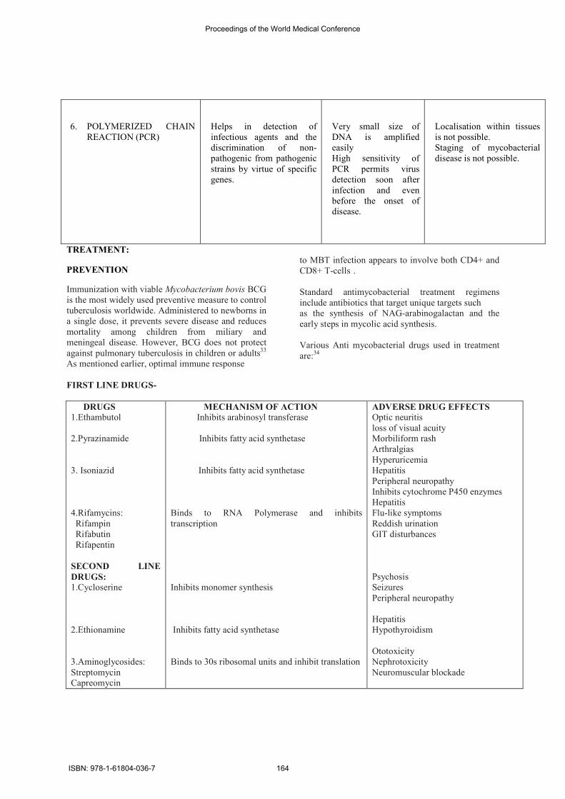
6. POLYMERIZED CHAIN
REACTION (PCR)
Helps in detection of
infectious agents and the
discrimination of non-
pathogenic from pathogenic
strains by virtue of specific
genes.
Very small size of
DNA is amplified
easily
High sensitivity of
PCR permits virus
detection soon after
infection and even
before the onset of
disease.
Localisation within tissues
is not possible.
Staging of mycobacterial
disease is not possible.
TREATMENT:
PREVENTION
Immunization with viable Mycobacterium bovis BCG
is the most widely used preventive measure to control
tuberculosis worldwide. Administered to newborns in
a single dose, it prevents severe disease and reduces
mortality among children from miliary and
meningeal disease. However, BCG does not protect
against pulmonary tuberculosis in children or adults33
As mentioned earlier, optimal immune response
to MBT infection appears to involve both CD4+ and
CD8+ T-cells .
Standard antimycobacterial treatment regimens
include antibiotics that target unique targets such
as the synthesis of NAG-arabinogalactan and the
early steps in mycolic acid synthesis.
Various Anti mycobacterial drugs used in treatment
are:34
FIRST LINE DRUGS-
DRUGS 1.Ethambutol
2.Pyrazinamide
3. Isoniazid
4.Rifamycins:
Rifampin
Rifabutin
Rifapentin
SECOND LINE
DRUGS:
1.Cycloserine
2.Ethionamine
3.Aminoglycosides:
Streptomycin
Capreomycin
MECHANISM OF ACTION Inhibits arabinosyl transferase
Inhibits fatty acid synthetase
Inhibits fatty acid synthetase
Binds to RNA Polymerase and inhibits
transcription
Inhibits monomer synthesis
Inhibits fatty acid synthetase
Binds to 30s ribosomal units and inhibit translation
ADVERSE DRUG EFFECTS
Optic neuritis
loss of visual acuity
Morbiliform rash
Arthralgias
Hyperuricemia
Hepatitis
Peripheral neuropathy
Inhibits cytochrome P450 enzymes
Hepatitis
Flu-like symptoms
Reddish urination
GIT disturbances
Psychosis
Seizures
Peripheral neuropathy
Hepatitis
Hypothyroidism
Ototoxicity
Nephrotoxicity
Neuromuscular blockade
Proceedings of the World Medical Conference
ISBN: 978-1-61804-036-7 164

Kanamycin
Amikacin
4.Fluoroquinolones:
Ciprofloxacin
Ofloxacin
Gatifloxacin
Levofloxacin
Moxifloxacin
5.Aminosalicylic acid
COMBINATION
DRUGS:
Rifamate
Rifater
Inhibits topo-isomerase II ( DNA Gyrase), thereby
releasing DNA with straggered double stranded
breaks
Competitive para-amino benzoic acid antagonist
Isoniazid+Rifampin
Isoniazid+Rifampin+pyrazinamide
Nausea
Abdominal rashes
Restlessness
Confusion
GIT disturbances
DOTS: Daily observed treatment schedule is also
being followed in TB cases.
CONCLUSION
To conclude, mouth lesions of tuberculosis are rare.
Nevertheless, the fact that tuberculosis may manifest
in oral tissues, together with its non-specific clinical
presentation and its infectious implications, demands
an adequate acquaintance with its oral lesions. The
dentist need to be aware that TB may occur in the
oral cavity and should be included in the differential
diagnosis of any ulcerated, indurated and non healing
lesion of the oral cavity, especially in the lower
socio-economic groups. Early diagnosis and
treatment planning may help in preventing
complications and death resulting due to this
common infectious multi systemic disorder.
REFERENCES
1.Memon GA, Khushk IA. Primary Tuberculosis of
tongue. J Coll Physicians Surg Pak 2003; 13; 604-5
2. Mignogna MD, Muzio LL, Favia G, Ruoppo E,
Sammartino G, Zarrelli C. Oral tuberculosis: a
clinical evaluation of 42 cases. Oral disease 2000; 6:
25-30.
3.Topazian RG, Goldberg MH: Oral and
maxillofacial Infections, 2nd
edition, WB Saunders
Co., pg 413, 1987
4. Eng H.L., Lu S.Y., Yang C.H., Chen W.J. Oral
tuberculosis. Oral Surg Oral Med Oral Pathol Oral
Radiol Endod 1996;81:415-20.
5. Hock-Liew E, Shin-Yu L, Chuang-HwaY, Wei-Jen
C. oral tuberculosis. Oral surgery, Oral Medicine,
Oral Pathology, Oral Radiology and Endodontics
1996; 81:415-420
6.Nwoku LA, Kekere-Ekun TA, Sawyer DR, Olude
OO. Primary tuberculous osteomyelitis of the
mandible. J Maxillofacial Surg 1983; 11; 46-48
7. S.R.Prabhu and S.k.Sengupta. Bacterial infections
due to mycobacteria A.Tuberculosis. Oral diseases in
the tropics. Oxford university press, 1993: 195-202
8.Mani NJ. Tuberculosis initially diagnosed by
asymptomatic oral lesions. Report of three cases. J
Oral Med 1985; 40; 39-42
9.Shafer WG, Hine MK, Levy BM. A Textbook of
Oral Pathology; 5th
edition; WB Saunders Company,
Philadelphia.
10. Martin S Greenberg, Micheal Glick. Burkitts Oral
Medicine Diagnosis and Treatment 10th
edition.
11. 1. Dye C, Scheele S, Dolin P et al. Global burden
of tuberculosis: estimated incidence, prevalence, and
mortality by country. JAMA 1999; 282: 677-- 86.
12. . The world health report 2006. http://
www.int/GlobalAtlas/predefined Reports/ TB/
index.asp?strselect edCountry=ind
13.Miziara ID. Tuberculosis affecting the oral cavity
in Brazilian HIV infected patients. Oral Surg Oral
Med Oral Path Oral Rad Endo 2005; 100; 179-182
14 A.P.Bhatt and Amrita Jayakrishnan. Tuberculous
osteomyelitis of the mandible: a case report.
International journal of Paediatric Dentistry 2001; 11:
304-308..
15.Das P, Suri V, Arora R, Kulkarni K, Kumar K.
Primary lingual tuberculosis mimicking malignancy:
a report of two cases and review of literature. Th e
Internet journal of Pathology 2007;6(2). available at:
htt p://www.ispub.com/ostia/index. php?xmlfi
lepath=journals/ijpa/vol6no2/lingual.xml
16. Ramesh V. Tuberculoma of the tongue presenting
as macroglossia. Cutis 1997; 60: 201-202.
Proceedings of the World Medical Conference
ISBN: 978-1-61804-036-7 165

17.Farber JE, Friedland E & Jacobs WF:
Tuberculosis of the tongue, Am Rev Tuber, 766:
1940-42
18.Thilander H, Wennstrom A: Tuberculosis of the
mouth and the surrounding tissues, Oral Surg Oral
Med Oral Path 1956; 9; 858-70
19.Bipin Kumar : Tuberculosis of the oral cavity
affecting Alveolus: A case report in Dentistry: 2011
20. Medhuri, Chander Mohan, Sharma ML. Posterior
oropharyngeal wall tuberculosis. Indian J of
Otolaryngol and Head and Neck Surg. 2002;54: 152-
153.
21.Bjorlin G : Oral Tuberculosis: Odontologisk
Revy, 18: 395-399
22. Bangalore Varadhan Karthikeyan, Avani Raju
Pradeep, C.G. Dileep Sharma. Primary tuberculous
Gingival Enlargement: A Rare Entity. Journal of
Canadian Dental Association 2006; 72(7): 645-648.
23.Tan KK. Tuberculosis lymphadenitis in
Singapore. Sing Med J 1988; 29; 441-416
24.Dandapat MC, Mishra BM, Dash SP, Kar PK.
Peripheral lymph node Tuberculosis : A Review of
80 cases. Br J Surg 1990; 77; 911-912
25.Van de Schoot L, Aronson DC, Behrendt H, Bras
J. The role of FNAC in children with persistent or
suspicious lymphadenopathy. J Pediat Surg 2001; 36;
7-11
26. K.B.Gupta, M.Manchanda, S.P.S.Yadav and
A.Mittal. Tubercular Osteomyelitis of Mandible.
Indian journal of tuberculosis 2005; 52:147-150.
27. Chapotel S. Tuberculose mandibularie. Rev
Odent 1930; 51:444-445
28.S elcuk A, Oruk V, Dere H, Boztepe F, Seckin S:
Tuberculous parotitis: Can be left silent for a long
time. KBB-Forum, 2006; 5(3):130-132.
29. Birkent H, Karahatay S, Akcam T, Durmaz A,
Ongoru O: Primary parotid Tuberculosis mimicking
parotid Neoplasm: A case report. Journal of Medical
Case Reports, 2008;2:62-63.
30. Suleiman AM: Tuberculous parotitis: report of 3
cases. British Journal of Oral and Maxillofacial
Surgery, 2001, 39(4):320-323.
31. Bakhshi GD, Shaikh AS, Borisa AD, Jamadar
NM, Singh AG, Vora SH: Primary Tuberculosis of
Paritid Gland Mimicking Parotid Tumour. Bombay
Hospital Journal, (Special Issue), 2009;14-16.
32.Kanwar Deep Singh Nanda, Anurag Mehta,
Mohita Marwaha, Manpreet Kalra, Jasmine Nanda: A
Disguised Tuberculosis of the oral Buccal mucosa.
Journal of Clinical and Diagnostic Research 2011:
Apr, vol -5 (2); 357-360
33.Rodregues LC, Diwan VK, Wheeler JG.
Protective effect of BCG against tuberculous
meningitis and miliary tuberculosis: a meta-analysis.
Int J Epidemiology 1993;22:1154-8.
34. Abramowicz M. (ed). Drugs for tuberculosis.
Treat Guidel Med Lett. 2004 Dec;2(28):83-8.
s
Proceedings of the World Medical Conference
ISBN: 978-1-61804-036-7 166