Embed Size (px)
Citation preview
assayed using the Illumina DASL microarray process specifically designed for use with FFPEsamples. The same treatment area was sampled in both active and quiescent stages inindividual UC patients in order to elucidate patterns of gene expression underlying UCdisease. 26/29 (90%) of the FFPE samples were successfully arrayed. RESULTS: Microarrayanalysis demonstrates that UC patients in the active stage present a transcriptional signaturedistinctive from the quiescent stage. Genes differentially expressed in the active stage (1.5fold or greater) were tested for functional enrichment analysis using the on-line tool DAVIDsponsored by the NIAID, and were found to be highly enriched for genes related to inflamma-tion (e.g., S100A8, S100A9, IL1B, IL6, IL8, and SELL; DAVID p , 4.9E-05), genes involvedwith chemotaxis and leukocyte migration (e.g. CXCL5, CCL8, CX3CL1, CXCL12, andDOCK2; p , 5E-06), and, interestingly, vitamin D response genes (ALPL, CYP27B1, PTGS2,CDKN2D, IL1B, PMF1, TGFB1; DAVID p=2.5E-4) suggesting a previously unappreciatedrole for the innate immune response in the ulcerative colitis inflammatory process. Inaddition, we found that some previously described cancer related genes such as MXI1and ANGPTL2 were differentially expressed between active and quiescent stages as well.CONCLUSIONS: Analysis of the gene expression signatures associated with the active andquiescent stage of ulcerative colitis at the precise sites of injury has allowed us to clearlydistinguish the two stages despite, in some cases, repeated flares occurring chronically atthe same site. The complete return to baseline transcription corresponding to an apparentlynon-inflammatory state common to all samples in the quiescent stage does not suggest anyintrinsic site-specific predisposition to UC injury. Further work will be to analyze tissuetaken from healthy controls and compare them directly to UC quiescent samples to confirmthis finding.
Tu1693
Indigenous Australians (IA) Have a Distinctive Gut Microbial ProfileCompared to Patients With Inflammatory Bowel Disease (IBD) and NonIndigenous ControlsGuru Iyngkaran, Seungha Kang, Chris McSweeney, Suresh Sivanesan, NadarajahKangaharan, Mark Morrison, Finlay A. Macrae
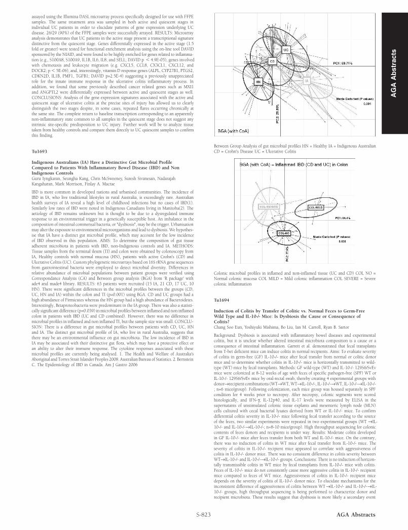
IBD is more common in developed nations and urbanised communities. The incidence ofIBD in IA, who live traditional lifestyles in rural Australia, is exceedingly rare. Australianhealth surveys of IA reveal a high level of childhood infections but no cases of IBD(1).Similarly low rates of IBD were noted in Indigenous Canadians living in Manitoba(2). Theaetiology of IBD remains unknown but is thought to be due to a dysregulated immuneresponse to an environmental trigger in a genetically susceptible host. An imbalance in thecomposition of intestinal commensal bacteria, or "dysbiosis", may be the trigger. Urbanisationmay alter the exposure to environmental microorganisms and lead to dysbiosis. We hypothes-ise that IA have a distinct gut microbial profile, which may account for the low incidenceof IBD observed in this population. AIMS: To determine the composition of gut tissueadherent microbiota in patients with IBD, non-Indigenous controls and IA. METHODS:Tissue samples from the terminal ileum (TI) and colon were obtained by colonoscopy fromIA, Healthy controls with normal mucosa (HN), patients with active Crohn's (CD) andUlcerative Colitis (UC). Custom phylogenetic microarrays based on 16S rRNA gene sequencesfrom gastrointestinal bacteria were employed to detect microbial diversity. Differences inrelative abundance of microbial populations between patient groups were verified usingCorrespondance Analysis (CA) and Between group analysis (BGA) from ‘R package' withade4 and made4 library. RESULTS: 63 patients were recruited (15 IA, 21 CD, 17 UC, 10HN). There were significant differences in the microbial profiles between the groups (CD,UC, HN and IA) within the colon and TI (p=0.001) using BGA. CD and UC groups had ahigh abundance of Firmicutes whereas the HN group had a high abundance of Bacteroidetes.Interestingly, Betaproteobacteria were predominant in the IA group. There was also a statisti-cally significant difference (p=0.039) inmicrobial profiles between inflamed and non inflamedcolon in patients with IBD (UC and CD combined). However, there was no difference inmicrobial profiles in inflamed and non inflamed TI, but the sample size was small. CONCLU-SION: There is a difference in gut microbial profiles between patients with CD, UC, HNand IA. The distinct gut microbial profile of IA, who live in rural Australia, suggests thatthere may be an environmental influence on gut microbiota. The low incidence of IBD inIA may be associated with their distinctive gut flora, which may have a protective effect oran ability to alter their immune response. The cytokine responses associated with thesemicrobial profiles are currently being analysed. 1. The Health and Welfare of Australia'sAboriginal and Torres Strait Islander Peoples 2008. Australian Bureau of Statistics. 2. BernsteinC. The Epidemiology of IBD in Canada. Am J Gastro 2006
S-823 AGA Abstracts
Between Group Analysis of gut microbial profiles HN = Healthy IA = Indigenous AustralianCD = Crohn's Disease UC = Ulcerative Colitis
Colonic microbial profiles in inflamed and non-inflamed tissue (UC and CD) COL NO =Normal colonic mucosa COL MILD = Mild colonic inflammation COL SEVERE = Severecolonic inflammation
Tu1694
Induction of Colitis by Transfer of Colitic vs. Normal Feces to Germ-FreeWild Type and IL-10-/- Mice: Is Dysbiosis the Cause or Consequence ofColitis?Chang Soo Eun, Yoshiyuki Mishima, Bo Liu, Ian M. Carroll, Ryan B. Sartor
Background: Dysbiosis is associated with inflammatory bowel diseases and experimentalcolitis, but it is unclear whether altered intestinal microbiota composition is a cause or aconsequence of intestinal inflammation. Garrett et al. demonstrated that fecal transplantsfrom T-bet deficient mice can induce colitis in normal recipients. Aims: To evaluate severityof colitis in germ-free (GF) IL-10-/- mice after fecal transfer from normal or colitic donormice and to determine whether colitis in IL-10-/- mice is horizontally transmitted to wild-type (WT) mice by fecal transplants. Methods: GF wild-type (WT) and IL-10-/- 129S6/SvEvmice were colonized at 8-12 weeks of age with feces of specific pathogen-free (SPF) WT orIL-10-/- 129S6/SvEv mice by oral-rectal swab, thereby creating 4 experimental groups withdonor→recipient combinations (WT→WT,WT→IL-10-/-, IL-10-/-→WT, IL-10-/-→IL-10-/-; n=6 mice/group). Following colonization, each mice group was housed separately in SPFcondition for 4 weeks prior to necropsy. After necropsy, colonic segments were scoredhistologically, and IFN-γ, IL-12p40, and IL-17 levels were measured by ELISA in thesupernatants of unstimulated colonic tissue explants and mesenteric lymph node (MLN)cells cultured with cecal bacterial lysates derived from WT or IL-10-/- mice. To confirmdifferential colitis severity in IL-10-/- mice following fecal transfer according to the sourceof the feces, two similar experiments were repeated in two experimental groups (WT →IL-10-/- and IL-10-/-→IL-10-/-; n=8-10 mice/group). High throughput sequencing for coloniccontents of feces donors and recipients is under way. Results: Moderate colitis developedin GF IL-10-/- mice after feces transfer from both WT and IL-10-/- mice. On the contrary,there was no induction of colitis in WT mice after fecal transfer from IL-10-/- mice. Theseverity of colitis in IL-10-/- recipient mice appeared to correlate with aggressiveness ofcolitis in IL-10-/- donor mice. There was no consistent difference in colitis severity betweenWT→IL-10-/- and IL-10-/-→IL-10-/- groups. Conclusions: There is no induction of horizon-tally transmissible colitis in WT mice by fecal transplants from IL-10-/- mice with colitis.Feces of IL-10-/- mice do not consistently cause more aggressive colitis in IL-10-/- recipientmice compared to feces of WT mice. Aggressiveness of colitis in IL-10-/- recipient micedepends on the severity of colitis of IL-10-/- donor mice. To elucidate mechanisms for theinconsistent difference of aggressiveness of colitis between WT→IL-10-/- and IL-10-/-→IL-10-/- groups, high throughput sequencing is being performed to characterize donor andrecipient microbiota. These results suggest that dysbiosis is more likely a secondary event
AG
AA
bst
ract
s
AG
AA
bst
ract
srather than a primary cause of colitis and that IBD is dependent on interacting microbialand genetic factors.
Tu1695
Saccharomyces Boulardii Prevents the Antibiotic Induced Changes in ColonicMicrobiotaAlexander Swidsinski, Vera Loening-Baucke, Sonja Swidsinski
Background: The impact of antibiotics on colonic microbiota is poorly characterized. Meth-ods: Three groups of women (N=10 each) treated for bacterial vaginosis were investigated. Metronidazole 3x400mg/day + ciprofloxacin 2x500mg/day were given for 2 weeks. GroupI received antibiotics only, Gr.II received Saccharomyces boulardii(Sb) concomitant to, andGr.III received Sb subsequent to antibiotic therapy. A 250 mg capsule Sb Perenterol®Biocodex was given 3xdaily for two weeks. Microbiota were investigated using structurefunctional FISH analysis of Carnoy fixated stool cylinders. Stools were collected at days-90,-60,-30,7,14,28,42,56, and 70 as related to the start of antibiotic therapy. 250 bacterialFISH probes were tested and 82 were selected for longitudinal analysis. Results: Bacteroides,F. prausnitzii and Roseburia (habitual groups) were present in all patients (10-30% of thefecal mass each). The occurrence of other bacteria was occasional. The composition ofmicrobiota prior to antibiotics was stable in repeated investigations of the same patient,despite high interindividual variability. Antibiotic therapy suppressed all bacterial groups,and some of the microbiota previously not detected, emerged in marginal concentrations(C.viridae, Streptococcus, Staphylococcus, Bif. longum ). Bifidobacteriaceae, Enterobacteriacae orC.difficile groups increased in the first two to four weeks after the end of the therapy (p ,0.01)and returned to initial levels with recovery of the habitual bacterial groups. Sb concomitantwith antibiotics reduced the antibiotic associated suppression of bacteria. After one weekof antibiotic therapy, concentrations of F.prausnitzii dropped in Gr.I, II, and III, respectively,from a median of 13.6±3.7 to 3.1±2.0, 8±4.1, and 3±2.1 x 10⁹bacteria/ml. The decrease inmicrobial concentrations in Gr. II receiving Sb concomitant to antibiotics was moderate anddiffered significantly from the decrease in Gr.I and III (p,0.001). The microbiota recoveredquicker in groups receiving Sb. The habitual bacteria reached initial values 4 weeks afterthe end of antibiotics in all groups treated with Sb In Group I, F. prausnitzi was still reducedto 9.8±2.8 x 10⁹bacteria/ml and was significantly lower than prior to antibiotics (p=0.04).The composition of microbiota returned quicker to the individual initial profiles in thegroups receiving Sb In regard to the 82 investigated bacterial groups, the median numberof mismatches in composition of microbiota prior to and after antibiotic therapy was 8.7;5.2 (p=0.05) and 2.4 (p,0.001) in Gr.I, II and III, respectively. The difference betweenpre- and postantibiotic period was significantly higher in Gr.I, receiving no Sb. Conclusions: S.boulardii prevents/reduces effectively the antibiotic-associated changes in colonic microbiota,when given concomitant/subsequent to antibiotic therapy.
Tu1696
Folate Prevents CEACAM6 Abnormal Expression and Subsequent Adherent-Invasive E. Coli- Induced Inflammation in Ceabac10 MiceJérémy Denizot, Alexis Desrichard, Allison Agus, Arlette Darfeuille-Michaud, NicolasBarnich
Background: Abnormal expression of CEACAM6 in Crohn's disease (CD) patients allowsAdherent-Invasive Escherichia coli (AIEC) to colonize gutmucosa, leading to the developmentof inflammation. Folate are key vitamins involved in maintenance of DNA methylationpatterns in mammalians and folate deficiency is often observed in CD patients, especiallythose with ileal involvement of the disease. In vitro, CEACAM6 expression is regulated byHypoxia Inductible Factor-1 (HIF-1) in a DNA methylation dependent manner. We thusinvestigated the importance of folate in the control of CEACAM6 expression in vivo and inAIEC-induced inflammation Methods: Global methylation status of CEACAM6 promoterwas analyzed using bisulfite/SnapShot sequencing in ileal or colonic enterocytes. TransgenicCEABAC10 mice expressing human CEACAM6 fed normal or methyl-donor deficient dietMDD (folate and B12 vitamin deficient) were supplemented with folic acid (FA). Colonicenterocytes were isolated and CEACAM6 and hif-1α expression were measured by RT-qPCRand Western-blot. AIEC colonization and gut inflammation were evaluated in mice orallychallenged with 10e9 bacteria. Results: Methyl-donor deficient diet (MDD) led to a significantdecrease in methylation levels of CpG-containing HIF-1 Responsive Elements (HRE) inCEACAM6 promoter, leading to significant increase in CEACAM6 mRNA and protein incolonic enterocytes of CEABAC10mice compared to a normal diet. After AIEC LF82 infection,important weight loss was observed in mice fed a MDD but not in mice fed a conventionaldiet. Three days post-infection, higher numbers of AIEC LF82 were associated with colonicmucosa of MDD-fed mice compared to normal diet-fed mice, due to a higher CEACAM6protein expression. This was associated with significant higher release of pro-inflammatorycytokines IL-6, KC and IL-1β from colonic mucosa in MDD-fed mice compared to normaldiet-fed mice. Folate supplementation increased methylation level of CpG-containing HREin CEACAM6 promoter, leading to decrease CEACAM6 expression. Conclusion: Folatedeficiency induces hypomethylation of HRE-containing CpG in CEACAM6 promoter, whichcorrelates with higher CEACAM6 expression. These findings indicate that abnormal CEA-CAM6 expression in ileal mucosa of CD patients could be related to folate deficiency. Folatesupplementation could be effective to achieve and maintain inflammation remission inpatients with ileal involvement of the disease by preventing CEACAM6 abnormal expressionand subsequent AIEC-mediated inflammation.
S-824AGA Abstracts
Tu1697
Viral Pathogens Pull the Necrotic Trigger in a TNF-Receptor IndependentManner in Caspase-8ΔIEC MiceClaudia Günther, Helmut Neumann, Guiwei He, Eva Martini, Markus F. Neurath,Christoph Becker
Background: Intestinal epithelial necroptosis has recently been described as a potentialpathogenic mechanism driving Crohn's disease. It was shown that lack of caspase-8 inintestinal epithelial cells leads to enhanced necroptosis and loss of immune homeostasis inthe gut, resulting in spontaneous development of terminal ileitis. However, the mechanismtriggering small intestinal epithelial necroptosis is still unknown. Aims: In this study weinvestigated the impact of death receptor ligands (DRL) and PAMPs (Pathogen-associatedmolecular pattern) on Rip3mediated intestinal necroptosis. Methods: Caspase-8ΔIEC, Rip3-/-Caspase-8ΔIEC, Tnf-R1-/-Caspase-8ΔIECmice and intestinal organoids were treated with PIC(PolyIC) and LPS (Lipopolysaccharide). Mice were examined bymeasuring body temperature,weight loss and monitoring development of diarrhoea. Tissue damage was investigated byimmunohistochemistry of gut specimen, western blot and qPCR. Results: We could demon-strate that programmed necrosis of intestinal epithelial cells, induced in the absence ofcaspase-8, is mediated via the Rip3 since Rip3-/-Caspase-8 ΔIEC double knock out micewere completely rescued regarding the loss of Paneth cells and intestinal inflammation aswell as cell death. We could further demonstrate that additional deletion of Tnf-R1 did notrescue the phenotype of Caspase-8ΔIEC mice, suggesting that caspase-8 in vivo is eitheractivated by a Tnf-R1 independent pathway or that redundant receptor signals are presentin the steady state gut. Indeed we could show that targeting of Toll like receptors (TLR) byinjection of PIC and LPS into mice induced a fast and dramatic villous atrophy and severedestruction of the small bowel of Caspase-8ΔIEC mice as compared to control littermates,leading to the death of the former mice within 6 hours. Immunohistochemistry analysisrevealed an excessive number of TUNEL positive but caspase-3 negative dying epithelialcells after TLR-stimulation in Caspase-8ΔIECmice, but not in Rip3-/-Caspase-8ΔIEC, indicat-ing that this form of cell death is due to Rip3-mediated necroptosis. Moreover we discoveredthat PIC triggered necroptosis was directly mediated via TLR3, since Tnf-R1-/-Caspase-8ΔIEC mice were not protected against PAMP induced cell death. At least we could showin vitro that PIC induced necroptosis was independent of immune cells since Caspase-8 ΔIECderived organoids, but not control organoids, were sensitive to PAMP induced Rip3-mediatedcell death. Conclusion: Our data demonstrate that Rip3-mediated cell death in the smallintestine of Caspase-8ΔIEC mice is triggered by Pattern-Recognition Receptors. Moreoverwe could show that dsRNA activation of the TLR3 induce epithelial cell death in a Tnf-independent manner. This indicates that viral pathogens are the main triggering effectorsfor small intestinal inflammation in Caspase-8ΔIEC mice.
Tu1698
Inflammation-Induced Regulatory Pathways of Oxidative Stress-Responses inCommensal Escherichia coli Impair Bacterial Motility, Biofilm Formation andAttenuate Experimental ColitisSandrine Y. Tchaptchet, Ting-Jia Fan, Laura E. Goeser, Ryan B. Sartor, Jonathan J. Hansen
BACKGROUND:Dysregulated immune responses to commensal intestinal bacteria contributeto the development of human inflammatory bowel diseases (IBDs) and experimental colitis,conditions that are characterized by increased reactive oxygen intermediates in inflamedtissues. Experimental colitis is associated with increased expression of the small regulatoryRNA oxyS in luminal Escherichia coli that protects bacteria from oxidative stress and inhibitsexpression of rpoS, a subunit of RNA polymerase that impairs growth of E. coli in mouseintestine. In addition to potentially enhancing growth of E. coli in the intestine, oxyS alsoreduces bacterial motility, which is linked to decreased biofilm formation. HYPOTHESIS:Expression of oxyS in commensal luminal E. coli during colitis inhibits rpoS expression,promotes bacterial survival, but decreases bacterial motility/biofilm formation and thereforehost inflammatory responses. METHODS: OxyS and rpoS expression in cecal bacteria fromwild-type (WT) and Il-10-/- (KO) germ-free mice monoassociated with the commensalmurine E. coli isolate, NC101, was measured by real-time PCR. Biofilm formation andmotility of NC101 and NC101 lacking oxyS (NC101 ΔoxyS) or rpoS (NC101ΔrpoS) weredetermined by crystal violet staining of biofilms on a polystyrene surface and by measuringcolony diameter on soft agar, respectively. Histological inflammation in colon sections,concentrations of NC101 in luminal contents, and adaptive immune responses to NC101antigens in ex vivo-stimulated mesenteric lymph node cells (MLN) from KO and WT micemonoassociated with NC101, NC101ΔoxyS or NC101ΔrpoS were determined using blindedscoring, quantitative culture, and ELISA for IFN- γ, respectively. RESULTS: Cecal E. colioxyS expression was proportional to the severity of colitis in monoassociated mice, butdiffering from our hypothesis did not correspond to decreased rpoS expression. Biofilmformation and motility rates of NC101 were significantly reduced compared to NC101 ΔoxySand NC101ΔrpoS (Table). Contrary to our hypothesis, luminal bacterial densities were notdifferent in mice monoassociated with NC101, NC101 ΔoxyS or NC101ΔrpoS. However,histological inflammation scores were lower in NC101- vs. NC101ΔoxyS- and NC101ΔrpoS-monoassociated KO mice and IFN-γ secretion by NC101 lysate-stimulated MLN cells fromNC101- vs. NC101ΔoxyS- and NC101ΔrpoS-monoassociated KO mice was decreased(Table). CONCLUSIONS: Intestinal inflammation causes commensal E. coli to increase oxySexpression, which does not affect luminal bacterial survival, but similar to rpoS, impairsbacterial motility and biofilm formation in vitro and decreases host immune responses in vivo.Further investigation of luminal microbial adaptation to immune-mediated inflammationmayprovide novel insights into the pathogenesis and treatment of IBDs.Table