Embed Size (px)
Citation preview
TSH Level in PregnancyGita Majdi
PGY4 Endocrinology FellowMay 2014
Outline:
• TFT in pregnancy• What is the normal range for TSH in pregnancy? • Trimester-specific reference ranges
• ATA Guidelines for:- Normal TSH in pregnancy- Who should be screened?
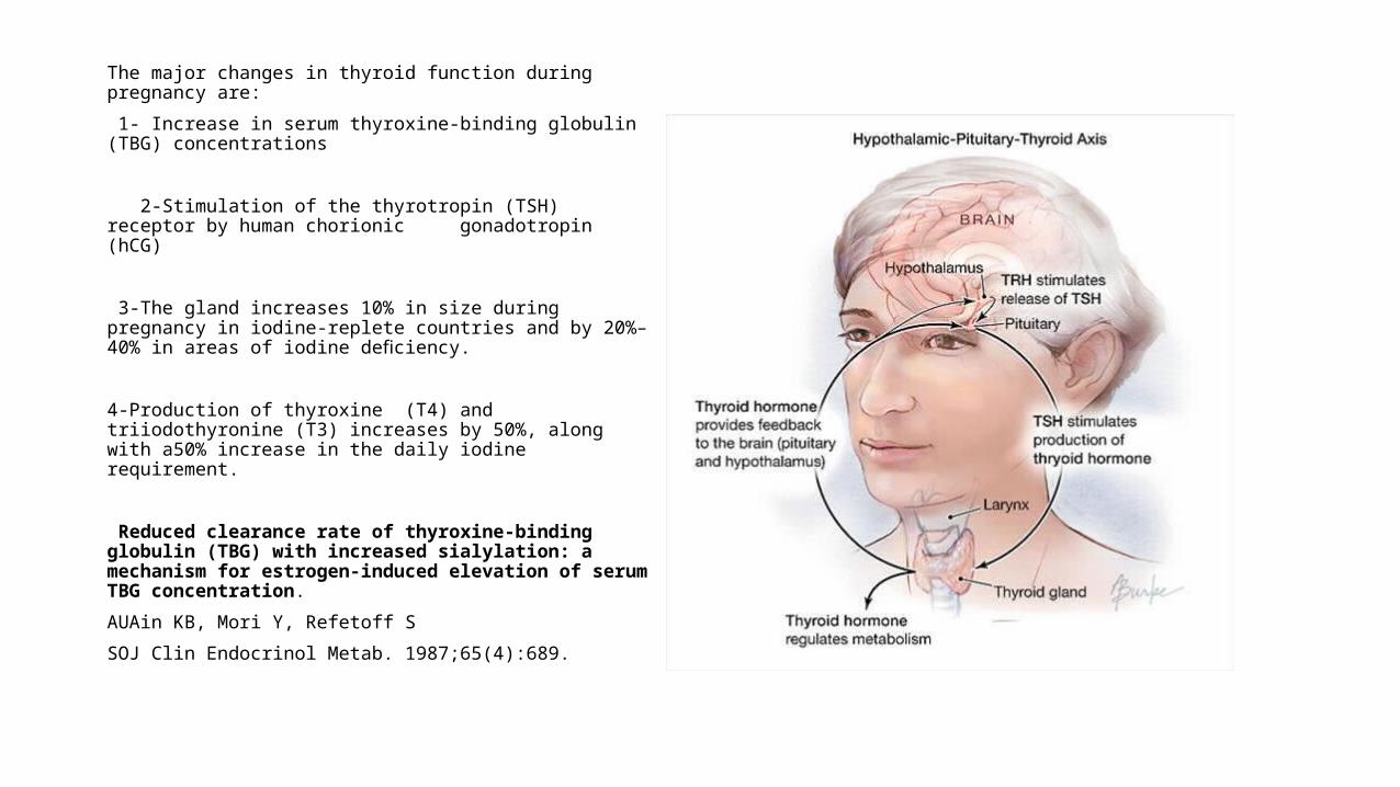
The major changes in thyroid function during pregnancy are:
1- Increase in serum thyroxine-binding globulin (TBG) concentrations
2-Stimulation of the thyrotropin (TSH) receptor by human chorionic gonadotropin (hCG)
3-The gland increases 10% in size during pregnancy in iodine-replete countries and by 20%–40% in areas of iodine deficiency.
4-Production of thyroxine (T4) and triiodothyronine (T3) increases by 50%, along with a50% increase in the daily iodine requirement.
Reduced clearance rate of thyroxine-binding globulin (TBG) with increased sialylation: a mechanism for estrogen-induced elevation of serum TBG concentration.
AUAin KB, Mori Y, Refetoff S
SOJ Clin Endocrinol Metab. 1987;65(4):689.
How do thyroid function tests changeduring pregnancy?• Following conception, circulating total T4 (TT4) and T4 binding globulin (TBG) concentrations
increase by 6–8 weeks and remain high until delivery. • Thyrotropic activity of hCG results in a decrease in serum TSH in the first trimester. • Most studies also report a substantial decrease in serum FT4 concentrations with progression
of gestation . • SerumFT4 measurements in pregnant women are complicated by increased TBG and
decreased albumin concentrations that can cause immunoassays to be unreliable . Therefore the analytical method used for serum FT4 analysis should be taken into consideration.
Glinoer D 1997 The regulation of thyroid function in Pregnancy. Pathways of endocrine adaptation from physiology to pathology. Endocr Rev 18:404–433.
• Timing and Magnitude of Increases in Levothyroxine Requirements during Pregnancy in Women with Hypothyroidism
• Erik K. Alexander, M.D., Ellen Marqusee, M.D., Jennifer Lawrence, M.D., Petr Jarolim, M.D., Ph.D., George A. Fischer, Ph.D., and P. Reed Larsen, M.D.
• N Engl J Med 2004; 351:241-249July 15, 2004DOI: 10.1056/NEJMoa040079
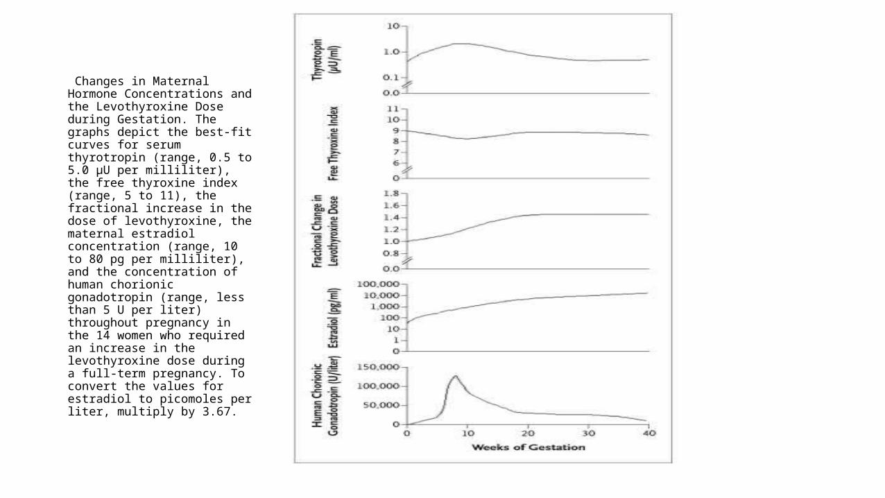
Changes in Maternal Hormone Concentrations and the Levothyroxine Dose during Gestation. The graphs depict the best-fit curves for serum thyrotropin (range, 0.5 to 5.0 μU per milliliter), the free thyroxine index (range, 5 to 11), the fractional increase in the dose of levothyroxine, the maternal estradiol concentration (range, 10 to 80 pg per milliliter), and the concentration of human chorionic gonadotropin (range, less than 5 U per liter) throughout pregnancy in the 14 women who required an increase in the levothyroxine dose during a full-term pregnancy. To convert the values for estradiol to picomoles per liter, multiply by 3.67.
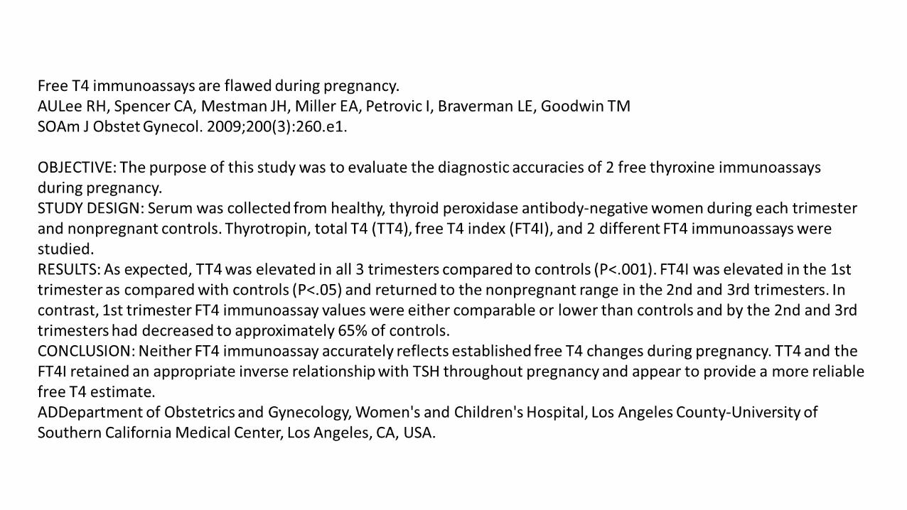
• Measurement of free T4 in the dialysate or ultrafiltrate of serum samples using liquid chromatography/tandem mass spectrometry appears to be the most reliable, and when this method is used, free T4 concentrations were shown to decrease gradually with advancing gestational age, particularly between the first and second trimester.
• Other free T4 assays (and probably free T3 assays) frequently fail to meet performance standards in pregnant patients, owing to increases in TBG and decreases in albumin concentrations that cause the immunoassay to be unreliable.
• When free T4 measurements appear discordant with TSH measurements, serum total T4 should be measured.
• Total T4 and T3 levels during pregnancy are 1.5-fold higher than in nonpregnant women due to TBG excess.
• Thus a normal reference range for pregnancy should be usedTo compensate, some kits have provided different free T4 normal ranges for pregnant patients, usually lower than those of nonpregnant patients.
Source: Uptoate
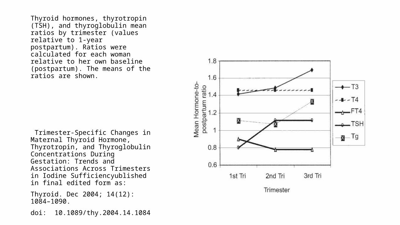
Thyroid hormones, thyrotropin (TSH), and thyroglobulin mean ratios by trimester (values relative to 1-year postpartum). Ratios were calculated for each woman relative to her own baseline (postpartum). The means of the ratios are shown.
Trimester-Specific Changes in Maternal Thyroid Hormone, Thyrotropin, and Thyroglobulin Concentrations During Gestation: Trends and Associations Across Trimesters in Iodine Sufficiencyublished in final edited form as:
Thyroid. Dec 2004; 14(12): 1084–1090.
doi: 10.1089/thy.2004.14.1084
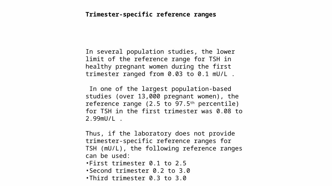
Trimester-specific reference ranges
In several population studies, the lower limit of the reference range for TSH in healthy pregnant women during the first trimester ranged from 0.03 to 0.1 mU/L .
In one of the largest population-based studies (over 13,000 pregnant women), the reference range (2.5 to 97.5th percentile) for TSH in the first trimester was 0.08 to 2.99mU/L .
Thus, if the laboratory does not provide trimester-specific reference ranges for TSH (mU/L), the following reference ranges can be used:•First trimester 0.1 to 2.5•Second trimester 0.2 to 3.0•Third trimester 0.3 to 3.0
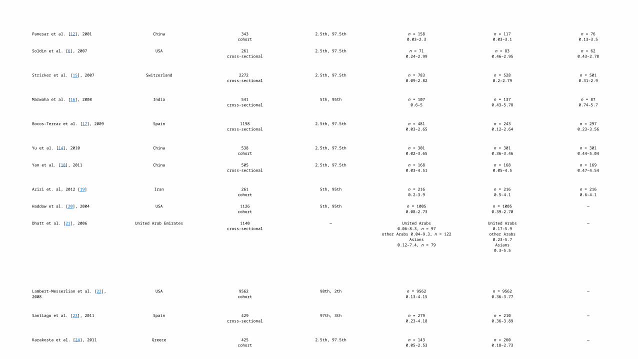
Panesar et al. [12], 2001 China 343cohort
2.5th, 97.5th n = 1580.03–2.3
n = 1170.03–3.1
n = 760.13–3.5
Soldin et al. [6], 2007 USA 261cross-sectional
2.5th, 97.5th n = 710.24–2.99
n = 830.46–2.95
n = 620.43–2.78
Stricker et al. [15], 2007 Switzerland 2272 cross-sectional
2.5th, 97.5th n = 7830.09–2.82
n = 5280.2–2.79
n = 5010.31–2.9
Marwaha et al. [16], 2008 India 541cross-sectional
5th, 95th n = 1070.6–5
n = 1370.43–5.78
n = 870.74–5.7
Bocos-Terraz et al. [17], 2009 Spain 1198cross-sectional
2.5th, 97.5th n = 4810.03–2.65
n = 2430.12–2.64
n = 2970.23–3.56
Yu et al. [14], 2010 China 538cohort
2.5th, 97.5th n = 3010.02–3.65
n = 3010.36–3.46
n = 3010.44–5.04
Yan et al. [18], 2011 China 505cross-sectional
2.5th, 97.5th n = 1680.03–4.51
n = 1680.05–4.5
n = 1690.47–4.54
Azizi et. al, 2012 [19] Iran 261cohort
5th, 95th n = 2160.2–3.9
n = 2160.5–4.1
n = 2160.6–4.1
Haddow et al. [20], 2004 USA 1126cohort
5th, 95th n = 10050.08–2.73
n = 10050.39–2.70
—
Dhatt et al. [21], 2006 United Arab Emirates 1140cross-sectional
— United Arabs0.06–8.3, n = 97
other Arabs 0.04–9.3, n = 122Asians
0.12–7.4, n = 79
United Arabs0.17–5.9
other Arabs0.23–5.7
Asians0.3–5.5
—
Lambert-Messerlian et al. [22], 2008 USA 9562 cohort
98th, 2th n = 95620.13–4.15
n = 95620.36–3.77
—
Santiago et al. [23], 2011 Spain 429cross-sectional
97th, 3th n = 2790.23–4.18
n = 2100.36–3.89
—
Karakosta et al. [24], 2011 Greece 425cohort
2.5th, 97.5th n = 1430.05–2.53
n = 2600.18–2.73
—
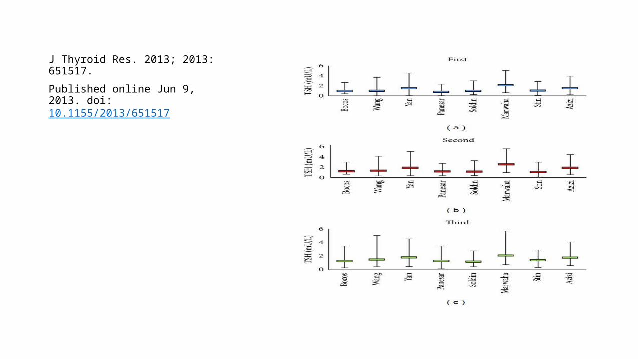
J Thyroid Res. 2013; 2013: 651517.
Published online Jun 9, 2013. doi: 10.1155/2013/651517
• The World Health Organization (WHO) recommends 250 mcg of iodine daily during pregnancy and lactation.
The Institute of Medicine recommends daily iodine intake of 220 mcg during pregnancy and 290 mcg during lactation. For women in the US to achieve this level of daily intake, the ATA recommends that women from the US receive a supplement of 150 mcg of iodine daily during pregnancy and lactation, which is the dose included in the majority of prenatal vitamins marketed in the US . The tolerable upper intake amount for iodine, as established by European and US expert committees, ranges from 600 to 1100 mcg daily for adults and pregnant women >19 years of age.
Guidelines of the American Thyroid Associationfor the Diagnosis and Management of Thyroid DiseaseDuring Pregnancy and PostpartumThe American Thyroid Association Taskforce on Thyroid Disease During Pregnancy and PostpartumAlex Stagnaro-Green (Chair),1 Marcos Abalovich,2 Erik Alexander,3 Fereidoun Azizi,4 Jorge Mestman,5Roberto Negro,6 Angelita Nixon,7 Elizabeth N. Pearce,8 Offie P. Soldin,9Scott Sullivan,10 and Wilmar Wiersinga11
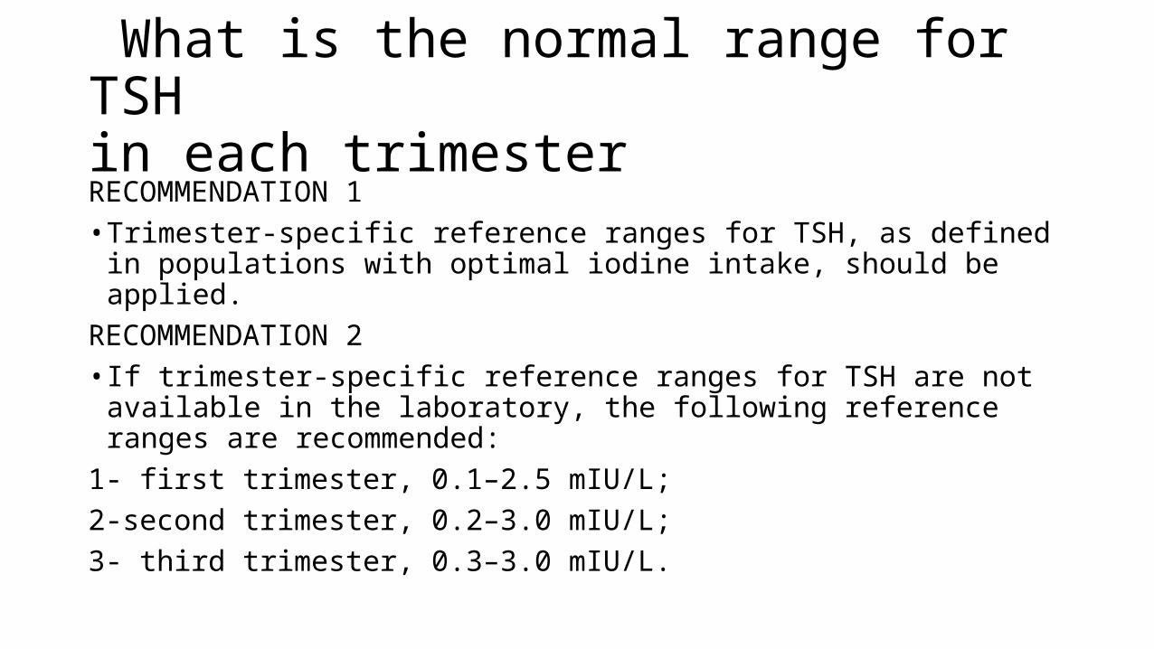
What is the normal range for TSHin each trimesterRECOMMENDATION 1• Trimester-specific reference ranges for TSH, as defined in populations
with optimal iodine intake, should be applied.RECOMMENDATION 2• If trimester-specific reference ranges for TSH are not available in the
laboratory, the following reference ranges are recommended:1- first trimester, 0.1–2.5 mIU/L; 2-second trimester, 0.2–3.0 mIU/L;3- third trimester, 0.3–3.0 mIU/L.
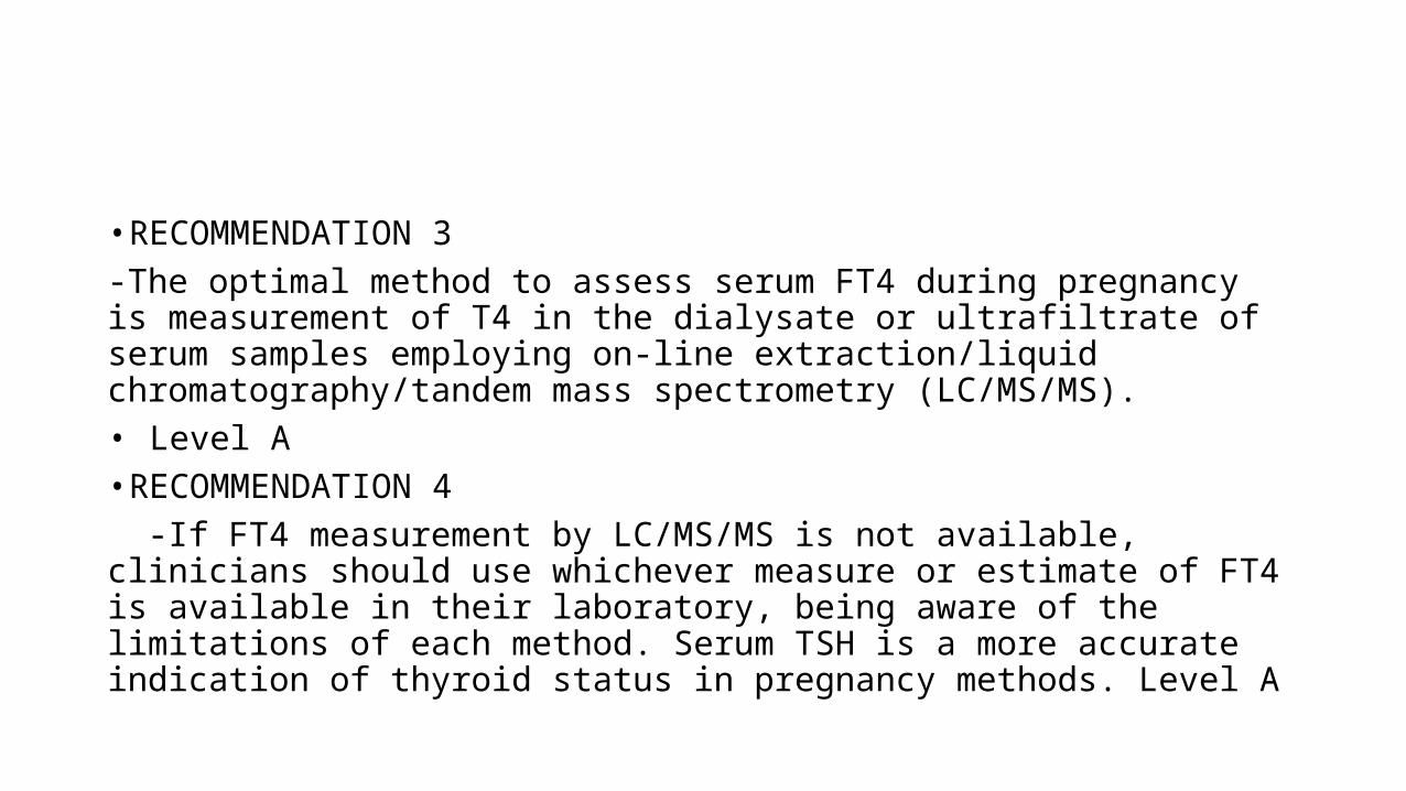
• RECOMMENDATION 3-The optimal method to assess serum FT4 during pregnancy is measurement of T4 in the dialysate or ultrafiltrate of serum samples employing on-line extraction/liquid chromatography/tandem mass spectrometry (LC/MS/MS).• Level A• RECOMMENDATION 4 -If FT4 measurement by LC/MS/MS is not available, clinicians should use whichever measure or estimate of FT4 is available in their laboratory, being aware of the limitations of each method. Serum TSH is a more accurate indication of thyroid status in pregnancy methods. Level A
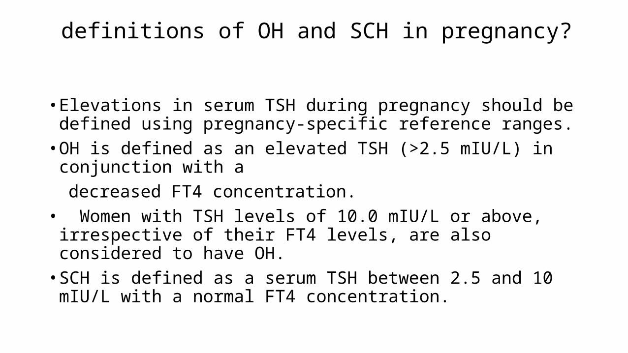
definitions of OH and SCH in pregnancy?
• Elevations in serum TSH during pregnancy should be defined using pregnancy-specific reference ranges.
• OH is defined as an elevated TSH (>2.5 mIU/L) in conjunction with a decreased FT4 concentration. • Women with TSH levels of 10.0 mIU/L or above, irrespective of their
FT4 levels, are also considered to have OH. • SCH is defined as a serum TSH between 2.5 and 10 mIU/L with a
normal FT4 concentration.
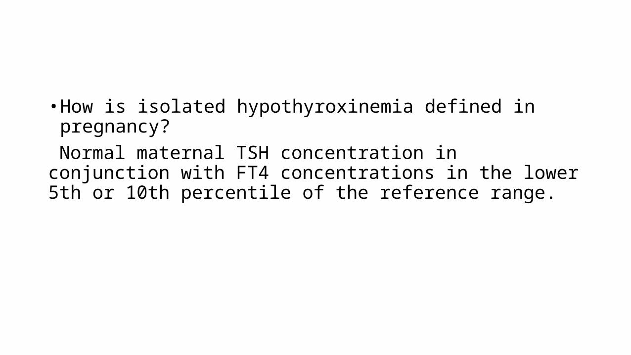
• How is isolated hypothyroxinemia defined in pregnancy? Normal maternal TSH concentration in conjunction with FT4 concentrations in the lower 5th or 10th percentile of the reference range.
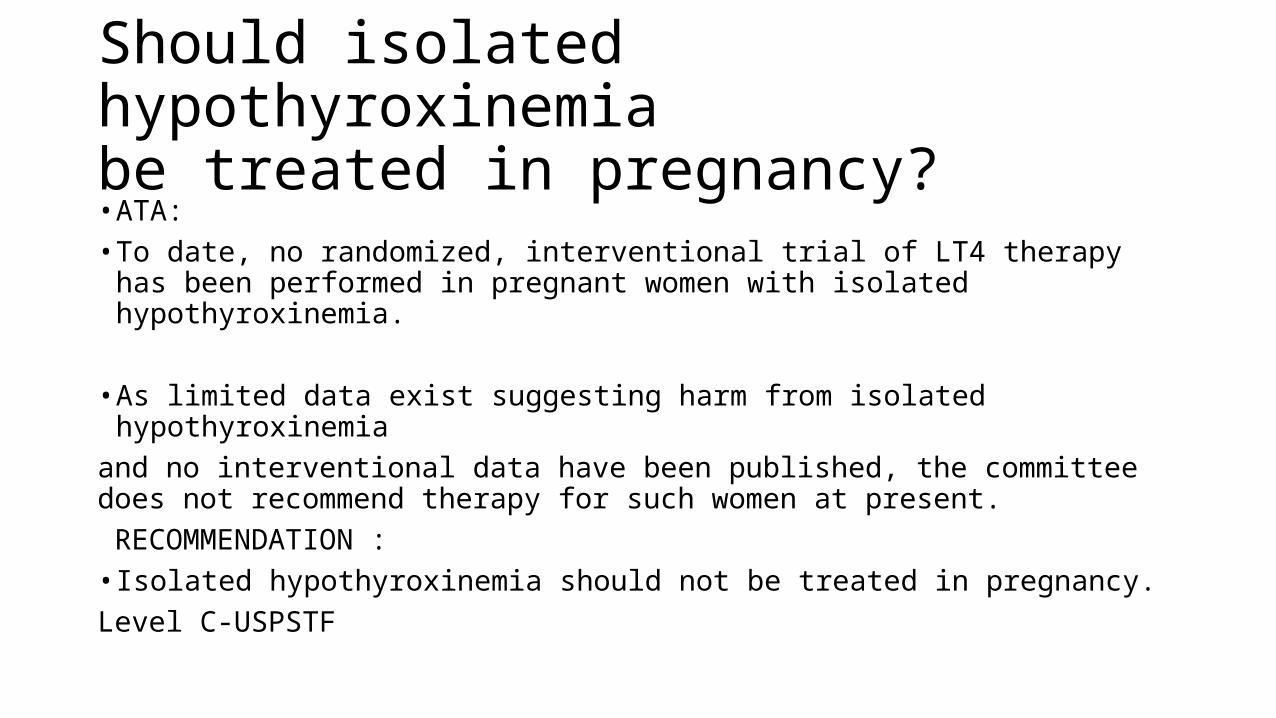
• ATA:• To date, no randomized, interventional trial of LT4 therapy has been
performed in pregnant women with isolated hypothyroxinemia.
• As limited data exist suggesting harm from isolated hypothyroxinemiaand no interventional data have been published, the committee does not recommend therapy for such women at present. RECOMMENDATION :• Isolated hypothyroxinemia should not be treated in pregnancy.Level C-USPSTF
Should isolated hypothyroxinemiabe treated in pregnancy?
• Antenatal Thyroid Screening and Childhood Cognitive Function• John H. Lazarus, M.D., Jonathan P. Bestwick, M.Sc., Sue Channon,
D.Clin.Psych., Ruth Paradice, Ph.D., Aldo Maina, M.D., Rhian Rees, M.Sc., Elisabetta Chiusano, M.Psy., Rhys John, Ph.D., Varvara Guaraldo, M.S.Chem., Lynne M. George, H.N.C., Marco Perona, M.S.Chem., Daniela Dall'Amico, M.D., Arthur B. Parkes, Ph.D., Mohammed Joomun, M.Sc., and Nicholas J. Wald, F.R.S.
• N Engl J Med 2012; 366:493-501February 9, 2012DOI: 10.1056/NEJMoa1106104
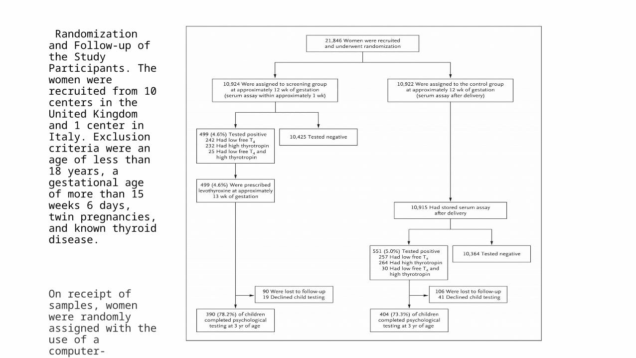
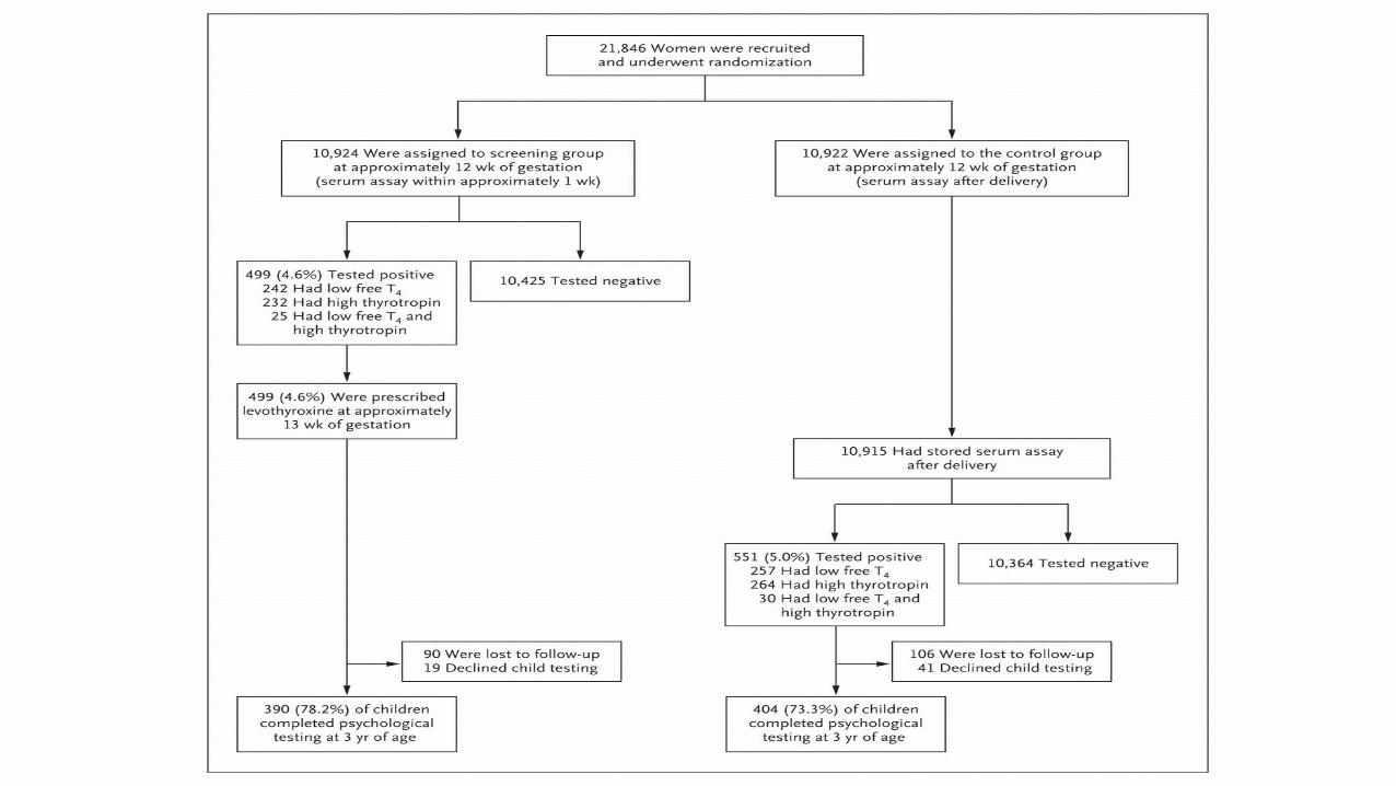
Randomization and Follow-up of the Study Participants. The women were recruited from 10 centers in the United Kingdom and 1 center in Italy. Exclusion criteria were an age of less than 18 years, a gestational age of more than 15 weeks 6 days, twin pregnancies, and known thyroid disease.
On receipt of samples, women were randomly assigned with the use of a computer-generated block design to the screening or control group.
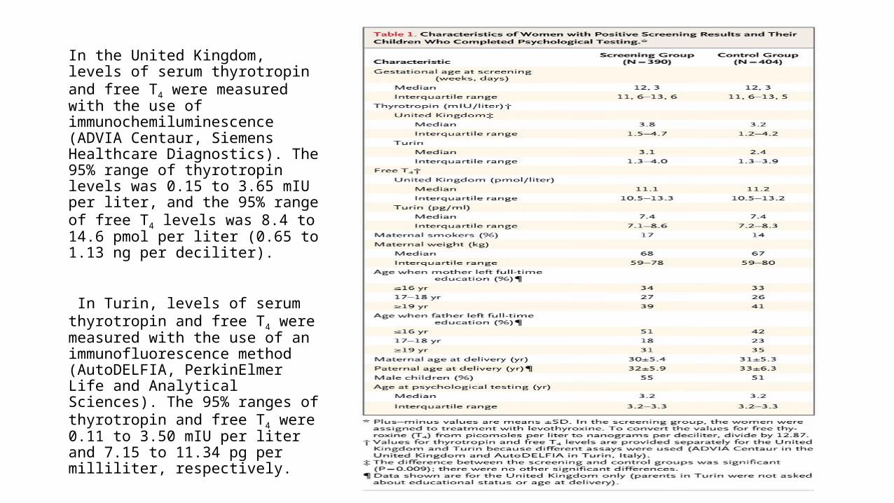
In the United Kingdom, levels of serum thyrotropin and free T4 were measured with the use of immunochemiluminescence (ADVIA Centaur, Siemens Healthcare Diagnostics). The 95% range of thyrotropin levels was 0.15 to 3.65 mIU per liter, and the 95% range of free T4 levels was 8.4 to 14.6 pmol per liter (0.65 to 1.13 ng per deciliter).
In Turin, levels of serum thyrotropin and free T4 were measured with the use of an immunofluorescence method (AutoDELFIA, PerkinElmer Life and Analytical Sciences). The 95% ranges of thyrotropin and free T4 were 0.11 to 3.50 mIU per liter and 7.15 to 11.34 pg per milliliter, respectively.
• Patients in the screening group who had positive results were treated with levothyroxine (recommended starting dose, 150 μg per day).
• Levels of thyrotropin and free T4 were checked 6 weeks after the start of levothyroxine therapy and at 30 weeks' gestation, with adjustment of the dose as necessary.
• The target thyrotropin level was 0.1 to 1.0 mIU per liter.
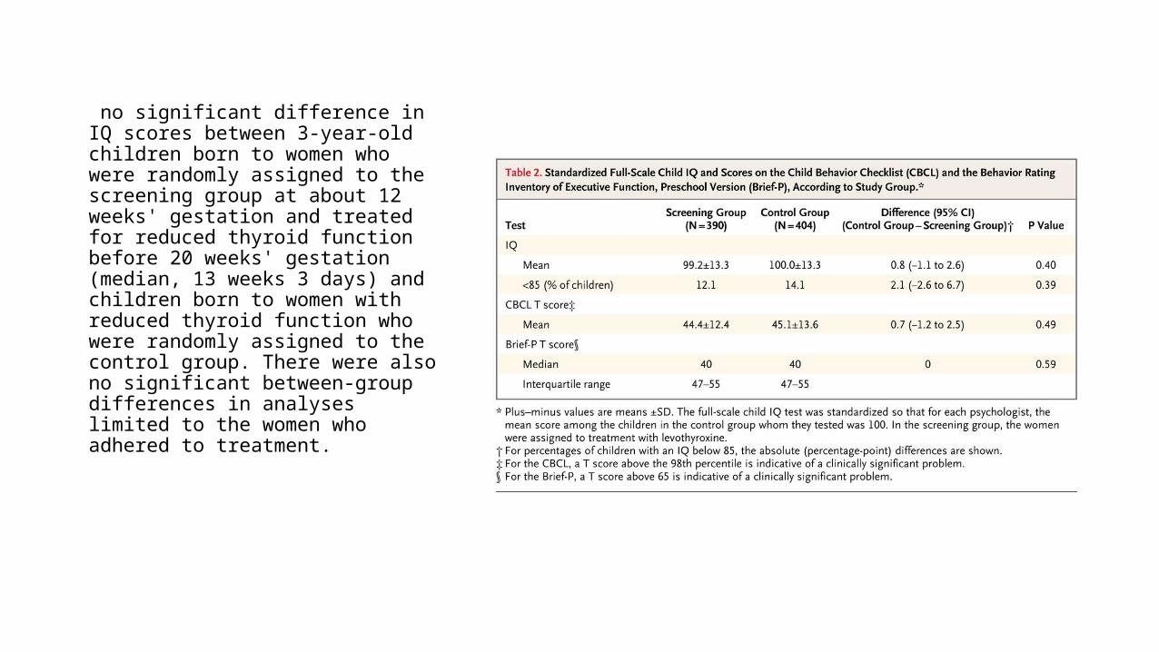
no significant difference in IQ scores between 3-year-old children born to women who were randomly assigned to the screening group at about 12 weeks' gestation and treated for reduced thyroid function before 20 weeks' gestation (median, 13 weeks 3 days) and children born to women with reduced thyroid function who were randomly assigned to the control group. There were also no significant between-group differences in analyses limited to the women who adhered to treatment.
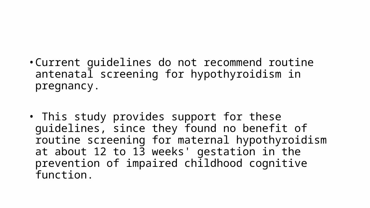
• Current guidelines do not recommend routine antenatal screening for hypothyroidism in pregnancy.
• This study provides support for these guidelines, since they found no benefit of routine screening for maternal hypothyroidism at about 12 to 13 weeks' gestation in the prevention of impaired childhood cognitive function.
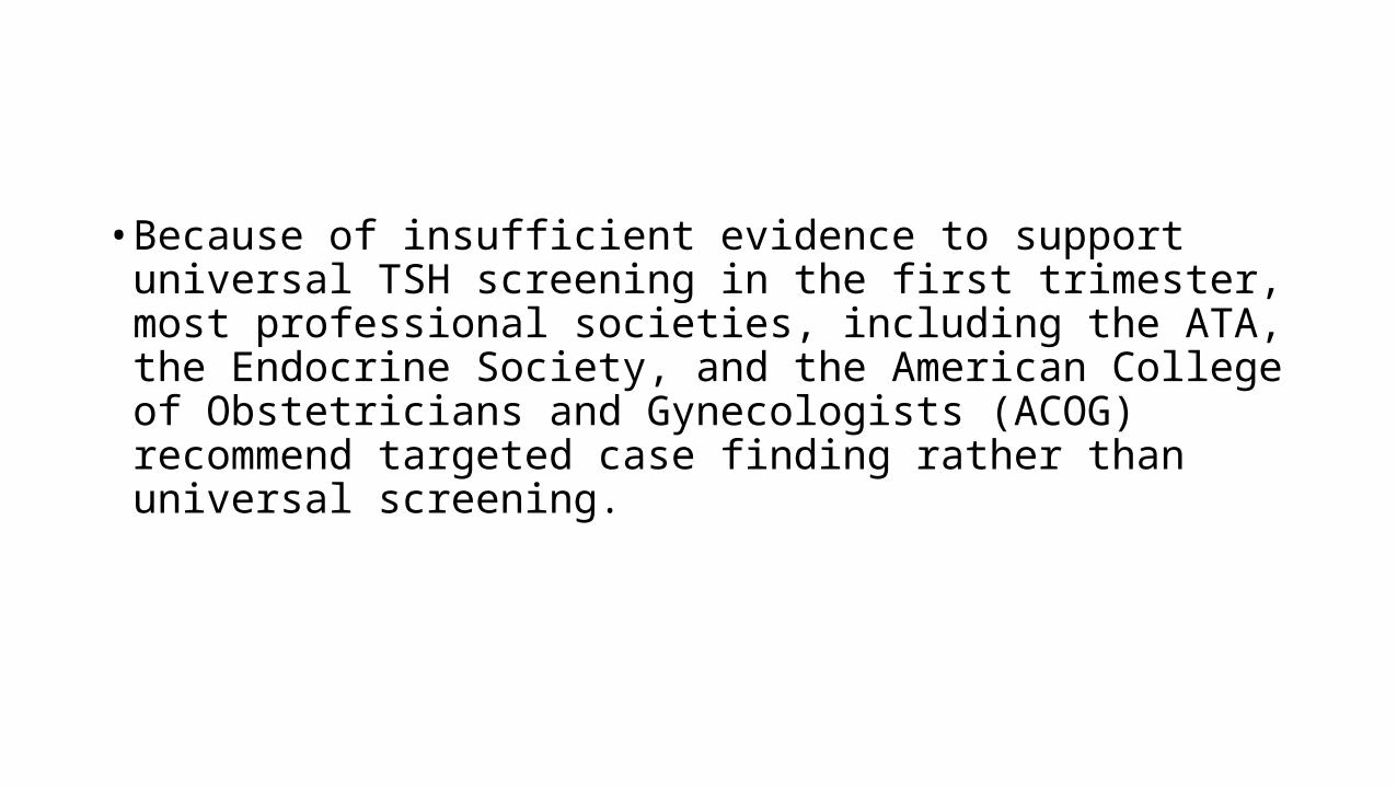
• Because of insufficient evidence to support universal TSH screening in the first trimester, most professional societies, including the ATA, the Endocrine Society, and the American College of Obstetricians and Gynecologists (ACOG) recommend targeted case finding rather than universal screening.
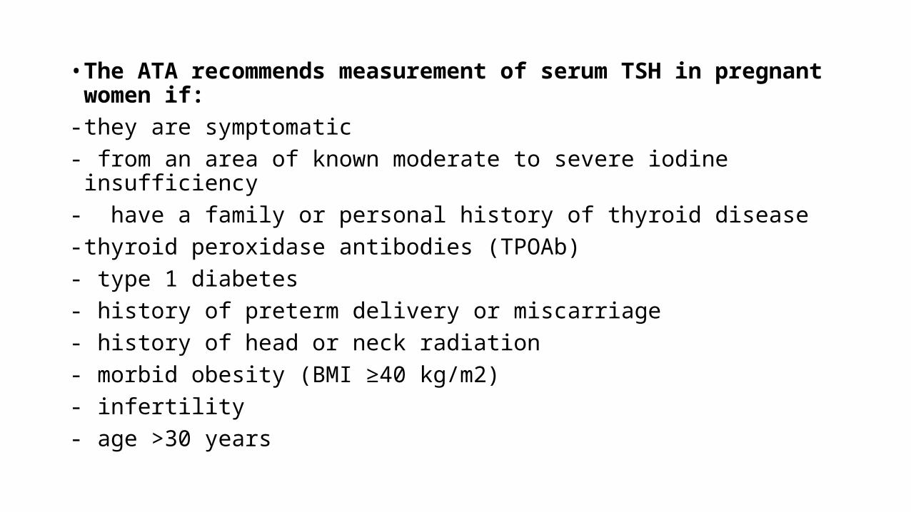
• The ATA recommends measurement of serum TSH in pregnant women if:- they are symptomatic- from an area of known moderate to severe iodine insufficiency- have a family or personal history of thyroid disease- thyroid peroxidase antibodies (TPOAb)- type 1 diabetes- history of preterm delivery or miscarriage- history of head or neck radiation- morbid obesity (BMI ≥40 kg/m2)- infertility- age >30 years
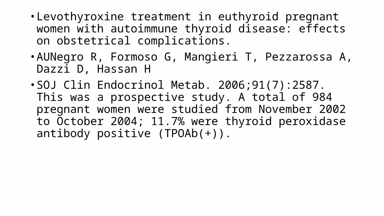
• Levothyroxine treatment in euthyroid pregnant women with autoimmune thyroid disease: effects on obstetrical complications.
• AUNegro R, Formoso G, Mangieri T, Pezzarossa A, Dazzi D, Hassan H• SOJ Clin Endocrinol Metab. 2006;91(7):2587. This was a prospective
study. A total of 984 pregnant women were studied from November 2002 to October 2004; 11.7% were thyroid peroxidase antibody positive (TPOAb(+)).
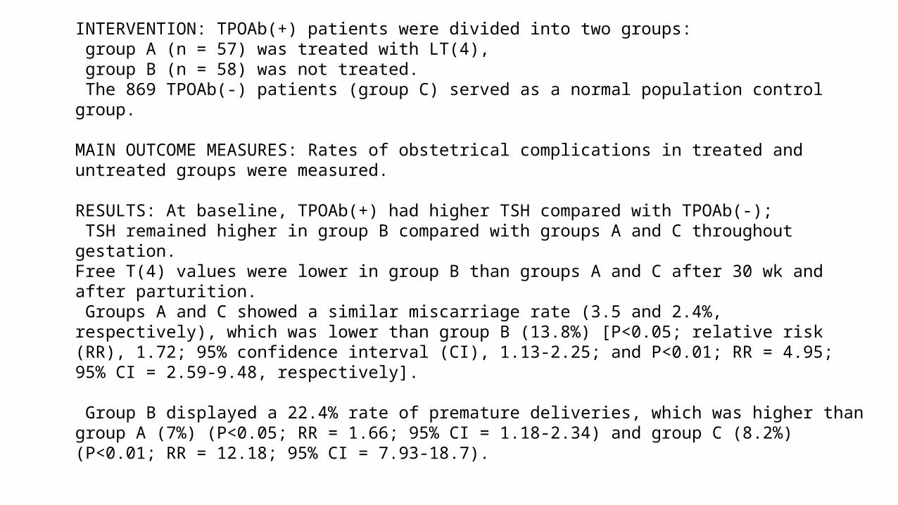
INTERVENTION: TPOAb(+) patients were divided into two groups: group A (n = 57) was treated with LT(4), group B (n = 58) was not treated. The 869 TPOAb(-) patients (group C) served as a normal population control group.
MAIN OUTCOME MEASURES: Rates of obstetrical complications in treated and untreated groups were measured.
RESULTS: At baseline, TPOAb(+) had higher TSH compared with TPOAb(-); TSH remained higher in group B compared with groups A and C throughout gestation. Free T(4) values were lower in group B than groups A and C after 30 wk and after parturition. Groups A and C showed a similar miscarriage rate (3.5 and 2.4%, respectively), which was lower than group B (13.8%) [P<0.05; relative risk (RR), 1.72; 95% confidence interval (CI), 1.13-2.25; and P<0.01; RR = 4.95; 95% CI = 2.59-9.48, respectively].
Group B displayed a 22.4% rate of premature deliveries, which was higher than group A (7%) (P<0.05; RR = 1.66; 95% CI = 1.18-2.34) and group C (8.2%) (P<0.01; RR = 12.18; 95% CI = 7.93-18.7).
CONCLUSIONS: Euthyroid pregnant women who are positive for TPOAb develop impaired thyroid function, which is associated with an increased risk of miscarriage and premature deliveries. Substitutive treatment with LT(4) is able to lower the chance of miscarriage and premature delivery.
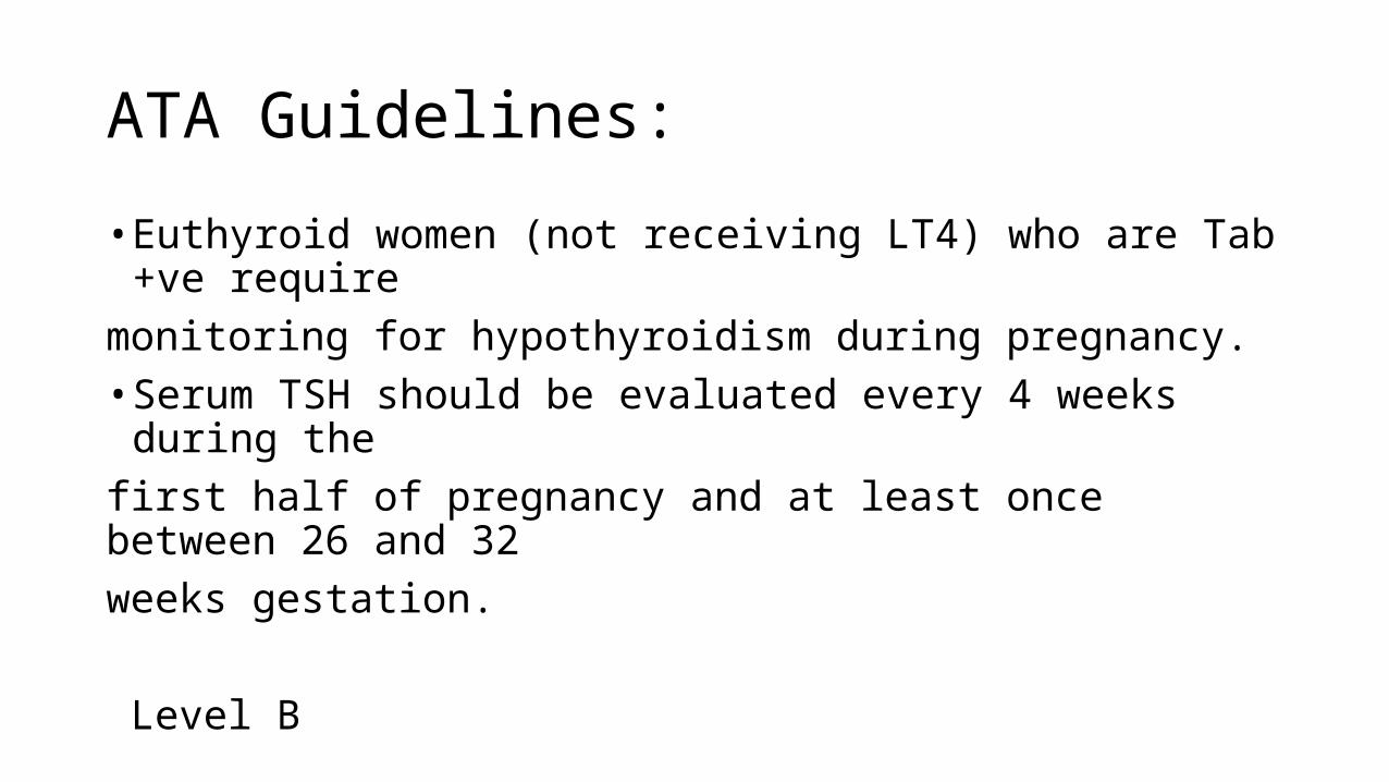
ATA Guidelines:
• Euthyroid women (not receiving LT4) who are Tab +ve requiremonitoring for hypothyroidism during pregnancy.• Serum TSH should be evaluated every 4 weeks during thefirst half of pregnancy and at least once between 26 and 32weeks gestation.
Level B
ATA Guidelines:
• In women being treated with ATDs in pregnancy, FT4 and TSH should be monitored approximately every 2–6 weeks.
• The primary goal is a serum FT4 at or moderately above the normal reference range. Level B
ATA Gidelines:
• Thyrotoxic women should be rendered euthyroid before attempting pregnancy. Level A
• PTU is preferred for the treatment of hyperthyroidism in the first trimester. Patients on MMI should be switched to PTU if pregnancy is confirmed in the first trimester.
• Following the first trimester, consideration should be given to switching to MMI.

THANK YOU