Embed Size (px)
Citation preview

Marginal-tryptophan deficiency was created by fe-eding the rats with 6% Casein and 12% gelatin. Ca-sein consists of 0.15% L-tryptophan which is abouthalf the minimal requirement reported for growingrats11-13. Gelatin comprises the entire range of aminoacids in the form of peptides except tryptophan14.
The Departmental Animal Ethics Committee hasapproved the animal care. The experiment was carriedout for three months. All the animals had free accessto food and water and were caged separately with si-milar ambience and the diurnal rhythm. Food intakeand body weights were monitored daily and weeklyrespectively.
Morphological and Behavioral studies
During the study period daily food intake and we-ekly body weights were recorded. Morphological(hair loss) and behavioral changes were observed pe-riodically.
Tissue extraction and processing: At the end of theexperiment animals were sacrificed by CO
2asphyxia-
tion and tissues were harvested and stored at -80o C tillfurther analysis. The liver and neuronal tissues wereextracted from each animal and liver was perfusedwith normal saline (0.9% NaCl) to reduce red bloodcell contamination. A 10% homogenate of hepatic andneuronal tissues was prepared and MDA levels wereestimated in total homogenate. The rest of the bioche-mical parameters were carried out with 25,000x g su-pernatant and necessary centrifugations depending onthe assays.
Biochemical estimation of oxidative stress markersand antioxidant enzymes: Malonadialdehyde (MDA)production was estimated by thiobarbituric acid-reac-tive substances (TBARS) as described by Buyan etal.15. Reduced glutathione was estimated by the spec-trofluorometric method of Hissin et al.16. Total Supe-roxide dismutase (SOD, E.C 1.15.1.2) activity was as-sayed by monitoring the rate of inhibition ofpyrogallol reduction17. One unit of SOD represents theamount of enzyme required for 50% inhibition of py-rogallol reduction/min. Catalase (CAT, E.C 1.11.1.6)activity of tissues was measured by the method of Ae-bi18 by monitoring the disappearance of H
2O
2. One unit
of catalase represents the decrease of 1 µmol H2O
2-
/min. The activity of Glutathione-s-transferase (GST,E.C 2.5.18) was assayed spectrophotometrically usingCDNB (1-Chloro-2, 4-dinitrobenzene) as substrate19.Protein carbonyl content of soluble protein of tissueswas measured spectrophotometrically using the 2, 4-dinitrophenyl-hydrazine20.
Advanced glycation related fluorescence: The Mai-llard reaction products i.e. advanced glycation endproducts (AGE) fluorescence was measured in solubleprotein (0.3 mg/2 ml in 0.05 M sodium phosphate buf-fer, pH 7.4). Fluorescence spectra were obtained from400-500 nm with excitation at 370 nm in a spectro-fluorometer21.
Tryptophan fluorescence
Tryptophan florescence was measured in the solu-ble protein fraction (0.15 mg/ml in 0.05 M sodiumphosphate buffer, pH 7.4) to determine the proteinoxidation and conformational changes in differentgroups of animals. The fluorescence spectra were ob-tained at excitation 295 nm and emission between310-400 nm22.
Protein estimation: The protein content of tissueswas estimated by the method of Lowry et al.23, usingBSA as the standard.
Statistical analysis: The differences between thecontrol and treated groups were analyzed using one-way ANOVA, followed by Post Hoc test (multiplecomparison). The differences were considered signifi-cant if p was at least < 0.05.
Results
Body weights and Food intake
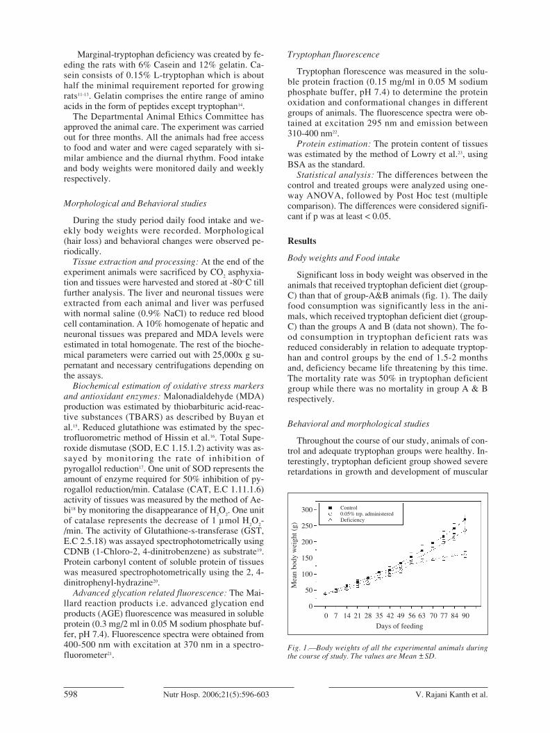
Significant loss in body weight was observed in theanimals that received tryptophan deficient diet (group-C) than that of group-A&B animals (fig. 1). The dailyfood consumption was significantly less in the ani-mals, which received tryptophan deficient diet (group-C) than the groups A and B (data not shown). The fo-od consumption in tryptophan deficient rats wasreduced considerably in relation to adequate tryptop-han and control groups by the end of 1.5-2 monthsand, deficiency became life threatening by this time.The mortality rate was 50% in tryptophan deficientgroup while there was no mortality in group A & Brespectively.
Behavioral and morphological studies
Throughout the course of our study, animals of con-trol and adequate tryptophan groups were healthy. In-terestingly, tryptophan deficient group showed severeretardations in growth and development of muscular
598 V. Rajani Kanth et al.Nutr Hosp. 2006;21(5):596-603
Fig. 1.—Body weights of all the experimental animals duringthe course of study. The values are Mean ±SD.
300
250
200
150
100
50
00 7 14 21 28 35 42 49 56 63 70 77 84 90
Days of feeding
Mea
n bo
dy w
eigh
t (g)
Control0.05% trp. administeredDeficiency
05. BEHAVIORAL 7/9/06 08:40 Página 598

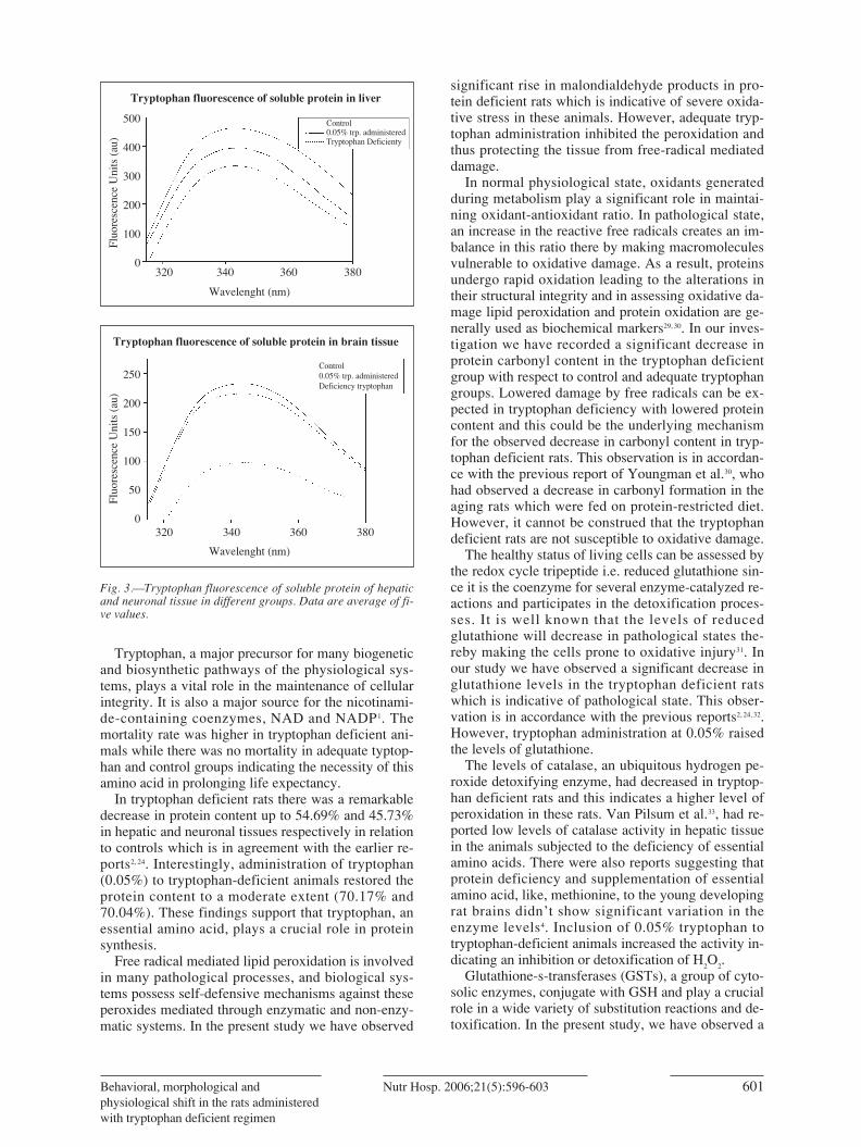
Advanced glycation end product (AGE) fluorescen-ce: AGE fluorescence (fig. 2) reveals an increase inAGEs formation in tryptophan deficient rats (group-C) compared to controls (group-A). Interestingly, thetryptophan-deficient animals which received 0.05%tryptophan had shown significant inhibition of AGEfluorescence (fig. 2).
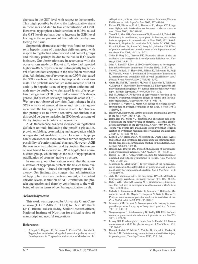
Tryptophan fluorescence: Tryptophan fluorescencespectra (fig. 3) showed a decrease in tryptophan fluo-rescence in tryptophan deficient rats (group-C) in rela-tion to controls (group-A). However, adequate tryp-tophan administration has been found to inhibit thedecrease in tryptophan fluorescence in comparison togroup-C (fig. 3).
Discussion
It is well documented that protein deficiency / res-triction will affect the morphological, physiologicalstatus of the cellular systems possibly through the ge-neration of free radicals / reactive oxygen species,which damage the integrity of biological systems lea-ding to several pathological states24, 25. In our presentstudy, we have noticed a significant decrease in foodintake, body weight, organ development and retarda-tion in muscular system in the rats that were fed ontryptophan deficient diet and this is in agreement withthe previous observations of protein deficiency5, 24, 26-28.However, these features were not seen in adequatetyptophan group and this is suggestive of a clear pro-tective influence of tryptophan in maintaining thephysiological status with respect to controls. In anearlier study carried out by John and Bhat6 no signifi-cant variation in the protein content, tryptophan le-vels and antioxidant enzymes between the controland pair-fed control groups was reported. In a pilotexperiment carried out by us we didn’t notice signifi-cant variation in the control and pair-fed controlgroups (data not shown). Therefore, in order to mini-
mize the number of animals due to ethical cons-traints, we could not maintain pair-fed control groupsin our investigation.
600 V. Rajani Kanth et al.Nutr Hosp. 2006;21(5):596-603
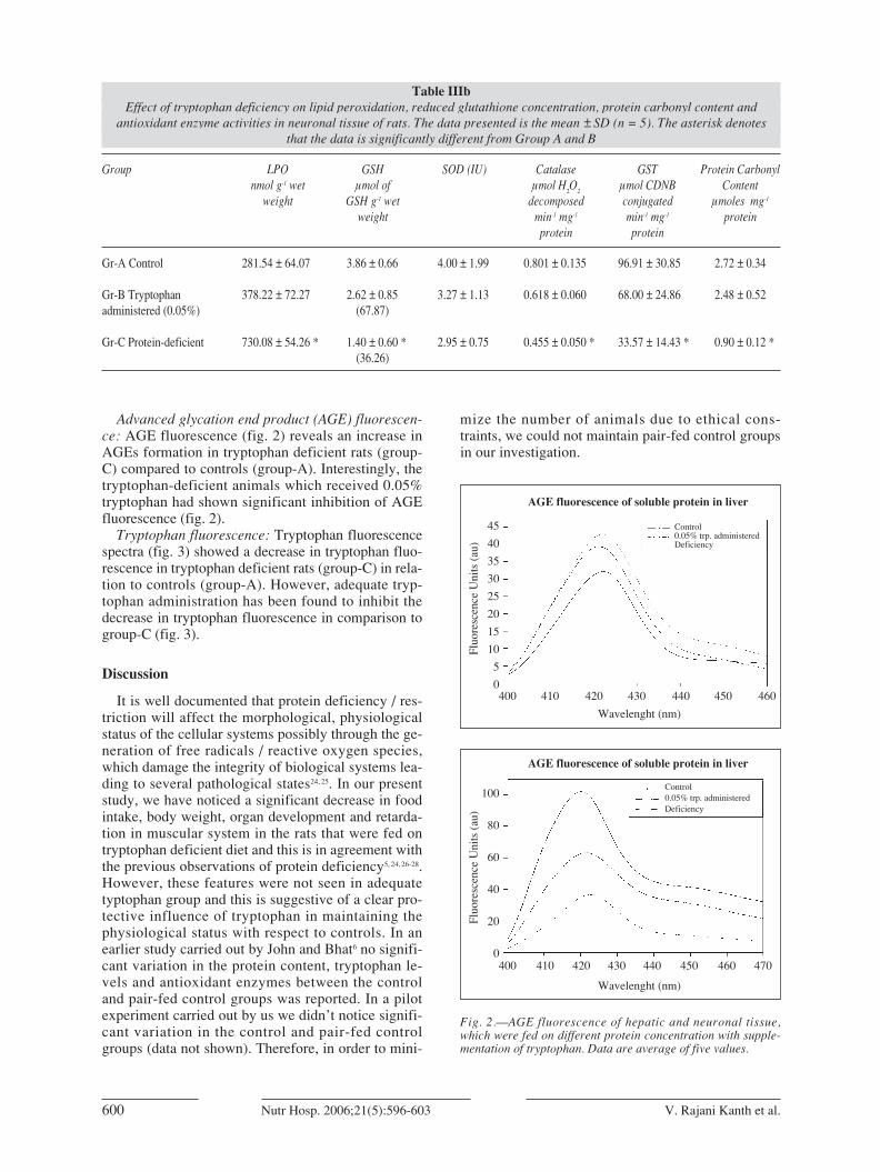
Table IIIbEffect of tryptophan deficiency on lipid peroxidation, reduced glutathione concentration, protein carbonyl content and
antioxidant enzyme activities in neuronal tissue of rats. The data presented is the mean ±SD (n = 5). The asterisk denotes that the data is significantly different from Group A and B
Group LPO GSH SOD (IU) Catalase GST Protein Carbonylnmol g-1 wet µmol of µmol H
2O
2µmol CDNB Content
weight GSH g-1 wet decomposed conjugated µmoles mg-1
weight min-1 mg-1 min-1 mg-1 proteinprotein protein
Gr-A Control 281.54 ± 64.07 3.86 ± 0.66 4.00 ± 1.99 0.801 ± 0.135 96.91 ± 30.85 2.72 ± 0.34
Gr-B Tryptophan 378.22 ± 72.27 2.62 ± 0.85 3.27 ± 1.13 0.618 ± 0.060 68.00 ± 24.86 2.48 ± 0.52administered (0.05%) (67.87)
Gr-C Protein-deficient 730.08 ± 54.26 * 1.40 ± 0.60 * 2.95 ± 0.75 0.455 ± 0.050 * 33.57 ± 14.43 * 0.90 ± 0.12 *(36.26)
Fig. 2.—AGE fluorescence of hepatic and neuronal tissue,which were fed on different protein concentration with supple-mentation of tryptophan. Data are average of five values.
45
40
35
30
25
20
15
10
5
0400 410 420 430 440 450 460
Wavelenght (nm)
AGE fluorescence of soluble protein in liver
Control0.05% trp. administeredDeficiency
Fluo
resc
ence
Uni
ts (
au)
100
80
60
40
20
0400 410 420 430 440 450 460 470
Wavelenght (nm)
AGE fluorescence of soluble protein in liver
Control0.05% trp. administeredDeficiency
Fluo
resc
ence
Uni
ts (
au)
05. BEHAVIORAL 7/9/06 08:40 Página 600
Tryptophan, a major precursor for many biogeneticand biosynthetic pathways of the physiological sys-tems, plays a vital role in the maintenance of cellularintegrity. It is also a major source for the nicotinami-de-containing coenzymes, NAD and NADP1. Themortality rate was higher in tryptophan deficient ani-mals while there was no mortality in adequate typtop-han and control groups indicating the necessity of thisamino acid in prolonging life expectancy.
In tryptophan deficient rats there was a remarkabledecrease in protein content up to 54.69% and 45.73%in hepatic and neuronal tissues respectively in relationto controls which is in agreement with the earlier re-ports2, 24. Interestingly, administration of tryptophan(0.05%) to tryptophan-deficient animals restored theprotein content to a moderate extent (70.17% and70.04%). These findings support that tryptophan, anessential amino acid, plays a crucial role in proteinsynthesis.
Free radical mediated lipid peroxidation is involvedin many pathological processes, and biological sys-tems possess self-defensive mechanisms against theseperoxides mediated through enzymatic and non-enzy-matic systems. In the present study we have observed
significant rise in malondialdehyde products in pro-tein deficient rats which is indicative of severe oxida-tive stress in these animals. However, adequate tryp-tophan administration inhibited the peroxidation andthus protecting the tissue from free-radical mediateddamage.
In normal physiological state, oxidants generatedduring metabolism play a significant role in maintai-ning oxidant-antioxidant ratio. In pathological state,an increase in the reactive free radicals creates an im-balance in this ratio there by making macromoleculesvulnerable to oxidative damage. As a result, proteinsundergo rapid oxidation leading to the alterations intheir structural integrity and in assessing oxidative da-mage lipid peroxidation and protein oxidation are ge-nerally used as biochemical markers29, 30. In our inves-tigation we have recorded a significant decrease inprotein carbonyl content in the tryptophan deficientgroup with respect to control and adequate tryptophangroups. Lowered damage by free radicals can be ex-pected in tryptophan deficiency with lowered proteincontent and this could be the underlying mechanismfor the observed decrease in carbonyl content in tryp-tophan deficient rats. This observation is in accordan-ce with the previous report of Youngman et al.30, whohad observed a decrease in carbonyl formation in theaging rats which were fed on protein-restricted diet.However, it cannot be construed that the tryptophandeficient rats are not susceptible to oxidative damage.
The healthy status of living cells can be assessed bythe redox cycle tripeptide i.e. reduced glutathione sin-ce it is the coenzyme for several enzyme-catalyzed re-actions and participates in the detoxification proces-ses. It is well known that the levels of reducedglutathione will decrease in pathological states the-reby making the cells prone to oxidative injury31. Inour study we have observed a significant decrease inglutathione levels in the tryptophan deficient ratswhich is indicative of pathological state. This obser-vation is in accordance with the previous reports2, 24, 32.However, tryptophan administration at 0.05% raisedthe levels of glutathione.
The levels of catalase, an ubiquitous hydrogen pe-roxide detoxifying enzyme, had decreased in tryptop-han deficient rats and this indicates a higher level ofperoxidation in these rats. Van Pilsum et al.33, had re-ported low levels of catalase activity in hepatic tissuein the animals subjected to the deficiency of essentialamino acids. There were also reports suggesting thatprotein deficiency and supplementation of essentialamino acid, like, methionine, to the young developingrat brains didn’t show significant variation in theenzyme levels4. Inclusion of 0.05% tryptophan totryptophan-deficient animals increased the activity in-dicating an inhibition or detoxification of H
2O
2.
Glutathione-s-transferases (GSTs), a group of cyto-solic enzymes, conjugate with GSH and play a crucialrole in a wide variety of substitution reactions and de-toxification. In the present study, we have observed a
Behavioral, morphological andphysiological shift in the rats administeredwith tryptophan deficient regimen
601Nutr Hosp. 2006;21(5):596-603
Fig. 3.—Tryptophan fluorescence of soluble protein of hepaticand neuronal tissue in different groups. Data are average of fi-ve values.
500
400
300
200
100
0320 340 360 380
Wavelenght (nm)
Tryptophan fluorescence of soluble protein in liver
Control0.05% trp. administeredTryptophan Deficienty
Fluo
resc
ence
Uni
ts (
au)
250
200
150
100
50
0320 340 360 380
Wavelenght (nm)
Tryptophan fluorescence of soluble protein in brain tissue
Control0.05% trp. administeredDeficiency tryptophan
Fluo
resc
ence
Uni
ts (
au)
05. BEHAVIORAL 7/9/06 08:40 Página 601
decrease in the GST level with respect to the controls.This might possibly be due to the high oxidative stressin these rats and due to low concentration of GSH.However, tryptophan administration at 0.05% raisedthe GST levels perhaps due to increase in GSH levelleading to the suppression of free radicals thereby pro-tecting the tissue.
Superoxide dismutase activity was found to increa-se in hepatic tissue of tryptophan deficient group withrespect to tryptophan administered and control groupsand this may perhaps be due to the rise in H
2O
2levels
in tissues. Our observations are in accordance with theobservations made by Rao et al.34, who had reportedhigher m-RNA expression and subsequently higher le-vel of antioxidant enzymes in the rats fed on restricteddiet. Administration of tryptophan at 0.05% decreasedthe SOD levels in relation to tryptophan deficient ani-mals. The probable mechanism in the increase of SODactivity in hepatic tissue of tryptophan deficient ani-mals may be attributed to decreased levels of tryptop-han dioxygenase (TDO) and Indoleamine 2,3- dioxy-genase (IDO) which scavenge superoxide anion (O
2–)1.
We have not observed any significant change in theSOD activity of neuronal tissue and this is in agree-ment with the findings of Bonatto et al.4. Our observa-tion prompts us to draw a tentative conclusion thatthis could be due to variation in IDO levels as some ofthe tryptophan metabolites are neurotoxic.
AGE fluorescence has been increased in tryptophandeficient animals indicating the possible glycation,protein unfolding, crosslinking and aggregation whichis suggestive of oxidative stress. Decrease in tryptop-han fluorescence in these animals further supports thepossibility of conformational changes. However, AGEfluorescence was inhibited and tryptophan fluorescen-ce was found to increase in 0.05% tryptophan admi-nistered group, which implies the role of tryptophan instabilization of proteins’ native structure.
In summary, our observations reveal that the admi-nistration of tryptophan protects the tissues from oxi-dative damage induced through tryptophan defi-ciency. Our findings also suggest that administrationof tryptophan restores protein content, antioxidantenzyme levels, inhibition of AGE formation and pro-tein aggregation and there by contributing to the well-being of rats in terms of combating oxidative insult.
Acknowledgments:
This work was supported by University Grant Com-mission (U.G.C.-MJRP F.3.123) to TNR. We thankDr. G. Bhanu Prakash Reddy, Senior Research officer,National Institute of Nutrition for critical review ofmanuscript and needful suggestions.
References
1. Allegri G, Ragazzi E, Bertazzo A, Costa CVL, Rocchi R.Tryptophan metabolism along the kynurenine pathway in rats,Developments in Tryptophan and serotonin Metabolism. In:
Allegri et al., editors. New York: Kluwer Academic/PlenumPublishers ed. Adv Exp Med Biol 2003; 527:481-96.
2. Petzke KJ, Elsner A, Proll J, Thielecke F , Metges CC. Long-term high protein intake does not increase oxidative stress inrats. J Nutr 2000; 130:2889-96.
3. Yen CLE, Mar MH, Craciunescu CN, Edwards LJ, Zeisel SH.Deficiency in methionine, tryptophan, isoleucine, or cholineinduces apoptosis in cultured cells. J Nutr 2002; 132:1840-7.
4. Bonatto F, Ploydoro M, Andrades ME, Junior MLCDF, Dal-Pizzol F, Rotta LN, Souza DO, Perry ML, Moreira JCF. Effectof protein malnutrition on redox state of the hippocampus ofrat. Brain Res 2005; 1042(1):17-22.
5. Sidhu P, Garg ML, Dhawan DK. Protective effects of zinc onoxidative stress enzymes in liver of protein deficient rats. NutrHosp 2004; 19:341-7.
6. John A, Bhat KS. Effect of riboflavin deficiency on low tryptop-han induced cataract in male rats. Nutr Res 1992; 12:1373-81.
7. Saito K, Fujigaki S, Heyes PM, Shibata K, Takemura M, FujjiH, Wada H, Noma A, Seishima M. Mechanism of increases inL-kynurenine and quinolinic acid in renal Insufficiency. Am JPhysiol Renal Physiol 2000; 279:F565- F572.
8. Grant SR, Naif H, Thuruthyil JS, Nasr N, Littlejohn T, TakikawaO, Kapoor V. Induction of Indoleamine 2, 3-Dioxygenase in pri-mary human macrophages by human immunodeficiency virustype 1 is strain dependent. J Virol 2000; 74:4110-5.
9. Bel N, Artigas F. Reduction of serotonergic function in ratbrain by tryptophan depletion: effects in control and fluvoxa-mine-treated rats. J Neurochem 1996; 67:669-76.
10. Sidransky H, Verney E, Murty CN. Effect of elevated dietarytryptophan on protein synthesis in rat liver. J Nutr 1981;111:1942-8.
11. Rogers QR, Harper AE. Amino acid diets and maximal growthin the rat. J Nutr 1965; 87:267-73.
12. Rama Rao PB, Metta VC, Johnson BC: The amino-acid com-position and the nutritive value of proteins. I. Essential amino-acid requirements of the growing rat. J Nutr 1959; 69:387-91.
13. Young VR, Munro HN. Plasma and tissue tryptophan levels inrelation to tryptophan requirements of weanling and adult rats.J Nutr 1973; 103:1756-63.
14. Lieben CKJ, Blokland A, Westerink B, Deutz NEP. Acutetryptophan and serotonin depletion using an optimized tryp-tophan-free protein-carbohydrate mixture in the adult rat. Neu-rochem Int 2004; 44:9-16.
15. Bhuyan KC, Bhuyan DK, Podos SM. Evidence of increased li-pid peroxidation in cataracts. IRCS Med Sci 1981; 9:126-7.
16. Hissin PJ, Hilf R. A fluorometric method for determination ofoxidized and reduced glutathione in tissues. Anal Biochem1976; 74:214-26.
17. Marklund S, Marklund G. Involvement of the superoxideanion radical in the autoxidation of pyrogallol and a conve-nient assay for superoxide dismutase. Eur J Biochem 1974;47(3):469-74.
18. Aebi H. Catalase in vitro. In: Bergmeyer HV, ed. Methods inEnzymology. Weinheim, Germany: Chemie 1984; 105:121-126.
19. Habig WH, Pabst MJ, Jokoby WB. Glutathione-S-transfera-ses. The first step in mercapturic acid formation. J Biol Chem1974; 249:7130-9.
20. Uchida K, Kanematsu M, Sakai K, Matsuda T, Hattori N, Mi-zuno Y, Suzuki D, Miyata T, Noguchi N, Niki E, Osawa T.Protein-bound acrolein: potential markers for oxidative stress.Proc Natl Acad Sci USA 1998; 95:4882-7.
21. Monnier VM, Cerami A. Nonenzymatic browning in vivo:possible process for aging of long-lived proteins. Science1981; 211:491-3.
22. Suryanarayana P, Krishnaswamy K, Reddy GB. Effect of cur-cumin on galactose-induced cataractogenesis in rats. Mol Vis2003; 9:223-30.
23. Lowry OH, Rosebrough NJ, Lewis Farr A, Randall RJ. Proteinmeasurement with Folin phenol reagent. J Biol Chem 1951;193:265-75.
24. Rana S, Sodhi CP, Mehta S, Vaiphei K, Katyal R, Thakur S,Mehta S K. Protein-energy malnutrition and oxidative injuryin growing rats. Hum Exp Toxicol 1996; 15:810-4.
602 V. Rajani Kanth et al.Nutr Hosp. 2006;21(5):596-603
05. BEHAVIORAL 7/9/06 08:40 Página 602

25. Huang CJ, Fwu ML. Protein insufficiency aggravates the en-hanced lipid peroxidation and reduced activities of antioxidati-ve enzymes in rats fed diets high in polyunsaturated fat. J Nutr1992; 122:1182-9.
26. Satyanarayana U, Narasinga Rao BS. Effect of diet restrictionon some key enzymes of tyrptophan-NAD pathway in rats. J Nutr 1977; 107:2213-8.
27. Wegener A, Golubnitschaja O, Breipohl W, Schild HH, Vren-sen GFJM. Effects of dietary deficiency of selective aminoacids on the function of the cornea and lens in rats. AminoAcids 2002; 23:337-342.
28. Perry ML, Gamallo JL, Bernard EA. Effect of protein malnu-trition on Glycoprotein synthesis in rat cerebral cortex slicesduring the period of brain growth spurt. J Nutr 1986; 116:2486-9.
29. Winterbourn CC, Chan T, Buss IH, Inder TE, Mogridge N,Darlow BA. Protein Carbonyls and Lipid Peroxidation Pro-ducts as Oxidation Markers in Preterm Infant Plasma: Asso-
ciations with Chronic Lung Disease and Retinopathy and Effects of Selenium Supplementation. Pediatric Res 2000; 48:84-90.
30. Youngman LD, Park JY, Ames BN. Protein oxidation associa-ted with aging is reduced by dietary restriction of protein orcalories. Proc Natl Acad Sci USA 1992; 89:9112-6.
31. Meister A. The gamma-glutamyl cycle. Diseases associatedwith specific enzyme deficiencies. Ann Intern Med 1974;81:247-53.
32. Hum S, Koski KG, Hoffer LJ. Varied protein intake alters glu-tathione metabolism in rats. J Nutr 1992; 122:2010-8.
33. Van Pilsum JF, Speyer JF, Samuels LT: Essential amino aciddeficiency and enzyme activity. Arch Biochem Biophys 1957;68:42-53.
34. Rao G, Xia E, Nadakavukaren MJ, Richardson A. Effect ofdietary restriction on the age-dependent changes in the ex-pression of antioxidant enzymes in rat liver. J Nutr 1990;120:602-9.
Behavioral, morphological andphysiological shift in the rats administeredwith tryptophan deficient regimen
603Nutr Hosp. 2006;21(5):596-603
05. BEHAVIORAL 7/9/06 08:40 Página 603