Embed Size (px)
Citation preview
Author's Accepted Manuscript
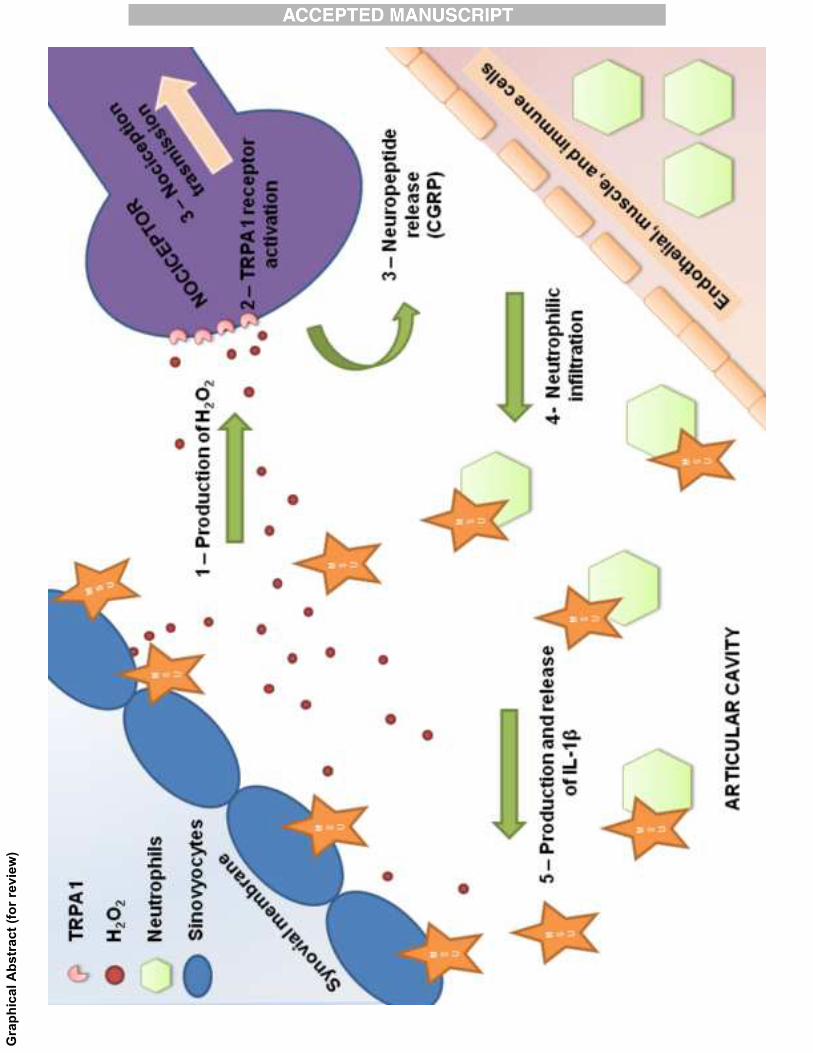
TRPA1 receptor stimulation by Hydrogenperoxide is critical to trigger hyperalgesiaand inflammation in a model of acute gout
Gabriela Trevisan, Carin Hoffmeister, MateusFortes Rossato, Sara Marchesan Oliveira, Mar-iane Arnoldi Silva, Cássia Regina Silva, CamillaFusi, Raquel Tonello, Daiana Minocci, GustavoPetri Guerra, Serena Materazzi, Romina Nassi-ni, Pierangelo Geppetti, Juliano Ferreira
PII: S0891-5849(14)00192-0DOI: http://dx.doi.org/10.1016/j.freeradbiomed.2014.04.021Reference: FRB11995
To appear in: Free Radical Biology and Medicine
Received date: 5 November 2013Revised date: 20 March 2014Accepted date: 21 April 2014
Cite this article as: Gabriela Trevisan, Carin Hoffmeister, Mateus FortesRossato, Sara Marchesan Oliveira, Mariane Arnoldi Silva, Cássia Regina Silva,Camilla Fusi, Raquel Tonello, Daiana Minocci, Gustavo Petri Guerra, SerenaMaterazzi, Romina Nassini, Pierangelo Geppetti, Juliano Ferreira, TRPA1receptor stimulation by Hydrogen peroxide is critical to trigger hyperalgesiaand inflammation in a model of acute gout, Free Radical Biology and Medicine,http://dx.doi.org/10.1016/j.freeradbiomed.2014.04.021
This is a PDF file of an unedited manuscript that has been accepted forpublication. As a service to our customers we are providing this early version ofthe manuscript. The manuscript will undergo copyediting, typesetting, andreview of the resulting galley proof before it is published in its final citable form.Please note that during the production process errors may be discovered whichcould affect the content, and all legal disclaimers that apply to the journalpertain.
www.elsevier.com/locate/freerad-
biomed
1
TRPA1 receptor stimulation by hydrogen peroxide is critical to trigger hyperalgesia
and inflammation in a model of acute gout
Gabriela Trevisana,f, Carin Hoffmeisterb, Mateus Fortes Rossatoa, Sara Marchesan
Oliveiraa, Mariane Arnoldi Silvaa, Cássia Regina Silvaa, Camilla Fusic, Raquel Tonelloa,
Daiana Minoccic, Gustavo Petri Guerrad, Serena Materazzic, Romina Nassinic, Pierangelo
Geppettic, Juliano Ferreiraa,b,e,*.
aGraduate Program in Biological Sciences: Toxicological Biochemistry, bGraduated
Program in Pharmacology, Federal University of Santa Maria, Santa Maria, RS, Brazil.
cDepartment of Health Sciences, University of Florence, Florence, Italy.
dDepartment of Food Technology, Federal Technological University of Paraná, Medianeira
Campus, Medianeira, PR, Brazil.
eDepartment of Pharmacology, Federal University of Santa Catarina (UFSC),
Florianópolis, SC, Brazil.
fLaboratório de Biologia Celular e Molecular, Programa de Pós-Graduacão em Ciências da
Saúde, Universidade do Extremo Sul Catarinense (UNESC), Criciúma, SC, Brazil
*Corresponding author: Juliano Ferreira, Department of Pharmacology, Biological
Sciences Centre, Block "D"/CCB, Federal University of Santa Catarina, Trindade, Zip
code: 88040-900, Florianópolis, SC, Brazil, Phone: +55 48 3721 9491, FAX: +55 48 3337
5479, email: [email protected].
2
Highlights
1. MSU-induced edema and hyperalgesia is largely mediated by TRPA1 receptor
activation
2. MSU i.a. injection increases TRPA1 expression in synovial tissue and CGRP
release
3. MSU increased H2O2 levels and NADPH-oxidase activity, but reduced CAT activity
4. MSU-induced responses are mediated by H2O2 production and subsequent TRPA1
activation
5. TRPA1 antagonism reduced MSU-induced neutrophil infiltration and IL-1β
production
Abbreviations
Allyl isothiocyanate, AITC; analysis of variance, ANOVA; calcitonin gene-related peptide,
CGRP; CGRP-like immunoreactivity, CGRP-LI; dithiothreitol, DTT; hydrogen peroxide,
H2O2; interleukin-1β, IL-1β; intra-articular, i.a.; myeloperoxidase, MPO; monosodium
urate, MSU; oral administration, p.o.; paw withdraw threshold, PWT; transient potential
receptor ankyrin 1, TRPA1; transient potential receptor vanilloid 1, TRPV1.
3
ABSTRACT
Acute gout attacks produce severe joint pain and inflammation associated with
monosodium urate (MSU) crystals leading to oxidative stress production. The transient
potential receptor ankyrin 1 (TRPA1) is expressed by a subpopulation of peptidergic
nociceptors and via its activation by endogenous reactive oxygen species, including
hydrogen peroxide (H2O2), contributes to pain and neurogenic inflammation. The aim of
the present study was to investigate the role of TRPA1 in hyperalgesia and inflammation in
a model of acute gout attack in rodents. Inflammatory parameters and mechanical
hyperalgesia were measured in male Wistar rats, wild-type (Trpa1+/+) or TRPA1-deficient
(Trpa1-/-) male mice. Animals received intra-articular (i.a., ankle) injection of MSU. The role
of TRPA1 was assessed by receptor antagonism, gene deletion or expression, sensory

4
fiber defunctionalization, and calcitonin gene-related peptide (CGRP) release. We found
that nociceptor defunctionalization, TRPA1 antagonist treatment (via i.a. or oral
administration), and TRPA1 gene ablation abated hyperalgesia and inflammatory
responses (edema, H2O2 generation, interleukin-1β release, and neutrophil infiltration)
induced by i.a. MSU injection. In addition, we showed that MSU evoked generation of
H2O2 in synovial tissue which stimulating TRPA1 producing CGRP release and plasma
protein extravasation. The MSU-elicited responses were also reduced by the H2O2-
detoxifying enzyme catalase and the reducing agent dithiothreitol. TRPA1 activation by
MSU challenge-generated H2O2 mediates the entire inflammatory response in an acute
gout attack rodent model, thus strengthening the role of the TRPA1 receptor and the H2O2
production as potential targets for treatment of acute gout attacks.
Keywords: gout; nociception; hydrogen peroxide; IL-1β; CGRP.
INTRODUCTION
Gout is the principal cause of inflammatory arthritis in men and postmenopausal
women. The identification of monosodium urate (MSU) crystals in the joints of gout
patients led to the clinical definition of gout as an inflammatory arthritic disease. However,
MSU crystals deposits in joints and periarticular tissues can also exists without associated
inflammatory response [1, 2]. Acute attacks of gout are accompanied by severe joint pain
and articular/periarticular inflammation, and specifically the presence of MSU crystals in
the interior of phagocytic cells [3] which is associated with neutrophil infiltration and
production of pro-inflammatory cytokines, mainly represented by interleukin-1β (IL-1β) [1,
2].
5
Regular control using urate lowering therapies and the reduction of the incidence of
acute gout burdens, by nonsteroidal anti-inflammatory drugs or colchicine (standard
therapy to control and prevent acute gout attacks), are the most popular therapies for gout,
which, however, may cause significant adverse effects, thus limiting their use. Although
new alternatives have been proposed, such as the modified uricases (pegloticase) and
interleukin-1 inhibitors [2, 4-5], gout patients are still undertreated, and novel strategies for
the relief of acute gout attacks with a good efficacy and safety profile are required.
Previous findings that endogenous reactive oxygen species are produced during the
process that results in acute gout attack [2, 6] and the observation that the antioxidant,
vitamin C, affords some beneficial effects on gout [7-9] have suggested the hypothesis that
oxidative stress, by still-unknown mechanisms, contributes to acute gout attacks. The
transient receptor potential ankyrin 1 (TRPA1) is a non-selective cation channel activated
by endogenous reactive oxygen species, including hydrogen peroxide (H2O2) [10]. TRPA1
is co-expressed in sensory neurons along with the hot chili pepper receptor, TRP vanilloid
1 (TRPV1), and the vasodilator and pro-inflammatory neuropeptide, calcitonin gene-
related peptide (CGRP) [11-13]. Independent preclinical studies using different rodent
models showed that TRPA1 antagonists inhibit nociception and inflammation [11-13].
Previously, we found that H2O2 production and subsequent TRPA1 activation
contribute significantly to painful and inflammatory responses induced by MSU
subcutaneous injection [14]. However, MSU injection into the articular tissue of the ankle,
seems to represent a more reliable model of acute gout attack than the injection into the
subcutaneous tissue, and is interesting to note that resident cells present in the synovial
cavity are considerable diverse from that expressed in the intraplantar tissue [14-19]. In
addition, there are poorly data showing the TRPA1 role in articular models of pain and
inflammation. Thus, the understanding of TRPA1 participation in the inflammatory and
6
painful processes after i.a. injection of MSU crystals is critical to guide the use of TRPA1
antagonists in this pathology. The aim of the present study was to investigate the role of
TRPA1 and H2O2 production in the mechanical hyperalgesia and inflammatory responses
in an articular model of acute gout attack in rodents
MATERIALS AND METHODS
Ethical statement
All experiments were carried out according to the current guidelines for the care of
laboratory animals (European Communities Council (ECC) guidelines for animal care
procedures and the Italian legislation (DL 116/92) application of the ECC directive
86/609/EEC) and ethical guidelines for investigations of experimental pain in conscious
animals [17]. All protocols were also approved by the Ethics Committees of the Federal
University of Santa Maria (process number 108/2011(2) and the University of Florence
(research permit number 143/2008-B and 204/2012-B). To describe the behavioral
studies, we have followed the ARRIVE guidelines [18]. Moreover, this study was
performed carried out in accordance with the Uniform Requirements for manuscripts
submitted to Biomedical journals.
Animals
Experiments were performed using adult male Wistar rats (150-200 g, bred in our
vivarium) and littermate wild-type (Trpa1+/+) or TRPA1-deficient (Trpa1-/-; B6;129P-
Trpa1tm1Kykw/J) mice (20-30 g) were generated by crossing heterozygous animals on a
C57BL/6 background (Jackson Laboratories, Bar Harbor, Maine, USA) [19]. Animals were
housed in a controlled-temperature environment in individually ventilated rat or mouse
7
cages (5 per cage for rats, 10 per cage for mice with wood shaving bedding and no
environment enrichment) maintained at 22±1°C. Animals were maintained with a 12 hours
light/dark cycle (lights on from 6:00 a.m. to 6 p.m.) and fed with rodent chow (Puro Lab 22
PB pelleted form, Puro Trato, Rio Grande do Sul, Brazil for rats or Global Diet 2018,
Harlan, Lombardia for mice) and tap water ad libitum. Before experiments, animals were
allowed to acclimatize to the experimental room for at least 1 hour and to their housing
environment for at least 72 hours after arrival.
Drugs
Unless otherwise indicated, all reagents were from Sigma (Sigma, St Louis, MO,
USA) and were dissolved in the appropriate vehicle solutions. The TRPA1-selective
antagonist HC-030031 was synthesized as previously described [20].
Study design
The primary outcome in the behavioral experiments was mechanical hyperalgesia,
and the secondary outcome was edema formation after the i.a. injection of MSU or H2O2.
These responses were evaluated in the same group of animals for all the treatments. For
behavioral experiments, we used a group size of six rats (or six samples) or seven mice
for all tests. The group size for each experiment was determined by sample size
estimation [21] (analysis of variance sample sizes, desired power 0.8, α = 0.05, standard
deviation = 4.5 and difference to detect = 6.5 for rats, or standard deviation = 0.25 and
difference to detect = 0.33 for mice) for each experiment, based on previous results
obtained in our laboratory, where we have observed mechanical hyperalgesia after MSU
i.a. injection. Each experiment was repeated 2 to 3 times. Allocation concealment was not
performed before the measure of the baseline threshold of animals, in order to yield
8
groups with similar range basal values in the initial phase of the experiment (before the i.a.
injection of MSU). Experimenters were blinded to the genotype and the drug treatment
when performing the tests and to the experimental group when performing analysis. The
inclusion and exclusion criteria for the behavioral test was the development of mechanical
hyperalgesia and edema formation that were changed at least 30% compared with the
baseline values. No animal or sample was excluded from the analysis. Experiments were
conducted between 8:00 a.m. and 5:00 p.m.
Procedures for MSU i.a. injection and behavioral experiments
Intra-articular injection of MSU crystals
An endotoxin-free MSU crystal suspension in PBS (with a mean length of 12 ± 2
μm) [22], vehicle, or drugs in a volume of 50 or 20 μL (1.25 mg/site) for rats and mice,
respectively, were injected into the medial side of the left tibiotarsal joint (ankle) under
isoflurane anesthesia [15, 16].
Mechanical hyperalgesia
Mechanical hyperalgesia, observed as an increase in nociceptive response, was
assessed according to a previously reported procedure and expressed as 50% mechanical
paw withdraw threshold (in g) [23, 24].
Edema formation
Edema formation was described as the difference (∆) between the basal value and
the test value measured using a digital caliper [22].
9
Evaluation of inflammatory cell accumulation and measurement of calcitonin gene-related
peptide (CGRP) and cytokine content
Hematoxylin-eosin (H&E) staining and histological evaluation of emigrated
neutrophils was performed following the i.a. injection of MSU (1.25 mg/site) in articular
tissue [25]. Furthermore, various inflammatory parameters were evaluated after MSU
injection in synovial lavage samples [26]. The total number of cells was counted using a
Neubauer chamber [26]. Myeloperoxidase (MPO) activity was determined as described
previously [27]. Protein content in the synovial fluid was determined as described
elsewhere [28]. We also measured the CGRP-like immunoreactivity (CGRP-LI) in the
synovial fluid as previously described using a commercial ELISA kit (Bertin Pharma,
France) [29]. Moreover, the synovial fluid was also assayed for IL-1β content using an
ELISA kit (PeproTech Inc., Rocky Hill, NJ, USA) [14].
Procedures for drug treatment
Here, we observed the antinociceptive and anti-inflammatory effects of drugs using
the time points of 1 and 4 hours after MSU injection. The 1-hour time point was chosen
because, at that time, we observed all the nociceptive signs without cellular infiltration;
however, at the 4-hour time point, we observed the nociceptive and inflammatory signs.
The selective and the poorly selective TRPA1 antagonists, HC-030031 (300 nmol/site) and
camphor (150 nmol/site), respectively, or a vehicle solution (50 μL/site, 0.1% DMSO in
PBS) were i.a. co-injected with MSU (1.25 mg/site), the TRPA1 agonists allyl
isothiocyanate (AITC, 1 nmol/site), or H2O2 (3 μmol/site), or vehicle. In addition, we tested
whether the systemic administration of HC-030031 (300 μmol/kg, p.o.) or vehicle (1%
DMSO in PBS, 1 mL/kg, p.o.) 1 hour before the i.a. injection of MSU (1.25 mg/site) or
vehicle (PBS, 50 μL) reduced MSU-mediated nociception and edema. In addition, MSU
10
crystals (1.25 mg/site, 20 μL) or PBS (20 μL/site) were i.a. injected into Trpa1-/- and
Trpa1+/+ mice, and mechanical hyperalgesia and edema were evaluated as described
above.
To explore the considerable role of TRPA1-positive fibers in MSU-induced
nociception and edema formation, we also employed an ablation protocol using a
perineural injection of capsaicin.[22] Animals were used after 7 days to observe the
responses to i.a. injection of MSU crystals (1.25 mg/site), AITC (1 nmol/site, a TRPA1
agonist used as a positive control), or vehicle (50 μL/site). In a different set of experiments,
catalase (a H2O2-detoxifying enzyme) from bovine liver (300 UI/site), dithiothreitol (DTT, 20
nmol/site), apocynin (a NADPH oxidase inhibitor, 1 μg/site), a CGRP antagonist (CGRP 8-
37, 1 nmol/site) or vehicle (50 μL/site) were i.a. co-injected with MSU (1.25 mg/site), H2O2
(3 μmol/site) or vehicle (50 μL/site). The treatment time and drug doses were based on
published data as well as on pilot experiments using positive controls (data not shown).
Animals were sacrificed with a high dose of intraperitoneal (i.p.) sodium pentobarbital (200
mg/kg).
Western blot analysis
Western blot analysis was carried out as described previously.[14, 22] Ponceau
staining served as a loading control. A specific anti-TRPA1 primary antibody (anti-TRPA1
polyclonal antibody; Santa Cruz Biotechnology, Inc., Santa Cruz, CA, USA) was used. The
results were normalized to the control group densitometry values and expressed as the
relative amount of TRPA1 immunoreactivity.
Real time PCR
11
Spinal cord and fresh blood were taken from sacrificed mice. Lymphocytes and
granulocytes were sepatarated by density gradient centrifugation using Ficoll Plaque (GE
Healthcare Bio-Sciences AB, Sweden). Total cellular RNA was extracted from spinal cord
by using the TRIZOL method (Invitrogen), cDNA was prepared from total RNA using the
iScript cDNA Synthesis kit (Biorad, Milan, Italy) and the real time PCR was performed
using Syber Green (SsoAdvanced SYBR Green Supermix, Biorad, Milan), to check the
TRPA1 mRNA levels compared to 18S.
Determination of hydrogen peroxide levels
H2O2 content in synovial tissue was assessed after i.a. injection of MSU (1.25
mg/site) or vehicle (50 μL/site) at different time points (1 and 4 hours) using the phenol
red-HRPO method [14].
Assessment of synovial production of hydrogen peroxide after MSU challenge in
vitro
Briefly, rat knee synovial membrane was removed and assayed as described
previously [30]. After a stabilization period (2 hours), tissues were incubated with MSU (25
mg/mL) or vehicle (assay buffer). Then, after different time points (0.25 to 4 hours),
aliquots (100 μL) were removed for H2O2 measurement as described above. We also
incubated the synovial membranes with colchicine (10 μM) or vehicle (assay buffer), and
after 1 hour, the tissues were treated with MSU (25 mg/mL) or vehicle [31].
Evaluation of catalase (CAT) activity
To evaluate CAT activity, we used the method described before [32]. Briefly, after
one or four hours after vehicle or MSU injection (with or without catalase co-injection, 300
12
UI/site), we collected samples of articular lavage using PBS (pH 7.4, 10 mM); and also
peri-articular tissue was collected and homogenized with PBS (pH 7.4, 10 mM). Both
samples were centrifuged at 5000 xg, 4°C during 10 minutes. The pellet of synovial fluid
was separated and resuspended in PBS, synovial fluid and tissue supernatants were also
separated for analysis. The reaction was started by mixing 25 uL samples with 150 uL of
PBS containing 0.02 mM H2O2 at 37°C. The reaction was monitored during 2 minutes
every 20 seconds (240 nm). The results were expressed as mmol of H2O2 consumed per
minute per uL sample (synovial fluid supernatant and pellet) or mg protein (tissue
supernatant).
Statistical analysis
All results were expressed as the mean ± S.E.M. Before performing statistical
significance analysis, data were tested for normality using the Kolmogorov-Smirnov test
and for homogeneity using the Bartlett test. Hyperalgesia data were log transformed to
meet parametrical assumptions. The difference between 2 groups at one time point were
analyzed by Student's t test; differences among 3 or more groups at one time point were
analyzed by one-way analysis of variance (ANOVA) followed by Bonferroni’s test;
differences among 3 or more groups at different times were analyzed by two-way ANOVA
followed by Bonferroni’s test. Statistical analysis was performed using GraphPad Software
5.0 (GraphPad Software, San Diego, CA, USA). The percentage inhibition values were
reported as the mean ± S.E.M. obtained in each experiment in relation to the control
values. P values less than 0.05 (P < 0.05) were considered significant. To meet the
ANOVA assumptions, the mechanical hyperalgesia data were log transformed prior to
statistical analysis.
13
RESULTS
MSU-induced edema and hyperalgesia after i.a. injection is largely mediated by
TRPA1 receptor activation
Before MSU injection, animals were healthy and without any detectable
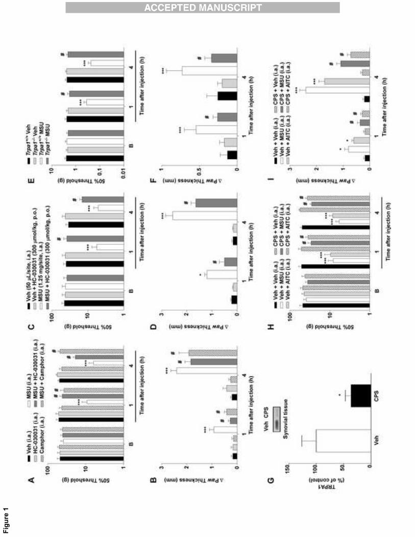
hyperalgesia or edema (data not shown). Local treatment with the TRPA1-selective
antagonist HC-030031 (300 nmol/site, i.a.) or with the poorly selective TRPA1 antagonist
camphor (150 nmol/site, i.a.) was able to decrease MSU-induced hyperalgesia and also
edema 1 and 4 hours after treatment (Fig. 1A-B). In addition, i.a. injection of the selective
TRPA1 agonist AITC caused nociceptive and edematogenic responses, which were
markedly reduced by the TRPA1 antagonists (HC-030031 or camphor, i.a.) (Table 1). Oral
administration of HC-030031 (300 μmol/kg, p.o.) also largely reduced the development of
MSU-elicited hyperalgesia and edema formation from 1 to 4 hours after treatment (Fig. 1C-
D). HC or camphor per se did not produce any measurable inflammatory response
compared to vehicle (Fig. 1).
Trpa1+/+ mice showed mechanical hyperalgesia and edema formation after i.a.
injection of MSU at all evaluated time points. However, Trpa1-/- mice presented a marked
reduction in MSU-trigged responses (Fig. 1E-F). Further, in naïve rats, ablation of TRPA1-
positive nerves by perineural injection of capsaicin reduced TRPA1 channel
immunoreactivity in the synovial tissue (64% reduction) 7 days after treatment (Fig. 2A). In
addition, ablation of TRPA1-positive fibers was associated with diminished MSU- and
AITC-induced hyperalgesia and edema from 1 to 4 hours (Fig. 2B-C).
MSU i.a. injection increases TRPA1 expression in synovial tissue and CGRP release
14
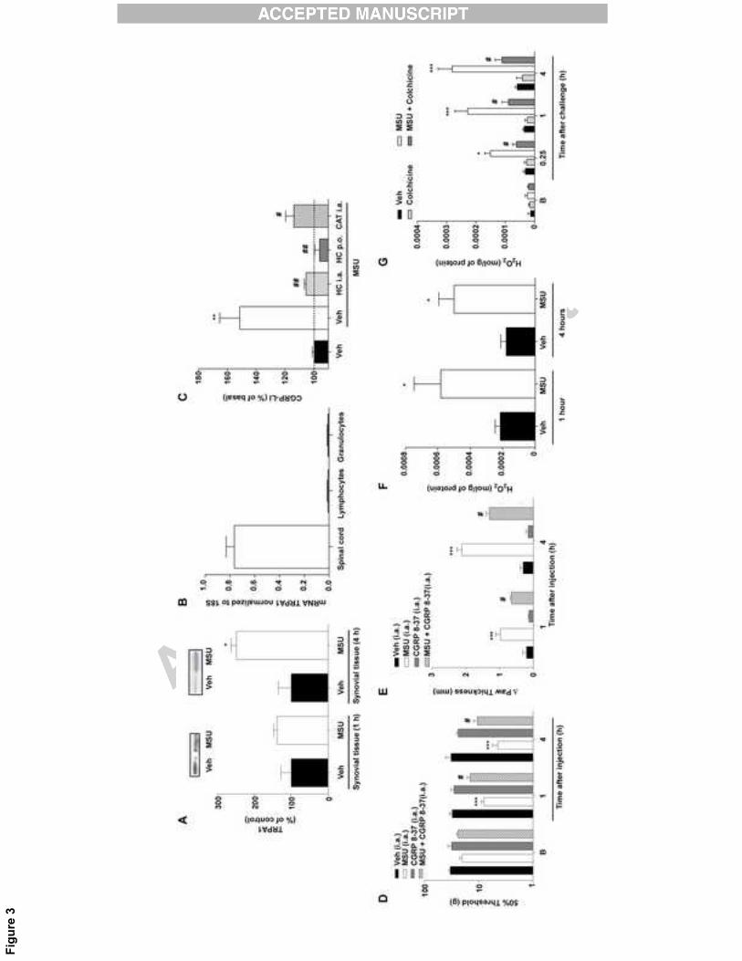
MSU increased TRPA1 expression 2.5-fold in synovial tissue 4 hours, but not 1
hour, after i.a. injection of MSU (Fig. 3A). However, we have not observed any TRPA1
mRNA expression in lymphocytes and granulocytes taken from rodent blood, when
compared to samples obtained from mouse spinal cord (Fig. 3B).
TRPA1 neuronal activation was assessed by measuring CGRP-LI release by MSU,
which produced a 1.5-fold increase in synovial lavage fluid compared to vehicle, HC-
030031 administration reduced CGRP-LI release (Fig. 3C). The H2O2-detoxifying enzyme
catalase abolished CGRP-LI release after MSU injection, suggesting a role of endogenous
H2O2 in the TRPA1-mediated MSU response (Fig. 3C).
In addition, the co-injection of the CGRP antagonist (CGRP8-37) was able to
decrease the nociception and edema formation induced by MSU i.a. injection (Fig. 3D and
E).
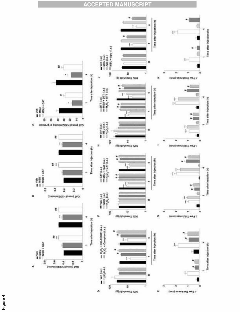
MSU challenge increased H2O2 production and reduced CAT activity
MSU injection (25 mg/mL) at 1 and 4 hours increased synovial tissue H2O2 levels
(Fig. 3F). The MSU challenge also increased synovial membrane H2O2 production at
different time points (0.25 to 4 hours). In addition, pretreatment of the synovial membranes
with colchicine (10 μM for 1 hour) reduced the H2O2 production (Fig. 3G).
Moreover, MSU i.a. injection increased the CAT activity in the synovial fluid (pellet
and supernatant, after 4 hours), and also in the synovial tissue (after 1 and 4 hours) (Fig.
4A, B, C). The i.a. co-injection of exogenous catalase reduced this effect in all measured
samples, and also increased the CAT activity after 1 hour in the synovial fluid samples,
when compared to vehicle treated animals (Fig. 4A, B, C).
15
The inflammatory responses elicited by i.a. injection of MSU were possibly mediated
by hydrogen peroxide production and subsequent TRPA1 activation
H2O2 (3 mol/site, i.a.) injection induced mechanical hyperalgesia and edema.
TRPA1 antagonists (HC-030031 and camphor) reduced the mechanical hyperalgesia and
edema formation induced by i.a. injection of H2O2 after 1 and 4 hours (Fig. 4A-B). Catalase
(300 UI/site) diminished either MSU- or H2O2-induced hyperalgesic and edematogenic
responses at 1 and 4 hours (Fig. 4C-D). The reducing agent DTT (20 nmol/site), which
reverses TRPA1 activation by H2O2 in vitro [33], also decreased MSU- or H2O2-induced
hyperalgesia and edema (Fig. 4E-F). Moreover, the NADPH-oxidase inhibitor apocynin
reduced the hyperalgesia and edema caused by MSU i.a. injection (Fig. 4 G-H).
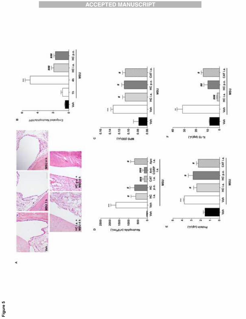
MSU-induced increases in neutrophil infiltration, plasma extravasation and IL-1β
levels were largely reduced by TRPA1 antagonism or by catalase
We found that MSU (1.25 mg/site, i.a.) significantly increased the number of
emigrated neutrophils in H&E-stained slices of articular tissue at 4 hours, but not 1 hour,
after injection (Fig. 5A-B). The inflammatory effect induced by MSU was reduced by co-
injection (300 nmol/site, i.a.) or oral pretreatment with HC-030031 (300 μmol/kg, 1 hour
before) (Fig. 5A-B). Finally, HC-030031 or catalase injection reduced the number of
neutrophils (Fig. 5C), the MPO activity (Fig. 5D), the plasma extravasation (Fig. 5E), and
the increase in IL-1β content (Fig. 5F) observed in the synovial tissue 4 hours after i.a.
MSU challenge. Moreover, the CGRP antagonist (CGRP 8-37) and NADPH-oxidase
inhibitor (apocynin) injection reduced the number of neutrophils induced by MSU i.a.
injection (Fig. 5C).
DISCUSSION
16
Hyperalgesia and edema are major symptoms in patients affected by acute gout
attacks, and the reduction of the pain hypersensitivity and the inflammation condition is a
main therapeutic goal for the gout treatment [34]. The major finding of the present study is
that pharmacological blockade or genetic ablation of the TRPA1 channel markedly
decreases the mechanical hyperalgesia and the entire inflammatory repertoire produced
by i.a. injection of MSU, a predictive rodent model of the acute gout attack. Moreover, we
showed that resident cells from the articular space are important to drive the initial
inflammatory process, an effect that we can not observed in our previous study by the
MSU injection in the intraplantar tissue (subcutaneous). Thus, our data added significant
novelty to the understanding of TRPA1 participation in a model of acute gout in rats and
mice.
A series of studies have reported the contribution of TRPA1 in hyperalgesia and
edema in inflammatory pain models [11-13]. TRPA1 is co-expressed with TRPV1 in
peptidergic sensory neurons [35]. This implies that the known ability of capsaicin to
produce desensitization of TRPV1 expressing neurons, results in profound desensitization
of TRPA1-positive, peptidergic afferents [36]. In addition, the use of capsaicin as a
counter-irritant to treat gout pain is probably based on the desensitization properties of the
drug [35]. Here, we showed that capsaicin desensitization produced antinociceptive and
anti-inflammatory effects by defunctionalizing TRPA1-expressing neurons. The
observation that capsaicin treatment decreased TRPA1 immunoreactivity in the synovial
tissue, may be explained by the ablation of sensory nerve terminals due to the
desensitizing action caused by intense TRPV1 stimulation.
Furthermore, we observed that the TRPA1 neuronal activation by MSU injection
increased CGRP release, and this effect was blocked by TRPA1 antagonism and the use
of the H2O2-detoxifying enzyme catalase. In accordance, with these data the CGRP
17
antagonist CGRP antagonist (CGRP8-37) reduced MSU-induced hyperalgesia and
edema, showing that TRPA1 activation produced CGRP release driving inflammation and
pain. However, MSU crystals or uric acid do not directly stimulate the TRPA1, whereas
H2O2 promotes TRPA1 activation [10, 14]. Injection of MSU (i.a.) increased the H2O2 levels
in synovial membranes in vitro, an effect that was reduced by colchicine, indicating a role
of MSU phagocytosis in this process. Previous findings show that MSU challenge
generated oxidative stress from resident cells and neutrophils, and both responses were
reduced by colchicine [31, 37, 38]. Thus, by analogy, we propose that resident synovial
cells, which have the potential to phagocytize MSU crystals, generate H2O2 when
challenged with MSU. In the normal synovial tissue, type A synoviocytes are known to
exert a macrophage-like behavior, with the associated phagocytic activity [39, 40, 41].
Previously, it has been described that MSU crystals injection in dog synovial space
produced an inflammatory response (marked infiltration of neutrophils) that is initiated by
the premature MSU crystal phagocytosis by resident cells (synovial lining cells) in the
synovial space [42].
Consistent with these findings, i.a. injection of H2O2 evoked nociception and
inflammation in a TRPA1-dependent manner. In addition, catalase or the reducing agent
DTT decreased these effects. Moreover, we observed that catalase activity was decreased
after MSU i.a. injection in the synovial fluid (supernatant, which represents the extracellular
enzyme, and pellet, which represents intracellular enzyme, after 4 hours), and also in the
synovial tissue (1 and 4 hours after MSU injection). In addition, the inhibition of NADPH
oxidase by apocynin was able to reduce MSU-induced hyperalgesia, edema, and
inflammatory cells infiltration. Then, the increase in NADPH oxidase and the decrease of
catalase activities after MSU i.a. injection may account for the observed H2O2 production,
which could in turn activate the TRPA1 receptor causing hyperalgesia and inflammation. In
18
this context, it should be underlined that gout patients show levels of reactive oxygen
species higher than those present in normal subjects [3, 6].
MSU-mediated inflammation has been linked to IL-1β production, which follows the
activation of infiltrating cells [43-45]. Indeed, neutralization of IL-1β has been explored as a
new strategy for the relief of gout pain [46, 47]. However, present data indicate that TRPA1
activation, presumably by H2O2, is upstream to neutrophil infiltration and the increased IL-
1β production. In fact, early blockade of TRPA1 receptor or of H2O2 production by catalase
reduced the MSU-evoked increase in IL-1β, MPO activity or neutrophil accumulation in the
synovial space. Also, CGRP antagonism and NADPH oxidase blockage reduced the
neutrophil accumulation in the synovial fluid.
The increase in TRPA1 expression after MSU injection was delayed and this fact is
consistent with the time lag usually required either for new protein generation or for
transport to the nerve terminals. The temporal association between the increase in TRPA1
expression and the continuation of the hypersensitivity and inflammatory condition
suggests that maintenance rather than onset of these phenomena is dependent from
augmented channel expression. It is possible, as observed by others [48], that
inflammatory mediators, including IL-1β, promote TRPA1 expression and contribute, by
this and other mechanisms, to increased firing of nociceptive neurons and nociception [11,
49]. As presented in Figure 3, TRPA1 is not expressed in leukocytes, indicating that
TRPA1 increase expression is not caused by TRPA1 expressed in infiltrating cells. Thus,
we speculate that the increase in TRPA1 expression is mediated by local protein synthesis
by a resident cell, especially sensory neurons since they may quickly synthesize protein in
their axons [50] and their defunctionalization is largely able to reduce either TRPA1
expression or MSU-trigged inflammation. However, we cannot exclude that the increased
expression of TRPA1 could also occur in other cell types present in the synovial cavity, as
19
synoviocytes [11]. Additional studies must be carried out to clarify the types of cells where
the TRPA1 receptor protein has an increased expression after MSU i.a. injection.
In conclusion, as previously observed in subcutaneous tissue, our present data
support the notion that early TRPA1 activation by endogenous reactive oxygen species in
the synovial tissue mediated nociceptive and inflammatory responses induced by MSU i.a.
injection. These findings, point to TRPA1 antagonism and the detoxifying of H2O2
production by catalase as a possible novel therapeutic option for gout treatment.
Acknowledgments: The authors thank the fellowships from CNPq and Coordenação de
Aperfeiçoamento de Pessoal de Nível Superior (CAPES). We are grateful to Dr. Delia Preti
(University of Ferrara, Italy) for providing HC-030031.
Competing interests: The authors declare no competing financial interests. P.G. is a
member of the editorial boards of Physiological Reviews and Pain and Molecular Pain and
receives research support from Chiesi Farmaceutici, Merck Sharp & Dohme, the Italian
Institute of Technology, the Regione Toscana, the Italian Ministry of University and
Research, and Ente Cassa di Risparmio di Firenze.
Funding: This study was supported by Conselho Nacional de Desenvolvimento Científico
(CNPq), Financiadora de Estudos e Projetos (FINEP), Programa de Apoio aos Núcleos de
20
Excelência (PRONEX) and Fundação de Amparo à Pesquisa do Estado do Rio Grande do
Sul (FAPERGS) (Brazil) to J.F. and in part by Ente Cassa di Risparmio di Firenze (Italy)
and the Italian Ministry of University and Research PRIN (Italy) to S.M.
REFERENCES
[1] Richette, P.; Bardin, T. Gout. Lancet 375:318-328; 2010.
[2] Terkeltaub, R. Update on gout: new therapeutic strategies and options. Nat. Rev.
Rheumatol. 6:30-38; 2010.
[3] Pascual, E.; Batlle-Gualda, E.; Martínez, A.; Rosas, J.; Vela, P. Synovial fluid
analysis for diagnosis of intercritical gout. Ann. Intern. Med. 131:756-759; 1999.
[4] Smith, H. S.; Bracken, D.; Smith, J. M. Gout: current insights and future
perspectives. J. Pain 12:1113-1129; 2011.
[5] Suresh, E.; Das, P. Recent advances in management of gout. Qjm 105:407-417;
2012.
[6] Amaral, F. A.; Costa, V. V.; Tavares, L. D.; Sachs, D.; Coelho, F. M.; Fagundes, C.
T.; Soriani, F. M.; Silveira, T. N.; Cunha, L. D.; Zamboni, D. S.; Quesniaux, V.; Peres, R.
21
S.; Cunha, T. M.; Cunha, F. Q.; Ryffel, B.; Souza, D. G.; Teixeira, M. M. NLRP3
inflammasome-mediated neutrophil recruitment and hypernociception depend on
leukotriene B(4) in a murine model of gout. Arthritis Rheum. 64:474-484; 2012.
[7] Huang, H. Y.; Appel, L. J.; Choi, M. J.; Gelber, A. C.; Charleston, J.; Norkus, E. P.;
Miller, E. R., 3rd. The effects of vitamin C supplementation on serum concentrations of uric
acid: results of a randomized controlled trial. Arthritis Rheum. 52:1843-1847; 2005.
[8] Juraschek, S. P.; Miller, E. R., 3rd; Gelber, A. C. Effect of oral vitamin C
supplementation on serum uric acid: a meta-analysis of randomized controlled trials.
Arthritis Care Res. (Hoboken) 63:1295-1306; 2011.
[9] Shen, L.; Ji, H. F. Potential of vitamin C in the prevention and treatment of gout.
Nat. Rev. Rheumatol. 7:368; 2011.
[10] Andersson, D. A.; Gentry, C.; Moss, S.; Bevan, S. Transient receptor potential A1 is
a sensory receptor for multiple products of oxidative stress. J. Neurosci. 28:2485-2494;
2008.
[11] Andrade, E. L.; Meotti, F. C.; Calixto, J. B. TRPA1 antagonists as potential
analgesic drugs. Pharmacol. Ther. 133:189-204; 2012.
[12] Baraldi, P. G.; Preti, D.; Materazzi, S.; Geppetti, P. Transient receptor potential
ankyrin 1 (TRPA1) channel as emerging target for novel analgesics and anti-inflammatory
agents. J. Med. Chem. 53:5085-5107; 2010.
[13] Bautista, D. M.; Pellegrino, M.; Tsunozaki, M. TRPA1: A gatekeeper for
inflammation. Annu. Rev. Physiol. 75:181-200; 2013.
[14] Trevisan, G.; Hoffmeister, C.; Rossato, M. F.; Oliveira, S. M.; Silva, M. A.; Ineu, R.
P.; Guerra, G. P.; Materazzi, S.; Fusi, C.; Nassini, R.; Geppetti, P.; Ferreira, J. TRPA1
receptor stimulation by hydrogen peroxide is critical to trigger pain during MSU-induced
inflammation. Arthritis Rheum. 2013:2984-2995; 2013.
22
[15] Coderre, T. J.; Wall, P. D. Ankle joint urate arthritis (AJUA) in rats: an alternative
animal model of arthritis to that produced by Freund's adjuvant. Pain 28:379-393; 1987.
[16] Coderre, T. J.; Wall, P. D. Ankle joint urate arthritis in rats provides a useful tool for
the evaluation of analgesic and anti-arthritic agents. Pharmacol. Biochem. Behav. 29:461-
466; 1988.
[17] Zimmermann, M. Ethical guidelines for investigations of experimental pain in
conscious animals. Pain 16:109-110; 1983.
[18] Kilkenny, C.; Browne, W.; Cuthill, I. C.; Emerson, M.; Altman, D. G. Animal
research: reporting in vivo experiments--the ARRIVE guidelines. J. Cereb. Blood Flow
Metab. 31:991-993; 2011.
[19] Kwan, K. Y.; Glazer, J. M.; Corey, D. P.; Rice, F. L.; Stucky, C. L. TRPA1 modulates
mechanotransduction in cutaneous sensory neurons. J. Neurosci. 29:4808-4819; 2009.
[20] Andre, E.; Campi, B.; Materazzi, S.; Trevisani, M.; Amadesi, S.; Massi, D.;
Creminon, C.; Vaksman, N.; Nassini, R.; Civelli, M.; Baraldi, P. G.; Poole, D. P.; Bunnett,
N. W.; Geppetti, P.; Patacchini, R. Cigarette smoke-induced neurogenic inflammation is
mediated by alpha,beta-unsaturated aldehydes and the TRPA1 receptor in rodents. J.
Clin. Invest. 118:2574-2582; 2008.
[21] Armitage, P.; Berry, G. The planning os statistical investigations. Statistical
methods in medical research. Oxford: Blackwell; 1987: 179-185.
[22] Hoffmeister, C.; Trevisan, G.; Rossato, M. F.; de Oliveira, S. M.; Gomez, M. V.;
Ferreira, J. Role of TRPV1 in nociception and edema induced by monosodium urate
crystals in rats. Pain 152:1777-1788; 2011.
[23] Chaplan, S. R.; Bach, F. W.; Pogrel, J. W.; Chung, J. M.; Yaksh, T. L. Quantitative
assessment of tactile allodynia in the rat paw. J. Neurosci. Methods 53:55-63; 1994.
23
[24] Dixon, W. J. Efficient analysis of experimental observations. Annu. Rev. Pharmacol.
Toxicol. 20:441-462; 1980.
[25] Nassini, R.; Materazzi, S.; Andre, E.; Sartiani, L.; Aldini, G.; Trevisani, M.; Carnini,
C.; Massi, D.; Pedretti, P.; Carini, M.; Cerbai, E.; Preti, D.; Villetti, G.; Civelli, M.; Trevisan,
G.; Azzari, C.; Stokesberry, S.; Sadofsky, L.; McGarvey, L.; Patacchini, R.; Geppetti, P.
Acetaminophen, via its reactive metabolite N-acetyl-p-benzo-quinoneimine and transient
receptor potential ankyrin-1 stimulation, causes neurogenic inflammation in the airways
and other tissues in rodents. Faseb J. 24:4904-4916; 2010.
[26] Pinto, L. G.; Cunha, T. M.; Vieira, S. M.; Lemos, H. P.; Verri, W. A., Jr.; Cunha, F.
Q.; Ferreira, S. H. IL-17 mediates articular hypernociception in antigen-induced arthritis in
mice. Pain 148:247-256; 2009.
[27] Suzuki, K.; Ota, H.; Sasagawa, S.; Sakatani, T.; Fujikura, T. Assay method for
myeloperoxidase in human polymorphonuclear leukocytes. Anal. Biochem. 132:345-352;
1983.
[28] Bradford, M. M. A rapid and sensitive method for the quantitation of microgram
quantities of protein utilizing the principle of protein-dye binding. Anal. Biochem. 72:248-
254; 1976.
[29] Materazzi, S.; Fusi, C.; Benemei, S.; Pedretti, P.; Patacchini, R.; Nilius, B.; Prenen,
J.; Creminon, C.; Geppetti, P.; Nassini, R. TRPA1 and TRPV4 mediate paclitaxel-induced
peripheral neuropathy in mice via a glutathione-sensitive mechanism. Pflugers Arch.
463:561-569; 2012.
[30] Hyc, A.; Osiecka-Iwan, A.; Dziunycz, P.; Moskalewski, S. Preparation of rat synovial
membrane for studies of cytokine secretion. Folia Histochem. Cytobiol. 45:57-60; 2007.
[31] Gaudry, M.; Roberge, C. J.; de Medicis, R.; Lussier, A.; Poubelle, P. E.; Naccache,
P. H. Crystal-induced neutrophil activation. III. Inflammatory microcrystals induce a distinct
24
pattern of tyrosine phosphorylation in human neutrophils. J. Clin. Invest. 91:1649-1655;
1993.
[32] Aebi, H. Catalase in vitro. Methods Enzymol. 105:121-126; 1984.
[33] Takahashi, N.; Mizuno, Y.; Kozai, D.; Yamamoto, S.; Kiyonaka, S.; Shibata, T.;
Uchida, K.; Mori, Y. Molecular characterization of TRPA1 channel activation by cysteine-
reactive inflammatory mediators. Channels (Austin) 2:287-298; 2008.
[34] Dalbeth, N.; Lindsay, K. The patient's experience of gout: new insights to optimize
management. Curr. Rheumatol. Rep. 14:173-178; 2012.
[35] Story, G. M.; Peier, A. M.; Reeve, A. J.; Eid, S. R.; Mosbacher, J.; Hricik, T. R.;
Earley, T. J.; Hergarden, A. C.; Andersson, D. A.; Hwang, S. W.; McIntyre, P.; Jegla, T.;
Bevan, S.; Patapoutian, A. ANKTM1, a TRP-like channel expressed in nociceptive
neurons, is activated by cold temperatures. Cell 112:819-829; 2003.
[36] Pecze, L.; Pelsoczi, P.; Kecskes, M.; Winter, Z.; Papp, A.; Kaszas, K.; Letoha, T.;
Vizler, C.; Olah, Z. Resiniferatoxin mediated ablation of TRPV1+ neurons removes TRPA1
as well. Can. J. Neurol. Sci. 36:234-241; 2009.
[37] Walker, R. A.; McCleane, G. J. The addition of glyceryltrinitrate to capsaicin cream
reduces the thermal allodynia associated with the application of capsaicin alone in
humans. Neurosci. Lett. 323:78-80; 2002.
[38] Chia, E. W.; Grainger, R.; Harper, J. L. Colchicine suppresses neutrophil
superoxide production in a murine model of gouty arthritis: a rationale for use of low-dose
colchicine. Br. J. Pharmacol. 153:1288-1295; 2008.
[39] Martin, W. J.; Grainger, R.; Harrison, A.; Harper, J. L. Differences in MSU-induced
superoxide responses by neutrophils from gout subjects compared to healthy controls and
a role for environmental inflammatory cytokines and hyperuricemia in neutrophil function
and survival. J. Rheumatol. 37:1228-1235; 2010.
25
[40] Malawista, S. E.; de Boisfleury, A. C.; Naccache, P. H. Inflammatory gout:
observations over a half-century. Faseb J. 25:4073-4078; 2011.
[41] Ando, A.; Hagiwara, Y.; Onoda, Y.; Hatori, K.; Suda, H.; Chimoto, E.; Itoi, E.
Distribution of type A and B synoviocytes in the adhesive and shortened synovial
membrane during immobilization of the knee joint in rats. Tohoku. J. Exp. Med. 221:161-
168; 2010.
[42] Schumacher, H. R.; Phelps, P.; Agudelo, C. A. Urate crystal induced inflammation
in dog joints: sequence of synovial changes. J. Rheumatol. 1:102-113; 1974.
[43] Matsukawa, A.; Miyazaki, S.; Maeda, T.; Tanase, S.; Feng, L.; Ohkawara, S.;
Yoshinaga, M.; Yoshimura, T. Production and regulation of monocyte chemoattractant
protein-1 in lipopolysaccharide- or monosodium urate crystal-induced arthritis in rabbits:
roles of tumor necrosis factor alpha, interleukin-1, and interleukin-8. Lab. Invest. 78:973-
985; 1998.
[44] Scanu, A.; Oliviero, F.; Gruaz, L.; Sfriso, P.; Pozzuoli, A.; Frezzato, F.; Agostini, C.;
Burger, D.; Punzi, L. High-density lipoproteins downregulate CCL2 production in human
fibroblast-like synoviocytes stimulated by urate crystals. Arthritis Res. Ther. 12:R23; 2010.
[45] Torres, R.; Macdonald, L.; Croll, S. D.; Reinhardt, J.; Dore, A.; Stevens, S.; Hylton,
D. M.; Rudge, J. S.; Liu-Bryan, R.; Terkeltaub, R. A.; Yancopoulos, G. D.; Murphy, A. J.
Hyperalgesia, synovitis and multiple biomarkers of inflammation are suppressed by
interleukin 1 inhibition in a novel animal model of gouty arthritis. Ann. Rheum. Dis.
68:1602-1608; 2009.
[46] Schlesinger, N.; Alten, R. E.; Bardin, T.; Schumacher, H. R.; Bloch, M.; Gimona, A.;
Krammer, G.; Murphy, V.; Richard, D.; So, A. K. Canakinumab for acute gouty arthritis in
patients with limited treatment options: results from two randomised, multicentre, active-
26
controlled, double-blind trials and their initial extensions. Ann. Rheum. Dis. 71:1839-1848;
2012.
[47] Schumacher, H. R., Jr.; Evans, R. R.; Saag, K. G.; Clower, J.; Jennings, W.;
Weinstein, S. P.; Yancopoulos, G. D.; Wang, J.; Terkeltaub, R. Rilonacept (interleukin-1
trap) for prevention of gout flares during initiation of uric acid-lowering therapy: Results
from a phase III randomized, double-blind, placebo-controlled, confirmatory efficacy study.
Arthritis Care Res. (Hoboken) 64:1462-1470; 2012.
[48] Hatano, N.; Itoh, Y.; Suzuki, H.; Muraki, Y.; Hayashi, H.; Onozaki, K.; Wood, I. C.;
Beech, D. J.; Muraki, K. Hypoxia-inducible Factor-1alpha (HIF1alpha) Switches on
Transient Receptor Potential Ankyrin Repeat 1 (TRPA1) Gene Expression via a Hypoxia
Response Element-like Motif to Modulate Cytokine Release. J. Biol. Chem. 287:31962-
31972; 2012.
[49] Lennertz, R. C.; Kossyreva, E. A.; Smith, A. K.; Stucky, C. L. TRPA1 mediates
mechanical sensitization in nociceptors during inflammation. PLoS One 7:e43597; 2012.
[50] Obata, K.; Katsura, H.; Mizushima, T.; Yamanaka, H.; Kobayashi, K.; Dai, Y.;
Fukuoka, T.; Tokunaga, A.; Tominaga, M.; Noguchi, K. TRPA1 induced in sensory
neurons contributes to cold hyperalgesia after inflammation and nerve injury. J. Clin.
Invest. 115:2393-2401; 2005.
27
LEGENDS
Fig. 1. Inflammatory responses induced by intra-articular (i.a.) injection of monosodium
urate crystals were mediated by TRPA1 channel activation in rodents. The (A) mechanical
hyperalgesia and (B) edema caused by i.a. injection of MSU were reduced by co-injection
with the selective and poorly selective TRPA1 antagonists HC-030031 (300 nmol/site, i.a.)
and camphor (150 nmol/site, i.a.), respectively. HC-030031 oral pretreatment (300
μmol/kg, 1 hour before MSU injection) also reduced the (C) mechanical hyperalgesia and
(D) edema induced by i.a. MSU injection. TRPA1 deficient mice (Trpa1-/-) showed reduced
(E) mechanical hyperalgesia and (F) edema formation to i.a. MSU injection compared with
Trpa1+/+ mice. The baseline threshold of animals was represented as B in the graphs.
Each column represents the mean ± S.E.M. of six rats or seven mice. The asterisks
denote the significance levels. *P < 0.05, ***P < 0.001 compared with the vehicle (Veh)-
treated group (Trpa1+/+ treated mice in E and F); or #P < 0.05 compared with the MSU-
28
treated group (Trpa1+/+ treated mice in E and F); two-way ANOVA followed by Bonferroni’s
post hoc test.
Fig. 2. Ablation of TRPA1-positive fibers largely reduced inflammatory responses elicited
by injection of monosodium urate crystals. (A) Western blot (inset) showing TRPA1
immunoreactivity in synovial tissue samples 7 days after the injection of capsaicin (CPS,
2%) or vehicle (Veh). Western blot results are expressed as % of control. The perineural
injection of CPS (2%) seven days before the intra-articular (i.a.) injection of monosodium
urate (MSU, 1.25 mg/site) crystals, the TRPA1 agonist AITC (1 nmol/site), or vehicle
reduced the (B) mechanical hyperalgesia and (C) edema formation induced by MSU or
AITC i.a. injection in rats. The baseline threshold of animals was represented as B in the
graphs. Each column represents the mean ± S.E.M. of six rats, except for western blot
when three to four samples were used. The asterisks denote the significance levels. *P <
0.05, ***P < 0.001 compared with the Veh-treated group (pretreated with Veh); #P < 0.05
compared with the MSU- or AITC-treated group (pretreated with Veh); Student's t test (in
A) or two-way ANOVA followed by Bonferroni’s post hoc test (in B-C).
Fig. 3. Monosodium urate crystal-elicited nociceptive and edematogenic responses were
accompanied by an increase in TRPA1 expression, release of calcitonin gene-related
peptide (CGRP), and H2O2 production and a reduction in catalase activity by the synovial
tissue. (A) Western blot (inset) showing TRPA1 immunoreactivity in synovial tissue at 1
and 4 hours after the i.a. injection of MSU (1.25 mg/site) or vehicle (Veh). Western blot
results are expressed % of control. (B) TRPA1 mRNA levels normalized to 18S in spinal
cord, lymphocytes and granulocytes. The expression in PBMCs is almost zero compared
to the TRPA1 expression in mouse spinal cord. (C) Calcitonin gene-related peptide-like
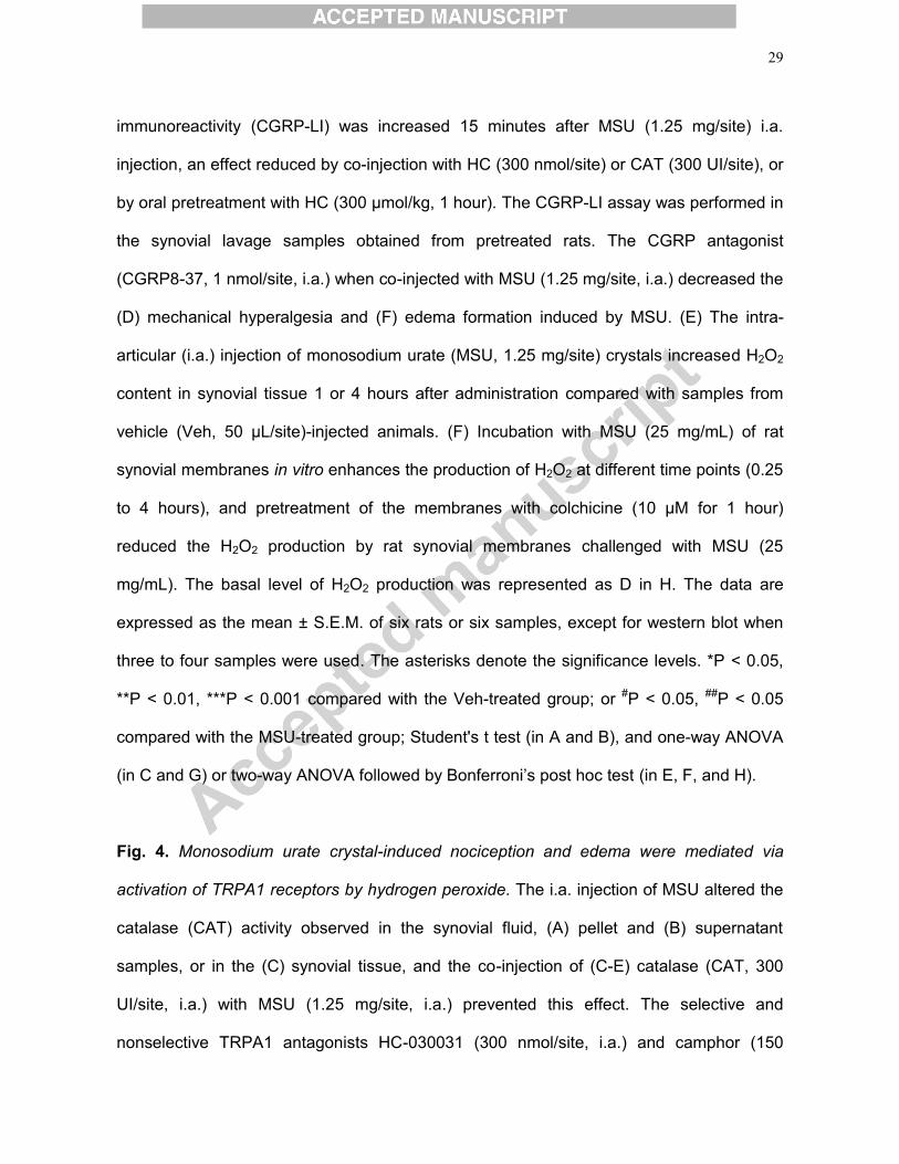
29
immunoreactivity (CGRP-LI) was increased 15 minutes after MSU (1.25 mg/site) i.a.
injection, an effect reduced by co-injection with HC (300 nmol/site) or CAT (300 UI/site), or
by oral pretreatment with HC (300 μmol/kg, 1 hour). The CGRP-LI assay was performed in
the synovial lavage samples obtained from pretreated rats. The CGRP antagonist
(CGRP8-37, 1 nmol/site, i.a.) when co-injected with MSU (1.25 mg/site, i.a.) decreased the
(D) mechanical hyperalgesia and (F) edema formation induced by MSU. (E) The intra-
articular (i.a.) injection of monosodium urate (MSU, 1.25 mg/site) crystals increased H2O2
content in synovial tissue 1 or 4 hours after administration compared with samples from
vehicle (Veh, 50 μL/site)-injected animals. (F) Incubation with MSU (25 mg/mL) of rat
synovial membranes in vitro enhances the production of H2O2 at different time points (0.25
to 4 hours), and pretreatment of the membranes with colchicine (10 μM for 1 hour)
reduced the H2O2 production by rat synovial membranes challenged with MSU (25
mg/mL). The basal level of H2O2 production was represented as D in H. The data are
expressed as the mean ± S.E.M. of six rats or six samples, except for western blot when
three to four samples were used. The asterisks denote the significance levels. *P < 0.05,
**P < 0.01, ***P < 0.001 compared with the Veh-treated group; or #P < 0.05, ##P < 0.05
compared with the MSU-treated group; Student's t test (in A and B), and one-way ANOVA
(in C and G) or two-way ANOVA followed by Bonferroni’s post hoc test (in E, F, and H).
Fig. 4. Monosodium urate crystal-induced nociception and edema were mediated via
activation of TRPA1 receptors by hydrogen peroxide. The i.a. injection of MSU altered the
catalase (CAT) activity observed in the synovial fluid, (A) pellet and (B) supernatant
samples, or in the (C) synovial tissue, and the co-injection of (C-E) catalase (CAT, 300
UI/site, i.a.) with MSU (1.25 mg/site, i.a.) prevented this effect. The selective and
nonselective TRPA1 antagonists HC-030031 (300 nmol/site, i.a.) and camphor (150
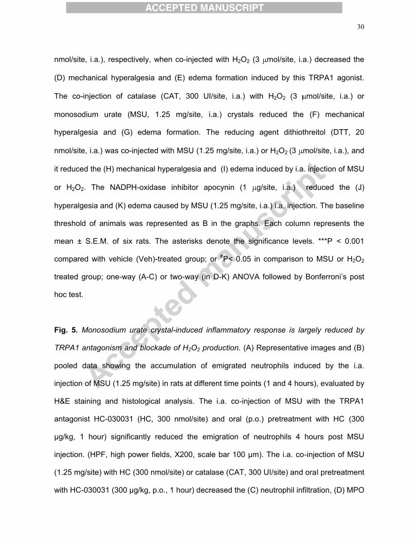
30
nmol/site, i.a.), respectively, when co-injected with H2O2 (3 mol/site, i.a.) decreased the
(D) mechanical hyperalgesia and (E) edema formation induced by this TRPA1 agonist.
The co-injection of catalase (CAT, 300 UI/site, i.a.) with H2O2 (3 mol/site, i.a.) or
monosodium urate (MSU, 1.25 mg/site, i.a.) crystals reduced the (F) mechanical
hyperalgesia and (G) edema formation. The reducing agent dithiothreitol (DTT, 20
nmol/site, i.a.) was co-injected with MSU (1.25 mg/site, i.a.) or H2O2 (3 mol/site, i.a.), and
it reduced the (H) mechanical hyperalgesia and (I) edema induced by i.a. injection of MSU
or H2O2. The NADPH-oxidase inhibitor apocynin (1 g/site, i.a.) reduced the (J)
hyperalgesia and (K) edema caused by MSU (1.25 mg/site, i.a.) i.a. injection. The baseline
threshold of animals was represented as B in the graphs. Each column represents the
mean ± S.E.M. of six rats. The asterisks denote the significance levels. ***P < 0.001
compared with vehicle (Veh)-treated group; or #P< 0.05 in comparison to MSU or H2O2
treated group; one-way (A-C) or two-way (in D-K) ANOVA followed by Bonferroni’s post
hoc test.
Fig. 5. Monosodium urate crystal-induced inflammatory response is largely reduced by
TRPA1 antagonism and blockade of H2O2 production. (A) Representative images and (B)
pooled data showing the accumulation of emigrated neutrophils induced by the i.a.
injection of MSU (1.25 mg/site) in rats at different time points (1 and 4 hours), evaluated by
H&E staining and histological analysis. The i.a. co-injection of MSU with the TRPA1
antagonist HC-030031 (HC, 300 nmol/site) and oral (p.o.) pretreatment with HC (300
μg/kg, 1 hour) significantly reduced the emigration of neutrophils 4 hours post MSU
injection. (HPF, high power fields, X200, scale bar 100 μm). The i.a. co-injection of MSU
(1.25 mg/site) with HC (300 nmol/site) or catalase (CAT, 300 UI/site) and oral pretreatment
with HC-030031 (300 μg/kg, p.o., 1 hour) decreased the (C) neutrophil infiltration, (D) MPO
31
activity, (E) plasma extravasation, and (F) interleukin-1β (IL-1β) levels observed 4 hours
after i.a. MSU injection. In addition, the co-injection the i.a. co-injection of MSU (1.25
mg/site) with the CGRP receptor antagonist (Ant-CGRP, CGRP8-37, 1 nmol/site) or
apocynin (Apo, 1 μg/site) decreased the (C) neutrophil infiltration. All the measurements
were performed using synovial lavage samples obtained from pretreated rats. Each
column represents the mean ± S.E.M. of six samples. The asterisks denote the
significance levels. ***P < 0.001 compared with the vehicle (Veh)-treated group; or #P <
0.05, ##P < 0.01, ###P < 0.001 compared with the MSU-injected group; one-way ANOVA
followed by Bonferroni's post hoc test.
32
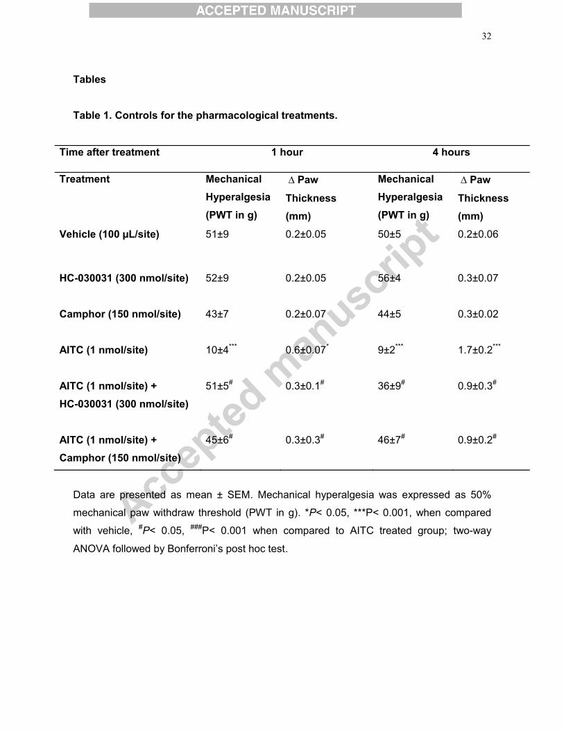
Tables Table 1. Controls for the pharmacological treatments.
Time after treatment 1 hour 4 hours
Treatment Mechanical Hyperalgesia
(PWT in g)
∆ Paw
Thickness (mm)
Mechanical Hyperalgesia
(PWT in g)
∆ Paw
Thickness (mm)
Vehicle (100 μL/site) 51±9 0.2±0.05 50±5 0.2±0.06
HC-030031 (300 nmol/site)
52±9
0.2±0.05
56±4
0.3±0.07
Camphor (150 nmol/site)
43±7
0.2±0.07
44±5
0.3±0.02
AITC (1 nmol/site)
10±4***
0.6±0.07*
9±2***
1.7±0.2***
AITC (1 nmol/site) + HC-030031 (300 nmol/site)
51±5#
0.3±0.1#
36±9#
0.9±0.3#
AITC (1 nmol/site) + Camphor (150 nmol/site)
45±6#
0.3±0.3#
46±7#
0.9±0.2#
Data are presented as mean ± SEM. Mechanical hyperalgesia was expressed as 50%
mechanical paw withdraw threshold (PWT in g). *P< 0.05, ***P< 0.001, when compared
with vehicle, #P< 0.05, ###P< 0.001 when compared to AITC treated group; two-way
ANOVA followed by Bonferroni’s post hoc test.
Gra
phic
al A
bstr
act (
for r
evie
w)
Figu
re 1
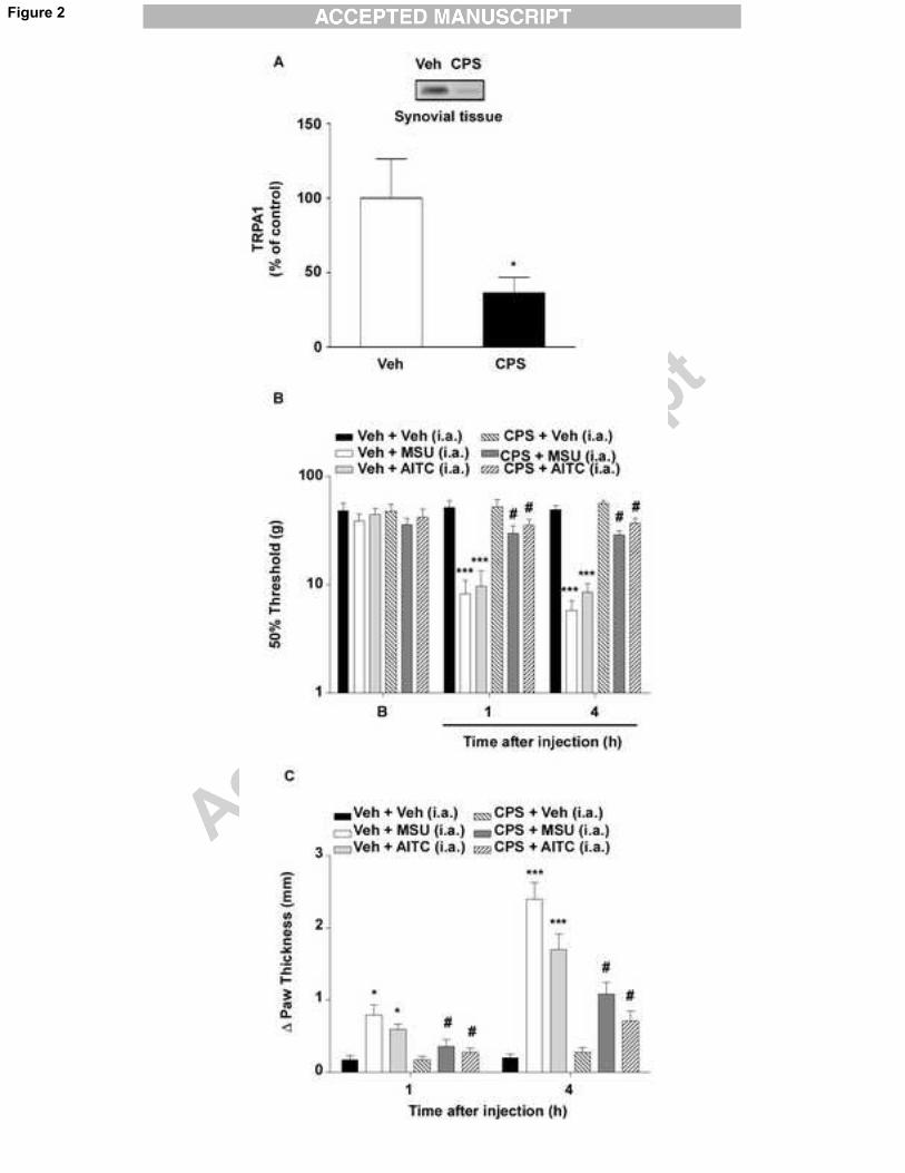
Figure 2
Figu
re 3
Figu
re 4
Figu
re 5